The Neurobiological Framework of Addiction: From Neural Circuits to Novel Therapeutics
This comprehensive review synthesizes current neuroscience research on addiction as a chronic brain disorder, examining the neuroadaptations in reward, stress, and executive control systems that drive the addiction cycle.
The Neurobiological Framework of Addiction: From Neural Circuits to Novel Therapeutics
Abstract
This comprehensive review synthesizes current neuroscience research on addiction as a chronic brain disorder, examining the neuroadaptations in reward, stress, and executive control systems that drive the addiction cycle. Targeting researchers, scientists, and drug development professionals, the article explores the three-stage neurobiological model of addiction (binge/intoxication, withdrawal/negative affect, preoccupation/anticipation) and their underlying neural substrates. It evaluates cutting-edge research methodologies, addresses persistent treatment challenges including relapse mechanisms, and discusses validation approaches for emerging interventions. The analysis bridges fundamental neurobiological discoveries with their translational applications in medication development and personalized treatment strategies for substance use disorders.
The Addicted Brain: Neural Circuits and Neuroadaptations in Substance Use Disorders
The conceptualization of addiction as a chronic brain disease represents a fundamental paradigm shift that has transformed both research and clinical practice over the past quarter century. This perspective emerged as a direct challenge to historical views that attributed addictive behaviors to moral failing or character flaws [1] [2]. Advances in neuroscience have demonstrated that addiction is instead marked by specific, measurable neuroadaptations that predispose individuals to pursue substances despite negative consequences [2]. The brain disease model has proven particularly valuable in reducing stigma, facilitating the integration of addiction treatment into mainstream healthcare, and guiding the development of novel therapeutics [3]. This whitepaper provides a comprehensive technical overview of the neurobiological framework underlying addiction, with specific emphasis on research methodologies, experimental protocols, and implications for drug development.
The foundational premise of the brain disease model rests on overwhelming scientific evidence that addiction is a health condition with biological, psychological, and social dimensions—comparable to other chronic conditions such as diabetes, hypertension, and asthma in its complexity, relapse rates, and treatment challenges [1] [3]. This paper synthesizes current neurobiological research to provide drug development professionals and researchers with a sophisticated understanding of addiction's underlying mechanisms and the experimental approaches used to investigate them.
The Neurobiological Framework of Addiction
Core Brain Circuits and the Addiction Cycle
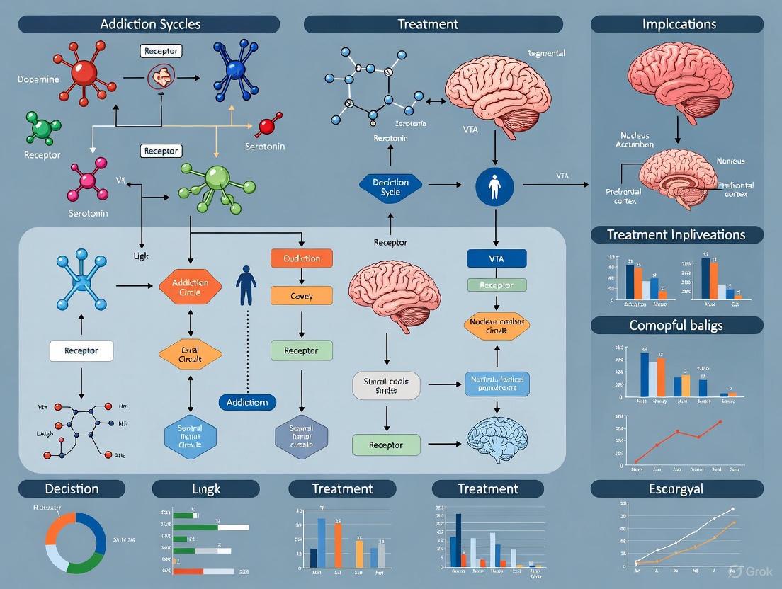
Research has identified three primary brain regions and their associated circuits that undergo specific neuroadaptations throughout the addiction cycle: the basal ganglia, extended amygdala, and prefrontal cortex [3] [2]. These regions form interconnected networks that mediate the transition from voluntary, controlled substance use to compulsive patterns of use that characterize addiction.
The addiction process occurs through a three-stage cycle that becomes more severe with repeated iterations, producing progressive changes in brain structure and function [3] [2]. The table below summarizes the characteristics and neurobiological underpinnings of each stage.
Table 1: The Three-Stage Cycle of Addiction: Neurobiological Substrates and Behavioral Manifestations
| Stage | Key Brain Regions | Primary Neurotransmitters | Behavioral Manifestations |
|---|---|---|---|
| Binge/Intoxication | Basal ganglia (particularly nucleus accumbens), ventral tegmental area | Dopamine, opioid peptides, GABA | Euphoria, reward, positive reinforcement, incentive salience |
| Withdrawal/Negative Affect | Extended amygdala (BNST, CeA), hypothalamus | CRF, dynorphin, norepinephrine | Irritability, anxiety, dysphoria, heightened stress sensitivity |
| Preoccupation/Anticipation | Prefrontal cortex (dlPFC, OFC), hippocampus | Glutamate, norepinephrine | Executive dysfunction, craving, impaired impulse control |
Signaling Pathways in the Addiction Cycle
The following diagram illustrates the primary signaling pathways and their interactions throughout the addiction cycle:
This diagram illustrates the cyclical nature of addiction, demonstrating how neuroadaptations in one stage prime the brain for progression to the next stage, creating a self-reinforcing cycle that becomes increasingly difficult to interrupt without intervention.
Experimental Models and Research Methodologies
Hierarchy of Evidence in Addiction Research
Multiple frameworks exist for evaluating evidence in addiction research, each with distinct advantages for different research questions. The table below compares prominent evidence evaluation models used in addiction science.
Table 2: Evidence Hierarchy Models in Addiction Research
| Model | Development Source | Key Stages/Levels | Application in Addiction Research |
|---|---|---|---|
| FDA Phase Model | U.S. Food and Drug Administration | Phase I: Safety/feasibility (10-100 participants)Phase II: Efficacy RCTsPhase III: Effectiveness RCTsPhase IV: Post-marketing surveillance | Primarily for pharmacotherapies; requires rigorous experimental design, double-blind RCTs |
| Stage Model for Behavioral Therapy | Onken et al., 1997 | Stage 1: Therapy development/feasibilityStage 2: Efficacy testing via RCTStage 3: Effectiveness testing in routine conditions | Used for behavioral intervention development; includes manual writing, adherence measures |
| Evidence-Based Medicine Model | Guyatt and Rennie, 2002 | 1. N-of-1 randomized trial2. Systematic reviews of RCTs3. Single RCT4. Systematic reviews of observational studies5. Single observational study6. Physiological studies7. Unsystematic clinical observations | Guides clinical decision-making; emphasizes patient-important outcomes |
| Clinical Psychology Framework | APA Division 12 Task Force | 1. Empirically validated (≥2 RCTs by independent teams)2. Probably efficacious (≥2 RCTs with waitlist control)3. Experimental | Categorizes behavioral treatments; requires treatment manuals, defined samples |
Key Experimental Protocols
Neuroimaging Protocol for Addiction Phenotyping
Objective: To characterize structural and functional brain alterations associated with different stages of addiction using multimodal neuroimaging.
Methodology:
- Participant Selection: Recruit individuals with substance use disorders (based on DSM-5 criteria) and matched healthy controls. Exclusion criteria typically include neurological disorders, contraindications for MRI, and certain psychiatric comorbidities.
- Image Acquisition:
- Structural MRI: T1-weighted images for voxel-based morphometry and cortical thickness analysis.
- Functional MRI (fMRI): Blood-oxygen-level-dependent (BOLD) signal during cue-reactivity tasks, reward processing tasks, and executive function paradigms.
- Diffusion Tensor Imaging (DTI): White matter integrity and connectivity analysis.
- Task Paradigms:
- Cue-Reactivity: Presentation of drug-related versus neutral cues while measuring BOLD response in reward regions.
- Monetary Incentive Delay Task: Assessment of reward anticipation and receipt.
- Go/No-Go or Stop-Signal Task: Evaluation of response inhibition and impulse control.
- Data Analysis:
- Preprocessing pipeline (motion correction, normalization, smoothing).
- First-level analysis for task-related activation.
- Second-level group analysis comparing patients versus controls.
- Connectivity analyses (seed-based, independent component analysis).
This protocol has been implemented in large-scale studies such as the Adolescent Brain Cognitive Development (ABCD) Study, which is gathering neuroimaging, biometric, and psychometric data to answer critical questions about substance use impacts on the developing brain [4].
Preclinical Self-Administration Protocol
Objective: To model addiction-like behaviors in laboratory animals and evaluate potential pharmacotherapies.
Methodology:
- Subjects: Typically rodents or non-human primates with intravenous catheter implantation.
- Apparatus: Operant conditioning chambers with levers/response devices, infusion pumps, and cue lights.
- Training Phase:
- Animals learn to self-administer drugs via lever pressing.
- Establishment of stable baseline responding.
- Testing Paradigms:
- Progressive Ratio Schedule: The response requirement increases exponentially to evaluate motivational strength (breakpoint).
- Cue-Induced Reinstatement: After extinction, presentation of drug-associated cues to model relapse.
- Drug-Primed Reinstatement: Administration of a small drug dose to trigger renewed responding.
- Data Collection:
- Number of infusions earned.
- Breakpoint values in progressive ratio.
- Reinstatement response rates.
This protocol allows researchers to investigate the neuropharmacological mechanisms underlying drug seeking and evaluate potential treatments before human trials [3].
Research Reagent Solutions
The table below outlines essential research reagents and their applications in addiction neuroscience research.
Table 3: Essential Research Reagents in Addiction Neuroscience
| Reagent/Category | Specific Examples | Research Application | Technical Function |
|---|---|---|---|
| Radioligands for Neuroimaging | [¹¹C]Raclopride, [¹⁸F]Fallypride | PET imaging of dopamine D2/D3 receptors | Quantification of receptor availability and dopamine release |
| Selective Receptor Agonists/Antagonists | SCH-23390 (D1 antagonist), Eticlopride (D2 antagonist) | Preclinical mechanistic studies | Target validation; circuit-specific manipulation |
| Chemogenetic Tools | DREADDs (Designer Receptors Exclusively Activated by Designer Drugs) | Circuit-specific manipulation in animal models | Precise temporal control over specific neural pathways |
| Viral Vector Systems | AAV-hSyn-hM3Dq, AAV-CaMKIIa-ChR2 | Optogenetics and chemogenetics | Targeted gene delivery for cell-type specific manipulation |
| Electrophysiology Reagents | Tetrodotoxin (TTX), Kynurenic acid | Brain slice electrophysiology | Synaptic transmission analysis; network activity recording |
| Behavioral Assay Kits | Conditioned Place Preference apparatus, Operant chambers | Preclinical addiction models | Quantification of reward, motivation, and relapse behaviors |
Implications for Treatment Development
Novel Pharmacotherapeutic Targets
Research on the neurobiology of addiction has identified numerous promising targets for medication development. The three-stage addiction cycle framework has been particularly valuable for developing interventions that target specific components of addiction pathology [2].
The following diagram illustrates the relationship between addiction stages and corresponding treatment approaches:
Several target classes show particular promise:
- D3 Receptor Partial Agonists/Antagonists: These compounds target the dopaminergic system with potentially greater selectivity for addiction-related pathways compared to non-selective dopamine antagonists [4].
- Orexin Antagonists: Target the orexin/hypocretin system involved in arousal, stress, and reward processing [4].
- GLP-1 Agonists: Originally developed for diabetes and obesity, these compounds show promise for multiple substance use disorders, potentially through effects on mesolimbic dopamine signaling [4].
- Kappa Opioid Receptor Antagonists: May normalize stress system dysregulation in the withdrawal/negative affect stage [2].
Neuromodulation Approaches
Recent advances in neuromodulation technologies offer promising non-pharmacological approaches for treatment-resistant addiction:
- Transcranial Magnetic Stimulation (TMS): FDA-approved as an adjunct treatment for smoking cessation and under investigation for other substance use disorders [4].
- Transcranial Direct Current Stimulation (tDCS): Investigational approach for modulating cortical excitability and reducing craving [4].
- Low-Intensity Focused Ultrasound: Non-invasive method that can reach deep brain targets; currently in clinical trials for cocaine use disorder and opioid use disorder [4].
Medication Development Workflow
The following diagram outlines a comprehensive medication development workflow for addiction therapeutics:
The conceptualization of addiction as a chronic brain disease has fundamentally transformed our approach to research and treatment development. The neurobiological framework outlined in this whitepaper provides a sophisticated understanding of the specific brain circuits, neurotransmitter systems, and neuroadaptations that drive the addiction cycle. This knowledge has enabled the development of targeted interventions at specific stages of addiction, from pharmacotherapies that restore biochemical balance to neuromodulation approaches that directly target dysfunctional circuits.
For researchers and drug development professionals, recognizing the complex, multi-stage nature of addiction is essential for developing effective therapeutics. Future directions include leveraging AI and computational approaches for higher-resolution analysis of neuroscience data, developing personalized treatment approaches based on individual neurobiological profiles, and creating novel delivery systems for medications and neuromodulation technologies [4]. The continued integration of basic neuroscience with clinical research promises to yield increasingly effective strategies for addressing this complex chronic disease.
Addiction is a chronic, relapsing brain disorder characterized by a compulsive cycle of drug seeking and use, despite harmful consequences. Groundbreaking research in neuroscience has fundamentally shifted our understanding from viewing addiction as a moral failing to recognizing it as a medical condition driven by specific neurobiological mechanisms [2] [3]. This whitepaper elaborates on the widely accepted neurobiological model of addiction, which frames the disorder as a recurring cycle of three distinct stages—binge/intoxication, withdrawal/negative affect, and preoccupation/anticipation [2] [5] [6]. Each stage is mediated by specific brain circuits, neurotransmitter systems, and neuroadaptations, which collectively promote the transition from controlled, impulsive use to compulsive, uncontrolled addiction [3] [6]. This review synthesizes the neurocircuitry, molecular mechanisms, and key behavioral correlates of each stage, providing a framework for understanding the pathophysiology of addiction and the development of novel treatment strategies.
The three-stage cycle of addiction provides a heuristic model for understanding the persistent nature of substance use disorders. The cycle is characterized by a shift from positive reinforcement (taking drugs for pleasure) to negative reinforcement (taking drugs to relieve negative emotional states) [6]. This transition is paralleled by a progression from impulsive to compulsive drug-seeking behavior [2] [6].
The stages are functionally linked to three primary brain regions:
- The basal ganglia are central to the binge/intoxication stage and the processing of reward and habit formation.
- The extended amygdala is critical to the withdrawal/negative affect stage and the stress responses associated with withdrawal.
- The prefrontal cortex is key to the preoccupation/anticipation stage and the executive control (and its failure) over craving and relapse [2] [3].
This cycle becomes more severe over time, driven by lasting neuroplastic changes that reduce an individual's ability to control substance use [3]. The following sections detail the neurobiological underpinnings of each stage.
Stage 1: Binge/Intoxication
The binge/intoxication stage is defined by the rewarding and reinforcing effects of a substance. This initial stage is primarily mediated by the basal ganglia, with a key role for the mesolimbic dopamine pathway connecting the ventral tegmental area (VTA) to the nucleus accumbens (NAcc) [2] [5] [3].
Neurocircuitry and Key Mechanisms
During this stage, all addictive substances directly or indirectly increase dopamine signaling in the NAcc [6]. This dopamine release is associated with the subjective experience of euphoria or a "high" [6]. With repeated use, neuroadaptations occur. The concept of incentive salience becomes critical, whereby dopamine firing shifts from responding to the drug itself to anticipating reward-related cues (people, places, paraphernalia) [2]. This process imbues these cues with powerful motivational properties, triggering drug-seeking behavior.
Concurrently, the nigrostriatal pathway in the dorsolateral striatum strengthens, underpinning the transition from voluntary, goal-directed drug use to automatic, habitual behavior—a hallmark of compulsion [2] [5].
Table 1: Key Neurotransmitter Changes in the Binge/Intoxication Stage
| Neurotransmitter/Neuromodulator | Change | Primary Brain Region(s) | Functional Consequence |
|---|---|---|---|
| Dopamine | Increase [6] | VTA, NAcc (ventral striatum) [2] | Euphoria, reinforcement, incentive salience [2] |
| Opioid Peptides | Increase [6] | NAcc, VTA | Modulation of dopamine release and reward [6] |
| GABA (γ-aminobutyric acid) | Increase [6] | VTA, NAcc | Modulation of neuronal excitability and reward [6] |
| Endocannabinoids | Increase [6] | VTA, NAcc | Modulation of reward and synaptic plasticity [6] |
Figure 1: Neurocircuitry of the Binge/Intoxication Stage. The diagram illustrates how drugs and associated cues activate the VTA, leading to dopamine release in the NAcc, which mediates reward and reinforcement. With repeated use, control over behavior shifts to the dorsolateral striatum, promoting compulsive habit formation.
Stage 2: Withdrawal/Negative Affect
The withdrawal/negative affect stage begins when drug concentration declines, leading to a aversive motivational state. This stage is driven by two major neuroadaptations: a within-system decrease in reward function and a between-system recruitment of brain stress systems [2] [6]. The extended amygdala (including the bed nucleus of the stria terminalis, the central nucleus of the amygdala, and the shell of the NAcc) is the key brain structure involved [2] [5] [3].
Neurocircuitry and Key Mechanisms
Chronic drug use leads to a hypofunction of the brain's reward systems, including a decreased baseline dopaminergic tone in the NAcc and a shift in the glutamate-GABA balance towards increased excitability [2]. This results in anhedonia—a diminished ability to experience pleasure from natural rewards.
Simultaneously, the brain's "anti-reward" system, centered on the extended amygdala, becomes hyperactive [2] [6]. This system releases stress neurotransmitters, creating a powerful negative emotional state that fuels further drug use through negative reinforcement—the compulsive taking of drugs to alleviate the dysphoria and distress of withdrawal [3] [6].
Table 2: Key Neurotransmitter Changes in the Withdrawal/Negative Affect Stage
| Neurotransmitter/Neuromodulator | Change | Primary Brain Region(s) | Functional Consequence |
|---|---|---|---|
| Corticotropin-Releasing Factor (CRF) | Increase [6] | Extended amygdala [2] | Anxiety, stress responses [6] |
| Dynorphin | Increase [6] | Extended amygdala, NAcc | Dysphoria, stress-like responses [6] |
| Norepinephrine | Increase [6] | Extended amygdala, BNST | Anxiety, autonomic hyperactivity [6] |
| Dopamine | Decrease [6] | NAcc, VTA | Anhedonia, reduced motivation [2] |
| Endocannabinoids | Decrease [6] | Extended amygdala | Reduced buffering of stress responses [2] |
Figure 2: Neurocircuitry of the Withdrawal/Negative Affect Stage. Chronic drug use triggers two major neuroadaptations: a deficit in the reward system and a surfeit in the brain's stress systems, centered on the extended amygdala. These changes converge to produce a negative emotional state, which drives drug seeking through negative reinforcement.
Stage 3: Preoccupation/Anticipation
The preoccupation/anticipation (craving) stage involves the relapse to drug-seeking behavior after a period of abstinence. This stage is characterized by a preoccupation with the drug and a deficit in executive control over the impulse to use. The prefrontal cortex (PFC) and its projections to the basal ganglia and extended amygdala are the primary neural substrates [2] [5].
Neurocircuitry and Key Mechanisms
This stage represents a critical failure of executive function. The PFC is responsible for planning, impulse control, and emotional regulation. In addiction, this region becomes dysregulated, leading to impaired decision-making and an inability to inhibit strong urges to use the drug [2] [3].
Two systems within the PFC are conceptualized: a "Go system" (involving the dorsolateral PFC and anterior cingulate) for goal-directed behaviors and a "Stop system" for inhibitory control. Addiction disrupts the balance, favoring the Go system for drug-seeking and weakening the Stop system [2]. Furthermore, brain regions like the insula are involved in interoception (awareness of bodily states) and contribute to conscious cravings, while the hippocampus and basolateral amygdala are critical for conditioning and memories associated with drug use [5] [6].
Table 3: Key Neurotransmitter Changes in the Preoccupation/Anticipation Stage
| Neurotransmitter/Neuromodulator | Change | Primary Brain Region(s) | Functional Consequence |
|---|---|---|---|
| Glutamate | Increase [6] | PFC to NAcc, Basolateral Amygdala [6] | Drives drug-seeking behavior, relapse [6] |
| Dopamine | Increase (in specific contexts) [6] | PFC | Altered salience attribution and executive function [6] |
| Corticotropin-Releasing Factor (CRF) | Increase [6] | Extended amygdala | Persistent stress and anxiety contributing to craving [6] |
Figure 3: Neurocircuitry of the Preoccupation/Anticipation Stage. During abstinence, exposure to drug cues, stress, or the drug context activates a distributed network involving the PFC, amygdala, hippocampus, and insula. This activation, coupled with dysregulated executive control and heightened craving, drives glutamate release that triggers relapse in downstream structures like the basal ganglia and extended amygdala.
Experimental Models and Research Tools
Understanding the three-stage cycle has been made possible through validated animal and human models that probe specific elements of addiction.
Key Animal Models and Methodologies
- Self-Administration: The gold standard model where animals (typically rodents or non-human primates) press a lever to intravenously self-administer a drug. This model directly assesses the reinforcing properties of a substance and can be used to study all stages of the addiction cycle [6] [7].
- Conditioned Place Preference (CPP): Measures the rewarding effects of a drug by pairing drug administration with a distinct environment. Animals later given a choice to enter either the drug-paired or a neutral environment will spend more time in the drug-paired one, indicating the drug's rewarding value [6].
- Intracranial Self-Stimulation (ICSS): Used to measure brain reward function. Animals perform a task to receive electrical stimulation to reward pathways like the medial forebrain bundle. During drug withdrawal, the reward threshold increases, reflecting a "reward deficit" akin to the anhedonia seen in the withdrawal/negative affect stage [6].
- Reinstatement Models: After a drug self-administration behavior is extinguished, drug-seeking is reinstated by exposure to a priming dose of the drug, drug-associated cues, or stress. This is a primary model for studying relapse (preoccupation/anticipation stage) [6] [7].
Human Research and Imaging Techniques
- Positron Emission Tomography (PET): Allows researchers to visualize and measure specific molecular targets in the living human brain, such as dopamine release, dopamine D2 receptor availability, and metabolic activity [3] [6].
- Functional Magnetic Resonance Imaging (fMRI): Measures brain activity by detecting changes in blood flow. fMRI studies have shown that drug-related cues activate the prefrontal cortex, amygdala, and striatum in individuals with addiction, providing a correlate for craving and the preoccupation/anticipation stage [5] [3].
Table 4: The Scientist's Toolkit: Essential Reagents and Models for Addiction Research
| Tool / Reagent / Model | Category | Primary Function in Research |
|---|---|---|
| Drug Self-Administration | Animal Model | Measures the reinforcing properties of a substance and models compulsion, escalation, and relapse [6]. |
| Conditioned Place Preference | Animal Model | Assesses the rewarding and aversive effects of drugs and the impact of contextual cues [6]. |
| Dopamine Receptor Ligands (e.g., Raclopride for D2) | Research Reagent | Used with PET imaging to quantify dopamine receptor levels and drug-induced dopamine release in the human brain [6]. |
| Corticotropin-Releasing Factor (CRF) Antagonists | Research Reagent | Pharmacological tools to test the role of the brain stress system in withdrawal and relapse [6]. |
| Optogenetics / Chemogenetics (DREADDs) | Neuromodulation Tool | Allows precise, cell-type-specific manipulation of neural circuits in animal models to establish causality in behavior [6]. |
Implications for Medication Development and Future Directions
The neurocircuitry analysis of addiction provides a rational blueprint for developing novel treatments. Instead of a "one-size-fits-all" approach, medications can be targeted to specific stages of the cycle from which a patient is suffering [6].
- Targeting Binge/Intoxication: Strategies include developing dopamine receptor partial agonists/antagonists, or modulating the opioid system (e.g., naltrexone for alcohol and opioid use disorders) to blunt the rewarding effects of the drug [3] [6].
- Targeting Withdrawal/Negative Affect: This is a major frontier. Medications aimed at restoring the brain's reward deficit (e.g., NMDA receptor modulators) or counteracting the overactive stress system (e.g., CRF antagonists, neurokinin-1 antagonists) hold promise for treating the negative emotional drive to relapse [6].
- Targeting Preoccupation/Anticipation: Approaches include cognitive enhancers to improve PFC function (e.g., modulators of glutamate, norepinephrine, or acetylcholine) and thus bolster inhibitory control, as well as medications that dampen cue-induced craving [3] [6].
Future research must continue to integrate findings across genetic, molecular, cellular, and circuit levels to fully elucidate the neuroadaptations underlying addiction. A deeper understanding of individual differences, developmental vulnerabilities (especially adolescence), and the neurobiology of behavioral addictions will be critical for advancing personalized medicine in the treatment of substance use disorders [3] [6].
Addiction is a chronic brain disorder characterized by compulsive drug seeking and use despite harmful consequences. Contemporary neurobiological research has established that addiction cannot be reduced to a single brain region or neurotransmitter system. Instead, it arises from dysfunctions within three interconnected brain networks: the basal ganglia, the extended amygdala, and the prefrontal cortex [3]. These regions form the core components of a cycle of addiction that progresses from binge/intoxication to withdrawal/negative affect to preoccupation/anticipation [3] [8]. Understanding the specific contributions of each region and their interactions provides a comprehensive framework for understanding the pathophysiology of addiction and developing targeted treatment interventions.
The following diagram illustrates how these three key brain regions interact throughout the addiction cycle:
Basal Ganglia: Reward Processing and Habit Formation
Structural and Functional Organization
The basal ganglia represent a collection of nuclei located deep within the cerebral hemispheres that play crucial roles in reward processing, habit formation, and motor control. The primary components include the nucleus accumbens (ventral striatum), dorsal striatum (caudate and putamen), globus pallidus, substantia nigra, and subthalamic nucleus [3] [9]. These structures are organized into parallel cortico-striato-pallido-thalamo-cortical loops that integrate associative, sensorimotor, and limbic information from the cortex [9]. The nucleus accumbens serves as a key interface between emotion and action, receiving dense dopaminergic projections from the ventral tegmental area that are central to reward processing [10] [11].
Dysfunctions in Addiction
In the initial stages of addiction, drugs of abuse produce powerful surges of dopamine in the basal ganglia, particularly the nucleus accumbens, generating intense euphoria and reinforcing drug-taking behavior [12]. With repeated drug exposure, neuroadaptations occur that fundamentally alter basal ganglia function. The table below summarizes the key dysfunctions:
Table 1: Basal Ganglia Dysfunctions in Addiction
| Functional Aspect | Normal Function | Addiction-Related Dysfunction | Key Neuroadaptations |
|---|---|---|---|
| Reward Processing | Processes natural rewards (food, sex) | Hyposensitivity to natural rewards; increased sensitivity to drug rewards | Reduced D2 receptor availability; decreased dopamine release in response to natural rewards |
| Habit Formation | Forms routines for efficient behavior | Compulsive drug-seeking habits | Shift from ventral to dorsal striatal control; strengthened stimulus-response associations |
| Motivational Salience | Attributes importance to biologically relevant stimuli | Enhanced incentive salience to drug cues | Dopamine hyperreactivity to drug-associated cues; cue-induced craving |
A critical transition occurs with prolonged drug use as control over drug seeking shifts from the ventral striatum (reward-based behavior) to the dorsal striatum (habit-based behavior) [10]. This ventral to dorsal striatum shift underlies the progression from voluntary drug use to compulsive drug-seeking habits that are resistant to negative consequences [10] [3]. Additionally, the basal ganglia undergoes changes that diminish sensitivity to natural rewards while enhancing the salience of drug-associated stimuli, creating a powerful bias toward drug seeking [12].
Experimental Approaches and Methodologies
Research on basal ganglia function in addiction employs diverse methodological approaches:
Self-Administration Paradigms: Animals are trained to perform operant responses (e.g., lever pressing) to receive intravenous drug infusions. This model allows investigation of reinforcing properties and motivation for drugs [9].
Fast-Scan Cyclic Voltammetry: This technique enables real-time measurement of dopamine concentration changes in specific basal ganglia regions (e.g., nucleus accumbens) during drug administration or presentation of drug-associated cues [9].
Chemogenetics and Optogenetics: These approaches use engineered receptors or light-sensitive ion channels to precisely control neuronal activity in specific basal ganglia pathways, establishing causal relationships between circuit function and drug-seeking behavior [13].
Extended Amygdala: Stress and Negative Affect
Structural and Functional Organization
The extended amygdala represents a macrostructure composed of several interconnected regions, including the central nucleus of the amygdala, bed nucleus of the stria terminalis, and a transition zone in the nucleus accumbens shell [8] [3]. This system serves as a primary brain stress center that integrates emotional and arousal-related information. It receives inputs from limbic, brainstem, and cortical regions and projects to hypothalamic and brainstem areas that control hormonal responses, autonomic nervous system activity, and behavioral stress responses [8].
Dysfunctions in Addiction
As addiction progresses, the extended amygdala becomes hyperactive, leading to the emergence of a negative emotional state during drug withdrawal. This negative affect powerfully motivates drug seeking through negative reinforcement mechanisms (i.e., taking drugs to relieve distress) [8] [3]. Key neurochemical systems involved in these processes include:
Table 2: Key Neurochemical Systems in the Extended Amygdala
| System | Normal Function | Dysfunction in Addiction | Behavioral Manifestation |
|---|---|---|---|
| Corticotropin-Releasing Factor (CRF) | Regulates stress response | CRF overexpression in extended amygdala | Heightened anxiety, irritability |
| Norepinephrine | Arousal, alertness | Increased norepinephrine release | Hypervigilance, stress reactivity |
| Dynorphin/κ-Opioid System | Counteracts reward | Enhanced dynorphin transmission | Dysphoria, anhedonia |
The extended amygdala shows progressive sensitization with repeated drug exposure and withdrawal, becoming increasingly reactive to stress and drug-related stimuli [3]. This creates a self-perpetuating cycle where stress triggers drug craving and relapse, while drug withdrawal further sensitizes the stress systems [8]. The CRF and norepinephrine systems engage in feed-forward interactions within the extended amygdala, mutually exciting each other and amplifying the stress response [8].
Experimental Protocols for Stress System Investigation
Research on extended amygdala function employs specific protocols to elucidate stress-related mechanisms:
Fear-Potentiated Startle: This behavioral paradigm measures the augmentation of the acoustic startle response in the presence of a conditioned fear stimulus, assessing anxiety-like responses mediated by the extended amygdala during drug withdrawal [8].
Intracranial Microinjection: Cannulae are implanted to allow localized administration of receptor agonists/antagonists (e.g., CRF₁ antagonists, α₂-adrenergic agonists like clonidine) into specific extended amygdala subregions to determine their role in stress-induced drug seeking [8].
In Vivo Microdialysis: This technique involves implanting a semi-permeable membrane into the extended amygdala to collect extracellular fluid and measure neurotransmitter dynamics (e.g., CRF, norepinephrine) during baseline, drug administration, and withdrawal states [8].
Prefrontal Cortex: Executive Control and Decision Making
Structural and Functional Organization
The prefrontal cortex (PFC) serves as the brain's primary executive control center, responsible for higher-order cognitive functions including decision-making, impulse control, emotional regulation, and goal-directed behavior [14] [3]. Key subregions implicated in addiction include the orbitofrontal cortex (OFC), anterior cingulate cortex (ACC), and dorsolateral prefrontal cortex (DLPFC) [14]. These regions form extensive connections with both the basal ganglia and extended amygdala, positioning the PFC to integrate motivational, emotional, and cognitive information to guide adaptive behavior [14] [15].
Dysfunctions in Addiction
Addiction is characterized by profound PFC dysfunction that manifests as impaired response inhibition and salience attribution (iRISA syndrome) [14] [15]. The iRISA model proposes that addiction involves attributing excessive salience to drug-related stimuli while decreasing sensitivity to non-drug rewards, coupled with a reduced ability to inhibit maladaptive behaviors [14]. The specific contributions of PFC subregions are detailed below:
Table 3: Prefrontal Cortex Subregional Dysfunctions in Addiction
| PFC Subregion | Normal Function | Dysfunction in Addiction | Consequence |
|---|---|---|---|
| Orbitofrontal Cortex (OFC) | Value representation, outcome expectation | Hyperactivity to drug cues; impaired reversal learning | Compulsive drug use despite negative consequences |
| Anterior Cingulate Cortex (ACC) | Conflict monitoring, error detection | Reduced activity; impaired performance monitoring | Diminished awareness of loss of control; perseveration |
| Dorsolateral PFC (DLPFC) | Executive control, working memory | Decreased activation; disrupted functional connectivity | Poor impulse control; impaired decision-making |
Neuroimaging studies consistently show reduced gray matter volume and hypoactivity in PFC regions across multiple substance use disorders [14] [15]. This PFC impairment undermines self-control and enables the automatic, habitual processes driven by the basal ganglia and the stress responses mediated by the extended amygdala to dominate behavior [3] [11]. The PFC is also the last brain region to mature fully, which helps explain why adolescence represents a period of heightened vulnerability for developing substance use disorders [3].
Methodologies for Assessing Prefrontal Cortex Function
Research investigating PFC dysfunction utilizes several sophisticated approaches:
Functional Magnetic Resonance Imaging (fMRI): This non-invasive technique measures brain activity by detecting changes in blood flow, revealing PFC hypoactivation during cognitive tasks (e.g., Stroop, Go/No-Go) in individuals with addiction [14] [15].
Transcranial Magnetic Stimulation (TMS): This neuromodulation technique applies magnetic fields to stimulate specific PFC regions, potentially normalizing activity patterns and showing promise for reducing craving and improving cognitive control in addiction [15].
Diffusion Tensor Imaging (DTI): This MRI method maps white matter tracts, identifying microstructural alterations in PFC connections (e.g., reduced integrity of pathways linking PFC with striatum) that contribute to impaired communication within control networks [14].
Integrated Circuitry: Neurobiological Basis of the Addiction Cycle
The three core brain regions do not operate in isolation but rather form an integrated circuit that drives the addiction cycle. The progression through binge/intoxication, withdrawal/negative affect, and preoccupation/anticipation stages reflects shifting dominance among these interconnected systems [3]. The following diagram illustrates the interactive pathways between these regions that create a self-perpetuating cycle of addiction:
During binge/intoxication, the basal ganglia dominate through their role in reward processing and reinforcement. In the withdrawal/negative affect stage, the extended amygdala becomes predominant, generating stress and discomfort that motivate drug seeking through negative reinforcement. In the preoccupation/anticipation stage, PFC dysfunctions contribute to intense craving and inability to control drug-seeking behavior despite adverse consequences [3]. This cycle becomes increasingly severe with repeated iterations, as neuroadaptations in each system deepen and interactions become more entrenched.
The Scientist's Toolkit: Research Reagent Solutions
The following table compiles essential research reagents and methodologies used in addiction neuroscience research, particularly for investigating the basal ganglia, extended amygdala, and prefrontal cortex:
Table 4: Essential Research Reagents and Methodologies
| Reagent/Methodology | Function/Application | Specific Examples |
|---|---|---|
| Receptor Antagonists | Pharmacological blockade of specific neurotransmitter receptors to assess their functional roles | CRF₁ antagonists (e.g., R121919); κ-opioid receptor antagonists; D₁/D2 dopamine receptor antagonists |
| Neurotransmitter Assays | Quantitative measurement of neurotransmitter levels and dynamics | HPLC for monoamines; microdialysis for in vivo sampling; ELISA for peptide quantification |
| Viral Vector Systems | Targeted gene delivery for manipulation of specific neural circuits | AAV-DREADDs (chemogenetics); AAV-ChR2 (optogenetics); AAV-Cre recombinase for cell-type specific targeting |
| Calcium Indicators | Real-time monitoring of neuronal activity using fluorescence signals | GCaMP6f/GCaMP7; jRCaMP1b; synthetic dyes (e.g., Fura-2) for in vivo imaging |
| Radioligands | Quantitative mapping of receptor distribution and density using PET imaging | [¹¹C]raclopride (D2/D3 receptors); [¹¹C]carfentanil (μ-opioid receptors); [¹¹C]LY2795050 (κ-opioid receptors) |
| Behavioral Assays | Assessment of addiction-relevant behaviors in animal models | Self-administration; conditioned place preference; elevated plus maze (anxiety); 5-choice serial reaction time (attention) |
These research tools have been instrumental in delineating the neurochemical and circuit-level mechanisms underlying addiction. For instance, CRF₁ antagonists have demonstrated efficacy in reducing stress-induced reinstatement of drug seeking in animal models, highlighting the therapeutic potential of targeting stress systems in the extended amygdala [8]. Similarly, optogenetic manipulation of specific basal ganglia pathways has established causal roles for these circuits in habit formation and compulsive drug seeking [10] [9].
Implications for Therapeutic Development and Future Research
The dysfunctions across the basal ganglia, extended amygdala, and prefrontal cortex provide multiple targets for therapeutic intervention. Approaches may include:
Pharmacotherapies targeting specific neurotransmitter systems: dopamine partial agonists for basal ganglia dysregulation, CRF₁ antagonists or norepinephrine modulators for extended amygdala hyperactivity, and cognitive enhancers for prefrontal cortex dysfunction [8] [15].
Neuromodulation Techniques such as transcranial magnetic stimulation or deep brain stimulation to normalize activity patterns in dysregulated circuits, particularly targeting PFC networks to enhance cognitive control [15].
Behavioral Interventions that leverage neuroplasticity to strengthen prefrontal regulatory capacity and reshape maladaptive habits and stress responses [11].
Future research directions include better understanding individual differences in vulnerability and resilience, elucidating the molecular mechanisms that transition occasional drug use to addiction, and developing biomarkers to identify specific circuit dysfunctions for personalized treatment approaches [15]. The integration of cross-species research, with complementary findings from human neuroimaging and animal model studies, continues to be essential for advancing our understanding of these complex brain disorders and developing more effective interventions.
This whitepaper synthesizes current research on the distinct roles of neurotransmitter systems in reward processing, with a specific emphasis on the incentive salience hypothesis of dopamine. A growing body of evidence indicates that dopamine does not primarily mediate the hedonic 'liking' of rewards or reward learning, but rather assigns motivational value, or 'wanting,' to reward-related cues [16] [17]. This neurobiological framework is fundamental to understanding the pathophysiology of addiction, wherein drugs of abuse short-circuit and sensitize these mesolimbic mechanisms, leading to compulsive 'wanting' [16] [18]. We detail the molecular signaling pathways, summarize key experimental data, and describe advanced methodologies for real-time neurochemical measurement. The concluding discussion focuses on the implications of this research for developing novel therapeutic strategies for Substance Use Disorders (SUDs).
Reward is not a unitary process but a constellation of dissociable psychological components, primarily 'liking' (hedonic impact), 'wanting' (incentive salience), and learning (predictive associations) [17]. The mesolimbic pathway, particularly the projection from the ventral tegmental area (VTA) to the nucleus accumbens (NAcc), is a cornerstone of the brain's reward system [18]. While multiple neurotransmitters modulate this circuit, dopamine has emerged as a critical substrate for the 'wanting' component of reward [16]. Other key neurotransmitters, including serotonin, opioids, GABA, and glutamate, interact with dopamine signaling to finely regulate overall reward perception and motivated behavior [19] [18]. The precise delineation of these roles is essential for deconstructing the neurobiological basis of addiction, characterized by a hijacking of the very systems that guide adaptive goal-directed behaviors.
The Central Role of Dopamine in Incentive Salience
The "Wanting" vs. "Liking" Distinction
The incentive salience hypothesis posits that dopamine is crucial for attributing motivational value to neutral stimuli that are associated with rewards, transforming them into potent "wanted" incentives that trigger craving and pursuit [16]. Landmark experiments demonstrated that dopamine depletion in rodents does not abolish their hedonic reactions to sweet tastes but profoundly impairs their motivation to work for them [17]. Conversely, enhancing dopamine signaling can increase effort expenditure for rewards without necessarily altering hedonic evaluations. This dissociation confirms that dopamine's primary role is not in pleasure generation but in motivation.
Signaling Pathways and Molecular Mechanisms
Dopamine exerts its effects by binding to G protein-coupled receptors, classified into D1-like (D1, D5) and D2-like (D2, D3, D4) families [19]. The synthesis of dopamine is a two-step process in the cytosol: Tyrosine hydroxylase converts tyrosine to L-DOPA, which is then converted to dopamine by aromatic L-amino acid decarboxylase [19]. Following synthesis, dopamine is packaged into synaptic vesicles via the vesicular monoamine transporter 2 (VMAT2). The schematic below illustrates the synthesis, signaling, and metabolism of dopamine.
Figure 1: Dopamine neurochemistry. The diagram outlines the key processes from synthesis and vesicular storage to receptor signaling and metabolic degradation. VMAT2: vesicular monoamine transporter 2; DAT: dopamine transporter; MAO-B: monoamine oxidase B; COMT: catechol-O-methyltransferase; DOPAL: 3,4-dihydroxyphenylacetaldehyde; HVA: homovanillic acid.
Quantitative Data on Dopamine and Behavior
Table 1: Key experimental evidence supporting the incentive salience hypothesis of dopamine.
| Experimental Paradigm | Key Manipulation | Behavioral Effect | Neurochemical Correlation | Source |
|---|---|---|---|---|
| 6-OHDA Lesion | ~99% dopamine depletion in nucleus accumbens/neostriatum | Aphagia/adipsia; intact hedonic taste reactivity; impaired instrumental seeking. | Near-total loss of dopamine terminals. | [17] |
| Drug Self-Administration | Dopamine receptor antagonists (e.g., haloperidol). | Increased instrumental response rate; decreased response vigor. | Blockade of D1/D2 receptors. | [17] [18] |
| Human Electrochemistry | Intraoperative measurement in substantia nigra during Ultimatum Game. | Rejection of unfair offers varies with social context. | Dopamine tracks offer value (RPE); higher baseline in social context. | [20] |
| Drugs of Abuse | Administration of cocaine, amphetamine, nicotine, alcohol. | Increased euphoria, reinforcement, and compulsive use. | Increased dopamine release in NAcc; synaptic plasticity. | [16] [18] |
Beyond Dopamine: A Multi-Transmitter System
The reward system is a complex network modulated by several neurotransmitters beyond dopamine.
- Serotonin: Serotonin appears to exert a restraining influence on impulsive behavior and social decision-making. Pharmacologically lowering serotonin increases the rejection of unfair offers in the ultimatum game, suggesting a role in regulating social norm enforcement and negative affect [20]. Recent fast electrochemical measurements show that serotonin tracks the current value of an offer, a distinct signal from dopamine's RPE encoding [20].
- Opioids: The endogenous opioid system is heavily implicated in the 'liking' component of reward. Opioids in the NAcc and hedonic hotspots in the brainstem enhance hedonic reactions to tastes [17]. The opioid antagonist naltrexone is used to treat alcoholism, as it is believed to dampen the pleasurable effects of alcohol [18].
- GABA and Glutamate: These primary fast neurotransmitters provide critical balance within reward circuits. GABAergic medium spiny neurons in the NAcc are a major output, while GABA and glutamate inputs from other regions exert inhibitory and excitatory control over VTA dopamine neuron firing [18].
Table 2: Roles of key neurotransmitters in reward and addiction.
| Neurotransmitter | Primary Role in Reward | Effect in Addiction | Example Pharmacotherapy |
|---|---|---|---|
| Dopamine | Incentive Salience ('Wanting'), motivation, RPE. | Sensitization of 'wanting' leads to compulsive drug seeking triggered by cues. | Bupropion (smoking cessation). |
| Serotonin | Mood, impulse control, social decision-making. | Dysregulation leads to increased impulsivity and negative mood. | Selective Serotonin Reuptake Inhibitors (SSRIs). |
| Opioids | Hedonic Impact ('Liking'), pain relief. | Enhanced 'liking' for the drug; contributes to positive reinforcement. | Naltrexone (alcohol, opioid use disorder). |
| GABA | Inhibition of dopamine neurons; reduction of anxiety. | Alcohol and benzodiazepines enhance GABA action, contributing to dependence. | Acamprosate (alcohol use disorder). |
| Glutamate | Excitatory drive; synaptic plasticity, learning. | Long-term potentiation underpins cue-drug associative memories. | N-acetylcysteine (under investigation). |
Advanced Methodologies for Real-Time Neurochemical Measurement
Human Electrochemistry Protocol
A breakthrough in human neuroscience is the ability to measure subsecond neuromodulator fluctuations during awake brain surgery [20]. The following diagram and protocol detail this methodology.
Figure 2: Workflow for real-time neurochemical measurement in humans. DBS: Deep Brain Stimulation; SNr: Substantia Nigra pars reticulata.
Detailed Experimental Protocol [20]:
- Participants: Patients (e.g., with Parkinson's disease) undergoing awake deep brain stimulation (DBS) electrode implantation.
- Electrode Placement: A carbon-fiber microelectrode is inserted into the target structure (e.g., substantia nigra pars reticulata (SNr) or striatum), constrained by the clinical procedure.
- Electrochemical Recording: The protocol involves the repeated delivery of a rapid change in electrical potential (Fast-Scan Cyclic Voltammetry, FSCV) to the electrode.
- Signal Acquisition: The induced oxidation and reduction currents from electrochemical reactions at the electrode tip are measured at a high temporal resolution (e.g., 10 samples per second).
- Signal Deconvolution: A signal prediction model, trained on wet-lab data where the chemical environment is controlled, is used to parse the current responses and provide subsecond estimates of dopamine and serotonin concentrations.
- Behavioral Task: Participants perform computerized tasks (e.g., the ultimatum game) during recording, allowing for trial-by-trial correlation of neurochemical dynamics with behavior.
The Scientist's Toolkit: Key Research Reagents and Materials
Table 3: Essential reagents and materials for research in reward neurobiology.
| Reagent / Material | Function / Application | Example Use in Research |
|---|---|---|
| 6-Hydroxydopamine (6-OHDA) | Selective neurotoxin for catecholaminergic neurons. | Creating animal models of dopamine depletion to study Parkinson's disease and motivation [17]. |
| Fast-Scan Cyclic Voltammetry (FSCV) | Electrochemical technique for high-resolution real-time measurement of neurotransmitters. | Measuring phasic dopamine release in the striatum of rodents or humans during behavioral tasks [20]. |
| Microdialysis | Technique for sampling extracellular fluid in the brain over minutes. | Measuring slower, tonic changes in baseline levels of dopamine, serotonin, and metabolites [17]. |
| Dopamine Receptor Antagonists | Pharmacological blockade of D1-like (e.g., SCH-23390) or D2-like (e.g., haloperidol) receptors. | Testing the necessity of dopamine signaling for specific reward-related behaviors [17]. |
| Viral Vector Systems (e.g., DREADDs, Optogenetics) | Cell-type-specific manipulation of neuronal activity. | Causally linking activity in specific neural projections (e.g., VTA→NAcc) to behavioral outcomes. |
Implications for Addiction and Treatment Development
The incentive salience theory reframes addiction as a pathology of aberrant attribution of motivational value, where dopamine systems become hypersensitive to drug-associated cues, driving compulsive 'wanting' even in the absence of 'liking' [16] [18]. This sensitization is long-lasting and contributes to high relapse rates.
Treatment Implications:
- Cue Exposure Therapy: Extinguishing the association between drug cues and the reward by repeated exposure in a safe context.
- Pharmacological Targets: Developing treatments that specifically dampen the sensitized dopamine 'wanting' system without blunting the capacity for experiencing pleasure from natural rewards. This could involve targeting downstream signaling molecules beyond the receptor level.
- Big Data and Network Neuroscience: The integration of large-scale datasets (genetics, neuroimaging, clinical assessments) is revolutionizing our understanding of addiction. Techniques like covSTATIS allow for the integration of multiple correlation tables to identify network-level biomarkers of addiction and predict treatment outcomes [21] [22]. This approach moves beyond single neurotransmitters to a systems-level understanding.
The intricate interplay of dopamine, serotonin, opioids, and other neurotransmitters within the mesolimbic circuit underpins the complex phenomena of reward and motivation. The robust empirical support for dopamine's role in incentive salience 'wanting' provides a powerful explanatory framework for the compulsive nature of addiction. Future research, leveraging advanced real-time measurement techniques and sophisticated computational models of brain networks, will continue to refine this framework. This deeper neurobiological understanding is paramount for pioneering targeted and effective therapeutic interventions for Substance Use Disorders.
HERE IS THE WHITEPAPER/IN-DEPTH TECHNICAL GUIDE. IT BEGINS WITH THE TITLE YOU SPECIFIED AND IS STRUCTURED ACCORDING TO YOUR CORE REQUIREMENTS.
Genetic and Developmental Vulnerabilities: Adolescence as a Critical Risk Period
An In-Depth Technical Guide for Researchers and Drug Development Professionals
Adolescence is recognized as a period of peak vulnerability for the initiation of substance use and the subsequent development of substance use disorders (SUDs). This susceptibility is not merely psychosocial but is deeply rooted in ongoing neurodevelopment. The contemporary neurobiological model posits that the adolescent brain undergoes an "imbalance" in maturation, where subcortical systems governing emotion and reward develop before the prefrontal cortical regions responsible for cognitive control [23] [24]. This developmental asynchrony creates a natural propensity for risk-taking and sensation-seeking behaviors, which can include experimentation with drugs and alcohol. Furthermore, a growing body of evidence from prospective, longitudinal studies indicates that a range of pre-existing neural, cognitive, and genetic vulnerabilities predate and predict substance use initiation, framing addiction within a developmental disorder paradigm [23] [25]. This whitepaper synthesizes the current state of research on these vulnerabilities, with a focus on quantifiable data, experimental protocols, and implications for targeted intervention and drug development within the broader context of the neurobiological basis of addiction.
The Genetic Landscape of Substance Use Vulnerability
Twin and family studies have long established the heritable component of SUDs, with estimates suggesting that genetics account for 40-60% of the population's variability in developing an addiction [26]. Recent large-scale genomic studies have begun to identify the specific variants and pathways underlying this risk.
Key Genetic Findings by Substance
Table 1: Key Genetic Associations for Substance Use Disorders
| Disorder | Heritability (h²) | Key Risk Genes/Loci | Primary Functions & Pathways |
|---|---|---|---|
| Alcohol Use Disorder (AUD) | ~50% (Twin); h²snp 5.6-10.0% [27] | ADH1B, ADH1C, ADH4, ADH5, ADH7, DRD2 [27] | Alcohol metabolism; Dopaminergic reward signaling |
| Cannabis Use Disorder (CUD) | ~50-60% [27] | CHRNA2, FOXP2 [27] | Nicotinic acetylcholine receptor signaling; Neural development and language |
| Tobacco Use Disorder (TUD) | ~30-70% [27] | CHRNA5-CHRNA3-CHRNB4, DNMT3B, MAGI2/GNAI1, TENM2 [27] | Nicotinic acetylcholine receptor signaling; DNA methylation; Synaptic organization |
Research Protocols and Mechanistic Insights
Genome-Wide Association Studies (GWAS) have been the primary tool for identifying common genetic variants (single nucleotide polymorphisms, or SNPs). The protocol involves:
- Genotyping: Using microarray technology to genotype hundreds of thousands to millions of SNPs across the genomes of large case-control cohorts (e.g., UK Biobank, Million Veteran Program).
- Association Analysis: Performing a statistical test for each SNP to compare allele frequencies between individuals with a diagnosed SUD (cases) and controls.
- Meta-Analysis: Combining results from multiple cohorts to increase statistical power, with recent studies exceeding hundreds of thousands of participants [27] [24].
- Functional Follow-up: Integrating GWAS hits with data from expression quantitative trait loci (eQTL) studies, epigenomic maps, and other functional genomics resources to identify putative causal genes and biological pathways.
A critical insight from multivariate GWAS is the substantial genetic correlation and pleiotropy (where one gene influences multiple traits) across different SUDs, pointing to shared neurobiological pathways, particularly within the mesolimbic dopamine system [27] [23]. Furthermore, gene-environment interactions are pivotal; for example, genetic influences on adolescent smoking can be moderated by factors like parental monitoring [26].
Neurodevelopmental Precursors Identified via Neuroimaging
Prospective longitudinal studies that scan substance-naïve adolescents before they initiate use have been instrumental in identifying pre-existing neural risk markers.
Structural and Functional Vulnerabilities
Table 2: Neural Precursors of Adolescent Substance Use Initiation
| Modality | Key Findings Predictive of Future Use | Implicated Brain Regions |
|---|---|---|
| Neuropsychology | Poorer performance on tasks of inhibition, working memory, and spatial planning [23] [24] | Prefrontal Cortex (PFC), Anterior Cingulate Cortex (ACC) |
| Structural MRI | Smaller volumes in orbitofrontal cortex, frontal gray matter, nucleus accumbens (NAcc), and anterior cingulate [23]. Lower white matter integrity in fronto-limbic tracts [23]. | Prefrontal Cortex, Nucleus Accumbens, Anterior Cingulate, White Matter Tracts |
| Functional MRI (fMRI) | Atypical activation during executive function: Both reduced [23] [24] and heightened [23] frontal lobe response during inhibition. Hyperactivity during reward processing in frontal regions [23]. Reduced activation during working memory tasks [23]. | Prefrontal Cortex, Striatum, Frontal Lobe |
| Family History (FHP) | Smaller amygdala volumes [23]; Sex-specific patterns in hippocampal and NAcc volume [23]; Reduced white matter integrity [23]; Altered brain response during inhibitory control [23]. | Amygdala, Hippocampus, Nucleus Accumbens, White Matter Tracts |
Experimental Protocol: Longitudinal Neuroimaging
The standard protocol for these studies, as exemplified by the Adolescent Brain Cognitive Development (ABCD) Study [28], involves:
- Baseline Recruitment: Enrolling a large cohort of children (e.g., ages 9-10) who are substance-naïve.
- Multimodal Assessment: Conducting comprehensive assessments at baseline and follow-ups (e.g., annually), including:
- Structural MRI: T1-weighted scans for volumetric analysis.
- Diffusion Tensor Imaging (DTI): For assessing white matter integrity (fractional anisotropy).
- Functional MRI (fMRI): During tasks of inhibitory control (e.g., Go/No-Go, Stop-Signal), working memory (e.g., n-back), and reward processing (e.g., Monetary Incentive Delay task).
- Clinical and Behavioral Phenotyping: Detailed interviews on substance use, family history of SUD, neuropsychological testing, and personality traits.
- Longitudinal Tracking: Following the cohort over time to identify a sub-group that naturally transitions into substance use, allowing for a case-control comparison using the pre-existing baseline data.
A seminal finding from this approach is that children who later initiate drug use before age 15 show pre-existing enlargements in many brain regions and larger brains overall, with a cortex having a larger surface area and more folds, potentially linked to traits like curiosity and sensation-seeking [28].
The Dopamine Signaling Pathway: A Core Circuit in Vulnerability
The mesolimbic dopamine pathway is the most coherent physiological theory underlying reward and addiction. The following diagram illustrates the key nodes and neurotransmission in this pathway, which is central to both genetic and developmental vulnerability models.
Diagram 1: The Mesolimbic Dopamine Reward Pathway. This circuit is central to addiction vulnerability. Genetic variations (e.g., in DRD2), structural differences (e.g., smaller NAcc volume), and the developmental imbalance between a hyperactive reward system (NAc) and an immature cognitive control system (PFC) create a neurobiological substrate of heightened risk during adolescence [23] [26] [2].
Experimental Workflow for Vulnerability Research
Integrating genetic, neuroimaging, and behavioral data requires a sophisticated experimental workflow. The following diagram outlines the key stages of a comprehensive longitudinal study design.
Diagram 2: Longitudinal Research Workflow for Identifying Vulnerability. This workflow, as implemented in large-scale studies like the ABCD study [28], involves tracking substance-naïve youth over time to distinguish pre-existing risk factors from consequences of use.
The Scientist's Toolkit: Key Research Reagent Solutions
Table 3: Essential Reagents and Tools for Addiction Vulnerability Research
| Item / Reagent | Function / Application in Research |
|---|---|
| High-Density SNP Microarrays | Genotyping platforms for conducting GWAS to identify common genetic risk variants [27]. |
| Magnetic Resonance Imaging (MRI) | Non-invasive in vivo imaging for assessing brain structure (sMRI), function (fMRI), and connectivity (DTI) [23] [28]. |
| Task-Based fMRI Paradigms | Standardized cognitive tasks (e.g., Go/No-Go, Monetary Incentive Delay) to probe inhibitory control and reward system function [23]. |
| Adverse Childhood Experiences (ACEs) Questionnaire | A standardized tool to quantify exposure to childhood trauma, a major environmental risk factor for SUDs [29]. |
| Polygenic Risk Scores (PGS) | A calculated metric that aggregates the effects of many genetic variants across the genome to estimate an individual's genetic susceptibility to a disorder [27]. |
| Electrophysiology (e.g., in vivo Fiber Photometry) | Measuring real-time neuronal activity and dopamine release in specific circuits in animal models, validating findings from human studies [2]. |
Implications for Prevention and Targeted Intervention
Understanding addiction as a developmental disorder with identifiable precursors opens avenues for mechanism-informed interventions.
- Personality-Targeted Prevention: A trial demonstrated that a personality-based intervention for adolescents high in impulsiveness or sensation-seeking led to a 35% reduction in the annual growth of substance use disorders and 87% lower odds of developing an SUD five years later [28]. This approach reframes "risky" traits as having strengths and teaches cognitive skills to manage them safely.
- Cognitive Training Interventions: Given that weaker executive functioning is a key precursor, interventions designed to bolster cognitive control, working memory, and inhibitory capacity could serve as a protective factor [23].
- Family-Focused Interventions: For youth with a family history of SUD (FHP), interventions that strengthen the family environment and improve parental monitoring can mitigate genetic risk [26] [25].
- Future Pharmacotherapies: As genetic studies pinpoint specific causal genes and pathways (e.g., in cholinergic or dopamine signaling), they reveal novel targets for medications aimed at preventing the transition from use to disorder in high-risk individuals [27].
The convergence of genetic, neuroimaging, and developmental research solidifies the view of adolescence as a critical period of vulnerability for addiction, characterized by pre-existing biological factors. The identification of specific neural precursors and genetic variants provides a roadmap for targeted, pre-emptive strategies rather than reactive treatments. Future research must focus on:
- Integrating Multi-Omics Data: Combining GWAS with epigenomic, transcriptomic, and proteomic data from developing brain tissues to build a more complete molecular model of risk.
- Elucidating Causal Mechanisms: Using human stem cell-derived models and genetically modified animals to functionally validate the roles of candidate risk genes identified in GWAS.
- Developing Biomarker Panels: Combining polygenic risk scores with neuroimaging and neuropsychological metrics to create clinically useful tools for identifying youth at the highest risk, enabling early and efficient intervention.
By framing addiction within this neurodevelopmental context, researchers and drug development professionals can shift the paradigm towards prevention and early intervention, potentially altering the trajectory of this debilitating disorder.
Addiction is a chronic and relapsing disorder characterized by a progressive shift in behavioral control, from impulsive to compulsive drug use. This transition represents a core pathophysiological mechanism underlying addiction, marked by a shift from positive reinforcement to negative reinforcement driving substance-seeking behavior. This whitepaper synthesizes current neurobiological evidence elucidating the neural circuits, molecular mechanisms, and behavioral markers that define this transition. We examine the triple-stage addiction cycle—binge/intoxication, withdrawal/negative affect, and preoccupation/anticipation—across which this shift occurs, involving specific neuroadaptations in the basal ganglia, extended amygdala, and prefrontal cortex. Furthermore, we explore emerging therapeutic approaches that target these neurobehavioral transitions, including novel pharmacotherapies and neuromodulation techniques. Understanding this impulsivity-compulsivity shift provides critical insights for developing targeted interventions that address the specific neurobiological stages of addiction.
Substance addiction represents a dynamic disorder characterized by a fundamental transition in behavioral control mechanisms. Initially, substance use is typically driven by impulsivity, defined as a predisposition toward rapid, unplanned actions without regard for negative consequences, primarily motivated by the rewarding effects of substances (positive reinforcement) [2] [30]. As addiction progresses, compulsivity emerges, characterized by repetitive, habitual substance use despite adverse consequences, primarily driven by the need to relieve negative emotional states or withdrawal symptoms (negative reinforcement) [30] [31].
This behavioral transition corresponds with a fundamental reorganization of brain circuits governing reward, motivation, stress, and executive control. Contemporary models of addiction utilize a neurobiological framework that defines addiction as a chronic, relapsing disorder marked by specific neuroadaptations that predispose individuals to pursue substances regardless of consequences [2]. The shift from impulsivity to compulsivity reflects the progression from a disorder of positive reinforcement to one of negative reinforcement, with the latter maintaining addiction even after the rewarding effects of the substance have diminished due to tolerance [30] [32].
Neurobiological Framework of the Addiction Cycle
The Three-Stage Addiction Cycle
Advances in neuroscience have established that addiction involves a repeating cycle of three distinct stages, each mediated by specific neural substrates and neurotransmitter systems [2]:
Table 1: Neurobiological Stages of Addiction
| Stage | Primary Driver | Key Brain Regions | Neurotransmitter Systems | Behavioral Manifestation |
|---|---|---|---|---|
| Binge/Intoxication | Positive reinforcement | Basal ganglia (ventral striatum, nucleus accumbens) | Dopamine, opioid peptides | Incentive salience; reward-seeking |
| Withdrawal/Negative Affect | Negative reinforcement | Extended amygdala (BNST, CeA) | CRF, dynorphin, norepinephrine | Irritability, anxiety, dysphoria |
| Preoccupation/Anticipation | Executive dysfunction | Prefrontal cortex | Glutamate, dopamine | Craving, impaired impulse control |
Binge/Intoxication Stage: The Impulsive Phase
The binge/intoxication stage begins with consumption of a rewarding substance and is primarily mediated by the basal ganglia [2]. During this stage, dopaminergic firing increases in response to substance-associated cues while diminishing for the substance itself—a process known as incentive salience [2]. The mesolimbic pathway, involving communication between the ventral striatum and nucleus accumbens (NAcc), is responsible for the reward and positive reinforcement via direct release of dopamine and opioid peptides [2]. Simultaneously, the nigrostriatal pathway, involving the dorsolateral striatum, controls habitual motor function and behavior [2]. As the addiction cycle repeats, dopamine firing patterns transform from responding to novel rewards to anticipating reward-related stimuli, progressively attributing greater motivational value to substance-associated cues than to the substance itself [2].
Withdrawal/Negative Affect Stage: The Transition Phase
The withdrawal/negative affect stage comprises both acute and post-acute withdrawal phenomenology and represents a critical transition point toward compulsivity [2]. Two primary neuroadaptations characterize this stage. First, within the reward system, chronic substance exposure decreases dopaminergic tone in the NAcc while shifting the glutaminergic-GABAergic balance toward increased glutaminergic tone and reduced GABAergic tone [2]. This in-system adaptation diminishes euphoria from the substance, reduces stress tolerance, and decreases satisfaction from natural rewards. Second, between-systems adaptation involves recruitment of brain stress circuits, particularly the extended amygdala (often termed the "anti-reward" system) [2]. This system includes the bed nucleus of the stria terminalis (BNST), central nucleus of the amygdala (CeA), and the shell of the NAcc [2]. Upregulation of this anti-reward system increases release of stress mediators including dynorphin, corticotropin-releasing factor (CRF), norepinephrine, and orexin, while positively modulating the hypothalamic-pituitary-adrenal (HPA) axis [2]. The clinical manifestation includes irritability, anxiety, and dysphoria, which drive further substance use through negative reinforcement.
Preoccupation/Anticipation Stage: The Compulsive Phase
The preoccupation/anticipation stage occurs during abstinence and is characterized by cravings and diminished executive control, representing the compulsive phase of addiction [2]. The prefrontal cortex (PFC) is primarily involved in this stage, responsible for executive functions including planning, task management, and regulation of thoughts, emotions, and impulses [2]. Research has identified two systems within the PFC: a "Go system" involving the dorsolateral prefrontal cortex and anterior cingulate for goal-directed behaviors, and a "Stop system" for behavioral inhibition [2]. In addiction, executive control systems are hijacked, presenting as diminished impulse control, executive planning, and emotional regulation [2]. This prefrontal dysfunction enables the compulsive substance-seeking that characterizes severe addiction, even in the face of significant negative consequences.
Diagram 1: The Three-Stage Neurobiological Cycle of Addiction. This diagram illustrates the cyclic progression through binge/intoxication, withdrawal/negative affect, and preoccupation/anticipation stages, with corresponding dominance of key brain regions and the transition from impulsivity to compulsivity.
Neurocircuitry of Behavioral Transition
The shift from impulsivity to compulsivity involves dynamic changes across multiple brain networks. Imaging studies have revealed that enhanced reactivity to drug-related cues involves neuronal structures responsible for attention, reward perception, action selection, decision making, and behavioral control, including the dorsolateral prefrontal cortex (DLPFC), ventral striatum (VS), amygdala, orbitofrontal cortex (OFC), and anterior cingulate cortex (ACC) [33] [34]. Specifically, the ventromedial prefrontal cortex (vmPFC), ACC, ventral striatum, and precuneus show increased activation in response to substance cues, with these activations correlating with higher craving and greater likelihood of relapse [33].
Dopamine plays a key role in the reinforcement of actions associated with reward. Repetitive drug consumption increases dopaminergic neuron activity, elevating dopamine concentration in the ACC, amygdala, and NAcc [34]. As addiction progresses, there is a shift from dopamine-driven reward processing to habit formation dependent on the dorsal striatum [2]. Simultaneously, the PFC undergoes changes that impair executive control, reducing the ability to inhibit compulsive substance-seeking behaviors [2] [33].
Experimental Models and Assessment Paradigms
Behavioral Phenotyping: Sign-Tracker vs Goal-Tracker Models
Individual vulnerability to the impulsivity-compulsivity transition can be studied through the Sign-Tracker (ST) and Goal-Tracker (GT) phenotypes in Pavlovian conditioning paradigms [32]. In these models, a neutral stimulus (e.g., lever-cue) is repeatedly paired with an unconditioned stimulus (US, e.g., food), becoming a conditioned stimulus (CS). ST individuals attribute both predictive and incentive value to the lever-cue itself, while GT individuals focus on the location of reward delivery [32]. ST individuals exhibit stronger Pavlovian-to-Instrumental Transfer (PIT) effects, continuing vigorous lever-pressing behavior even without reward, aligning with the incentive-sensitization theory of addiction [32]. Research indicates ST individuals are more likely to develop compulsive drug-use behaviors as measured by the 3-CRIT criteria: (1) resistance to punishment during continued drug responses, (2) continued responses when the drug is unavailable, and (3) motivation to seek the drug under progressive ratio schedules [32].
Behavioral Economic Approaches
Delay discounting tasks measure the decline in value of a reinforcer as a function of the delay to its receipt and serve as a behavioral biomarker for addiction [35]. Individuals with substance use disorders typically show steeper delay discounting, preferring smaller immediate rewards over larger delayed rewards, reflecting impulsivity [35]. Reinforcer pathology is a concept from behavioral economics focused on how the window of time over which reinforcers are integrated determines the relative value of immediate versus delayed rewards [35]. This approach provides a quantitative framework for understanding the decision-making impairments in addiction.
Neuroimaging and Electrophysiological Biomarkers
Neuroimaging and electrophysiological techniques provide objective measures of addiction-related neural changes. Functional magnetic resonance imaging (fMRI) studies have mapped neural correlates of cue-reactivity, demonstrating that increased activation in limbic cortico-striatal dopamine systems in response to drug cues predicts craving and relapse [33]. Activation-likelihood estimation (ALE) meta-analyses consistently show increased activity in the amygdala, ventral striatum, and orbitofrontal cortex across various substance addictions in response to drug cues [33].
Electroencephalography (EEG) provides high temporal resolution measurements of addiction-related neural processing through Event-Related Potentials (ERP) [34]. Key ERP components in addiction research include:
Table 2: Electrophysiological Biomarkers in Addiction Research
| ERP Component | Latency (ms) | Neural Correlates | Alteration in Addiction | Functional Interpretation |
|---|---|---|---|---|
| N170 | 130-200 | Occipito-temporal cortex | Prolonged latencies, decreased amplitudes in alcohol use disorder | Altered visual/emotional processing |
| N2/Mismatch Negativity (MMN) | 100-350 | Frontal cortex | Reduced amplitudes in alcohol, tobacco, cannabis use disorders | Impaired cognitive control, change detection |
| P300 | 300-600 | Parietal cortex | Enhanced response to drug cues | Attentional bias to salient stimuli |
| Error-Related Negativity (ERN) | 50-100 | Anterior cingulate cortex | Enhanced amplitudes in substance users | Increased performance monitoring |
These electrophysiological measures serve as potential biomarkers for treatment response, with specific patterns predicting relapse susceptibility [34]. For instance, alcohol-dependent individuals who relapsed showed pre-quit alcohol cue-reactivity-related activation in the medial prefrontal cortex (mPFC) and diminished volumes of the mPFC, OFC, and ACC, whereas abstinent individuals showed increased cue-reactivity-related activation in the midbrain and ventral striatum [33].
The Scientist's Toolkit: Research Methods and Reagents
Table 3: Essential Research Reagents and Methodologies for Addiction Neuroscience
| Category | Specific Tool/Method | Research Application | Key Functional Assessment |
|---|---|---|---|
| Behavioral Paradigms | Pavlovian Conditioned Approach | Sign-tracker vs Goal-tracker phenotyping | Incentive salience attribution |
| Delay Discounting Tasks | Decision-making impulsivity | Preference for immediate vs delayed rewards | |
| Go/No-Go Task | Response inhibition | Motor impulsivity and cognitive control | |
| Pavlovian-to-Instrumental Transfer (PIT) | Cue-triggered motivation | Compulsive seeking behavior | |
| Neuroimaging Techniques | Functional MRI (fMRI) | Neural circuit activation | Cue-reactivity, executive function |
| Positron Emission Tomography (PET) | Neurotransmitter system function | Dopamine receptor availability | |
| Structural MRI | Brain volume and connectivity | Gray matter alterations | |
| Electrophysiological Methods | Event-Related Potentials (ERP) | Cognitive processing | Attentional bias, cognitive control |
| Electroencephalography (EEG) | Brain state dynamics | Resting state and task-related activity | |
| Molecular Assays | Microdialysis | Neurotransmitter measurement | Extracellular dopamine dynamics |
| Immunohistochemistry | Protein expression | Neural plasticity markers | |
| Pharmacological Tools | D1/D2 Dopamine Receptor Agonists/Antagonists | Dopamine pathway manipulation | Reward processing modulation |
| CRF Receptor Antagonists | Stress system manipulation | Withdrawal/negative affect states | |
| GLP-1 Receptor Agonists | Appetitive behavior regulation | Craving and consumption behavior |
Key Experimental Protocols
Pavlovian Conditioned Approach (PCA) Protocol
The PCA procedure involves repeated pairings of a neutral conditioned stimulus (CS; e.g., lever extension) with delivery of an unconditioned stimulus (US; e.g., food pellet) into a food magazine [32]. Each session consists of 25 trials with variable inter-trial intervals. The critical measures include: (1) probability of approaching the CS, (2) latency to approach the CS, (3) probability of approaching the US, (4) latency to approach the US, and (5) number of CS contacts [32]. Animals are classified as Sign-Trackers or Goal-Trackers based on a composite score incorporating these measures, with ST showing higher probability and shorter latency to approach the CS [32].
Cue-Reactivity fMRI Protocol
Participants undergo fMRI scanning while viewing substance-related cues and neutral cues in block or event-related designs [33]. Substance cues may include images of the substance, paraphernalia, or simulated scenarios. The primary outcome measure is BOLD signal change in predefined regions of interest (ROIs) including the ventral striatum, amygdala, OFC, and PFC [33]. Simultaneous subjective craving ratings are collected to correlate with neural activation. This protocol identifies neural predictors of relapse risk, with increased activation in reward and craving regions predicting poorer treatment outcomes [33].
Diagram 2: Comprehensive Research Workflow for Addiction Neuroscience. This diagram outlines the sequential experimental approaches from initial behavioral phenotyping through neuroimaging assessment, biomarker analysis, and therapeutic intervention development.
Therapeutic Implications and Future Directions
Targeting the Impulsivity-Compulsivity Transition
Understanding the neurobehavioral transition from impulsivity to compulsivity has profound implications for developing stage-specific treatments. For early-stage addiction dominated by impulsivity, interventions might focus on enhancing prefrontal executive control through cognitive training, neuromodulation techniques such as transcranial Direct Current Stimulation (tDCS), or medications that improve inhibitory control [34]. For later-stage addiction characterized by compulsivity, treatments might target the stress systems and habit circuits, including CRF antagonists, dynorphin modulators, or therapies designed to disrupt maladaptive habit formation [2] [31].
Emerging evidence suggests that medications like methadone may reduce compulsivity by normalizing dopamine and serotonin imbalances in the brain, thereby alleviating compulsive tendencies in opioid use disorder [31]. Importantly, research indicates that heightened compulsivity may persist during abstinence and potentially recur after ending medication treatment, suggesting the need for sustained therapeutic approaches [31].
GLP-1 Agonists and Novel Pharmacotherapeutic Approaches
GLP-1 receptor agonists, initially developed for diabetes and weight loss, show promising effects in reducing cravings across multiple substances including alcohol, opioids, and stimulants, as well as behavioral addictions like gambling [36]. These medications work by binding to receptors in key reward regions including the ventral tegmental area, nucleus accumbens, and prefrontal cortex, where they blunt dopamine release and reduce reward signaling [36]. Preclinical models demonstrate that GLP-1 receptor agonists reduce voluntary alcohol consumption, prevent relapse, and blunt stress-induced substance seeking [36]. Their once-weekly administration format addresses adherence challenges common in addiction treatment [36].
Biomarker-Guided Treatment and Closed-Loop Systems
Future directions include developing biomarker-guided therapies and closed-loop systems that detect addiction-related neurophysiological parameters and deploy targeted neuromodulation [34]. Real-time fMRI neurofeedback has shown potential as a clinical intervention by training individuals to self-regulate brain activations associated with craving [33]. Similarly, EEG biomarkers could be integrated with neuromodulation devices to create adaptive systems that deliver intervention only when pathological patterns are detected [34]. Such approaches represent a paradigm shift toward personalized, biomarker-guided addiction treatment.
Evolving Endpoints in Clinical Trials
The understanding of addiction as a chronic disorder with complex behavioral transitions has prompted reconsideration of clinical trial endpoints. There is growing recognition that reduction in substance use, rather than exclusively abstinence, represents a clinically meaningful endpoint [37]. The FDA has begun to accept reduced use as a valid outcome measure, particularly for alcohol use disorder where reduction in heavy drinking days is an accepted endpoint [37]. Similar approaches are being developed for other substance use disorders, with evidence showing that reduced use is associated with improved psychosocial functioning and decreased addiction severity [37]. This paradigm shift may accelerate development of novel pharmacotherapies by establishing more achievable endpoints that still reflect meaningful clinical improvement.
The transition from impulsivity to compulsivity represents a fundamental reorganization of behavioral control mechanisms in addiction, supported by specific neuroadaptations in distinct brain circuits. The progression from impulsive, reward-driven substance use to compulsive, habit-driven use despite negative consequences reflects a shift from positive to negative reinforcement mechanisms, mediated by changing dominance of basal ganglia, extended amygdala, and prefrontal cortical circuits. Contemporary research approaches leveraging behavioral phenotyping, neuroimaging, and electrophysiological biomarkers provide powerful tools for elucidating the mechanisms underlying this transition. This neurobehavioral framework enables development of targeted interventions for specific addiction stages, from cognitive enhancers for impulsive early-stage addiction to stress system modulators for compulsive later-stage addiction. Emerging treatments such as GLP-1 agonists and closed-loop neuromodulation systems hold promise for addressing the multifaceted nature of addiction across its developmental trajectory. Furthermore, the recognition that reduction in substance use represents a valid therapeutic outcome marks significant progress in developing accessible, effective treatments that acknowledge the chronic, relapsing nature of addictive disorders.
Research Methods and Translational Applications in Addiction Neuroscience
Addiction is a chronic and relapsing neuropsychiatric disorder characterized by a loss of control over substance use despite harmful consequences [38] [2]. Understanding its complex etiology and developing effective treatments requires sophisticated research approaches that can dissect the neurobiological mechanisms underlying addictive behaviors. Animal models serve as indispensable tools in this endeavor, providing experimental access to brain circuits, synaptic physiology, and molecular mechanisms that would be impossible to study in humans [38]. Among these models, drug self-administration paradigms stand as the gold standard for investigating addiction-like behaviors in controlled laboratory settings, allowing researchers to examine the transition from casual drug use to compulsive drug-seeking and taking [38].
This technical guide explores the core principles, methodologies, and applications of self-administration paradigms in addiction research, framed within the context of advancing our understanding of the neurobiological basis of addiction and its treatment implications. We will examine the experimental protocols, behavioral assessments, and key research tools that enable scientists to model different aspects of human addiction, from initial drug-taking to relapse vulnerability. Furthermore, we will discuss how these preclinical models inform medication development and clinical translation, with particular attention to individual differences and emerging research trends that reflect the complex nature of substance use disorders.
Theoretical Foundations of Addiction Models
Addiction research utilizes animal models based on well-established neurobiological theories that frame the disorder as a cycle comprising distinct stages. The contemporary understanding conceptualizes addiction as a three-stage cycle: binge/intoxication, withdrawal/negative affect, and preoccupation/anticipation [2]. Each stage involves specific brain regions and neurotransmitter systems, with the basal ganglia driving the rewarding effects during intoxication, the extended amygdala contributing to negative emotional states during withdrawal, and the prefrontal cortex governing executive dysfunction and craving during anticipation [2].
Several neurobiological theories provide the framework for interpreting findings from self-administration paradigms. The Opponent-Process Theory, proposed by Solomon and Corbit, suggests that repeated drug use strengthens a counteracting opponent process that diminishes pleasurable drug effects (tolerance) and produces withdrawal symptoms, ultimately driving compulsive use to alleviate negative states [39]. The Dopaminergic Hypothesis of Addiction identifies the mesolimbic dopamine pathway—particularly projections from the ventral tegmental area to the nucleus accumbens—as the central reward system that all addictive substances hijack, though often through different initial molecular targets [39]. Contemporary models have evolved into more comprehensive frameworks such as the Incentive-Sensitization Theory and Allostasis Model, which incorporate learning mechanisms and brain stress systems to explain the persistent vulnerability to relapse [38] [39].
These theoretical foundations guide the design and interpretation of self-administration studies, allowing researchers to model specific aspects of the addiction cycle and investigate underlying neuroadaptations at cellular, circuit, and system levels.
Self-Administration Paradigms: Methodologies and Applications
Self-administration represents the most direct animal analogue of human drug-taking behavior, with animals voluntarily pressing a lever or nose-poking to receive intravenous drug infusions [38]. This contingent model, where drug delivery depends on the animal's behavior, captures the motivational component of addiction that non-contingent experimenter-administered models cannot assess.
Fundamental Self-Administration Procedures
The basic self-administration paradigm involves surgical implantation of an intravenous catheter into the jugular vein, which is connected to a delivery system that administers a drug solution when the animal performs a designated operant response [38] [40]. Several schedule of reinforcement parameters can be manipulated to answer different research questions:
- Fixed-Ratio (FR) Schedules: The animal must complete a set number of responses to receive a single drug infusion. FR schedules are typically used for acquisition and maintenance studies, as well as for determining dose-response curves [41].
- Progressive-Ratio (PR) Schedules: The response requirement for each subsequent infusion increases according to a predetermined formula (e.g., exponential progression). The breaking point—the highest ratio completed—serves as a measure of the drug's reinforcing efficacy or motivation to seek the drug [41].
Session duration represents another critical variable, with short-access (typically 1-2 hours) and long-access (typically 6+ hours) paradigms modeling different patterns of drug use. Long-access conditions consistently lead to escalation of drug intake, modeling the transition from controlled to compulsive use that characterizes addiction [38].
Advanced Self-Administration Models
More sophisticated paradigms have been developed to model specific aspects of addiction:
- Reinstatement Models: After animals learn self-administration and subsequently undergo extinction (where responses no longer produce drug), various stimuli can reinstate drug-seeking behavior. These include drug-associated cues, stress, or a priming dose of the drug itself, modeling relapse triggers in humans [38].
- Punishment Models: Drug-seeking is challenged by the possibility of negative consequences such as mild footshock or the addition of bitter-tasting quinine to the drug solution. Persistence despite punishment models the compulsive nature of addiction [40].
- Choice Paradigms: Animals choose between a drug reward and an alternative natural reward (e.g., sweet solution, social interaction). This approach quantifies the relative value of the drug compared to other reinforcers [38].
Table 1: Key Self-Administration Paradigms and Their Experimental Applications
| Paradigm | Key Features | Experimental Measures | Human Addiction Analogue |
|---|---|---|---|
| Fixed-Ratio (FR) | Set number of responses required per infusion | Response rates, infusion patterns | Acquisition and maintenance of drug use |
| Progressive-Ratio (PR) | Increasing response requirements | Break point, final ratio completed | Motivation to obtain drug |
| Reinstatement | Drug-seeking after extinction | Responses during cue-, stress-, or drug-primed sessions | Relapse vulnerability |
| Punishment Paradigms | Drug delivery paired with adverse outcome | Percentage of animals continuing to self-administer | Compulsive use despite negative consequences |
| Choice Procedures | Option between drug and natural rewards | Preference ratios, choice latency | Drug preference over alternative activities |
Behavioral Assessment in Animal Models
Comprehensive addiction phenotyping in animals requires the integration of self-administration with specialized behavioral assessments that capture different dimensions of addiction-like behavior.
Impulsivity and Decision-Making Assessments
Impulsivity represents a multifaceted construct with particular relevance to addiction. The relationship between impulsivity and substance use is complex, with evidence supporting both impulsivity as a predisposing vulnerability and as a consequence of chronic drug exposure [40].
- Delay Discounting Tasks (DDT): Measure impulsive choice by quantifying preference for smaller immediate rewards over larger delayed rewards. Steeper discounting of delayed rewards indicates higher impulsivity [40].
- 5-Choice Serial Reaction Time (5-CSRT) Task: Measures impulsive action through premature responses to visual stimuli, as well as attention and response control [40].
- Risky Decision-Making Tasks: Animals choose between small, safe rewards and larger rewards associated with probabilistic punishment (e.g., footshock) [40].
Recent research has challenged the simple view that pre-existing impulsivity universally predicts addiction vulnerability. A 2025 study by Shen et al. found that while baseline impulsivity did not predict cocaine self-administration, chronic cocaine exposure selectively increased impulsive decision-making in normally low-impulsive rats, associated with reduced functional connectivity in mesocorticolimbic networks and decreased dopamine receptor expression [40].
Models of Individual Vulnerability
Individual differences in addiction susceptibility are well-documented in humans, and animal models have successfully captured this variability:
- High-Responder/Low-Responder Model: Animals are classified based on their locomotor response to a novel environment or initial drug exposure, with high responders showing greater propensity to develop addiction-like behaviors [38].
- Sign-Tracker/Goal-Tracker Model: In a Pavlovian conditioning paradigm, sign-trackers approach and interact with cue lights predicting reward delivery, while goal-trackers approach the location where reward is delivered. Sign-trackers show enhanced cue-induced reinstatement of drug-seeking, modeling cue-driven relapse vulnerability [38].
Relapse and Reinstatement Models
A critical feature of addiction is high relapse rates even after extended abstinence. Animal models of relapse typically utilize the reinstatement procedure, where previously extinguished drug-seeking behavior is precipitated by:
- Drug-Associated Cues: Presentation of a light or tone previously paired with drug infusion [38].
- Stress: Exposure to mild stressors such as footshock or pharmacological stress inducers [38].
- Drug Priming: Administration of a small, non-contingent dose of the drug [38].
These models have identified neural substrates of relapse vulnerability, including specific prefrontal cortical regions, the amygdala, and neurotransmitter systems (dopamine, glutamate, corticotropin-releasing factor) [38].
Experimental Protocols: Key Methodologies
This section provides detailed methodologies for core experiments in addiction research using animal models.
Cocaine Self-Administration Protocol (Fixed-Ratio)
The following protocol is adapted from recent research investigating cocaine self-administration in rats [40]:
Subjects: Adult male and female Long-Evans rats (250-350g), individually housed under a reversed 12-hour light/dark cycle with food restriction to approximately 85% of free-feeding weight during behavioral testing.
Apparatus: Operant test chambers (30.5 × 24.1 × 21.0 cm) equipped with two response levers, cue lights above each lever, a tone generator, and a house light. The active lever responses result in drug delivery, while inactive lever responses are recorded but have no programmed consequence.
Surgical Procedure:
- Anesthetize rats with ketamine (100 mg/kg, i.p.) and xylazine (10 mg/kg, i.p.).
- Implant a sterile silicone catheter into the right jugular vein, externalized at the scapular region.
- Administer postoperative analgesia (e.g., meloxicam, 1-2 mg/kg, s.c.) and antibiotics (e.g., cefazolin, 10 mg/kg, i.v.) daily for 5-7 days post-surgery.
- Maintain catheter patency with daily flushing of heparinized saline (100 IU/ml).
Training Procedure:
- Acquisition: Train rats to self-administer cocaine (e.g., 0.5-1.0 mg/kg/infusion) under a fixed-ratio 1 (FR1) schedule during daily 2-hour sessions. Each infusion is paired with a 5-second cue light illumination and tone presentation, followed by a 20-second timeout period.
- Stabilization: After stable responding is established (typically 5-7 days), increase the response requirement to FR5 and eventually to target FR values (e.g., FR20) used in the experimental phase.
- Dose-Response Determination: Test a range of cocaine doses (0, 0.125, 0.25, 0.5, 1.0 mg/kg/infusion) in a counterbalanced order across sessions, with each dose available for 2-3 consecutive sessions until stability criteria are met.
Table 2: Quantitative Outcomes from Recent Self-Administration Studies
| Study Model | Drug/Dose | Schedule | Key Outcome Measures | Significant Findings |
|---|---|---|---|---|
| Rat Cocaine SA [40] | 0.125-1.0 mg/kg/infusion | FR20 | Response rates, infusion patterns | No difference in acquisition between high- and low-impulsive rats |
| Monkey Cocaine SA [41] | 0.001-0.1 mg/kg/infusion | FR20, PR | Response rates, break points | MALT monkeys showed higher peak response rates in adulthood vs. adolescence |
| Rat Impulsivity & Cocaine [40] | 0.5 mg/kg/infusion | FR | Delay discounting post-SA | Cocaine increased impulsivity selectively in low-impulsive rats |
| Monkey Early Life Stress [41] | 0.003-0.3 mg/kg/infusion | FR, PR | Response rates, break points | Higher adolescent cocaine intake predicted higher adult breakpoints |
Delay Discounting Task Protocol
This protocol measures impulsive choice decision-making and can be implemented before or after self-administration to assess trait-like impulsivity or drug-induced changes in impulsive behavior [40]:
Apparatus: The same operant chambers used for self-administration can be employed.
Behavioral Training:
- Lever Press Training: Initially train rats to press both levers for food reward (e.g., 45 mg pellets) under a fixed-ratio 1 schedule.
- Choice Training: Introduce a choice between a small immediate reward (one pellet delivered after 0-second delay) and a larger delayed reward (two pellets delivered after a variable delay).
- Delay Manipulation: Systematically increase the delay to the larger reward across sessions according to the following typical progression: 0, 2, 5, 10, 20, 40, 60 seconds.
- Session Structure: Each session consists of 5 blocks of 10 trials, with each block featuring a different delay value presented in either ascending or descending order.
Data Analysis:
- Calculate the percentage of choices for the larger reward at each delay.
- Fit choice data to a hyperbolic discounting function: V = A/(1 + kD), where V is the subjective value of the reward, A is the reward amount, D is the delay, and k is the discounting rate parameter (higher k values indicate steeper discounting/more impulsive choice).
- Compare k values between experimental groups or before/after drug exposure.
The Scientist's Toolkit: Essential Research Reagents and Materials
Successful implementation of addiction research protocols requires specialized reagents and equipment. The following table details essential research tools and their applications:
Table 3: Key Research Reagent Solutions in Addiction Research
| Reagent/Material | Specifications | Research Application | Experimental Function |
|---|---|---|---|
| Operant Chambers | Sound-attenuating, with levers, cue lights, tone generators | Self-administration, behavioral assessment | Controlled environment for measuring operant responses |
| Intravenous Catheters | Silicone or polyurethane, custom-sized for species | Self-administration studies | Chronic vascular access for drug delivery |
| Infusion Pumps | Precision syringe pumps with programmable parameters | Self-administration studies | Accurate delivery of drug solutions contingent on behavior |
| Cocaine HCl | 0.125-1.0 mg/kg/infusion (rats); 0.001-0.3 mg/kg/infusion (monkeys) | Psychostimulant self-administration | Prototypical stimulant drug of abuse |
| RNAscope Assay | Multiplex fluorescent in situ hybridization | Tissue analysis post-behavior | Measurement of gene expression (e.g., dopamine receptors) in brain tissue |
| fMRI Equipment | High-field MRI systems (e.g., 7T for rodents) | Functional connectivity studies | Assessment of neural circuit function and connectivity |
Recent Advances and Research Implications
Longitudinal and Developmental Perspectives
Recent research has emphasized the importance of developmental timing and longitudinal approaches in addiction modeling. A 2025 study with rhesus monkeys demonstrated that after adolescent cocaine self-administration followed by prolonged abstinence (>3 years), sensitivity to cocaine reinforcement increased in adulthood, particularly in monkeys who experienced early life stress through maternal maltreatment (MALT) [41]. This highlights how both developmental stage and adverse early experiences can shape long-term vulnerability to addiction, with implications for targeted prevention strategies.
Neurobiological Insights from Recent Studies
Advanced neuroimaging and molecular techniques have yielded new insights into how chronic drug exposure alters brain function. Research using resting-state fMRI in rats has revealed that chronic cocaine self-administration selectively impairs functional connectivity between the midbrain and frontal cortex, as well as between the thalamus and frontal regions, in normally low-impulsive rats [40]. These neural changes were associated with reduced expression of dopamine D1, D2, and D3 receptor mRNA in corticostriatal regions, providing a potential mechanism for cocaine-induced increases in impulsive choice.
Implications for Treatment Development
Animal models of addiction directly inform medication development and treatment approaches. The recognition that recovery often involves reductions in drug use rather than immediate abstinence has prompted calls for endpoints in clinical trials that capture meaningful changes in drug use patterns, similar to how reduction in heavy drinking days is an accepted endpoint for alcohol use disorder trials [37]. This shift in perspective acknowledges the chronic, relapsing nature of addiction while validating incremental improvements that significantly reduce harm and improve functioning.
Experimental Workflow in Addiction Neuroscience Research
Animal models employing self-administration paradigms and sophisticated behavioral assessments continue to provide critical insights into the neurobiological basis of addiction. These models have evolved from simple drug delivery systems to complex paradigms that capture individual differences, developmental trajectories, and specific addiction endophenotypes such as compulsivity, relapse vulnerability, and choice impulsivity. Recent research emphasizes that addiction involves dynamic interactions between pre-existing vulnerabilities, drug-induced neuroadaptations, and environmental factors across the lifespan.
The future of addiction research using animal models lies in further refining these paradigms to increase their translational validity, incorporating more complex decision-making scenarios, social dimensions, and individual differences. Additionally, the integration of cutting-edge neuroscience techniques—including circuit-specific manipulations, in vivo neuroimaging, and molecular profiling—will continue to elucidate the precise mechanisms through which addictive substances hijack brain circuits to produce persistent behavioral pathology. These advances will undoubtedly inform the development of more effective, personalized interventions for substance use disorders.
Advanced neuroimaging techniques, particularly Positron Emission Tomography (PET) and functional Magnetic Resonance Imaging (fMRI), have revolutionized our understanding of the neurobiological basis of addiction. This technical guide delineates how these modalities elucidate the structural and functional alterations within the brain's reward, stress, and executive control circuits in substance use disorders (SUDs). By integrating findings from molecular PET studies and network-based fMRI analyses, researchers can map the neuroplastic adaptations that underlie the transition from voluntary drug use to compulsive addiction. This whitepaper details the specific neurocircuitry involved, summarizes key quantitative findings, provides standardized experimental protocols for data acquisition, and discusses the implications of these insights for developing novel treatment strategies, thereby framing addiction within the context of a chronic, relapsing brain disorder.
Drug addiction is characterized as a chronically relapsing disorder defined by a compulsion to seek and take the drug, loss of control over intake, and emergence of a negative emotional state during withdrawal [6] [5]. Research has progressively shifted from studying the acute rewarding effects of drugs to understanding the chronic, enduring brain changes that decrease the threshold for relapse [6]. Modern neuroimaging allows for the in vivo investigation of these changes, providing a heuristic framework to conceptualize addiction as a three-stage cycle—binge/intoxication, withdrawal/negative affect, and preoccupation/anticipation (craving)—that involves specific and interacting neurocircuits [6] [5]. This guide explores how PET, fMRI, and MR imaging are used to visualize and quantify the dysfunction within these circuits.
The Neurocircuitry Framework of Addiction
Addiction can be conceptualized as a disorder that involves a progressive shift from impulsivity to compulsivity, mediated by dysregulation in three core neurobiological circuits: the basal ganglia (reward), the extended amygdala (stress), and the prefrontal cortex (executive control) [6].
The Three-Stage Addiction Cycle
The following diagram illustrates the interconnected neurocircuits and neurotransmitter dynamics that drive the recurring cycle of addiction.
The Three-Stage Addiction Cycle and Its Neurobiological Substrates. This diagram illustrates the recurrent stages of addiction and their associated key brain regions and neurotransmitter changes. The binge/intoxication stage (green) primarily involves dopamine release from the ventral tegmental area (VTA) to the nucleus accumbens, reinforcing drug use. The withdrawal/negative affect stage (red) is mediated by the extended amygdala, characterized by increased stress neurotransmitters like corticotropin-releasing factor (CRF) and dynorphin, and a drop in dopamine function. The preoccupation/anticipation stage (yellow) involves prefrontal cortex dysregulation, leading to craving and compromised executive function, driven in part by glutamate [6] [5].
Neurotransmitter Dynamics Across the Addiction Cycle
Table 1: Key Neurotransmitter Changes in the Stages of Addiction [6]
| Stage of Addiction Cycle | Neurotransmitter/Neuromodulator | Direction of Change | Primary Brain Region(s) |
|---|---|---|---|
| Binge/Intoxication | Dopamine | Increase | Ventral Tegmental Area, Ventral Striatum |
| Opioid Peptides | Increase | Ventral Striatum | |
| γ-aminobutyric acid (GABA) | Increase | Ventral Tegmental Area, Nucleus Accumbens | |
| Withdrawal/Negative Affect | Corticotropin-Releasing Factor (CRF) | Increase | Extended Amygdala |
| Dynorphin | Increase | Extended Amygdala | |
| Dopamine | Decrease | Ventral Striatum | |
| Endocannabinoids | Decrease | Extended Amygdala | |
| Preoccupation/Anticipation | Glutamate | Increase | Prefrontal Cortex to Striatum/Extended Amygdala |
| Dopamine | Increase (in some contexts) | Prefrontal Cortex | |
| Corticotropin-Releasing Factor (CRF) | Increase | Extended Amygdala |
Multi-Modal Neuroimaging of Addiction
Positron Emission Tomography (PET)
PET imaging utilizes radioligands to quantify molecular and neurochemical processes in the living brain. In addiction research, it is crucial for measuring receptor availability, neurotransmitter release, and glial cell activity [42].
- Dopamine Signaling: PET studies with radioligands for dopamine D1 and D2 receptors have shown that intoxicating doses of drugs like alcohol, cocaine, and amphetamines cause a fast, steep release of dopamine in the ventral striatum, which is associated with the subjective "high" [6]. Importantly, lower baseline levels of striatal D2/3 receptors have been linked to a predisposition to drug self-administration.
- Neuroinflammation and Glial Cells: Glial cells (astrocytes, microglia) are increasingly recognized for their role in the neuroinflammatory processes of addiction. PET imaging targeting translocator protein (TSPO) can visualize glial activation. However, there is a noted need for more selective radioligands to differentiate between glial cell types [42].
- Glucose Metabolism: Using [^18F]-fluorodeoxyglucose (FDG), PET can measure regional cerebral metabolic rate of glucose (rCMRGlu), a proxy for neuronal energy consumption. Patterns of hypometabolism or hypermetabolism can reveal circuit-wide dysfunctions associated with chronic drug use [43].
Functional Magnetic Resonance Imaging (fMRI)
fMRI, primarily Blood-Oxygen-Level-Dependent (BOLD) imaging, measures indirect hemodynamic changes linked to neural activity. It excels at mapping brain networks and functional connectivity [44].
- Resting-State Functional Connectivity (rsFC): This technique examines the temporal coherence of low-frequency neural oscillations between different brain regions at rest. A major meta-analysis of SUD patients revealed consistent dysconnectivity within the cortical-striatal-thalamic-cortical circuit, including hyperconnectivity between the prefrontal cortex and striatum, and hypoconnectivity between the thalamus and superior frontal gyrus [45].
- Analytical Metrics:
- Regional Homogeneity (ReHo): Measures the similarity of a given voxel's time series to its nearest neighbors, reflecting local functional connectivity [46].
- Amplitude of Low-Frequency Fluctuations (fALFF): Quantifies the power of low-frequency oscillations in the BOLD signal, thought to reflect spontaneous neuronal activity [46].
- Eigenvector Centrality (ECM): A graph-theory measure that identifies hub regions highly connected to other hub regions within a network [46].
Integrated PET/MRI Systems
Simultaneous PET/MRI acquisition represents a significant technological advance, providing precisely matched structural, functional, and metabolic data from a single scanning session, thereby eliminating confounds from intra-individual differences between separate scans [43] [44].
- Advantages: Allows for the direct investigation of the relationship between glucose metabolism (from FDG-PET) and hemodynamic response (from BOLD-fMRI) during the same neural activity [44]. This is critical for understanding neurometabolic and neurovascular coupling in the addicted brain.
- Methodological Considerations: Acoustic noise from MRI sequences can cause reproducible increases in FDG uptake in the primary auditory cortex. Therefore, for quantitative metabolic studies, it is preferred to run fMRI sequences during the plateau phase (30-40 minutes post-FDG injection) of the FDG uptake curve to minimize this interference [43].
The following workflow chart outlines a standard protocol for a simultaneous resting-state FDG-PET/fMRI study.
Simultaneous PET/fMRI Data Acquisition Workflow. This chart outlines a standardized protocol for integrated neuroimaging studies. Following participant preparation and FDG injection via a constant infusion, a prolonged simultaneous scan is conducted [44]. The protocol includes initial steps for attenuation correction and anatomical localization, followed by an experimental scan split into periods with (MRI-on) and without (MRI-off) fMRI pulsing to assess its impact on quantitative PET [43]. Blood sampling throughout the scan allows for precise quantification of plasma radioactivity for metabolic rate calculation.
Experimental Protocols and Methodologies
Detailed Protocol: Simultaneous Resting-State BOLD-fMRI and Constant Infusion FDG-PET
This protocol is adapted from recent studies to investigate metabolic and hemodynamic connectivity in the resting brain [43] [44].
- Participants: Healthy adults (e.g., n=27, 18-23 years), screened for psychiatric, neurological, metabolic (e.g., diabetes), and cardiovascular conditions. Exclude for MRI contraindications and recent radiation exposure.
- Pre-scan Procedures:
- Participants should fast for at least 6 hours and maintain hydration.
- Cannulate a vein in each forearm (one for FDG infusion, one for blood sampling).
- Obtain a 10 mL baseline blood sample.
- Radiopharmaceutical: [^18F]-FDG, average dose of 233 MBq, administered via a constant infusion over the entire scan duration (e.g., 95 minutes) using an MR-compatible infusion pump.
- Simultaneous Data Acquisition:
- Scanner: Integrated 3T PET-MRI system (e.g., Siemens Biograph mMR).
- MRI Sequences:
- Localizer & Ultra-short echo time (UTE) sequence: For PET attenuation correction.
- 3D T1-weighted FSPGR sequence: High-resolution anatomical reference (voxel size = 1.0 mm³).
- BOLD-fMRI: Gradient-echo echo-planar imaging (EPI) sequence (TR = 3000 ms, TE = 30 ms, flip angle = 90°, voxel size = 3.0 mm³). Acquired for ~10-11 minutes during the "MRI-on" mode.
- PET Data: Acquired in list-mode simultaneously with MRI for the entire scan duration (e.g., 95 minutes). Dynamic data is reconstructed into frames (e.g., 2-minute frames) using an OSEM algorithm with corrections for attenuation, scatter, randoms, and dead time.
- Blood Sampling: Collect 10 mL blood samples at regular intervals (e.g., every 10 minutes) from the second cannula. Centrifuge samples, pipette plasma, and count in a well counter to derive an arterial input function for quantitative kinetic modeling.
- Instruction to Participants: Instruct participants to rest with their eyes open, think of nothing in particular, and remain as still as possible.
Key fMRI Preprocessing and Analysis Pipeline
A standard preprocessing pipeline for the acquired BOLD data, implemented in tools like AFNI or FSL, includes [46]:
- Discarding initial volumes: Removing the first 4 frames to allow for magnetization stabilization.
- Slice timing correction: Correcting for acquisition time differences between slices.
- Motion correction: Realigning volumes to a base image to correct for head motion.
- Co-registration & Spatial normalization: Aligning functional images to the individual's high-resolution T1 scan and then warping to a standard stereotaxic space (e.g., MNI).
- Spatial smoothing: Applying a Gaussian kernel (e.g., 6 mm FWHM) to increase signal-to-noise ratio.
- Temporal filtering: Applying a bandpass filter (e.g., 0.01–0.1 Hz) to retain low-frequency fluctuations of interest.
- Nuisance regression: Removing signals from non-neural sources, including the 6 rigid-body motion parameters, their derivatives, and average signals from white matter and cerebrospinal fluid (CSF) masks.
The Scientist's Toolkit: Essential Research Reagents and Materials
Table 2: Key Materials and Reagents for Addiction Neuroimaging Research
| Item | Function/Application | Example/Specification |
|---|---|---|
| Radiopharmaceuticals | ||
| [^18F]-FDG | Measures regional cerebral glucose metabolism as a marker of synaptic activity. | 3.7 MBq/kg body weight; constant infusion protocol for fPET [43] [44]. |
| [^11C]-(+)-PHNO | Dopamine D2/D3 receptor agonist radioligand, particularly sensitive to dopamine release in limbic regions. | Used to measure drug-induced dopamine release. |
| [^11C]-PBR28 | Radioligand for the Translocator Protein (TSPO) to image neuroinflammation and glial activation [42]. | Requires genetic screening for TSPO binding affinity. |
| MRI Contrast Agents | ||
| Gadolinium-based Contrast | Used in MR Angiography and perfusion studies to assess blood-brain barrier integrity, which can be compromised in addiction [42]. | e.g., Gadavist, Dotarem. |
| Software & Analytical Tools | ||
| AFNI | A comprehensive software suite for analyzing and displaying functional and structural MRI data. | Used for preprocessing, connectivity analysis (ReHo), and spectral analysis (fALFF) [46]. |
| FSL | FMRIB Software Library for MRI data analysis, including FEAT for fMRI analysis and MELODIC for ICA. | Common tool for resting-state network identification. |
| SDM-PSI | Seed-based d Mapping with Permutation of Subject Images. A robust statistical tool for coordinate-based meta-analysis of neuroimaging studies [45]. | Used to synthesize findings across multiple studies to identify consistent neural patterns in SUD. |
| Other Materials | ||
| MR-compatible Infusion Pump | For safe and controlled administration of radiotracer during simultaneous PET/MR scanning. | e.g., BodyGuard 323 MR-compatible infusion pump [44]. |
| High-performance Computing Cluster | Essential for processing and storing large-volume neuroimaging datasets (PET, fMRI, structural MRI). |
The detailed mapping of addiction neurocircuitry through advanced neuroimaging provides a solid foundation for developing novel, targeted treatment interventions. Understanding that the prefrontal cortex is compromised in its executive function supports behavioral therapies that strengthen cognitive control. Similarly, the documented hyperactivity of the extended amygdala and its stress neurotransmitters like CRF provides a compelling rationale for testing pharmacological agents that block CRF receptors to alleviate the negative emotional state of withdrawal and thus prevent relapse [6] [5].
Furthermore, the identification of common network abnormalities across different substance use disorders, such as the dysfunctional cortical-striatal-thalamic-cortical circuit, suggests that neuromodulation techniques like transcranial magnetic stimulation (TMS) or deep brain stimulation (DBS) targeted at key nodes (e.g., dorsolateral prefrontal cortex) could have transdiagnostic therapeutic benefits by restoring normal connectivity patterns [45]. Future research, leveraging simultaneous multi-modal imaging and more selective radioligands, will continue to refine our neurobiological models of addiction, moving the field toward personalized medicine approaches for this devastating disorder.
The neurobiological basis of addiction involves complex alterations in the brain's reward system, particularly in how dopamine signaling encodes learning and expectation. Central to this process is the reward prediction error (RPE)—a fundamental teaching signal representing the discrepancy between expected and actual rewards [47] [48]. Computational models built upon this concept provide a powerful framework for understanding the transition from controlled drug use to compulsive addiction phenotypes. These models enable researchers to formalize precise, testable hypotheses about the neural computations that become dysregulated in addiction, moving beyond descriptive accounts to mechanistic explanations [47] [49].
The RPE hypothesis posits that phasic dopamine signals encode this prediction error, driving reinforcement learning by updating the expected value of rewards and cues that predict them [47] [50]. In substance and behavioral addictions alike, drugs of abuse or addictive behaviors directly or indirectly cause massive, aberrant dopamine release that disrupts normal RPE signaling [47] [49]. This disruption creates a powerful teaching signal that artificially reinforces drug-seeking behaviors and establishes maladaptive learning patterns that persist despite negative consequences [47]. Computational approaches allow researchers to quantify these aberrations, model their development over time, and predict how specific interventions might restore normal function.
Theoretical Foundations: Computational Frameworks for Reward Learning
Temporal Difference Learning and Dopamine Encoding
Temporal difference reinforcement learning (TDRL) provides the dominant computational framework for understanding dopamine's role in reward processing [50]. In this model, the RPE (δ) is calculated as:
δt = [Rewardt + γV(St+1)] - V(St)
where Rewardt represents the reward received at time t, V(St) is the value expected at the current state, V(St+1) is the value of the next state, and γ is a discount factor that weights immediate versus future rewards [50]. This temporal difference reward prediction error (TD-RPE) is then used to update value estimates according to:
V'(St) ← V(St) + (α • δt)
where α is a learning rate parameter controlling how strongly the prediction error updates existing value estimates [50].
Dopamine neurons exhibit precisely the response properties predicted by this framework [47] [48]. They show phasic bursts of activity when rewards occur unexpectedly, no response when rewards are perfectly predicted, and depressed activity when predicted rewards fail to materialize [47]. During learning, dopamine responses gradually transfer from the reward itself to the earliest predictive cue, mirroring the process by which animals learn cue-reward associations [47].
Model-Free versus Model-Based Control in Addiction
Reinforcement learning systems can be broadly categorized into two complementary types:
Table 1: Comparison of Model-Free and Model-Based Control Systems
| Feature | Model-Free Control | Model-Based Control |
|---|---|---|
| Computational Basis | Cached value estimates based on past experience | Internal model of environment dynamics |
| Flexibility | Rigid, habit-like | Flexible, goal-directed |
| Computational Demand | Low | High |
| Primary Neural Substrate | Dorsal striatum, dopamine | Prefrontal cortex, hippocampus |
| Role in Addiction | Promotes compulsive drug-seeking habits | Underperforms, failing to inhibit maladaptive behaviors |
Addiction is characterized by a shift toward dominant model-free control at the expense of model-based control, resulting in compulsive behaviors that persist despite negative consequences [49]. Computational models suggest this imbalance may arise from drug-induced sensitization of dopamine signals, which powerfully reinforce model-free habits, combined with impaired prefrontal function necessary for flexible model-based planning [49].
Computational Modeling of Addictive Processes
Aberrant RPE Signaling in Addiction Pathology
Drugs of abuse create a fundamental perturbation in the RPE system by causing massive dopamine release that cannot be canceled by reward predictions because their pharmacological effects are not properly predicted by the internal model [47] [49]. Redish (2004) first proposed that drugs act as "fictitious RPEs" that indefinitely increase the estimated value of drug-associated cues and actions [49]. This creates a self-reinforcing cycle where:
- Drugs generate persistent positive RPEs despite repeated administration
- These signals over-reinforce drug-seeking behaviors
- Drug-associated cues acquire excessive motivational value
- The system becomes biased toward model-free habitual control
These computational insights explain key features of addiction, including the persistence of craving and drug-seeking despite conscious awareness of negative consequences, and the powerful triggering of relapse by drug-associated cues [47] [49].
Extension to Behavioral Addictions
Computational frameworks originally developed for substance addiction have been successfully extended to non-pharmacological behavioral addictions including pathological gambling, compulsive videogaming, social networking, and binge eating [49]. These conditions share with substance use disorders aberrant functioning in reward processing regions and similar behavioral manifestations, suggesting common computational principles [49]. For instance, individuals with binge eating disorder show a similar bias toward model-free control, and people with problematic social network use display heightened sensitivity to social rewards and reliance on RPE updates [49].
Experimental Protocols and Methodologies
Probing RPE Signaling: Key Experimental Paradigms
Researchers employ several well-established experimental protocols to investigate RPE signaling and its disruption in addiction:
Classical Conditioning Tasks: These paradigms directly probe how animals learn associations between neutral cues and rewards. Dopamine responses are tracked as learning progresses, showing the characteristic transfer from reward delivery to cue onset [47] [48]. These tasks can be adapted to study drug-related cues in addiction models.
Reversal Learning Tasks: These protocols assess behavioral flexibility by reversing cue-reward contingencies after initial learning. Addicted individuals typically show perseverative errors and impaired adaptation to the new contingencies, reflecting disrupted RPE signaling [48].
Sensory Preconditioning: This sophisticated paradigm demonstrates that dopamine neurons can signal inferred value, not just directly experienced rewards [51]. In this task, rats first learn that a neutral cue (clicker) predicts another neutral cue (tone). Later, the tone is paired with reward. When tested, rats show dopamine responses to the clicker despite never having experienced it paired directly with reward, indicating model-based inference in dopamine signaling [51].
Blocking Paradigms with Optogenetic Manipulation: The classic "blocking" procedure, where a redundant cue fails to gain associative strength, has been combined with optogenetics to establish causal roles for dopamine neurons in RPE encoding [48]. Steinberg et al. (2013) demonstrated that optogenetic stimulation of VTA dopamine neurons during presentation of a blocked cue can unblock learning, while optogenetic inhibition prevents normal learning, confirming their essential role in RPE signaling [48].
The Scientist's Toolkit: Essential Research Reagents and Methods
Table 2: Key Research Reagents and Methods for Studying Dopamine RPE Signaling
| Research Tool | Function/Application | Key Insights Enabled |
|---|---|---|
| Optogenetics | Precise activation/inhibition of specific neuronal populations | Causal relationship between dopamine neuron activity and learning [48] |
| Fibre Photometry | Recording population-level calcium or dopamine signals in behaving animals | Measurement of dopamine dynamics during reward learning tasks [48] |
| Fast-Scan Cyclic Voltammetry | Real-time detection of dopamine release with high temporal resolution | Correlation of phasic dopamine signals with RPE computations [48] |
| Computational Modeling (TDRL) | Formal description of learning algorithms and neural computations | Quantitative predictions of behavior and neural activity [47] [50] |
| fMRI with Computational Modeling | Linking brain activity to computational variables | Identification of RPE signals in human brain regions [52] |
Visualization of Key Concepts and Pathways
Dopamine RPE Encoding in Classical Conditioning
Neural Circuits of Reward and Addiction
Implications for Treatment Development
Computational models of dopamine RPE signaling provide specific, testable targets for addiction treatments. Rather than broadly targeting dopamine function, these models suggest more precise interventions:
Normalizing RPE Signaling: Treatments could aim to restore typical RPE patterns rather than simply suppressing dopamine activity. This might involve pharmacological agents that modulate dopamine receptor sensitivity or behavioral interventions that retrain prediction mechanisms [47] [49].
Strengthening Model-Based Control: Interventions that enhance prefrontal function or facilitate access to model-based representations could help rebalance the model-free/model-based arbitration that becomes biased in addiction [49]. Cognitive training paradigms and neuromodulation approaches like TMS or tDCS targeting prefrontal regions show promise in this regard.
Retraining Maladaptive Associations: Computational principles can inform exposure-based therapies designed to update the excessive value assigned to drug cues by creating new, non-drug associations that generate negative prediction errors to these cues [47].
Biomarker Identification: Model-based analysis of behavior and neural signals provides potential computational biomarkers for diagnosis and treatment monitoring. For example, reduced neural encoding of utility prediction errors has been demonstrated in cocaine addiction [52], suggesting measurable targets for treatment development.
Future Directions and Challenges
The computational psychiatry approach to addiction faces several important challenges and opportunities. Future research must better integrate punishment processing into current models, which have predominantly focused on reward [50]. Additionally, more work is needed to understand how individual differences in baseline computational parameters predispose to addiction, and how these parameters change across different stages of the addiction cycle.
The growing availability of large datasets and advanced machine learning techniques presents opportunities to develop more sophisticated models that capture the complexity of addictive processes across multiple timescales and levels of analysis. Integrating computational models with circuit-level neuroscience and human neuroimaging will be essential for translating these insights into improved treatments for addiction.
As computational approaches continue to evolve, they offer the promise of truly mechanistic, brain-based understanding of addiction that can generate targeted, effective interventions rooted in the neurobiological computations that underlie this devastating disorder.
This whitepaper examines the evolution of Mechanisms of Behavior Change (MOBC) science from establishing correlational relationships to demonstrating causal inference in addiction treatment research. With numerous evidence-based treatments for alcohol and other drug (AOD) use disorders demonstrating similar modest effects, the field has reached a "plateau in the knowledge base" [53]. MOBC science represents a paradigm shift toward understanding how change occurs to enhance treatment efficacy, efficiency, and personalization [53] [54]. We synthesize the current MOBC knowledge base, methodological considerations for establishing causality, and translational frameworks for applying these insights to improve direct patient care within the context of neurobiological addiction research.
The proliferation of evidence-based AOD treatments without clear guidance on optimal frontline care created the impetus for MOBC science [53]. While the clinical trials era produced many manualized treatments, meta-analyses rarely identify a single uniquely efficacious approach, and effect sizes typically range from small-to-moderate at early follow-up to small for maintenance of change [53]. This "embarrassment of riches" prompted funding bodies like the National Institute on Alcohol Abuse and Alcoholism (NIAAA) to prioritize understanding treatment processes through mediators and moderators [53].
MOBC science aims to improve direct patient care by (a) enhancing existing treatment efficacy and efficiency, (b) informing provider matching of evidence-based treatments to patient sub-populations, (c) improving training and supervision approaches, and (d) identifying essential treatment elements for community implementation [53]. This aligns with trans-behavioral efforts like the National Institutes of Health's Science of Behavior Change (SOBC) initiative [53].
Key Mechanisms of Change in Addiction Treatment
Research has identified several putative mechanisms with varying levels of empirical support, with recent investigations focusing on the timing and magnitude of their change during treatment.
Mechanisms with Substantial Empirical Support
Table 1: Key Mechanisms of Behavior Change in Substance Use Disorder Treatment
| Mechanism | Level of Support | Key Findings | Neurobiological Correlates |
|---|---|---|---|
| Self-Efficacy | Strong | Sudden improvement (d=0.47) coincides with abstinence initiation; predictive of outcomes in CBT [55] | Prefrontal cortex regulation of limbic reward systems |
| Coping Skills | Strong | Gradual improvement throughout treatment with sudden gain (d=0.27) at abstinence initiation [55] | Enhanced executive functioning and cognitive control networks |
| Craving | Strong | Reduces significantly corresponding to timing of abstinence initiation [55] | Diminished amygdala and ventral striatum reactivity to cues |
| Protective Behavioral Strategies | Moderate | Emerging evidence supports role in natural recovery and treatment contexts [54] | Prefrontal inhibitory control over automatic responses |
| Substance-Free Rewards | Moderate | Increasing alternative reinforcers supports recovery maintenance [54] | Restoration of reward system sensitivity to natural reinforcers |
Mechanisms with Emerging or Limited Support
Research shows minimal support for motivation and identity as direct mechanisms, though they may function as upstream factors [54]. Therapeutic alliance demonstrates complex relationships with outcomes, predicting next-week drinking reductions but not showing sudden improvements with abstinence initiation [55].
Methodological Approaches: From Correlation to Causation
Establishing Causal Inference
Traditional MOBC research has relied heavily on mediation tests to establish mechanisms, but this approach has limitations for establishing causality [54]. Recommended methodological advances include:
- Experimental Designs to Manipulate Putative Mechanisms: Controlled studies that directly target and manipulate hypothesized mechanisms to observe effects on outcomes [54].
- Intensive Longitudinal Measurement: Expanding temporal units of analysis through ecological momentary assessment and session-by-session measurement [54] [55].
- Advanced Analytic Approaches: Moving beyond simple mediation to methods like interrupted time-series analysis, which can quantify the magnitude and timing of change in MOBC variables in relation to key events like abstinence initiation [55].
Experimental Protocols for MOBC Research
Table 2: Methodological Protocols for MOBC Investigation
| Research Component | Protocol Specification | Application Example |
|---|---|---|
| Study Design | Randomized controlled trials with repeated measures; interrupted time-series analysis | Participants classified based on timing of abstinence initiation; MOBC measures collected at every treatment session [55] |
| Participant Recruitment | Community recruitment via advertisements, flyers, referrals, media; DSM criteria for AUD | Female participants with past-year DSM-IV alcohol dependence; exclusion of contraindicated conditions [55] |
| Assessment Schedule | Session-by-session measurement throughout treatment; follow-up periods | Abstinence self-efficacy, coping skills, and therapeutic alliance measured at each of 12 CBT sessions [55] |
| Primary Measures | Patient-reported outcomes; behavioral assessments; biological indicators | Abstinence self-efficacy scale; drinking-related coping skills measure; therapeutic alliance inventory [55] |
| Statistical Analysis | Interrupted time-series; multilevel modeling; causal mediation analysis | Within-subjects effect sizes calculated for changes in MOBC variables relative to abstinence initiation timing [55] |
The Scientist's Toolkit: Essential Research Reagents
Table 3: Key Research Reagents and Methodological Solutions for MOBC Science
| Research Reagent | Function/Application | Implementation Example |
|---|---|---|
| Repeated Measures Designs | Captures temporal dynamics of change processes | Session-by-session measurement of hypothesized mechanisms throughout treatment [55] |
| Interrupted Time-Series Analysis | Quantifies magnitude and timing of change in relation to events | Assessing sudden improvements in self-efficacy coinciding with abstinence initiation [55] |
| Causal Inference Frameworks | Establishes directional relationships between mechanisms and outcomes | Manipulation designs that directly target hypothesized mechanisms [54] |
| Ecological Momentary Assessment | Measures mechanisms in real-world contexts | Studying behavior change outside formal treatment settings [54] |
| Molar Behavioral Approaches | Examines patterns in extended environmental contexts | Investigating how community-level variables and social determinants influence recovery [54] |
Visualizing MOBC Research Frameworks and Workflows
MOBC Research Design Workflow
Temporal Dynamics of Key MOBCs
Translational MOBC Science: Implementation and Future Directions
The transition from basic MOBC science to translational applications requires integration with implementation science [53]. This intersection represents the next phase of MOBC research—Translational MOBC Science—with several critical pathways:
Implementation Science and MOBC
Implementation science focuses on the integration, adoption, and sustainment of research innovations within specific settings [53]. The intersection occurs in two primary scenarios:
- MOBC Science on Implementation Strategy Outcomes: Studying mechanisms through which implementation strategies produce their effects [53].
- Implementation Science on MOBC: Applying implementation frameworks to integrate MOBC knowledge into clinical care systems [53].
Research Recommendations for Translational MOBC Science
- Identify Implementable MOBCs: Prioritize mechanisms with strong empirical support and clear clinical applications for implementation [53].
- Inform Broad Behavior Change Theory: Use MOBC research results to refine overarching health behavior change theories [53].
- Methodological Triangulation: Employ diverse research methodologies to build a translational MOBC knowledge base [53].
- Multi-Level Approaches: Expand beyond individual-level change to include organizational, system, and policy levels [53].
- Bridge Research-Practice Divides: Eliminate siloes between MOBC science and implementation science to accelerate clinical impact [53].
MOBC science represents a critical evolution in addiction treatment research, moving beyond questions of efficacy to investigate how change occurs. The strongest evidence supports self-efficacy, coping skills, craving, protective behavioral strategies, and substance-free rewards as key mechanisms, while the timing and magnitude of their change—particularly in relation to abstinence initiation—provides insights for clinical application. Future research must employ methodologically rigorous designs to establish causal mechanisms, integrate with implementation science, and ultimately translate these findings to improve direct patient care for substance use disorders.
Addiction is a chronic, relapsing brain disorder characterized by compulsive drug seeking and use despite harmful consequences. Understanding its neurobiological underpinnings is essential for developing effective circuit-based interventions. The neurobiology of addiction can be framed within a heuristic three-stage cycle: binge/intoxication, withdrawal/negative affect, and preoccupation/anticipation [56]. Each stage involves specific neuroadaptations in corresponding brain domains: increased incentive salience in the basal ganglia, decreased reward and increased stress in the extended amygdala, and compromised executive function in the prefrontal cortex [56].
At the core of addiction pathophysiology is the usurpation of motivational circuits, particularly the dopamine system. Addictive drugs highjack the brain's reward pathways by increasing dopamine levels in the nucleus accumbens (NAc), a key node in reward neurocircuitry [56]. Neuroadaptations consequent to repeated drug exposure include mechanisms driving incentive salience, compulsive habits, deficits in reward processing, recruitment of stress systems, and modulation of executive function [56]. Critically, addiction is not a disease of neuronal loss but rather one of maladaptive synaptic plasticity, where circuits change their function, leading to compulsive drug use despite negative consequences [57]. This plasticity-based understanding provides a theoretical foundation for the reversibility of addiction-related changes and informs the development of targeted interventions.
Optogenetics: Principles and Tools
Optogenetics is a neuromodulation technique that combines optical and genetic methods to control specific cell types in living animals with millisecond precision [58] [59]. The core mechanism involves the genetic delivery of light-sensitive proteins called opsins, which convert light into intracellular signals that modulate neuronal activity [58].
Key Optogenetic Tools:
- Channelrhodopsin-2 (ChR2): A light-activated cation channel that permits sodium and potassium ion flow when illuminated with blue light (∼460 nm), resulting in neuronal depolarization and activation [58].
- Halorhodopsin (NpHR): A light-activated chloride pump that inhibits neuronal activity when illuminated with yellow light (∼580 nm) by pumping chloride ions into cells [58].
- Engineered Variants: Advanced opsins include ChETA (faster kinetics), red-shifted opsins (better tissue penetration), and Jaws (red-light sensitive inhibition) for improved experimental capabilities [58].
Optogenetic manipulation requires a genetic construct containing opsins, a fluorescent protein, and regulatory elements packaged into a viral vector (typically adeno-associated virus) delivered to target brain regions via stereotaxic surgery [58]. Subsequently, implanted optical fibers allow light delivery to the opsin-expressing cells, enabling precise temporal control of neuronal activity.
Chemogenetics: Principles and Tools
Chemogenetics refers to the engineering of macromolecules to respond to previously unrecognized small molecules, with Designer Receptors Exclusively Activated by Designer Drugs (DREADDs) representing the most prominent platform [59]. Unlike optogenetics, chemogenetics does not require implanted hardware for light delivery and is better suited for longer-term modulation.
Key Chemogenetic Approaches:
- DREADDs: Engineered G-protein coupled receptors that respond to inert compounds like clozapine-N-oxide (CNO) to modulate neuronal activity through G-protein signaling pathways [59].
- Synthetic Physiology: A recent advancement involving engineered ion channels gated by specific drugs of abuse, such as cocaine-activated channels [60] [61].
- Luminopsins: Fusion proteins combining light-emitting luciferase and light-sensing opsins that enable both optogenetic and chemogenetic control through the same molecule [59].
Table 1: Comparison of Optogenetics and Chemogenetics
| Feature | Optogenetics | Chemogenetics |
|---|---|---|
| Temporal Precision | Millisecond-second | Minutes-hours |
| Spatial Resolution | High (cell-type specific) | High (cell-type specific) |
| Invasiveness | Higher (requires fiber implantation) | Lower (viral delivery only) |
| Light Requirement | Yes | No |
| Pharmacological Agent | No | Yes (e.g., CNO) |
| Simultaneous Multi-region Manipulation | Challenging | Easier |
| Best Applications | Acute, precise circuit mapping | Chronic, long-term modulation |
Experimental Protocols and Workflows
Biomimetic Optogenetics for Investigating Opiate Reward
Recent advances in optogenetics have revealed the critical importance of temporal firing patterns in neural coding. Traditional tonic stimulation approaches may not adequately reconstitute naturalistic activity patterns, potentially explaining conflicting findings in the literature [62]. The following protocol demonstrates a biomimetic approach to investigate ventral tegmental area (VTA) GABA neurons in acute opiate reward:
Protocol: Biomimetic Investigation of VTA GABA Neurons
- In vivo Electrophysiology: Implant electrode hyperdrives into the VTA of drug-naïve mice to record single-unit activity of putative GABA neurons based on established electrophysiological properties (firing frequencies, waveforms, peak-to-peak times) [62].
- Pattern Recording: Record baseline activity followed by responses to intraperitoneal morphine injection (5-15 mg/kg) to capture drug-induced firing patterns [62].
- Pattern Conversion: Convert temporal occurrences of action potentials to binary laser pulse-triggering events using MATLAB, creating stimulation files where laser pulses (3 ms) directly mirror native firing patterns [62].
- Viral Delivery: Perform bilateral stereotaxic injections of double-floxed ChR2 variant (ChETAA) into the VTA of GAD65-cre mice for cell-type specific targeting [62].
- Fiber Implantation: Implant bilateral fiber optics above VTA for light delivery [62].
- Behavioral Assessment: Test animals in real-time place preference (RT-PP) paradigms using biomimetic stimulation patterns to assess motivational effects [62].
Key Finding: Stimulation of VTA GABA neurons using morphine-induced firing patterns is rewarding, while continuous light or shuffled patterns are aversive or neutral, demonstrating the critical importance of temporal encoding [62].
Diagram 1: Biomimetic optogenetics workflow for studying opiate reward. MP: morphine pattern; BLP: baseline pattern.
Cocaine-Activated Chemogenetic Receptors
A groundbreaking synthetic physiology approach has recently been developed to interfere with the positive-feedback cycle of addiction by creating cocaine-dependent opposing signaling processes [60] [61]. This represents a closed-loop intervention that precisely mirrors the temporal dynamics of drug ingestion.
Protocol: Development of Cocaine-Gated Ion Channels
- Protein Engineering: Employ structure-guided mutagenesis of α7 nicotinic acetylcholine receptor ligand-binding domains (LBDs) fused to ion pore domains of cation-selective serotonin receptors (5HT3) or anion-selective glycine receptors (GlyR) [60].
- Mutagenesis Screening: Screen for cocaine agonist activity using fluorescence membrane potential assays, focusing on residues proximal to the cocaine-binding pocket (e.g., Leu141, Gly175, Tyr210, Tyr217) [60].
- Characterization: Evaluate optimized receptors (coca-5HT3 and coca-GlyR) for cocaine potency, specificity over metabolites and other drugs, and activity in transfected neurons [60].
- In vivo Validation: Express excitatory cocaine-gated channels (coca-5HT3) in the lateral habenula of rats, a region normally inhibited by cocaine [60].
- Behavioral Testing: Assess effects on cocaine self-administration and natural reward motivation using operant paradigms [60].
- Neurochemical Measurement: Measure cocaine-induced dopamine changes in the nucleus accumbens using techniques like fast-scan cyclic voltammetry [60].
Key Finding: Expression of cocaine-gated excitatory channels in the lateral habenula suppressed cocaine self-administration without affecting food motivation, and reduced cocaine-induced extracellular dopamine rises in the NAc [60] [61].
Table 2: Key Research Reagent Solutions for Featured Experiment (Cocaine Chemogenetics)
| Reagent/Tool | Function/Application | Key Characteristics |
|---|---|---|
| coca-5HT3 | Cocaine-gated cation channel | EC~50~ = 1.5 ± 0.3 µM; selective for cocaine over metabolites; activates lateral habenula neurons |
| coca-GlyR | Cocaine-gated chloride channel | Inhibitory chemogenetic receptor; provides complementary approach to coca-5HT3 |
| α7-5HT3 Chimera | Parent scaffold for engineering | Fusion of α7 nAChR LBD with 5HT3 ion pore domain |
| L141G/G175K/Y210F/Y217F Mutations | Key mutations conferring cocaine sensitivity | Reduce steric clash with cocaine ester; reduce ACh potency |
| AAV Vectors | In vivo gene delivery | Enables targeted expression in specific brain regions (e.g., lateral habenula) |
Diagram 2: Development workflow for cocaine-activated chemogenetic receptors.
Recent Advances and Findings
Glucagon-like Peptide-1 (GLP-1) Circuit Manipulation
Recent research has identified a critical brain circuit involving glucagon-like peptide-1 (GLP-1) that regulates cocaine-seeking behavior [63]. Chronic cocaine use reduces GLP-1 levels, suggesting that enhancing central GLP-1 signaling could attenuate cocaine seeking [63].
Key Findings:
- GLP-1-producing neurons in the nucleus tractus solitarius (NTS) project to the ventral tegmental area (VTA) [63].
- GLP-1 receptors are primarily located on GABA neurons within the VTA [63].
- Activation of GLP-1 receptors increases GABA neuron activity, which subsequently reduces dopamine neuron firing [63].
- Manipulation of this NTS→VTA GLP-1 circuit significantly reduces cocaine-seeking behavior in animal models [63].
This research provides a promising avenue for developing GLP-1-based treatments for cocaine use disorder and exemplifies how circuit discovery can inform novel therapeutic approaches.
Deep Brain Stimulation as a Circuit-Based Intervention
Deep brain stimulation (DBS) represents a translationally advanced circuit-based intervention that builds on knowledge derived from optogenetic and chemogenetic studies [57] [64]. While less cell-type specific than optogenetics, DBS can modulate dysregulated neural circuits in addiction through several complementary mechanisms:
Mechanisms of DBS Action:
- GABA Release: High-frequency stimulation increases extracellular GABA in target regions like the NAc, calming hyperactive circuits [64].
- Oscillation Disruption: DBS disrupts pathological beta oscillations in basal ganglia-thalamocortical networks that underlie compulsive behaviors [64].
- Neuroplasticity Restoration: DBS induces long-lasting neuroplastic changes, including normalization of drug-adaptive synaptic transmission and structural connectivity [64].
Clinical evidence, though preliminary, shows promise. In alcohol addiction, three of five patients significantly reduced consumption over eight years with DBS, and two maintained total abstinence [64]. Experimental DBS to the NAc reduces dopamine flow by nearly half, potentially altering the neurochemical basis of addictive behaviors [64].
Clinical Translation and Future Directions
The translation of circuit-based interventions from animal models to human therapies presents both challenges and opportunities. Direct optogenetic intervention in humans remains experimental, though a proof-of-concept study has demonstrated that optogenetics applied to the human central nervous system can treat blindness [65]. This milestone underscores the curative potential of direct optogenetic intervention while highlighting the need for careful ethical consideration.
Two primary translational pathways are emerging:
1. Indirect Translation: Utilizing knowledge from optogenetic circuit discoveries to refine existing neuromodulation approaches like DBS, transcranial magnetic stimulation, and pharmacological treatments [65]. This pathway leverages causal knowledge from optogenetics without requiring direct genetic intervention in humans.
2. Direct Translation: Applying optogenetics directly in humans for disorders where benefits outweigh risks, particularly conditions without current curative therapies [65]. Current challenges include optimizing opsin selection, immune interactions, vector design, and light delivery strategies.
Future directions in the field include:
- Developing chemogenetic receptors for additional addictive drugs beyond cocaine [60]
- Advancing closed-loop stimulation systems that respond to neural or pharmacological biomarkers [64]
- Creating less invasive optogenetic delivery systems, including red-shifted opsins with better tissue penetration [58]
- Targeting specific debilitating symptoms transdiagnostically rather than focusing on disorder categories [65]
As these technologies mature, they offer the potential to transform addiction treatment by directly targeting the maladaptive neural circuits that sustain addictive behaviors, ultimately providing hope for one of the most challenging and devastating public health problems worldwide.
Addressing Treatment Challenges: Relapse Mechanisms and Intervention Optimization
Relapse, the resumption of drug-taking behavior after a period of abstinence, represents the most significant challenge in the long-term treatment of substance use disorders (SUDs). The chronically relapsing nature of addiction is supported by specific and enduring neuroadaptations that perpetuate vulnerability to relapse despite extended drug-free periods. Contemporary neurobiological research has shifted from viewing addiction as a moral failing to understanding it as a chronic brain disorder characterized by measurable pathophysiological changes. This whitepaper synthesizes current evidence on the neurobiological mechanisms underpinning relapse vulnerability, focusing on three primary precipitating factors: drug-associated cues, stress, and the drug themselves. Understanding these mechanisms is crucial for developing targeted therapeutic interventions that can effectively mitigate relapse risk and improve long-term recovery outcomes.
Core Mechanisms of Relapse
The Three-Stage Addiction Cycle
Addiction is understood as a chronic, relapsing disorder that progresses through a repeating cycle of three distinct neurobiological stages, each mediated by specific brain regions and neurotransmitter systems [2].
- Binge/Intoxication Stage: This initial stage involves the acute rewarding effects of substances, primarily mediated by increased dopaminergic transmission from the ventral tegmental area (VTA) to the nucleus accumbens (NAc) within the basal ganglia. With repeated drug use, a shift occurs where dopamine release begins to respond more strongly to drug-predictive cues than to the drug itself, a phenomenon known as incentive salience [2].
- Withdrawal/Negative Affect Stage: During abstinence, a "within-system" adaptation leads to reduced basal dopaminergic tone in the reward system, while a "between-systems" adaptation involves the recruitment of brain stress systems. Key structures include the extended amygdala (bed nucleus of the stria terminalis and central nucleus of the amygdala), which shows increased release of stress mediators like corticotropin-releasing factor (CRF), dynorphin, and norepinephrine. This creates a negative emotional state (dysphoria, anxiety, irritability) that fuels drug-seeking through negative reinforcement [2].
- Preoccupation/Anticipation (Craving) Stage: This stage involves a preoccupation with the drug and impaired executive control, primarily governed by the prefrontal cortex (PFC). The PFC is responsible for executive functions, including planning, impulse control, and emotional regulation. In addiction, this region shows functional deficits, leading to intense cravings and an inability to resist drug-seeking impulses, even despite negative consequences [2].
Primary Triggers for Relapse
Behavioral studies in humans and laboratory animals have consistently identified three primary categories of events that precipitate relapse: re-exposure to drug-associated cues, exposure to stressors, and re-exposure to the drug itself (a "priming" dose) [66]. These triggers are not mutually exclusive and can interact to potentiate relapse vulnerability.
Table 1: Primary Triggers for Relapse
| Trigger | Description | Key Neurobiological Correlates |
|---|---|---|
| Drug-Associated Cues | Environmental stimuli previously paired with drug use (e.g., paraphernalia, locations, people). Acquire conditioned incentive value through associative learning. | Mesocorticolimbic dopamine system; glutamate projections from PFC to NAc; basolateral amygdala for memory consolidation [66] [67]. |
| Stress | Exposure to physical or psychological stressors, which can provoke drug-seeking to alleviate the resulting negative affective state. | CRF and norepinephrine systems in the extended amygdala; dysregulation of the HPA axis [66] [2]. |
| Drug Priming | A single, re-exposure to the previously used drug, which can rapidly re-initiate compulsive drug-seeking behavior. | Mesolimbic dopamine system; believed to restore the incentive salience of drug-associated cues [66]. |
A 2022 meta-analysis of 237 studies, representing 51,788 participants, provided overwhelming quantitative evidence for the role of cues and craving in relapse. The analysis found a significant prospective association between cue/craving indicators and subsequent drug use or relapse (Odds Ratio = 2.05), meaning that a one-unit increase in these indicators more than doubled the odds of future drug use. The strongest associations were found for cue-induced craving and studies using ecological momentary assessment [68].
Neurocircuitry of Relapse
The neural pathways mediating relapse are complex and partially dissociable depending on the trigger, yet they converge on a final common pathway for drug-seeking behavior.
Common Pathways and Key Brain Regions
The mesocorticolimbic dopaminergic system and its glutamatergic connections form the core circuitry of relapse. The VTA, NAc, PFC, and amygdala are central hubs, with their interactions determining behavioral output [66].
Diagram 1: Neurocircuitry of Relapse Triggers. This diagram illustrates the primary brain pathways activated by stress (yellow), drug-associated cues (green), and executive control systems (blue), and their convergence on the nucleus accumbens to drive drug-seeking behavior (red).
Molecular and Neurochemical Mechanisms
At the molecular level, enduring vulnerability to relapse is mediated by neuroadaptations that arise from chronic drug exposure.
- Glutamatergic Plasticity: A key adaptation is the disruption of glutamate homeostasis in the NAc and PFC. Chronic drug use weakens prefrontal cortical control (hypofrontality) while strengthening glutamate release from other afferents to the NAc. This results in a state where drug cues can trigger an explosive release of glutamate in the NAc, driving compulsive drug-seeking. This synaptic plasticity is believed to underpin the long-lasting nature of addiction memories [66] [67].
- Brain-Derived Neurotrophic Factor (BDNF): Increased expression of BDNF is a mechanism for the "incubation" of craving, where cue-induced craving intensifies over the first weeks of abstinence. BDNF supports synaptic strengthening and long-term potentiation, contributing to the persistence of cue-reward associations [67].
- Corticotropin-Releasing Factor (CRF) and Norepinephrine: In the withdrawal/negative affect stage, the CRF and noradrenergic systems in the extended amygdala become hyperactive. This upregulation of the brain's "anti-reward" system creates a persistent state of anxiety and dysphoria, making the individual more susceptible to stress-induced relapse and motivating drug use to achieve relief [66] [2].
Quantitative Evidence and Lasting Neural Changes
Meta-Analytic Evidence for Cue and Craving Effects
The 2022 meta-analysis by provides robust, quantitative evidence that drug cues and craving are core mechanisms underlying drug use and relapse [68]. The study synthesized 656 statistics from 237 studies.
Table 2: Meta-Analysis of Cue and Craving Indicators on Drug Use/Relapse
| Analysis | Odds Ratio (OR) | 95% CI | Interpretation |
|---|---|---|---|
| Omnibus Association | 2.05 | 1.94 - 2.15 | All cue/craving indicators double the odds of future drug use. |
| After Trim-and-Fill Adjustment | 1.31 | 1.25 - 1.38 | Association remains significant after accounting for potential unpublished null studies. |
| Key Moderators | Strongest associations for: | Cue-induced craving, real cues/images, drug use (vs. relapse) outcome, same-day time lag, Ecological Momentary Assessment (EMA) studies. |
Accelerated Brain Aging in Addiction
Beyond functional changes, addiction is associated with profound structural alterations. A 2017 neuroimaging study provided quantitative evidence for accelerated brain aging in alcohol dependence (AD). The study found a striking cross-regional correlation between patterns of age-related and alcohol-related grey matter loss (GML) across 110 brain regions (r = 0.54, p < 10⁻⁸) [69]. A brain age model revealed that the brain age of AD subjects was increased by up to 11.7 years compared to healthy controls. Furthermore, alcohol-related brain aging was not detected in the youngest subjects (20-30 years) but systematically increased with age, supporting an increased vulnerability hypothesis in older individuals [69].
Experimental Models and Methodologies
The Reinstatement Model
The most widely used animal model for studying relapse is the reinstatement model [66]. This model allows researchers to operationalize and dissect the different triggers of relapse in a controlled laboratory setting.
Diagram 2: Reinstatement Model Workflow. The standard experimental protocol for modeling relapse in animals, progressing from drug self-administration to extinction and finally testing responses to specific triggers.
Behavioral Economic Demand
Behavioral economics provides a quantitative framework for assessing the motivation for drugs in both humans and animals, aligning human and preclinical research. In laboratory settings, an animal's "demand" for a drug is quantified by measuring how much work (e.g., lever presses) it is willing to perform to receive a single drug infusion at increasing costs (fixed or variable ratios) [70]. This generates a demand curve, where animals with high demand (a "inelastic" curve) are willing to work excessively for the drug, reflecting a higher addiction-like phenotype. This model is strongly associated with addiction-like behavior and is used to predict individual vulnerability to relapse [70].
The Scientist's Toolkit: Key Research Reagents and Materials
Table 3: Essential Reagents and Materials for Relapse Research
| Item | Function in Research | Example Use |
|---|---|---|
| Operant Conditioning Chambers | Controlled environments for behavioral testing. Feature levers, nose-poke holes, cue lights, tone generators, and drug infusion systems. | Used for self-administration training, extinction, and reinstatement testing in rodent models [70]. |
| Virus-Mediated Gene Expression (e.g., DREADDs, CRISPR) | Allows targeted manipulation of specific neuronal populations or genes. | To test causal roles of specific circuits or proteins (e.g., BDNF) in relapse behavior [67]. |
| Microdialysis / Fast-Scan Cyclic Voltammetry (FSCV) | In vivo measurement of neurotransmitter dynamics (e.g., dopamine, glutamate) in specific brain regions. | To correlate neurotransmitter release in the NAc with cue presentation or drug-seeking behavior [66]. |
| Receptor-Specific Agonists/Antagonists | Pharmacological tools to activate or block specific neurotransmitter receptors. | To determine the role of receptors (e.g., mGluR2/3, D1/D2 dopamine receptors) in mediating reinstatement [66] [67]. |
| Structural & Functional MRI (fMRI) | Non-invasive imaging of brain structure (grey matter volume) and function (regional activation). | To identify neural correlates of craving in humans and measure alcohol-related accelerated brain aging [69] [68]. |
Implications for Treatment Development
The neurobiological framework of relapse has direct implications for developing novel treatment strategies. Targeting the specific pathways and neuroadaptations outlined above offers promising avenues:
- Targeting Glutamate Systems: Given the central role of glutamatergic transmission in the NAc for cue-induced relapse, medications that normalize prefrontal glutamate output or modulate glutamate receptors (e.g., mGluR2/3 agonists, N-acetylcysteine) are under active investigation [67].
- Modulating Stress Systems: CRF1 receptor antagonists and alpha-1 adrenergic receptor antagonists could be developed to blunt the impact of stress on relapse vulnerability by dampening the hyperactive stress circuitry in the extended amygdala [66] [2].
- Cognitive and Neuromodulatory Interventions: Therapies like Cognitive Behavioral Therapy (CBT) and Contingency Management aim to strengthen top-down executive control from the PFC and weaken the conditioned associations processed by the amygdala. Neuromodulation techniques like Transcranial Magnetic Stimulation (TMS) directly target PFC activity to reduce craving and improve cognitive control [2].
- Harnessing Telemedicine and Novel Delivery Systems: The demonstrated efficacy of telemedicine for delivering medications for opioid use disorder (e.g., tele-buprenorphine) underscores the potential of technology to increase treatment access and adherence, which is critical for relapse prevention [71].
Relapse in substance use disorders is not a failure of willpower but a consequence of specific, enduring neuroadaptations that create a persistent state of vulnerability. The convergence of evidence from animal models, human neuroimaging, and meta-analyses confirms that drug-associated cues, stress, and drug priming exert powerful effects on behavior through defined neural circuits, including the mesocorticolimbic dopamine system, extended amygdala, and prefrontal cortex. Molecular changes, including glutamatergic plasticity and upregulation of stress neurotransmitters, underpin the long-lasting nature of this vulnerability. Quantitative approaches, such as behavioral economics and brain age modeling, provide robust tools for measuring this risk. Future research that continues to deconstruct these mechanisms on an individual level will be paramount for developing personalized, neurobiologically-informed therapies that effectively prevent relapse and break the cycle of addiction.
Addiction is a chronic relapsing brain disorder characterized by a loss of control over substance use despite adverse consequences. A cornerstone of its chronicity lies in the concept of persistent neuroadaptations—long-lasting molecular, cellular, and circuit-level changes in the brain that persist long after substance use has ceased and acute withdrawal symptoms have subsided [3] [2]. These adaptations are believed to underpin the enduring vulnerability to relapse, a hallmark of substance use disorders, where a majority of individuals treated for a substance use disorder experience relapse within the first year after discharge from treatment [3].
This whitepaper delves into the neurobiological basis of these persistent changes, framing them within the context of a chronic brain disease model. This modern understanding has shifted historical perspectives that viewed addiction as a moral failing or character flaw, instead supporting its integration into mainstream healthcare and motivating research into targeted biological interventions [3] [2]. We will explore the specific brain regions and neurotransmitter systems involved, detail the experimental methodologies used to uncover these mechanisms, and discuss the critical implications for developing novel treatment strategies aimed at promoting long-term recovery.
Neurobiological Framework of Addiction and Persistence
The Three-Stage Addiction Cycle and Key Brain Regions
The addiction process is conceptualized as a recurring three-stage cycle: binge/intoxication, withdrawal/negative affect, and preoccupation/anticipation [3] [2]. Each stage involves specific brain regions whose function is progressively disrupted through repeated substance use, leading to the persistent changes observed post-abstinence.
- Binge/Intoxication: This stage involves the rewarding and reinforcing effects of substances, primarily mediated by the basal ganglia, specifically the nucleus accumbens (NAc). All addictive substances directly or indirectly increase dopamine levels in this region, reinforcing drug-taking behavior and facilitating the formation of habits [3] [2] [39].
- Withdrawal/Negative Affect: When substance use stops, the extended amygdala—a key brain stress system—becomes hyperactive. This leads to feelings of unease, anxiety, irritability, and a general state of dysphoria, which negatively reinforces further drug use to alleviate this discomfort [3] [2].
- Preoccupation/Anticipation: This stage, central to relapse, is characterized by craving and a loss of executive control over drug-seeking. It is primarily governed by the prefrontal cortex (PFC), which is responsible for executive functions like decision-making, impulse control, and emotional regulation. Substance use disrupts the PFC's ability to restrain impulsive urges, leading to compulsive drug-seeking [3] [2].
Disruptions in these three brain regions enable substance-associated cues to trigger intense craving, reduce sensitivity to natural rewards, heighten activation of brain stress systems, and impair executive control. Critically, evidence confirms that these changes in the brain persist long after substance use stops [3].
Mechanisms of Persistence and the "Incubation of Craving"
The phenomenon of "incubation of craving," a rat model for the persistence of vulnerability to relapse, provides powerful evidence for long-term neuroadaptations. Research on methamphetamine has shown that cue-induced craving not only persists but can intensify ("incubate") over extended periods of forced abstinence, lasting for at least 100 days in both male and female rats [72]. This extremely long-lasting vulnerability is supported by specific synaptic changes, such as the elevation of calcium-permeable AMPA receptors (CP-AMPARs) in the NAc core, which was observed to persist alongside the incubated craving [72].
Beyond receptor-level changes, persistent neuroadaptations involve complex alterations in gene expression related to fundamental neuronal maintenance. For instance, chronic voluntary alcohol intake in rats, followed by 3 weeks of abstinence, induces region-specific changes in the expression of genes involved in cholesterol homeostasis in the PFC, NAc, mesencephalon, and amygdala [73] [74]. Given cholesterol's crucial role in maintaining neuronal morphology, synaptogenesis, and synaptic communication, these durable deregulations suggest a mechanism for the long-lasting structural and functional changes that support relapse risk [73].
Table 1: Key Brain Regions Implicated in Persistent Neuroadaptations
| Brain Region | Primary Function in Addiction Cycle | Nature of Persistent Neuroadaptation |
|---|---|---|
| Prefrontal Cortex (PFC) | Executive control, regulation of impulses and emotions [3] [2] | Reduced functioning, executive dysfunction, impaired decision-making [3] [2] |
| Basal Ganglia (incl. Nucleus Accumbens) | Reward, pleasure, and formation of habitual substance taking [3] [2] | Enabled incentive salience; synaptic incorporation of CP-AMPARs [3] [72] |
| Extended Amygdala | Stress, feelings of anxiety and irritability during withdrawal [3] [2] | Heightened activation of brain stress systems (e.g., CRF, norepinephrine) [3] [2] [75] |
Experimental Evidence and Methodologies
Key Experimental Models and Protocols
Research into persistent neuroadaptations relies on well-established preclinical models that allow for controlled investigation of craving and relapse mechanisms.
1. Incubation of Craving Model for Methamphetamine [72]:
- Subjects: Adult male and female Sprague Dawley rats.
- Surgery: Intravenous catheter implantation into the jugular vein for self-administration.
- Self-Administration Training:
- Duration: 10 sessions over 12 days.
- Session Length: 6 hours per session.
- Schedule: Fixed-ratio-1 (FR1) reinforcement schedule.
- Substance: Methamphetamine (0.1 mg/kg/infusion) vs. saline control.
- Cue: Active nose-poke delivers drug infusion paired with a 20-second light cue.
- Abstinence Period: Forced abstinence in home cages for periods extending beyond 100 days.
- Testing: Cue-induced seeking tests are conducted at various withdrawal time points (e.g., WD1, WD15, etc.) in extinction conditions, where responses activate the previously paired light cue but do not deliver the drug.
2. Chronic Intermittent Alcohol Consumption and Protracted Abstinence [73] [74]:
- Subjects: Adult male Long-Evans rats.
- Paradigm: Intermittent access to a two-bottle choice procedure (alcohol vs. water).
- Abstinence Period: 3 weeks of forced abstinence, corresponding to a period of protracted abstinence in rodents when physical withdrawal symptoms have subsided.
- Tissue Collection: Brains are harvested after abstinence, and key regions (PFC, NAc, amygdala, mesencephalon) are dissected for molecular analysis.
- Analysis: Real-time PCR (RT-PCR) is used to measure mRNA levels of candidate genes involved in cholesterol metabolism and other pathways.
Quantitative Data on Lasting Changes
Table 2: Summary of Persistent Neuroadaptations from Preclinical Studies
| Substance | Abstinence Period | Key Finding | Quantitative / Measured Outcome |
|---|---|---|---|
| Methamphetamine [72] | Up to 100-135 days | Incubation of cue-induced craving & elevated CP-AMPARs in NAc core | Craving (lever presses in test): Remained high. CP-AMPARs: Significantly higher in methamphetamine rats vs. saline controls across withdrawal. |
| Alcohol [73] [74] | 3 weeks | Altered cholesterol homeostasis gene expression | PFC, NAc, Mesencephalon: Increased expression of synthesis genes (HMGCoA reductase, FDFT1, FDPS). Amygdala: Decreased cholesterol metabolism. |
| Cannabis [76] | Chronic Use (vs. Acute) | Persistent network reconfiguration & tolerance | Chronic users: Displayed decreased segregation of brain networks independent of acute intoxication. Attentional impairment: Most impaired in occasional, not chronic, users. |
Research Reagent Solutions and Methodological Toolkit
Table 3: Essential Research Reagents and Materials for Investigating Neuroadaptations
| Reagent / Material | Primary Function in Research | Example Application |
|---|---|---|
| Intravenous Catheters | Allows for chronic intravenous drug self-administration in rodent models [72]. | Central for studies on incubation of craving for drugs like methamphetamine, cocaine, and heroin [72]. |
| Positron Emission Tomography (PET) Tracers | Enables non-invasive visualization and quantification of specific neurochemical targets in the living human brain [75]. | Used to probe targets like dopamine release, neuroinflammation, and glucocorticoid receptors in addiction [75]. |
| Calcium-Permeable AMPA Receptor (CP-AMPAR) Antagonists | Pharmacological tools to investigate the role of specific receptor subtypes in addictive behaviors [72]. | Used in ex vivo electrophysiology and in vivo behavioral tests to confirm the causal role of CP-AMPARs in expressed craving [72]. |
| Real-Time PCR (RT-PCR) | Quantifies changes in gene expression (mRNA levels) in specific brain regions post-mortem [73]. | Applied to measure persistent changes in genes involved in cholesterol homeostasis, neurotransmission, and plasticity after abstinence [73]. |
| Whole-Cell Patch-Clamp Electrophysiology | Provides detailed information on the electrical properties and synaptic transmission of individual neurons [72]. | Used to measure synaptic levels of CP-AMPARs and other receptors in medium spiny neurons of the NAc after incubation [72]. |
Signaling Pathways and Neural Circuitry
The following diagram illustrates the key neuroadaptations in the nucleus accumbens (NAc) that persist during extended abstinence, contributing to the incubation of craving, as identified in research on methamphetamine [72].
Persistent Synaptic Change in the NAc
The brain's stress systems, centered on the extended amygdala, also undergo profound and persistent adaptations. The following diagram outlines the key components of this "anti-reward" system that is upregulated during the withdrawal/negative affect stage and contributes to long-term relapse vulnerability [3] [2] [75].
Upregulation of Brain Stress Systems
Implications for Treatment Development
Understanding persistent neuroadaptations provides a critical roadmap for developing novel therapeutic strategies. The goal shifts from short-term detoxification to targeting the long-lasting biological underpinnings of relapse.
- Targeting Specific Neuroadaptations: Evidence that elevated CP-AMPARs in the NAc core mediate incubated craving for methamphetamine suggests that antagonists of these receptors could be a potential therapeutic strategy to reduce cue-induced craving and prevent relapse [72].
- Modulating Brain Stress Systems: The upregulation of stress neurotransmitters like CRF and norepinephrine in the extended amygdala presents a direct target for intervention. Medications such as CRF1 receptor antagonists or the α2-adrenergic agonist guanfacine (which improves PFC function and attenuates stress-related substance use) have shown promise in preclinical and some clinical studies [2] [75].
- Addressing Metabolic Dysregulation: The discovery of persistent alterations in genes regulating cholesterol metabolism in reward-related brain circuits after alcohol abstinence opens a new avenue for pharmacotherapy. Brain-penetrating statins, which inhibit the rate-limiting enzyme in cholesterol synthesis (HMGCoA reductase), have been shown to reduce drug-seeking for cocaine and nicotine in rodent models [73] [74].
- Cognitive and Behavioral Interventions: The cognitive pathway to persistence—where individuals fail to recognize or form incorrect beliefs about the adverse consequences of their drug use [77]—supports the use of therapies like Motivational Interviewing and Cognitive-Behavioral Therapy (CBT). These approaches can help correct faulty contingency knowledge and improve cognitive control, which is compromised by PFC dysfunction.
Persistent neuroadaptations represent the core biological substrate of addiction's chronic and relapsing nature. The evidence is clear that changes in brain function—including synaptic plasticity in the nucleus accumbens, hyperactivity of stress circuits in the extended amygdala, and executive dysfunction from prefrontal cortex impairment—can endure for months, and potentially years, after the cessation of substance use [3] [72] [2]. These are not short-term toxicological effects but rather fundamental maladaptive learning processes that hijack neural circuits dedicated to reward, motivation, stress, and executive control.
Future research must continue to delineate the precise molecular and genetic mechanisms behind these long-lasting changes, leveraging advanced tools like PET neuroimaging with novel tracers and detailed electrophysiological and transcriptomic analyses in preclinical models [75] [73]. The translation of these findings into clinical practice is paramount. A new generation of treatments that directly target these persistent neuroadaptations—be it CP-AMPARs, brain stress systems, or metabolic pathways like cholesterol homeostasis—holds the promise of effectively reducing the burden of relapse and promoting sustained recovery for individuals suffering from substance use disorders.
The development of effective pharmacological treatments for substance use disorders is predicated on a deep understanding of the evolving neurobiological pathology that characterizes addiction. Contemporary models define addiction as a chronic, relapsing disorder marked by specific neuroadaptations that drive compulsive drug use despite negative consequences [2]. This pathological state is conceptualized as a three-stage cycle—binge/intoxication, withdrawal/negative affect, and preoccupation/anticipation—that worsens over time and involves specific neurocircuitry disruptions [5] [6]. The transition through these stages involves a progression from impulsivity to compulsivity, with a corresponding shift from positive reinforcement (seeking the pleasurable effects of the drug) to negative reinforcement (seeking relief from the negative emotional state of withdrawal) [6].
Targeting medication development to specific stages of this cycle represents a promising but challenging approach. The neuroplastic changes that occur throughout the addiction cycle involve multiple brain regions, neurotransmitter systems, and molecular pathways that evolve as the disorder progresses [5]. This whitepaper examines the specific challenges in developing medications that effectively target the distinct neurobiological mechanisms underlying each stage of addiction, within the context of advancing our understanding of the neurobiological basis of addiction and its treatment implications.
Neurocircuitry of the Addiction Cycle: Stage-Specific Targets
Extensive research utilizing animal models and human neuroimaging studies has identified discrete neural circuits that mediate the three stages of the addiction cycle, with key neuroadaptations occurring in specific brain regions at each stage [5]. The transition to addiction involves neuroplasticity across all these structures, beginning with changes in the mesolimbic dopamine system and progressing to a cascade of neuroadaptations extending from the ventral striatum to dorsal striatum and orbitofrontal cortex, eventually dysregulating prefrontal cortex, cingulate gyrus, and extended amygdala circuitry [5]. The table below summarizes the primary neural substrates and neurotransmitter systems involved in each stage of the addiction cycle.
Table 1: Neurocircuitry and Neurotransmitter Systems in the Three Stages of Addiction
| Addiction Stage | Key Brain Regions | Primary Neurotransmitter Changes | Behavioral Manifestations |
|---|---|---|---|
| Binge/Intoxication | Ventral tegmental area (VTA), ventral striatum (nucleus accumbens) | ↑ Dopamine, ↑ opioid peptides, ↑ GABA [5] [6] | Euphoria, loss of control over intake |
| Withdrawal/Negative Affect | Extended amygdala (BNST, CeA), shell of NAcc | ↓ Dopamine, ↑ CRF, ↑ dynorphin, ↑ norepinephrine [2] [6] | Dysphoria, anxiety, irritability, motivational withdrawal syndrome |
| Preoccupation/Anticipation | Prefrontal cortex (dlPFC, OFC), basolateral amygdala, hippocampus, insula | ↑ Glutamate, ↑ CRF, dysregulated dopamine [5] [2] | Craving, impaired executive function, compulsivity |
Binge/Intoxication Stage
The binge/intoxication stage is primarily mediated by the basal ganglia, with the ventral tegmental area (VTA) and ventral striatum serving as focal points [5]. During this stage, drugs of abuse robustly activate brain reward systems, with the ascending mesocorticolimbic dopamine system playing a key role in the rewarding properties of nearly all drugs of abuse [6]. In humans, neuroimaging studies have shown that intoxicating doses of drugs release dopamine and opioid peptides into the ventral striatum, with fast and steep dopamine release associated with the subjective "high" [6].
The specific circuitry associated with drug reward has expanded to include multiple neurotransmitters beyond dopamine, including GABA, glutamate, serotonin, acetylcholine, and endocannabinoid systems that act at the level of either the VTA or nucleus accumbens [6]. As the addiction cycle repeats, dopaminergic firing patterns transform from responding to the drug itself to anticipating drug-related cues (* incentive salience* ), a process that involves a progressive shift in control from the ventral to dorsal striatum [2]. This shift underscores the development of habitual drug-seeking behaviors that become less dependent on the actual drug reward and more automatic.
Withdrawal/Negative Affect Stage
The withdrawal/negative affect stage is characterized by the activation of brain stress systems orchestrated by the extended amygdala (including the bed nucleus of the stria terminalis, central nucleus of the amygdala, and shell of the nucleus accumbens) [5] [2]. This stage involves two primary neuroadaptations: within-system changes in the reward circuitry and between-system recruitment of stress circuits [2].
The within-system adaptation involves decreased dopaminergic tone in the nucleus accumbens and a shift in the glutamatergic-GABAergic balance toward increased glutamatergic tone, leading to diminished drug-induced euphoria and reduced pleasure from natural rewards [2]. The between-system adaptation involves upregulation of the "anti-reward" system, leading to increased release of stress mediators including corticotropin-releasing factor (CRF), dynorphin, norepinephrine, and orexin, along with positive modulation of the hypothalamic-pituitary-adrenal (HPA) axis [2] [6]. The clinical manifestation includes irritability, anxiety, and dysphoria that drive further drug use through negative reinforcement.
Preoccupation/Anticipation Stage
The preoccupation/anticipation stage (craving) involves a widely distributed network including the prefrontal cortex, orbitofrontal cortex, basolateral amygdala, hippocampus, and insula [5]. This stage is characterized by executive dysfunction involving compromised inhibitory control and emotional regulation [6]. The prefrontal cortex, responsible for executive functioning including planning, task management, and impulse regulation, becomes dysregulated in addiction [2].
Researchers have conceptualized two systems within the PFC relevant to addiction: a "Go system" involving the dorsolateral prefrontal cortex and anterior cingulate for goal-directed behaviors, and a "Stop system" involving the ventromedial prefrontal cortex and inferior frontal cortex for behavioral inhibition [2]. The imbalance between these systems, particularly hyperactivity of the "Go" system combined with hypoactivity of the "Stop" system, contributes to the intense craving and impaired decision-making that characterize this stage [2]. Glutamatergic projections from the prefrontal cortex to the basal ganglia and extended amygdala are critically involved in relapse phenomena [6].
Figure 1: The Three-Stage Addiction Cycle Neurocircuitry Model. This diagram illustrates the primary brain regions and neurotransmitter systems involved in each stage of addiction, highlighting the cyclical nature of the disorder with each stage predisposing to the next. VTA: ventral tegmental area; NAcc: nucleus accumbens; BNST: bed nucleus of the stria terminalis; CeA: central nucleus of the amygdala; OFC: orbitofrontal cortex; dlPFC: dorsolateral prefrontal cortex; CRF: corticotropin-releasing factor; DA: dopamine.
Methodological Approaches in Addiction Neuroscience Research
Experimental Models and Their Validation
The study of addiction neurobiology relies on multiple complementary approaches, each with distinct advantages and limitations. Animal models permit investigations of specific signs or symptoms associated with addiction and allow for controlled manipulation and measurement of neural systems [6]. Modern animal models take advantage of individual and strain diversity in responses to drugs, incorporate complex environments with access to alternative reinforcers, and test effects of stressful stimuli, thereby investigating neurobiological processes underlying addiction risk and resilience factors [6].
For a model to have construct validity, it must adequately mimic the phenomenology observed in humans during the transition from experimentation to addiction [6]. Key behavioral paradigms include drug self-administration (measuring the reinforcing properties of drugs), conditioned place preference (measuring drug-context associations), escalation of intake models (modeling the transition to compulsive use), and reinstatement models (modeling relapse) [5] [6]. These models can be paralleled by human laboratory models and studied with neuroimaging to enhance translational validity [6].
The Scientist's Toolkit: Essential Research Reagents and Methodologies
Table 2: Key Research Reagents and Methodologies in Addiction Neuroscience
| Research Tool | Primary Application | Key Functions and Relevance |
|---|---|---|
| Microdialysis | Neurotransmitter measurement | In vivo measurement of extracellular neurotransmitter levels in specific brain regions during drug administration and withdrawal |
| Fast-Scan Cyclic Voltammetry | Dopamine signaling | High temporal resolution measurement of dopamine dynamics in response to drugs and drug-associated cues |
| Electrophysiology | Neuronal activity recording | Measurement of firing patterns and synaptic plasticity in identified neural circuits during addiction behaviors |
| DREADDs | Circuit manipulation | Chemogenetic control of specific neuronal populations to establish causal links between circuit activity and behavior |
| Optogenetics | Precise neural control | Millisecond-timescale manipulation of specific neural circuits using light-sensitive opsins to dissect addiction circuitry |
| CRISPR/Cas9 | Genetic manipulation | Targeted gene editing to study the role of specific genes and epigenetic mechanisms in addiction vulnerability |
| Radioligands (for PET) | Human neuroimaging | Quantification of receptor availability, neurotransmitter release, and metabolic activity in human addiction |
Advanced techniques such as optogenetics and chemogenetics (DREADDs) allow for precise manipulation of specific neural circuits to establish causal relationships between circuit activity and addiction-related behaviors [5]. These tools have been particularly valuable in dissecting the serial connectivity between ventral and dorsal striatum in the development of compulsive drug-seeking habits [5]. Human neuroimaging approaches, including fMRI, PET, and MEG, provide complementary data on neural structure, function, and neurochemistry in individuals with substance use disorders, allowing for correlation between neural measures and clinical phenomena such as craving and impulsivity [6] [78].
Medication Development Challenges and Innovative Approaches
Key Challenges in Anti-Addiction Medication Development
The development of effective medications for addiction faces numerous scientific and translational challenges. The unknown pathophysiology for many aspects of addiction makes target identification particularly challenging [79]. While significant progress has been made in understanding the neurocircuitry of addiction, the molecular and cellular mechanisms that underlie the transition from controlled to compulsive drug use remain incompletely understood [79].
Animal models often cannot recapitulate the entire disorder, and their predictive validity for medication efficacy in humans has been limited [79]. This is likely due to several factors, including the inability of animal models to fully mimic the human condition, potential mismatch of preclinical and clinical endpoints, and the complexity of human addiction which involves cognitive, social, and environmental dimensions not captured in animal models [79] [80]. Additional challenges include the heterogeneity of patient populations, which can be addressed through increased clinical phenotyping and endotyping, and the lack of validated diagnostic and therapeutic biomarkers to objectively detect and measure treatment effects [79].
The drug development process itself is lengthy, complex, and costly, entrenched with a high degree of uncertainty that a drug will succeed [79]. The traditional pathway from target identification to clinical approval can take 10-15 years and cost billions of dollars, with fewer than 14% of drug candidates entering Phase 1 trials ultimately gaining FDA approval [80]. This high failure rate represents a significant barrier to investment in addiction medication development.
Stage-Specific Medication Challenges
Table 3: Stage-Specific Medication Development Challenges
| Addiction Stage | Primary Medication Targets | Key Development Challenges |
|---|---|---|
| Binge/Intoxication | Dopamine receptors, opioid receptors, GABA receptors | • Avoiding blockade of natural reward processing• Managing compensatory increases in drug intake• Abuse potential of agonist medications |
| Withdrawal/Negative Affect | CRF receptors, dynorphin/KOR systems, norepinephrine | • Timing of intervention relative to withdrawal onset• Individual differences in stress system reactivity• Comorbidity with anxiety and mood disorders |
| Preoccupation/Anticipation | Glutamate receptors, dopamine D3 receptors, nicotinic receptors | • Targeting cognitive processes without detrimental effects• Individual differences in executive function deficits• Maintaining efficacy over prolonged treatment |
Targeting the binge/intoxication stage presents the challenge of reducing drug reward without affecting natural reward processing. Dopamine receptor antagonists can reduce drug reward but may also produce anhedonia and lack motivation for natural rewards, limiting adherence [6]. Agonist therapies (e.g., methadone for opioid use disorder) stabilize the reward system but carry abuse potential and require careful dosing [78].
For the withdrawal/negative affect stage, medications targeting stress systems must alleviate negative emotional states without completely blocking adaptive stress responses. CRF antagonists have shown promise in animal models for reducing stress-induced drug seeking but face challenges in human trials related to individual differences in stress system engagement and comorbidity with affective disorders [2] [6].
Treating the preoccupation/anticipation stage involves enhancing prefrontal cortical function and reducing craving without producing unwanted cognitive or emotional side effects. Medications targeting glutamate systems, such as modafinil or N-acetylcysteine, aim to restore cortical control over drug-seeking behavior but must navigate the complexity of glutamatergic signaling across multiple brain regions [78].
Innovative Approaches and Future Directions
Several innovative approaches are being explored to address these challenges. Epigenetic therapeutics represent a promising avenue, targeting the persistent changes in gene expression that underlie addiction [81]. Evidence suggests that excessive dopamine signaling during drug use modulates gene expression through epigenetic mechanisms, altering synaptic function and circuit activity and leading to maladaptive behaviors [81]. On a longer timescale, life experiences can shape the epigenetic landscape in the brain, potentially contributing to individual vulnerability by amplifying drug-induced changes in gene expression [81].
Advanced disease modeling using induced pluripotent stem cells (iPSCs) may help overcome limitations of animal models by providing more accurate human cellular models for target identification and toxicity screening [80]. iPSCs can be differentiated into specific neural cell types that more accurately mirror the molecular and cellular phenotypes observed in patients, potentially providing better prediction of drug safety profiles in humans [80].
Artificial intelligence and machine learning approaches are being applied to drug discovery for addiction, with platforms developed for small molecule discovery, analysis of cellular behaviors, and insights into disease mechanisms from population-scale data [80]. These technologies can help identify novel targets, predict compound efficacy and toxicity, and stratify patient populations based on neurobiological profiles.
Figure 2: Drug Development Pipeline for Addiction Medications. This workflow illustrates the stages of medication development from target identification to clinical trials, highlighting key innovative approaches (green) addressing major challenges (red) at each stage. iPSC: induced pluripotent stem cells; AI: artificial intelligence.
The development of medications that target specific stages of the addiction cycle and their underlying neurocircuits represents a promising but challenging frontier in addiction treatment. The neurobiological framework of addiction as a three-stage cycle provides a heuristic basis for understanding the evolving nature of the disorder and for targeting treatments to specific neuroadaptations [5] [2] [6]. However, the complexity of addiction neurobiology, the limitations of current animal models, patient heterogeneity, and the high costs of drug development present significant obstacles [79] [80].
Future progress will likely depend on several key approaches. First, improved patient stratification based on neurobiological phenotypes rather than simply substance class or diagnostic criteria may help match specific medications to individuals most likely to benefit [79] [78]. The Addictions Neuroclinical Assessment (ANA) represents one such approach, translating the three neurobiological stages of addiction into three neurofunctional domains: incentive salience, negative emotionality, and executive dysfunction [2]. Second, advanced biomarker development including neuroimaging, genetic, and epigenetic markers may provide objective measures of target engagement and treatment response [79]. Third, innovative clinical trial designs that incorporate human laboratory models, ecological momentary assessment, and adaptive designs may improve the efficiency of medication development [78].
Finally, a translational perspective that integrates basic neuroscience discoveries with clinical observations will be essential for advancing medication development for addiction. As our understanding of the neurocircuitry of addiction continues to evolve, so too will opportunities for developing more effective and targeted treatments that address the specific neuroadaptations underlying each stage of this complex and devastating disorder.
The gap between proven addiction treatments and their real-world application represents a critical failure in healthcare delivery, with profound human costs. Contemporary models define addiction not as a moral failing but as a chronic, relapsing brain disorder marked by specific, measurable neuroadaptations in key brain circuits [2]. This neurobiological framework is foundational for developing effective treatments; however, the existence of evidence-based interventions is insufficient if they do not reach the populations in need. Implementation science—the systematic study of methods to integrate research findings into healthcare policy and practice—is therefore not merely an administrative concern but a fundamental ethical and scientific necessity in addiction medicine. The staggering statistics, including over 107,941 drug overdose deaths in the U.S. in 2022 alone, underscore the urgent need to bridge this chasm [82]. The field's central challenge is to align treatment systems with the complex neurobiology of addiction, ensuring that interventions which are effective in controlled trials are delivered faithfully, sustainably, and equitably in diverse community settings.
The Neurobiological Framework of Addiction: A Foundation for Treatment
To effectively implement treatments, one must first understand the disorder's biological underpinnings. Advances in neuroscience have fundamentally shifted the paradigm from viewing addiction as a character flaw to understanding it as a medical condition characterized by a three-stage cycle: binge/intoxication, withdrawal/negative affect, and preoccupation/anticipation [2]. Each stage is mediated by distinct brain regions and neuroadaptations, which create a self-reinforcing pattern that becomes increasingly difficult to break without intervention.
The Three-Stage Addiction Cycle: Key Neurocircuitry and Mechanisms
The following diagram illustrates the interconnected neurobiological stages of addiction, highlighting the primary brain regions, neurotransmitters, and behavioral manifestations involved in this repeating cycle:
Table 1: Neurobiological Characteristics of the Addiction Cycle
| Stage | Primary Brain Regions | Key Neurotransmitters/Mediators | Clinical Manifestations |
|---|---|---|---|
| Binge/Intoxication | Basal ganglia, nucleus accumbens, ventral tegmental area | Dopamine ↑, Opioid peptides ↑, GABA | Euphoria, incentive salience, positive reinforcement |
| Withdrawal/Negative Affect | Extended amygdala (BNST, CeA), hypothalamus | CRF ↑, Dynorphin ↑, Norepinephrine ↑, Dopamine ↓ | Irritability, anxiety, dysphoria, anhedonia |
| Preoccupation/Anticipation | Prefrontal cortex (dlPFC, ACC), hippocampus | Glutamate ↑, Norepinephrine modulation | Cravings, impaired impulse control, executive dysfunction |
During the binge/intoxication stage, consumption of addictive substances activates dopaminergic pathways in the basal ganglia, particularly the mesolimbic pathway connecting the ventral tegmental area to the nucleus accumbens. This surge produces euphoria and positively reinforces substance use. With repeated use, dopamine firing shifts from responding to the substance itself to anticipating substance-associated cues (people, places, paraphernalia)—a phenomenon termed incentive salience [2].
The withdrawal/negative affect stage emerges as chronic substance exposure depletes basal dopamine levels and disrupts the balance between GABAergic inhibition and glutamatergic excitation. This triggers recruitment of brain stress systems in the extended amygdala, increasing release of corticotropin-releasing factor (CRF), dynorphin, and norepinephrine. Clinically, this presents as a hyperkatifeia—a pronounced negative emotional state during abstinence that drives substance use through negative reinforcement [2].
In the preoccupation/anticipation stage, the prefrontal cortex exhibits significant functional impairment. The "Go system" (dorsolateral PFC, anterior cingulate) becomes hyperactive toward substance-seeking, while the "Stop system" (orbitofrontal cortex, ventromedial PFC) shows reduced capacity for impulse control. This executive dysfunction manifests as intense cravings and an inability to cease substance use despite adverse consequences [2].
Addiction Epidemiology: The Scale of the Implementation Challenge
The population impact of these neurobiological processes is substantial, creating an immense implementation challenge for healthcare systems. The following table summarizes key epidemiological data that define the scope of this public health issue:
Table 2: Substance Use and Addiction Statistics in the United States
| Category | Statistic | Population Impact |
|---|---|---|
| Overall Substance Use | 47.7 million current illegal drug users (2023) | 16.8% of Americans aged 12+ [82] |
| Lifetime Prevalence | 145.1 million people aged 12+ have used illicit drugs | 51.2% of people 12+ [82] |
| Substance Use Disorders | 38.6% of illegal drug users have a drug disorder | 21.6% of these have opioid disorders [82] |
| Opioid Misuse | 8.9 million misuse opioids annually | 3.4% of Americans aged 12+ [82] |
| Adolescent Use | 36.8% use illegal drugs by 12th grade | 5.4% of 8th graders, 16.5% of 12th graders [82] |
| Overdose Mortality | 107,941 drug overdose deaths (2022) | 75.6% involved opioids [82] |
Implementation Science Framework: From Neurobiology to Systems
Implementation science provides theoretical frameworks and methodological approaches to address the complex process of integrating evidence-based treatments (EBTs) into routine care. The neurobiological understanding of addiction directly informs which implementation strategies are likely to succeed.
Core Implementation Concepts and Definitions
Implementation science operates through several key constructs that describe the pathway from research to practice:
- Evidence-Based Treatments (EBTs): Interventions with proven efficacy in controlled trials, such as medication-assisted treatment (MAT) for opioid use disorder, contingency management, and cognitive-behavioral therapy tailored to addiction neurobiology.
- Implementation Strategies: Systematic methods for adopting, implementing, and sustaining EBTs. These include provider training, audit and feedback, clinical reminder systems, and financial incentives.
- Implementation Outcomes: Measures of implementation success including acceptability, adoption, appropriateness, feasibility, fidelity, penetration, and sustainability.
The C-DIAS Fellowship in Addiction Implementation Science exemplifies structured approaches to building capacity in this field, focusing on training researchers to "use rigorous methods in Implementation science to improve public access to high quality addiction treatment" [83].
The Implementation Science Workflow: From Evidence to Sustainment
The process of translating neurobiologically-informed addiction treatments into sustained practice involves multiple phases, each with distinct activities and objectives, as illustrated below:
Methodologies and Experimental Protocols in Addiction Implementation Science
Implementation research employs diverse methodological approaches to study and improve the integration of services into care systems. These methods prioritize external validity and practical applicability while maintaining scientific rigor.
Core Implementation Study Designs and Protocols
- Hybrid Effectiveness-Implementation Designs: These trials simultaneously evaluate clinical interventions and implementation strategies, accelerating translation. Protocol: (1) Randomize clinics/sites to implementation strategies; (2) Measure patient-level clinical outcomes; (3) Assess implementation outcomes; (4) Analyze mediation and moderation effects.
- Stepped-Wedge Cluster Randomized Trials: All participants eventually receive the intervention, but the timing is randomized. Protocol: (1) Recruit heterogeneous treatment programs; (2) Randomize implementation sequence; (3) Collect baseline data across all sites; (4) Roll out intervention in steps; (5) Analyze both implementation and clinical outcomes.
- Mixed-Methods Approaches: Integration of quantitative and qualitative data provides comprehensive understanding. Protocol: (1) Concurrent quantitative implementation outcome measurement; (2) Concurrent qualitative interviews with providers/patients; (3) Data integration analysis; (4) Interpretation of convergent and divergent findings.
Table 3: Essential Research Materials and Measures for Implementation Science
| Tool/Resource | Function/Purpose | Application Context |
|---|---|---|
| Consolidated Framework for Implementation Research (CFIR) | Systematic assessment of implementation context across multiple domains | Identifying barriers and facilitators pre-implementation |
| Addictions Neuroclinical Assessment (ANA) | Translates 3 neurobiological addiction stages into clinical domains | Patient stratification and treatment matching in implementation [2] |
| RE-AIM Framework | Evaluates Reach, Effectiveness, Adoption, Implementation, Maintenance | Comprehensive implementation outcome assessment |
| Behavioral Change Wheel | Links behavioral analysis to implementation strategy selection | Designing targeted implementation interventions |
| Implementation Climate Scale | Measures organizational support for evidence-based practices | Assessing organizational readiness for implementation |
Data Synthesis: Integrating Neurobiology and Implementation Outcomes
Successful implementation requires alignment between neurobiological mechanisms of addiction and treatment delivery systems. The following table illustrates this integration:
Table 4: Neurobiology-Implementation Alignment Framework
| Neurobiological Stage | Evidence-Based Treatment Target | Implementation Challenge | Implementation Strategy |
|---|---|---|---|
| Binge/Intoxication | Mu-opioid receptor antagonists (naltrexone), incentive salience reduction | Provider knowledge of neurobiology, medication access | Academic detailing, integrated prescribing protocols |
| Withdrawal/Negative Affect | CRF antagonists, alpha-2 agonists (clonidine), stress system modulation | Stigma toward medication treatments, workflow integration | Clinical decision support, shared decision-making tools |
| Preoccupation/Anticipation | Cognitive remediation, executive function training, glutamate modulation | Resource intensity, staff training requirements | Technology-facilitated delivery, consultation models |
The integration of implementation science with the neurobiology of addiction represents a paradigm shift with transformative potential. By systematically addressing the gap between research and practice, the field can ensure that advances in understanding addiction mechanisms translate to improved population health outcomes. Future directions must include: (1) Development of precision implementation strategies matched to specific neurobiological addiction subtypes; (2) Increased focus on sustainment and scale-up of effective interventions; (3) Enhanced measurement of implementation outcomes through novel technologies; and (4) Greater attention to health equity in implementation processes. As the field evolves, the ongoing dialogue between neuroscience and implementation science will be essential for reducing the substantial burden of addiction on individuals, families, and communities.
Substance use disorders (SUDs) represent a significant global public health challenge, affecting millions and imposing substantial economic and well-being burdens [84]. Historically, addiction was mischaracterized as a manifestation of personal ethical or moral shortcomings; however, contemporary models utilize a neurobiological framework for understanding its onset, development, and maintenance [2]. Advances in neuroscience have fundamentally changed this perception, establishing addiction as a chronic, relapsing disorder marked by specific neuroadaptations that compel an individual to pursue substances despite negative consequences [2]. This understanding forms the critical foundation for developing targeted, effective treatments. The high rates of relapse following treatment—up to 85% within one year—highlight the limitations of a one-size-fits-all approach and underscore the urgent need for personalized treatment strategies [85]. Personalized medicine, which tailors intervention to patient-specific characteristics, offers a promising avenue to address the profound heterogeneity in treatment response observed across individuals with SUDs [86] [87]. This guide details how emerging research on the neurobiological basis of addiction is being translated into precision medicine approaches that match treatments to individual neurobiological profiles.
The Neurobiological Stages of Addiction and Treatment Implications
The addiction cycle is a repeating process comprising three distinct, interconnected neurobiological stages, each mediated by specific brain regions and neurotransmitter systems [2]. Understanding these stages is essential for identifying targets for personalized interventions.
- Stage 1: Binge/Intoxication: This stage begins with the consumption of a rewarding substance. The reinforcing effects are primarily mediated by the mesolimbic pathway, involving dopaminergic transmission from the midbrain to the nucleus accumbens (NAcc) in the basal ganglia [2]. The release of dopamine and opioid peptides produces euphoria and positively reinforces substance use. With repeated cycles, dopaminergic firing shifts from responding to the substance itself to anticipating substance-associated cues (people, places, things); this process, known as incentive salience, creates powerful motivational urges to use [2].
- Stage 2: Withdrawal/Negative Affect: When the substance is absent, a withdrawal state emerges driven by two key neuroadaptations. First, chronic reward exposure decreases tonic dopaminergic tone in the NAcc, reducing the effects of natural rewards. Second, brain stress circuits, primarily the extended amygdala (the "anti-reward" system), become upregulated [2]. This leads to increased release of stress mediators like corticotropin-releasing factor (CRF), dynorphin, and norepinephrine, resulting in clinical presentations of irritability, anxiety, dysphoria, and a diminished baseline capacity for pleasure [2].
- Stage 3: Preoccupation/Anticipation: This stage, occurring during abstinence, is characterized by intense cravings and a preoccupation with re-attaining the substance. It is primarily governed by the prefrontal cortex (PFC), the brain's center for executive function [2]. In addiction, executive control systems are "hijacked," leading to diminished impulse control, disrupted emotional regulation, and impaired executive planning, which collectively predispose an individual to relapse [2]. The PFC contains competing "Go" systems (for goal-directed behavior) and "Stop" systems (for inhibitory control), the balance of which is disrupted in SUDs [2].
The following diagram illustrates the interconnected nature of this cycle and the primary brain regions involved.
Table 1: Neurobiological Stages of Addiction and Their Characteristics
| Stage | Primary Brain Region | Key Neuroadaptations | Behavioral Manifestation |
|---|---|---|---|
| Binge/Intoxication | Basal Ganglia (Mesolimbic Pathway) | Increased dopamine for substance-associated cues (incentive salience); diminished dopamine for the substance itself [2]. | Euphoria, positive reinforcement, habitual substance-seeking. |
| Withdrawal/Negative Affect | Extended Amygdala | Upregulated "anti-reward" system; increased CRF, dynorphin, norepinephrine; decreased dopaminergic tone [2]. | Irritability, anxiety, dysphoria, anhedonia, negative reinforcement. |
| Preoccupation/Anticipation | Prefrontal Cortex | Hijacked executive control systems; disrupted "Go" and "Stop" systems [2]. | Craving, preoccupation, diminished impulse control, relapse. |
A Precision Medicine Framework: From Neurobiology to Subtyping
The variability in SUD treatment response arises from complex interactions among behavioral, environmental, and biological factors unique to each individual [86]. Precision medicine addresses this by moving beyond generic diagnoses to define subtypes based on underlying mechanistic pathways.
Identifying Neurobehavioral Subtypes
Emerging research demonstrates that individuals with SUDs can be categorized into neurobehaviorally distinct subtypes, each defined by specific impairments in core functional domains. A seminal latent profile analysis study identified three primary subtypes [85]:
- Reward Type: Characterized by heightened approach-related behavior and incentive salience. Individuals in this subtype are primarily driven by the rewarding and euphoric effects of substances.
- Cognitive Type: Marked by significant executive dysfunction, including impairments in impulse control, working memory, and planning. For these individuals, substance use may persist due to a reduced capacity for inhibitory control.
- Relief Type: Defined by high negative emotionality and the use of substances to cope with negative affective states such as anxiety, depression, or stress. This subtype aligns with the negative reinforcement stage of the addiction cycle.
These subtypes are equally distributed across different primary substances of abuse (e.g., alcohol, cannabis, multiple substances) and gender, confirming they represent trans-diagnostic, mechanism-based classifications [85]. The following workflow outlines the process for deriving these subtypes from raw data.
The Addictions Neuroclinical Assessment (ANA)
A key tool for translating the three-stage neurobiological model into clinical practice is the Addictions Neuroclinical Assessment (ANA) [2]. Developed by the National Institute on Alcohol Abuse and Alcoholism (NIAAA), the ANA translates the three neurobiological stages into three measurable neurofunctional domains for assessment:
- Incentive Salience: Corresponding to the binge/intoxication stage.
- Negative Emotionality: Corresponding to the withdrawal/negative affect stage.
- Executive Function: Corresponding to the preoccupation/anticipation stage.
By profiling an individual's functioning across these domains, clinicians can move beyond a substance-focused diagnosis to a mechanism-based characterization, enabling the selection of targeted treatments that address an individual's specific vulnerabilities [2].
Methodologies and Biomarkers for Profiling
Implementing personalized medicine requires robust methodologies to assess the neurobiological and cognitive profiles of individuals with SUDs.
Neuroimaging and Neurocognitive Assessment
Advanced neuroimaging techniques are central to identifying the neural correlates of the functional domains and subtypes.
- Resting-State Functional Magnetic Resonance Imaging (fMRI): This technique has revealed that each neurobehavioral subtype exhibits distinct patterns of brain connectivity [85]. For example, the Reward type shows substance-use-related connectivity in the Value/Reward and Ventral-Frontoparietal networks; the Cognitive type in the Frontoparietal and Parietal Association networks; and the Relief type in the Parietal Association and Higher Visual networks [85]. The Salience network is implicated across all subtypes, suggesting its role as a common node [85].
- Positron Emission Tomography (PET): PET imaging can be used to quantify receptor availability and neurotransmitter function. For instance, alterations in striatal dopamine D2/3 receptor binding have been identified as a potential biomarker for predicting treatment response [86].
- Neurocognitive Tasks: Well-established behavioral tasks are used to assess the integrity of the three core domains. While a systematic review found that less than half of studies showed neurocognition significantly predicted outcomes, there is evidence that reward-related processes may be particularly important for detecting early risk and treatment response [88]. Tasks measuring impulsivity, delay discounting, and emotional processing are commonly employed.
Table 2: Key Biomarker Classes in SUD Personalized Medicine
| Biomarker Class | Examples | Function in Personalization | Assessment Method |
|---|---|---|---|
| Neuroimaging | Resting-state network connectivity, Dopamine D2/3 receptor availability [86] [85]. | Patient subtyping; predicting relapse vulnerability; identifying targets for neuromodulation. | fMRI, PET |
| Genetic | Variations in dopaminergic (DRD2, ANKK1), serotoninergic (5-HTTLPR), and opioidergic (OPRM1) systems [86]. | Predicting medication response (e.g., naltrexone, acamprosate) and susceptibility to side effects. | Genetic sequencing, SNP arrays |
| Epigenetic | DNA methylation patterns of stress-related genes (e.g., FKBP5). | Understanding how environmental exposures (e.g., stress) shape disease trajectory and treatment response [2]. | Bisulfite sequencing |
| Molecular Blood-Based | Neurofilament Light Chain (NfL), inflammatory cytokines (e.g., TNF-α, IL-6) [86] [89]. | Non-invasive monitoring of neuronal damage, inflammatory status, and relapse risk. | Immunoassays (ELISA) |
| Neurocognitive | Performance on tasks of inhibitory control, reward learning, and emotional reactivity [88] [85]. | Profiling domain-specific deficits (Executive, Reward, Relief) for behavioral treatment matching. | Computerized tasks, clinical scales |
Statistical and Machine Learning Approaches
Modern methods for personalized medicine draw upon advanced statistical and machine learning techniques to handle the complexity and high dimensionality of biomarker data.
- Predicted Individual Treatment Effects (PITEs): This method uses baseline patient characteristics to provide predictions of which treatment an individual is most likely to benefit from [87]. For example, a secondary analysis of Project MATCH demonstrated that individual differences in the likelihood of abstinence one year after treatment were significant, with 37% of the outpatient sample having a better chance of abstinence with Cognitive Behavioral Therapy (CBT) than Motivational Enhancement Therapy (MET), and 16% having a better chance with MET [87].
- Machine Learning Models: Techniques such as random forests and support vector machines can capture complex, multi-way interactions between baseline variables and treatment outcomes that are difficult to detect with standard statistical approaches [87]. These models are designed to maximize predictive accuracy for clinical decision-making, even if the underlying mechanisms are not fully elucidated.
- Multifactorial Integration: Models that integrate multiple types of biomarkers (e.g., combining behavioral, neural, and genetic markers) have been shown to outperform single-factor approaches in predicting treatment success [86].
The Scientist's Toolkit: Research Reagent Solutions
The following table details key reagents and tools essential for conducting research in the field of addiction personalized medicine.
Table 3: Essential Research Reagents and Materials
| Item Name | Specification / Example | Primary Research Function |
|---|---|---|
| Structured Clinical Interview | Structured Clinical Interview for DSM-5 (SCID-5) | Gold-standard for establishing clinical SUD and comorbid psychiatric diagnoses to ensure sample purity [85]. |
| Phenotypic Assessment Battery | UPPS-P Impulsivity Scale, Behavioral Inhibition/Activation System (BIS/BAS), Beck Depression Inventory (BDI) [85]. | Comprehensive profiling of the three core functional domains (Incentive Salience, Executive Function, Negative Emotionality). |
| fMRI Scanner | 3 Tesla MRI Scanner with high-resolution EPI sequences | Acquisition of resting-state and task-based functional connectivity data to map subtype-specific neural circuits [85]. |
| DNA Genotyping Kit | TaqMan SNP Genotyping Assays for OPRM1 (A118G), DRD2/ANKK1 (Taq1A) | Genotyping of candidate genes to investigate pharmacogenetic predictors of treatment response [86]. |
| ELISA Kit for Serum Biomarkers | Human NF-L (Neurofilament Light) ELISA Kit | Quantification of blood-based biomarkers for non-invasive monitoring of neuronal injury and treatment response [86] [89]. |
| Cognitive Task Software | CANTAB, E-Prime, Psychopy | Administration of standardized neurocognitive tasks (e.g., Stop-Signal, Delay Discounting, Emotional Stroop) to assess executive and reward function [88]. |
The paradigm for treating substance use disorders is shifting from a one-size-fits-all model to a precision approach grounded in the neurobiology of addiction. The framework of three core neurofunctional domains—Incentive Salience, Negative Emotionality, and Executive Function—provides a powerful lens through which to deconstruct the heterogeneity of SUDs into mechanistically distinct subtypes, such as the Reward, Relief, and Cognitive types. Leveraging advanced tools from neuroimaging, genetics, and computational analytics allows researchers and clinicians to profile individuals according to these underlying dysfunctions. The ultimate application of this knowledge is the development of personalized intervention strategies, whether pharmacological, behavioral, or neuromodulatory, that are matched to an individual's specific neurobiological profile. While translational challenges remain, this mechanistic, biomarker-driven approach holds transformative potential for improving long-term treatment outcomes and alleviating the immense personal and societal burden of addiction.
Combining Pharmacological and Psychosocial Interventions for Enhanced Efficacy
Substance use disorders (SUDs) represent a significant global public health challenge, characterized as chronic, relapsing conditions with profound neurobiological underpinnings. Contemporary treatment paradigms have evolved beyond singular intervention strategies, moving toward integrated approaches that synergistically target the distinct neural circuits implicated in addiction. This technical review examines the evidence for combining pharmacological and psychosocial interventions, framing their enhanced efficacy within the context of the neurobiological stages of addiction—binge/intoxication, withdrawal/negative affect, and preoccupation/anticipation. We summarize quantitative outcomes from clinical research, provide detailed methodological protocols for key integrated interventions, and delineate the specific neural pathways modulated by these combined treatments. The synthesis of evidence indicates that a multi-modal, neurobiologically-informed treatment approach, which concurrently addresses the motivational, emotional, and cognitive dysregulations of addiction, yields superior patient outcomes compared to either modality alone.
Addiction is currently understood as a chronic brain disorder marked by specific and enduring neuroadaptations, not a moral failing or simple lack of willpower [2]. These neuroadaptations drive a repetitive, three-stage cycle: binge/intoxication, dominated by reward and positive reinforcement; withdrawal/negative affect, characterized by stress and negative reinforcement; and preoccupation/anticipation, governed by executive dysfunction and craving [90] [2]. This cycle involves dysregulation across three core neurofunctional domains: incentive salience (attribution of excessive value to drug cues), negative emotionality (elevated stress and anhedonia), and executive function (impaired inhibitory control and decision-making) [2].
Effective intervention requires simultaneously targeting these interconnected neural systems. Pharmacological agents primarily act on neurotransmitter systems to dampen reward, relieve negative affect, or reduce craving. Psychosocial interventions, in contrast, leverage the brain's capacity for neuroplasticity to reshape maladaptive learning, build cognitive control, and reinforce alternative, non-drug behaviors [91]. When combined, they produce a synergistic effect, addressing the neuropathology of addiction with greater breadth and depth than monotherapies. This whitepaper details the evidence, mechanisms, and methodologies for this integrated approach, providing a resource for researchers and drug development professionals.
Quantitative Evidence for Combined Treatment Efficacy
Clinical research consistently demonstrates that the combination of pharmacological and psychosocial interventions yields superior outcomes across various substance use disorders. The tables below summarize key efficacy data from meta-analyses and clinical studies.
Table 1: Efficacy of Combined vs. Single-Modality Treatments for Alcohol Use Disorder (AUD)
| Treatment Modality | Key Medications | Key Psychosocial Interventions | Relative Efficacy vs. Monotherapy | Primary Outcome Measures |
|---|---|---|---|---|
| Combined Pharmacotherapy & Psychotherapy | Naltrexone, Acamprosate, Disulfiram [92] | CBT, Motivational Interviewing, Contingency Management [92] | 27% more likely to respond than psychotherapy alone; 25% more likely to respond than pharmacotherapy alone [93] | Increased abstinence rates, reduced heavy drinking days, longer time to relapse |
| Pharmacotherapy Alone | Naltrexone (reduces rewarding effects), Acamprosate (aids abstinence maintenance) [92] | - | Effective, but lower sustained abstinence rates compared to combined approaches | Reduction in craving, modulation of reward response |
| Psychotherapy Alone | - | CBT (coping skills), MET (enhancing motivation) [92] | Comparable efficacy to pharmacotherapy alone, but with higher patient acceptability [93] | Improved self-efficacy, development of relapse prevention skills |
Table 2: Neurobiological Targets and Synergistic Mechanisms of Combined Interventions
| Addiction Stage | Brain Circuitry / Neurotransmitters | Pharmacological Target | Psychosocial Intervention | Synergistic Mechanism |
|---|---|---|---|---|
| Binge/Intoxication | Mesolimbic Dopamine Pathway; Opioid peptides [2] [94] | Naltrexone (opioid receptor antagonism) [92] | Contingency Management (alternative reinforcement) [91] | Medication blunts the hedonic impact of alcohol, while psychotherapy strengthens competing, non-drug rewards. |
| Withdrawal/Negative Affect | Extended Amygdala (CRF, Dynorphin); Low Dopamine tone [2] | Acamprosate (GABA/Glutamate modulation) [92] | Community-Reinforcement Approach [91] | Medication stabilizes emotional baseline, reducing distress, while psychotherapy builds a rewarding sober environment to counteract negative affect. |
| Preoccupation/Anticipation | Prefrontal Cortex (executive control); Glutamate systems [2] | Nalmefene (opioid receptor antagonism for reduction) [92] | Cognitive Bias Modification, CBT [91] | Medication reduces cue-induced craving, while psychotherapy directly trains cognitive control and modifies automatic attentional biases towards drug cues. |
The efficacy of this combined approach extends beyond AUD. A network meta-analysis for Obsessive-Compulsive Disorder, a condition sharing neural circuitry with addiction, found that combined pharmacological treatment (e.g., SSRIs) and Cognitive Behavioral Therapy (CBT) was significantly more effective than pharmacotherapy alone [95]. Furthermore, emerging treatments for stimulant use disorders highlight contingency management as the most effective intervention, with its efficacy potentially enhanced when combined with medications that target underlying craving or anhedonia [4] [91].
Experimental Protocols for Key Combined Interventions
Translating the theoretical synergy into clinical practice requires standardized, evidence-based protocols. Below are detailed methodologies for two potent combined interventions.
Protocol: Naltrexone Integrated with Cognitive Behavioral Therapy for AUD
This protocol is designed to synergistically target both the neurobiology of reward (via naltrexone) and the learned patterns of use (via CBT).
- Objective: To achieve and maintain abstinence from alcohol by reducing the rewarding effects of drinking and developing robust cognitive-behavioral coping skills.
- Population: Adults diagnosed with moderate to severe Alcohol Use Disorder.
- Pharmacological Component:
- Medication: Naltrexone.
- Dosing: 50 mg orally once daily, or 380 mg intramuscular injection once monthly.
- Duration: Minimum of 12 weeks, extendable based on clinical response.
- Monitoring: Assess liver function tests at baseline and periodically during treatment. Evaluate medication adherence and side effects at each visit.
- Psychosocial Component:
- Intervention: Weekly, manualized, individual CBT sessions (60 minutes each) for 12 weeks.
- Core Modules:
- Functional Analysis: Identify triggers (people, places, things, emotions) for alcohol use.
- Coping Skills Training: Teach and rehearse skills for managing cravings (e.g., distraction, urge surfing).
- Refusal Skills Training: Role-play strategies to decline alcohol in social situations.
- Problem-Solving Training: Address life problems that contribute to substance use.
- Relapse Prevention: Identify warning signs and develop a plan to interrupt a lapse.
- Integration Method: The therapist actively links the patient's experience with the medication to the CBT material. For example, if a patient reports that alcohol "didn't feel the same" after taking naltrexone, the therapist uses this to reinforce the practice of coping skills, as the pharmacological "buffer" provides an opportunity to implement new behaviors when cues are encountered.
Protocol: Contingency Management with Agonist Therapy for Opioid Use Disorder (OUD)
This protocol uses positive reinforcement to promote medication adherence and abstinence, leveraging the neurobiological principle of alternative reinforcement.
- Objective: To increase retention in treatment and achieve verified abstinence from illicit opioids.
- Population: Individuals with OUD initiating buprenorphine or methadone maintenance therapy.
- Pharmacological Component:
- Medication: Buprenorphine/Naloxone or Methadone.
- Dosing: Stabilized on a once-daily dose as per clinical guidelines.
- Duration: Concurrent with CM intervention (typically 12-24 weeks).
- Psychosocial Component:
- Intervention: Prize-Based Contingency Management.
- Procedure: Patients provide urine samples under observation 2-3 times per week.
- Reinforcement Schedule: Each negative sample for illicit opioids and other drugs (e.g., cocaine, methamphetamine) earns a draw from a prize bowl.
- Prize System: The bowl contains slips of paper with the following distribution:
- "Good Job!" (no prize, 50% of slips)
- Small prize (e.g., $1-20 value, 41.8%)
- Large prize (e.g., $100 value, 8%)
- Jumbo prize (e.g., $500 value, 0.2%)
- Escalation: The number of draws earned per negative sample escalates with consecutive negative samples (e.g., 1 draw for the first, 2 for the second, etc., resetting to 1 after a positive sample or missed test).
- Integration Method: The CM protocol is embedded within the medication management visit. The clinician directly links the positive reinforcement (winning prizes) to both medication adherence and drug-free urine screens, reinforcing the medication as a tool for success. This addresses the executive dysfunction stage by providing a tangible, immediate reward for behavior that aligns with long-term recovery goals.
Neurobiological Mechanisms of Action
The enhanced efficacy of combined treatments is rooted in their complementary actions on the dysregulated neural circuits of addiction.
The Three-Stage Addiction Cycle and Treatment Modulation
The following diagram illustrates the addictive cycle and how combined interventions target each stage.
During the binge/intoxication stage, the basal ganglia, particularly the nucleus accumbens, is activated. Dopamine release reinforces drug-taking behavior, a process known as incentive salence [2]. Pharmacological agents like naltrexone block opioid receptors, indirectly dampening dopamine release and blunting the rewarding "high" [92]. Concurrently, psychosocial interventions like contingency management provide competing, alternative reinforcers, activating the same reward pathways for non-drug behaviors and thus weakening the relative value of the substance [91].
The withdrawal/negative affect stage is mediated by the extended amygdala, where stress neurotransmitters like corticotropin-releasing factor (CRF) and dynorphin become hyperactive, while dopamine tone drops [2]. This creates a state of hyperkatifeia—a heightened negative emotional state. Medications such as acamprosate (thought to restore GABA-glutamate balance) or buprenorphine (preventing opioid withdrawal) work to stabilize this dysregulated system [92] [4]. The community-reinforcement approach in psychotherapy complements this by helping patients construct a social environment and lifestyle that is inherently more rewarding than the drug-using one, thereby directly countering the anhedonia and distress that drive negative reinforcement [91].
In the preoccupation/anticipation stage, the prefrontal cortex (PFC), responsible for executive control, is hijacked. This leads to diminished impulse control, heightened emotional reactivity, and intense craving, often triggered by drug-associated cues [2]. Emerging medications, such as GLP-1 agonists (e.g., semaglutide) currently under investigation, may modulate craving and reward circuits [4]. Psychosocial interventions are critical here: Cognitive Behavioral Therapy trains top-down PFC control to manage thoughts and cravings, while Cognitive Bias Modification uses repetitive training to retrain automatic attentional biases away from drug cues and towards neutral stimuli, effectively rewiring the maladaptive learning that underlies craving [91].
Neuroplasticity and Molecular Mechanisms
The success of combined treatments is underpinned by neuroplasticity—the brain's ability to reorganize its structure and function in response to experience. Psychosocial interventions are a powerful inducer of experience-dependent neuroplasticity. When pharmacological treatment creates a "therapeutic window" by reducing craving, withdrawal, or the rewarding effects of the drug, the individual is better able to engage in and learn from psychotherapy. This learning is reflected in molecular changes, such as protein phosphorylation and alterations in gene expression, which strengthen new, adaptive neural connections and weaken old, drug-associated ones [94] [91]. This synergy promotes long-term recovery by fostering enduring neurobiological change, making the brain more resilient to relapse.
The Scientist's Toolkit: Research Reagents & Methodologies
To advance research in this field, a standardized set of tools and paradigms is essential. The following table details key resources for preclinical and clinical investigation.
Table 3: Essential Research Reagents and Methodologies for Investigating Combined Treatments
| Tool / Reagent | Category | Primary Function in Research | Example Application |
|---|---|---|---|
| Naltrexone / Nalmefene | Pharmacological Probe | Opioid receptor antagonist; blunts reward from alcohol/opioids. | Used in rodent models and human lab studies to assess how reducing drug reward enhances the efficacy of alternative reinforcement (e.g., contingency management). |
| GLP-1 Agonists (e.g., Semaglutide) | Novel Pharmacological Target | Modulates mesolimbic dopamine pathways; anorexigenic; reported to reduce reward from multiple substances. | In preclinical and emerging clinical trials (NIDA-funded) to evaluate efficacy for opioid and stimulant use disorders, potentially by reducing craving and drug value. |
| Operant Conditioning Chambers | Preclinical Model | Allows precise measurement of drug self-administration and choice behavior against non-drug alternatives. | Modeling contingency management by studying if animals choose a sweet solution or social interaction over a drug when both are available, with/without a candidate medication. |
| fMRI / BOLD Imaging | Neuroimaging Technique | Measures brain activity by detecting changes in blood flow. | Mapping neural circuitry changes (e.g., in PFC, striatum, amygdala) before and after combined treatment with CBT and naltrexone to identify biomarkers of treatment response. |
| Dot-Probe Task / Attentional Bias Tasks | Cognitive Paradigm | Quantifies automatic attentional orientation towards drug-related cues. | Serving as an outcome measure in trials of Cognitive Bias Modification, testing if training reduces bias scores and if this correlates with reduced craving and relapse. |
| Dopamine & CRF Receptor Ligands | Radioactive Tracers | Allows for in vivo visualization and quantification of receptor availability (e.g., via PET imaging). | Investigating neuroadaptations in dopamine D2/D3 or CRF receptor density in patients undergoing treatment, linking receptor changes to clinical outcomes. |
The evidence is compelling: combining pharmacological and psychosocial interventions provides a powerful, neurobiologically-grounded strategy for treating substance use disorders. By simultaneously targeting the distinct but interconnected circuits of reward, stress, and executive control, this integrated approach achieves a therapeutic synergy that surpasses the efficacy of either treatment modality in isolation. The future of addiction treatment lies in further refining this combination, moving towards personalized medicine where specific neurofunctional profiles (e.g., high incentive salience vs. high negative emotionality) guide the selection of specific medication-therapy pairs.
Future research must focus on several key areas:
- Elucidating Neurobiological Mechanisms: Deeper investigation into the molecular and cellular changes induced by combined treatments using advanced imaging, electrophysiology, and molecular biology techniques [91].
- Developing Novel Pharmacotherapies: Accelerating the pipeline for new medications that target circuits shared across addictions, such as the orexin, D3, and GLP-1 systems [4].
- Leveraging Technology: Integrating digital therapeutics, AI-driven analytics of large datasets (e.g., ABCD study), and neuromodulation techniques (e.g., TMS, tDCS) to enhance and scale the delivery of combined interventions [4].
- Improving Implementation: Overcoming systemic barriers to make evidence-based combinations like contingency management and medications for OUD standard practice in all care settings, including justice systems [4] [71].
By continuing to build upon the neurobiological framework of addiction, researchers and clinicians can develop and deliver ever more effective, personalized, and compassionate treatments for those suffering from substance use disorders.
Validating Addiction Models and Comparing Therapeutic Mechanisms
The historical perception of addiction as a moral failing or character flaw has been fundamentally overturned by decades of neuroscientific research. Contemporary models now define addiction as a chronic brain disorder characterized by clinically significant impairments in health, social function, and voluntary control over substance use or behaviors, driven by specific neurobiological adaptations [3] [2]. This understanding provides a unified framework for examining both substance-based and behavioral addictions.
This whitepaper delineates the shared neurobiological mechanisms and distinct features between substance and behavioral addictions, framing this knowledge within the context of developing targeted therapeutic interventions. We synthesize evidence from structural, functional, and molecular neuroimaging studies, alongside key findings from preclinical models, to provide a comprehensive resource for researchers and drug development professionals.
Core Neuroanatomy of Addiction
Addictive processes, whether substance- or behavior-related, converge on a common set of brain regions and circuits primarily involved in reward, motivation, emotional regulation, and executive control [3] [2]. The following table summarizes the key brain structures and their functional roles in the addiction cycle.
Table 1: Key Brain Regions in the Addiction Cycle
| Brain Region | Primary Function in Addiction | Associated Stage of Addiction Cycle |
|---|---|---|
| Basal Ganglia (includes Nucleus Accumbens, NAcc) | Mediates the rewarding, pleasurable effects (euphoria); formation of habitual substance taking and incentive salience [3]. | Binge/Intoxication [2] |
| Prefrontal Cortex (PFC) | Governs executive function (planning, decision-making, impulse control); its dysfunction is linked to cravings and loss of control [3] [2]. | Preoccupation/Anticipation [2] |
| Extended Amygdala | Central to stress responses; involved in the negative emotional state (dysphoria, anxiety, irritability) characteristic of withdrawal [3] [2]. | Withdrawal/Negative Affect [2] |
| Ventral Tegmental Area (VTA) | Source of dopaminergic neurons that project to the NAcc and PFC, forming the mesolimbic pathway, which is critical for reward processing [39]. | Binge/Intoxication [39] |
The interplay between these regions forms a recurrent three-stage cycle—binge/intoxication, withdrawal/negative affect, and preoccupation/anticipation—that becomes more severe over time and is central to the pathology of addiction [3] [2].
Diagram 1: Core addiction neurocircuitry model.
Shared Neurobiological Mechanisms
The Dopaminergic Reward Pathway
The mesolimbic dopaminergic pathway is the cornerstone of the brain's reward system. All addictive substances, including alcohol, nicotine, cocaine, and opioids, directly or indirectly cause a surge of dopamine in the NAcc, producing feelings of pleasure and reinforcing the substance-taking behavior [39] [3]. This process, known as positive reinforcement, is a critical initial step in the addiction cycle [2].
Crucially, evidence indicates that behavioral addictions, such as gambling disorder (GD), activate this same reward circuitry. Individuals with GD and those with cocaine use disorder (CUD) show similar patterns of neural activation during reward anticipation and processing [96]. This suggests that the behavior itself, and the associated cues, can provoke a dopamine release comparable to that of a psychoactive substance, thereby reinforcing the behavior through the same neurochemical pathway [97] [39].
Neuroadaptations and the Allostatic State
Repeated stimulation of the reward system leads to compensatory neuroadaptations. The brain reduces dopamine receptor availability and sensitivity to counteract the constant dopamine surges, a phenomenon described in the Opponent-Process Theory and later refined in the Allostasis model [39] [2]. This leads to:
- Tolerance: The need for more substance or more intense behavior to achieve the same effect [98] [2].
- Diminished Pleasure Response: A reduced capacity to experience pleasure from natural rewards (e.g., food, social interaction), a state known as anhedonia [2].
Concurrently, chronic exposure to addictive stimuli upregulates brain stress systems, particularly within the extended amygdala, increasing the release of molecules like corticotropin-releasing factor (CRF) and dynorphin [2]. This creates a persistent negative emotional state during abstinence, driving substance use or behavioral engagement to relieve discomfort (negative reinforcement) [2].
Structural and Microstructural Alterations
Advanced neuroimaging reveals that both substance and behavioral addictions share common microstructural alterations in white matter. A diffusion MRI (dMRI) study comparing individuals with GD, CUD, and healthy controls found shared reductions in the integrity of secondary fiber pathways in regions with complex, crossing fiber architecture, such as the superior longitudinal fasciculus and frontal-striatal tracts [96]. These shared microstructural features suggest a common neurobiological vulnerability that may underlie traits like impulsivity and compulsivity, which are central to the clinical presentation of both addiction types [96].
Table 2: Summary of Key Shared Neurobiological Mechanisms
| Mechanism | Substance Addiction Evidence | Behavioral Addiction Evidence |
|---|---|---|
| Dopamine Signal in NAcc | Direct increase from drug use [39] [3]. | Increase from behavior/reward anticipation [97] [96]. |
| Incentive Salience | Cues trigger craving and drug-seeking [2]. | Gambling/shopping cues trigger craving [97]. |
| Withdrawal/Negative Affect | CRF/dynorphin drive negative emotion [2]. | Psychic discomfort, irritability upon cessation [97]. |
| Executive Function Deficit | PFC dysfunction leads to loss of control [3] [2]. | PFC dysfunction in GD similar to CUD [96]. |
| White Matter Alterations | Reduced integrity in CUD [96]. | Reduced integrity in GD, shared with CUD [96]. |
Distinct Neurobiological Features
While shared mechanisms are prominent, critical differences exist, primarily concerning the direct pharmacological impact of substances.
The most significant distinction is the presence of pronounced physical withdrawal symptoms and direct neurotoxicity in many substance addictions. For example, chronic alcohol, opioid, or stimulant use can lead to profound physical withdrawal (tremors, seizures, nausea) and cause measurable brain damage and atrophy, which are generally not observed in behavioral addictions [97] [39]. The direct action of substances on the brain often produces more rapid and severe neuroadaptations compared to most behavioral addictions.
Furthermore, the rate and intensity of dopamine increase are typically more potent and direct with drugs of abuse. For instance, cocaine and amphetamine directly block dopamine reuptake or cause massive dopamine release, resulting in a faster, more intense dopamine surge than what is elicited by natural rewards or behaviors [39].
Emerging Research and Therapeutic Implications
The Role of GLP-1 and Novel Pharmacological Targets
Recent research has identified the glucagon-like peptide-1 (GLP-1) system as a promising target for addiction treatment. A seminal 2025 study on cocaine use disorder revealed that chronic cocaine use reduces central GLP-1 levels [63]. The study identified a critical circuit where GLP-1-producing neurons in the nucleus tractus solitarius (NTS) project to the VTA. Activating this circuit reduced cocaine-seeking behavior by stimulating GLP-1 receptors on GABA neurons in the VTA, which subsequently inhibited nearby dopamine neurons, thereby attenuating the reward signal driving addiction [63]. This mechanism is being explored for both substance and behavioral addictions, with GLP-1 receptor agonists (e.g., semaglutide) showing unexpected benefits in reducing alcohol and nicotine use [98].
Diagram 2: GLP-1 circuit inhibits dopamine activity.
The Scientist's Toolkit: Essential Research Reagents and Methodologies
Table 3: Key Research Reagents and Methodologies for Addiction Neurobiology
| Tool/Reagent | Function/Application | Experimental Context |
|---|---|---|
| dMRI (diffusion MRI) | Models white matter microstructure by measuring water diffusion; identifies integrity of fiber pathways using metrics like Fractional Anisotropy (FA) and Partial Volume Estimates (PVEs) [96]. | Human studies comparing GD, CUD, and HC [96]. |
| GLP-1 Receptor Agonists | Pharmacological tools to activate GLP-1 receptors; used to test hypothesis that enhancing GLP-1 signaling reduces drug-seeking [98] [63]. | Preclinical models of cocaine seeking; clinical observations for alcohol/nicotine [63]. |
| Chemogenetics (DREADDs) | Genetically engineered receptors allow precise activation of specific neural circuits; used to causally link NTS→VTA GLP-1 circuit to behavior [63]. | Preclinical rodent models to manipulate specific neuron populations [63]. |
| Positron Emission Tomography | Quantifies receptor availability (e.g., dopamine D2 receptors) and brain metabolism in living human subjects [3]. | Human studies measuring neuroadaptations in substance use disorders [3]. |
Detailed Experimental Protocol: dMRI for Cross-Addiction Comparison
The following protocol is derived from a study investigating shared microstructural features in gambling and cocaine use disorders [96].
- Objective: To identify shared and unique white matter characteristics in behavioral (GD) and substance (CUD) addictions compared to healthy controls (HC).
- Participants: Three age- and gender-matched cohorts: individuals with GD, individuals with CUD, and HC. Participants undergo two complete dMRI acquisitions to increase the signal-to-noise ratio.
- Image Acquisition:
- Scanning: A 3T MRI scanner with a 32-channel head coil.
- dMRI Parameters: Multi-slice diffusion-weighted echo-planar imaging sequence. Multiple diffusion-weighted volumes are acquired with different diffusion gradient directions and a non-diffusion-weighted volume.
- Data Analysis:
- Preprocessing: Data are corrected for eddy currents and head motion.
- Tract-Based Spatial Statistics (TBSS): All subjects' FA data are aligned into a common space (FMRIB58_FA template). A mean FA skeleton is created, representing the centers of all white matter tracts common to the group.
- Crossing Fiber Modeling: The
tbss_xpipeline is used to account for regions with complex fiber orientations. This providesPVE1(primary fiber contribution) andPVE2(secondary fiber contribution) maps for each subject. - Statistical Analysis: Voxelwise ANOVA and ANCOVA are performed on the skeletonized FA, PVE1, and PVE2 data, controlling for multiple comparisons, to identify significant group differences.
This protocol robustly identified that GD and CUD share microstructural deficits in secondary fiber pathways, distinguishing them from HC and pointing toward a common neurobiological substrate [96].
The neurobiological landscapes of substance and behavioral addictions are deeply intertwined, sharing a common foundation in the dysregulation of core brain circuits governing reward, stress, and executive control. The shared microstructural findings and the responsiveness of both addiction types to interventions targeting systems like GLP-1 underscore the utility of a trans-diagnostic approach in research and drug development.
Future work should focus on leveraging these shared mechanisms to develop broad-spectrum pharmacotherapies while simultaneously refining diagnostics to account for distinct features, such as direct neurotoxicity. For treatment, this synthesis of evidence strongly supports the use of abstinence-based resets to facilitate the recalibration of brain reward systems [98], integrated with cognitive-behavioral therapies [97] and emerging pharmacological agents that target shared stress and reward pathways. Ultimately, recognizing addiction as a brain disease [3] [2] is paramount for reducing stigma and advancing effective, neuroscience-based interventions.
The three-stage model of addiction—encompassing binge/intoxication, withdrawal/negative affect, and preoccupation/anticipation stages—provides a robust neurobiological framework for understanding substance use disorders as chronic brain diseases. This whitepaper synthesizes evidence from human and animal studies validating this model, detailing the specific neural circuits, neurotransmitter systems, and neuroadaptations that drive the addictive cycle. Groundbreaking research in neuroscience has fundamentally shifted the paradigm from viewing addiction as a moral failing to understanding it as a treatable medical condition characterized by specific brain changes [2] [99]. The validation of this model has direct implications for developing targeted pharmacological and behavioral interventions that address the distinct neurobiological mechanisms underlying each stage of addiction.
Neurobiological Framework of the Three-Stage Model
Based on decades of animal and human research, the scientifically validated neurobiological model of addiction consists of a repeating cycle of three distinct stages that tend to amplify over time, leading to increasing biological, sociological, and psychological harm [2]. This model explains the transition from impulsive to compulsive substance use through specific neuroadaptations in key brain regions.
Stage 1: Binge/Intoxication
The binge/intoxication stage begins when an individual consumes a rewarding substance and experiences its pleasurable effects [99]. This stage is primarily mediated by the basal ganglia and its associated reward circuits [2] [99].
- Key Neural Circuitry: The mesolimbic pathway, involving communication between the ventromedial striatum and nucleus accumbens (NAcc), is responsible for the reward and positive reinforcement via direct release of dopamine and opioid peptides [2]. Simultaneously, the nigrostriatal pathway involving the dorsolateral striatum controls habitual motor function and behavior, synergistically linking reward with reward-seeking behavior through dopaminergic transmission [2].
- Neurotransmitter Dynamics: At baseline, there is tonic release of dopamine from the midbrain into the striatum and prefrontal cortex, primarily impacting high-affinity dopamine-2 (D2) receptors [2]. When a rewarding substance is present, dopaminergic transmission increases significantly, stimulating dopamine-1 (D1) receptors which leads to the subjective experience of euphoria [2].
- Incentive Salience: As the addiction cycle repeats, dopaminergic firing patterns transform from responding to the reward itself to anticipating reward-related cues (people, places, things), a phenomenon known as incentive salience [2]. This mechanism explains why environmental triggers can provoke intense cravings even after tolerance to the substance itself has diminished.
Stage 2: Withdrawal/Negative Affect
The withdrawal/negative affect stage comprises both acute and post-acute withdrawal phenomenology and is characterized by a negative emotional state when the substance is absent [2] [99]. This stage primarily involves the extended amygdala, often termed the "anti-reward" system [2].
- Neuroadaptations in Reward System: Chronic exposure to substances of abuse decreases dopaminergic tone in the NAcc and creates a glutaminergic-GABAergic imbalance characterized by increased glutaminergic tone and diminished GABAergic activity [2]. This in-system adaptation leads to diminished euphoria from the substance, reduced tolerance for stress, increased agitation, and decreased satisfaction from natural rewards like food, sex, and social interactions [2].
- Activation of Stress Systems: The extended amygdala (comprising the bed nucleus of the stria terminalis, central nucleus of the amygdala, and shell of the NAcc) becomes increasingly sensitized, leading to elevated release of stress mediators including dynorphin, corticotropin-releasing factor (CRF), norepinephrine, orexin, and positive modulation of the hypothalamic-pituitary-adrenal (HPA) axis [2].
- Clinical Manifestations: The upregulated anti-reward system presents clinically as irritability, anxiety, dysphoria, and a general state of psychological and physical discomfort that drives further substance use through negative reinforcement—taking the substance to alleviate withdrawal symptoms [2].
Stage 3: Preoccupation/Anticipation
The preoccupation/anticipation stage occurs during periods of abstinence and is characterized by intense cravings and preoccupation with reacquiring and using the substance [2] [99]. This stage is primarily mediated by the prefrontal cortex (PFC), which is responsible for executive functions including planning, impulse control, and emotional regulation [2].
- Executive Function Dysregulation: The PFC contains competing "Go" and "Stop" systems that regulate goal-directed behaviors and impulse control [2]. In addiction, this balance is disrupted, leading to diminished executive control over substance seeking despite potential negative consequences [99].
- Craving Mechanisms: Cravings represent the signature phenomenon of this stage and predispose individuals to repeat the addiction cycle [2]. The length of this stage varies with addiction severity, potentially lasting only hours in severe cases before the cycle repeats [2].
- Vulnerability Factors: Prefrontal cortex maturation occurs relatively late in brain development, making adolescents particularly vulnerable to addictive processes [99]. Additionally, genetic predispositions and environmental factors such as trauma, early substance exposure, and high-risk environments further increase vulnerability to addiction [99].
Quantitative Data Synthesis
Table 1: Neurobiological Correlates of the Three-Stage Addiction Cycle
| Stage | Primary Brain Regions | Key Neurotransmitters/Neuromodulators | Behavioral Manifestations | Validating Evidence |
|---|---|---|---|---|
| Binge/Intoxication | Basal ganglia, nucleus accumbens, ventral tegmental area | Dopamine, opioids, endocannabinoids, GABA | Euphoria, positive reinforcement, incentive salience | Animal self-administration studies [2], human imaging showing dopamine release in striatum [99] |
| Withdrawal/Negative Affect | Extended amygdala (BNST, CeA), hypothalamus | CRF, dynorphin, norepinephrine, orexin | Anxiety, irritability, dysphoria, negative reinforcement | Intracranial stimulation studies [2], measurable HPA axis dysfunction [2] |
| Preoccupation/Anticipation | Prefrontal cortex (dlPFC, ACC), hippocampus, insula | Glutamate, norepinephrine, CRF | Craving, impaired impulse control, executive dysfunction | Human fMRI during cue exposure [2], cognitive testing showing executive function deficits [99] |
Table 2: Quantitative Research Designs for Validating Addiction Models
| Research Design | Application in Addiction Research | Hierarchy of Evidence Level | Key Strengths | Key Limitations |
|---|---|---|---|---|
| Cross-Sectional Studies | Prevalence of substance use in populations, correlation studies | Descriptive (Lower) | Efficient, cost-effective, assesses multiple variables [100] | Cannot establish causality or temporal sequence [100] |
| Case-Control Studies | Identifying risk factors for addiction development | Analytical (Medium) | Efficient for rare outcomes, examines multiple risk factors [100] | Recall bias, confounding variables [100] |
| Cohort Studies | Natural history of addiction, progression of SUD | Analytical (Medium) | Establishes temporal sequence, multiple outcomes [100] | Time-consuming, expensive, attrition issues [100] |
| Quasi-Experimental | Evaluating treatment interventions in real-world settings | Experimental (High) | High ecological validity, ethical feasibility [101] | Non-random assignment introduces selection bias [101] |
| Randomized Controlled Trials | Testing efficacy of pharmacological treatments | Experimental (Highest - "Gold Standard") | Highest internal validity, controls confounding [100] | May lack generalizability, high cost, ethical constraints [100] |
Experimental Methodologies and Protocols
Animal Models Validating the Three-Stage Framework
Self-Administration Protocol (Binge/Intoxication Stage)
Purpose: To model the binge/intoxication stage and measure the reinforcing properties of addictive substances [2].
Detailed Methodology:
- Surgical Preparation: Animals are surgically implanted with intravenous catheters connected to a substance delivery system, allowing for precise substance administration.
- Operant Conditioning Chamber: Animals are placed in chambers equipped with levers or nose-poke devices. Active responses on one device result in substance infusion, while responses on another device are recorded but have no programmed consequence.
- Reinforcement Schedule: Training typically begins on a fixed-ratio 1 (FR1) schedule where each response delivers substance infusion. Progressive ratio schedules may subsequently be implemented where increasing responses are required for each subsequent infusion, measuring motivational strength.
- Control Procedures: Yoked control animals receive substance infusions paired with those of experimental animals but without response contingency, controlling for nonspecific substance effects.
- Data Collection: Primary measures include number of infusions earned, response rates, breakpoint on progressive ratio schedules, and patterns of self-administration.
Validation: This protocol directly measures the rewarding effects of substances and has demonstrated neural activation in the basal ganglia and dopamine release in the nucleus accumbens, validating the neurobiological basis of the binge/intoxication stage [2].
Conditioned Place Aversion Protocol (Withdrawal/Negative Affect Stage)
Purpose: To quantify the negative affective state associated with substance withdrawal [2].
Detailed Methodology:
- Apparatus: A chamber with distinct compartments differentiated by visual, tactile, and sometimes olfactory cues.
- Baseline Preference: Animals are allowed free access to all compartments to establish baseline preference.
- Conditioning: During withdrawal induction, animals are confined to the preferred compartment. Control animals may receive saline injections or be placed in the non-preferred compartment.
- Testing: After multiple conditioning sessions, animals are again allowed free access to all compartments.
- Data Collection: Time spent in each compartment is measured. A significant decrease in time spent in the previously preferred compartment indicates conditioned place aversion.
Validation: This protocol has demonstrated involvement of the extended amygdala and stress neurotransmitters (CRF, dynorphin), validating the neuroadaptations in the withdrawal/negative affect stage [2].
Human Experimental Paradigms
Cue-Reactivity Neuroimaging Protocol (Preoccupation/Anticipation Stage)
Purpose: To measure neural correlates of craving and preoccupation in humans [2] [99].
Detailed Methodology:
- Participant Selection: Individuals with substance use disorders and matched control participants are recruited.
- Cue Development: Substance-related cues (images, paraphernalia) and neutral control cues are presented in blocked or event-related designs.
- Neuroimaging: Functional magnetic resonance imaging (fMRI) is conducted during cue presentation to measure brain activity.
- Self-Report Measures: Participants rate subjective craving levels during cue exposure.
- Data Analysis: Contrasts between substance cue and neutral cue activation are analyzed, with focus on prefrontal cortex regions, striatum, and amygdala.
Validation: This paradigm consistently shows heightened activation in the prefrontal cortex, particularly in regions involved in executive function and emotional regulation, during cue exposure in addicted individuals, validating the neural basis of the preoccupation/anticipation stage [2] [99].
Visualization of Neurobiological Pathways
Three-Stage Addiction Cycle Neurocircuitry
Dopaminergic Pathway Adaptations in Addiction
Research Reagent Solutions
Table 3: Essential Research Tools for Addiction Neuroscience
| Reagent/Technology | Primary Application | Specific Utility in Addiction Research | Key Experimental Considerations |
|---|---|---|---|
| Microdialysis | Neurotransmitter measurement in specific brain regions | Real-time monitoring of dopamine, glutamate, and other neurotransmitters during drug administration and withdrawal [2] | High temporal resolution but limited spatial resolution; invasive procedure requiring surgical implantation |
| Optogenetics | Neural circuit manipulation | Precise control of specific neural populations in reward and stress pathways to establish causality [2] | Requires genetic manipulation; enables temporal precision in circuit manipulation not possible with pharmacological methods |
| Chemogenetics (DREADDs) | Remote neural circuit control | Non-invasive manipulation of neural activity in specific addiction-related circuits over longer timeframes [2] | Less temporal precision than optogenetics but easier implementation and longer duration of effect |
| CRISPR-Cas9 Gene Editing | Genetic manipulation of molecular targets | Investigation of specific gene products in addiction vulnerability and neuroadaptations [99] | Enables precise genetic manipulation but requires careful control for off-target effects |
| Fast-Scan Cyclic Voltammetry | Real-time dopamine detection | Measurement of rapid dopamine fluctuations during drug administration and cue exposure with subsecond temporal resolution [2] | Excellent temporal resolution but limited to few neurotransmitters; primarily used in animal models |
| fMRI/BOLD Imaging | Human brain activity mapping | Non-invasive measurement of neural circuit activation during cue reactivity, decision-making, and intoxication states [99] | Indirect measure of neural activity; excellent spatial resolution but poor temporal resolution |
| PET Imaging | Receptor quantification and neurotransmitter release | Measurement of receptor availability, drug binding, and dopamine release in human subjects [99] | Radioactive tracers required; provides direct neurochemical data but limited temporal resolution |
| 3D Organoids/Brain Organoids | Human cellular models of addiction | Modeling human-specific neurobiology and screening pharmacological treatments without animal models [102] | Limited circuit complexity; does not fully recapitulate integrated brain systems but offers human-specific data |
| Quantitative Structure-Activity Relationship (QSAR) | In silico drug screening | Predicting abuse liability and pharmacological properties of novel compounds prior to animal testing [103] | Computational model requiring validation with biological data; supports Reduction and Replacement principles |
Implications for Therapeutic Development
The validation of the three-stage model through convergent evidence from human and animal studies has profound implications for developing targeted addiction treatments. Understanding the distinct neurobiological mechanisms underlying each stage enables precisely timed interventions that address the specific neural dysfunctions characterizing each phase of the addiction cycle.
First, the binge/intoxication stage suggests therapeutic targets including dopamine receptor partial agonists to normalize reward processing, opioid receptor antagonists to blunt the rewarding effects of substances, and GABA-enhancing medications to counter substance-induced excitation [2]. Second, the withdrawal/negative affect stage points to treatments targeting the stress system, including CRF antagonists, norepinephrine inhibitors, kappa opioid receptor antagonists, and neuropeptide Y enhancers to alleviate the negative emotional state driving negative reinforcement [2]. Finally, the preoccupation/anticipation stage indicates the need for cognitive enhancers, glutamate modulators, and medications that restore prefrontal cortex function to improve executive control and reduce cravings [2] [99].
The three-stage model also provides a framework for combining pharmacological and behavioral interventions timed to specific stages of the addiction cycle. This neurobiologically-informed approach represents a significant advance over undifferentiated treatment strategies and offers promise for developing more effective, personalized interventions for substance use disorders.
The opioid crisis remains a critical global public health challenge, driven by the proliferation of highly potent synthetic opioids such as fentanyl and its analogues. Understanding the efficacy of existing opioid agonist therapies (OAT) and the development of novel therapeutic compounds is paramount within the broader research context of the neurobiological basis of addiction. This whitepaper provides a technical evaluation of current OAT medications—methadone, buprenorphine, and slow-release oral morphine (SROM)—synthesizing population-level data on their dosing efficacy, retention rates, and mortality outcomes. Furthermore, it details the molecular mechanisms of opioid receptor signaling underlying tolerance and dependence, and profiles emerging therapeutic compounds and neuromodulation techniques at the forefront of addiction treatment research. Designed for researchers, scientists, and drug development professionals, this review integrates recent epidemiological findings with experimental protocols and neuropharmacological data to inform future research directions and clinical translation.
Established Opioid Agonist Therapies: Efficacy and Dosing
Current Clinical Dosing Landscape
Opioid agonist treatment (OAT) is the cornerstone of pharmacological intervention for opioid use disorder (OUD). The therapeutic goal is to stabilize neuronal function by providing a longer-acting, supervised opioid agonist, thereby reducing withdrawal symptoms, craving, and the use of illicit opioids. The medications most commonly used are methadone (a full μ-opioid receptor agonist), buprenorphine (a partial μ-opioid receptor agonist), and slow-release oral morphine (SROM). Their efficacy is critically dependent on appropriate dosing, a challenge that has been magnified in the era of fentanyl and other highly potent synthetic opioids [104] [105].
Recent population-level observational studies from British Columbia, Canada, have been instrumental in evaluating real-world dosing strategies. These studies leverage linked health administrative databases to compare the effectiveness of different initial and maintenance dosing strategies on two primary outcomes: treatment discontinuation and all-cause mortality [104] [105]. This data is particularly valuable as much of the evidence underpinning existing clinical guidelines was established prior to the widespread contamination of the drug supply with fentanyl.
Table 1: Recommended Maintenance Dose Ranges for OAT in International Guidelines
| Guideline Source | Methadone (mg/day) | Buprenorphine/Naloxone (mg/day) |
|---|---|---|
| British Columbia, CAN (2023) | 60–120 mg or higher; >150 mg may be required for fentanyl | Up to 32 mg/8 mg to address high tolerance [105] |
| CRISM, CAN (2018) | ≥80 mg; >120 mg may be required for full opioid blockade | Up to 24 mg buprenorphine [105] |
| SAMHSA, USA (2021) | >60 mg; 80–120 mg is typical | 4 mg/1 mg–24 mg/6 mg; target 16 mg/4 mg [105] |
| ASAM, USA (2020) | 60–120 mg; some require higher for fentanyl | 16 mg–24 mg; limited evidence for doses >24 mg [105] |
| UK (2017) | 60–120 mg; some require higher | 12–16 mg, up to 32 mg [105] |
| Australia (2014) | 60–120 mg; >150 mg offers little benefit | 12–24 mg, up to 32 mg [105] |
Clinical practice is adapting to the increased potency of the illicit drug supply. Prescribers in British Columbia have been initiating both new and experienced OAT clients at higher doses, titrating more rapidly, and maintaining clients on higher doses than previously recommended, despite the lack of formal guideline updates [105]. This practice is supported by emerging evidence suggesting that higher maintenance doses, particularly of methadone (≥100 mg), may be necessary for individuals using fentanyl daily to improve retention and reduce mortality risk [105].
Quantitative Outcomes and Retention Data
The comparative effectiveness of different OAT dosing strategies is quantitatively assessed through large-scale retrospective studies. The core methodology involves emulating a "target trial" using observational data to compare specified dosing strategies over time.
Table 2: Key Outcomes in OAT Dosing Studies
| Study Parameter | Methadone | Buprenorphine/Naloxone | Slow-Release Oral Morphine (SROM) |
|---|---|---|---|
| Primary Outcomes | Time to OAT discontinuation; All-cause mortality [104] [105] | Time to OAT discontinuation; All-cause mortality [104] [105] | Time to OAT discontinuation; All-cause mortality [104] [105] |
| Typical Analysis Method | Clone-censor-weight approach to adjust for time-dependent confounding [105] | Propensity score weighting; Instrumental variable analyses [104] | Propensity score weighting; Instrumental variable analyses [104] |
| Reported Retention (vs. others) | Highest treatment retention rates among OAT medications [106] | Lower retention compared to methadone [106] | Limited evidence for the fentanyl era [104] |
| Fentanyl-Era Challenge | Higher doses likely required for adequate retention [105] | Micro-dosing induction to avoid precipitated withdrawal [104] | Limited evidence on initial dosing and effectiveness [104] |
Experimental Protocol for Population-Level Dosing Studies: The following workflow outlines the methodology used in recent comparative effectiveness studies [104] [105]:
- Cohort Creation: Identify all residents in a universal healthcare system (e.g., British Columbia) initiating OAT within a specified period (e.g., Jan 1, 2010, to Dec 31, 2022). Apply exclusion criteria (age <18, pregnancy, cancer, palliative care, incarceration).
- Data Linkage: Link individual-level data across multiple provincial health administrative databases (e.g., pharmacy dispensations, hospitalizations, medical services, vital statistics, enrollment files).
- Exposure Definition: Define exposure groups based on the initial dose or maintenance dose strategy for each OAT type (e.g., methadone: <60 mg, 60-100 mg, >100 mg).
- Outcome Measurement: Follow individuals from OAT initiation until the first occurrence of a primary outcome (treatment discontinuation or mortality), end of study period, or administrative censoring.
- Statistical Analysis: To account for confounding, emulate a per-protocol trial using a clone-censor-weight approach. This involves:
- Cloning: Creating copies of each individual at the start of follow-up, with each copy assigned to a potential dosing strategy.
- Censoring: Censoring individuals when their follow-up deviates from their assigned strategy.
- Weighting: Applying time-varying inverse probability weights to the censored population to adjust for time-dependent confounding, creating a weighted population where confounding is balanced.
- Sensitivity Analyses: Assess the robustness of results through a range of sensitivity analyses, such as varying outcome definitions, exposure classifications, and model assumptions.
OAT Dosing Study Workflow: This diagram outlines the core methodology for comparative effectiveness research on OAT dosing using linked administrative data.
Neuropharmacology of Opioid Dependence and Treatment
Molecular Signaling and Adaptive Mechanisms
The efficacy of OAT and the development of OUD are rooted in the neuroadaptations induced by chronic opioid exposure at the molecular and cellular levels. The primary molecular target for both addictive opioids and OAT medications is the mu-opioid receptor (MOR), a Gi/o-protein coupled receptor (GPCR) [107].
Key Signaling Pathway Experimental Protocol: The following steps are central to in vitro and in vivo investigations of opioid receptor signaling and adaptation:
- Cell Culture/Model System: Use transfected cell lines (e.g., HEK293) stably expressing MOR, or neuronal cultures from key brain regions (e.g., locus ceruleus, ventral tegmental area).
- Receptor Stimulation: Apply opioid agonists (e.g., fentanyl, morphine, buprenorphine) at varying concentrations and durations to model acute and chronic exposure.
- Signal Measurement:
- G-protein Activation: Measure inhibition of forskolin-stimulated cAMP production using ELISA or BRET-based assays.
- β-arrestin Recruitment: Utilize BRET or FRET biosensors to quantify agonist-induced β-arrestin mobilization to the receptor.
- Receptor Trafficking: Employ confocal microscopy with fluorescently tagged MORs to track agonist-induced internalization and recycling.
- Kinase Activity: Use Western blotting with phospho-specific antibodies to detect phosphorylation of MOR, ERK, and other signaling nodes.
- Assessment of Cellular Adaptation:
- Tolerance: In tissue or animal models, measure the rightward shift in the dose-response curve for analgesia after chronic opioid exposure.
- Withdrawal: Precipitate withdrawal with an antagonist (e.g., naloxone) and measure behavioral signs (e.g., jumping, wet-dog shakes in mice) or biochemical correlates (e.g., increased cAMP "overshoot" in the locus ceruleus) [108].
Chronic MOR activation triggers a cascade of adaptive mechanisms that underlie tolerance and dependence. Initially, agonist binding promotes Gαi/o protein signaling, inhibiting adenylyl cyclase (AC) and reducing intracellular cAMP levels, which contributes to analgesia and euphoria. With repeated exposure, compensatory mechanisms emerge, including upregulation of AC activity and cAMP pathway signaling. Upon cessation of opioid use, this results in a cAMP "overshoot," contributing to the hyperarousal state characteristic of withdrawal [108] [107].
A critical process is receptor desensitization and internalization, mediated by GRK-mediated phosphorylation of the MOR C-terminus and subsequent β-arrestin recruitment. β-arrestin uncouples the receptor from G-proteins and targets it for internalization. While this process dampens signaling, the β-arrestin pathway itself has been linked to certain adverse effects, such as respiratory depression [107]. These neuroadaptations are not uniform across the brain. The locus ceruleus (LC), a major noradrenergic nucleus, becomes hyperactive during withdrawal due to upregulated cAMP signaling, driving physical withdrawal symptoms. Simultaneously, dysregulation of the mesolimbic dopamine system—specifically reduced tonic dopamine release in the nucleus accumbens (NAc)—underlies the anhedonia and negative affect that drive compulsive drug seeking [108] [2].
MOR Signaling and Adaptation: This diagram illustrates the primary G-protein mediated signaling responsible for the therapeutic and acute effects of opioids, and the β-arrestin-mediated and compensatory adaptations that lead to tolerance and withdrawal.
The Scientist's Toolkit: Key Research Reagents
Table 3: Essential Research Reagents for Investigating Opioid Receptor Function
| Reagent / Material | Function / Application in Research |
|---|---|
| MOR-Expressing Cell Lines (e.g., HEK293, CHO) | In vitro system for studying receptor pharmacology, signaling, and trafficking without endogenous receptor interference. |
| Phospho-Specific Antibodies (e.g., pMOR, pERK) | Detect activation-dependent phosphorylation of MOR and downstream kinases via Western blot or immunofluorescence. |
| cAMP Assay Kits (e.g., ELISA, BRET-based) | Quantify intracellular cAMP levels to measure Gi-protein activity and AC superactivation. |
| BRET/FRET Biosensors (e.g., for β-arrestin recruitment) | Monitor real-time, protein-protein interactions in live cells to study GPCR signaling bias. |
| Radiolabeled Ligands (e.g., [³H]-DAMGO) | Perform binding assays to determine receptor affinity (Kd), density (Bmax), and competitive binding. |
| Conditional Knockout Mice (e.g., OPRM1⁻/⁻) | Determine the specific roles of MOR in distinct neuronal circuits for behaviors like analgesia, reward, and dependence. |
Emerging Compounds and Novel Therapeutic Approaches
Long-Acting Opioid Antagonists and New Molecules
The limitations of existing treatments, particularly against potent synthetic opioids, have spurred the development of novel compounds. A primary research focus is on long-acting opioid antagonists to address the short duration of action of naloxone, which often requires multiple administrations to reverse fentanyl overdoses due to fentanyl's high potency and kinetic profile [109].
A leading candidate is NCWR-10, a molecule developed through a partnership between the University of Arizona and the National Center for Wellness and Recovery. Experimental Protocol for NCWR-10 Characterization [109]:
- In Vitro Binding and Functional Assays:
- Competitive Binding: Use cell membranes expressing human MOR with a radiolabeled antagonist to determine the IC50 and Ki of NCWR-10.
- Functional cAMP Assay: Assess antagonist potency by its ability to inhibit agonist-induced suppression of forskolin-stimulated cAMP production.
- In Vivo Efficacy (Rodent Models):
- Respiratory Depression Model: Anesthetized mice or rats are administered fentanyl (or carfentanil) to induce respiratory depression, measured via whole-body plethysmography. NCWR-10 or naloxone is administered to compare the speed and duration of respiratory recovery.
- Antinociception Assay: Use the tail-flick or hot-plate test to confirm that NCWR-10 blocks the analgesic effects of fentanyl.
- Withdrawal Assessment: In opioid-dependent animals, administer NCWR-10 and score for precipitated withdrawal signs (e.g., jumps, teeth chattering, ptosis) and compare the severity to that precipitated by naloxone.
Early results for NCWR-10 are promising, showing it works as quickly as naloxone in returning respiratory rates to normal but lasts longer. Intriguingly, it may possess mild agonist activity, which could potentially reduce the severe withdrawal symptoms triggered by pure antagonists like naloxone, improving patient tolerability in a crisis [109].
Neuromodulation and Other Innovative Strategies
For treatment-resistant OUD and stimulant use disorder (where no FDA-approved medications exist), neuromodulation represents a promising frontier. These techniques aim to directly correct the dysfunctional neurocircuitry identified in the three-stage addiction cycle: binge/intoxication (impaired basal ganglia), withdrawal/negative affect (hyperactive extended amygdala), and preoccupation/anticipation (weakened prefrontal cortex control) [2] [106].
Repetitive Transcranial Magnetic Stimulation (rTMS) is the most studied non-invasive technique. Standard rTMS Experimental Protocol for Addiction [106]:
- Targeting: The most common target is the left dorsolateral prefrontal cortex (DLPFC), a region critical for executive control and downregulated in addiction. Neuronavigation using the subject's MRI scan ensures precise coil placement.
- Stimulation Parameters: High-frequency stimulation (e.g., 10 Hz) is typically used to increase cortical excitability. Treatment courses often involve daily sessions for several weeks.
- Outcome Measures: Primary outcomes include reductions in cue-induced craving (measured by self-report scales and/or fMRI) and drug consumption (verified by urine toxicology). A recent large study of intermittent theta-burst stimulation (an efficient form of rTMS) in methamphetamine use disorder demonstrated a significant decline in cue-induced craving compared to sham treatment [106].
Other emerging approaches include Deep Brain Stimulation (DBS), which involves surgical implantation of electrodes into deep brain targets like the nucleus accumbens, and the development of biased MOR agonists. These agonists are designed to selectively activate G-protein signaling over β-arrestin recruitment, with the goal of providing analgesia and therapeutic effect with reduced respiratory depression and tolerance [106] [107].
The Evolving Threat of Novel Synthetic Opioids
The public health response is further challenged by the continuous emergence of novel synthetic opioids (NSOs) in the illicit drug market. Following the international control of brorphine in 2022, several analogues, including chlorphine and R-6890 (Spirochlorphine), have been reported in North America and Europe [110].
Experimental Protocol for NSO Risk Assessment [110]:
- In Vitro Characterization:
- Receptor Affinity and Efficacy: Use competitive binding and functional cAMP assays to determine potency and efficacy at MOR, KOR, and DOR relative to standard opioids.
- Receptor Activation profiling: Assess whether the NSO acts as a full or partial agonist.
- In Vivo Pharmaco-toxicology:
- Antinociception: Determine the analgesic efficacy and potency in mouse models (e.g., tail flick).
- Respiratory Depression: Measure the degree of respiratory suppression using plethysmography.
- Naloxone Reversibility: Test the sensitivity of the NSO's effects to reversal by naloxone, which can vary among analogues and may require higher or repeated doses.
In vitro characterization of brorphine analogues like chlorphine has shown them to be potent MOR agonists, and in vivo mouse experiments have demonstrated they induce significant antinociception and pronounced respiratory depression, confirming their substantial harm potential [110]. A critical finding for public health safety is that fentanyl test strips do not detect brorphine analogues, necessitating the development of new chemical-specific test strips and updated analytical methods for forensic and clinical labs [110].
The neurobiological understanding of addictive disorders has evolved significantly, moving beyond traditional substance-based classifications to encompass a spectrum of behavioral addictions. The Component Model of Addiction Treatment (CMAT) represents a pragmatic, transdiagnostic framework that conceptualizes behavioral and substance use addictions as different expressions of a common underlying disorder [111]. This model targets shared neurobiological and psychological mechanisms rather than focusing on specific addictive behaviors or substances, representing a paradigm shift in addiction treatment research and therapeutic development.
CMAT addresses the considerable overlap in etiological, phenomenological, and clinical presentations across addictive behaviors [111]. Evidence indicates that behavioral addictions such as gambling, video gaming, and compulsive shopping share fundamental characteristics with substance use disorders, including similar onset patterns (typically late teens or early twenties), variable courses of lapses and recoveries, and common risk factors such as adverse childhood experiences [111]. This convergence suggests that a unified treatment approach targeting shared mechanisms may prove more efficient and effective than disorder-specific protocols.
Theoretical Foundations and Neurobiological Basis
Historical Context and Theoretical Precedents
The conceptual foundation for transdiagnostic approaches to addiction began emerging decades before CMAT's formalization. Jacobs' General Theory of Addictions (1980s) proposed that predisposing factors including chronic hypo/hyperarousal and maladaptive self-schemas create vulnerability across addictive behaviors [111]. This model identified a three-phase process: initial discovery of negative affect relief, over-learning of reinforcing effects, and active avoidance of aversive states—patterns consistent across substance and behavioral addictions [111].
Orford's Excessive Appetites Theory further advanced the field by emphasizing psychological processes common to both substance and behavioral addictions, highlighting how various appetitive behaviors can become excessive through similar mechanisms [111]. The Syndrome Model of Addiction introduced the concept of multiple interacting biopsychosocial antecedents, manifestations, and consequents, conceptualizing addiction as a cluster of signs and symptoms related to common underlying dysfunction [111].
Shared Neurobiology of Addictive Disorders
Research consistently demonstrates that both behavioral and substance addictions involve dysregulation of the dopamine reward system [111]. Engaging in addictive behaviors activates this system, with continued engagement producing structural and functional changes that mirror those observed in substance dependence [111]. While neurological differences exist—for instance, substance use disorders typically show increased activation in the ventral striatum during reward processing, while gambling demonstrates decreased activation in the dorsal striatum—the overarching reward pathway involvement remains consistent [111] [112].
Executive functioning deficits represent another shared neurobiological feature across addictive disorders. Decision-making impairments and difficulties delaying rewards manifest similarly in both behavioral and substance addictions [111]. These deficits reflect dysregulation in prefrontal cortical regions responsible for cognitive control, response inhibition, and value-based decision making—core components compromised across the addiction spectrum.
Table 1: Neurobiological Commonalities Across Addictive Disorders
| Neurobiological System | Substance Addiction Manifestations | Behavioral Addiction Manifestations | Research Evidence |
|---|---|---|---|
| Dopamine Reward Pathways | Robust neurotransmitter involvement, especially dopamine in stimulant use | Less clear neurotransmitter role but similar reward system activation | Meta-analysis of 25 studies on reward processing [111] |
| Executive Function Networks | Deficits in decision-making, delay discounting | Similar executive functioning deficits | Comparative studies of gambling, gaming, and substance disorders [111] |
| Emotional Processing Systems | Emotional dysregulation, low distress tolerance | Similar emotional regulation deficits in gambling, shopping, binge eating | Studies across multiple addictive behaviors [111] |
| Prefrontal Cortical Regulation | Deficits in self-control, impulsivity | Similar deficits across behavioral addictions | Research on gambling, video games, sex, shopping addictions [111] |
The CMAT Framework: Core Components and Vulnerabilities
The CMAT identifies six core component vulnerabilities that represent enduring yet malleable psychological, cognitive, and neurobiological characteristics common across addictive disorders [111] [113]. These components provide specific targets for therapeutic intervention and research.
Component Vulnerabilities and Intervention Strategies
1. Lack of Motivation: This component reflects deficits in intrinsic motivation for change, often manifested as treatment ambivalence or low readiness to modify addictive patterns. Neurobiologically, this relates to dysfunction in reward valuation systems and prefrontal cortical regions involved in goal-directed behavior [111].
Intervention Possibilities: Motivational Enhancement Therapy (MET), Contingency Management, Values Clarification Exercises [111]
2. Urgency: Defined as the tendency to act rashly in response to intense positive or negative emotions, urgency represents a form of emotion-driven impulsivity. This component links to amygdala hyperactivity and prefrontal cortical dysregulation during emotional arousal [111].
Intervention Possibilities: Emotion Regulation Training, Distress Tolerance Skills, Mindfulness-Based Relapse Prevention [111]
3. Maladaptive Expectancies: This cognitive component involves distorted beliefs about the effects of addictive behaviors (e.g., "Gambling will make me feel better") and impaired outcome expectations. These expectancies reflect learned associations mediated by cortico-striatal circuits [111].
Intervention Possibilities: Cognitive Restructuring, Expectancy Challenge Procedures, Psychoeducation about Addiction Effects [111]
4. Deficits in Self-Control: Encompassing difficulties with impulse control, delayed gratification, and response inhibition, this component maps directly onto prefrontal cortical dysfunction, particularly in the dorsolateral and ventromedial prefrontal regions [111].
Intervention Possibilities: Cognitive Remediation Therapy, Impulse Control Training, Self-Monitoring Techniques [111]
5. Deficits in Social Support: Reflecting interpersonal isolation, poor social networks, and impaired social functioning, this component associates with neurobiological systems underlying social connection and attachment [111].
Intervention Possibilities: Interpersonal Therapy, Social Skills Training, Couples/Family Therapy, Connection to Support Groups [111]
6. Compulsivity: Manifesting as repetitive addictive behaviors despite adverse consequences, compulsivity involves progressive loss of behavioral control mediated by a shift from ventral to dorsal striatal control [111].
Intervention Possibilities: Exposure and Response Prevention, Habit Reversal Training, Medication-Assisted Treatment [111]
Table 2: CMAT Component Vulnerabilities and Assessment Approaches
| Component Vulnerability | Neurobiological Correlates | Behavioral Assessment Methods | Neuroimaging Measures |
|---|---|---|---|
| Lack of Motivation | Anterior cingulate cortex, ventral striatum | Readiness Ruler, University of Rhode Island Change Assessment | fMRI reward task activation, connectivity in reward networks |
| Urgency | Amygdala reactivity, prefrontal-amygdala connectivity | UPPS-P Impulsive Behavior Scale | Emotional Stroop task during fMRI, heart rate variability |
| Maladaptive Expectancies | Prefrontal cortical regions, semantic networks | Alcohol Expectancy Questionnaire (adapted) | fMRI during cue reactivity tasks, implicit association tests |
| Deficits in Self-Control | Dorsolateral prefrontal cortex, inferior frontal gyrus | Barratt Impulsiveness Scale, Go/No-Go tasks | fMRI during inhibitory control tasks, EEG/ERP measures |
| Deficits in Social Support | Social brain network (temporoparietal junction, medial PFC) | Social Support Questionnaire, interpersonal functioning scales | fMRI during social exclusion tasks, oxytocin measurements |
| Compulsivity | Dorsal striatum, orbitofrontal cortex | Yale-Brown Obsessive Compulsive Scale (adapted) | fMRI during habit learning tasks, reversal learning paradigms |
Experimental Methodologies and Research Protocols
Neurobiological Assessment Protocols
fMRI Reward Processing Protocol:
- Task Design: Monetary Incentive Delay (MID) task with addiction-relevant and neutral cues
- Scanning Parameters: 3T MRI, T2*-weighted echo planar imaging (TR=2000ms, TE=30ms, flip angle=90°, voxel size=3×3×3mm)
- Analysis Pipeline: Preprocessing (motion correction, normalization, smoothing), General Linear Model analysis, Region of Interest (ROI) approach for ventral and dorsal striatum, functional connectivity analysis using psychophysiological interaction (PPI)
- Clinical Application: Measure neural response to reward anticipation and receipt across different addiction types; assess treatment-related changes in reward system function [111] [112]
Executive Function Assessment Battery:
- Cognitive Tasks: Stop-Signal Task (response inhibition), Delay Discounting Task (impulsive choice), Intradimensional/Extradimensional Set-Shifting (cognitive flexibility), Spatial Working Memory (planning)
- Procedure: Computerized administration in controlled laboratory setting, standardized instructions, practice trials followed by test trials
- Data Analysis: Calculation of primary metrics (Stop-Signal Reaction Time, discounting rate k, errors per stage, between-search errors), comparison to normative data, composite executive function score creation [111] [112]
CMAT Intervention Testing Protocol
Randomized Controlled Trial Design:
- Participants: Transdiagnostic addiction sample (n=200) with varied primary addictions (alcohol, stimulants, gambling, gaming)
- Screening: Structured clinical interview for addiction diagnosis, assessment of all six CMAT vulnerabilities
- Randomization: Stratified by primary addiction type and severity
- Intervention Arm: 16-week CMAT-informed treatment targeting identified vulnerabilities
- Control Arm: Treatment as usual (disorder-specific protocol)
- Assessment Timepoints: Baseline, mid-treatment (8 weeks), post-treatment (16 weeks), follow-up (3, 6, 12 months)
- Primary Outcomes: Days of abstinence/controlled use, Addiction Severity Index scores
- Secondary Outcomes: Change in targeted vulnerability measures, neuroimaging metrics, quality of life measures [111] [114]
CMAT Mechanistic Pathways: This diagram illustrates the proposed neurobiological mechanisms underlying CMAT component vulnerabilities and their targeted interventions, demonstrating the transdiagnostic treatment approach.
Research Reagent Solutions and Methodological Tools
Table 3: Essential Research Reagents and Assessment Tools for CMAT Investigation
| Research Tool Category | Specific Examples | Research Application | Vendor/Source |
|---|---|---|---|
| Behavioral Assessment Platforms | CANTAB, Psychology Experiment Building Language (PEBL) | Computerized cognitive testing for executive function, decision-making, impulsivity measures | Cambridge Cognition, PEBL Project |
| Neuroimaging Task Paradigms | Monetary Incentive Delay Task, Emotional Stroop, Stop-Signal Task, Dot Comparison Task | fMRI activation and connectivity analysis for reward processing, inhibitory control, spatial working memory | NIH Task Bank, OpenNeuro |
| Psychometric Instruments | UPPS-P Impulsive Behavior Scale, Barratt Impulsiveness Scale, Addiction Severity Index | Quantification of CMAT vulnerability components, addiction severity, treatment outcomes | Professional assessment publishers |
| Biochemical Assays | ELISA kits for cortisol, BDNF, inflammatory markers; DNA extraction and genotyping kits | Stress physiology, neuroplasticity, genetic vulnerability factors in addiction | R&D Systems, Thermo Fisher Scientific |
| Neurostimulation Equipment | TMS (transcranial magnetic stimulation), tDCS (transcranial direct current stimulation) | Non-invasive modulation of neural circuits underlying CMAT vulnerabilities for mechanistic studies | MagVenture, Neuroelectrics |
| Data Analysis Software | FSL, SPM, AFNI for neuroimaging; R, Python for statistical analysis | Processing and analysis of multimodal research data | Open source and commercial platforms |
Implications for Drug Development and Future Directions
The CMAT framework offers significant implications for pharmaceutical development in addiction treatment. Rather than developing compounds targeting specific substance use disorders, this transdiagnostic approach encourages drug development focused on shared neurobiological vulnerabilities across addictive disorders [111]. Compounds enhancing cognitive control, modulating reward sensitivity, or improving emotion regulation may demonstrate efficacy across multiple addiction types.
Future research directions should include:
- Biomarker Identification: Multimodal studies integrating genetics, neuroimaging, and behavioral measures to identify biomarkers predicting treatment response across addictions
- Mechanism-Targeted Trials: Clinical trials testing interventions specifically targeting CMAT-identified vulnerabilities in transdiagnostic samples
- Developmental Studies: Longitudinal research examining the emergence and progression of CMAT vulnerabilities across the lifespan
- Personalized Medicine Approaches: Development of algorithms matching specific CMAT vulnerability profiles to optimized treatment protocols
- Integrated Intervention Development: Creation of combination therapies simultaneously addressing multiple CMAT vulnerabilities
The CMAT framework represents a significant advancement in addiction neuroscience and treatment development, providing a scientifically-grounded approach for addressing the complex, multifaceted nature of addictive disorders through targeted mechanism-based interventions.
Opioid use disorder (OUD) has reached pandemic proportions, with approximately 60 million people globally engaging in non-medical opioid use in 2022, and opioid-related deaths accounting for approximately 450,000 of the 600,000 total deaths attributed to drug use according to World Health Organization (WHO) estimates [115]. The profound health and societal burdens imposed by OUD demand evidence-based treatment guidelines grounded in a sophisticated understanding of the neurobiological mechanisms underlying addiction. For researchers and drug development professionals, effective interventions require decoding the complex neural circuitry and molecular adaptations that characterize OUD. The WHO is currently updating its guidelines for the psychosocially assisted pharmacological treatment of opioid dependence and community management of opioid overdose, with a guideline development group meeting scheduled for October 2025 to review systematic reviews, evidence summaries, and propose new recommendations [115]. This technical guide examines these evolving evidence-based recommendations through the critical lens of the neurobiological basis of addiction, providing a comprehensive framework for advancing therapeutic innovation.
Neurobiological Foundations of Opioid Addiction
Neural Circuitry of Addiction: A Three-Stage Cycle
Addiction is now understood as a chronic brain disorder characterized by functional changes in specific neural circuits. Research from the National Institute on Alcohol Abuse and Alcoholism (NIAAA) reveals that addiction follows a three-stage cycle—binge/intoxication, withdrawal/negative affect, and preoccupation/anticipation—each mediated by distinct but interacting brain regions and neurotransmitter systems [116]. This framework provides critical insights for understanding OUD pathophysiology and treatment targets.
The binge/intoxication stage primarily involves circuits in the basal ganglia, where opioids activate reward processing systems. Opioids bind to mu-opioid receptors (MORs) in the ventral tegmental area (VTA), triggering dopamine release in the nucleus accumbens (NAc), producing pleasurable effects and reinforcing drug use through dopaminergic signaling [116] [108]. This process engages "incentive salience" circuits that associate drug use with environmental cues, strengthening habitual use patterns.
During the withdrawal/negative affect stage, activity in the extended amygdala becomes predominant. When opioid use ceases, reward circuit activity decreases while stress circuits activate, resulting in a hypersensitive negative emotional state termed hyperkatifeia [116]. This state is characterized by dysphoria, malaise, irritability, pain, and sleep disturbances, driven by increased release of stress neurotransmitters including corticotropin-releasing factor, dynorphin, norepinephrine, and vasopressin, alongside possible proinflammatory immune agents [116].
The preoccupation/anticipation stage involves prefrontal cortex circuits, where executive function becomes dysregulated. This impairment in impulse control, decision-making, and emotional regulation creates powerful cravings, especially in response to stress, negative emotions, and drug-related cues [116]. Glutamate emerges as a key neurotransmitter in this stage, mediating connections between the prefrontal cortex and basal ganglia that can trigger relapse [116].
Molecular Mechanisms: Mu-Opioid Receptor Adaptation
The mu-opioid receptor (MOR) serves as the primary molecular target for both therapeutic and addictive effects of opioids [117]. MORs belong to the inhibitory G-protein coupled receptor (GPCR) superfamily and are distributed throughout the nervous system in multiple splice variants that may influence addiction vulnerability and treatment response [117]. Chronic opioid exposure induces profound molecular adaptations in MOR signaling that drive addiction progression.
The development of tolerance (needing increased drug amounts to achieve the same effect) and dependence (withdrawal upon drug cessation) stems from neuroadaptive changes in MOR function [108]. With repeated opioid exposure, neurons in the locus ceruleus (LC)—a brain region regulating arousal and stress responses—adjust to opioid suppression by increasing their baseline activity [108]. When opioids are present, their suppressive effect is offset by this heightened activity, but when opioids are absent, the LC neurons release excessive noradrenaline, triggering withdrawal symptoms including jitters, anxiety, muscle cramps, and diarrhea [108].
The "changed set point" model explains how opioid abuse alters the brain's reward system baseline [108]. Repeated heroin use enhances "braking" mechanisms on VTA dopamine neurons, inhibiting their normal dopamine release. The dependent individual then requires more opioid to overcome this deficit, creating a cycle of escalating use. Upon cessation, a dopamine-deprived state manifests as dysphoria, agitation, and other withdrawal symptoms that promote relapse [108].
Table 1: Key Neuroadaptations in Opioid Use Disorder
| Neurobiological System | Acute Opioid Effect | Chronic Adaptation | Behavioral Manifestation |
|---|---|---|---|
| Mesolimbic Dopamine Pathway | Increased dopamine release in NAc | Reduced baseline dopamine function | Anhedonia, diminished reward sensitivity |
| Locus Ceruleus (NA system) | Suppressed noradrenaline release | Compensatory increase in NA activity | Withdrawal symptoms: anxiety, agitation |
| Prefrontal Cortex | Mild alterations in activity | Significant executive function impairment | Poor impulse control, heightened craving |
| Mu-Opioid Receptors | Normal receptor trafficking | Receptor desensitization, internalization | Tolerance, requiring higher doses |
WHO Treatment Guidelines: Evidence-Based Interventions
Pharmacological Interventions: Targeting Neurobiological Systems
WHO recommends a range of pharmacological treatments for opioid dependence, with opioid agonist maintenance treatment (OAMT) recommended for most patients as the intervention with the strongest evidence base for multiple outcomes [115]. These medications directly target the neurobiological systems underlying OUD.
Methadone, a full MOR agonist, activates opioid receptors sufficiently to suppress withdrawal and cravings without producing significant euphoria, allowing normalization of brain function [118]. It is typically administered in specialized clinics under supervision, particularly in the United States [118].
Buprenorphine, a partial MOR agonist, binds strongly to MORs but activates them less intensely than full agonists, creating a ceiling effect that reduces overdose risk [118]. The elimination of the X-Waiver requirement through the Mainstreaming Addiction Treatment (MAT) Act has significantly expanded access to buprenorphine treatment in the United States, as all DEA-registered practitioners with Schedule III prescribing authority may now prescribe it for OUD [118].
Naltrexone, a MOR antagonist, blocks the effects of opioids by competitively binding to receptors without activating them [115] [118]. This mechanism prevents the rewarding effects of opioid use, potentially extinguishing conditioned drug-seeking behaviors over time.
Table 2: WHO-Recommended Pharmacotherapies for Opioid Use Disorder
| Medication | Mechanism of Action | Evidence Strength | Key Outcomes |
|---|---|---|---|
| Methadone | Full mu-opioid receptor agonist | Strongest evidence base per WHO | Reduces non-medical opioid use, mortality, morbidity, risk behaviors for HIV/hepatitis |
| Buprenorphine | Partial mu-opioid receptor agonist | Strong evidence, improved access | Lower overdose risk than methadone, reduced cravings, improved treatment retention |
| Naltrexone | Mu-opioid receptor antagonist | Effective for specific patient groups | Blocks opioid effects, prevents relapse in abstinent patients |
The effectiveness of these medications is substantially enhanced when combined with appropriate psychosocial treatments [108]. This combination approach addresses both the biological and behavioral components of addiction, targeting the multifaceted nature of OUD.
Psychosocial and Harm Reduction Interventions
WHO guidelines emphasize that psychosocial support represents an essential component of comprehensive OUD treatment [115]. Cognitive behavioral therapy helps patients develop coping strategies, identify triggers, and manage cravings by modifying maladaptive thought patterns and behaviors associated with drug use [118]. Group therapy provides social reinforcement and cost-effective support, helping patients maintain self-control and develop drug-free social networks [118].
For overdose prevention, WHO recommends that people likely to witness an opioid overdose, including people who use opioids, their family, and friends, should have access to naloxone and training in its use [115]. Naloxone is a competitive MOR antagonist that rapidly reverses opioid overdose by displacing opioids from receptors, restoring normal respiration [115]. Community-based naloxone distribution programs represent a critical harm reduction strategy with substantial evidence for reducing overdose mortality.
Experimental Models and Research Methodologies
Neurocircuitry Mapping Techniques
Understanding the neural substrates of opioid addiction requires sophisticated experimental approaches for circuit-level analysis. Animal models, particularly rodent studies, have been instrumental in delineating the specific pathways involved in addiction behaviors. The conditioned place preference paradigm measures drug reward by assessing an animal's preference for environments paired with drug administration, while self-administration models examine drug-seeking behavior [13]. These behavioral tests can be combined with advanced techniques to establish causal relationships between neural activity and behavior.
Optogenetics allows precise control of specific neuronal populations using light-sensitive proteins. By expressing these proteins in defined cell types (e.g., dopamine neurons in the VTA or neurons in the prefrontal cortex projecting to the NAc), researchers can experimentally activate or inhibit these circuits during behavioral tasks to determine their necessity and sufficiency for drug-seeking behaviors [13].
Chemogenetics (Designer Receptors Exclusively Activated by Designer Drugs - DREADDs) provides remote control of neural activity using engineered GPCRs that respond to otherwise inert ligands. This technique enables longer-term modulation of circuit function than optogenetics, allowing investigation of how prolonged alterations in specific pathways affect addiction-related behaviors over days or weeks [13].
In vivo electrophysiology records electrical activity from neurons in awake, behaving animals, revealing how drug exposure alters firing patterns during various stages of addiction. Multi-electrode arrays can monitor ensemble activity across multiple brain regions simultaneously, providing insights into how coordinated activity across networks underlies drug-seeking and relapse [13].
Molecular and Cellular Investigation Methods
At the cellular level, several advanced techniques elucidate the molecular adaptations driving OUD:
Receptor autoradiography maps the distribution and density of opioid receptors in brain sections using radiolabeled ligands, revealing how chronic opioid exposure alters receptor availability in different regions [117].
Immunohistochemistry visualizes protein expression and post-translational modifications (e.g., phosphorylation) of signaling molecules using antibodies, providing spatial information about molecular adaptations at synaptic resolution [117].
Western blotting and ELISA quantify expression levels of key proteins in reward-related brain regions, allowing comparison between opioid-exposed and control animals to identify addiction-related molecular changes [117].
Electrophysiological slice recordings measure synaptic transmission and plasticity in ex vivo brain preparations, revealing how opioids alter long-term potentiation (LTP) and depression (LTD) in circuits critical for learning and reward processing [117].
Table 3: Key Research Reagent Solutions for Opioid Addiction Research
| Research Reagent | Function/Application | Example Use in OUD Research |
|---|---|---|
| Selective MOR Agonists (DAMGO) | Activate MOR with high specificity | Studying receptor signaling and trafficking in heterologous cells |
| MOR Antagonists (Naloxone, Naltrexone) | Block MOR activation | Precipitating withdrawal to study adaptive mechanisms |
| Radiolabeled Ligands ([³H]-DAMGO) | Quantify receptor binding | Measuring receptor density and affinity in brain tissue |
| Phospho-Specific Antibodies | Detect phosphorylated signaling proteins | Assessing GPCR kinase-mediated regulation of MOR |
| Transgenic Mouse Models | Alter expression of specific genes | Determining roles of specific receptors/signaling molecules |
| Viral Vector Systems (AAV) | Deliver genes to specific cell populations | Manipulating gene expression in defined neural circuits |
Emerging Research Directions and Novel Therapeutic Targets
Social Reward Interactions and the Oxytocin System
Groundbreaking research is investigating how social interactions influence drug reward pathways, potentially revealing novel treatment approaches. A recent $3.7 million NIH-funded study at Florida State University is examining how peer partnerships facilitate drug avoidance and reduce drug-seeking behavior [13]. This research focuses on the interaction between oxytocin and dopamine in the nucleus accumbens, investigating how social affiliation dampens the brain's response to drugs like amphetamine.
The research model examines three key questions: how amphetamine use impairs social affiliation, how social affiliation facilitates drug extinction (resisting drugs despite their presence), and the role of oxytocin in mediating these drug- and social-reward interactions [13]. Preliminary evidence suggests that positive social interactions release dopamine in the same brain regions activated by drugs, potentially providing natural competition for drug rewards. This line of investigation may lead to novel pharmacological therapies targeting the oxytocin system or new behavioral approaches that strategically leverage social support to enhance treatment outcomes.
Neuroimmune Mechanisms in Addiction
Emerging evidence indicates that neuroimmune signaling contributes to opioid addiction processes. Chronic opioid exposure can activate microglia and astrocytes, releasing proinflammatory cytokines that alter synaptic function and contribute to negative affective states during withdrawal. These neuroimmune mechanisms may underlie the persistent vulnerability to relapse long after acute withdrawal has resolved. Compounds that target specific immune signaling pathways are being investigated for their potential to normalize addiction-related neural adaptations and reduce relapse rates.
Research Workflows and Signaling Pathways
Experimental Workflow for Evaluating Opioid Reward and Relapse
The following Graphviz diagram illustrates a comprehensive experimental workflow for investigating opioid reward and relapse behaviors in rodent models, integrating both pharmacological and circuit-manipulation approaches:
Mu-Opioid Receptor Signaling Pathway
The molecular signaling cascade downstream of MOR activation involves complex intracellular events that mediate both therapeutic and addictive effects:
The WHO evidence-based recommendations for opioid use disorders represent a critical integration of neurobiological knowledge and clinical practice. Understanding OUD as a chronic brain disorder with specific circuit-level and molecular underpinnings allows for more targeted and effective interventions. The ongoing WHO guideline update process, incorporating the latest evidence on psychosocially assisted pharmacological treatment and overdose management, promises to further refine this integration [115].
For researchers and drug development professionals, several key priorities emerge: First, advancing medications that more precisely target specific components of the addiction cycle while minimizing side effects. Second, developing biomarkers that can predict individual treatment response based on neurobiological characteristics. Third, elucidating the mechanisms by which psychosocial interventions induce neuroplastic changes that support recovery. Finally, exploring novel targets beyond the opioid system, including stress, reward, and executive control pathways.
As our neurobiological understanding deepens, OUD treatment continues evolving toward more personalized, mechanism-based approaches. The integration of pharmacological agents that normalize brain function with behavioral interventions that promote adaptive plasticity represents the most promising path forward for addressing this devastating disorder.
The treatment of addictive disorders represents a significant public health challenge, with current interventions often yielding only modest success and high relapse rates. This whitepaper explores two emerging paradigms in addiction therapeutics: psychedelic-assisted therapy and circuit-targeted neuromodulation. A growing body of evidence indicates that these approaches can directly target and remodel the neural circuits dysregulated in addiction, including reward, executive control, and emotional processing systems. Psilocybin and other serotonergic psychedelics promote neuroplasticity via 5-HT2A receptor activation, facilitating structural and functional neural changes that may disrupt addictive patterns. Complementary to this, neuromodulation techniques like deep brain stimulation (DBS) and vagus nerve stimulation (VNS) directly alter activity in addiction-related circuits. This review synthesizes current mechanistic understanding, preclinical and clinical evidence, detailed experimental methodologies, and essential research tools driving these innovative therapeutic directions forward.
Addictive disorders are chronic, relapsing conditions characterized by compulsive drug use despite negative consequences. The neurobiological basis of addiction involves dysregulation in multiple brain circuits, including the mesolimbic dopamine system, prefrontal cortical regions governing executive control, and stress-related pathways in the extended amygdala [119] [39]. These alterations lead to the core behavioral manifestations of addiction: impaired inhibitory control, heightened drug sensitivity, and increased stress reactivity.
Traditional pharmacothepies have limited efficacy in addressing these widespread circuit abnormalities. The high relapse rates (50-70% within one year post-treatment) highlight the urgent need for novel interventions that can directly target and remodel affected neural networks [120] [119]. Psychedelic-assisted therapy and neuromodulation approaches represent promising directions that operate through fundamentally different mechanisms than conventional treatments, potentially offering more durable recovery by resetting pathological neural adaptations.
Psychedelic-Assisted Therapy for Addiction
Mechanisms of Action
Psychedelic compounds like psilocybin exert their therapeutic effects through multiple synergistic mechanisms that target the neural underpinnings of addiction:
Serotonergic System Modulation: Psilocybin is metabolized to psilocin, which acts as a potent partial agonist at serotonin 5-HT2A receptors [121]. These receptors are abundantly expressed in cortical and limbic regions, including the prefrontal cortex, anterior cingulate, and hippocampus. 5-HT2A activation initiates a cascade of intracellular signaling events that promote neuronal growth and plasticity.
Enhanced Neuroplasticity: Psilocybin administration rapidly increases dendritic spine density and promotes spinogenesis in cortical neurons. Animal studies demonstrate a ~10% increase in spine size and density within 24 hours of administration, an effect that can persist for at least one month [122]. This structural plasticity provides the substrate for functional reorganization of neural circuits.
Neurotrophic Factor Signaling: Psilocybin upregulates brain-derived neurotrophic factor (BDNF) expression and signaling through its receptor TrkB [123] [122]. BDNF is a critical regulator of neuronal survival, differentiation, and synaptic strengthening, facilitating long-term adaptive changes in brain circuits affected by addiction.
Functional Network Reorganization: Neuroimaging studies show that psilocybin alters functional connectivity patterns, particularly in default mode, salience, and executive control networks [124]. These changes may disrupt rigid maladaptive thought and behavior patterns characteristic of addiction, creating a window of opportunity for therapeutic intervention.
Anti-inflammatory Effects: Psilocybin demonstrates significant anti-inflammatory properties, reducing pro-inflammatory cytokines (TNF-α, IL-1β) and promoting a shift in microglial activation from pro-inflammatory M1 to anti-inflammatory M2 phenotypes [122]. This effect is particularly relevant given the role of neuroinflammation in sustaining addictive processes.
Table 1: Key Neurobiological Mechanisms of Psilocybin in Addiction Treatment
| Mechanism | Biological Process | Experimental Evidence | Therapeutic Implication |
|---|---|---|---|
| 5-HT2A Receptor Activation | Altered cortical excitation; intracellular signaling cascades | Receptor binding studies; blockade by ketanserin [121] | Altered conscious perception; neural plasticity initiation |
| Neuroplasticity Enhancement | Dendritic spinogenesis; synaptogenesis | In vivo microscopy in rodents (10% spine density increase) [122] | Circuit rewiring potential; lasting behavioral change |
| BDNF/TrkB Upregulation | Neuronal growth, survival & differentiation | Immunoassays of BDNF levels; TrkB phosphorylation [123] | Support for neuronal health; synaptic strengthening |
| Network Connectivity Changes | Altered functional connectivity between brain regions | fMRI studies showing decreased DMN connectivity [124] | Disruption of rigid thought patterns; cognitive flexibility |
| Immunomodulation | Reduced pro-inflammatory cytokine release | In vitro macrophage cultures; rodent LPS models [122] | Reduced neuroinflammation contributing to addiction |
Experimental Models and Protocols
Preclinical Models for Psilocybin Research
Animal models provide essential platforms for investigating psilocybin's efficacy and mechanisms in addiction contexts:
Rodent Self-Administration Models: Rats or mice are trained to self-administer drugs of abuse (e.g., cocaine, alcohol) via operant lever pressing. Following stable drug-seeking behavior establishment, animals receive psilocybin (dose range: 1-3 mg/kg, i.p.) in conjunction with extinction training. Reduction in drug-seeking behavior is measured during subsequent reinstatement tests triggered by drug-associated cues or stress [121].
Conditioned Place Preference (CPP): Animals develop preference for environments paired with drug rewards. Psilocybin's effect on extinction and reinstatement of CPP is evaluated, measuring its potential to disrupt drug-context associations.
Behavioral Sensitization: Repeated psychostimulant administration produces progressive enhancement of locomotor activity. Studies examine psilocybin's ability to reverse established sensitization, indicating normalization of neuroadaptive changes.
Zebrafish Models: Larval zebrafish offer advantages for real-time neural imaging due to transparency. Psilocybin effects on neural activity and stress-related behaviors can be visualized using calcium imaging in serotonergic circuits, particularly the dorsal raphe nucleus [121].
Clinical Research Protocols
Clinical trials of psilocybin-assisted therapy for substance use disorders typically employ standardized protocols:
Dosing Regimen: Most protocols utilize 1-3 sessions with moderate to high psilocybin doses (20-30 mg/70kg), administered in carefully controlled clinical settings. Sessions are spaced several weeks apart and preceded by extensive preparatory therapy [124].
Therapeutic Framework: Psilocybin sessions are embedded within a broader psychotherapeutic context, typically employing a supportive, non-directive approach. Therapists provide continuous presence and support throughout the acute drug effects (4-6 hours).
Outcome Measures: Primary endpoints typically include changes in drug use (e.g., urine toxicology, self-report), craving scales, abstinence rates, and measures of psychological wellbeing. Neuroimaging and biomarker assessments are increasingly incorporated as secondary outcomes.
Diagram 1: Psilocybin signaling pathway in addiction treatment
Neuromodulation Approaches for Addiction
Deep Brain Stimulation (DBS)
DBS involves the surgical implantation of electrodes into specific brain regions to modulate neural activity through electrical stimulation:
Target Structures: The nucleus accumbens (NAc) is the most extensively studied DBS target for addiction, though other regions including the subthalamic nucleus (STN), lateral habenula (LHb), medial prefrontal cortex (mPFC), and hypothalamus show promise [120]. These regions represent key nodes in addiction circuitry.
Mechanisms of Action: DBS exerts complex effects on neural circuits, including:
- Depolarization blockade of neuronal firing in stimulated regions
- Activation of inhibitory interneurons leading to synaptic inhibition
- Modulation of network oscillations and restoration of normal rhythmic activity
- Induction of neuroplasticity through release of trophic factors and modulation of glutamatergic transmission [120]
Evidence Base: Clinical studies of NAc-DBS for addiction, though limited, show promising results. One study of 28 patients with bilateral NAc lesions (a permanent form of ablation analogous to DBS effects) reported 11 patients remained relapse-free at 15-month follow-up, with 7 showing excellent and 10 good therapeutic outcomes [120].
Table 2: Deep Brain Stimulation Targets in Addiction Treatment
| Target Region | Circuit Function | Proposed Mechanism in Addiction | Clinical Evidence |
|---|---|---|---|
| Nucleus Accumbens (NAc) | Reward processing; motivation | Normalizes drug-induced synaptic plasticity in D1R MSNs; reduces craving | Case series (n=28) showing 39% abstinence at 15 months [120] |
| Subthalamic Nucleus (STN) | Response inhibition; impulse control | Enhances inhibitory control over drug-seeking behaviors | Limited evidence from Parkinson's patients with addiction comorbidity |
| Lateral Habenula (LHb) | Aversion; reward prediction error | Modulates negative reinforcement in withdrawal | Preclinical models show reduced reinstatement |
| Medial Prefrontal Cortex (mPFC) | Executive control; decision-making | Restores top-down regulation of drug cues | Early-stage human trials ongoing |
| Ventral Tegmental Area (VTA) | Dopamine cell bodies; reward initiation | Modulates dopamine release in target regions | Primarily preclinical evidence |
Non-Invasive and Peripheral Neuromodulation
Beyond invasive DBS, several non-invasive or peripheral approaches show promise:
Vagus Nerve Stimulation (VNS): VNS delivers electrical pulses to the vagus nerve, which projects to key addiction-related regions including the nucleus of the solitary tract, amygdala, and prefrontal cortex. Preclinical research demonstrates that VNS enhances extinction learning when paired with cue exposure, an effect dependent on BDNF-TrkB signaling [123]. Rats receiving VNS during extinction training showed significantly reduced drug-seeking behavior, with BDNF requirement confirmed through TrkB receptor blockade experiments.
Transcranial Magnetic Stimulation (TMS): Repetitive TMS applied to the dorsolateral prefrontal cortex (DLPFC) modulates cortical excitability and connectivity with subcortical reward regions. Multiple sessions of high-frequency TMS to DLPFC reduce craving and substance use across various substances, potentially by restoring impaired inhibitory control [119].
Transcranial Direct Current Stimulation (tDCS): This technique applies weak direct currents to scalp electrodes to modulate cortical excitability. Anodal stimulation of DLPFC generally increases excitability, potentially strengthening cognitive control over drug urges, though effects are more transient than TMS [119].
Experimental Protocols in Neuromodulation Research
Preclinical DBS Methodology
Animal models of DBS for addiction employ sophisticated stereotactic surgical approaches:
Electrode Implantation: Rats or mice undergo stereotactic surgery under anesthesia for bilateral electrode implantation into target regions (e.g., NAc coordinates: AP +1.6 mm, ML ±1.5 mm, DV -6.8 mm from bregma). Electrodes are connected to a subcutaneous stimulator.
Stimulation Parameters: Typical parameters include monophasic square-wave pulses (frequency: 130-160 Hz, pulse width: 60-90 μs, amplitude: 50-200 μA). Stimulation is often applied during behavioral testing or continuously.
Behavioral Assessment: Animals are trained in self-administration paradigms, followed by extinction training. DBS effects are evaluated during reinstatement tests induced by drug primes, cues, or stress. Additional assessments include measures of anxiety, depression, and cognitive function to evaluate overall behavioral impact.
Vagus Nerve Stimulation Protocol
Surgical Preparation: Rats are implanted with cuff electrodes around the left cervical vagus nerve connected to a subcutaneous stimulator.
Stimulation Timing: VNS is delivered during extinction sessions (typically 30s on/30s off, 0.5 mA, 30 Hz), precisely paired with extinction trials to enhance consolidation of new learning.
Mechanistic Investigation: To probe BDNF dependence, TrkB antagonists are administered intracranially prior to VNS extinction sessions. Biochemical analyses measure BDNF levels and downstream signaling in prefrontal regions.
Diagram 2: Neuromodulation targets and pathways for addiction
The Scientist's Toolkit: Essential Research Reagents and Materials
Table 3: Key Research Reagents for Investigating Novel Addiction Therapies
| Reagent/Material | Research Application | Function/Mechanism | Example Use Cases |
|---|---|---|---|
| Psilocybin (≥99% purity) | Psychedelic mechanism studies | 5-HT2A receptor partial agonist; prodrug to psilocin | Rodent self-administration models; human clinical trials [121] |
| Ketanserin | 5-HT2A receptor antagonist | Blocks 5-HT2A receptors; confirms mechanism of action | Control condition to isolate 5-HT2A-mediated effects [121] |
| BDNF ELISA Kits | Neuroplasticity assessment | Quantifies BDNF protein levels in brain tissue or serum | Measure psilocybin-induced neurotrophic effects [123] |
| TrkB Antagonists (e.g., ANA-12) | BDNF pathway investigation | Selective inhibition of TrkB receptor signaling | Confirm BDNF dependence in VNS extinction learning [123] |
| DBS Electrodes (e.g., platinum-iridium) | Circuit modulation studies | Focal electrical stimulation of deep brain structures | Target NAc, STN, or other addiction-related nodes [120] |
| Vagus Nerve Cuff Electrodes | Peripheral neuromodulation | Stimulation of vagal afferents to central targets | Pair VNS with extinction training in rodent models [123] |
| Calcium Indicators (e.g., GCaMP) | Neural activity imaging | Reports neuronal calcium dynamics as proxy for activity | Real-time imaging of circuit responses in zebrafish [121] |
| Positron Emission Tomography (PET) ligands | Receptor quantification | In vivo imaging of receptor availability and binding | Measure mGluR5, dopamine D2 receptor changes [125] |
Comparative Therapeutic Mechanisms and Future Directions
While psychedelic-assisted therapy and neuromodulation approaches differ in their implementation, they share common therapeutic objectives and partially overlapping mechanisms:
Circuit Reorganization: Both approaches ultimately aim to restore normal function in addiction-affected neural circuits, particularly those involving reward valuation (NAc, VTA), executive control (PFC), and emotional processing (amygdala).
Synaptic Plasticity: Psilocybin and VNS both enhance BDNF signaling and promote structural neural changes, though through different initial triggers (5-HT2A activation versus vagal afferent stimulation).
Extinction Enhancement: Both modalities can strengthen the extinction of drug-associated memories when applied in conjunction with cue exposure or abstinence contexts.
Future research directions should focus on:
- Optimizing combination approaches that leverage synergistic effects between pharmacological and circuit-targeted interventions
- Developing biomarkers to predict individual treatment response and guide personalized therapy selection
- Refining stimulation parameters and psychedelic dosing protocols for maximal durability of therapeutic effects
- Addressing ethical implementation of these powerful neuromodulatory approaches in vulnerable addiction populations
Psychedelic-assisted therapy and neuromodulation approaches represent paradigm-shifting directions in addiction treatment that directly target the circuit-level abnormalities underlying addictive disorders. Through distinct but complementary mechanisms—serotonergically-mediated neuroplasticity and electrically-modulated neural activity—these interventions offer promise for addressing the chronic, relapsing nature of addiction. While both approaches require further investigation to optimize efficacy and safety, they collectively represent a significant advance beyond conventional pharmacotherapies by targeting the core neurobiological substrates of addiction rather than merely managing symptoms. Continued research integrating these modalities with psychosocial support holds potential to fundamentally transform outcomes for individuals with treatment-resistant addictive disorders.
Conclusion
The neurobiological understanding of addiction has fundamentally transformed from viewing it as a moral failing to recognizing it as a chronic brain disorder characterized by specific neural circuit dysfunctions. The three-stage model provides a comprehensive framework for understanding the progression from initial drug use to addiction, highlighting disruptions in reward (basal ganglia), stress (extended amygdala), and executive control (prefrontal cortex) systems. Future research must focus on translating these neurobiological insights into targeted interventions, with priorities including developing stage-specific treatments, advancing personalized medicine approaches through biomarkers, addressing implementation barriers to ensure evidence-based care reaches affected individuals, and exploring novel mechanisms such as glutamatergic systems and epigenetic modifications. The integration of advanced computational models, circuit-level manipulations, and implementation science will be crucial for developing more effective, durable interventions that address the complex neurobiological underpinnings of addictive disorders.
References
- 1. Addiction as a brain disease revised: why it still matters, ... [https://www.nature.com/articles/s41386-020-00950-y]
- 2. Neurobiology of Addiction - StatPearls - NCBI Bookshelf [https://www.ncbi.nlm.nih.gov/books/NBK597351/]
- 3. THE NEUROBIOLOGY OF SUBSTANCE USE, MISUSE, AND ... [https://www.ncbi.nlm.nih.gov/books/NBK424849/]
- 4. Reflecting on NIDA's 50th year and looking to 2025 - NIH [https://nida.nih.gov/about-nida/noras-blog/2025/01/reflecting-nidas-50th-year-looking-to-2025]
- 5. Neurocircuitry of Addiction | Neuropsychopharmacology [https://www.nature.com/articles/npp2009110]
- 6. Neurobiology of addiction: a neurocircuitry analysis - PMC [https://pmc.ncbi.nlm.nih.gov/articles/PMC6135092/]
- 7. Review Neurobiology of addiction: a neurocircuitry analysis [https://www.sciencedirect.com/science/article/abs/pii/S2215036616001048]
- 8. Brain stress systems in the amygdala and addiction - PMC [https://pmc.ncbi.nlm.nih.gov/articles/PMC2774745/]
- 9. The basal ganglia as a substrate for the multiple actions ... [https://www.sciencedirect.com/science/article/abs/pii/S2210533611001080]
- 10. Parallel and interactive learning processes within the basal ... ganglia [https://pubmed.ncbi.nlm.nih.gov/18950658/]
- 11. Addiction and the Brain [https://www.psychologytoday.com/us/basics/addiction/addiction-and-the-brain]
- 12. Drugs, Brains, and Behavior: The Science of Addiction [https://nida.nih.gov/publications/drugs-brains-behavior-science-addiction/drugs-brain]
- 13. FSU researchers awarded $3.7M grant to investigate ... [https://news.fsu.edu/news/health-medicine/2025/10/13/fsu-researchers-awarded-3-7m-grant-to-investigate-neural-interactions-influencing-drug-and-social-reward-interaction/]
- 14. Dysfunction of the prefrontal cortex in addiction [https://pmc.ncbi.nlm.nih.gov/articles/PMC3462342/]
- 15. The neurobiology of drug addiction: cross-species insights ... [https://www.nature.com/articles/s41386-021-01153-9]
- 16. The debate over dopamine's role in reward - PubMed [https://pubmed.ncbi.nlm.nih.gov/17072591/]
- 17. What is the role of dopamine in reward: hedonic impact ... [https://www.sciencedirect.com/science/article/abs/pii/S0165017398000198]
- 18. Addiction and the brain: the role of neurotransmitters in the ... [https://pmc.ncbi.nlm.nih.gov/articles/PMC80880/]
- 19. Dopamine: Functions, Signaling, and Association with ... [https://pmc.ncbi.nlm.nih.gov/articles/PMC11469830/]
- 20. Dopamine and serotonin in human substantia nigra track ... [https://www.nature.com/articles/s41562-024-01831-w]
- 21. covSTATIS: a multi-table technique for network neuroscience [https://arxiv.org/abs/2403.14481]
- 22. The evolution of Big Data in neuroscience and neurology - PMC [https://pmc.ncbi.nlm.nih.gov/articles/PMC10333390/]
- 23. Adolescence and Drug Use Vulnerability: Findings from ... [https://pmc.ncbi.nlm.nih.gov/articles/PMC5241101/]
- 24. Adolescence and drug use vulnerability: findings from ... [https://www.sciencedirect.com/science/article/abs/pii/S2352154616302674]
- 25. Understanding Addiction as a Developmental Disorder [https://pmc.ncbi.nlm.nih.gov/articles/PMC4628052/]
- 26. Understanding the genetics and neurobiological pathways ... [https://pmc.ncbi.nlm.nih.gov/articles/PMC8014976/]
- 27. The genetic landscape of substance use disorders [https://www.nature.com/articles/s41380-024-02547-z]
- 28. Massive Study Flips Our Story of Addiction and the Brain [https://www.scientificamerican.com/article/addiction-risk-shows-up-in-childrens-brain-scans-before-drug-use-starts/]
- 29. The intersection of childhood trauma and addiction [https://www.counseling.org/publications/counseling-today-magazine/article-archive/article/legacy/the-intersection-of-childhood-trauma-and-addiction]
- 30. Relating Compulsivity and Impulsivity With Severity of ... [https://pmc.ncbi.nlm.nih.gov/articles/PMC9248365/]
- 31. Impulsivity, Compulsivity & The Brain During Opioid ... [https://www.recoveryanswers.org/research-post/impulsivity-compulsivity-and-brain-structure-in-individuals-with-opioid-use-disorder-are-they-different-during-periods-of-active-addiction-methadone-treatment-and-abstinence-based-recovery/]
- 32. Understanding the shift to compulsion in addiction: insights ... [https://www.frontiersin.org/journals/psychology/articles/10.3389/fpsyg.2024.1416222/full]
- 33. Neuroimaging and Biomarkers in Addiction Treatment - PMC [https://pmc.ncbi.nlm.nih.gov/articles/PMC4427893/]
- 34. Biomarkers and neuromodulation techniques in substance ... [https://bioelecmed.biomedcentral.com/articles/10.1186/s42234-020-0040-0]
- 35. Dimensional Approaches to Understanding Addiction [https://www.sciencedirect.com/science/article/abs/pii/S1471491417302307]
- 36. A turning point in addiction psychiatry? [https://sph.brown.edu/news/2025-07-24/brain-science-glp-1s-addiction]
- 37. Advancing reduction of drug use as an endpoint in addiction ... [https://nida.nih.gov/about-nida/noras-blog/2025/03/advancing-reduction-drug-use-endpoint-in-addiction-treatment-trials]
- 38. Understanding Addiction Using Animal Models [https://www.frontiersin.org/journals/behavioral-neuroscience/articles/10.3389/fnbeh.2019.00262/full]
- 39. Neurobiological Theories of Addiction: A Comprehensive ... [https://www.mdpi.com/2813-1851/3/1/3]
- 40. Cocaine Self-Administration Increases Impulsive Decision ... [https://www.eneuro.org/content/12/3/ENEURO.0408-24.2025]
- 41. Cocaine self-administration in adult female and male ... [https://www.nature.com/articles/s41386-025-02161-9]
- 42. Glial dysfunction in substance use disorders. New insights ... [https://www.sciencedirect.com/science/article/pii/S2772392523000780]
- 43. Integrated Positron Emission Tomography/Magnetic ... [https://pmc.ncbi.nlm.nih.gov/articles/PMC8926297/]
- 44. Simultaneous BOLD-fMRI and constant infusion FDG-PET ... [https://www.nature.com/articles/s41597-020-00699-5]
- 45. Common neural patterns of substance use disorder: a seed ... [https://www.nature.com/articles/s41398-025-03396-2]
- 46. The colors of our brain: an integrated approach for ... [https://pmc.ncbi.nlm.nih.gov/articles/PMC9107439/]
- 47. Dopamine prediction errors in reward learning and addiction [https://pmc.ncbi.nlm.nih.gov/articles/PMC4760620/]
- 48. Reward prediction error in learning-related behaviors [https://www.frontiersin.org/journals/neuroscience/articles/10.3389/fnins.2023.1171612/full]
- 49. Computational models of behavioral addictions: state of the ... [https://pmc.ncbi.nlm.nih.gov/articles/PMC10214928/]
- 50. Computational reinforcement learning, reward (and ... [https://www.frontiersin.org/journals/psychiatry/articles/10.3389/fpsyt.2022.886297/full]
- 51. Prediction Error: The expanding role of dopamine [https://elifesciences.org/articles/15963]
- 52. Article Reduced neural encoding of utility prediction errors ... [https://www.sciencedirect.com/science/article/pii/S0896627323007006]
- 53. Addictions treatment mechanisms of change science and ... [https://pmc.ncbi.nlm.nih.gov/articles/PMC10314994/]
- 54. Mechanisms of Behavior Change in Substance Use Disorder ... [https://pubmed.ncbi.nlm.nih.gov/35138868/]
- 55. Changes in Hypothesized Mechanisms of Change Before ... [https://pmc.ncbi.nlm.nih.gov/articles/PMC6866668/]
- 56. The neurobiology of addiction - PMC - PubMed Central - NIH [https://pmc.ncbi.nlm.nih.gov/articles/PMC6767400/]
- 57. The neuroscience of addiction and possible paths to a cure [https://alleninstitute.org/news/lab-notes-the-neuroscience-of-addiction/]
- 58. Optogenetics and chemogenetics: key tools for modulating ... [https://www.frontiersin.org/journals/neural-circuits/articles/10.3389/fncir.2025.1516839/full]
- 59. Optogenetic investigation of neural mechanisms for alcohol ... [https://www.sciencedirect.com/science/article/abs/pii/S0741832918300454]
- 60. Cocaine chemogenetics blunts drug-seeking by synthetic ... [https://www.nature.com/articles/s41586-025-09427-8]
- 61. Cocaine chemogenetics blunts drug-seeking by synthetic ... [https://pubmed.ncbi.nlm.nih.gov/40866713/]
- 62. Drug-free activation of acute opiate reward - PubMed Central [https://pmc.ncbi.nlm.nih.gov/articles/PMC12478058/]
- 63. A new path to recovery: Scientists uncover key brain circuit ... [https://www.sciencedaily.com/releases/2025/02/250226163231.htm]
- 64. How Deep Brain Stimulation Repairs Neural Pathways To ... [https://globalrph.com/2025/06/how-deep-brain-stimulation-repairs-neural-pathways-to-end-the-addiction-cycle/]
- 65. Roadmap for direct and indirect translation of optogenetics ... [https://www.nature.com/articles/s41593-025-02097-9]
- 66. Psychological and neural mechanisms of relapse - PMC [https://pmc.ncbi.nlm.nih.gov/articles/PMC2607321/]
- 67. Neurobiology of craving, conditioned reward and relapse [https://www.sciencedirect.com/science/article/abs/pii/S147148920400205X]
- 68. Association of Drug Cues and Craving With Drug Use ... [https://pmc.ncbi.nlm.nih.gov/articles/PMC9161117/]
- 69. Quantitative neurobiological evidence for accelerated brain ... [https://www.nature.com/articles/s41398-017-0037-y]
- 70. Understanding the Behavioral and Neurobiological ... [https://www.unh.edu/inquiryjournal/blog/2023/03/understanding-behavioral-neurobiological-mechanisms-relapse-alcohol-use-disorder]
- 71. Research Roundup - 2025 | Center for Mental Health and ... [https://publichealth.jhu.edu/center-for-mental-health-and-addiction-policy/research-roundup-2025]
- 72. Persistent Neuroadaptations in the Nucleus Accumbens ... [https://pmc.ncbi.nlm.nih.gov/articles/PMC10016192/]
- 73. Persistent Neuroadaptations in the Expression of Genes ... [https://www.frontiersin.org/journals/molecular-neuroscience/articles/10.3389/fnmol.2018.00457/full]
- 74. Persistent Neuroadaptations in the Expression of Genes ... [https://pmc.ncbi.nlm.nih.gov/articles/PMC6300585/]
- 75. Targeting Stress Neuroadaptations for Addiction Treatment [https://pmc.ncbi.nlm.nih.gov/articles/PMC5440362/]
- 76. Cannabis perturbs dynamic brain states [https://pubmed.ncbi.nlm.nih.gov/41130555/?utm_source=FeedFetcher&utm_medium=rss&utm_campaign=None&utm_content=1ncuFVMRo4HvVSDxkE8buGo0yxkEmrUpESaASXF7OZZyMEfQCn&fc=None&ff=20251028173946&v=2.18.0.post22+67771e2]
- 77. Pathways to the persistence of drug use despite its adverse ... [https://www.nature.com/articles/s41380-023-02040-z]
- 78. Changing brain function in substance addiction [https://www.sciencedirect.com/science/article/abs/pii/S0149763413002261]
- 79. Drug Development Challenges - NCBI [https://www.ncbi.nlm.nih.gov/books/NBK195047/]
- 80. Dealing with the challenges of drug discovery [https://www.cas.org/resources/cas-insights/dealing-challenges-drug-discovery]
- 81. The Molecular Basis of Drug Addiction: Linking Epigenetic ... [https://www.sciencedirect.com/science/article/pii/S0896627319300431]
- 82. NCDAS: Substance Abuse and Addiction Statistics [2025] [https://drugabusestatistics.org/]
- 83. C-DIAS Fellowship in Addiction Implementation Science [https://www.c-dias.org/educational-programs/c-dias-fellowship-in-addiction-di/]
- 84. Neurobiological Basis of Substance Use Disorders [https://www.frontiersin.org/research-topics/64372/neurobiological-basis-of-substance-use-disorders-new-findings-and-perspectivesundefined]
- 85. Subtypes in addiction and their neurobehavioral profiles ... [https://www.nature.com/articles/s41398-023-02426-1]
- 86. Precision medicine in substance use disorders [https://www.sciencedirect.com/science/article/pii/S0149763425003124]
- 87. Applying Methods for Personalized Medicine to the ... [https://pmc.ncbi.nlm.nih.gov/articles/PMC8284918/]
- 88. Neurocognitive predictors of addiction-related outcomes [https://www.sciencedirect.com/science/article/pii/S0149763423002646]
- 89. Evaluating Novel Biomarkers for Personalized Medicine [https://www.mdpi.com/2075-4418/14/6/587]
- 90. Neuroscience of behavioral and pharmacological treatments ... [https://pmc.ncbi.nlm.nih.gov/articles/PMC3063555/]
- 91. Can we enhance the clinical efficacy of cognitive and ... [https://www.sciencedirect.com/science/article/abs/pii/S0149763422003888]
- 92. Combined pharmacological and psychosocial interventions ... [https://pmc.ncbi.nlm.nih.gov/articles/PMC9994458/]
- 93. Combined drug and psychological therapies may be most ... [https://evidence.nihr.ac.uk/alert/combined-drug-and-psychological-therapies-may-be-most-effective-for-depression/]
- 94. Neuroscience - Pathways of Addiction - NCBI Bookshelf - NIH [https://www.ncbi.nlm.nih.gov/books/NBK232964/]
- 95. Comparing the efficacy of pharmacological and ... [https://www.sciencedirect.com/science/article/abs/pii/S0022395622000668]
- 96. Shared Microstructural Features of Behavioral and ... [https://www.sciencedirect.com/science/article/abs/pii/S2451902216300039]
- 97. Behavioral Addiction versus Substance Addiction [https://pmc.ncbi.nlm.nih.gov/articles/PMC3354400/]
- 98. Why our brains are wired for addiction: What the science says [https://med.stanford.edu/news/insights/2025/08/addiction-science-human-brain-ancient-wiring.html]
- 99. The Brain Disease Model of Addiction [https://www.hazeldenbettyford.org/research-studies/addiction-research/brain-disease-model]
- 100. Quantitative Research Designs, Hierarchy of Evidence and ... [https://pmc.ncbi.nlm.nih.gov/articles/PMC12056466/]
- 101. Types of Quantitative Research Methods and Designs [https://www.gcu.edu/blog/doctoral-journey/types-quantitative-research-methods-designs]
- 102. Three Rs (animal research) [https://en.wikipedia.org/wiki/Three_Rs_(animal_research)]
- 103. Strategies to apply 3Rs in preclinical testing - PMC [https://pmc.ncbi.nlm.nih.gov/articles/PMC8491455/]
- 104. Comparative effectiveness of alternative initial doses ... [https://pmc.ncbi.nlm.nih.gov/articles/PMC12406891/]
- 105. Comparative effectiveness of maintenance doses of opioid ... [https://pmc.ncbi.nlm.nih.gov/articles/PMC12359485/]
- 106. Emerging neuromodulation treatments for opioid and ... [https://www.frontiersin.org/journals/human-neuroscience/articles/10.3389/fnhum.2025.1570555/full]
- 107. Molecular and cellular basis of mu-opioid receptor signaling [https://www.frontiersin.org/journals/neuroscience/articles/10.3389/fnins.2025.1597922/full]
- 108. The Neurobiology of Opioid Dependence: Implications for ... [https://pmc.ncbi.nlm.nih.gov/articles/PMC2851054/]
- 109. Researchers testing molecules that may reduce opioid ... [https://research.arizona.edu/news/researchers-testing-molecules-may-reduce-opioid-overdose-deaths]
- 110. News: May 2025 – Emerging analogues of brorphine [https://www.unodc.org/LSS/Announcement/Details/55d787ca-cec7-4204-90b0-68236305dcaa]
- 111. Component Model of Addiction Treatment - PubMed Central [https://pmc.ncbi.nlm.nih.gov/articles/PMC6127248/]
- 112. The Differential Role of Verbal and Spatial Working ... [https://pmc.ncbi.nlm.nih.gov/articles/PMC4142562/]
- 113. Component Model of Addiction Treatment: A Pragmatic ... [https://pubmed.ncbi.nlm.nih.gov/30233427/]
- 114. A Pragmatic Transdiagnostic Treatment Model of ... [https://www.addictioncounselorce.com/courses/102414/component-model-of-addiction-treatment-a-pragmatic-transdiagnostic-treatment-model-of-behavioral-and-substance-addictions]
- 115. WHO updates guidelines on opioid dependence treatment ... [https://www.who.int/news/item/09-02-2025-who-updates-guidelines-on-opioid-dependence-treatment-and-overdose-prevention]
- 116. Neuroscience: The Brain in Addiction and Recovery [https://www.niaaa.nih.gov/health-professionals-communities/core-resource-on-alcohol/neuroscience-brain-addiction-and-recovery]
- 117. Mechanism of opioid addiction and its intervention therapy [https://pmc.ncbi.nlm.nih.gov/articles/PMC9218544/]
- 118. Opioid Use Disorder: Evaluation and Management - NCBI - NIH [https://www.ncbi.nlm.nih.gov/books/NBK553166/]
- 119. Neuromodulation interventions for addictive disorders [https://pmc.ncbi.nlm.nih.gov/articles/PMC6059187/]
- 120. Role of deep brain stimulation (DBS) in addiction disorders [https://pmc.ncbi.nlm.nih.gov/articles/PMC10783698/]
- 121. Neurobiology of psilocybin: a comprehensive overview and ... [https://www.frontiersin.org/journals/systems-neuroscience/articles/10.3389/fnsys.2025.1585367/full]
- 122. - Psilocybin Recovery from Orthopedic Injuries: Assisted ... Mechanisms [https://www.linkedin.com/pulse/psilocybin-assisted-recovery-from-orthopedic-injuries-graham--hao2c]
- 123. Neuroscientists Show How VNS May Enhance Drug ... [https://news.utdallas.edu/health-medicine/vns-drug-addiction-treatment-2024/]
- 124. Psychedelic- Assisted for Substance Use Disorders and... Therapy [https://pubmed.ncbi.nlm.nih.gov/34910289/]
- 125. The role of metabotropic glutamate receptor 5 in the ... [https://pmc.ncbi.nlm.nih.gov/articles/PMC4364244/]