Stereotaxic Surgery in Parkinson's Disease: A Research and Clinical Review from Foundations to Future Frontiers
This article provides a comprehensive analysis of the evolving role of stereotaxic surgery in Parkinson's disease (PD) management, tailored for researchers and drug development professionals.
Stereotaxic Surgery in Parkinson's Disease: A Research and Clinical Review from Foundations to Future Frontiers
Abstract
This article provides a comprehensive analysis of the evolving role of stereotaxic surgery in Parkinson's disease (PD) management, tailored for researchers and drug development professionals. It explores the foundational principles and historical evolution of surgical interventions, detailing current methodological applications including Deep Brain Stimulation (DBS), lesioning procedures, and advanced imaging techniques. The content addresses critical challenges in surgical precision, optimization strategies, and comparative efficacy of different surgical modalities. By synthesizing evidence from recent clinical studies and technological innovations, this review aims to inform future research directions and the development of next-generation therapeutic strategies for PD, bridging the gap between neurosurgical practice and neurobiological research.
From Lesioning to Neuromodulation: The Historical Evolution and Pathophysiological Basis of Stereotaxy in PD
Stereotaxic surgery has played a pivotal role in shaping our understanding and treatment of Parkinson's disease (PD), representing a fascinating convergence of neuroanatomy, clinical observation, and technological innovation. The evolution from early lesioning procedures to modern neuromodulation techniques reflects a century of progressive refinement in targeting specific brain structures to alleviate motor symptoms. The basic principle underpinning this surgical approach is that Parkinson's disease causes loss of specific brain cells, creating imbalances in brain circuitry that lead to motor symptoms. By surgically targeting overactive areas deep within the brain, neurosurgeons can reestablish functional balance and significantly reduce tremor, rigidity, and other disabling manifestations [1].
The historical trajectory from pallidotomy to Deep Brain Stimulation (DBS) represents more than just technical progression; it embodies fundamental advances in our comprehension of basal ganglia circuitry and its role in motor control. This evolution has been guided by both empirical clinical observation and growing theoretical understanding of the complex networks involved in movement disorders. Within the broader context of stereotaxic surgery research, this journey reveals how therapeutic innovation can stem from the interplay between surgical technique development and deepening physiological insight [2].
Historical Development of Surgical Interventions
Early Lesioning Procedures (1930s-1950s)
The earliest surgical interventions for movement disorders began in the early 1900s, initially targeting the cerebral cortex with significant side effects [1]. American neurosurgeon Russel Meyers pioneered a transformative shift in the 1940s by focusing on the basal ganglia, demonstrating that lesioning this structure could improve tremor, rigidity, and walking difficulties in Parkinson's patients [1]. This crucial observation established the basal ganglia as the central focus of surgical intervention for PD.
The 1950s witnessed the emergence of more refined stereotactic procedures:
Pallidotomy: Involved surgically lesioning the globus pallidus, often through alcohol injection [1]. Between 1939 and the late 1950s, numerous surgical procedures targeting the globus pallidus and ansa lenticularis were performed to alleviate rigidity and tremor [3]. Over time, targets within the ventral and posterior portions of the internal pallidum were identified as most effective [3].
Thalamotomy: Targeted a tiny area of the thalamus and became the most common surgical intervention for PD by the mid-1970s, with over 70,000 operations performed worldwide [1]. This procedure was particularly effective for tremor control [4].
Table 1: Historical Surgical Interventions for Parkinson's Disease
| Procedure | Time Period | Primary Target | Main Indications | Key Limitations |
|---|---|---|---|---|
| Cortical Excision | Early 1900s | Cerebral Cortex | Tremor, Rigidity | Significant side effects |
| Pallidotomy | 1939-1950s | Globus Pallidus | Rigidity, Tremor | Variable efficacy, side effects |
| Thalamotomy | 1950s-1970s | Thalamus | Tremor | Less effective for other symptoms |
The Rise and Fall of Pallidotomy
Pallidotomy experienced a period of significant popularity from 1939 through the late 1950s, with surgeons reporting beneficial effects using various techniques and targets within the pallidum and its projections [3]. However, based on anatomic studies, surgical experiences, and empirical observations, neurosurgeons abruptly shifted their focus to thalamic targets for treating parkinsonian tremor in the 1960s, largely abandoning pallidotomy [3].
Two major developments accelerated this transition:
Advent of L-dopa: The striking clinical benefits of L-dopa, introduced in the mid-1960s, led to the cessation of virtually all surgery for Parkinson's disease within 5-10 years [3].
Technical limitations: Early pallidotomy procedures were permanent, could not be fine-tuned over time, and were mostly limited to unilateral application, restricting their ability to treat symptoms affecting both sides of the body [1].
Despite its decline, the knowledge gained from pallidotomy procedures provided crucial insights into basal ganglia circuitry that would later inform the development of more advanced neuromodulation approaches.
The Paradigm Shift: From Ablative Lesions to Neuromodulation
Rediscovery of Pallidal Targets and the Emergence of DBS
The limitations of pharmacological treatment became increasingly apparent over time, as patients experienced the shortcomings of long-term medication therapy, including motor fluctuations and dyskinesias. This led to a rediscovery of surgical approaches in the late 1980s and 1990s [3]. Simultaneously, pioneering work in electrical brain stimulation laid the foundation for a paradigm shift from destructive lesions to reversible neuromodulation.
The concept of therapeutic brain stimulation dates back surprisingly far, with Roman physician Scribonius Largo documenting the use of electric eels applied to the head for headache treatment in 46 AD [1]. Modern brain stimulation research emerged in the 19th century, with Ugo Cerletti's introduction of electroshock therapy in 1938 representing the first modern therapeutic application [1]. By the 1950s, electrical stimulation was being successfully used for pain control, establishing principles that would later underpin DBS development [1].
Critical observations in the 1960s revealed that while low-frequency stimulation (5-10 Hz) could worsen tremor, high-frequency stimulation (50-100 Hz) effectively reduced this symptom [1]. The first report of implanted electrodes for movement disorders was published by Russian researcher Natalia Petrovna Bekthereva in 1963, though this went largely unnoticed at the time [1]. It wasn't until 1991 that the first major studies on DBS for tremor emerged, demonstrating superior effectiveness compared to previous lesioning surgeries and catalyzing a fundamental shift in the surgical approach to Parkinson's disease [1].
Resolving the "Paradox of Stereotaxic Surgery"
The transition from ablative lesions to DBS raised important theoretical questions about basal ganglia function. The remarkable similarity in clinical effects between pallidotomy (removing neural tissue) and DBS (electrically modulating the same area) created what researchers termed the "paradox of stereotaxic surgery" [2].
Standard "rate models" of basal ganglia function proposed that competing direct and indirect pathways determined firing rates in output nuclei (GPi/SNr), which in turn suppressed or released thalamic regions to drive movement [2]. According to this model, parkinsonian symptoms resulted from excessive basal ganglia output inhibiting the thalamus and cortex. While this framework explained why lesions might improve symptoms, it couldn't adequately explain why electrical stimulation—presumably increasing output further—produced similar benefits.
Contemporary research has proposed resolutions to this paradox by reconceptualizing basal ganglia-thalamocortical interactions. Rather than simply gating thalamic activity, the basal ganglia may modulate the timing of thalamic perturbations to cortical activity [2]. In this model, motor cortex exhibits rotational dynamics during movement, allowing the same thalamocortical perturbation to affect motor output differently depending on its timing within the rotational cycle. Both lesions and high-frequency stimulation may serve to disrupt pathological patterns, restoring more normal timing in these circuits regardless of whether overall firing rates increase or decrease [2].
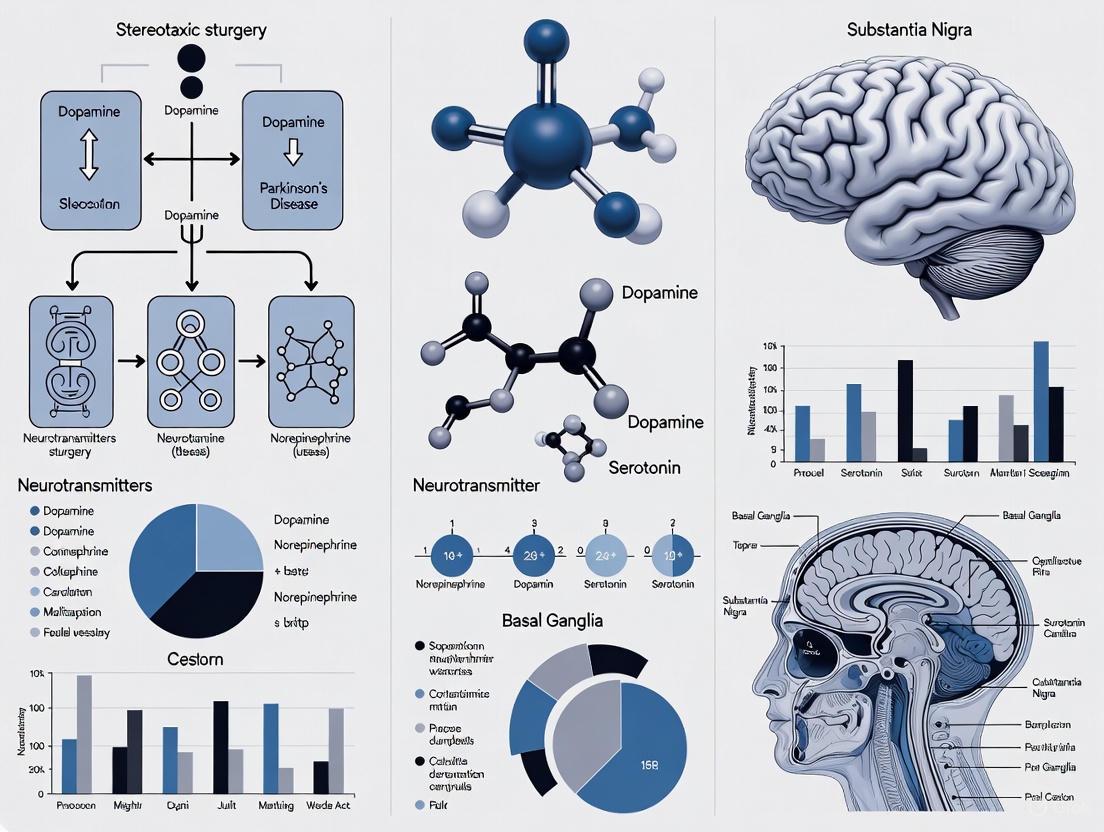
Diagram 1: Conceptual evolution from rate models to dynamic models explaining the "paradox of stereotaxic surgery." Both pallidotomy and DBS disrupt pathological oscillatory patterns to restore more normal timing dynamics in basal ganglia-thalamocortical circuits.
Modern Deep Brain Stimulation: Mechanisms and Methodologies
Contemporary DBS Approaches and Targets
Deep brain stimulation has become the most commonly performed surgical treatment for Parkinson's disease, typically considered for patients who have had PD for at least four years, experience significant "off" time or dyskinesias, but still respond beneficially to medication [5]. The procedure involves implanting thin metal wires (electrodes) into specific brain areas, connected to a battery-operated neurostimulator placed under the skin below the collarbone [5].
The primary targets for DBS in Parkinson's disease are:
- Subthalamic Nucleus (STN): Effective for addressing the full range of parkinsonian motor symptoms and allowing significant medication reduction.
- Globus Pallidus pars interna (GPi): Particularly effective for managing dyskinesias while providing good symptom control.
The exact mechanisms of DBS action remain incompletely understood, but many experts believe it regulates abnormal electrical signaling patterns in the brain [5]. In Parkinson's disease, normal communicative electrical signals between brain cells become irregular and uncoordinated, leading to motor symptoms. DBS appears to interrupt these irregular signaling patterns, enabling more normalized cellular communication and symptom reduction [5].
Table 2: Modern Deep Brain Stimulation Approaches for Parkinson's Disease
| Parameter | STN DBS | GPi DBS | Notes |
|---|---|---|---|
| Primary Benefits | Reduces full spectrum of motor symptoms; allows significant medication reduction | Excellent dyskinesia control; good symptom improvement | Choice depends on individual patient profile |
| Medication Reduction | Substantial | Moderate | |
| Cognitive Considerations | Higher risk of neurocognitive side effects | Possibly better cognitive safety profile | |
| Bilateral Safety | Safe for bilateral implantation | Safe for bilateral implantation | Significant advantage over bilateral pallidotomy |
Stereotactic Surgical Technique and Targeting
Modern DBS surgery represents the culmination of decades of refinement in stereotactic technique. The procedure typically involves:
Preoperative Planning: High-resolution MRI or CT imaging is performed with a stereotactic frame attached to the patient's head. For functional targeting, systems like the CRW stereotaxic system or Leksell system are utilized [6].
Target Localization: Surgical targets are identified relative to the anterior commissure-posterior commissure (AC-PC) line, with standardized coordinates (e.g., for pallidal targets: X = ±10–±13 mm, Y = −1 to −2 mm, Z = −2 to −6 mm from the midpoint of AC-PC) [6].
Intraoperative Confirmation: Patients are typically awake during surgery to provide feedback, though some centers now use imaging-guided placement while the patient is asleep [5]. Microelectrode recording may be used to map neuronal activity and confirm optimal target location [4].
Stimulator Implantation: After electrode placement, the neurostimulator is implanted and connected, with programming typically initiated several weeks postoperatively [5].
Advanced Targeting Technologies
Recent technological advances have significantly improved the precision of DBS targeting:
Three-Dimensional Mark Point Positioning: Advanced MRI algorithms enable precise feature positioning on patient images, improving surgical accuracy and outcomes [6]. Research has demonstrated that this approach yields markedly effective results in 92.5% of cases, compared to 87.5% with conventional methods [6].
Deep-Learning Based Segmentation: Automated segmentation techniques like GP-net use convolutional neural networks to precisely identify GPi and GPe boundaries from 7 Tesla MRI images, providing patient-specific segmentation without relying on atlas-based registration [7]. This approach accounts for interpatient variability more effectively than template-based methods.
High-Field MRI: The use of 7 Tesla MRI provides superior resolution for visualizing deep brain structures, enabling more accurate target identification [7].
Diagram 2: Modern DBS surgical workflow integrating advanced imaging, stereotactic targeting, and postoperative programming for optimal therapeutic outcomes.
Comparative Effectiveness and Safety Profiles
Outcomes of Pallidotomy Versus DBS
Rigorous comparison between pallidotomy and DBS reveals important differences in their effectiveness and risk profiles. Although no randomized clinical trials comparing these interventions have controlled for patient bias, analysis of available evidence suggests that unilateral pallidotomy provides approximately 20-30% reduction in 'off' total motor UPDRS scores, similar to the effects of unilateral GPi or STN DBS, though less than bilateral STN DBS [8].
The safety profiles of these procedures differ significantly. At experienced centers, the safety of unilateral pallidotomy appears equivalent to unilateral DBS. However, bilateral DBS is considerably safer than bilateral pallidotomy, which has limited its application for patients with symptoms affecting both sides of the body [8]. For dystonia, limited uncontrolled series suggest that bilateral pallidotomy is similar to GPi DBS in both effectiveness and safety [8].
Contemporary Role of Pallidotomy
Despite the dominance of DBS in modern surgical practice, pallidotomy remains a viable alternative in specific circumstances. It may be considered when DBS is not available, not feasible due to economic constraints, or when patients cannot tolerate implanted hardware [8]. Recent technical innovations, particularly focused ultrasound, have renewed interest in lesioning approaches by offering a less invasive method for creating precise lesions without open surgery [1].
Table 3: Quantitative Outcomes Comparison Between Surgical Modalities
| Assessment Measure | Unilateral Pallidotomy | Unilateral GPi DBS | Unilateral STN DBS | Bilateral STN DBS |
|---|---|---|---|---|
| UPDRS Motor Improvement ('off' medication) | 20-30% reduction | 20-30% reduction | 20-30% reduction | >30% reduction |
| Dyskinesia Reduction | Moderate | Significant | Significant | Significant |
| Medication Reduction | Minimal | Moderate | Substantial | Substantial |
| Risk of Serious Adverse Events | Low (unilateral) | Low (unilateral) | Low (unilateral) | Moderate |
Future Directions and Research Applications
Emerging Technologies and Approaches
The evolution of surgical therapies for Parkinson's disease continues with several promising developments:
Adaptive DBS: This next-generation approach measures brain waves to adapt stimulation parameters in real-time based on a patient's immediate symptoms, moving beyond constant stimulation to more targeted, efficient therapy [1]. Early clinical trials have demonstrated the feasibility of this closed-loop approach.
Early Intervention Studies: Recent research has suggested the potential of DBS to possibly slow the rate of Parkinson's progression, prompting investigations into the benefits of earlier surgical intervention [1].
Improved Battery Technology and Miniaturization: Efforts to extend battery life, design smaller devices, and integrate wireless technology aim to enhance patient experience and reduce the need for replacement surgeries [1].
Research Toolkit for Stereotactic Surgery
Modern investigations into stereotactic procedures utilize a sophisticated array of research tools:
Table 4: Essential Research Toolkit for Advanced Stereotactic Surgery Studies
| Research Tool | Application in Stereotactic Surgery | Research Utility |
|---|---|---|
| 7 Tesla MRI | High-resolution imaging of deep brain structures | Enables precise visualization of GPi, GPe, and lamina boundaries |
| Microelectrode Recording | Intraoperative neuronal activity mapping | Validates target location; research on pathological firing patterns |
| Deep-Learning Segmentation (e.g., GP-net) | Automated identification of target structures | Provides patient-specific segmentation; reduces atlas registration errors |
| Three-Dimensional Mark Point Positioning | Surgical planning and navigation accuracy | Improves targeting precision; reduces procedural variability |
| Adaptive DBS Platforms | Closed-loop stimulation systems | Enables research on dynamic neural correlates of symptoms |
The historical trajectory from pallidotomy to modern DBS represents more than simply technical progression in surgical technique. It exemplifies how therapeutic innovation can evolve through the interplay of clinical observation, theoretical modeling, and technological advancement. The journey has transformed our fundamental understanding of basal ganglia function, moving from simplistic rate-based models to more nuanced concepts of dynamic network interactions and timing-dependent modulation of cortical activity [2].
Within the broader context of stereotaxic surgery research, this evolution underscores several fundamental principles. First, it demonstrates the importance of reversible interventions in establishing causal relationships between brain circuits and behavior. Second, it highlights how therapeutic advances can drive basic scientific understanding, with clinical observations from both pallidotomy and DBS challenging and refining theoretical models of basal ganglia function. Finally, it illustrates the iterative nature of medical progress, where older approaches (like lesioning) may see renewed utility through technological innovation (such as focused ultrasound).
As research continues to refine DBS techniques and explore next-generation neuromodulation approaches, the historical lessons from this trajectory remain relevant. They remind us that today's standard therapies emerged from yesterday's paradoxes, and that current clinical challenges will likely drive the next transformative innovations in the surgical management of Parkinson's disease and other movement disorders.
The basal ganglia circuitry represents a complex network whose dysfunction underlies the pathophysiology of Parkinson's disease (PD). This whitepaper examines the pathophysiological basis for surgical targeting within this network, contextualized within the broader role of stereotaxic surgery in PD research. We synthesize current evidence detailing how distinct nodes within the cortico-basal ganglia-thalamo-cortical (CBTC) circuit contribute to PD symptomatology and how advanced neurosurgical interventions, particularly deep brain stimulation (DBS), achieve therapeutic effects by modulating these pathological networks. The analysis encompasses established targets including the subthalamic nucleus (STN) and globus pallidus internus (GPi), emerging targets such as the dentato-rubro-thalamic tract (DRTt), and the computational models that elucidate the "push-pull" dynamics of network regulation. This resource provides researchers, scientists, and drug development professionals with a technical framework for understanding the mechanistic foundations of surgical interventions in PD.
Parkinson's disease is characterized by the progressive degeneration of dopaminergic neurons in the substantia nigra pars compacta, initiating a cascade of pathological changes throughout the basal ganglia network [9]. The consequent dopamine depletion triggers an imbalance between the direct and indirect pathways of the basal ganglia, leading to excessive inhibitory output from the GPi to the thalamus and reduced thalamocortical excitation [10]. This model provides the fundamental pathophysiological rationale for surgical intervention within specific nodes of the CBTC circuit.
The clinical manifestations of PD, including bradykinesia, rigidity, and tremor, reflect these underlying circuit abnormalities. Traditionally, pharmacological treatments with levodopa have aimed to restore dopaminergic transmission. However, long-term treatment often leads to motor complications including dyskinesias and the "wearing-off" phenomenon, limitations that have driven the development of surgical alternatives [11]. Stereotaxic surgery, enabled by precise targeting and advanced neuroimaging, allows for direct modulation of the dysfunctional circuitry, offering symptomatic control when medications prove insufficient [10].
Pathophysiological Models of Basal Ganglia Dysfunction
The Classical Model: Direct and Indirect Pathway Imbalance
The classical model of PD pathophysiology centers on an imbalance between the direct (facilitatory) and indirect (inhibitory) pathways within the basal ganglia. Dopamine depletion in the striatum results in reduced activity in the direct pathway and increased activity in the indirect pathway. This leads to disinhibition of the STN and subsequent overactivation of the GPi, which excessively inhibits thalamocortical neurons, ultimately reducing motor cortex activity and producing the hallmark hypokinetic features of PD [10].
Network-Level Dysfunction and the "Push-Pull" Mechanism
Recent functional MRI studies have revealed a more complex, network-level dysfunction characterized by a "push-pull" effect within the CBTC circuit. Multi-scale computational models integrating fMRI data from PD patients have demonstrated that dopamine deficiency induces chain coupling variations that "push" the network into an abnormal state. Specifically, increased GABAergic projection from the basal ganglia to the thalamus worsens rigidity, while reduced GABAergic projection within the cortex exacerbates bradykinesia [12].
Conversely, DBS appears to "pull" the network toward a healthy state by alleviating rigidity through enhanced GABAergic projections within the basal ganglia and improving bradykinesia by reducing cortical projections to the basal ganglia [12]. This model provides a more nuanced understanding of how different PD motor symptoms arise from distinct circuit-level abnormalities and how targeted interventions can address these specific pathophysiological mechanisms.
Expanding the Circuitry: Cerebellar Integration
Growing evidence indicates that PD pathophysiology extends beyond the basal ganglia to include cerebellar connections. The DRTt, a cerebellar efferent pathway, has emerged as a significant contributor to tremor generation, particularly in tremor-dominant PD. This pathway begins in the dentate nucleus of the cerebellum, ascends via the superior cerebellar peduncle, and projects to the ventral lateral thalamus [10]. The identification of this pathway explains why some patients with predominant tremor symptoms may benefit from surgical targeting that extends beyond traditional basal ganglia nuclei.
Surgical Targets: Pathophysiological Rationale and Outcomes
Established Surgical Targets
Table 1: Established Surgical Targets in Parkinson's Disease
| Surgical Target | Primary Indications | Pathophysiological Rationale | Key Outcomes |
|---|---|---|---|
| Subthalamic Nucleus (STN) | Medication-resistant tremors, levodopa-induced dyskinesias, motor fluctuations [11] | Reduces pathological overactivity of STN that contributes to excessive GPi inhibition of thalamocortical pathways [10] | Improves UPDRS-III scores by >50%; reduces levodopa requirements; ameliorates bradykinesia, rigidity, and tremor [10] |
| Globus Pallidus Internus (GPi) | Dyskinesia-dominant PD, patients with cognitive or mood concerns [11] | Directly modulates excessive inhibitory output from GPi to thalamus, normalizing thalamocortical drive [11] | Effective for drug-induced dyskinesias (70-90% reduction); improves appendicular symptoms; potentially fewer neuropsychiatric effects than STN-DBS [11] |
| Ventral Intermediate Nucleus (VIM) | Tremor-dominant PD, medication-resistant tremor [11] | Interrupts tremor-generating circuitry in the cerebello-thalamo-cortical pathway [11] | Significant tremor reduction (up to 89% for essential tremor); less effective for other PD motor symptoms [10] |
Emerging and Adjunctive Targets
Table 2: Emerging Surgical Targets in Parkinson's Disease
| Surgical Target | Primary Indications | Pathophysiological Rationale | Key Outcomes |
|---|---|---|---|
| Dentato-Rubro-Thalamic Tract (DRTt) | Tremor-predominant PD, especially when conventional targeting is suboptimal [10] | Modulates cerebellar efferent pathways involved in tremor generation and coordination [10] | Simultaneous STN and DRTt stimulation produces superior tremor control; distance between DRTt and electric field predicts efficacy (AUC >0.9) [10] |
| Pedunculopontine Nucleus (PPN) | Axial symptoms (freezing of gait, postural instability) refractory to STN/GPi DBS [10] | Activates mesencephalic locomotor region with extensive connections to basal ganglia, thalamus, cerebellum, and spinal cord [10] | Improves gait initiation and reduces falls with low-frequency stimulation; results inconsistent due to anatomical variability and technical challenges [10] |
| Zona Incerta | Tremor-dominant PD, often combined with other targets [10] | Modulates tremor-related activity in cerebello-thalamo-cortical pathways; precise mechanism under investigation [10] | Emerging evidence for tremor control; often used as adjunctive target rather than primary intervention [10] |
Experimental Models and Methodologies for Circuit Investigation
Preclinical Models of Basal Ganglia Circuitry
Table 3: Experimental Models for Studying Basal Ganglia Circuitry in PD
| Model Type | Key Features | Utility for Circuit Investigation | Limitations |
|---|---|---|---|
| 6-OHDA Lesioned Rat | Unilateral dopaminergic lesion; robust rotational behavior | Classic model for screening therapeutic interventions; assessment of motor asymmetry [9] | Limited representation of progressive neurodegeneration and non-motor symptoms |
| MPTP-Treated Non-Human Primate | Bilateral dopaminergic depletion; development of motor symptoms and dyskinesias | Gold standard for predictive efficacy of surgical and pharmacological interventions [9] | High cost and ethical considerations; limited availability |
| Alpha-Synuclein Pre-Formed Fibrils (PFF) | Progressive neurodegeneration; Lewy body-like pathology | Models disease progression and proteinopathy spread through connected circuits [9] | Variable timeline of pathology development between subjects |
| Genetic Models (LRRK2, GBA, Parkin) | Monogenic forms of PD; specific molecular pathways | Investigation of specific pathogenic mechanisms underlying circuit dysfunction [9] | Most models do not fully recapitulate sporadic PD pathology and progression |
Neuroimaging and Electrophysiological Protocols
High-Resolution MRI and Diffusion Tensor Imaging Tractography Protocol:
- Application: Preoperative surgical planning for DBS electrode placement [10]
- Methodology: High-resolution structural MRI (3T or higher) combined with diffusion-weighted imaging for fiber tracking
- Target Visualization: Enables precise mapping of STN borders and reconstruction of adjacent white matter tracts, including DRTt
- Implementation: Integration with surgical navigation systems for stereotactic planning; patient-specific models of electric field propagation
Local Field Potential (LFP) Recording Protocol:
- Application: Identification of pathological biomarkers for adaptive DBS [13]
- Methodology: Intraoperative or chronic recording of oscillatory activity from implanted DBS electrodes
- Key Biomarkers: Beta band (13-30 Hz) oscillations in STN correlated with bradykinesia and rigidity; tremor-frequency oscillations
- Therapeutic Application: Closed-loop algorithms that modulate stimulation intensity based on real-time biomarker fluctuations
Functional MRI (fMRI) Network Analysis Protocol:
- Application: Investigation of large-scale network alterations in PD and DBS effects [12]
- Methodology: Resting-state fMRI to assess functional connectivity between nodes of the CBTC circuit
- Analytical Approach: Seed-based connectivity or independent component analysis to identify network-level changes
- Multi-Scale Integration: Combination with computational modeling to infer microscopic coupling parameters from macroscopic connectivity data
Advanced Surgical Technologies and Future Directions
Stereotactic Targeting and Surgical Delivery
Modern stereotactic surgery for PD employs frameless or frame-based systems with submillimeter accuracy. Surgical workflow typically includes:
- Preoperative Planning: Fusion of high-resolution MRI with CT for coordinate determination and trajectory planning
- Intraoperative Confirmation: Microelectrode recording to map electrophysiological signatures of target nuclei and identify borders
- Therapeutic Delivery: Implantation of DBS electrodes or performance of lesioning procedures (pallidotomy, thalamotomy)
- Postoperative Verification: CT or MRI confirmation of accurate lead placement [11]
Focused ultrasound (FUS) represents a non-invasive alternative for creating precise lesions, particularly for unilateral procedures such as thalamotomy for tremor control. However, long-term data on FUS outcomes remain limited compared to established DBS approaches [11].
Adaptive Deep Brain Stimulation
Adaptive DBS (aDBS) represents a significant advancement in neuromodulation technology, delivering dynamic, symptom-contingent stimulation rather than continuous fixed-parameter stimulation. aDBS systems utilize real-time feedback from neural biomarkers (typically local field potentials) to adjust stimulation parameters [13].
According to a recent Delphi consensus study among DBS experts, aDBS is expected to become clinical routine within the next decade. Key research priorities include:
- Simplification of implantation and programming procedures
- Development of more reliable biomarkers for diverse PD phenotypes
- Integration with artificial intelligence for automated parameter optimization
- Expansion to control more complex symptoms, including non-motor features [13]
Gene Therapy Approaches
Gene therapy strategies represent an emerging frontier in surgical treatment of PD, though most remain in early clinical stages. Approaches include:
- AAV2-hAADC: Enhances expression of aromatic L-amino acid decarboxylase to improve conversion of levodopa to dopamine
- ProSavin: Uses viral vectors to deliver genes for dopamine synthesis enzymes Preliminary studies have demonstrated early-phase safety and efficacy, but larger-scale controlled trials are needed to establish long-term outcomes [11].
Visualization of Key Circuitry and Surgical Approaches
Surgical Targets in Basal Ganglia Circuitry
This diagram illustrates the key nodes and connections within the cortico-basal ganglia-thalamo-cortical circuit, highlighting established and emerging surgical targets for Parkinson's disease. The direct pathway (green) facilitates movement, while the indirect pathway (red) inhibits movement. Dopamine from the substantia nigra pars compacta (SNc) modulates both pathways. Surgical targets (STN, GPi, and the cerebellar DRTt) are indicated with dashed connections, representing sites for deep brain stimulation or lesioning procedures to restore circuit balance.
The Scientist's Toolkit: Research Reagent Solutions
Table 4: Essential Research Reagents for Basal Ganglia Circuit Investigation
| Reagent/Technology | Application | Function in Research |
|---|---|---|
| Alpha-Synuclein Pre-Formed Fibrils (PFF) | Modeling PD pathology | Induces progressive, spreading synucleinopathy in neuronal cultures and animal models [9] |
| AAV Vectors (e.g., AAV2-hAADC) | Gene therapy research | Delivers therapeutic genes to specific nodes of the basal ganglia circuitry [11] |
| 6-Hydroxydopamine (6-OHDA) | Dopaminergic lesioning | Creates selective nigrostriatal pathway lesions in rodent models for circuit dysfunction studies [9] |
| Izhikevich Neuron Model | Computational modeling | Simulates neural dynamics in brain areas including basal ganglia and thalamus with biological realism [12] |
| Local Field Potential (LFP) Recording Systems | Electrophysiological monitoring | Captures oscillatory activity from deep brain structures to identify pathological biomarkers [13] |
| Diffusion Tensor Imaging (DTI) | Tractography | Reconstructs white matter pathways connecting nodes of the basal ganglia circuit [10] |
The pathophysiological rationale for surgical targets in Parkinson's disease continues to evolve from a focus on discrete nuclei to a network-level understanding of basal ganglia circuitry. The classical model of direct/indirect pathway imbalance provides the foundation for targeting the STN and GPi, while emerging research highlights the importance of cerebellar connections through the DRTt for tremor control and network-level "push-pull" dynamics that differentiate symptom-specific mechanisms. Stereotaxic surgery serves not only as a therapeutic modality but as a research tool for elucidating the complex pathophysiology of PD. Future directions include closed-loop adaptive stimulation, gene therapy approaches, and the integration of multi-scale computational models with patient-specific data to optimize surgical targeting and outcomes. For researchers and drug development professionals, understanding these circuit-level mechanisms provides crucial insights for developing next-generation interventions that more precisely address the network dysfunction underlying Parkinson's disease.
The surgical treatment of Parkinson's disease (PD) has undergone a significant revival over recent decades, driven by advancements in stereotaxic surgery and a deeper understanding of basal ganglia-thalamocortical circuits [2] [11]. The core motor symptoms of PD—tremor, bradykinesia, rigidity, and axial symptoms like postural instability—arise from dysfunctional neural networks, making them suitable targets for precise neuromodulation [14]. This technical guide examines the key anatomical targets—the subthalamic nucleus (STN), globus pallidus internus (GPi), and thalamus—within the context of stereotaxic surgery, detailing their roles in symptom management, experimental methodologies for investigation, and quantitative outcomes that inform modern therapeutic strategies.
Anatomical and Physiological Foundations of PD Targets
Basal Ganglia-Thalamocortical Circuitry in PD
The basal ganglia (BG) are interconnected subcortical nuclei acting as the primary receptive nucleus for cortical input [2]. Standard "rate" models describe BG function via direct and indirect pathways from the striatum to BG output nuclei (GPi and SNr). In PD, dopaminergic neuron loss in the substantia nigra pars compacta (SNc) causes imbalanced pathway activity, leading to excessive inhibitory output from GPi/SNr to the motor thalamus (BG-Mthal) and suppressed cortical motor drive [2] [10]. This results in bradykinesia and rigidity. The STN, a glutamatergic nucleus within the indirect pathway, becomes overactive in PD, further exciting GPi/SNr and exacerbating motor suppression [10]. While foundational, the rate model faces a challenge known as the "paradox of stereotaxic surgery": both lesions (e.g., pallidotomy) and high-frequency stimulation of GPi improve PD symptoms, suggesting mechanisms beyond simple rate changes are involved [2].
Key Surgical Targets and Their Roles
- Subthalamic Nucleus (STN): A small, lens-shaped glutamatergic nucleus. Its pathological overactivity in PD contributes to bradykinesia, rigidity, and tremor. Modulation via DBS disrupts this aberrant activity, making it the most common DBS target [10].
- Globus Pallidus internus (GPi): The primary inhibitory output nucleus of the BG. Its overactivity in PD excessively inhibits the thalamus. DBS or lesioning of GPi reduces this inhibition, alleviating hypokinetic and hyperkinetic symptoms (e.g., dyskinesias) [2] [15].
- Motor Thalamus (specifically the Ventral Intermediate Nucleus - VIM): The BG-recipient zone of the thalamus (BG-Mthal) and the cerebellar-recipient zone (CB-Mthal) are critical hubs. While not a primary target for multi-symptom PD management, the VIM nucleus of the thalamus is effectively targeted for medication-resistant tremor [16] [11].
Diagram: Simplified Basal Ganglia Pathways in Normal and Parkinsonian States. The model shows balanced direct (green, movement-promoting) and indirect (red, movement-suppressing) pathways under normal conditions. In Parkinson's disease (dashed outline), dopaminergic loss from SNc leads to an overactive indirect pathway and increased inhibitory output from GPi to thalamus, suppressing movement. STN overactivity further exacerbates this. Targets for stereotaxic surgery (STN, GPi) are highlighted.
Quantitative Outcomes of Stereotaxic Interventions
Comparative Efficacy of STN-DBS vs. GPi-DBS
Systematic evaluation of stereotaxic interventions relies on standardized metrics like the Unified Parkinson's Disease Rating Scale (UPDRS). Meta-analyses of randomized controlled trials provide robust quantitative comparisons between targets.
Table 1: Meta-Analysis of Bilateral STN-DBS vs. GPi-DBS Outcomes (6-12 Month Follow-up)
| Outcome Measure | STN-DBS Improvement | GPi-DBS Improvement | Comparative Notes |
|---|---|---|---|
| UPDRS-III (Motor) | 50.5% reduction (OFF-med) [17] | 29.8% reduction (OFF-med) [17] | STN-DBS shows greater improvement in motor scores [17]. |
| UPDRS-II (ADL) | 47% reduction (OFF-med) [17] | 18.5% reduction (OFF-med) [17] | STN-DBS shows greater improvement in activities of daily living in the OFF-medication state [17]. |
| Levodopa Equivalent Daily Dose (LEDD) | ~50% reduction [15] [17] | No significant reduction [15] | A key advantage of STN-DBS is enabling major medication reduction [15]. |
| Dyskinesia | ~64% improvement [17] | Significant improvement, potentially superior to STN [15] | GPi-DBS may have an advantage in direct dyskinesia suppression [15]. |
| Postoperative ADLs (ON-med) | — | Better improvement than STN-DBS [15] | GPi-DBS may offer better ON-medication daily function [15]. |
Target-Specific Symptom Management Profiles
Choosing a surgical target involves matching its symptom profile to the patient's most burdensome symptoms.
Table 2: Symptom-Specific Management Profiles of Key Surgical Targets
| Primary Target | Most Responsive Symptoms | Key Clinical Considerations |
|---|---|---|
| Subthalamic Nucleus (STN) | Bradykinesia, Rigidity, Tremor [16] [10] | Allows significant reduction of dopaminergic medication; may be preferable for patients with significant medication-related side effects [15] [17]. |
| Globus Pallidus internus (GPi) | Dyskinesias, Dystonia, All cardinal motor symptoms [15] [11] | Superior for direct dyskinesia suppression; less associated with speech/ cognitive declines; may be preferred for patients with significant dyskinesia or cognitive concerns [15] [17]. |
| Thalamus (VIM) | Tremor [16] [11] | Highly effective for PD tremor but has little effect on bradykinesia, rigidity, or other symptoms; typically reserved for patients with severe, medication-resistant tremor as the dominant feature [16]. |
Advanced Experimental Protocols and Network-Based Targeting
Protocol for Mapping Symptom-Specific Networks with DBS Fiber Filtering
The recognition that different PD symptoms map to distinct brain networks enables more personalized DBS. The following protocol, derived from a large-scale multicenter study, details the methodology for identifying symptom-response tracts [14].
Objective: To identify the white matter tracts associated with improvement in tremor, bradykinesia, rigidity, and axial symptoms following STN-DBS.
Materials & Methods:
- Cohort: N = 129 patients with PD from three independent centers undergoing bilateral STN-DBS.
- Clinical Assessment: Preoperative and postoperative (6-12 months) UPDRS-III motor scores, with items subdivided into symptom subscales (tremor, bradykinesia, rigidity, axial).
- Imaging: Preoperative T1-weighted and diffusion-weighted MRI (dMRI) for tractography. Postoperative CT for precise localization of DBS leads.
- Computational Modeling:
- Lead Localization & Volume of Tissue Activated (VTA) Modeling: Co-register postoperative CT with preoperative MRI. Reconstruct electrode positions and model the VTA for each patient's stimulation settings.
- Tractography Reconstruction: Use dMRI to reconstruct a whole-brain connectome. Employ an extended DBS tractography atlas to define fibers passing through the subthalamic region.
- DBS Fiber Filtering: Calculate the overlap between the patient-specific VTA and the predefined fiber tracts. Correlate the amount of overlap with the clinical improvement for each symptom domain.
- Statistical Analysis & Cross-Validation: Apply False Discovery Rate (FDR) correction for multiple comparisons. Validate the resulting symptom-tract models using permutation testing and 10-fold cross-validation.
Key Findings: The analysis revealed a distinct rostrocaudal gradient of symptom-specific tracts within the STN region [14]:
- Tremor: Associated with stimulation of tracts connected to the primary motor cortex and the cerebellothalamic pathway.
- Rigidity: Linked to tracts connected to the pre-supplementary motor area (pre-SMA).
- Bradykinesia: Associated with tracts from the SMA entering the medial STN.
- Axial Symptoms: Mapped to tracts from the SMA terminating laterally in the STN and extending to the brainstem near the pedunculopontine nucleus (PPN).
Diagram: Workflow for DBS Fiber-Filtering Analysis. This experimental protocol integrates patient imaging, stimulation parameters, and clinical scores to identify white matter tracts whose modulation leads to improvement in specific Parkinson's disease symptoms.
Table 3: Key Research Reagents and Resources for DBS Investigation
| Resource / Tool | Function / Application | Specific Examples / Notes |
|---|---|---|
| High-Field MRI & dMRI | Provides high-resolution anatomical imaging and reconstruction of white matter tracts via tractography. | Critical for visualizing STN, GPi, and the dentato-rubro-thalamic tract (DRTt); used for surgical targeting and research [14] [10]. |
| Stereotactic Planning Software | Platforms for surgical planning, electrode localization, and computational modeling of stimulation fields. | Lead-DBS; allows for VTA modeling, fiber filtering, and integration with tractography data [14]. |
| Unified Parkinson's Disease Rating Scale (UPDRS) | The gold standard clinical tool for quantifying PD severity and motor symptom improvement. | Part III (motor examination) is essential for evaluating DBS outcomes; sub-scores used for symptom-specific analysis [17] [14]. |
| DBS Tractography Atlas | A predefined atlas of white matter pathways relevant to DBS targets. | Serves as a reference for the "DBS fiber-filtering" method to identify symptom-response circuits [14]. |
| Computational Models (VTA/PAM) | Simulates the electrical field generated by DBS and its effect on neural tissue. | Volume of Tissue Activated (VTA) or Pathway Activation Models (PAM) are used to estimate the neural substrate modulated by stimulation [14] [10]. |
Emerging Targets and Future Directions in Stereotaxy
Beyond the Classical Targets: The Dentato-Rubro-Thalamic Tract (DRTt)
While STN and GPi remain primary targets, recent evidence highlights the importance of the dentato-rubro-thalamic tract (DRTt), a cerebellar efferent pathway, for tremor control [10]. Co-stimulation of the STN and the nearby DRTt leads to superior motor outcomes and greater tremor reduction than STN stimulation alone. The distance between the stimulation field and the DRTt is a robust predictor of efficacy, underscoring the value of patient-specific tractography-guided planning to optimize outcomes, especially in tremor-dominant PD [10].
Network-Based Paradigms and Adaptive DBS
The field is shifting from focal stimulation to network-based neuromodulation. The identification of symptom-specific tracts enables "network blending," where stimulation parameters are optimized to modulate multiple circuits based on a patient's unique symptom profile [14]. Furthermore, the integration of sensing technologies allows for the development of adaptive DBS (aDBS) systems. These closed-loop systems can record neural biomarkers (e.g., beta oscillations) and adjust stimulation in real-time, promising more effective and energy-efficient therapy [18].
Stereotaxic surgery, epitomized by DBS, represents a cornerstone in the management of advanced Parkinson's disease. The STN, GPi, and thalamus serve as key anatomical nodes whose modulation effectively alleviates specific motor symptoms. While quantitative outcomes demonstrate the robust efficacy of these targets, the "paradox of stereotaxic surgery" indicates that our understanding of the underlying mechanisms continues to evolve. The future of the field lies in moving beyond a one-size-fits-all approach. By leveraging advanced neuroimaging, computational modeling, and a nuanced understanding of symptom-specific brain networks, researchers and clinicians are advancing toward a paradigm of truly personalized, network-based neuromodulation for Parkinson's disease.
The introduction of levodopa in the late 1960s marked a revolutionary turning point in the management of Parkinson's disease (PD), fundamentally altering the treatment landscape and reshaping surgical indications for decades to follow. Before this medical breakthrough, functional neurosurgery, primarily consisting of ablative procedures such as pallidotomy and thalamotomy, represented the only intervention offering symptomatic relief for debilitating parkinsonian symptoms [11] [19]. The profound efficacy of levodopa in ameliorating core motor symptoms led to an abrupt decline in surgical interventions for PD, effectively ending what is often termed the "pre-levodopa era" of Parkinson's surgery [11] [20]. However, the long-term limitations of levodopa therapy, particularly the emergence of motor fluctuations and levodopa-induced dyskinesias (LID), eventually uncovered a new therapeutic niche for surgical approaches [19]. This whitepaper examines how the evolution of medical therapy for PD catalyzed the development of modern stereotactic neurosurgical techniques, transforming surgical indications from primary symptomatic treatment to management of medication-resistant complications, thereby fostering innovation in neuromodulation and precise brain circuit intervention.
The Pre-Levodopa Surgical Landscape
Early Ablative Procedures
Prior to the levodopa era, surgical management of Parkinson's disease relied exclusively on lesioning specific brain structures within the basal ganglia-thalamocortical pathways. These early procedures were pioneered with limited understanding of basal ganglia circuitry but were encouraged by serendipitous observations, such as the accidental ligation of the anterior choroidal artery by Cooper in 1953, which unexpectedly alleviated tremor and rigidity on the contralateral side despite a concerning mortality rate of approximately 10% [11] [19]. The initial surgical targets included:
- Cerebral cortex: Early attempts by Bucy and Case in the 1930s involved removal of the cerebral cortex to treat Parkinsonian tremors, but these procedures were abandoned due to unacceptable side effects like hemiparesis [11].
- Globus Pallidus (Pallidotomy): Lesioning of the globus pallidus internus (GPi) emerged as a common procedure in the 1950s and 1960s to treat PD, dystonia, and other movement disorders [11].
- Thalamus (Thalamotomy): Thalamotomy gained popularity for its efficacy in reducing Parkinsonian tremor, particularly before the widespread understanding of basal ganglia circuitry [11].
These early ablative procedures were performed using rudimentary stereotactic techniques with limited precision. The introduction of stereotactic frames by Spiegel et al. represented a significant advancement, allowing for more accurate targeting of deep brain structures through electrical coagulation of the globus pallidus, thalamus, and ansa lenticularis [11]. However, these procedures faced criticism due to insufficient long-term follow-up, concerns about morbidity, and limited engagement from medical neurologists, which contributed to inaccurate reporting of outcomes [11].
Physiological Insights from Early Surgery
Paradoxically, these early surgical interventions provided crucial insights into the pathophysiology of Parkinson's disease, despite their limited therapeutic success. The observation that lesions to specific basal ganglia structures could ameliorate certain motor symptoms without causing catastrophic neurological deficits suggested the potential for targeted circuit manipulation. This formed the conceptual foundation for what would later be recognized as the "paradox of stereotaxic surgery" – how both lesions and stimulation of similar targets could produce therapeutic effects apparently contradicting standard rate models of basal ganglia function [2]. The clinical experiences from this era gradually revealed that the motor circuit dysfunction in PD could be modified by interrupting pathological signaling at specific nodes, a principle that would later underpin the development of deep brain stimulation.
Table 1: Historical Evolution of Surgical Approaches in Parkinson's Disease
| Era | Primary Surgical Approach | Key Limitations | Major Technological Advances |
|---|---|---|---|
| 1930s-1950s | Open ablative procedures (cortical removal) | High morbidity, hemiparesis, ~10% mortality | Development of early stereotactic principles |
| 1950s-1960s | Stereotactic pallidotomy & thalamotomy | Inconsistent outcomes, limited follow-up | Stereotactic frames, electrical coagulation techniques |
| 1970s-1980s | Sharp decline in all surgical interventions | Near-complete abandonment of surgery | N/A (Medical therapy dominance) |
| 1990s-Present | Deep brain stimulation (DBS) | Hardware-related risks, cost limitations | Implantable pulse generators, directional leads |
The Levodopa Revolution and Surgical Decline
Mechanism of Action and Initial Impact
Levodopa's mechanism of action addresses the core neurochemical deficit in Parkinson's disease – the progressive loss of dopaminergic neurons in the substantia nigra pars compacta and the consequent depletion of striatal dopamine. As a metabolic precursor to dopamine, levodopa crosses the blood-brain barrier and is converted to dopamine via the enzyme aromatic L-amino acid decarboxylase (AADC), thereby restoring dopaminergic neurotransmission in the striatum [21]. The addition of carbidopa, a peripheral decarboxylase inhibitor, in 1975 significantly enhanced levodopa's efficacy by preventing its premature conversion to dopamine in the periphery, increasing brain bioavailability and reducing peripheral side effects [21].
The dramatic symptomatic improvement provided by levodopa therapy led to an rapid and nearly complete abandonment of surgical procedures for Parkinson's disease throughout the 1970s and 1980s [11] [22]. The "honeymoon period" characterized by stable reduction of motor symptoms with dopaminergic therapy rendered the risk-benefit ratio of ablative surgeries unacceptable to most patients and clinicians [19]. Neurosurgical programs focused on movement disorders experienced a sharp decline, and research into improved surgical techniques stagnated during this period as the medical community embraced levodopa as the definitive treatment for PD.
Emergence of Long-Term Complications
Despite its remarkable efficacy, long-term levodopa therapy revealed significant limitations that would eventually reopen the door for surgical interventions. As PD progresses, the therapeutic window of levodopa narrows considerably, such that previously effective doses no longer provide adequate motor symptom control without negative side effects [21]. The key long-term complications include:
- Motor fluctuations: Alternating states between periods when motor symptoms are well-controlled ("On" time) and when symptoms reappear ("Off" time) [21].
- Levodopa-induced dyskinesias (LID): Involuntary muscle movements that emerge after several years of treatment, affecting nearly 90% of patients after 10 years of therapy [19].
- Wearing-off phenomena: The gradual shortening of levodopa's duration of effect, leading to end-of-dose deterioration and recurrence of parkinsonian symptoms [21].
The pathophysiology of these complications involves both progressive nigrostriatal denervation and pulsatile dopaminergic stimulation. As the disease advances, the loss of dopaminergic terminals reduces the brain's capacity to buffer fluctuations in synaptic dopamine levels, leading to discontinuous stimulation of dopamine receptors and subsequent alterations in basal ganglia circuitry [19]. Pharmacological strategies to manage these complications, including adjusting dosing regimens, employing controlled-release formulations, and adding adjunctive medications, often provide only partial relief and may introduce new side effects [21].
Renaissance of Surgical Approaches
Deep Brain Stimulation: From Concept to Clinical Application
The limitations of long-term levodopa therapy catalyzed a renaissance in surgical approaches to Parkinson's disease, culminating in the development and refinement of deep brain stimulation (DBS). The modern era of DBS began with Benabid and colleagues' pioneering work in the late 1980s and early 1990s, demonstrating that high-frequency stimulation of the ventral intermediate nucleus (VIM) of the thalamus could effectively suppress Parkinsonian tremor [11] [22]. This breakthrough represented a paradigm shift from destructive lesions to reversible, adjustable neuromodulation.
DBS hardware consists of electrodes (leads) implanted stereotactically into specific brain targets, connected to an implantable pulse generator (IPG) typically placed in a subclavicular pocket [19]. The procedure involves microelectrode recording and intraoperative macrostimulation to optimize target localization and assess clinical benefits and potential adverse effects [19]. Unlike ablative procedures, DBS offers the advantages of reversibility, adjustability through programmer settings, and the ability to perform bilateral procedures without the high risk of permanent side effects associated with bilateral lesions [19].
The established targets for DBS in PD include:
- Subthalamic nucleus (STN): The most common target worldwide, effective for tremor, rigidity, bradykinesia, and allowing significant reduction of dopaminergic medication [11] [19].
- Globus pallidus internus (GPi): Particularly effective for managing levodopa-induced dyskinesias while providing good motor symptom control [11] [19].
- Ventral intermediate nucleus (VIM) of thalamus: Primarily for tremor-predominant PD, with limited effect on other cardinal symptoms [19].
Diagram 1: Evolution of surgical approaches through the levodopa era. The therapeutic revolution initially caused a surgical decline, but long-term complications ultimately drove a renaissance of refined surgical approaches.
Contemporary Surgical Indications in the Levodopa Era
The emergence of DBS has established clear indications for surgical intervention in Parkinson's disease, all directly related to limitations of medical therapy:
- Medication-resistant tremors: Patients with tremors that persist despite optimal dopaminergic therapy [11] [19].
- Motor complications: Significant "wearing-off" phenomena, unpredictable motor fluctuations, and disabling dyskinesias that cannot be adequately managed through medication adjustments [11] [19].
- Intolerance to dopaminergic medications: Patients who develop unacceptable side effects from levodopa or other Parkinson's medications [11].
- Dose-limiting dyskinesias: Patients whose anti-parkinsonian treatment is limited by the emergence of dyskinesias before adequate motor symptom control is achieved [19].
Critical to appropriate patient selection is the concept of "levodopa responsiveness" – patients must demonstrate significant improvement in motor symptoms with levodopa challenges to be considered good surgical candidates, as DBS primarily improves the motor features that respond to dopaminergic therapy [19]. Additional selection criteria include the absence of significant cognitive impairment or psychiatric comorbidities, reasonable surgical risk, and realistic patient expectations [19].
Table 2: Modern Surgical Options for Advanced Parkinson's Disease
| Procedure | Primary Indications | Advantages | Limitations | Impact on Levodopa Therapy |
|---|---|---|---|---|
| STN DBS | Motor fluctuations, tremor, rigidity, bradykinesia | Allows significant medication reduction (40-60%) | Cognitive/psychiatric risks; worsening of axial symptoms long-term | Often enables lower levodopa doses |
| GPi DBS | Levodopa-induced dyskinesias, motor fluctuations | Direct antidyskinetic effect; stable medication requirements | Less medication reduction possible compared to STN | May allow optimization of levodopa dosing |
| Pallidotomy | Unilateral dyskinesias, asymmetric symptoms (when DBS not feasible) | Single procedure, no hardware maintenance | Irreversible; bilateral procedures risk speech/swallowing deficits | May improve response to existing levodopa doses |
| Focused Ultrasound (FUS) | Medication-resistant tremor | Non-invasive, no implants | Limited long-term data; primarily for unilateral symptoms | Adjunctive to ongoing medical therapy |
Advanced Surgical Technologies and Methodologies
Stereotactic Surgical Protocol for DBS Implantation
The modern surgical approach to DBS electrode implantation represents a sophisticated integration of imaging, electrophysiology, and clinical assessment. The following protocol outlines the key methodological steps:
Preoperative Imaging and Targeting:
- High-resolution MRI (T1, T2, SWI sequences) acquired with stereotactic fiducials
- Direct and indirect targeting methods combined to identify STN (dorsolateral), GPi (posteroventral), or VIM
- Coordinates relative to midcommissural point: STN: 1-3mm posterior, 9-12mm lateral, 4-5mm inferior; GPi: 2-3mm anterior, 18-22mm lateral, 4-6mm inferior [19]
Intraoperative Physiological Confirmation:
- Microelectrode recording (MER) to identify characteristic neuronal activity patterns:
- STN: Irregular, high-frequency (25-45 Hz) activity with kinesthetic cells
- GPi: High-frequency discharge with tremor-related cells
- Border regions: Decreased activity transitioning from GPe to GPi [19]
- Macrostimulation for clinical effect assessment:
- Test stimulation (typically 60-180 μs, 1-10V, 130-185 Hz)
- Assess tremor reduction, rigidity improvement, capsular side effects (muscle contractions), visual phenomena (optic tract stimulation) [19]
- Microelectrode recording (MER) to identify characteristic neuronal activity patterns:
Lead Placement and Pulse Generator Implantation:
- Final DBS lead placement with 4-8 contacts
- Intraoperative fluoroscopy to confirm lead stability
- Connection to extension cables and implantation of IPG in infraclavicular pocket [19]
Postoperative Programming and Medication Adjustment:
- Initial programming 2-4 weeks postoperatively
- Systematic testing of contacts, parameters (voltage, pulse width, frequency)
- Gradual medication reduction (especially for STN DBS) over subsequent weeks [19]
The Scientist's Toolkit: Key Research Reagents and Technologies
Table 3: Essential Research Materials for Parkinson's Disease Surgical Research
| Research Tool | Function/Application | Technical Notes |
|---|---|---|
| Stereotactic Frames | Precise targeting of deep brain structures | Modern frameless systems available; compatibility with MRI/CT essential |
| Microelectrodes | Single-unit recording for physiological confirmation | Tungsten or platinum-iridium; impedance 0.5-1.5 MΩ; multiple parallel trajectories possible |
| Deep Brain Stimulation Leads | Chronic neuromodulation delivery | Directional leads with segmented electrodes now available for current steering |
| Implantable Pulse Generators | Power source and parameter control | Rechargeable and non-rechargeable options; current-controlled or voltage-controlled |
| Unified Parkinson's Disease Rating Scale (UPDRS) | Standardized assessment of motor and non-motor symptoms | Part III (motor examination) critical for pre/post-operative assessment |
| Levodopa Challenge Test | Assessment of dopaminergic responsiveness | Administer supramaximal levodopa dose; measure UPDRS improvement pre-operatively |
| Seed Amplification Assays (SAA) | Detection of pathological α-synuclein in CSF | Emerging biomarker for biological diagnosis; used in research frameworks [23] |
Future Directions: Integration of Medical and Surgical Therapies
Emerging Technologies and Approaches
The evolution of surgical indications for Parkinson's disease continues to advance beyond conventional DBS, with several emerging technologies poised to further reshape the therapeutic landscape:
- Directional DBS Leads: These advanced leads with segmented electrodes allow for more precise current steering, potentially improving therapeutic benefits while reducing side effects through selective stimulation of specific fiber pathways [22].
- Adaptive DBS (aDBS): Closed-loop systems that adjust stimulation parameters in real-time based on recorded neural signals (such as beta oscillations) may provide more effective symptom control while optimizing battery consumption [22].
- Focused Ultrasound (FUS): Magnetic resonance-guided focused ultrasound offers a non-invasive alternative for creating precise lesions (thalamotomy, pallidotomy) without craniotomy, though long-term data remains limited [11] [24].
- Gene Therapy Approaches: Investigational strategies including AAV2-hAADC (to enhance dopamine production), ProSavin (lentiviral vector containing GCH1-TH-AADC), and AAV2-GAD (to modulate neurotransmitter balance) represent novel biological interventions, though these remain in early clinical stages [25].
Diagram 2: Causal relationship between medical therapy limitations and surgical innovation. Long-term complications of levodopa directly stimulated development of advanced surgical approaches, now evolving toward integrated treatment paradigms.
Biological Redefinition of Parkinson's Disease and Surgical Implications
Recent proposals for biological definitions of Parkinson's disease, including the "Neuronal Synuclein Disease Integrated Staging System" (NSD-ISS) and "SynNeurGe" classification system, represent a paradigm shift from clinically-defined to biologically-defined disease entities [23]. These frameworks prioritize the presence of α-synucleinopathy, neurodegeneration, and genetic factors over clinical manifestations, aiming to identify disease in its biologically early stages [23]. This approach has profound implications for future surgical interventions:
- Earlier Intervention: As biological diagnoses enable identification of PD before significant neurodegeneration occurs, surgical interventions might eventually be considered at earlier disease stages to prevent circuit-level abnormalities.
- Targeted Therapies: The recognition of biological subtypes may lead to more personalized surgical approaches, with specific targets or stimulation parameters tailored to individual patterns of network dysfunction.
- Disease Modification: Future surgical strategies may combine neuromodulation with targeted delivery of neuroprotective agents (e.g., via convection-enhanced delivery or viral vectors) to alter disease progression rather than merely alleviate symptoms.
The journey through the levodopa era has fundamentally transformed the surgical management of Parkinson's disease, creating a dynamic interplay between medical and surgical approaches that continues to evolve. Levodopa therapy initially rendered ablative surgeries nearly obsolete but ultimately created new indications for surgical intervention by revealing its own long-term limitations. This therapeutic challenge catalyzed the development of deep brain stimulation, which has established itself as a powerful treatment for medication-resistant symptoms. The current landscape is characterized by sophisticated patient selection criteria centered on levodopa responsiveness, advanced stereotactic techniques for precise targeting, and a growing array of surgical options. As we enter an era of biological disease definition and targeted therapies, the integration of medical and surgical approaches will likely become increasingly sophisticated, potentially intervening earlier in the disease course and offering more personalized therapeutic strategies based on individual patterns of network dysfunction and underlying pathology.
The management of Parkinson's disease (PD) has long been dominated by pharmacological strategies, primarily levodopa, which remains the gold standard for symptomatic control. However, the progressive nature of PD and the eventual emergence of medication-resistant symptoms and complications—including refractory tremors, motor fluctuations, and levodopa-induced dyskinesias—have necessitated a rediscovery of surgical interventions. This whitepaper examines the re-emergence and evolution of stereotactic surgery as a pivotal therapeutic strategy within the broader context of PD research, addressing the critical gap left by the declining efficacy of long-term dopaminergic therapy. The limitations of medication are well-documented; while levodopa provides dramatic initial benefits, its long-term utility is often compromised as the degeneration of dopamine-producing neurons continues. A 10-year follow-up study noted that the remarkable effect of L-dopa on bradykinesia often becomes progressively ineffective after 3 to 5 years, with negligible long-term benefit on other disease manifestations [26]. Similarly, a contemporary review confirms that side effects such as dyskinesias and motor fluctuations frequently limit the long-term effectiveness of pharmacological treatments [11]. It is within this therapeutic challenge that stereotactic surgery has been rediscovered and refined, evolving from early ablative procedures to sophisticated neuromodulation techniques that offer new hope for patients with advanced, medication-resistant PD.
Historical Context and Modern Rediscovery
The foundation of modern surgical intervention for movement disorders was laid in the mid-20th century. Early surgical approaches for PD primarily involved open ablation procedures, such as cortical excisions, which were eventually abandoned due to significant morbidity, including hemiparesis [11]. The advent of stereotactic techniques in the 1950s, facilitated by the development of stereotactic frames and improved anatomical targeting, marked a significant advancement. Key milestones in the evolution of PD surgery are outlined in Table 1.
Table 1: Historical Evolution of Surgical Interventions for Parkinson's Disease
| Time Period | Surgical Approach | Key Features | Major Limitations |
|---|---|---|---|
| 1930s-1950s | Open Ablative Surgery (e.g., cortical excision, anterior choroidal artery ligation) | Accidental discovery of tremor relief from basal ganglia lesions [11]. | High mortality (~10%), hemiparesis, non-selective lesions [11]. |
| 1950s-1960s | Stereotactic Ablative Procedures (Pallidotomy, Thalamotomy) | Use of stereotactic frames for precise, minimally invasive lesioning of GPi or thalamus; effective for tremor and rigidity [11]. | Morbidity from inaccurate lesions, bilateral procedures risked speech and cognitive deficits [11]. |
| 1960s-1980s | Era of Levodopa | Introduction of levodopa led to a dramatic decline in surgical interventions for PD [11]. | Surgical expertise and development stagnated. |
| 1990s-Present | Deep Brain Stimulation (DBS) | FDA approval for PD; reversible, adjustable high-frequency stimulation of STN or GPi [11]. | Hardware-related risks (infection, lead breakage), cost, need for specialized programming [27]. |
| 2000s-Present | Advanced Stereotactic Techniques (e.g., MRI-guided, Focused Ultrasound) | Incorporation of advanced imaging for precision; incisionless ablation with focused ultrasound [6] [28]. | Long-term data for newer techniques (e.g., FUS) is still being gathered [11]. |
The 1990s witnessed a pivotal resurgence of surgery with the introduction and FDA approval of deep brain stimulation (DBS). DBS represented a paradigm shift from permanent ablation to adjustable, reversible neuromodulation. This rediscovery was fueled by the recognition that while levodopa manages symptoms, it does not halt disease progression, and its long-term side effects are a major source of disability for patients [11]. Contemporary research continues to refine these surgical techniques, with a focus on improving precision, reducing invasiveness, and expanding the pool of eligible patients through innovations like focused ultrasound and adaptive DBS systems [28].
Quantitative Outcomes of Modern Surgical Techniques
The efficacy of modern surgical interventions is demonstrated through robust clinical data. The following tables summarize key quantitative outcomes from recent studies and trials, providing a comparative overview of their impact on motor symptoms, medication usage, and safety profiles.
Table 2: Five-Year Outcomes of Bilateral Subthalamic Nucleus Deep Brain Stimulation (STN-DBS)
| Outcome Measure | Baseline (Pre-DBS, OFF Medication) | 1-Year Post-DBS | 5-Year Post-DBS | P-Value |
|---|---|---|---|---|
| UPDRS-III Motor Score (OFF Med) | 42.8 (mean) [27] | 21.1 (51% improvement) [27] | 27.6 (36% improvement) [27] | < .001 |
| UPDRS-II ADL Score (OFF Med) | 20.6 (mean) [27] | 12.4 (41% improvement) [27] | 16.4 (22% improvement) [27] | < .001 |
| Dyskinesia Score | 4.0 (mean) [27] | 1.0 (75% improvement) [27] | 1.2 (70% improvement) [27] | < .001 |
| Levodopa Equivalent Daily Dose (LEDD) | Baseline | 28% reduction [27] | 28% reduction (stable) [27] | < .001 |
Table 3: Comparative Outcomes of Different Surgical Interventions for Parkinson's Disease
| Procedure | Primary Targets | Key Efficacy Outcomes | Common Adverse Events / Risks |
|---|---|---|---|
| Unilateral Pallidotomy | Globus Pallidus internus (GPi) | Improves tremor, rigidity, bradykinesia, and drug-induced dyskinesias contralaterally [11]. | Risk of hypophonia, cognitive decline, and urinary incontinence with bilateral procedures [11]. |
| Unilateral Thalamotomy | Ventral intermediate nucleus (Vim) | Highly effective for contralateral medication-resistant tremor [11]. | Primarily effective for tremor, less impact on other PD symptoms [11]. |
| Focused Ultrasound (FUS) | Thalamus (Vim) / GPi | Significant improvement in tremor in nearly two-thirds of patients; newly approved for bilateral treatment [29] [28]. | Transient and mild side effects such as headache and nausea; risk of sensory loss [29]. |
| STN-DBS | Subthalamic Nucleus | Sustained improvement in motor function and ADLs at 5 years; allows significant medication reduction [27]. | Postoperative delirium (21% incidence); hardware-related infection (~9% in some studies) [11] [27]. |
| GPi-DBS | Globus Pallidus internus | Similar motor improvement to STN-DBS; may better suppress levodopa-induced dyskinesias [11]. | Typically does not allow for same degree of medication reduction as STN-DBS [11]. |
Technical and Methodological Deep Dive: Surgical Protocols
Preoperative Targeting with Advanced MRI
The success of any stereotactic procedure hinges on precise target localization. Modern protocols employ high-resolution magnetic resonance imaging (MRI) coregistered with computed tomography (CT) to define the anterior commissure (AC) and posterior commissure (PC) line, the fundamental reference plane for basal ganglia targeting [6].
Experimental/Methodology Protocol: MRI-Guided Stereotactic Targeting
- Patient Preparation: Patients typically cease levodopa medications 24 hours pre-operatively to reduce confounding motor fluctuations [6].
- Stereotactic Frame Application: A rigid stereotactic headframe (e.g., Leksell G-frame) or a frameless thermoplastic mask system is applied for head immobilization and coordinate definition [6] [29].
- Image Acquisition: Using a 1.5T or 3T MRI scanner, a specific protocol is executed:
- Sequence: T1-weighted and T2-weighted fast spin echo sequences.
- Parameters: Slice thickness ≤1.5 mm, no interslice gap, TR/TE = 3500-3800/130 ms for T2W, FOV = 26 cm [6].
- Plane: Scans are acquired parallel to the AC-PC line to minimize distortion.
- Target Coordinate Calculation: The AC-PC midpoint is defined as the origin of the coordinate system. Initial indirect targeting is based on standard stereotactic atlases (e.g., ±10–13 mm lateral, -1 to -2 mm posterior, -2 to -6 mm ventral to the mid-commissural point for the STN) [6].
- 3D Marker Point Positioning Algorithm: This advanced computational method enhances precision. It involves:
- Feature Detection: Using Hough transform algorithms to detect linear and circular fiducial markers on the images [6].
- 3D Reconstruction: Creating a 3D volumetric model of the brain from 2D MRI slices [6].
- Coordinate Transformation: Converting the anatomical target points into stereotactic frame coordinates, reducing manual measurement errors and accounting for individual anatomical variation [6].
The following diagram illustrates the workflow for this precise surgical planning.
Intraoperative Procedure and Physiologic Confirmation
Following preoperative planning, the physical surgical procedure is conducted.
Experimental/Methodology Protocol: Deep Brain Stimulation Lead Implantation
- Burr Hole Trephination: Under local anesthesia, a small burr hole is made in the skull at the calculated entry point.
- Microelectrode Recording (MER): A microelectrode is advanced slowly to the target region. This technique records extracellular neuronal activity to physiologically map the target:
- Macrostimulation: After MER, test stimulation is performed through the macroelectrode to assess therapeutic effects and side-effect thresholds (e.g., muscle contractions, paresthesia). This confirms the optimal final target while avoiding critical structures like the internal capsule.
- Lead Placement and IPG Implantation: The DBS lead is implanted at the confirmed target and secured. The internal pulse generator (IPG) is then implanted in a subcutaneous pectoral or abdominal pocket and connected to the leads via extension wires [11].
The following flowchart summarizes the core intraoperative process.
The Scientist's Toolkit: Essential Research Reagents and Materials
The development and refinement of surgical techniques rely on a suite of specialized tools and reagents. The following table details key components essential for preclinical and clinical research in this field.
Table 4: Key Research Reagent Solutions for Stereotactic Surgery Research
| Tool/Reagent | Function/Application | Research Context |
|---|---|---|
| Stereotactic Frames (e.g., Leksell, CRW) | Provides a rigid coordinate system fixed to the skull for precise instrument guidance to deep brain targets [6]. | Essential for all preclinical large-animal studies modeling DBS and for human clinical trials and procedures. |
| Microelectrodes | Records single-neuron activity during surgery to physiologically identify target nuclei (e.g., STN, GPi) based on firing patterns [11]. | Used in intraoperative mapping in clinical studies to validate targeting accuracy and understand neurophysiology. |
| High-Field MRI (3T and above) | Provides high-resolution anatomical imaging for direct target visualization and pre-operative planning; functional MRI (fMRI) can help map neural circuits [6]. | Critical for non-invasive target delineation in clinical practice and for developing new imaging-based targeting algorithms. |
| DBS Pulse Generators (IPGs) | Implantable devices that deliver programmed electrical stimulation to the brain target; newer models allow for adaptive stimulation [28]. | The core implant in therapeutic DBS trials. Used to test efficacy and safety of different stimulation paradigms (e.g., constant current). |
| Therapeutic Antibodies (e.g., Prasinezumab) | Monoclonal antibodies designed to target and clear pathological alpha-synuclein aggregates, the hallmark of PD pathology [28]. | Used in clinical trials (Phase III) to investigate disease-modifying effects, often in cohorts also receiving surgical therapy. |
| Stem Cell-Derived Therapies (e.g., Bemdaneprocel) | Dopaminergic neuron precursors derived from pluripotent stem cells, intended to replace lost neurons and restore dopamine production upon transplantation [28]. | Subject of ongoing clinical trials (Phase I/II) as a potential restorative surgical therapy, representing the next frontier. |
Future Directions and Integrative Therapeutic Strategies
The future of surgical intervention in PD is oriented towards greater precision, personalization, and integration with other therapeutic modalities. Key emerging trends include:
- Adaptive DBS: This represents a significant leap from open-loop to closed-loop systems. The recently FDA-approved adaptive DBS systems can record neural signals and automatically adjust stimulation parameters in real-time based on a patient's fluctuating symptoms, potentially improving efficacy and reducing side effects [28].
- Focused Ultrasound (FUS): As a completely non-invasive ablative technique, FUS is gaining traction. Recent approvals now allow for treatment on both sides of the body (in staged procedures), expanding its utility for bilateral symptom control [28].
- Gene Therapy: Strategies like AAV2-hAADC (designed to enhance the brain's conversion of levodopa to dopamine) and ProSavin (a gene therapy that enables local dopamine production) have shown early-phase safety and efficacy, offering a potential one-time surgical intervention to modify disease course [11].
- Cell Replacement Therapy: Therapies like bemdaneprocel, which involves the surgical transplantation of embryonic stem cell-derived dopaminergic neurons, are advancing through clinical trials. This approach aims to biologically reconstruct the damaged nigrostriatal pathway and represents a potentially transformative restorative surgery [28].
The convergence of these advanced surgical techniques with novel pharmacological approaches, such as the selective D1 dopamine receptor agonist tavapadon, creates a powerful, multi-pronged strategy for managing PD [30]. The ongoing research into environmental and genetic triggers, such as the potential role of the Human Pegivirus, may further refine patient selection and timing for these interventions [31]. As these technologies mature, the role of surgery will continue to expand from a palliative measure for medication-resistant symptoms to an integral component of a comprehensive, disease-modifying treatment strategy.
Precision in Practice: Contemporary Stereotactic Techniques, Targets, and Technological Integration
Stereotaxic surgery has been instrumental in advancing our understanding and treatment of Parkinson's disease (PD), providing both a therapeutic tool and a research platform for investigating brain circuitry. Deep Brain Stimulation (DBS), as the modern embodiment of stereotaxic neurosurgery, has evolved from an antiparkinsonian rescue intervention into a flexible neuromodulatory therapy with potential for personalized, adaptive, and enhancement-focused interventions [32]. Within the context of PD research, DBS has served as both a treatment modality and an investigative tool, allowing researchers to record neural activity and test circuit-based hypotheses in humans. The procedure has enabled significant advances in understanding the network pathophysiology of PD, particularly through the ability to record local field potentials from deeply implanted electrodes [33]. This whitepaper examines the current state of DBS research, focusing on mechanisms of action, target selection, patient considerations, and methodological approaches that are driving the field forward.
Mechanisms of Action: From Local Effects to Network Modulation
Historical and Contemporary Theoretical Frameworks
The therapeutic mechanism of DBS remains multifactorial, with several competing but complementary theories attempting to explain its effects. Common theories include local suppression and "informational lesion" hypotheses, where DBS may suppress pathological neural activity to relieve PD symptoms similar to how stereotactic ablation would function [32]. Early observations noted that lesions in GPi and STN produced comparable effects to DBS, supporting a functional deafferentation hypothesis [32]. At the cellular level, neuronal firing suppression in the STN, GPi, and thalamus has been linked to GABAergic activation, synaptic depression, or depolarization blockade [32].
The standard "rate model" of basal ganglia function has been influential in understanding PD pathophysiology and DBS mechanisms. This model proposes that dopamine depletion in PD leads to increased output from the GPi/SNr, which excessively inhibits thalamocortical projections, resulting in bradykinesia [2]. According to this model, DBS is thought to suppress this pathological overactivity. However, this model fails to fully explain several clinical observations, including why lesions and stimulation of the same structure can produce similar therapeutic benefits—the "paradox of stereotaxic surgery" [2]. This paradox has prompted the development of more sophisticated models that consider temporal patterns of neural activity rather than simply firing rates.
Circuit-Level Mechanisms and Pathway-Specific Modulation
Recent research has revealed that DBS effects operate at multiple spatial scales, from local neural populations to distributed brain networks. A 2025 study combining multisite recordings with connectomics demonstrated that dopamine and DBS exert distinct mesoscale effects but share macroscale network mechanisms [33]. Specifically, while dopamine and DBS differentially modulate local oscillatory activity, they both suppress excessive interregional network synchrony associated with indirect and hyperdirect cortex-basal ganglia pathways [33].
At the local level, dopamine and DBS show distinct spectrally specific effects. Dopamine administration suppresses canonical high beta (20-30 Hz) power in the cortex and low beta (12-20 Hz) power in the STN, while DBS predominantly suppresses high beta power in the STN without significantly modulating cortical power spectra [33]. These differential effects highlight the complex spatio-spectral patterns of modulation and suggest that both therapies ultimately normalize pathological network synchrony in the cortico-basal ganglia-thalamic (CBGT) loop, albeit through different initial mechanisms [33] [34].
Table 1: Comparative Effects of Dopamine and DBS on Neural Oscillatory Activity
| Brain Region | Dopaminergic Medication Effects | DBS Effects | Functional Significance |
|---|---|---|---|
| Motor Cortex | Suppresses high beta (20-30 Hz) power [33] | No significant modulation of cortical power spectra [33] | High beta associated with impaired movement preparation |
| Subthalamic Nucleus (STN) | Suppresses low beta (12-20 Hz) power [33] | Suppresses high beta (24.5-27.5 Hz) power [33] | Low beta suppression correlates with clinical improvement |
| Sensory/Parietal Cortex | Increases mu/alpha (8-12 Hz) power [33] | Not reported | Potential sensory processing effects |
The hyperdirect pathway from cortex to STN has been proposed as a primary target for clinical DBS effects [33]. This monosynaptic pathway is thought to provide rapid inhibition of movement and has been implicated in the origin of pathological subthalamic synchrony. Research indicates that DBS mimics dopamine in its suppression of excessive synchrony in both the hyperdirect and indirect pathways, suggesting shared network-level mechanisms despite distinct local effects [33].
Experimental Workflow for Investigating DBS Mechanisms
The following diagram illustrates a comprehensive experimental approach for studying DBS mechanisms, combining invasive recordings with computational modeling:
Diagram 1: Experimental workflow for DBS mechanism research
This methodology enables researchers to characterize neural circuit effects of levodopa and DBS through fully invasive cortex-STN multisite intracranial EEG recordings in patients undergoing DBS electrode implantation for PD, combined with normative MRI-based whole-brain connectivity mapping [33].
Target Selection: STN vs. GPi
Anatomical Targets and Clinical Considerations
The sensorimotor GPi and STN are the main targets of DBS in PD, with several studies indicating that the pedunculopontine nucleus (PPN) and globus pallidus externus (GPe) might be effective targets as well [32]. For dystonia, the posteroventral GPi has become the preferred stimulation site, while the thalamus's ventralis intermediate nucleus (Vim) remains the primary target for essential tremor [32]. Both STN and GPi improve the treatment of PD's tremor [32].
The choice between STN and GPi DBS involves careful consideration of clinical profiles, patient characteristics, and treatment goals. STN DBS generally allows for greater reduction in dopaminergic medication (approximately 50% reduction on average), while GPi DBS has pronounced anti-dyskinetic effects without significant medication reduction [35]. For tremor-dominant Parkinsonism in the off-medication phase, Vim-targeted DBS has been associated with better improvement in UPDRS scores [36].
Table 2: Comparative Efficacy of DBS Targets for Parkinson's Disease Symptoms
| Symptom Domain | STN DBS Effects | GPi DBS Effects | Comparative Evidence |
|---|---|---|---|
| Overall Motor Symptoms | 30-60% improvement in UPDRS-III [35] | 30-60% improvement in UPDRS-III [35] | Similar efficacy in on-medication phase [36] |
| Levodopa Medication | ~50% reduction in LEDD [35] | Minimal change [35] | Significant advantage for STN [32] |
| Dyskinesias | Moderate improvement | Marked improvement through direct suppression | Significant advantage for GPi [32] [35] |
| Action Tremor | Significant improvement at 6-12 months [37] | Significant improvement at 6-12 months [37] | STN superior at 6 months, equivalent at 12 months [37] |
| Rest Tremor | Significant improvement at 6-12 months [37] | Significant improvement at 6-12 months [37] | Equivalent improvement at 6-12 months [37] |
| Cognitive/Mood Effects | Higher risk of decline in patients with pre-existing issues [35] | Generally better tolerated | GPi preferred with neurocognitive concerns [35] |
Novel Targeting Approaches and Future Directions
Recent advancements in target selection include the development of dual-target DBS approaches. A study conducted by Schmidt et al. (2024) on a cohort of six patients with PD showed that dual-target DBS of both STN and globus pallidus led to improvements in motor symptoms (UPDRS-III), reduced levodopa requirement, and increased "on" time without dyskinesia over 2 years [32]. Moreover, dual-target DBS showed more than 8 hours of dyskinesia absence during the "on" time, compared to approximately 4.5 hours in single-target DBS [32].
Connectomic approaches to DBS targeting are also emerging. The dentatorubrothalamic tract (DRTT) has been identified as an important and efficient target in the treatment of essential tremor [32]. Research has shown that the efficiency of DBS between the posterior subthalamic area and Vim depends on the proximity of the lead to the DRTT, with shorter distances to this target resulting in better clinical outcomes, even at lower amplitudes [32]. This suggests the importance of tractographic location rather than simpler anatomic location.
The following diagram illustrates the key circuitry involved in PD and DBS targets:
Diagram 2: Basal ganglia-thalamocortical circuitry and DBS targets
Patient Selection Criteria and Methodological Considerations
Established Selection Criteria
Careful patient selection represents the most important step for successful DBS outcomes. The Core Assessment Program for Surgical Interventional Therapies in Parkinson's Disease (CAPSIT-PD) provides foundational criteria for patient selection [32] [35]. Key criteria include confirmed idiopathic PD with at least 5 years of disease duration, significant dopaminergic responsiveness (at least 33% improvement in UPDRS-III score with levodopa challenge), severe motor fluctuations, dyskinesias, or medication-resistant tremors that significantly impair quality of life, and absence of severe cognitive impairment or dementia [32] [35].
The CAPSIT-PD criteria have become increasingly restrictive over time, estimating that only 1.6% of persons with PD would be eligible for DBS, though more flexible and inclusive criteria estimate eligibility at 4.5% [32]. The Florida Surgical Questionnaire for Parkinson's Disease (FLASQ-PD) represents a more contemporary screening tool that assigns higher scores to better surgical candidates, with a score of approximately 25 without red flags indicating a potentially good surgical candidate [38].
Table 3: Patient Selection Criteria for DBS in Parkinson's Disease
| Selection Factor | Ideal Candidate Profile | Exclusion Criteria | Evidence Base |
|---|---|---|---|
| Diagnosis | Idiopathic PD | Atypical parkinsonism (MSA, PSP, CBD) [35] | CAPSIT-PD [32] |
| Disease Duration | ≥5 years [35] [39] | <5 years (unless early disabling symptoms) [35] | CAPSIT-PD [32] |
| Levodopa Response | ≥30% UPDRS-III improvement [35] | <30% improvement or no response [39] | CAPSIT-PD [32] |
| Motor Symptoms | Fluctuations, dyskinesias, refractory tremor [39] | Isolated gait freezing or postural instability [35] | CAPSIT-PD [32] |
| Cognitive Status | No significant impairment [35] [39] | Dementia or significant cognitive deficits [35] | Mattis DRS cutoff: 130/120 [32] |
| Psychiatric Status | Stable or well-controlled | Active major depression, psychosis [32] | MADRS cutoff: 7-19 [32] |
| Age | <70-75 years (relative) [35] | No strict limit, but caution with elderly [35] | Individualized assessment [35] |
Preoperative Assessment Protocol
A comprehensive multidisciplinary assessment is essential before proceeding with DBS surgery. This includes evaluation by a movement disorders neurologist, neurosurgeon, neuropsychologist, and often a psychiatrist [35] [38]. Neuropsychological testing is strongly recommended to establish baseline cognitive function and identify potential risk factors for postoperative decline [35] [39]. Brain MRI is necessary to rule out structural abnormalities and assist with surgical planning [35]. Additional evaluations may include speech and swallowing assessment, particularly for patients with bulbar symptoms [38].
Medical optimization is crucial before elective DBS surgery. Patients with cardiovascular comorbidities should receive appropriate clearance, and antithrombotic medications need to be managed perioperatively [35]. Importantly, patient expectations must be carefully managed, with clear communication that DBS improves motor symptoms but does not halt disease progression, and that benefits vary across symptom domains [35].
Research Methods and Technical Approaches
Stereotactic Surgical Protocol
Modern DBS implantation relies on sophisticated stereotactic techniques. At the University of Florida, a typical protocol involves atlas-based anatomical mapping of the target location on a preoperative CT scan fused with a 3T MRI image [37]. Further guidance for lead implantation is obtained from intraoperative microelectrode recordings and macrostimulation testing performed immediately after lead implantation [37]. The DBS lead is implanted under local anesthesia, and postoperative CT is fused with preoperative MRI for confirmation of lead location [37].
MRI plays a crucial role in stereotactic surgery for PD. Research has shown that MRI under three-dimensional mark point positioning algorithms significantly improves surgical accuracy [6]. This approach allows multiple viewing angles, slices, and directions, with computer systems converting anatomical mark points into coordinate values to minimize measurement errors [6].
The Scientist's Toolkit: Essential Research Reagents and Materials
Table 4: Key Research Reagents and Materials for DBS Investigation
| Research Tool | Application in DBS Research | Key Function | Representative Examples |
|---|---|---|---|
| Medtronic Percept DBS System | Neural sensing and stimulation [34] | Captures local field potentials during stimulation and naturalistic activities | Clinical DBS research [34] |
| Microelectrode Recording Systems | Intraoperative neurophysiology [37] | Single-unit recording for target verification and refinement | Lead implantation guidance [37] |
| 3T MRI with Stereotactic Sequences | Preoperative planning and lead localization [37] | High-resolution anatomical imaging for target identification | CRW stereotaxic system, Leksell system [6] [37] |
| Normative Connectomic Databases | Network analysis of DBS effects [33] | Provides reference maps for structural and functional connectivity | Whole-brain MRI connectivity [33] |
| Local Field Potential (LFP) Analysis | Investigation of oscillatory activity [33] | Records population-level neural oscillations | Beta band (12-30 Hz) analysis in STN [33] |
| Electrocorticography (ECoG) | Cortical recording during DBS [33] | Measures cortical activity and connectivity | Sensorimotor cortex recording [33] |
Emerging Research Directions and Future Perspectives
DBS research is entering a "third wave" focused on better understanding of neural circuits, the integration of AI-based adaptive technologies, and emphasis on cost-effectiveness [32]. Key future directions include connectomic approaches to targeting, closed-loop sensing, and explainable machine learning pipelines that may transform patient selection, programming, and long-term stewardship [32].
Closed-loop DBS represents a particularly promising frontier. By recording neural signals that correlate with symptom severity, these systems can adjust stimulation parameters in real-time based on patient state [33]. The identification of specific biomarkers, such as beta band oscillations in the STN, provides potential control signals for these adaptive systems [33]. Research combining DBS with other interventions, such as aerobic exercise, may also yield novel insights into neural mechanisms and potential synergistic effects [34].
From a healthcare perspective, long-term cost-effectiveness analyses increasingly support DBS. Long-horizon models show positive incremental net monetary benefit for Parkinson's disease, with rechargeable devices demonstrating particular cost-effectiveness in treatment-resistant depression and obsessive-compulsive disorder [32]. A meta-analysis conducted by Lannon et al. (2024) showed that DBS presents a positive incremental net benefit of USD 40,504.81 across studies with horizons of 15 years or longer, compared to best medical therapy [32].
The continued refinement of DBS technologies and approaches ensures that stereotaxic surgery will remain both a valuable therapeutic tool and a research platform for investigating the circuit basis of Parkinson's disease and other neurological disorders.
Stereotactic surgery has fundamentally reshaped the therapeutic landscape for Parkinson's disease (PD), with ablative procedures experiencing a significant resurgence after a period of decline following the introduction of levodopa [11] [40]. The contemporary era of PD management is characterized by a renewed interest in precise, lesion-based treatments, driven by technological innovations that enhance safety and efficacy. The global stereotactic surgery devices market, valued at USD 28.54 billion in 2025 and projected to grow at a CAGR of 4.1%, reflects this trend, underpinned by the rising prevalence of neurological disorders and a transition towards minimally invasive procedures [41]. This whitepaper provides an in-depth technical analysis of three cornerstone ablative procedures—Focused Ultrasound (FUS), Pallidotomy, and Thalamotomy—framed within the context of modern stereotaxic surgical research. We will explore their methodologies, outcomes, and the essential tools that enable their application in both clinical and research settings, providing drug development professionals and scientists with a detailed overview of their role in altering the course of PD symptomatology.
Ablative procedures involve the precise, irreversible destruction of hyperactive neural circuits implicated in PD symptomatology. The targets are deep within the brain structures of the cortico-basal ganglia-thalamo-cortical circuit [11].
Figure 1: Hierarchical classification of ablative procedures for Parkinson's disease, showing their key characteristics and ablation mechanisms.
The following table provides a structured, quantitative comparison of these three ablative procedures, highlighting key parameters critical for research and clinical planning.
Table 1: Technical and Procedural Comparison of Ablative Techniques for Parkinson's Disease
| Parameter | Focused Ultrasound (FUS) | Pallidotomy | Thalamotomy |
|---|---|---|---|
| Invasiveness | Non-invasive [40] | Invasive/Minimally Invasive [11] | Invasive/Minimally Invasive [11] |
| Procedure Duration | ~3 hours [40] | Varies; historically longer | Varies; historically longer |
| Primary Target(s) | VIM, GPi, Pallidothalamic Tract (PTT) [40] [42] | Globus Pallidus Internus (GPi) [11] | Ventral Intermediate (VIM) nucleus of thalamus [11] [43] |
| Ablation Mechanism | Thermal coagulation (55-60°C) via focused sonic beams [40] | Radiofrequency or physical lesioning [11] | Radiofrequency or physical lesioning [11] |
| Energy Required to Reach 50°C | 10.9 kJ (GPi Pallidotomy), 5.7 kJ (VIM Thalamotomy) [44] | Not Applicable (Non-thermal primary mechanism) | Not Applicable (Non-thermal primary mechanism) |
| Key Symptom Indications | Tremor, rigidity, bradykinesia (target-dependent) [40] [42] | Tremor, rigidity, bradykinesia, drug-induced dyskinesias [11] | Parkinsonian resting tremor (unilateral, medication-resistant) [11] |
| Skull Density Ratio (SDR) Consideration | Critical; SDR <0.4 associated with reduced efficacy, especially for off-center targets like GPi [44] | Not Applicable | Not Applicable |
Detailed Experimental and Clinical Protocols
Magnetic Resonance-Guided Focused Ultrasound (MRgFUS) Thalamotomy Protocol
The following workflow details the standard clinical and research protocol for MRgFUS thalamotomy, a key experimental model for studying incisionless ablation.
Figure 2: Standardized experimental workflow for MRgFUS thalamotomy, highlighting the critical SDR screening step.
Pre-Procedural Phase:
- Patient Selection: Enroll patients with medication-resistant, tremor-dominant PD or essential tremor. Key exclusion criteria include standard MRI contraindications and cognitive impairment that would prevent providing intra-procedural feedback [43].
- Skull Density Ratio (SDR) Screening: Calculate the SDR from a pre-procedural CT scan. An SDR ≥0.4 is generally required for treatment eligibility, as lower values are associated with greater energy requirements and reduced treatment efficiency [44].
- Target Planning: Using high-resolution MRI (e.g., 3T), identify the Ventral Intermediate (VIM) nucleus of the thalamus. This can be done using standard atlas-based stereotactic coordinates relative to the anterior and posterior commissures, often supplemented by patient-specific anatomy [40].
Intra-Procedural Phase:
- Patient Positioning: The patient's head is shaved and fixed within a stereotactic frame embedded in the MRI table, which is coupled to a hemispherical ultrasound transducer array [40].
- Low-Power Test Sonications: Administer escalating doses of low-power sonication to gradually increase the target temperature to approximately 45°C. This sub-therapeutic temperature allows for:
- Radiological confirmation: Real-time MR thermometry verifies the precise location of the focal point [40].
- Clinical confirmation: The patient, typically awake, is assessed for tremor reduction and any adverse effects (e.g., sensory changes). This biofeedback is crucial for final target refinement [43] [42].
- Ablative Sonication: Once the target is confirmed, perform a series of high-power sonications. The acoustic power is gradually increased until the target temperature reaches 55–60°C, the threshold for irreversible thermal coagulation. The procedure uses continuous MR thermometry to monitor the temperature rise and ensure safety [40].
- Cooling Mechanism: Chilled water is circulated through the transducer helmet to protect the scalp and skull from excessive heat during sonication [40].
Post-Procedural Phase:
- Immediate Imaging: A post-procedure MRI (typically T2-weighted) is performed to confirm the location and size of the ablation lesion [43].
- Clinical Assessment: Patients are monitored for immediate symptom improvement and any short-term adverse effects. Standardized rating scales like the Fahn-Tolosa-Marin Clinical Rating Scale for Tremor (CRST) or the Unified Parkinson's Disease Rating Scale (UPDRS) are administered [43].
Pallidotomy and Thalamotomy Protocol
While sharing similarities with FUS in terms of pre-operative planning, traditional Pallidotomy and Thalamotomy are invasive procedures.
Pre-Procedural Phase:
- This phase is identical to that of FUS, involving patient selection, MRI/CT-based stereotactic planning for the Globus Pallidus Internus (GPi) or VIM, and determination of target coordinates [11].
Intra-Procedural Phase:
- Access: Unlike FUS, these procedures require the creation of a burr hole in the skull under local anesthesia.
- Electrode Insertion and Neurophysiological Mapping: A recording or stimulating electrode is advanced towards the target. This is a critical differentiator from FUS.
- Microelectrode recording (MER) is used to identify characteristic neuronal discharge patterns of the target structure (e.g., GPi) and its borders.
- Macrostimulation is performed to assess the therapeutic effect (e.g., tremor suppression) and to identify the proximity of critical structures (e.g., the internal capsule), which helps minimize side effects.
- Lesion Creation: After optimal target confirmation, a permanent lesion is made using radiofrequency energy delivered through the tip of the electrode [11].
Post-Procedural Phase:
- Involves similar clinical and radiological follow-up to assess outcome and complications.
Table 2: Quantitative Clinical Outcomes of Ablative Procedures from Recent Studies
| Procedure | Study Design | Primary Outcome Measure | Result | Common Adverse Effects |
|---|---|---|---|---|
| MRgFUS Thalamotomy [43] | Prospective trial (n=13 PD/ET patients) | CRST/UPDRS tremor scores | Significant tremor alleviation post-treatment (p<0.001) | Gait disturbance (23%), paresthesia/numbness (37%) at 1 month |
| MRgFUS Pallidotomy [44] | Technical efficiency study (n=20 PD patients) | Maximum average temperature, energy requirement | Lower max temp (55.0°C vs 56.7°C) and higher energy requirement vs. thalamotomy | Not specified in efficiency analysis |
| Unilateral Pallidotomy [11] | Narrative review of multiple studies | UPDRS motor scores | Improvement in tremor, rigidity, bradykinesia, and dyskinesia | Hypophonia, cognitive decline, urinary incontinence (risk higher with bilateral procedures) |
The Scientist's Toolkit: Essential Research Reagents and Materials
The execution and study of ablative procedures require a sophisticated integration of imaging, surgical, and computational tools. The following table details key components of the research toolkit.
Table 3: Essential Research Reagents and Materials for Ablative Procedure Studies
| Item | Function/Application in Research | Specific Examples/Notes |
|---|---|---|
| 3T MRI Scanner | Provides high-resolution anatomical imaging for pre-operative target planning (VIM, GPi) and post-operative lesion confirmation [40]. | Essential for visualizing deep brain structures; used with T2-weighted, MPRAGE sequences. |
| Stereotactic Planning Software | Enables fusion of CT/MRI images, 3D coordinate calculation for targets, and surgical trajectory planning while avoiding critical vasculature and structures. | Frameless neuromavigation systems are increasingly preferred for their patient comfort [41]. |
| MRgFUS System | Non-invasive ablation device. The ExAblate Neuro system is a mid-frequency (650 kHz) transducer integrated with an MRI for real-time thermometry [44] [43]. | Key for FUS procedures; allows for precise thermal dose control. |
| Skull Density Ratio (SDR) Algorithm | Software tool that calculates the ratio of cancellous to cortical bone density from a pre-procedural CT scan. Critical for predicting FUS treatment efficacy and patient eligibility [44]. | SDR <0.4 is associated with significantly higher energy requirements and may contraindicate treatment, especially for off-center targets like GPi [44]. |
| Microelectrode Recording (MER) System | (For invasive procedures) Records single-neuron activity to physiologically confirm the target location (e.g., characteristic firing patterns of STN or GPi) and define its boundaries [11]. | Used in DBS implantation and invasive lesioning to refine anatomical targeting. |
| Clinical Rating Scales | Standardized tools to quantitatively assess treatment efficacy and symptom progression in clinical trials. | Fahn-Tolosa-Marin Clinical Rating Scale for Tremor (CRST) [43]; Unified Parkinson's Disease Rating Scale (UPDRS) [11] [43]. |
| MR Thermometry Sequence | A specialized MRI protocol that provides real-time, non-invasive temperature mapping at the target focus during FUS sonications, ensuring safety and efficacy [40]. | Monitors the thermal dose delivered to ensure it remains within the planned target volume. |
Ablative procedures, underpinned by advanced stereotaxic technology, have secured a definitive role in the management of advanced Parkinson's disease. The contemporary landscape is marked by the non-invasive precision of MRgFUS and the refined practice of traditional Pallidotomy and Thalamotomy. The choice of procedure and target is highly specific, dictated by the patient's predominant symptoms, anatomy (e.g., SDR), and overall health profile [11] [44] [42]. For the research community, these interventions are not merely therapeutic endpoints but are powerful tools for probing the neural circuits of movement and disease.
Future progress will be driven by several key trends visible in the market and research data. The stereotactic devices sector is prioritizing AI-driven surgical navigation and robotic-assisted systems to enhance precision and efficiency [41] [45]. Furthermore, research is expanding beyond traditional single targets like the VIM to explore the efficacy of multi-target (e.g., VIM + PTT) or alternative target (e.g., GPi, CTT) ablation for a broader range of PD symptoms [40] [46]. As these technologies converge, the role of stereotactic ablative procedures will continue to evolve, offering increasingly personalized and effective interventions for Parkinson's disease and solidifying their critical place in neuroscience research and therapeutic development.
Stereotactic surgery represents a cornerstone in the management of advanced Parkinson's disease (PD), with its efficacy fundamentally dependent on the precise localization of deep brain structures. The evolution of this field has been propelled by advancements in advanced imaging technologies, particularly three-dimensional magnetic resonance imaging (3D MRI) and sophisticated algorithmic processing. These innovations have transformed neurosurgical research and practice by enabling direct anatomical visualization of targets such as the subthalamic nucleus (STN) and the globus pallidus internus (GPi), which are critical for procedures like deep brain stimulation (DBS) [47] [48]. This technical guide examines the integral role of 3D MRI, computational algorithms, and enhanced visualization in refining stereotactic techniques, thereby improving surgical outcomes and propelling PD research forward. The integration of these technologies allows for a more nuanced, patient-specific approach, moving beyond standardized atlases to target individual patient anatomy and specific symptom-response circuits [14] [48].
Advanced 3D MRI Sequences for Surgical Planning
The transition from two-dimensional to three-dimensional MRI has been pivotal for stereotactic surgery, providing volumetric data that allows for multi-planar reconstruction and precise spatial localization of surgical targets.
Core 3D MRI Sequences for Target Delineation
Different MRI sequences exploit varied tissue properties to visualize the STN and other basal ganglia structures. The STN appears relatively hypointense compared to surrounding white matter due to its high iron content, but the degree of contrast varies significantly across pulse sequences [49]. A quantitative analysis of 3D stereotactic MRI at 3.0 Tesla provides critical metrics for comparing these sequences, as shown in the table below.
Table 1: Quantitative Comparison of 3D MRI Sequences for STN Delineation in Medication-Refractory PD (N=45)
| MR Pulse Sequence | Signal-to-Noise Ratio (SNR) | Contrast | Signal Difference-to-Noise Ratio (SDNR) | Primary Utility in DBS Planning |
|---|---|---|---|---|
| T2-Weighted Imaging (T2WI) | 94.23 ± 31.63 (Lowest) | Intermediate | 32.14 ± 17.23 (Lowest) | General anatomical overview |
| T2-Fluid-Attenuated Inversion Recovery (FLAIR) | Intermediate | 0.33 ± 0.07 (Highest) | 98.65 ± 51.37 (Highest) | Optimal for STN delineation |
| Susceptibility-Weighted Imaging (SWI) | 276.16 ± 115.5 (Highest) | Lowest | Intermediate (Lower with long-term meds) | Sensitive to iron-laden nuclei |
This data demonstrates that T2-FLAIR provides the highest contrast and SDNR for the STN, making it the most suitable sequence for direct targeting [49]. In contrast, while SWI offers an exceptionally high SNR, its contrast is lower, and its SDNR can be significantly reduced in patients with longer durations of medication treatment (≥13 years).
Emerging MRI Techniques and Algorithmic Processing
Beyond conventional sequences, several advanced techniques are enhancing bone and soft tissue visualization:
- Short Echo Time (TE) Sequences: Techniques like Zero TE (ZTE) and Ultrashort TE (UTE) are capable of capturing signals from tissues with very short T2 relaxation times, such as bone. This is particularly valuable for creating "black bone" MRI, which can generate synthetic CT (sCT) scans for precise stereotactic frame registration without exposing patients to ionizing radiation. ZTE is emerging as the sequence of choice for preoperative skull imaging and is a preferred base for neural networks in sCT generation [50].
- Spoiled Gradient Echo (GRE) Sequences: Variations like Volumetric Interpolated Breath-hold Examination (VIBE) and STAR-VIBE provide enhanced bone-to-soft tissue contrast, useful in scenarios where patient motion is a concern [50].
- Three-Dimensional Mark Point Positioning: This algorithm improves stereotactic accuracy by processing MRI data to create a precise 3D coordinate system for the surgical target. The process involves detecting circular and linear fiducial markers on the images via algorithms like the Hough transform, which converts image space features into a parameter space for robust detection of shapes despite noise. This allows for the conversion of anatomical landmarks into coordinate values, minimizing measurement errors and providing a clear 3D roadmap for the surgeon [6].
Algorithmic Targeting and Symptom-Specific Circuitry
The application of artificial intelligence (AI) and machine learning (ML) algorithms represents a paradigm shift in stereotactic surgery, moving from a one-target-fits-all approach to a personalized, symptom-specific strategy.
AI and Machine Learning in Patient Stratification
AI-driven platforms are increasingly used to analyze large, multimodal datasets (including clinical, genetic, and proteomic data) to address critical challenges in PD research. A primary application is the identification of biomarkers for early disease detection and tracking progression, which is currently a significant hurdle [47]. For instance, one study utilized mass spectrometry-based plasma proteomics and a machine learning model to identify a panel of proteins that could distinguish de novo PD patients from healthy controls with 100% accuracy in the study cohort [47]. Furthermore, ML algorithms can be trained on morphological features (e.g., regional brain volumes, surface area) to differentiate PD from other parkinsonian syndromes like Progressive Supranuclear Palsy (PSP), which is a common diagnostic challenge [47].
Defining Symptom-Specific Networks for DBS
A major breakthrough in personalizing DBS has been the mapping of distinct white matter tracts associated with the improvement of specific PD symptoms. Research on a large multi-center cohort (N=237) has identified that stimulating specific tracts connected to the STN correlates with improvements in different symptom domains [14].
Table 2: Symptom-Specific White Matter Tracts for Deep Brain Stimulation
| Symptom Domain | Associated White Matter Tracts and Connected Brain Regions | Spatial Location Relative to STN |
|---|---|---|
| Tremor | Tracts connected to the primary motor cortex and cerebellothalamic pathway [14] | Most posterior region of the motor STN |
| Bradykinesia | Tracts connected to the supplementary motor area (SMA) [14] | Anteroposteriorly overlapping with axial symptoms, but entering from the medial surface of the STN |
| Rigidity | Tracts connected to the pre-supplementary motor area (pre-SMA) [14] | Anterior part of the subthalamic premotor region |
| Axial Symptoms | Tracts connected to the SMA and brainstem (near the pedunculopontine nucleus, PPN) [14] | Anteroposteriorly overlapping with bradykinesia, but terminating at the lateral aspect of the STN |
This "symptom-response multi-tract model" reveals a distinct rostrocaudal gradient of symptom improvements at the subthalamic level. An algorithm that uses this library of tracts can be applied to suggest optimal, patient-specific DBS parameters based on an individual's pre-operative symptom profile, a process termed "network blending" [14].
The following diagram illustrates the experimental workflow for developing and validating such a symptom-specific tract model.
Diagram 1: Workflow for Symptom-Specific Tract Model Development
Experimental Protocols for 3D Stereotactic MRI Analysis
To ensure reproducibility and rigor in pre-operative research, standardized experimental protocols are essential. The following methodology details a quantitative approach for comparing MRI sequences, as validated in recent literature [49].
Patient Cohort and MRI Acquisition
- Participants: Recruit medication-refractory PD patients who are candidates for STN-DBS surgery. A typical study may include 45 consecutive patients [49].
- Inclusion/Exclusion Criteria: Establish clear protocols, including evaluation by a neurologist using the Unified Parkinson's Disease Rating Scale (UPDRS), complete clinical data, and absence of other significant neurological or psychiatric conditions [6].
- MRI Scanning Parameters: Perform MRI on a 3.0T scanner using a multi-channel head coil. Acquire whole-brain 3D sequences including T1WI, T2WI, FLAIR, and SWI, with slices oriented parallel to the anterior commissure-posterior commissure (AC-PC) line. Example parameters for T2-FLAIR (from [49]): Turbo spin-echo sequence; Voxel size = 0.43 x 0.43 x 1.2 mm³; Acquisition matrix = 568 x 568; Slice thickness = 1.2 mm (160 slices); Echo Time (TE)/Repetition Time (TR) = 355 ms/4800 ms.
Data Postprocessing and Quantitative Analysis
- Image Selection: On a postprocessing workstation, review T2WI, FLAIR, and SWI images to select the axial slice that shows optimal visualization of both STN and red nuclei. Use oblique coronal and sagittal reformations to confirm the anatomy.
- Region of Interest (ROI) Placement: Manually place ROIs on the selected image for the following structures [49]:
- Bilateral STN.
- Adjacent white matter structure (e.g., corona radiata).
- Background noise (a large rectangular ROI in the phase-encoding direction).
- Calculation of Quantitative Metrics: For each sequence and for each patient, calculate the following:
- Signal-to-Noise Ratio (SNR):
SNR = SI_STN / SD_background(where SI is mean signal intensity, SD is standard deviation). - Contrast:
Contrast = |SI_STN - SI_white_matter| / SI_white_matter. - Signal Difference-to-Noise Ratio (SDNR):
SDNR = |SI_STN - SI_white_matter| / SD_background.
- Signal-to-Noise Ratio (SNR):
- Statistical Analysis: Use one-way repeated measures ANOVA to compare SNR, contrast, and SDNR across the different pulse sequences (T2WI, FLAIR, SWI). Perform post-hoc tests (e.g., dependent t-test) for pairwise comparisons, correcting for multiple comparisons as needed.
Direct Anatomical Visualization in Neurosurgical Planning
The culmination of advanced imaging and algorithmic processing is the creation of intuitive, high-fidelity 3D visualizations that enhance a surgeon's ability to plan and execute complex procedures.
From 2D Slices to 3D Models: Reconstruction Techniques
The process of converting 2D clinical scan data into 3D models is foundational to modern visualization. Two primary techniques are employed:
- Marching Cubes Algorithm: This is a mesh-based geometry technique that constructs a 3D surface from a set of 2D image slices. The algorithm generates a polygonal mesh (composed of triangles) that represents a specific iso-value (intensity threshold) within the data. This method is computationally intensive but offers great flexibility, as the resulting mesh can be exported, textured, and animated within 3D computer graphics software [51] [52].
- Volume 3D Reconstruction: This technique directly uses voxels (3D pixels) from the 2D slices to create a 3D representation. It is a faster and less computationally demanding process than marching cubes but offers less scope for detailed artistic augmentation and manipulation [51].
Application in Surgical Education and Planning
Realistically rendered 3D models are vital for multiple aspects of neurosurgery:
- Surgical Planning and Rehearsal: Patient-specific 3D models derived from MRI/CT data allow surgeons to visualize the individual's unique anatomy, plan the optimal trajectory for electrode placement, and rehearse the procedure virtually. This is crucial for minimizing risk in minimally invasive neurosurgery where margins for error are exceptionally small [52].
- Enhanced Intra-operative Guidance: Fused 3D models can be integrated with surgical navigation systems, providing real-time guidance during the operation. Furthermore, the use of 3D visualization aids in communicating the surgical plan to the entire team and to the patient [51].
The following diagram outlines the technical pipeline for creating these surgical planning visualizations.
Diagram 2: 3D Visualization Pipeline for Surgical Planning
Table 3: Essential Research Reagents and Software for Advanced Imaging and Targeting Studies
| Item / Resource | Function / Application | Example Tools / Notes |
|---|---|---|
| 3.0 Tesla MRI Scanner | High-field MRI is essential for achieving sufficient resolution and contrast for direct STN visualization. | Scanners from Philips, Siemens, GE, etc. [49] |
| Multi-Channel Head Coil | Improves signal-to-noise ratio and image quality during MRI acquisition. | dStream HeadSpine coil (Philips) [49] |
| Stereotactic Planning Software | Fuses MRI/CT data, allows for direct target identification, and plans surgical trajectories. | BrainLab, Medtronic StealthStation, Open-Source: Horos, 3D Slicer [49] [52] |
| Lead-DBS Software | Open-source platform for post-operative electrode localization, field modeling, and tractography analysis. | Critical for DBS fiber filtering and symptom-response modeling research [14] |
| DICOM Processing Tools | Converts and processes raw medical imaging data for analysis and 3D reconstruction. | Osirix, Horos, 3D Slicer [51] |
| 3D Computer Graphics Software | Used for mesh processing, applying visual enhancements (textures, lighting), and creating final visualizations. | Maya, Blender; used for "artisan" enhancement of clinical data [51] |
| Proteomic/Kits | For biomarker discovery studies aimed at early diagnosis and patient stratification. | Mass spectrometry kits for plasma proteomic phenotyping [47] |
The integration of advanced 3D MRI, sophisticated algorithmic targeting, and high-fidelity direct anatomical visualization has fundamentally refined the practice and research of stereotactic surgery for Parkinson's disease. The ability to directly visualize critical nuclei, to map and target symptom-specific neural circuits and to create patient-specific 3D surgical roadmaps represents a significant leap from the indirect, atlas-dependent methods of the past. These technological advancements not only improve the precision and safety of current surgical interventions but also open new avenues for exploring the pathophysiology of PD and developing more personalized neurotherapeutic strategies. As these imaging and computational technologies continue to evolve, they will further blur the lines between diagnostic imaging, surgical planning, and therapeutic delivery, solidifying the role of stereotactic surgery as a precision medicine discipline within Parkinson's disease research.
Stereotactic surgery is a cornerstone of modern neurosurgical interventions for Parkinson's disease (PD), enabling precise targeting of deep brain structures for therapeutic applications. The evolution of stereotactic platforms from traditional frame-based systems to contemporary frameless and fiducial-free technologies represents a significant advancement in the surgical management of neurodegenerative disorders. These platforms facilitate critical procedures such as deep brain stimulation (DBS), which has demonstrated sustained benefits in improving motor symptoms and quality of life for PD patients over five-year periods [53]. Within PD research, these stereotactic systems provide the methodological foundation for delivering novel therapeutic agents, implanting recording devices for pathophysiological studies, and creating precise lesion models of disease pathology. The accuracy, reliability, and practicality of these surgical platforms directly impact both clinical outcomes and the quality of scientific data derived from preclinical and clinical investigations [11] [54]. This technical guide examines the operational principles, comparative performance metrics, and research applications of the three predominant stereotactic platforms—frame-based, frameless fiducial, and frameless fiducial-free systems—within the context of Parkinson's disease research.
Technical Specifications and Comparative Accuracy
The fundamental requirement for any stereotactic system is precise navigation to predetermined coordinates within the brain. The three platform types achieve this through different technical approaches, each with distinct operational characteristics and accuracy profiles relevant to Parkinson's disease research applications.
Operational Principles
Frame-Based Systems: Representing the historical gold standard, these systems utilize a rigid stereotactic frame fixed to the patient's skull to establish a stable three-dimensional coordinate system. The CRW (Cosman-Roberts-Wells) and Leksell frames are common implementations used in functional neurosurgery [54] [55]. The frame provides a rigid reference for both preoperative imaging and surgical intervention, ensuring high positional stability throughout the procedure.
Frameless Fiducial Systems: These systems eliminate the rigid frame through the use of bone-implanted fiducial markers (typically 4-7 markers) placed prior to imaging. The fiducials create reference points that allow co-registration between preoperative image datasets and the patient's intracranial anatomy during surgery. Systems typically employ a skull-mounted trajectory guidance device (e.g., NexFrame) in conjunction with optical tracking for instrument localization [54] [56].
Frameless Fiducial-Free Systems: The most technologically advanced approach, these systems completely eliminate both frames and pre-implanted fiducials. Instead, they utilize intraoperative imaging systems (e.g., O-Arm) to directly coregister the patient's anatomy with preoperative plans. A reference frame is secured to the patient's head during surgery, and high-definition 3D scans provide real-time registration without physical markers [54] [56].
Quantitative Accuracy Assessment
Accuracy is paramount in Parkinson's disease research, where millimeter-scale deviations can compromise both experimental outcomes and patient safety. Comparative studies provide essential quantitative data on the performance characteristics of each platform.
Table 1: Comparative Accuracy of Stereotactic Platforms for DBS Electrode Implantation
| Platform Type | Radial Error (mm) | Vector Error (mm) | ΔX (mm) | ΔY (mm) | ΔZ (mm) | Study Reference |
|---|---|---|---|---|---|---|
| Frame-Based | 1.82 ± 0.29 | 3.14 ± 0.35 | 1.30 ± 0.91 | 0.95 ± 0.98 | N/R | Ricciuti et al. 2025 [54] |
| Frameless Fiducial | 1.71 ± 0.36 | 4.92 ± 0.54 | 1.05 ± 0.93 | 1.11 ± 1.17 | N/R | Ricciuti et al. 2025 [54] |
| Frameless Fiducial-Free | 1.91 ± 1.49 | 4.42 ± 1.22 | 1.33 ± 1.09 | 1.28 ± 1.14 | N/R | Ricciuti et al. 2025 [54] |
| Frameless Fiducial-Free | 1.52 ± 0.60 (left) | 1.61 ± 0.49 (right) | 0.72 ± 0.37 (left) | 0.78 ± 0.56 (left) | 0.77 ± 0.71 (left) | Picciano et al. 2024 [56] |
| Frameless Fiducial | 1.44 ± 0.65 (left) | 1.66 ± 0.69 (right) | 0.75 ± 0.33 (left) | 0.80 ± 0.51 (left) | 0.73 ± 0.64 (left) | Picciano et al. 2024 [56] |
The accuracy data demonstrate that all three platforms provide sufficient precision for deep brain stimulation targeting, with no statistically significant differences in overall accuracy between approaches [54] [56]. The consistency of these findings across multiple studies suggests that fiducial-free methods have matured to provide reliability equivalent to established techniques.
Table 2: Procedural and Practical Characteristics of Stereotactic Platforms
| Characteristic | Frame-Based | Frameless Fiducial | Frameless Fiducial-Free |
|---|---|---|---|
| Patient Comfort | Low (rigid frame fixation) | Moderate (fiducial placement required) | High (minimal hardware) |
| Setup Time | Prolonged (frame application) | Moderate (fiducial placement) | Reduced (no fiducials) |
| Operational Flexibility | Limited (fixed coordinate system) | Moderate (tracking dependent) | High (intraoperative adjustment) |
| Hardware Requirements | Stereotactic frame, imaging | Fiducial markers, optical tracking | Intraoperative CT/O-Arm, navigation |
| Learning Curve | Established technique | Moderate | Steeper (advanced technology) |
Detailed Experimental Protocols
Standardized experimental protocols are essential for ensuring reproducibility in both clinical applications and preclinical research using stereotactic platforms. The following methodologies detail the implementation of each system for deep brain stimulation electrode implantation, a core procedure in Parkinson's disease research.
Frame-Based DBS Implantation Protocol
The frame-based approach remains the benchmark against which newer techniques are compared, particularly for procedures requiring maximal mechanical stability.
Preoperative Planning Phase:
- Frame Application: Fix the stereotactic frame (e.g., CRW frame) to the patient's skull under local anesthesia on the day of surgery [54].
- Imaging Acquisition: Perform computed tomography (CT) imaging with the frame in place. Slice thickness should be 1mm or less for optimal precision.
- Image Fusion and Targeting: Fuse preoperative MRI (volumetric 3D T1 Gd-enhanced sequences for planning, T2 turbo spin echo for subthalamic nucleus visualization) with the frame-based CT using planning software (e.g., StealthStation S8) [54].
- Coordinate Determination: Calculate target coordinates relative to the mid-commissural point of the anterior commissure-posterior commissure (AC-PC) line. Initial STN targeting typically uses coordinates 12mm lateral, 2mm posterior, and 4mm inferior to the AC-PC midpoint, with adjustments based on direct visualization of the dorsolateral STN on T2-weighted images [54].
Surgical Implementation Phase:
- Patient Positioning: Secure the frame to a Mayfield fixation system with the patient in supine position [54].
- Burr Hole Creation: Create bilateral frontal burr holes under local anesthesia, followed by dural opening.
- Microelectrode Recording: Advance microelectrodes using the frame's guidance system to perform intraoperative recordings for physiological confirmation of the STN target.
- Lead Placement and Fixation: Implant DBS electrodes following successful physiological confirmation, followed by secure fixation using anchoring devices (e.g., Stimloc) [54].
- IPG Implantation: Internal pulse generator (e.g., Percept PC) is implanted in the subclavicular region under general anesthesia [54].
Frameless Fiducial DBS Implantation Protocol
Frameless techniques with fiducial markers balance accuracy with improved patient comfort and workflow efficiency.
Preoperative Planning Phase:
- Fiducial Placement: Implant seven bone fiducial markers in the skull under local anesthesia 24 hours prior to surgery [54].
- Imaging Acquisition: Perform fine-cut CT scanning following fiducial placement.
- Image Fusion and Planning: Fuse fiducial CT with preoperative MRI datasets using surgical planning software. Determine trajectory planning identical to frame-based protocols.
- Trajectory Optimization: Design surgical trajectories to avoid cortical veins, dural venous lakes, and ventricular spaces [54].
Surgical Implementation Phase:
- Registration: Position patient supine and secure a noninvasive reference frame (Head Tracker Frame) to the forehead. Perform registration by touching each fiducial marker with a tracked probe to link image space with surgical space (registration error < 0.4mm) [54].
- Burr Hole Creation: Make bilateral skin incisions and create burr holes after sterile preparation.
- Guidance System Setup: Place the lead anchoring device and NexFrame base. Perform a second sterile registration.
- Trajectory Alignment: Align the NexFrame tower to the planned trajectory using navigation software (NexProbe and FrameLink).
- Electrode Implantation: Insert guide tubes and microelectrodes along the registered trajectory, followed by physiological confirmation and DBS lead placement [54].
Frameless Fiducial-Free DBS Implantation Protocol
The fiducial-free approach represents the most technologically advanced implementation, streamlining the surgical workflow through intraoperative imaging.
Preoperative Planning Phase:
- Imaging Acquisition: Obtain preoperative volumetric CT and MRI scans using standard protocols for STN targeting [54].
- Trajectory Planning: Fuse imaging datasets and plan trajectories using navigation software, identical to other methods.
Surgical Implementation Phase:
- Initial Setup: Position patient supine and secure a noninvasive reference frame. Perform a preliminary low-dose 3D CT scan using an O-Arm system to identify and mark planned entry points [54].
- Burr Hole Creation: Make bilateral linear skin incisions and create burr holes after sterile draping.
- Registration Scan: Place lead anchoring devices and NexFrame base, then obtain a high-definition 3D O-Arm scan with the optic system fixed to the skull.
- Image Coregistration: Coregister the intraoperative 3D scan with preoperative planning data using the volumetric CT as reference imaging (target registration error < 0.5mm) [54].
- Target Alignment: Align the NexFrame tower to the target through the planned trajectory using navigation software.
- Depth Calculation and Electrode Implantation: Calculate target depth, set the electrode microguide, and proceed with microelectrode recording and DBS lead implantation [54].
Research Applications in Parkinson's Disease
Stereotactic platforms serve as enabling technologies for multiple research domains in Parkinson's disease, facilitating both basic science investigations and clinical translation.
Therapeutic Delivery and Device Implantation
The precision of modern stereotactic systems enables targeted delivery of experimental therapeutics to specific basal ganglia circuits affected in Parkinson's disease:
- Gene Therapy Vectors: Early-phase clinical trials of AAV2-hAADC and ProSavin utilize stereotactic platforms for precise intracerebral delivery, attempting to restore dopaminergic function in defined neuroanatomical regions [11].
- Cell-Based Therapies: Stem cell and progenitor cell transplantation protocols require accurate placement within striatal targets, with platform selection influencing graft distribution and viability.
- Device-Based Neuromodulation: DBS electrode implantation for the subthalamic nucleus and globus pallidus represents the best-established application, with research focusing on optimal targeting, closed-loop systems, and differential circuit modulation [11] [53].
Pathophysiological Investigation
Stereotactic platforms enable direct interrogation of Parkinson's disease neurobiology through precise sampling and recording:
- Microelectrode Recording: Intraoperative physiological mapping during DBS procedures provides unique insights into basal ganglia network dynamics in parkinsonian states, revealing characteristic firing patterns in the STN and GPi [54].
- Deep Brain Sampling: Advanced platforms enable safe procurement of tissue and fluid samples from specific brain regions for molecular analysis, potentially identifying novel biomarkers [57].
- Circuit Analysis: The combination of DBS with simultaneous recording capabilities (e.g., sensing-enabled systems) allows investigation of pathological oscillations and their modification by therapeutic stimulation [53].
Technology Development and Validation
Stereotactic systems provide the foundation for evaluating novel surgical technologies and approaches:
- Robotic Integration: Robotic assistance platforms (e.g., SINO surgical robot) are being evaluated for stereotactic procedures, demonstrating equivalent accuracy (target point error: 1.10 ± 0.30mm robotic vs. 1.63 ± 0.41mm frame-based) with reduced operative times [57] [58].
- Ablative Technique Development: Focused ultrasound thalamotomy and pallidotomy represent incisionless ablative approaches that still rely on stereotactic principles for targeting, though long-term data remain limited [11].
- Multi-Modal Integration: Advanced platforms increasingly incorporate functional MRI, diffusion tensor imaging, and computational modeling to optimize targeting based on individual connectomics [55].
Visualization of Workflows
The procedural workflows for each stereotactic platform share common elements but differ significantly in their registration and targeting methodologies. The following diagram illustrates the comparative processes:
Stereotactic Platform Comparison
This workflow diagram illustrates the procedural differences between the three stereotactic platforms, highlighting their distinctive approaches to bridging preoperative planning with surgical implementation. The frame-based method relies on mechanical stability, the frameless fiducial approach uses marker-based registration, and the fiducial-free method utilizes intraoperative imaging for direct registration.
The implementation and optimization of stereotactic platforms in Parkinson's disease research requires specialized equipment, software, and analytical tools. The following table details essential components of the stereotactic research toolkit.
Table 3: Essential Research Resources for Stereotactic Parkinson's Disease Research
| Resource Category | Specific Examples | Research Application | Technical Considerations |
|---|---|---|---|
| Stereotactic Hardware | CRW Frame, Leksell Frame, NexFrame, SINO Robot | Provides mechanical platform for precise trajectory guidance | Accuracy (radial error: 1.52-1.91mm), compatibility with imaging modalities [54] [57] [56] |
| Neuroimaging Systems | 3T MRI, O-Arm, Intraoperative CT | Enables target identification, trajectory planning, and registration | Sequence optimization (T2 for STN visualization), minimal distortion for accuracy [54] [55] |
| Surgical Planning Software | StealthStation S8, FrameLink, Sinoplan | Facilitates multi-modal image fusion, trajectory planning, and avoidance of critical structures | Integration with hardware platforms, support for advanced imaging (DTI, fMRI) [54] [57] |
| Intraoperative Monitoring | Microelectrode recording systems, physiological monitoring | Provides real-time physiological confirmation of target nuclei | Signal processing capabilities, compatibility with recording electrodes [54] |
| Therapeutic Devices | DBS electrodes (e.g., Medtronic), internal pulse generators | Enables chronic neuromodulation for therapeutic assessment | Programmability, sensing capabilities, compatibility with MRI [11] [53] |
| Data Analysis Platforms | MATLAB, Python with scientific libraries | Supports analysis of electrophysiological data, clinical outcomes, and imaging data | Custom algorithm development, statistical analysis capabilities [59] |
The evolution of stereotactic platforms from frame-based to frameless fiducial-free systems represents a significant technical advancement with profound implications for Parkinson's disease research. Quantitative evidence demonstrates that all three platforms provide clinically sufficient accuracy, with recent studies showing no statistically significant differences in targeting precision between approaches [54] [56]. This technological progression has important consequences for both basic science and clinical research in Parkinson's disease.
The selection of an appropriate stereotactic platform involves balancing multiple factors including accuracy requirements, procedural efficiency, patient comfort, and available institutional resources. While frame-based systems offer proven reliability and mechanical stability, frameless approaches provide enhanced patient comfort and workflow advantages. Fiducial-free systems represent the most technologically advanced option, eliminating invasive marker placement while maintaining targeting precision through intraoperative imaging registration [54] [56].
For the Parkinson's disease research community, these platforms enable increasingly sophisticated investigations into disease mechanisms and therapeutic interventions. The precision offered by modern stereotactic systems facilitates targeted delivery of novel biological therapies, precise placement of recording and stimulation devices, and refined ablation procedures. As these technologies continue to evolve through integration with robotic assistance, advanced imaging, and computational modeling, they will further enhance the precision and safety of neurosurgical interventions in Parkinson's disease research [11] [57] [58].
Stereotaxic surgery has evolved from its origins in crude ablative procedures to become a cornerstone of innovative therapeutic development for Parkinson's disease (PD). This transformation positions stereotaxic technique not merely as a treatment modality but as an indispensable platform for delivering next-generation interventions directly to affected neural circuits [11]. The convergence of gene therapy vectors and robotic-assisted stereotaxy represents a paradigm shift in how researchers approach PD treatment, enabling unprecedented precision in targeting the complex pathophysiology of this neurodegenerative disorder [60] [61].
The limitations of conventional pharmacological treatments for PD have driven this technological evolution. While levodopa remains the gold standard for symptomatic management, long-term treatment leads to refractory tremors, dyskinesias, and motor fluctuations in a significant proportion of patients [11]. Furthermore, systemic medications cannot address the progressive nature of PD or target specific dysfunctional neural circuits with sufficient specificity [61]. These therapeutic gaps have catalyzed the development of sophisticated local delivery approaches that combine biological therapeutics with precision neurosurgical techniques [60].
This review examines the synergistic relationship between advanced gene therapy vectors and robotic-assisted stereotaxic systems, framing them as interdependent technologies driving progress in PD research. We analyze technical specifications, quantitative performance metrics, and experimental methodologies that define the current state of these innovative approaches, providing researchers with a comprehensive resource for advancing therapeutic development in Parkinson's disease.
Gene Therapy Vectors for Parkinson's Disease
Gene therapy offers the potential for durable modification of neuronal function by introducing genetic material to correct dysfunctional circuits or protect vulnerable neurons in PD [60] [61]. The successful implementation of gene therapy relies fundamentally on the selection of appropriate viral vectors, each with distinct characteristics that influence transduction efficiency, cargo capacity, and safety profiles.
Vector Serotypes and Characteristics
Table 1: Comparison of Viral Vectors Used in Parkinson's Disease Gene Therapy
| Vector Type | Genome Type | Capacity | Integration | Primary Applications in PD |
|---|---|---|---|---|
| AAV2 | Single-stranded DNA | ~4.7 kb | Non-integrating (episomal) | AADC, GAD, GDNF/NRTN delivery [60] [62] |
| Lentivirus | Single-stranded RNA | ~8 kb | Integrating | ProSavin (GCH1-TH-AADC) [61] |
| AAV9 | Single-stranded DNA | ~4.7 kb | Non-integrating (episomal) | Emerging use for enhanced CNS penetration [61] |
| Adenovirus | Double-stranded DNA | ~8-36 kb | Non-integrating | Limited use due to immunogenicity [62] |
Adeno-associated viruses (AAVs), particularly AAV2, have emerged as the predominant vector for PD gene therapy clinical trials due to their favorable safety profile, neuronal tropism, and ability to mediate long-term transgene expression without integrating into the host genome [60] [62]. The AAV2 vector was used in the pioneering trial of glutamic acid decarboxylase (GAD) gene delivery to the subthalamic nucleus, demonstrating not only feasibility but also measurable clinical improvements in motor function [60]. Lentiviral vectors offer the advantage of a larger cargo capacity, enabling the delivery of multiple genes, as exemplified by ProSavin, which delivers three enzymes (AADC, TH, and GCH1) critical for dopamine synthesis [61].
Strategic Approaches and Clinical Outcomes
Gene therapy strategies for PD have evolved along several conceptual pathways, each with distinct molecular targets and therapeutic goals.
Dopamine restoration strategies aim to enhance striatal dopamine levels through enzymatic approaches. AADC gene therapy focuses on expressing the aromatic L-amino acid decarboxylase (AADC) enzyme, which converts levodopa to dopamine, thereby enhancing the efficacy of standard levodopa medication [60] [61]. Clinical trials of AAV2-AADC have demonstrated increased putaminal F-DOPA uptake on PET imaging and reduced levodopa-equivalent daily dose (LEDD) requirements by up to 422.31 mg [63] [61]. The more comprehensive ProSavin approach utilizes a lentiviral vector to deliver three genes (AADC, TH, GCH1) required for dopamine synthesis, attempting to create an autonomous dopamine production system independent of exogenous levodopa [61].
Circuit modulation strategies seek to rebalance the disturbed neural networks in PD. The introduction of the GAD gene into the subthalamic nucleus enhances GABA production, effectively inhibiting this overactive structure. In a phase 2 double-blind trial, AAV2-GAD delivery resulted in significant improvements in UPDRS motor scores compared to sham surgery (8.1 points vs. 4.7 points improvement) [60] [61].
Neuroprotective and disease-modifying approaches represent the frontier of PD gene therapy. Delivery of glial cell line-derived neurotrophic factor (GDNF) or its cousin neurturin (NRTN) aims to support the survival of dopaminergic neurons [64] [61]. Recent phase 1b trial data for AB-1005 (AAV2-GDNF) showed promising safety results and improvements in ON/OFF time, with ongoing monitoring for potential disease modification [64]. For genetic forms of PD, particularly those associated with GBA1 mutations, gene therapy approaches aim to correct the underlying enzymatic deficiency responsible for pathology [60] [61].
Table 2: Quantitative Outcomes from Selected Gene Therapy Clinical Trials in Parkinson's Disease
| Therapy | Vector | Target | Key Efficacy Outcomes | Safety Profile |
|---|---|---|---|---|
| AAV2-AADC | AAV2 | Putamen | 21-42% putaminal coverage; Increased F-DOPA uptake; Reduced LEDD [61] | Met safety and tolerability criteria in Phase 1 [61] |
| ProSavin | Lentivirus | Striatum | Improved UPDRS-III "off"; Lower LEDD [61] | Safety/tolerability criteria met [61] |
| AAV2-GAD | AAV2 | STN | UPDRS-III improvement: 8.1 points (23.1%) vs. 4.7 points (12.7%) in sham group [61] | Met safety/tolerability criteria [61] |
| AAV2-GDNF (AB-1005) | AAV2 | Putamen | Improvements in ON/OFF time; Asymptomatic T1 hypointensity on MRI in 3/11 patients [64] | No serious adverse events attributed to therapy [64] |
Technical Considerations for Vector Delivery
The effectiveness of gene therapy depends not only on vector selection but also on precise surgical delivery. Current approaches require direct intraparenchymal injection via stereotaxic surgery, as AAV vectors cannot cross the blood-brain barrier [61]. Key technical considerations include:
Coverage and Distribution: Adequate coverage of the target structure (typically the putamen) is critical for clinical efficacy. Optimized infusion systems using convection-enhanced delivery have been developed to improve distribution, with coverage of 21-42% of the putamen achieved in AAV2-AADC trials [61].
Immunological Considerations: While AAV vectors generally elicit minimal immune responses, pre-existing immunity to certain serotypes can reduce transduction efficiency. Strategies to mitigate this include screening for neutralizing antibodies and potentially using immunosuppression [60].
Monitoring Gene Expression: Advanced imaging techniques allow researchers to monitor transgene expression and engagement. F-DOPA PET scanning provides an indirect measure of AADC activity, while other radiotracers are in development for different transgenes [60].
Robotic-Assisted Stereotaxic Systems
The precision required for effective gene therapy delivery and deep brain stimulation (DBS) electrode placement has driven the development of increasingly sophisticated robotic-assisted stereotaxic systems. These platforms enhance the accuracy, reproducibility, and safety of stereotaxic procedures essential for PD research and treatment.
System Architectures and Workflows
Robotic systems for stereotactic neurosurgery, including the ROSA, neuromate, and Remebot platforms, integrate preoperative imaging, trajectory planning, and automated instrument guidance into a unified workflow [63] [65]. The fundamental operational principles shared across systems include:
Image-Guided Planning: Preoperative MRI and CT images are fused to create a three-dimensional reconstruction of the patient's neuroanatomy. Surgeons then plan optimal trajectories to targets such as the subthalamic nucleus (STN) or globus pallidus internus (GPi), while avoiding critical structures like blood vessels and ventricles [65].
Registration and Navigation: Modern systems employ optical tracking with videometric registration, using target patterns with high-contrast regions called "X points" that are recognized by stereoscopic cameras. This approach achieves submillimeter accuracy in aligning the patient's anatomy with the preoperative plan [65].
Robotic Execution: The robotic arm positions itself along the planned trajectory with exceptional precision, eliminating human tremor and ensuring consistent placement of cannulas or electrodes according to the surgical plan [63] [65].
Quantitative Accuracy Assessment
The performance of robotic stereotaxic systems is quantifiably superior to traditional frame-based approaches, with meta-analyses demonstrating consistent submillimeter to low-millimeter accuracy.
Table 3: Accuracy Metrics for Robotic-Assisted Stereotaxic Systems in Parkinson's Disease Procedures
| System | Radial Error (mm) | Vector Error (mm) | Axial Error (mm) | Study Details |
|---|---|---|---|---|
| ROSA/neuromate | - | 1.09 (95% CI: 0.87-1.30) | - | Meta-analysis of 15 studies, 732 patients [63] |
| Remebot | 1.28 ± 0.36 | - | 1.20 ± 0.40 | 30 DBS implantations [65] |
| Frame-based (comparator) | - | - | - | Considered historical "gold standard" [63] |
A comprehensive meta-analysis of robot-assisted DBS procedures found a pooled vector error of 1.09 mm (95% CI: 0.87 to 1.30), demonstrating significantly enhanced precision compared to conventional frame-based techniques [63]. The Remebot system, guided by a novel frameless videometric registration workflow, achieved a radial error of 1.28 ± 0.36 mm and axial error of 1.20 ± 0.40 mm in clinical implementation [65].
Beyond accuracy, robotic systems offer practical advantages in operational efficiency. They reduce the time required to achieve acceptable stereotactic precision, which can be "tedious, error-prone, and time-consuming" with manual methods [63]. This enhanced efficiency potentially improves patient comfort and safety by minimizing anesthesia and operating room time [63].
Integration with Functional Neurosurgery
Robotic-assisted stereotaxy has proven particularly valuable for DBS procedures, which require exceptional precision in targeting specific nuclei within the basal ganglia [63] [65]. The two most common targets for PD are the STN, which allows for reduction of levodopa dosage, and the GPi, which has a more favorable cognitive side effect profile [65]. Robotic systems enable consistent targeting of these structures, with accuracy maintained across multiple surgical trajectories.
The application of robotic systems extends beyond DBS to include stereoelectroencephalography (SEEG), biopsy procedures, and the precise delivery of gene therapy vectors to predetermined brain regions [63]. The same principles of precision trajectory planning and execution apply across these applications, establishing robotic stereotaxy as a versatile platform for both diagnostic and therapeutic interventions in PD research.
Integrated Experimental Protocols
The synergy between advanced gene therapy vectors and robotic delivery systems enables sophisticated experimental approaches for PD research. Below, we outline representative protocols that exemplify this integration.
Protocol: AADC Gene Therapy Delivery via Robotic Assistance
This protocol details the methodology used in clinical trials of AAV2-AADC gene therapy for advanced PD, adapted for use with robotic stereotactic systems [61].
Preoperative Planning Phase
- High-Resolution Imaging: Acquire preoperative 3T MRI using T1WI-3D-MPRAGE (slice thickness 1.0 mm) and volumetric T2-weighted sequences (slice thickness 2.0 mm) for optimal visualization of the putamen and surrounding structures.
- Stereotactic CT: On the day of surgery, acquire CT images (slice thickness 0.625 mm) with the robotic fiducial marker system in place.
- Image Fusion and Trajectory Planning: Fuse MRI and CT datasets using mutual information registration algorithms in the robotic planning software. Define bilateral trajectories to the posterior putamen, ensuring avoidance of ventricles and cerebral vasculature.
Robotic Registration and Alignment
- System Registration: Employ automatic optical registration using the videometric tracker to align the patient's actual position with the preoperative imaging data. Verify registration accuracy before proceeding.
- Trajectory Verification: Confirm the planned trajectory using the robotic system's simulation mode, ensuring the path avoids critical structures.
- Robotic Alignment: Position the robotic arm along the validated trajectory, with the system maintaining this position throughout the procedure.
Surgical Delivery Phase
- Cannula Placement: Under robotic guidance, advance the infusion cannula to the target coordinates within the putamen.
- Convection-Enhanced Delivery: Infuse AAV2-AADC vector using a stepped perfusion protocol to maximize distribution volume. Co-infuse gadoteridol (0.5 mM) to allow real-time MRI monitoring of infusate distribution.
- Post-Infusion Monitoring: Maintain the cannula in position for several minutes after infusion to prevent backflow along the trajectory.
Postoperative Assessment
- Immediate Postoperative MRI: Acquire within 24 hours to assess initial infusate distribution and monitor for complications.
- Long-Term Follow-Up: Evaluate transgene expression using F-DOPA PET at 6 and 12 months post-treatment. Assess motor function using UPDRS Part III in the practical OFF state.
The Scientist's Toolkit: Essential Research Reagents and Systems
Table 4: Key Research Reagents and Systems for Gene Therapy and Robotic Stereotaxy Research
| Reagent/System | Function/Application | Research Utility |
|---|---|---|
| AAV2 Vectors | Delivery of transgenes to neurons | Preferred vector for CNS gene therapy due to neuronal tropism and safety profile [60] [62] |
| Lentiviral Vectors | Delivery of larger genetic payloads | Enables multi-gene approaches like ProSavin (GCH1-TH-AADC) [61] |
| F-DOPA PET Tracers | Imaging assessment of AADC activity | Quantitative measure of target engagement in AADC gene therapy trials [60] [61] |
| ROSA Robot | Robotic stereotactic assistance | Provides submillimeter accuracy for DBS and gene therapy delivery [63] |
| Remebot System | Frameless robotic assistance | Videometric tracking enables accurate registration without traditional frame [65] |
| Gadoteridol | MRI contrast agent | Co-infused with therapeutic agents to monitor distribution in real-time [61] |
Future Directions and Technical Challenges
The continued evolution of gene therapy and robotic stereotaxy faces several technical challenges that represent opportunities for innovation. Vector development remains a priority, with research focusing on novel capsids that enhance blood-brain barrier penetration, thereby potentially eliminating the need for invasive intracranial delivery [60] [61]. The disruption of the blood-brain barrier using focused ultrasound is being explored as a method to facilitate non-invasive vector delivery [60].
The field is moving toward more personalized approaches, including gene therapies targeting specific genetic forms of PD, such as GBA1-associated Parkinson's [60] [61]. These approaches represent the first attempts to correct fundamental genetic causes of PD rather than addressing downstream symptoms.
Technical improvements in robotic systems continue to enhance their capabilities. Future developments may include real-time intraoperative imaging integration, adaptive trajectory planning that accounts for brain shift during procedures, and increasingly automated safety systems that further improve the reliability of stereotactic interventions [63] [65].
The convergence of gene therapy and robotic stereotaxy represents a transformative development in Parkinson's disease research, offering unprecedented opportunities to intervene directly in the disease process with growing precision. As these technologies continue to evolve and integrate, they promise to accelerate the development of effective treatments that address not only the symptoms but potentially the progression of Parkinson's disease.
Navigating Precision: Mitigating Technical Challenges and Optimizing Surgical Outcomes
Stereotaxic surgery is a cornerstone of both clinical treatment and basic research for Parkinson's disease (PD), enabling precise interventions in deep brain structures. Its role is pivotal in advancing our understanding of PD pathophysiology and in developing novel therapeutic strategies, such as deep brain stimulation (DBS) and targeted drug delivery [11] [66]. The efficacy of these interventions, however, is fundamentally dependent on surgical precision. This technical guide examines the three major sources of error in stereotaxic procedures—image distortion, frame application, and brain shift. For researchers in PD drug development, a thorough understanding of these pitfalls is essential for the accurate interpretation of preclinical data, the refinement of animal models, and the successful translation of neurosurgical innovations from the laboratory to the clinic.
The accuracy of any stereotaxic procedure is compromised by a chain of potential errors. Meticulous attention to detail is required at every stage, from preoperative planning to the final surgical intervention, to ensure the precision demanded by functional neurosurgery [67]. The following sections detail the primary technical challenges.
Image Distortion
Geometric inaccuracies in medical imaging, particularly Magnetic Resonance Imaging (MRI), present a significant challenge for stereotaxic targeting. Unlike computed tomography (CT), which offers excellent geometric fidelity, MRI is susceptible to spatial distortion due to inhomogeneities in the magnetic field [67]. These distortions are exacerbated at the periphery of the magnetic field and can lead to a false spatial representation of brain anatomy, rendering images unreliable for accurate surgical targeting if uncorrected.
Quantitative Impact of Image Distortion:
| Factor | Impact on Accuracy | Mitigation Strategy |
|---|---|---|
| Magnetic Field Inhomogeneity | Causes nonlinear geometric distortion; error magnitude increases with distance from isocenter [67]. | Use stereotactic MRI with dedicated distortion-correction algorithms; center the target region within the bore [67]. |
| Fiducial Localization | Uncorrected distortion can misplace the anterior middle fiducial of a Leksell frame posteriorly by a measurable margin [67]. | Perform manual correction during fiducial registration based on phantom calibration data [67]. |
| Image Fusion Error | Introducing errors during the fusion of different image modalities (e.g., MRI with CT) [67]. | Prefer direct stereotactic MRI over image fusion to avoid registration inaccuracies [67]. |
Frame Application
The physical fixation of the stereotactic frame to the patient's skull is a foundational step where errors can originate. The principle is that the frame defines the coordinate system for the entire procedure; any movement between image acquisition and surgery will therefore result in a targeting error [67].
Critical considerations include:
- Secure Fixation: The frame must be firmly secured to prevent movement. However, over-tightening the fixation pins can penetrate the inner table of the skull and damage underlying structures [67]. The use of a torque wrench is recommended to ensure consistent and safe pin pressure.
- Frame Placement: The position of the frame on the head must be planned to ensure that the securing pins do not obstruct the planned surgical trajectory. Furthermore, for MRI-guided procedures, the "base ring" of the frame should be placed low on the head, as larger magnetic inhomogeneities exist near its base, which are difficult to correct [67].
- Equipment Integrity: The stereotactic system itself must be meticulously maintained. A single loose screw can introduce geometrical inaccuracies that compromise the entire procedure [67].
Brain Shift
Brain shift refers to the displacement of intracranial structures after the skull has been opened and during the procedure itself. This dynamic change invalidates the static coordinates obtained from preoperative imaging. A primary cause is the leakage of cerebrospinal fluid and the subsequent introduction of air (pneumocephalus) into the cranial vault [68].
The following diagram illustrates the workflow for mitigating core sources of stereotactic error.
Quantitative Data on Placement Accuracy
Empirical studies have quantified the accuracy of stereotactic electrode placement, highlighting the practical implications of the errors discussed above. The following table synthesizes key findings from clinical studies.
Stereotactic Electrode Placement Accuracy in Clinical Studies:
| Study Description | Methodology | Mean Radial/Trajectory Deviation | Key Findings |
|---|---|---|---|
| 4-Lead DBS Implantation (n=12 patients, 48 leads) [69] | Analysis of pre/post-op imaging; bilateral (n=40) vs. four-lead (n=12) techniques. | Not explicitly stated for 4-lead group; trend of increasing deviation from 1st to 4th lead. | Accuracy decreases for subsequent leads in a single session; recommends implanting most critical lead first or using staged procedures [69]. |
| Bilateral DBS Implantation (n=40 patients, 80 leads) [69] | Pre/post-op imaging assessment for bilateral implantations. | Mean radial deviation: 1.40 mm | A significant difference in accuracy was found between the first and second implanted leads [69]. |
| Frame-Based CT Study (n=23 patients, 46 targets) [68] | Pre- and postoperative CT with frame on; measurement vs. planned target. | Mean spatial deviation: 1.32 mm ± 0.75 mm (perpendicular to electrode) | Demonstrates the high accuracy achievable with frame-based CT, avoiding MRI distortion and image fusion errors [68]. |
Experimental Protocols for Error Mitigation
To ensure the highest level of precision in stereotactic research, the following protocols are recommended.
Protocol for High-Fidelity Stereotactic MRI
This protocol is designed to minimize errors from image distortion [67].
- Equipment Setup: Collaborate with neuroradiology to establish optimized MRI sequences for the target (e.g., T2-weighted for subthalamic nucleus visualization).
- Patient/Subject Positioning: Align the axes of the stereotactic frame with the scanner planes using a spirit level. Center the target region within the bore of the magnet.
- Image Acquisition: Acquire thin-slice contiguous images that cover from the intended entry point to the target, ensuring all fiducial markers are included. Apply the scanner's built-in distortion correction algorithms.
- Quality Control: Manually verify fiducial registration accuracy, correcting for any known systematic errors (e.g., anterior fiducial displacement).
Protocol for Secure Frame Application
This protocol ensures a stable mechanical foundation [67].
- Pre-Procedure Check: Inspect the stereotactic frame and arc for any loose components or signs of damage.
- Anesthesia and Preparation: Apply the frame under appropriate anesthesia.
- Pin Placement: Select pin sites that avoid the surgical trajectory and major cranial vessels. Use a torque wrench to tighten fixation pins to a specified, safe torque to prevent skull penetration.
- Final Verification: Confirm the frame is rigidly fixed without movement on the head.
Protocol for Managing Brain Shift
This protocol aims to account for intraoperative brain deformation [68] [67].
- Preoperative Planning: Identify patients or subjects at high risk for brain shift (e.g., those with significant cortical atrophy or large ventricles).
- Surgical Technique: Minimize CSF loss during the procedure by sealing the dura when possible.
- Intraoperative Verification: Employ intraoperative imaging, such as a postoperative CT scan performed while the stereotactic frame is still attached. This allows direct comparison of electrode position with the planned target without image fusion errors [68].
- Data Adjustment: Use the verification scan to calculate the actual vs. planned lead location. This data is critical for auditing surgical performance and refining techniques.
The Scientist's Toolkit: Research Reagent Solutions
Advancing stereotactic techniques and PD research relies on a suite of specialized tools and reagents. The table below details key resources for related experimental workflows.
Essential Materials for Stereotactic and PD Research:
| Research Reagent / Solution | Function in Experimental Workflow |
|---|---|
| Stereotactic Frames (Arc-Centered) | Provides a rigid coordinate system for navigating to deep brain targets with high precision, maximizing accuracy at the target point [67]. |
| Adeno-Associated Virus (AAV) Vectors | Enables targeted gene delivery in animal models (e.g., AAV-hA53T for PD modeling) for functional studies or potential gene therapy (e.g., AAV2-hAADC) [11] [70]. |
| Microelectrode Recording (MER) | Used for neurophysiological confirmation of target structures (e.g., STN) by recording characteristic neural firing patterns, refining targeting based on functional data [67]. |
| Diffusion Tensor Imaging (DTI) Tractography | A neuroimaging technique that visualizes white matter tracts (e.g., Dentato-Rubro-Thalamic Tract). Critical for patient-specific, network-based DBS targeting [10] [14]. |
| Optogenetic Tools (e.g., optoRET) | Allows precise, light-controlled modulation of specific signaling pathways (e.g., c-RET) in freely behaving animal models, facilitating causal studies of neural circuits [70]. |
Within the context of Parkinson's disease (PD) research, stereotactic surgery has proven to be an indispensable intervention for patients exhibiting inadequate response to pharmacologic therapy [71]. The evolution from ablative procedures to deep brain stimulation (DBS) has represented a significant advancement, offering reversibility, programmability, and the ability to perform bilateral procedures safely [71]. The effectiveness of these surgical treatments, however, hinges on a fundamental relationship: the connection between the accuracy of surgical targeting and the resulting clinical efficacy for the patient.
This technical guide explores the critical intersection of targeting precision and therapeutic outcomes in functional neurosurgery for Parkinson's disease. While technological advancements have enabled remarkable stereotactic accuracy, the relationship between sub-millimeter errors and patient improvement reveals considerable complexity. By examining quantitative data, methodological protocols, and theoretical frameworks, this analysis provides researchers and drug development professionals with a comprehensive understanding of how targeting successes and shortcomings ultimately translate to functional outcomes in the human brain.
Quantitative Landscape of Targeting Accuracy and Clinical Outcomes
The relationship between physical electrode placement and clinical improvement can be quantified through specific metrics, providing a foundation for surgical goals and outcome assessments.
Table 1: Quantitative Measures of Targeting Accuracy and Clinical Improvement
| Metric | Definition | Measurement Method | Reported Values | Clinical Correlation |
|---|---|---|---|---|
| Radial Error | Distance between planned and implanted electrode position [72]. | Intraoperative O-arm scan coregistered with preoperative planning MRI [72]. | Final Error: 0.86 ± 0.29 mm (Revision), 0.91 ± 0.43 mm (Non-Revision) [72]. | Errors ≤ 1.5-2.0 mm generally preserve clinical efficacy [72]. |
| MDS-UPDRS III Improvement | Percentage improvement in motor score post-DBS [72]. | (Pre-op On-med score - Post-op On-med/On-stim score) / Pre-op score [72]. | 38 ± 17% (Revision), 40 ± 26% (Non-Revision) [72]. | Primary indicator of motor symptom improvement; similar despite repositioning [72]. |
| Subthalamic Nucleus (STN) Contrast | Ability to differentiate STN from adjacent white matter on MRI [73]. | (SICorona Radiata - SISTN) / SICorona Radiata [73]. | Highest on FLAIR: 0.33 ± 0.07 [73]. | Critical for direct target visualization; higher contrast improves delineation [73]. |
| STN Signal Difference-to-Noise Ratio (SDNR) | Delineation ability accounting for background noise [73]. | (SICorona Radiata - SISTN) / SDBackground [73]. | Highest on FLAIR: 98.65 ± 51.37 [73]. | Optimizes target visibility for surgical planning, potentially improving accuracy [73]. |
The data reveals a critical insight: extreme sub-millimeter accuracy may not be the sole determinant of clinical success. A meta-analysis of 1,391 patients found that targeting errors up to 1.86 mm did not have a negative effect on motor improvement in PD [72]. Furthermore, intraoperative repositioning of electrodes that initially exhibited placement errors can achieve final accuracy and clinical outcomes comparable to cases not requiring revision [72]. This suggests the existence of a therapeutic window or volume around the ideal target, rather than a single point, wherein stimulation can yield effective results.
Methodological Protocols in Stereotactic Targeting and Outcome Assessment
High-Accuracy Surgical Workflow with Intraoperative Verification
The "asleep" DBS implantation technique, performed under general anesthesia, relies on rigorous imaging-guided and imaging-verified protocols to achieve precision [72].
- Preoperative Imaging and Planning: Patients undergo whole-brain 3D MRI (including T2WI, FLAIR, and SWI sequences) at a 3.0 T scanner [73]. The head is fixed in a stereotactic frame (e.g., Leksell frame), and an initial O-arm scan is used for registration. The surgical target (e.g., STN or GPi) is identified on the preoperative MRI [72].
- Electrode Implantation: A robotic arm positions the DBS electrode. A craniostomy is created, and the electrode is advanced along the planned trajectory [72].
- Intraoperative Verification and Repositioning Logic: An intraoperative O-arm scan is obtained after electrode placement and coregistered with the preoperative plan. Decision Point: If the radial error exceeds 1.5 mm or the electrode position suggests potential adverse effects (e.g., placement in the internal capsule), a repositioning sequence is initiated [72].
- Repositioning Technique: A new trajectory is planned that accounts for the observed deviation. For example, a 1.5 mm posteromedial error on the first pass is corrected by planning a new target 1.5 mm anterolateral. A second craniostomy is typically made for the new trajectory [72].
- Final Verification and Securing: A final O-arm scan confirms the improved electrode position. The electrode is then secured with a titanium plate [72].
Quantitative MRI Protocol for Optimal Target Delineation
Direct targeting for DBS requires clear visualization of the STN, which is achieved through optimized MRI protocols [73].
- Image Acquisition: Using a 3.0 T MRI scanner with a 15-channel head coil, whole-brain 3D sequences are acquired: T2WI, FLAIR, and SWI. Parameters should include 160 slices with 1 mm thickness and no gap, oriented parallel to the anterior commissure-posterior commissure (AC-PC) line [73].
- Region of Interest (ROI) Analysis: On the axial image that best visualizes both STN and red nuclei, ROIs are placed on the bilateral STN and the adjacent corona radiata (white matter). A large rectangular ROI (>10 cm²) is also placed in the background noise area [73].
- Quantitative Calculation:
- Sequence Selection: Based on quantitative comparisons, FLAIR typically provides the highest contrast and SDNR for the STN, suggesting its optimal use for delineation [73].
Diagram 1: Surgical workflow for high-accuracy DBS implantation, illustrating the intraoperative verification and repositioning logic that enables correction of targeting errors.
The Theoretical Paradox: Why Do Lesions and Stimulation Show Similar Efficacy?
A fundamental paradox in functional neurosurgery challenges a purely accuracy-centric view: how can a destructive lesion (e.g., pallidotomy) and therapeutic electrical stimulation (DBS) in the same brain structure produce similar clinical benefits? [2]. This observation contradicts classic "rate model" explanations of basal ganglia function.
The standard rate model posits that PD symptoms arise from over-inhibition of the motor thalamus by the GPi/SNr. Thus, a lesion (pallidotomy) should work by reducing this inhibitory output, while DBS should work by inhibiting the overactive GPi/SNr, achieving the same net effect [2]. However, this model fails to fully explain the complexity of outcomes.
An emerging resolution to this paradox involves a shift in focus from the basal ganglia as a simple gate for movement to the thalamocortical loops as primary drivers of motor output [2]. In this model, the basal ganglia do not simply suppress or release the cortex. Instead, they modulate the timing of thalamic perturbations to ongoing cortical activity. Motor cortex exhibits rotational dynamics during movement, and the same thalamic input can have dramatically different effects depending on when it arrives within this cycle. Therefore, both a lesion and DBS may serve to disrupt pathological, timing-specific signals from the basal ganglia, allowing the thalamocortical circuit to function more normally [2]. This explains why clinical efficacy can be maintained even with minor targeting inaccuracies, as the goal shifts from hitting an exact point to influencing a broader dysfunctional network.
Table 2: Essential Research and Clinical Materials for Stereotactic Surgery Research
| Item / Reagent | Function / Application | Specific Example / Protocol Note |
|---|---|---|
| 3.0 T MRI Scanner | High-resolution imaging for direct target visualization. | Enables delineation of small structures like the STN; used with specialized head coils [73]. |
| Pulse Sequences (T2WI, FLAIR, SWI) | Provides contrast for differentiating subcortical nuclei. | FLAIR offers highest contrast and SDNR for STN delineation [73]. |
| Stereotactic System | Provides a coordinate system for accurate instrument guidance. | Leksell system (Elekta AB) or Cosman-Roberts-Wells (CRW) frame [6]. |
| Intraoperative Verification Imaging | Confirms accurate device placement during surgery. | O-arm scanning for real-time assessment of electrode location [72]. |
| Unified Parkinson's Disease Rating Scale (UPDRS/MDS-UPDRS) | Gold standard for quantifying PD severity and treatment outcomes. | Part III (Motor Examination) used pre- and post-op to assess efficacy [6] [72]. |
| Microelectrode Recording (MER) | Used in "awake" surgery to map electrophysiological activity of target structures. | Helps confirm target location based on neuronal firing patterns [74]. |
| Checklists | Standardizes procedure and mitigates errors from planning to closure. | A 58-point intraoperative checklist can improve stereotactic localization precision [74]. |
The pursuit of perfect accuracy in stereotactic surgery for Parkinson's disease is a necessary but not sufficient condition for optimizing clinical efficacy. Quantitative evidence demonstrates that a therapeutic window exists, where final targeting errors below approximately 2 mm can yield excellent motor outcomes [72]. The ability to intraoperatively detect and correct suboptimal placements ensures that clinical results need not be compromised when initial passes are imperfect.
The paradoxical equivalence of lesions and stimulation in treating PD symptoms underscores a deeper principle: the ultimate goal of stereotactic intervention is the restoration of functional network dynamics, not merely anatomical precision. The basal ganglia-thalamocortical circuit operates as a complex system where timing and information processing are likely as critical as firing rates. For researchers and drug development professionals, this implies that evaluations of novel therapies must look beyond simple locational accuracy and consider the broader impact on network function and resilience. As the field advances, the integration of superior imaging, real-time verification, and a refined understanding of neural circuits will continue to blur the line between a targeting error and a therapeutically effective outcome.
Stereotaxic surgery represents a cornerstone technique in modern neuroscience research, providing the foundational methodology for precise intracranial interventions. Within the context of Parkinson's disease research, the role of stereotaxic surgery is indispensable, enabling investigators to model disease pathology, deliver therapeutic agents, and modulate neural circuitry with sub-millimeter accuracy [66] [11]. The efficacy of these preclinical investigations hinges critically on the implementation of robust strategies for mitigating technical error across the surgical continuum. This technical guide delineates three core pillars of surgical precision—distortion correction, trajectory planning, and intraoperative monitoring—framing them within the specific requirements of Parkinson's disease research. The integration of these strategies ensures the accurate targeting of deep brain structures central to Parkinson's pathology, such as the striatum, subthalamic nucleus, and globus pallidus, thereby enhancing the validity and translational potential of experimental outcomes [11] [2].
Distortion Correction in Imaging Data
The fidelity of stereotaxic surgery is fundamentally dependent on the accuracy of preoperative imaging data. Image distortion, arising from various physical and technical factors, introduces systematic errors that can compromise targeting accuracy. Implementing rigorous distortion correction protocols is therefore paramount.
Image distortions in magnetic resonance imaging (MRI) can be geometrically categorized into two primary types. Geometric distortions occur due to nonlinearities in the gradient fields and static field inhomogeneities, leading to spatial warping of the image. Intensity distortions, such as signal dropouts or shading, can obscure anatomical boundaries crucial for planning [75]. In Parkinson's disease research, where targets are often small, deep-seated nuclei with indistinct borders on standard imaging, uncorrected distortions can lead to misplaced injections or electrode implants, potentially confounding experimental results [2].
Correction Methodologies
A multi-modal approach is recommended for comprehensive distortion correction.
- Phantom-Based Correction: This method uses a phantom with a known, precise geometry. By scanning the phantom and comparing the acquired images to its known structure, a distortion map is generated. This map can then be applied to correct subsequent experimental subject scans. This is considered a robust method for correcting system-related distortions.
- Sequence-Based Minimization: Choosing imaging sequences less susceptible to distortion is a proactive strategy. For instance, spin-echo sequences are generally less prone to distortion than gradient-echo sequences. Furthermore, increasing the receiver bandwidth can reduce geometric distortion at the cost of signal-to-noise ratio.
- Software-Based Unwarping: Post-processing algorithms can correct distortions using theoretical models of the scanner's gradients or by leveraging dual-echo sequences to estimate field maps. These field maps characterize the spatial variation of the magnetic field, allowing for pixel-wise correction of the geometric distortion.
Table 1: Quantitative Impact of Distortion Correction on Surgical Targeting
| Correction Method | Reported Reduction in Target Registration Error (TRE) | Key Technical Consideration | Applicable Imaging Modality |
|---|---|---|---|
| Phantom-Based Mapping | 60-70% | Requires regular re-calibration | MRI |
| Field Map Unwarping | 50-65% | Sensitive to motion artifacts | MRI |
| Sequence Optimization | 30-50% | Balances correction with scan time | MRI, CT |
Experimental Protocol: MRI Distortion Mapping for Preclinical Models
Objective: To characterize and correct for geometric distortions in a 7T MRI scanner for rodent Parkinson's disease models. Materials:
- Custom-designed MRI phantom with a grid of known dimensions filled with contrast solution.
- Rodent stereotaxic setup with MRI-compatible head holder.
- 7T preclinical MRI scanner.
Methodology:
- Phantom Imaging: Secure the phantom in the head holder. Acquire a high-resolution T2-weighted 3D dataset using a standard sequence for rodent brain imaging.
- Distortion Map Generation: Co-register the acquired phantom image to a digital model of its ideal geometry using a rigid-body registration. The resultant displacement vector field constitutes the distortion map.
- Validation: Apply the distortion map to an MRI scan of a cadaveric rodent brain implanted with fiducial markers at known stereotaxic coordinates.
- Accuracy Assessment: After correction, compare the imaged location of the fiducials to their known physical coordinates to calculate the residual TRE. This protocol should be performed quarterly or following any major scanner service.
Trajectory Planning and Surgical Navigation
Advanced trajectory planning moves beyond simple coordinate targeting to encompass a holistic approach that avoids critical neurovascular structures and optimizes the surgical path for the specific research objective.
Principles of Risk-Averse Trajectory Planning
Effective trajectory planning involves a multi-stage process. Multi-planar Reconstruction allows the surgeon to visualize the proposed trajectory in axial, sagittal, and coronal planes simultaneously, providing a comprehensive understanding of the anatomical context. Critical Structure Avoidance is a non-negotiable principle; trajectories must be planned to avoid piercing ventricles, major blood vessels (e.g., the sagittal sinus), and fiber tracts that could lead to unintended functional deficits. In the context of Parkinson's research, a trajectory to the subthalamic nucleus, for instance, would be planned to avoid the internal capsule medially and vasculature on the pial surface [11].
The Role of Surgical Navigation and Emerging Technologies
Surgical navigation systems fuse preoperative imaging with the physical space of the surgical field, providing real-time guidance.
- Optical Navigation: These systems, which track the position of surgical instruments in space using infrared cameras, are the clinical gold standard and provide high spatial accuracy [75]. They are increasingly adapted for high-precision preclinical research.
- Mixed Reality (MR) Navigation: An emerging technology with significant promise for research applications. Systems like the one described by [76] use a head-mounted display (e.g., HoloLens 2) to overlay a preoperatively reconstructed 3D model of the target anatomy (e.g., the rodent brain) directly onto the surgeon's view of the surgical field. This creates an intuitive, "immersive" navigation experience without requiring the surgeon to look away at a separate screen.
Table 2: Comparison of Surgical Navigation Modalities in Preclinical Research
| Modality | Key Feature | Reported Accuracy (TRE) | Advantages for PD Research |
|---|---|---|---|
| Optical Navigation | External camera tracks instruments | ~0.5 mm [75] | High accuracy; established protocols |
| Mixed Reality Navigation | Head-mounted display projects 3D model | 1.5 - 1.8 mm [76] | Intuitive visualization; no hand-eye separation |
| Ultrasound Navigation | Real-time intraoperative imaging | 2-3 mm [75] | True real-time imaging; low cost |
Experimental Protocol: Mixed Reality-Guided Cannulation of the Striatum
Objective: To employ a mixed reality navigation system for accurate delivery of a viral vector into the mouse striatum, a common intervention in Parkinson's disease gene therapy research. Materials:
- Mixed Reality Navigation System (e.g., HoloLens 2 with custom software) [76].
- Stereotaxic frame.
- Preoperative micro-CT scanner.
- MRI-compatible guide tubes and injection cannulae.
Methodology:
- Preoperative Modeling: Perform a high-resolution micro-CT scan of the subject mouse skull. Segment the skull and the striatum to create a precise 3D model. Export the model in a compatible format (e.g., OBJ).
- System Registration: Load the 3D model into the MR navigation system. Secure the animal in the stereotaxic frame. Using a tracked probe, perform a surface-based registration by touching predefined anatomical landmarks on the exposed skull (e.g., Bregma, Lambda). The system aligns the virtual 3D model with the physical animal.
- Trajectory Planning and Guidance: Visualize the planned trajectory to the striatum as a virtual channel overlaid onto the real-world view. Adjust the trajectory in real-time to ensure an optimal path.
- Execution: Advance the guide tube and injection cannula along the virtual trajectory under direct MR visual guidance. The system provides real-time feedback on depth and position.
Diagram 1: MR Surgical Navigation Workflow.
Intraoperative Monitoring and Verification
Intraoperative monitoring provides real-time feedback to verify target engagement and assess the functional impact of the intervention, closing the loop between preoperative planning and surgical execution.
Physiologic Validation and Electrophysiology
While anatomical imaging provides a roadmap, the functional identity of neurons at the target site is confirmed through electrophysiological recording. Microelectrode Recording (MER) involves advancing a fine-tipped electrode and recording the extracellular activity of neurons. In a Parkinson's disease model, this technique is used to identify the sensorimotor region of the target nucleus (e.g., STN or GPi) based on its characteristic firing patterns—such as the high-frequency, irregular bursts of the STN—and its response to passive limb movements [11] [2]. This is crucial for distinguishing the target from surrounding structures like the thalamus or zona incerta.
Real-Time Imaging and Feedback
Technologies that provide real-time anatomical feedback are becoming integral to the research operating room.
- Intraoperative Ultrasound (iOUS): iOUS allows for real-time visualization of brain structures and instruments. Its utility in research includes confirming the position of an injection cannula tip prior to infusion and detecting any immediate complications, such as hemorrhage [75].
- Fluorescence Imaging: The use of fluorescent markers or labeled therapeutics can provide visual confirmation of delivery. For example, co-injecting a fluorescent dye with a therapeutic agent allows the surgeon to visually confirm the location and spread of the injection intraoperatively.
Table 3: Intraoperative Monitoring Modalities and Their Research Applications
| Monitoring Modality | Measured Parameter | Application in PD Research | Key Limitation |
|---|---|---|---|
| Microelectrode Recording | Neuronal firing patterns | Functional identification of STN, GPi | Invasive; requires expert interpretation |
| Intraoperative Ultrasound | Real-time anatomical visualization | Cannula placement verification; hemorrhage detection | Lower resolution than MRI |
| Fluorescence Imaging | Spatial distribution of agents | Visual confirmation of injection site/spread | Superficial penetration only |
Experimental Protocol: Electrophysiological Identification of the Subthalamic Nucleus
Objective: To functionally localize the sensorimotor subthalamic nucleus (STN) in a parkinsonian non-human primate model using microelectrode recording. Materials:
- Multi-channel microelectrode recording system.
- Motorized microdrive.
- Data acquisition and analysis software.
- Equipment for passive joint manipulation.
Methodology:
- Approach: Based on preoperative planning, a recording electrode is advanced towards the stereotaxic coordinates of the STN.
- Data Acquisition: As the electrode descends, neural signals are amplified, filtered, and displayed. The electrophysiological signature of each structure is noted:
- Striatum: Characterized by low-frequency activity and intermittent bursts.
- Globus Pallidus externa (GPe): Displays high-frequency discharges with pauses.
- Subthalamic Nucleus (STN): Identified by high-frequency (20-30 Hz), irregularly bursting neurons.
- Sensorimotor Mapping: Upon entering a region with STN-like activity, passive manipulation of the contralateral limbs is performed. Neurons whose firing rate modulates with movement are classified as belonging to the sensorimotor territory, which is the optimal target for intervention.
- Documentation: The depth and characteristics of all recorded cells are logged to create a functional map of the trajectory, which is used to finalize the target for the therapeutic intervention (e.g., lesioning, drug infusion).
Diagram 2: Intraoperative Monitoring Logic Flow.
The Scientist's Toolkit: Essential Research Reagents and Materials
The successful implementation of the aforementioned strategies relies on a suite of specialized reagents and tools. The following table details key items essential for stereotaxic surgery in Parkinson's disease research.
Table 4: Key Research Reagent Solutions for Stereotaxic Surgery in PD Research
| Item | Function/Benefit | Example Application |
|---|---|---|
| High-Titer AAV Vectors | Efficient gene delivery to specific neural populations. Serotypes (e.g., AAV2, AAV5, AAV9) dictate tropism. | Overexpression of GDNF in the striatum; silencing of pathological genes [11]. |
| Precise Neurotoxins | Selective ablation of dopaminergic neurons to model Parkinson's. | 6-OHDA injection into the medial forebrain bundle or striatum to create a dopamine-deficient state [2]. |
| MRI-Compatible Stereotaxic Frame | Allows for preoperative imaging with fiducial markers for direct coordinate planning. Reduces error from atlas-brain mismatch. | Accurate targeting of the mouse STN or rat striatum based on individual subject anatomy. |
| Mixed Reality Navigation System | Provides intuitive, 3D visual guidance by overlaying virtual models onto the surgical field. Improves intuitiveness and can reduce procedural time [76]. | Cannulation of the rodent striatum or ventricular system for drug delivery. |
| Microelectrode Recording System | Enables functional identification of deep brain structures based on electrophysiological signatures. Critical for target verification. | Distinguishing the subthalamic nucleus from adjacent white matter tracts in a primate model [11] [2]. |
| Fluorescent Tracers & Histology Kits | Post-mortem verification of injection location and assessment of cellular changes. | Confirming AAV transduction in the STN; quantifying dopaminergic neuron survival in the substantia nigra. |
The management of Parkinson's disease (PD) has evolved beyond addressing motor symptoms to encompass a complex landscape of non-motor comorbidities and their impact on disease trajectory. Cognitive impairment and cardiovascular dysautonomia, particularly hypertension, represent critical determinants of patient outcomes, quality of life, and therapeutic efficacy. Within the context of stereotaxic surgery research, understanding these comorbidities is paramount for refining patient selection, optimizing surgical outcomes, and developing next-generation interventions. This technical review examines the intricate relationships between cognitive decline, age, and blood pressure regulation in PD, providing a scientific framework for researchers and drug development professionals working at the intersection of neurology, neurosurgery, and cardiovascular medicine. The assessment and management of these factors are increasingly recognized as vital components in the development of stereotaxic surgical protocols and the advancement of neuromodulation therapies, enabling more precise targeting and personalized treatment approaches for complex PD presentations.
Clinical Epidemiology and Risk Stratification
Cognitive impairment and cardiovascular dysautonomia are highly prevalent in Parkinson's disease, with significant implications for disease progression and therapeutic decision-making. Understanding their epidemiology provides a foundation for risk stratification in both clinical practice and surgical research cohorts.
Table 1: Epidemiological Profile of Cognitive Impairment in Parkinson's Disease
| Cognitive Stage | Prevalence at Diagnosis | Cumulative Prevalence (15-20 years) | Key Risk Factors | Common Cognitive Domains Affected |
|---|---|---|---|---|
| Mild Cognitive Impairment (PD-MCI) | 5-20% [77] [78] | Not specified | Older age, specific genetic profiles [78] | Executive function, visuospatial skills, memory [78] |
| Parkinson's Disease Dementia (PD-D) | <5% | Up to 80% [78] | Visual hallucinations, older age, cortical atrophy [78] | Multiple domains including executive, memory, visuospatial, language [78] |
Dementia manifests in approximately 17% of PD patients within 5 years of diagnosis, increasing to 46% at 10 years, and reaching up to 80% at 20 years [79]. This progression is not uniform, with some patients experiencing rapid cognitive decline while others maintain relatively stable function over extended periods. The heterogeneity of cognitive progression underscores the need for biomarkers that can predict individual trajectories, particularly when considering invasive interventions such as stereotaxic surgery.
Table 2: Blood Pressure Abnormalities in Parkinson's Disease
| Abnormality | Definition | Reported Prevalence in PD | Association with Cognitive Status |
|---|---|---|---|
| Orthostatic Hypotension (OH) | Reduction in SBP ≥20 mmHg or DBP ≥10 mmHg after standing [79] | 30% (increasing with age, disease duration, LEDD) [79] | Correlates with impairment in verbal memory, visuospatial, language, and executive functions [79] |
| Supine Hypertension (SH) | SBP ≥140 mmHg or DBP ≥90 mmHg in supine position [79] | 31% [79] | Significantly associated with dementia (Cramer's V = 0.2562, p = 0.0473) [79] |
| Nocturnal Reverse Dipping | <0% change between daytime and night-time BP values [79] | 14% (coexistence with OH) [79] | Correlates with cognitive impairment, especially visuospatial, language and executive functions [79] |
| Short-Term Blood Pressure Variability | Increased SD of daytime DBP and ARV of SBP [79] | Higher in PD vs. healthy subjects [79] | Significantly higher in PD-D vs. cognitively intact patients [79] |
The co-occurrence of hypertension in PD presents a complex clinical picture. While hypertension is generally less prevalent in PD populations secondary to the antihypertensive effects of levodopa, its presence significantly contributes to cognitive morbidity [77]. Recent research has identified that specific patterns of blood pressure dysregulation, particularly short-term variability and reverse dipping, are associated with more severe cognitive impairment independent of disease duration or dopaminergic therapy [79].
Pathophysiological Mechanisms
Neurobiological Substrates of Cognitive Impairment
The pathophysiology of cognitive impairment in PD involves multiple interconnected mechanisms. Cortical involvement of Lewy body pathology and Alzheimer-type co-pathologies are key features, with degeneration of dopaminergic frontal-striatal circuits and cholinergic systems playing central roles [77] [78]. Frontally-mediated executive dysfunction results primarily from dopamine depletion in cortico-striatal circuits, while memory impairment involves medial temporal lobe structures and is associated with co-morbid Alzheimer's disease pathology [77]. The heterogeneity of cognitive syndromes in PD reflects variations in the relative burden and distribution of these pathological processes.
Hypertension and Cerebrovascular Dysregulation
Mid-life hypertension establishes a trajectory of cerebrovascular dysregulation that persists even after blood pressure normalization, permanently elevating dementia risk [80]. The mechanistic link involves irreversible structural damage to critical cognitive networks, particularly the prefrontal cortex-hippocampal circuit. Animal models of transient hypertension demonstrate decreased blood vessel density, myelin loss, and neuronal degeneration in these regions, accompanied by disrupted theta-gamma phase amplitude coupling—a key mechanism for memory encoding and retrieval [80]. This network dysfunction persists despite restoration of normotension, explaining why antihypertensive treatment reduces but does not eliminate dementia risk.
Integrated Pathophysiological Model
The convergence of PD pathology and hypertension-induced cerebrovascular damage creates a synergistic model of cognitive decline. Hypertensive injury to the cerebral microvasculature and white matter tracts may reduce neural reserve, lowering the threshold for expression of cognitive impairment from accumulating synuclein pathology. This model explains the observed associations between blood pressure variability, reverse dipping patterns, and specific cognitive deficits in PD patients [79]. The compromised blood-brain barrier integrity and impaired clearance of toxic proteins further accelerate neurodegenerative processes.
Figure 1: Pathophysiological Integration of Hypertension and PD Cognitive Decline. This diagram illustrates the convergent mechanisms through which mid-life hypertension and Parkinson's pathology interact to accelerate cognitive decline, informing stereotaxic surgery considerations.
Assessment Methodologies and Experimental Protocols
Neuropsychological Assessment Battery
Comprehensive cognitive assessment in PD research requires a multidomain approach. Standardized test batteries should evaluate five key cognitive domains: attention/working memory, delayed episodic memory (verbal), language, executive skills, and processing speed [77]. The Unified Parkinson's Disease Rating Scale (UPDRS) serves as the benchmark for evaluating PD severity and progression, with part III specifically assessing motor function [11]. For clinical trials and surgical candidate evaluation, the Dementia Rating Scale-II (DRS-II) provides a reliable screening tool, with scores below the 5th percentile indicating significant impairment warranting exclusion from certain interventions [77].
Table 3: Standardized Neuropsychological Tests for PD Cognitive Assessment
| Cognitive Domain | Assessment Tool | Output Variables | Application in PD |
|---|---|---|---|
| Working Memory | Digit Span (WAIS-III) | Forward span, backwards span scores | Assesses auditory attention and working memory [77] |
| Episodic Memory | Hopkins Verbal Learning Test-R (HVLT-R) | Number of items recalled after 20-minute delay | Verbal memory assessment [77] |
| Episodic Memory | Logical Memory Stories II (WMS-III) | Number of units recalled after 30-minute delay | Story memory measurement [77] |
| Language | Boston Naming Test (BNT) | Total items correctly named | Confrontation naming ability [77] |
| Executive Function | Trail Making Test, Part B (TMT-B) | Total time for completion | Set-shifting and cognitive flexibility [77] |
| Executive Function | Stroop Color-Word Test | Number of items within 45 seconds on color-word trial | Cognitive inhibition assessment [77] |
| Executive Function | Controlled Oral Word Association Test (COWA) | Number of words produced | Letter fluency and executive function [77] |
| Processing Speed | Trail Making Test, Part A (TMT-A) | Total time for completion | Visual search and motor speed [77] |
| Processing Speed | Stroop Single Word Reading | Number of words read in 45 seconds | Reading speed assessment [77] |
Cardiovascular Autonomic Assessment Protocol
Comprehensive evaluation of cardiovascular autonomic function in PD requires a standardized protocol:
Office Blood Pressure Measurement: After at least five minutes of rest in a supine position using an electronic inflatable brachial sphygmomanometer. Supine hypertension is defined as SBP ≥140 mmHg or DBP ≥90 mmHg [79].
Orthostatic Testing: BP and heart rate measurements after 3, 5, and 10 minutes of standing from a supine position. Orthostatic hypotension is defined as a reduction in SBP ≥20 mmHg or ≥10 mmHg in DBP [79].
24-hour Ambulatory Blood Pressure Monitoring (ABPM): Using devices such as the Watch BP03 on the non-dominant arm with recordings every 20 minutes between 6:00 am and 10:00 pm and every 30 minutes overnight [79]. Key calculated indices include:
- Standard deviation (SD) and coefficient of variation (CV) for daytime, night-time, and 24-hour SBP and DBP
- Average real variability (ARV) of 24-hour SBP and DBP
- Nocturnal BP profiles classified as: reverse dipper (<0%), non-dipper (0-10%), dipper (10-20%), extreme-dipper (>20%)
Laboratory Assessments: Fasting blood samples collected for vascular and dementia risk factors (vitamin B12, folic acid, thyrotropic hormone, glucose) plus renal and hepatic function tests [79].
Translational Animal Model of Transient Hypertension
The following protocol characterizes a rodent model of transient hypertension with extended normotensive recovery, relevant for studying the long-term cognitive effects of mid-life hypertension:
Animals: Age-matched F344 rats maintained on a 12-hour light/dark cycle with ad libitum access to food and water [80].
Hypertension Induction: Four-month-old rats treated with L-NAME (10 mg/kg/day for males, 7.5 mg/kg/day for females) in drinking water for 1 month. Doses established to induce hypertension without affecting normal cage behavior [80].
Confirmation of Hypertension: Systolic blood pressure measured at 5, 6, and 9 months of age using the CODA-HT2 tail-cuff system under isoflurane anesthesia [80].
Behavioral Assessment - Barnes Maze:
- Conducted in a behavioral suite with spatial cues and aversive light
- Training to location of escape followed by 3 days of acquisition (2 trials/day)
- Spatial memory probe trial 3 days post-acquisition
- Reversal learning (5 days, 2 trials/day) with escape location switched
- Latency to find escape hole measured and search strategies scored as: Direct (1), corrected (0.75), long correction (0.5), focused search (0.5), serial (0.25), random (0) [80]
Tissue Processing and Analysis:
- Transcardial perfusion with PBS-0.1% heparin followed by PBS-4% paraformaldehyde
- Brain extraction, post-fixation, cryopreservation in PBS-30% sucrose
- Sectioning at 40μm thickness between AP +4.0 to -6.0mm
- Immunofluorescence for AQP4, MBP, NeuN with tomato lectin counterstaining
- Myelin staining with Black Gold II [80]
Figure 2: Experimental Workflow for Transient Hypertension Model. This protocol evaluates long-term cognitive consequences of brief hypertensive episodes, revealing persistent deficits in cognitive flexibility despite spatial memory recovery.
The Scientist's Toolkit: Essential Research Reagents and Materials
Table 4: Key Research Reagent Solutions for PD Comorbidity Investigations
| Reagent/Material | Application | Function/Utility | Example Specifications |
|---|---|---|---|
| L-NAME (N-nitro-L-arginine methyl ester) | Induction of transient hypertension in animal models | Nitric oxide synthase inhibitor that modulates vascular tone to induce hypertension [80] | 7.5-10 mg/kg/day in drinking water for 1 month [80] |
| CODA-HT2 Tail-Cuff System | Non-invasive blood pressure monitoring in rodents | Measures systolic blood pressure under isoflurane anesthesia to confirm hypertension induction [80] | Kent Scientific; measurements at 5, 6, and 9 months of age [80] |
| Watch BP03 Device | 24-hour ambulatory blood pressure monitoring in humans | Records BP every 20 min (day) and 30 min (night) for BPV analysis [79] | Measures SD, CV, ARV of SBP/DBP; classifies nocturnal BP profiles [79] |
| Stereotaxic Surgery Apparatus | Precise intracranial interventions in rodent models | Enables targeted brain injections for mechanistic studies [66] | Applicable to intracranial injections of viruses and drugs [66] |
| Anti-aquaporin-4 Antibody | Immunofluorescence tissue analysis | Labels astrocytic endfeet and cerebrovascular structures [80] | Millipore #AB3594, 1:500 dilution [80] |
| Anti-myelin basic protein Antibody | Myelin integrity assessment in brain tissue | Identifies white matter damage and myelin loss post-hypertension [80] | Millipore #MAB386, 1:50 dilution [80] |
| Anti-NeuN Antibody | Neuronal quantification in brain sections | Marks neuronal nuclei for assessing neuronal density and loss [80] | Millipore #ABN90, 1:250 dilution [80] |
| Black Gold II | Myelin staining in brain sections | Histochemical staining of myelin fibers to assess white matter integrity [80] | Histo-Chem #1BGII; 0.3% solution at 60°C for 5min [80] |
Implications for Stereotaxic Surgery and Therapeutic Development
Patient Selection and Risk Assessment
The presence and severity of cognitive impairment and cardiovascular comorbidities significantly influence candidacy for stereotaxic surgical interventions such as deep brain stimulation (DBS). Preoperative cognitive status predicts postoperative outcomes, with existing dementia often constituting a relative contraindication to DBS due to risks of accelerated cognitive decline and poorer functional outcomes [11] [78]. Comprehensive neuropsychological assessment is therefore essential in surgical workups, with particular attention to frontally-mediated executive functions that may be further compromised by surgical intervention.
Blood pressure dysregulation presents additional surgical considerations. Orthostatic hypotension increases perioperative risks including falls, syncope, and cerebral hypoperfusion [79]. The high prevalence of supine hypertension (31%) and reverse dipping patterns (14%) in PD patients necessitates careful perioperative blood pressure management to avoid both cerebral hypoperfusion and hypertensive crises [79]. Preoperative autonomic cardiovascular assessment should be standard in DBS evaluation protocols.
Advanced Neuromodulation Approaches
Recent technological advances in stereotaxic surgery offer promising approaches for addressing complex PD presentations with comorbidities:
Adaptive Deep Brain Stimulation (aDBS): This closed-loop system provides dynamic, symptom-contingent stimulation rather than continuous stimulation [13]. aDBS uses local field potentials as feedback biomarkers to adjust stimulation parameters in real-time, potentially improving control of motor fluctuations and reducing stimulation-induced side effects such as speech impairments [13]. Expert consensus indicates aDBS will enter clinical routine within 10 years, with potential benefits for patients with significant motor fluctuations or dyskinesias on conventional DBS [13].
Focused Ultrasound (FUS): This non-invasive stereotaxic technique recently received FDA approval for bilateral treatment of PD symptoms (in separate procedures at least six months apart) [28]. FUS provides an alternative for patients with comorbidities that increase risks associated with implanted hardware or anesthesia.
Novel Pharmacological Approaches
Concurrent with surgical advances, new pharmacological therapies are emerging that may complement stereotaxic interventions:
Tavapadon: This first-in-class D1 dopamine receptor agonist represents the first novel PD drug treatment in over half a century [30]. As a selective D1 receptor activator, tavapadon avoids many side effects associated with D2-targeting agonists and demonstrates efficacy comparable to levodopa in clinical trials [30]. Its once-daily dosing and potential for use as monotherapy or with levodopa may simplify medication regimens for surgical candidates.
Disease-Modifying Therapies: Investigational agents targeting alpha-synuclein pathology, such as prasinezumab, are advancing through clinical trials [28]. While still unproven, such therapies may eventually alter the progressive cognitive decline that complicates long-term management of surgical patients.
The management of comorbidities in Parkinson's disease, particularly cognitive impairment and hypertension, requires integrated approaches that span pharmacological, surgical, and device-based therapies. Stereotaxic surgery research continues to evolve toward more personalized, adaptive technologies that can accommodate the complex interplay between motor symptoms and non-motor comorbidities. Comprehensive preoperative assessment of cognitive and cardiovascular function is essential for optimal patient selection and outcomes. Future directions include the development of biomarkers to predict cognitive decline, refinement of closed-loop neuromodulation systems that can adapt to fluctuating symptoms, and combination therapies that address both motor and non-motor aspects of PD. As these advanced therapies emerge, the role of stereotaxic surgery will continue to expand, offering new hope for patients with complex Parkinson's disease presentations.
The successful management of Parkinson's disease (PD) following stereotaxic surgery extends far beyond the operating room, encompassing a critical and continuous postoperative phase. This phase is dedicated to optimizing therapeutic outcomes and managing the progressive nature of the disease through sophisticated device programming, careful medication adjustment, and comprehensive long-term care strategies. Within the broader research context of stereotaxic surgery for PD, the postoperative period represents a dynamic interface where advanced technology meets individualized patient neurology. The efficacy of powerful interventions like deep brain stimulation (DBS) is not solely determined by precise electrode placement but is profoundly influenced by the subsequent titration of stimulation parameters and dopaminergic therapy [18] [81]. This guide details the established and emerging protocols that define this complex process, providing researchers and clinicians with a framework for translating surgical intervention into sustained symptomatic control and improved quality of life.
Device Programming for Deep Brain Stimulation
Postoperative DBS programming is a systematic and iterative process aimed at delivering optimal therapeutic stimulation while minimizing side effects. It involves selecting the active electrodes and adjusting electrical parameters—amplitude, pulse width, and frequency [18].
Programming Strategies and Parameter Optimization
Traditional programming relies on a methodical, manual testing of parameters. However, new technologies are revolutionizing this approach, enabling more precise and efficient personalization of therapy.
- Initial Programming and "Goldilocks Dosing": The goal is to find the "Goldilocks dose" of stimulation—a parameter set that is neither too low to be ineffective nor too high to cause adverse effects [18]. This is typically initiated several weeks after surgery to allow for resolution of perioperative edema.
- Innovative Guidance Systems:
- Image-Guided Programming: Acting as a "GPS" for the brain, this technique uses pre- and post-operative imaging to visualize the lead location relative to anatomical structures. This allows for faster, more informed determination of stimulation parameters by modeling the electrical field and predicting its interaction with neural circuits [18].
- Physiology-Guided Programming: Leveraging sensing-based technology embedded in modern DBS systems, this approach uses recorded neural signals, such as local field potentials from the target nuclei, to guide programming. For instance, the beta-band oscillatory activity in the subthalamic nucleus (STN) can serve as a biomarker for tuning stimulation to suppress pathological activity [18].
- Adaptive and Automated Programming: A frontier in DBS technology, adaptive DBS (aDBS) delivers stimulation in a closed-loop manner. The device automatically adjusts stimulation intensity in real-time based on feedback from sensed neural signals. This approach aims to provide optimal symptom control while improving energy efficiency, potentially extending battery life and reducing side effects of continuous stimulation [18].
Table 1: Key Stimulation Parameters and Their Clinical Correlates
| Parameter | Typical Range | Primary Impact | Considerations for Adjustment |
|---|---|---|---|
| Amplitude (Voltage) | 1 - 4 V | Determines the spatial reach and strength of the electrical field. | Increased amplitude broadens the volume of tissue activated (VTA). Side effects often occur at higher amplitudes due to current spread to adjacent structures. |
| Pulse Width | 60 - 120 µs | Influences the temporal footprint and axonal activation. | Wider pulse widths can recruit a larger number of neural elements. Can be adjusted to fine-tune the therapeutic window. |
| Frequency | 130 - 185 Hz | Critical for achieving the therapeutic effect on motor symptoms. | High-frequency stimulation is typically necessary for symptom suppression in PD. Lower frequencies may be explored for specific symptoms like gait. |
The following diagram illustrates the logical workflow integrating these modern programming approaches:
Troubleshooting and Device Management
Long-term DBS management requires vigilance for potential issues. Key considerations include:
- Side Effects: Symptoms like muscle pulling, tingling, visual flashes, or changes in phonation may indicate excess stimulation affecting nearby structures and typically require parameter adjustment [82].
- System Dysfunction: A sudden loss of benefit should prompt checks for device activation, recent trauma (e.g., falls that could displace leads), or battery depletion. Interrogation via telemetry by a specialist is often necessary [82].
- Battery Life: Non-rechargeable pulse generators typically last three to five years. Rechargeable systems offer longer life but require patient compliance with charging routines [18] [82].
Medication Adjustment and Regimen Optimization
Pharmacological therapy remains a cornerstone of PD management post-surgery, and its adjustment is intimately linked with DBS programming.
Postoperative Medication Management
The overarching goal is to achieve a synergistic effect between stimulation and medication, reducing motor complications while maintaining control over a broad range of symptoms.
- Levodopa Reduction: DBS, particularly of the subthalamic nucleus (STN), often allows for a significant reduction in dopaminergic medications—typically by 30-50% or more. This reduction is a primary strategy for mitigating levodopa-induced dyskinesias [18] [82].
- Synergistic Effects: DBS and levodopa have complementary effects. While DBS provides continuous symptom control, levodopa can be used to manage residual fluctuations or symptoms less responsive to stimulation, such as some axial features [82].
- Non-Motor Symptoms: It is critical to recognize that DBS primarily improves motor symptoms. Medications for non-motor symptoms (e.g., depression, dementia) often need to be continued and managed independently [82].
Protocol for Medication Titration
A structured approach ensures safety and efficacy when adjusting medications after DBS activation.
- Stable Baseline: Maintain the patient's preoperative medication regimen during the initial DBS programming phase to accurately assess the effects of stimulation.
- Stimulate First: Optimize DBS parameters to achieve the best possible motor control before initiating significant medication changes.
- Gradual Taper: Slowly reduce levodopa equivalents, focusing first on agents that contribute most to dyskinesias and motor fluctuations. Reductions should be made in small increments over weeks to months.
- Continuous Monitoring: Use tools like the Unified Parkinson's Disease Rating Scale (UPDRS) to objectively assess motor function in both "on" and "off" states, ensuring that medication reduction does not lead to unacceptable "off" time [18] [11].
Table 2: Long-Term Outcomes and Management Considerations for Surgical Therapies in PD
| Therapy | Primary Motor Benefits | Impact on Medication | Common Long-Term Considerations |
|---|---|---|---|
| Deep Brain Stimulation (DBS) | Improves tremor, rigidity, bradykinesia, dyskinesia; may help gait and freezing [82]. | Significant reduction possible, especially with STN-DBS [18]. | Disease progression may require parameter adjustments; hardware-related risks (infection, lead breakage); management of non-motor symptoms [82]. |
| Ablative Procedures (Pallidotomy, Thalamotomy) | Improves tremor, rigidity, bradykinesia, and drug-induced dyskinesias [11]. | Less documented reduction compared to DBS; medications often maintained. | Effects are irreversible; bilateral procedures carry higher risks of speech and cognitive deficits [11]. |
| Stereotactic Radiosurgery (e.g., GK Thalamotomy) | Significant tremor reduction in 63-71% of patients at 1 year [83]. | Medication reductions reported in 6-75% of cases [84]. | Delayed treatment effect; potential for tremor recurrence (3-24%) [84] [83]. |
Long-Term Care and Multidisciplinary Support
The long-term management of PD patients after stereotaxic surgery requires a proactive, multidisciplinary approach to address the evolving nature of the disease.
Monitoring Disease Progression and Symptom Evolution
DBS does not halt PD progression. Over 5-15 years, patients may develop new or worsening symptoms that are less responsive to both medication and stimulation [18] [82].
- Axial Symptoms: Gait disturbances, postural instability, and freezing of gait often emerge as major challenges. These may be only partially responsive to DBS and require dedicated physical therapy [18].
- Technology-Assisted Assessment: Wearable sensor technology enables the quantitative longitudinal assessment of symptoms like gait in real-life settings, moving beyond clinic-based evaluations and providing richer data for management decisions [18].
The Role of the Multidisciplinary Team
A team-based model is essential for optimal long-term outcomes [18].
- Neurology and Neurosurgery: For ongoing device management and monitoring of neurological status.
- Psychiatry/Neuropsychology: To manage potential postoperative neurobehavioral side effects or pre-existing psychiatric conditions.
- Rehabilitation Therapies: Physical, occupational, and speech therapy are critical for maintaining mobility, function, and communication.
- Social Work: Provides invaluable support by connecting patients and caregivers to resources, helping navigate healthcare systems, and addressing psychosocial challenges [18].
The Scientist's Toolkit: Research Reagents and Materials
Research into postoperative management and DBS mechanisms relies on a suite of specialized tools and models.
Table 3: Key Research Reagents and Experimental Tools for Investigating Stereotaxic Therapies
| Research Tool / Reagent | Function in Experimental Protocols | Application Example |
|---|---|---|
| Stereotaxic Surgery Apparatus | Provides precise 3D coordinate-based navigation for intracranial injections or electrode implantation in animal models [66]. | Used in mouse models to study the effects of targeted neuronal manipulation or to create toxin-based (e.g., 6-OHDA) PD models [2]. |
| Optogenetics (e.g., ChR2) | Allows for millisecond-precise excitation or inhibition of specific neuronal populations using light-activated opsins [2]. | Used to dissect the roles of direct vs. indirect pathway striatal neurons in motor behavior, testing predictions of basal ganglia models [2]. |
| CORIN Antibody | Cell surface marker used to sort and enrich for midbrain dopamine progenitors from a mixed population of pluripotent stem cell-derived cells [85]. | Critical for generating purified dopaminergic neuron grafts for transplantation therapy, reducing the risk of tumor formation from contaminating undifferentiated cells [85]. |
| Local Field Potential (LFP) Recording | Measures aggregate synaptic and neural activity from a population of cells near a DBS electrode [18]. | Used to identify pathological biomarkers (e.g., beta oscillations in STN) for guiding closed-loop DBS algorithms in both human and animal studies [18]. |
| Unified Parkinson's Disease Rating Scale (UPDRS/MDS-UPDRS) | The gold-standard clinical scale for quantifying PD disability and motor symptom severity in therapeutic trials [11] [83]. | Used to objectively assess the efficacy of DBS programming changes or medication adjustments in clinical research and practice [18] [83]. |
Evidence-Based Assessment: Validating Outcomes and Comparing Surgical Modalities
Stereotactic surgery, particularly Deep Brain Stimulation (DBS), has established itself as a cornerstone in the treatment of advanced Parkinson's disease (PD). The evaluation of its efficacy relies on a triad of core metrics: objective motor assessment using the Unified Parkinson's Disease Rating Scale (UPDRS), quantifiable reduction in dopaminergic medication, and patient-reported quality of life (QoL) measures. These metrics are indispensable for researchers and clinicians in determining the success of interventions targeting the subthalamic nucleus (STN) or the globus pallidus internus (GPi). Within the broader thesis on the role of stereotaxic surgery in PD research, these endpoints provide the critical, standardized data necessary to compare outcomes across surgical targets, technological advancements, and patient cohorts, thereby guiding future research and clinical practice.
Quantitative Efficacy Data from Clinical Studies
The efficacy of DBS is demonstrated through significant improvements in motor symptoms, substantial reductions in medication, and enhanced quality of life. The following tables consolidate key quantitative findings from meta-analyses and clinical studies.
Table 1: Motor and Medication Outcomes following Deep Brain Stimulation
| Outcome Measure | Surgical Target | Preoperative Baseline (Mean) | Postoperative Outcome (Mean) | Percentage Improvement | Source/Study Details |
|---|---|---|---|---|---|
| UPDRS III (Motor) | STN | 42.6 | 21.6 | 49.5% | Meta-analysis of 39 studies (n=2035); OFF-medication/ON-stimulation state [86] |
| UPDRS III (Motor) | GPi | 42.7 | 30.5 | 28.4% | Meta-analysis of 5 studies (n=292); OFF-medication/ON-stimulation state [86] |
| UPDRS II (ADL) | STN | 21.6 | 12.8 | 40.9% | Meta-analysis; OFF-medication/ON-stimulation state [86] |
| UPDRS II (ADL) | GPi | 19.2 | 15.6 | 18.5% | Meta-analysis; OFF-medication/ON-stimulation state [86] |
| Levodopa Equivalent Daily Dose (LEDD) | STN | ~800 mg | ~400 mg | 50.0% | Meta-analysis showing sustained reduction [86] |
| Levodopa Equivalent Daily Dose (LEDD) | STN | 803.8 mg | 313.6 mg | 61.0% | Single-center study (n=33) at 6-month follow-up [87] |
| Dyskinesia | STN | - | - | 64.0% | Significant reduction as per meta-analysis [86] |
| Daily OFF Time | STN | - | - | 69.1% | Significant reduction as per meta-analysis [86] |
| Quality of Life (PDQ-39 SI) | STN | - | - | 22.2% | Meta-analysis showing improved quality of life [86] |
Table 2: Long-term Efficacy of STN-DBS (8-15 Year Follow-up)
| Outcome Measure | Preoperative Baseline | 1-Year Post-Op | 8-15 Years Post-Op | Notes |
|---|---|---|---|---|
| UPDRS III Improvement | - | 61% | 39% | OFF-medication/ON-stimulation vs. OFF-medication/OFF-stimulation [88] |
| LEDD Reduction | - | 55% | 44% | Sustained reduction from baseline [88] |
Unified Parkinson's Disease Rating Scale (UPDRS) Scoring
Protocol for UPDRS Assessment in Surgical Trials
The UPDRS is the most widely accepted rating scale for Parkinson's disease. Its application in surgical trials requires a rigorous and standardized protocol to ensure valid and comparable results.
- Part III (Motor Examination): This is the primary endpoint in most DBS efficacy studies. It is a clinician-rated scale comprising 14 items scored from 0 (normal) to 4 (severe). These items evaluate tremor, rigidity, bradykinesia, and postural stability. The total score ranges from 0 to 108 [89] [86].
- Assessment Conditions: The protocol mandates evaluations in specific states to isolate the effects of stimulation and medication [88]:
- Preoperative: Assessed in the practical OFF state (after ≥12-hour withdrawal of antiparkinsonian medication) and the ON state following a suprathreshold dose of levodopa.
- Postoperative: Assessed in the OFF-medication/ON-stimulation state and compared to the preoperative OFF score to determine the effect of DBS. The OFF-medication/OFF-stimulation state may also be tested to measure the isolated effect of stimulation, though this can be poorly tolerated in advanced patients.
- Levodopa Challenge: The preoperative response to levodopa, calculated as the percentage improvement in UPDRS III from OFF to ON states, is a critical predictive factor for DBS outcome. A robust response (typically >30%) is a key inclusion criterion and is highly predictive of postoperative motor improvement [86].
Key Findings and Interpretation
Meta-analyses confirm that STN-DBS provides a 49.5% improvement in the UPDRS III score in the OFF-medication state, while GPi-DBS provides a 28.4% improvement [86]. This motor benefit is long-lasting, with one study showing a 39% improvement persisting 8-15 years after surgery [88]. Furthermore, the efficacy of DBS in improving motor symptoms (UPDRS III) is similar between young-onset and late-onset PD patients, indicating that age of onset should not be a primary deterrent for surgery [89].
Medication Reduction as an Efficacy Metric
Methodology for Calculating Drug Reduction
The reduction of antiparkinsonian medication, particularly levodopa, is a vital efficacy metric, as it directly correlates with the amelioration of drug-induced side effects like dyskinesias.
- Levodopa Equivalent Daily Dose (LEDD): To standardize the reporting of complex drug regimens, all antiparkinsonian medications are converted to an LEDD using established formulas [87]. This allows for a quantitative comparison of total drug load before and after surgery.
- Postoperative Tapering Protocol: Following STN-DBS implantation, patients typically continue their preoperative medication dosages initially. The dosages are then systematically reduced during subsequent programming sessions as stimulation parameters are optimized. The goal is to find the combination that provides optimal motor control with the minimal necessary medication, thereby reducing side effects. The LEDD is usually re-evaluated after the patient's condition stabilizes, often at 6 months post-surgery [87].
Key Findings and Predictive Factors
STN-DBS is consistently associated with a significant reduction in LEDD, typically ranging from 50% to 61% [86] [87]. In contrast, GPi-DBS does not typically allow for a substantial reduction in medication, as its antidyskinetic effect is direct and independent of drug dosage [90]. A key preoperative predictor for successful medication reduction after STN-DBS is the patient's non-motor symptom profile. One study found that a higher number of preoperative psychiatric/cognitive symptoms was significantly correlated with a greater LEDD reduction rate, whereas traditional factors like age, disease duration, or preoperative LEDD were not [87].
Quality of Life and Other Patient-Centric Measures
While motor scores and medication reduction are objective metrics, the ultimate goal of therapy is to improve the patient's day-to-day life.
- PDQ-39: The 39-item Parkinson's Disease Questionnaire is the most validated health-related quality of life measure in PD. It covers eight domains: mobility, activities of daily living, emotional well-being, stigma, social support, cognition, communication, and bodily discomfort. The results are often summarized as a single index score (PDQ-39 SI). Meta-analyses show that STN-DBS improves the PDQ-39 SI by approximately 22% [86].
- Dyskinesia and OFF-Time Reduction: Patient-reported diary data quantifying the amount of time spent in the "OFF" state (periods of poor mobility) and the severity of levodopa-induced dyskinesias are critical functional outcomes. STN-DBS has been shown to reduce daily OFF time by nearly 70% and dyskinesia severity by 64% [86]. These reductions directly contribute to improved quality of life and functional independence.
The Scientist's Toolkit: Research Reagents and Materials
Table 3: Essential Materials and Tools for DBS Efficacy Research
| Item/Tool | Function in Research | Application Example |
|---|---|---|
| Unified Parkinson's Disease Rating Scale (UPDRS) | Standardized tool for semi-quantitative assessment of Parkinson's disease signs and symptoms. | The primary outcome measure for motor symptoms (Part III) in most clinical trials [89] [86]. |
| Levodopa Equivalent Dose (LEDD) Formula | Algorithm to convert doses of various antiparkinsonian drugs into a value equivalent to levodopa. | Allows for quantitative comparison of total medication load pre- and post-surgery across diverse patient regimens [87]. |
| PDQ-39 Questionnaire | Patient-reported outcome measure to assess health-related quality of life across multiple domains. | A key secondary endpoint to evaluate the holistic impact of DBS on a patient's life [86]. |
| DBS Programming System | Clinical tool for non-invasive adjustment of stimulation parameters (amplitude, pulse width, frequency). | Used postoperatively to optimize therapeutic effect and minimize side effects, directly influencing all efficacy metrics [88]. |
| Stereotactic Planning Software | Neuroimaging software used to precisely localize the STN or GPi target for electrode implantation. | Critical for surgical accuracy; influences the magnitude of clinical improvement and reduction in adverse events [88]. |
The rigorous assessment of stereotactic surgery for Parkinson's disease hinges on a triad of interdependent efficacy metrics: the UPDRS for objective motor control, medication reduction for managing side effects, and quality of life measures for holistic patient benefit. The evidence demonstrates that DBS, particularly of the STN, produces robust, long-lasting improvements across all these domains. The choice of target (STN vs. GPi) involves a trade-off, with STN offering greater medication reduction and GPi providing a direct, medication-independent effect on dyskinesia. For researchers, the consistent application of these standardized metrics and experimental protocols is paramount for validating the role of existing and future stereotactic interventions in the ongoing battle against Parkinson's disease.
Stereotaxic surgery has fundamentally transformed the therapeutic landscape for Parkinson's disease (PD), providing intervention strategies when pharmacological management alone becomes insufficient. The evolution from early ablative procedures to sophisticated neuromodulation techniques represents a paradigm shift in how clinicians address the complex circuitry of basal ganglia disorders [11]. This progression began with pallidotomy and thalamotomy in the 1950s, was temporarily overshadowed by the introduction of levodopa, and resurged with the development of deep brain stimulation (DBS) in the 1990s [11]. Within this context, the comparative analysis of DBS versus lesioning procedures remains critically relevant for researchers and clinicians aiming to optimize surgical outcomes. Lesioning techniques, which create permanent, irreversible ablation of specific brain nuclei, and DBS, which delivers adjustable, reversible electrical stimulation to targeted areas, constitute the two principal stereotaxic approaches for managing PD symptoms [11]. This review provides a contemporary technical analysis of both modalities, examining their respective benefits, risks, and limitations to inform therapeutic development and clinical decision-making.
Historical Evolution and Technical Foundations
The contemporary era of stereotaxic surgery for movement disorders originated with interventions targeting the basal ganglia, based on growing understanding of its anatomy and pathological role in PD [11]. Early ablative procedures, though initially crude, demonstrated that targeted disruption of specific circuits could alleviate Parkinsonian symptoms. Cooper's accidental discovery in 1953 that ligation of the anterior choroidal artery reduced contralateral tremor and rigidity provided crucial impetus for developing more precise lesioning techniques [11]. The subsequent development of stereotactic frames enabled more accurate targeting of structures like the globus pallidus (pallidotomy) and thalamus (thalamotomy) [11].
The transition from open procedures to minimally invasive techniques was facilitated by the work of Spiegel et al., who introduced stereotactic approaches combined with electrical coagulation of target structures [11]. However, the surgical management of PD declined temporarily following the introduction of levodopa, only to resurge when the long-term limitations of dopaminergic therapy became apparent [11]. The modern era of neuromodulation began with Benabid and colleagues' pioneering work on chronic deep brain stimulation, demonstrating that high-frequency stimulation of the ventral intermediate nucleus of the thalamus could effectively suppress tremors [11]. This established DBS as a powerful alternative to permanent lesioning, offering comparable efficacy with adjustable and reversible effects.
Deep Brain Stimulation: Mechanisms and Methodologies
Deep brain stimulation involves the surgical implantation of electrodes into specific brain targets connected to implantable pulse generators (IPGs) typically placed in the subclavicular region [11]. The primary targets for PD include the subthalamic nucleus (STN), globus pallidus internus (GPi), and ventral intermediate nucleus (Vim) of the thalamus [11] [32]. The procedure entails precise stereotactic placement of bilateral leads, followed by connection to IPGs that deliver high-frequency electrical stimulation to modulate pathological neural circuits [11].
The selection of specific targets depends on symptom profile and therapeutic goals. STN-DBS and GPi-DBS both effectively address core motor symptoms, including tremor, rigidity, and bradykinesia, with evidence supporting sustained benefits over at least five years [91]. STN-DBS typically enables significant reduction in dopaminergic medication (approximately 28% reduction in levodopa equivalent dose), while GPi-DBS offers particular advantage for managing levodopa-induced dyskinesias [32] [91]. Emerging targets include the pedunculopontine nucleus (PPN) for gait disturbances, and dual-target approaches (concurrent STN and GPi stimulation) that may provide enhanced symptom control [32].
Mechanisms of Action
The therapeutic mechanisms of DBS involve complex modulation of neural circuits rather than simple inhibition or excitation. Early theories proposed a "reversible lesion" effect, where high-frequency stimulation functionally inhibits targeted structures [92]. However, contemporary research reveals more nuanced mechanisms involving differential effects on various neural elements.
Recent investigations using fiber photometry with genetically encoded sensors demonstrate that STN-DBS produces contrasting presynaptic and postsynaptic effects: while afferent axon terminals are activated, local STN neuronal activity is inhibited [92]. This inhibition arises from differential synaptic depression, with a greater decrease in glutamate release than GABA, shifting the excitation/inhibition balance toward net inhibition [92]. This mechanism is supported by findings that chemogenetic inhibition of STN neurons mimics the therapeutic effects of electrical DBS in PD models [92]. DBS also exerts widespread network effects by modulating activity throughout the cortico-basal ganglia-thalamo-cortical loop, normalizing pathological oscillatory activity characteristic of PD [32].
Table 1: Key Experimental Findings on DBS Mechanisms from Preclinical Studies
| Experimental Approach | Key Findings | Implications for Mechanism |
|---|---|---|
| Spectrally resolved fiber photometry with GCaMP6f in STN neurons | Sustained decrease in calcium activity after brief initial rise during DBS | Supports net inhibitory effect on STN neuronal bodies [92] |
| Paired photometry in STN somata and SNr terminals | Inhibition observed at both somata and axonal terminals | Challenges soma-axon "decoupling" hypothesis [92] |
| Glutamate and GABA sensing with iGluSnFR and iGABASnFR | Greater depression of glutamate release than GABA release | Differential synaptic depression shifts E/I balance toward inhibition [92] |
| Chemogenetic inhibition of STN neurons | Restoration of motor function in PD mouse models | Mimics therapeutic effect of electrical DBS [92] |
Research Reagents and Methodologies
Technical advances in neuroscience tools have been instrumental in elucidating DBS mechanisms. The following research reagents represent essential methodologies for investigating DBS effects:
Genetically Encoded Calcium Indicators (GECIs):
- GCaMP6f/GCaMP8f: Used for monitoring neuronal activity in specific cell populations or projections during DBS [92].
- Application: Expressed in STN neurons or afferent terminals to record presynaptic and postsynaptic activity during stimulation.
Neurotransmitter Fluorescent Sensors:
Chemogenetic Tools:
- DREADDs (Designer Receptors Exclusively Activated by Designer Drugs): Used to selectively inhibit or excite neuronal populations [92].
- Application: Tests causal relationships between neuronal inhibition/excitation and behavioral effects, validating mechanisms of electrical DBS.
Viral Vector Systems:
- AAV9-hSyn-DIO-GCaMP6f-WPRE, AAVretro-syn-jGCaMP7f-WPRE, and similar constructs: Enable cell-type-specific and projection-specific expression of sensors and actuators [92].
- Application: Provides anatomical precision for investigating circuit-specific effects of DBS.
Hybrid Electrode-Optical Fiber Probes:
- Custom-built integrated devices: Combine stimulating electrodes with optical fibers for simultaneous stimulation and photometry [92].
- Application: Allows real-time recording of neural activity during DBS delivery in the same brain region.
Lesioning Procedures: Mechanisms and Methodologies
Lesioning procedures involve creating precise, irreversible ablations in specific brain nuclei to disrupt pathological circuitry. The primary lesioning approaches for PD include pallidotomy (targeting the globus pallidus internus) and thalamotomy (targeting the ventral intermediate nucleus of the thalamus) [11]. These procedures are typically performed unilaterally to minimize risks, though bilateral procedures may be considered in select cases [11].
Pallidotomy is indicated for managing tremors, motor fluctuations, wearing-off phenomena, and drug-induced dyskinesias in patients who maintain responsiveness to levodopa [11]. Thalamotomy is predominantly reserved for severe, medication-resistant unilateral resting tremor in tremor-dominant PD [11]. Contemporary lesioning techniques utilize advanced neuroimaging for precise targeting and employ methods such as radiofrequency ablation or focused ultrasound (FUS) to create controlled lesions [11] [28].
Mechanisms of Action
The mechanism of action for lesioning procedures is conceptually straightforward: permanent ablation of hyperactive or dysfunctional neural structures disrupts pathological circuitry responsible for PD symptoms [11]. For instance, the STN demonstrates hyperactivity in PD models, and its lesioning reduces excessive inhibitory output from the GPi to the thalamus, thereby improving thalamocortical drive and motor function [92].
Unlike DBS, which modulates neural activity through complex temporal patterns of stimulation, lesioning produces a permanent alteration of brain circuitry. This fundamental difference accounts for both the advantages (simplicity, no hardware maintenance) and limitations (irreversibility, inability to adjust) of lesioning approaches.
Comparative Outcomes: Efficacy, Safety, and Limitations
Motor Symptom Improvement
Both DBS and lesioning procedures demonstrate significant efficacy in improving cardinal motor symptoms of PD. The following table summarizes comparative outcomes based on current literature:
Table 2: Comparative Outcomes of DBS and Lesioning Procedures for Parkinson's Disease
| Parameter | Deep Brain Stimulation (DBS) | Lesioning Procedures |
|---|---|---|
| Motor Symptom Improvement | UPDRS-III improvement: 51% at 1 year, 36% at 5 years (STN-DBS) [91] | Effective for tremor, rigidity, bradykinesia, and drug-induced dyskinesias [11] |
| Medication Reduction | 28% reduction in levodopa equivalent dose sustained at 5 years (STN-DBS) [91] | Generally does not enable significant medication reduction |
| Dyskinesia Control | 75% reduction in dyskinesia scores at 1 year, 70% at 5 years [91] | Effective control of drug-induced dyskinesias [11] |
| Axial Symptoms | Variable effects on gait, balance, and speech; may decline over time [11] | Unilateral procedures show less reliable improvement; bilateral procedures higher risk [11] |
| Procedure Invasiveness | Requires permanent implantation of hardware (leads, IPG) [11] | No permanent hardware; minimally invasive approaches available (e.g., FUS) [11] [28] |
| Adjustability | Stimulation parameters adjustable post-operatively to optimize therapy and manage side effects [32] [13] | Non-adjustable; effects permanent once lesion created |
| Reversibility | Reversible by turning off stimulation or removing device | Irreversible effects |
| Hardware-related Risks | Infection (~4-9%), lead malposition/fracture (~3%), IPG replacement [32] [91] | No hardware-related risks |
| Procedure-specific Risks | Intracranial hemorrhage (∼2%), postoperative delirium (21% incidence) [32] [91] | Similar surgical risks; bilateral procedures risk speech, cognitive impairments [11] |
| Long-term Data | Extensive long-term data up to 5-15 years supporting durability [91] | Limited long-term comparative data, especially for newer techniques like FUS [11] |
Technological Advancements and Future Directions
The field of stereotaxic surgery for PD continues to evolve with significant technological innovations. For DBS, these include:
- Adaptive DBS (aDBS): Closed-loop systems that modulate stimulation parameters in response to real-time biomarkers (e.g., local field potentials) [13]. Expert consensus suggests aDBS will become clinical routine within 10 years, potentially improving symptom control while reducing side effects and energy consumption [13].
- Directional Leads: Electrodes with segmented contacts enabling more precise current steering to optimize therapeutic effects while minimizing stimulation-induced side effects [32] [13].
- Connectomic Targeting: Utilization of brain connectivity maps to optimize lead placement based on individual brain architecture rather than standardized anatomical coordinates [32].
For lesioning procedures, the primary advancement has been the development of focused ultrasound (FUS), which creates precise lesions without cranial incision [11] [28]. Recent approvals have expanded FUS applications to include bilateral treatment (in separate procedures) for symptoms affecting both sides of the body [28].
Within the context of stereotaxic surgery for Parkinson's disease, both DBS and lesioning procedures offer effective intervention for medication-refractory symptoms. The choice between these modalities involves careful consideration of multiple factors, including symptom profile, disease characteristics, risk tolerance, and individual patient priorities.
DBS provides adjustable, reversible neuromodulation with proven long-term efficacy and the flexibility to adapt to disease progression and medication changes. However, it requires permanent hardware implantation with associated risks and ongoing management. Lesioning procedures offer a single-intervention solution without hardware maintenance but lack adjustability and carry permanent risks, particularly with bilateral procedures.
Future directions include more personalized target selection, closed-loop adaptive systems for DBS, and refined lesioning techniques with improved precision. As both modalities continue to evolve, their complementary roles in the stereotaxic management of Parkinson's disease will likely expand, offering enhanced therapeutic options for this complex neurodegenerative disorder.
Appendix: Experimental Visualization
Diagram 1: Comparative procedural workflows for DBS and Lesioning.
Diagram 2: Comparative mechanisms of DBS and Lesioning procedures.
Stereotactic surgery represents a cornerstone of neurosurgical intervention for Parkinson's disease (PD), enabling precise modulation of deep brain structures. The evolution from traditional frame-based to contemporary frameless stereotactic systems has prompted rigorous investigation into their comparative accuracy, workflow efficiency, and clinical outcomes. This technical review synthesizes current evidence demonstrating equivalent targeting accuracy between these methodologies, with both achieving sub-2mm radial errors in clinical practice. While frameless techniques offer distinct advantages in patient comfort and workflow integration, frame-based procedures maintain a reputation for robust mechanical stability. The selection of surgical approach should be guided by institutional resources, surgical expertise, and specific patient considerations rather than presumed superiority of either technique.
Stereotactic surgery has fundamentally transformed the therapeutic landscape for Parkinson's disease, enabling neurosurgeons to accurately target deep brain structures with sub-millimeter precision. Deep brain stimulation (DBS) of targets such as the subthalamic nucleus (STN) requires exceptional accuracy to maximize therapeutic benefit and minimize side effects [54]. Historically, frame-based stereotaxy established itself as the gold standard for these procedures, utilizing a rigid coordinate system fixed to the patient's skull [11].
The development of frameless stereotactic systems represents a significant technological advancement, leveraging contemporary neuronavigation and imaging technologies to guide electrode placement without the need for invasive frame fixation [93] [94]. These systems have gained increasing clinical adoption due to potential advantages in patient comfort and procedural workflow. However, the comparative accuracy, efficacy, and practical implementation of these approaches remain subjects of critical importance within the functional neurosurgery community, particularly within the broader context of optimizing stereotactic interventions for Parkinson's disease research and treatment.
Technical Methodologies and Experimental Protocols
Frame-Based Stereotactic Technique
The frame-based protocol employs a rigid stereotactic frame (e.g., CRW frame) affixed to the patient's skull under local anesthesia [54]. A high-resolution computed tomography (CT) scan is obtained after frame placement and fused with preoperative magnetic resonance imaging (MRI) using planning software (e.g., StealthStation). The anatomical target (typically STN) is identified relative to the mid-commissural point, with standard initial coordinates of 12 mm lateral, 2 mm posterior, and 4 mm inferior to this point [54]. Trajectories are planned to avoid cortical veins, dural venous lakes, and ventricles. Intraoperative physiological confirmation often employs microelectrode recording (MER) and macrostimulation before definitive lead placement.
Frameless Stereotactic Techniques
Frameless techniques eliminate the rigid frame through alternative registration methods. Two predominant approaches have been developed:
- Frameless with Fiducial Markers (F+F): Several bone fiducial markers are implanted in the patient's skull prior to preoperative imaging [54]. During surgery, a non-invasive reference frame is secured to the forehead, and registration is performed using a navigation probe to link the surgical space to the preoperative images. A skull-mounted trajectory guide (e.g., NexFrame) is then aligned to the planned trajectory.
- Frameless Fiducial-Less (F-F): This approach utilizes intraoperative imaging (e.g., O-Arm CT) without fiducial markers [54]. After positioning and draping, a preliminary 3D scan is obtained and co-registered with preoperative planning data. The trajectory guide is then aligned based on this integrated navigation data.
Innovative protocols have further enhanced frameless visualization. Some centers employ intraoperative X-ray control with radiopaque fiducial rings to verify electrode progression and final placement, providing real-time feedback comparable to frame-based workflows [94].
Comparative Accuracy and Clinical Outcomes
Quantitative Accuracy Metrics
Multiple studies have systematically evaluated the targeting accuracy of frame-based and frameless techniques. The table below summarizes key accuracy metrics from comparative investigations.
Table 1: Comparative Targeting Accuracy of Stereotactic Techniques
| Technique | Radial Error (mm, mean ± SD) | Vector Error (mm, mean ± SD) | Single-Pass Success Rate | Study |
|---|---|---|---|---|
| Frame-Based (FB) | 1.82 ± 0.29 | 3.14 ± 0.35 | ~97% [95] | [54] |
| Frameless with Fiducials (F+F) | 1.71 ± 0.36 | 4.92 ± 0.54 | Not specified | [54] |
| Frameless Fiducial-Less (F-F) | 1.91 ± 1.49 | 4.42 ± 1.22 | Not specified | [54] |
| iMRI-Verified Frame-Based | 0.9 ± 0.3 | Not reported | 97% | [95] |
Statistical analyses reveal no significant differences in the absolute deviations along the x, y, and z axes between frame-based and frameless methodologies [54]. Intraoperative MRI verification in frame-based procedures demonstrates exceptionally high precision, with a mean final targeting error of 0.9±0.3 mm across 650 procedures [95]. Notably, immediate lead relocation was required in only 3% of cases, highlighting the accuracy of initial targeting [95].
Long-Term Clinical Efficacy
Long-term outcome studies confirm sustained therapeutic benefits with both technical approaches. A 5-year follow-up study of frameless STN-DBS demonstrated significant improvement in UPDRS III scores in the off-medication/on-stimulation condition (37.6% improvement, P<0.001) alongside a substantial reduction in dopaminergic medication (21.6% reduction in LEDD, P=0.036) [93] [96]. These findings corroborate earlier studies showing comparable motor improvement and medication reduction between techniques, with no statistically significant intergroup differences in clinical outcomes [54].
Table 2: Long-Term Clinical Outcomes of Frameless STN-DBS
| Follow-up Period | UPDRS III Improvement (Off-med/On-stim) | LEDD Reduction | Axial Sub-score Improvement | Study |
|---|---|---|---|---|
| 1 Year | 30.1% (P<0.00001) | 32.3% | 38.7% (P<0.00001) | [96] |
| 3 Years | Sustained improvement | Sustained reduction | Sustained improvement | [93] |
| 5 Years | 37.6% (P<0.001) | 21.6% (P=0.036) | Not specified | [93] |
Workflow and Procedural Considerations
Clinical Workflow Comparison
The surgical workflow differs substantially between frame-based and frameless approaches, impacting patient experience, resource utilization, and surgical planning.
Stereotactic Technique Selection Workflow
Safety Profiles and Adverse Events
Both techniques demonstrate favorable safety profiles, with serious adverse events being uncommon. Frame-based procedures with intraoperative MRI verification report hemorrhage rates of 0.6%, with transient neurological symptoms in 0.4% of procedures [95]. Notably, hemorrhage risk was significantly higher when multiple brain passes were required (5.4% vs. 0.2% for single-pass implantation, p=0.0058) [95].
Frameless techniques exhibit comparable safety, with no serious intraoperative or perioperative adverse events reported in several series [93]. Stimulation-related adverse effects such as dysarthria were most frequently reported during long-term follow-up, while device-related issues were uncommon [93].
The Scientist's Toolkit: Essential Research Reagents and Materials
Table 3: Key Research Reagents and Surgical Materials
| Item | Function/Application | Example Products/Models |
|---|---|---|
| Stereotactic Frame | Establishes rigid coordinate system for frame-based targeting | CRW Frame (Radionics), Leksell Frame (Elekta) |
| Frameless Trajectory Guide | Skull-mounted platform for guiding electrode placement | NexFrame (Medtronic) |
| Microelectrodes | Intraoperative recording of neuronal activity | Platinum-Iridium Microelectrodes (FHC Corp.) |
| Navigation System | Integrates preoperative images with surgical space | StealthStation (Medtronic) |
| Intraoperative Imaging | Real-time verification of lead placement | O-Arm (Medtronic), intraoperative MRI |
| DBS Leads | Chronic stimulation of deep brain targets | Model 3389 (Medtronic), Vercise (Boston Scientific) |
The contemporary evidence demonstrates that both frame-based and frameless stereotactic techniques achieve clinically equivalent accuracy and long-term efficacy for DBS in Parkinson's disease. The historical distinction favoring frame-based systems as more accurate is not supported by current quantitative data, which shows comparable radial errors below 2mm for both approaches. The choice between techniques should be guided by specific clinical scenarios: frame-based systems offer proven mechanical stability, while frameless techniques provide enhanced patient comfort and potential workflow advantages. Future developments in intraoperative imaging, registration algorithms, and robotic assistance will likely further blur the distinctions between these approaches, ultimately enhancing precision and expanding the therapeutic potential of stereotactic neurosurgery for Parkinson's disease.
Stereotaxic surgery has reemerged as a cornerstone in the management of advanced Parkinson's disease (PD), offering therapeutic options when pharmacological therapies yield inadequate control of motor complications. The role of these neurosurgical interventions extends beyond symptomatic relief, with growing evidence investigating their potential impact on disease modification. Deep brain stimulation (DBS), lesioning procedures, and emerging ablation techniques represent key modalities whose long-term benefits and limitations require systematic evaluation. This review synthesizes current evidence on the durability of symptom control afforded by stereotaxic approaches and examines their influence on the natural progression of PD, providing researchers and drug development professionals with a comprehensive analysis of outcomes beyond the immediate postoperative period.
Understanding the long-term trajectory of patients undergoing stereotaxic procedures is crucial for optimizing patient selection, refining surgical targets, and developing next-generation therapies. While these interventions are primarily symptomatic, their ability to provide sustained benefit over decades raises important questions about how modulation of neural networks may alter the clinical course of PD. This review examines the multi-year outcomes of established and emerging stereotaxic procedures, with particular focus on quantitative measures of motor symptom control, medication requirements, and quality of life metrics.
Long-Term Efficacy of Stereotaxic Procedures
Deep Brain Stimulation
Table 1: Long-Term Outcomes of Deep Brain Stimulation for Parkinson's Disease
| Outcome Measure | 3-5 Year Outcomes | 7-10 Year Outcomes | 15+ Year Outcomes | Notes |
|---|---|---|---|---|
| Motor Function (UPDRS-III) | 37-66% improvement [10] | "Considerable" benefit maintained [97] | Limited data available | Bilateral DBS provides greater improvement than unilateral [10] [98] |
| Levodopa Equivalent Daily Dose (LEDD) | ~50% reduction with STN-DBS [99] | Data inconsistent | Limited data | GPi-DBS typically does not permit significant medication reduction [99] |
| Tremor Control | 60-70% improvement with VIM-DBS [99] | Benefit maintained >10 years [99] | Limited data | VIM target specific for tremor; other symptoms progress |
| Dyskinesias | Significant improvement [11] [100] | Long-term benefit demonstrated [97] | Limited data | GPi-DBS has direct anti-dyskinetic effect [100] [99] |
| Axial Symptoms | Variable response | Often progressive despite DBS [11] | Limited data | Bilateral stimulation shows better improvement [98] |
| Battery Longevity | 3-5 years (non-rechargeable) [97] | Typically requires replacement | Multiple replacements often needed | Rechargeable systems offer longer service life [97] |
DBS demonstrates sustained motor benefit for PD patients throughout long-term follow-up. A scoping review of DBS longevity found that patients with PD maintain considerable benefit in motor scores 7-10 years after implantation, although the percentage improvement decreases over time [97]. Stimulation-OFF scores in PD show worsening consistent with disease progression, indicating that DBS does not halt the underlying neurodegenerative process but provides sustained symptomatic control [97].
Target selection influences long-term outcomes. While both subthalamic nucleus (STN) and globus pallidus interna (GPi) DBS provide comparable overall motor benefit, medication management differs substantially. STN-DBS typically allows for approximately 50% reduction in dopaminergic medication, whereas GPi-DBS results in minimal change to medication regimens [99]. Conversely, GPi stimulation directly improves dyskinesias regardless of medication adjustments, while STN-DBS reduces dyskinesias primarily through medication reduction [100] [99].
The longevity of DBS systems presents practical considerations for long-term management. Non-rechargeable implantable pulse generators (IPGs) typically require replacement every 3-5 years, while rechargeable systems offer extended service life [97]. One review of 1537 unique implants found that subsequent pulse generator replacement surgery does not increase infection rates, supporting the long-term safety of the hardware maintenance [97].
Lesioning Procedures and Novel Techniques
Table 2: Long-Term Outcomes of Ablative Procedures for Parkinson's Disease
| Procedure | Target | Tremor Outcomes | Other Motor Symptoms | Durability | Limitations |
|---|---|---|---|---|---|
| Radiofrequency Thalamotomy | VIM | 70-90% improvement [99] | Minimal effect on other PD symptoms | Long-lasting | Bilateral procedures high risk for speech complications |
| Gamma Knife Thalamotomy | VIM | 70-90% improvement [99] | Minimal effect | Recurrence rate 2.8% at 24 months [99] | Delayed effect (1-12 months); adverse effects appear weeks to months post-procedure |
| Focused Ultrasound (FUS) | VIM | 36% improvement in active vs. sham at 3 months [99] | Limited data | Benefit sustained at 6 months [99] | Transient and persistent paresthesia, ataxia |
| Pallidotomy | GPi | Improves tremor, rigidity, bradykinesia [11] | Improves drug-induced dyskinesias, dystonia [11] | Long-lasting for appendicular symptoms [11] | Axial symptoms decline over time; bilateral procedures risk speech complications |
Lesioning procedures provide durable symptom control, though with different risk-benefit profiles compared to DBS. Unilateral pallidotomy provides comprehensive improvement for all primary motor symptoms of PD, including tremor, rigidity, and bradykinesia, as well as drug-induced dyskinesias and dystonia [11]. Axial symptoms show less reliable improvement after unilateral pallidotomy compared to appendicular symptoms, with many patients experiencing decline over time [11]. Bilateral procedures increase efficacy for axial symptoms but carry higher risks, including hypophonia, urinary incontinence, and cognitive deterioration [11].
Stereotactic radiotherapy (SRT) represents a non-invasive alternative for medically refractory PD tremor. A systematic review found that SRT resulted in tremor improvement in 71%-100% of cases, with severity reduction of 38%-67% and tremor elimination in 0%-70% of patients at 12-month follow-up [84]. However, tremor recurrence rates ranged from 3%-24%, highlighting the need for further long-term efficacy studies [84].
Focused ultrasound (FUS) provides another incisionless approach, though long-term data remains limited. The most extensive FUS study for PD tremor enrolled 27 subjects randomized to active treatment or sham, demonstrating a significant difference of 36% improvement of on-medication tremor scores in the active treatment group at 3 months [99]. Benefits were sustained at 6-month follow-up, though longer-term outcomes require further investigation [99].
Impact on Disease Progression
The potential for stereotaxic surgery to modify PD progression remains a subject of ongoing investigation. Recent research on connectivity-based DBS targeting suggests that precise electrode placement may influence disease trajectory. A 2023 study demonstrated that stimulation of a specific "sweet spot" within the dorsolateral subthalamic nucleus—encompassing fibers connected to M1/SMA primary motor and supplementary motor areas while avoiding the pre-supplementary motor region—was associated with slowed motor progression in early-stage PD patients [101]. Patients receiving stimulation outside this target area followed a similar course of motor progression as control subjects on optimal drug therapy alone [101].
This finding was subsequently validated in advanced-stage PD patients, with variance in motor function changes after an average of 5.5 years of DBS strongly correlating with stimulation of the M1/SMA pathway while avoiding the pre-supplementary motor region [101]. The mechanism through which targeted stimulation might slow motor progression may involve improved long-term plasticity within the sensorimotor network, suggesting that "the treated brain undergoing lasting changes in neural connections and activity patterns in response to the sustained stimulation" [101].
Neuroimaging studies provide insights into how bilateral DBS may produce superior outcomes compared to unilateral approaches. fMRI during active DBS demonstrates that bilateral STN stimulation produces reciprocal fMRI responses compared to unilateral stimulation in key hubs including the ipsilateral ventrolateral thalamus, ipsilateral sensorimotor cortex, and contralateral cerebellum [98]. Bilateral stimulation elicits a greater magnitude of effect within the primary sensorimotor cortex while simultaneously reducing unintended responses in the frontal lobe, suggesting a possible synergistic effect of concurrent bilateral stimulation [98]. The degree of stimulation-dependent activity within these regions correlates with clinical improvement, most strongly in the sensorimotor cortex [98].
Experimental Protocols and Methodologies
Presurgical Evaluation Protocol
Comprehensive patient selection represents a critical determinant of long-term success in stereotaxic procedures for PD. Standardized presurgical evaluation includes:
- Diagnostic Confirmation: Movement disorder specialist confirmation of idiopathic PD using UK Brain Bank Criteria, with exclusion of atypical parkinsonism evidenced by absence of early postural instability, supranuclear gaze palsy, or severe early dysautonomia [100].
- Levodopa Responsiveness Assessment: Formal Unified Parkinson's Disease Rating Scale (UPDRS) evaluation in practically defined OFF state (typically after 8-12 hour medication withdrawal) and ON state after suprathreshold levodopa dose (usually 1.5 times usual morning dose) [100]. A minimum of 30% improvement in UPDRS-III score is typically required, except for tremor-dominant phenotypes where tremor may be medication-resistant [100].
- Neuropsychological Evaluation: Comprehensive cognitive and psychiatric assessment including tests of executive function, memory, attention, language, and visuospatial abilities, with specific screening for depression, anxiety, apathy, and impulsivity [100] [99]. Dementia and manifest psychosis represent contraindications for surgery [99].
- Neuroimaging: High-resolution MRI with stereotactic protocol including T1-weighted volumetric sequences, T2-weighted images, and sometimes susceptibility-weighted imaging (SWI) for direct target visualization [10]. Increasingly, diffusion tensor imaging (DTI) tractography is incorporated for connectomic targeting [10].
- Multidisciplinary Review: Case discussion with movement disorder neurologist, neurosurgeon, neuropsychologist, and often additional team members to reach consensus on suitability, target selection, and laterality [100] [99].
Intraoperative Targeting and Verification
Surgical methodology varies by procedure type but shares common elements for precision targeting:
- Stereotactic Frame Application: Placement of stereotactic headframe under local anesthesia with imaging compatibility for coordinate transformation [99].
- Target Coordinate Calculation: Indirect targeting using standard stereotactic atlases relative to anterior-posterior commissure line, supplemented by direct visualization of target structures on appropriate MRI sequences [10] [99].
- Microelectrode Recording (for DBS and radiofrequency lesioning): Single-unit extracellular recording along planned trajectories to identify characteristic neuronal activity patterns of target structures (e.g., STN sensorimotor region) and surrounding structures [99]. Typically involves 1-5 parallel trajectories based on surgical philosophy.
- Macrostimulation Testing (awake procedures): Test stimulation through DBS lead or lesioning electrode to assess therapeutic effects and stimulation-induced adverse effects (e.g., corticospinal tract activation causing muscle contraction, corticobulbar tract affecting speech, sensory effects) [99].
- Lead Fixation and Internal Pulse Generator Implantation (DBS): Secure fixation of DBS lead to skull using specialized plate, tunneling of extension wires, and subcutaneous implantation of IPG in infraclavicular region [99].
Figure 1: Surgical Decision Pathway for Parkinson's Disease
Postoperative Management and Outcome Assessment
Standardized protocols for postoperative management include:
- DBS Programming Initiation: Typically begins 2-4 weeks postoperatively to allow resolution of microlesion effect. Systematic testing of each contact using monopolar review to identify therapeutic window for each electrode [99].
- Medication Adjustment: Gradual optimization of dopaminergic medications based on therapeutic response to stimulation, typically more substantial reduction with STN-DBS compared to GPi-DBS [99].
- Long-term Follow-up Schedule: Standardized assessments at 3, 6, and 12 months postoperatively, then annually using UPDRS Parts I-IV, dyskinesia scales, quality of life measures (PDQ-39), and neuropsychological assessments [97].
- Advanced Programming Strategies: For suboptimal response, consideration of interleaved stimulation, directional current steering, and in research settings, closed-loop adaptive stimulation based on physiological biomarkers [99].
Signaling Pathways and Neural Circuits
The therapeutic effects of stereotaxic procedures derive from modulation of precisely defined neural networks. Understanding these circuits is essential for optimizing surgical outcomes.
Basal Ganglia-Thalamocortical Circuits
The classic model of PD pathophysiology involves imbalance between direct and indirect pathways through the basal ganglia. Dopamine depletion in the substantia nigra pars compacta leads to reduced activity in the direct pathway (striatum → GPi/SNr) and increased activity in the indirect pathway (striatum → GPe → STN → GPi/SNr), resulting in excessive inhibitory output from GPi/SNr to thalamus and reduced thalamocortical excitation [10]. DBS likely acts by disrupting this pathological activity and replacing it with a more regular pattern that allows better information transmission through these circuits [10].
Cerebello-Thalamo-Cortical Pathways
The dentato-rubro-thalamic tract (DRTt) has emerged as an important pathway for tremor control. This cerebellar efferent pathway begins in the dentate nucleus, ascends via the superior cerebellar peduncle, and projects to the ventral lateral thalamus [10]. Recent evidence demonstrates that simultaneous stimulation of both STN and DRTt results in significantly better motor outcomes than stimulation limited to STN alone, with the distance between the DRTt and the electric field emerging as a robust predictor of clinical efficacy (AUC > 0.9) [10].
Figure 2: Neural Circuits Modulated by Stereotaxic Surgery
The Scientist's Toolkit: Research Reagent Solutions
Table 3: Essential Research Materials for Stereotaxic Surgery Studies
| Research Tool | Application in PD Surgery Research | Function and Relevance |
|---|---|---|
| High-Field MRI | Preoperative targeting; postoperative lead localization [10] | Provides detailed anatomical visualization for surgical planning and outcome correlation |
| Diffusion Tensor Imaging (DTI) | Connectomic targeting; tractography [10] | Visualizes white matter pathways (e.g., DRTt) for personalized targeting |
| Unified Parkinson's Disease Rating Scale (UPDRS/MDS-UPDRS) | Standardized outcome assessment [11] [98] | Gold standard for quantifying motor and non-motor symptoms in PD |
| Microelectrode Recording Systems | Intraoperative neurophysiological mapping [99] | Identifies characteristic neuronal firing patterns for target confirmation |
| Electric Field Modeling Software | DBS programming optimization; outcome analysis [10] | Computational models predicting volume of tissue activated for dose-response relationship |
| 3D Stereotactic Atlases | Surgical planning; target coordinate calculation [100] | Integrated with patient imaging for individualized targeting based on standard neuroanatomy |
| Quality of Life Measures (PDQ-39) | Comprehensive outcome assessment [102] [97] | Captures patient-reported outcomes beyond motor symptoms |
The field of stereotaxic surgery for PD continues to evolve with several promising directions. Connectomic targeting using DTI tractography represents a shift from anatomical to connectivity-based approaches, potentially improving outcomes by accounting for individual variations in functional neuroanatomy [10]. The identification of stimulation "sweet spots" associated with better long-term outcomes enables more precise targeting, with evidence suggesting this approach may slow motor progression in both early and advanced PD [101].
Advanced DBS technologies including directional leads that steer current away from unwanted regions and closed-loop systems that adjust stimulation based on neurophysiological biomarkers are under active investigation [99]. These systems use local field potentials or other physiological signals to deliver adaptive stimulation, potentially improving efficacy while reducing side effects and extending battery life [99].
Future research should address the substantial gap in literature regarding outcomes beyond 10 years post-implantation [97]. Additionally, the impact of stereotaxic procedures on non-motor symptoms requires further elucidation, as does the potential for disease modification through precise network modulation. Integration of genetic profiling may help identify patients at risk for poorer outcomes, particularly those with mutations associated with cognitive decline (e.g., GBA mutation) [99].
In conclusion, stereotaxic procedures provide durable symptom control for PD patients, with DBS demonstrating maintained motor benefit for 7-10 years and potentially longer. While these interventions do not halt the underlying neurodegenerative process, emerging evidence suggests that precise targeting may influence disease progression in selected patients. Future advances in targeting, technology, and patient selection promise to further enhance the long-term outcomes of stereotaxic surgery for Parkinson's disease.
Stereotactic surgery has revolutionized the treatment of Parkinson's disease (PD), offering precise interventions for patients with medication-resistant symptoms. As these advanced neurosurgical techniques evolve from specialized procedures to more widely adopted therapies, understanding their economic impact and market accessibility becomes crucial for researchers, healthcare systems, and drug development professionals. The global market for stereotactic surgical devices is experiencing significant transformation, driven by technological innovation and increasing prevalence of neurological disorders [41]. Within this context, deep brain stimulation (DBS) has emerged as the most established stereotactic technique for PD, with growing evidence supporting its cost-effectiveness compared to best medical therapy alone [103]. This whitepaper analyzes the economic and access considerations of stereotactic surgery for PD, providing researchers with comprehensive data on global market trends, cost-effectiveness analyses, and regional variations that shape the adoption and implementation of these technologies.
Global Market Analysis of Stereotactic Surgery Devices
Market Size and Growth Projections
The global market for stereotactic surgery devices demonstrates robust growth, fueled by increasing prevalence of Parkinson's disease and technological advancements in minimally invasive procedures. Table 1 summarizes the current market valuation and future projections across different geographic regions and segments.
Table 1: Stereotactic Surgery Devices Market Size and Growth Projections
| Market Segment | 2024/2025 Value | 2035 Projection | CAGR | Primary Growth Drivers |
|---|---|---|---|---|
| Global Stereotactic Surgery Devices Market | $21.8B (2024) [104] / $28.54B (2025) [41] | $28.84B (2029) [104] / $42.66B (2035) [41] | 2.9-6.5% [104] / 4.1% [41] | Minimally invasive procedures, rising neurological disorders, precision demand |
| Stereotactic System Market | $2.5B (2024) [105] | $4.8B (2033) [105] | 7.5% [105] | Imaging advancements, Parkinson's prevalence, robotic-assisted systems |
| Asia-Pacific Neuro-Navigation | $170.7M (2024) [106] | $702.2M (2035) [106] | 14.05% [106] | Healthcare infrastructure, neurological disorder rise, AI integration |
This growth trajectory is primarily driven by the rising global burden of Parkinson's disease, with approximately 90,000 new diagnoses annually in the United States alone—a 50% increase from previous estimates [104]. The stereotactic surgery devices market is further propelled by the transition toward minimally invasive surgical procedures that reduce patient recovery time and improve outcomes [105]. Technological innovations, particularly the integration of artificial intelligence (AI), machine learning, and augmented reality into surgical planning and navigation systems, represent additional significant growth factors [106].
Regional Market Variations
Significant regional variations exist in the adoption and development of stereotactic technologies. North America currently dominates the market, attributed to rapid adoption of AI and robotics, favorable reimbursement policies, and established healthcare infrastructure [41] [107]. The United States maintains particularly strong growth due to complex brain surgery volumes and supportive insurance reimbursement models for stereotactic procedures [41].
The Asia-Pacific region represents the fastest-growing market, projected to expand at a remarkable CAGR of 14.05% through 2035 [106]. This growth is concentrated in China, Japan, and South Korea, with China emerging as the leading contributor to regional expansion [106]. The region's growth is fueled by increasing healthcare expenditures, rising neurological disorder prevalence, and government-supported initiatives in countries like South Korea that are investing in AI-driven surgical robotics through public-private partnerships [41].
European markets maintain significant shares with emphasis on sustainability regulations and compliance with the EU Medical Device Regulation (MDR) 2024 framework, which mandates stricter CE marking requirements and clinical trials [41]. Western European countries show particular focus on sustainable manufacturing processes and recyclable materials, with 55% of respondents preferring biodegradable polymers for disposable components to meet environmental goals [41].
Cost-Effectiveness Analysis of Deep Brain Stimulation
Cost-Effectiveness in Parkinson's Disease
DBS has demonstrated significant cost-effectiveness for Parkinson's disease treatment, particularly when analyzed over extended time horizons. A 2025 meta-analysis revealed that DBS provides a positive incremental net monetary benefit of $40,504.81 compared to best medical therapy across studies with horizons of 15 years or longer [103]. Registry-based modeling from Sweden further corroborates these findings, indicating that DBS delivers more quality-adjusted life years (QALYs) with reduced total costs compared to best medical therapy, continuous subcutaneous apomorphine injection, and levodopa-carbidopa intestinal gel when caregiver and nursing home costs are included in the model [103].
The timing of DBS intervention significantly impacts clinical outcomes and economic value. A 2025 multicenter cohort study of 1,717 PD patients found that those with mid-duration PD (5-10 years) achieved the greatest improvements in motor outcomes, neuropsychological evaluations, and quality of life after DBS [108]. This suggests that the 5-10 year window may represent the optimal surgical timing for maximizing both clinical and economic benefits.
Cost-Effectiveness Methodology and Threshold Analysis
Research into the cost-effectiveness of DBS employs sophisticated modeling approaches to evaluate its economic value. The standard methodology for these analyses involves:
Model Framework: Decision-analytic models comparing DBS to treatment-as-usual (TAU), typically over a 5-year time horizon for novel surgical interventions [109]
Effectiveness Measures: Calculation of quality-adjusted life years (QALYs) using utility values derived from established quality of life instruments, ranging from 1 (perfect health) to 0 (death) [109]
Cost Perspectives: Analysis from both healthcare sector and societal perspectives, with the latter including productivity losses, transportation costs, and caregiver burdens [109]
Probabilistic Sensitivity Analysis: Monte Carlo simulations to estimate incremental cost-effectiveness ratios (ICERs) and account for parameter uncertainty [109]
Table 2: Cost-Effectiveness of DBS Across Neurological and Psychiatric Indications
| Condition | Device Type | ICER (USD/QALY) | Cost-Effectiveness Threshold Met? | Key Factors |
|---|---|---|---|---|
| Parkinson's Disease | Both rechargeable and non-rechargeable | $40,504.81 incremental net benefit [103] | Yes (long-term) | Caregiver cost inclusion, nursing home avoidance |
| Treatment-Resistant Depression | Rechargeable | $31,878 (healthcare), -$43,924 (societal) [103] | Yes | Societal perspective shows cost-saving |
| Treatment-Resistant OCD | Rechargeable vs. Non-rechargeable | $41,495 vs. $203,202 [103] | Yes (rechargeable only) | Device longevity, battery replacement needs |
| Refractory Epilepsy | Standard DBS | €46,640 [103] | Borderline | Competitive with vagus nerve stimulation |
Threshold analyses for DBS in treatment-resistant depression establish that rechargeable devices require significantly lower remission rates (8-19%) than non-rechargeable systems (35-85%) to achieve cost-effectiveness, depending on the perspective (healthcare sector vs. societal) [109]. This highlights the critical importance of device selection in economic outcomes, particularly for psychiatric indications where DBS remains investigational.
Technological Advancements and Research Directions
Emerging Technologies and Their Economic Impact
The integration of artificial intelligence and robotics represents the most significant technological trend in stereotactic surgery, with profound implications for procedure precision, efficiency, and economic viability. Optical navigation systems are emerging as the leading technology segment due to their enhanced precision and ease of integration with existing surgical workflows [106]. The development of AI-driven surgical navigation systems incorporating machine learning algorithms enables improved surgical planning and intraoperative decision-making, potentially reducing complication rates and associated costs [41] [106].
Robotic-assisted stereotactic systems are experiencing expanded adoption for complex procedures including brain tumor biopsies and deep brain stimulation for Parkinson's disease [41]. These systems enhance surgical precision while potentially reducing procedure times and surgeon fatigue. The economic impact of these technologies is moderated by their substantial capital costs, with significant regional variation in willingness to invest. While 65% of healthcare providers in the U.S. and Western Europe are willing to pay a 20% premium for AI-driven and robotic-assisted systems, only 15% in Japan and South Korea consider premium models, favoring lower-cost alternatives instead [41].
Research Gaps and Future Directions
Despite significant advances in stereotactic surgery, important research gaps remain in economic and access considerations. The cost-effectiveness of newer neurosurgical approaches like focused ultrasound (FUS) requires further investigation, as most studies currently lack long-term data [11]. Gene therapy strategies including AAV2-hAADC and ProSavin show early-phase safety and efficacy but remain in early clinical stages, with their economic viability still unproven [11].
Future research directions should prioritize long-term, prospective studies comparing the cost-effectiveness of different stereotactic approaches, analysis of the economic impact of adaptive DBS technologies, and investigation of strategies to reduce upfront device costs in resource-limited settings. The development of standardized cost-effectiveness methodologies specific to neurosurgical interventions would enhance comparability across studies and facilitate more meaningful economic evaluations.
Research Toolkit
Experimental Protocols for Economic Analysis
Researchers conducting economic evaluations of stereotactic surgery should implement the following standardized protocols:
Data Collection Framework:
- Healthcare sector perspective: Direct medical costs including device acquisition, implantation procedure, follow-up care, and medication costs
- Societal perspective: Adds productivity losses, transportation, caregiver time, and informal care costs
- Use validated instruments (EQ-5D, SF-6D) for health utility measurement at baseline and regular intervals post-intervention
Modeling Approach:
- Implement decision-analytic models (Markov, discrete-event simulation) with appropriate time horizons (minimum 5 years for surgical interventions)
- Conduct probabilistic sensitivity analysis to account for parameter uncertainty
- Perform threshold analyses to identify efficacy requirements for cost-effectiveness
Outcome Measures:
- Primary: Incremental cost-effectiveness ratio (ICER) in USD per quality-adjusted life year (QALY)
- Secondary: Cost per responder, net monetary benefit, budget impact analysis
Diagram 1: Economic Evaluation Workflow for Stereotactic Surgery. This flowchart illustrates the standardized methodology for conducting cost-effectiveness analyses of stereotactic procedures, from data collection through modeling to result interpretation.
Key Research Reagent Solutions
Table 3: Essential Materials and Tools for Stereotactic Surgery Research
| Research Tool | Function | Application in Economic Analysis |
|---|---|---|
| Stereotactic Surgical Devices | Precise intracranial navigation and intervention | Device cost calculation, procedure efficiency measurement |
| AI-Integrated Navigation Systems | Enhanced surgical planning and precision | Impact assessment on complication rates and resource utilization |
| DBS Rechargeable vs. Non-rechargeable Implants | Long-term neuromodulation therapy | Comparative analysis of device longevity and replacement costs |
| Diffusion Tensor Imaging Tractography | Preoperative target visualization | Analysis of targeting accuracy impact on clinical outcomes |
| Quality of Life Assessment Instruments (PDQ-39, EQ-5D) | Health utility measurement | QALY calculation for cost-effectiveness analysis |
| Healthcare Resource Utilization Databases | Real-world cost and outcome tracking | Longitudinal economic evaluation beyond clinical trials |
Stereotactic surgery for Parkinson's disease represents a clinically effective and increasingly cost-effective intervention, particularly when implemented with rechargeable DBS systems in appropriately selected patients. The global market for stereotactic devices shows robust growth, especially in the Asia-Pacific region, though significant regional variations in adoption drivers and barriers persist. Future research should focus on long-term economic evaluations, comparison of emerging technologies, and strategies to enhance global access to these transformative neurosurgical interventions. As stereotactic techniques continue to evolve with AI integration and connectivity-based targeting, ongoing economic assessment will be essential to ensure sustainable implementation within healthcare systems worldwide.
Conclusion
Stereotaxic surgery has firmly re-established itself as a pivotal intervention for advanced Parkinson's disease, evolving from crude lesioning to sophisticated neuromodulation and precise ablation techniques. The convergence of advanced neuroimaging, refined surgical platforms, and a deeper understanding of basal ganglia pathophysiology has significantly improved precision and expanded therapeutic possibilities. Future progress hinges on the integration of emerging technologies—including AI-driven surgical planning, robotic assistance, and gene therapy—to further enhance accuracy and develop disease-modifying interventions. For researchers and drug developers, stereotaxic procedures offer not only a powerful therapeutic tool but also a unique window into PD's neural circuitry, presenting opportunities for targeted biologic delivery and the validation of novel treatment paradigms in the pursuit of precision neurology.
References
- 1. A brief history of brain surgery in Parkinson's [https://www.parkinsons.org.uk/research/research-blog/research-explained/brief-history-brain-surgery-parkinsons]
- 2. Revisiting the “Paradox of Stereotaxic Surgery” [https://www.frontiersin.org/journals/systems-neuroscience/articles/10.3389/fnsys.2021.725876/full]
- 3. A brief history of pallidotomy [https://pubmed.ncbi.nlm.nih.gov/9361073/]
- 4. Role of stereotaxic surgery in treatment of Parkinson's ... [https://link.springer.com/chapter/10.1007/978-3-7091-9146-0_19]
- 5. Deep Brain Stimulation | Parkinson's Disease [https://www.michaeljfox.org/deep-brain-stimulation]
- 6. Stereotactic Surgery of Parkinson's Disease with Magnetic ... [https://pmc.ncbi.nlm.nih.gov/articles/PMC9251154/]
- 7. Deep‐learning based fully automatic segmentation of the ... [https://pmc.ncbi.nlm.nih.gov/articles/PMC8127160/]
- 8. What Happened to Posteroventral Pallidotomy for ... [https://www.sciencedirect.com/science/article/pii/S1878747923005639]
- 9. Drug discovery and development for Parkinson's disease [https://www.frontiersin.org/journals/aging-neuroscience/articles/10.3389/fnagi.2025.1692592/full]
- 10. Deep Brain Stimulation for Parkinson's Disease—A ... [https://www.mdpi.com/2227-9059/13/10/2430]
- 11. The Next Frontier in Parkinson's Disease Treatment [https://pmc.ncbi.nlm.nih.gov/articles/PMC12391019/]
- 12. Push-pull effects of basal ganglia network in Parkinson's ... [https://www.nature.com/articles/s41531-024-00835-7]
- 13. Will adaptive deep brain stimulation for Parkinson's ... [https://www.nature.com/articles/s41531-025-00974-5]
- 14. Deep brain stimulation of symptom-specific networks in ... [https://www.nature.com/articles/s41467-024-48731-1]
- 15. STN versus GPi deep brain stimulation for dyskinesia ... [https://www.sciencedirect.com/science/article/abs/pii/S0303846720307939]
- 16. Deep Brain Stimulation for Parkinson's Disease [https://www.sciencedirect.com/science/article/pii/S1878747923005585]
- 17. Subthalamic and pallidal deep brain stimulation for ... [https://www.nature.com/articles/s41531-021-00223-5]
- 18. Patient, target, device, and program selection for DBS in ... [https://www.nature.com/articles/s41531-025-01015-x]
- 19. Surgical Treatment of Levodopa-induced Dyskinesia in ... [https://pmc.ncbi.nlm.nih.gov/articles/PMC5586111/]
- 20. The modern pre-levodopa era of Parkinson's disease [https://pmc.ncbi.nlm.nih.gov/articles/PMC4163032/]
- 21. Maximizing "On" Time: Longer-Lasting Levodopa Therapy ... [https://www.neurologylive.com/view/maximizing-on-time-longer-lasting-levodopa-therapy-options-for-parkinson-s-disease]
- 22. A comprehensive review of deep brain stimulation for ... [https://www.sciencedirect.com/science/article/pii/S259009352500013X]
- 23. Are we ready for a biological diagnosis of Parkinson's ... [https://www.nature.com/articles/s41419-025-08076-z]
- 24. Non-invasive brain stimulation to modulate neural activity ... [https://www.nature.com/articles/s41531-025-00908-1]
- 25. Investigational Gene Therapies for Parkinson's Disease [https://link.springer.com/article/10.1007/s40263-025-01203-6]
- 26. The long-term results of stereotaxic surgery and L-dopa ... [https://pubmed.ncbi.nlm.nih.gov/6999132/]
- 27. Five-Year Outcomes from Deep Brain Stimulation of the ... [https://pubmed.ncbi.nlm.nih.gov/40952750/]
- 28. Advances in Parkinson's Therapies: Five Key Areas to Watch [https://www.michaeljfox.org/news/advances-parkinsons-therapies-five-key-areas-watch]
- 29. What Is Stereotactic Radiosurgery? [https://www.brainandlife.org/articles/stereotactic-radiosurgery-targeted-radiation-surgery-no-incisions]
- 30. Research Breakthroughs Lead to Potential New ... [https://newsroom.uvahealth.com/2025/10/27/research-breakthroughs-lead-to-potential-new-parkinsons-drug/]
- 31. Scientists Discover a Possible Environmental Trigger for ... [https://news.feinberg.northwestern.edu/2025/07/17/scientists-discover-a-possible-environmental-trigger-for-parkinsons-disease/]
- 32. Deep Brain Stimulation: Mechanisms, Cost-Effectiveness ... [https://pmc.ncbi.nlm.nih.gov/articles/PMC12650342/]
- 33. Shared pathway-specific network mechanisms of ... [https://www.nature.com/articles/s41467-025-58825-z]
- 34. DBS, Exercise and Parkinson's - Consult QD [https://consultqd.clevelandclinic.org/dbs-trial-expected-to-yield-novel-insights-into-exercise-and-parkinsons]
- 35. Considerations for Patient and Target Selection in Deep Brain ... [https://www.ncbi.nlm.nih.gov/books/NBK536714/]
- 36. Comparison of Efficacy of Deep Brain Stimulation of Different ... [https://pmc.ncbi.nlm.nih.gov/articles/PMC6395396/]
- 37. STN Versus GPi Deep Brain Stimulation for Action and ... [https://www.frontiersin.org/journals/human-neuroscience/articles/10.3389/fnhum.2020.578615/full]
- 38. Who is a Candidate for DBS? [https://movementdisorders.ufhealth.org/for-patients/deep-brain-stimulation-information/uf-what-a-patient-needs-to-know-about-dbs/who-is-a-candidate-for-dbs/]
- 39. Considering Deep Brain Stimulation [https://www.parkinson.org/library/fact-sheets/deep-brain-stimulation]
- 40. MRI-Guided Focused Ultrasound in Parkinson's Disease and ... [https://pmc.ncbi.nlm.nih.gov/articles/PMC12580729/]
- 41. Stereotactic Surgery Devices Market Trends 2025 to 2035 [https://www.futuremarketinsights.com/reports/stereotactic-surgery-devices-market]
- 42. OHSU first in nation to use new, incisionless surgery ... [https://news.ohsu.edu/2025/10/28/ohsu-first-in-nation-to-use-new-incisionless-surgery-to-treat-parkinsons]
- 43. Focused ultrasound thalamotomy for tremor treatment ... [https://www.nature.com/articles/s41531-023-00543-8]
- 44. Technical Comparison of Treatment Efficiency of Magnetic ... [https://www.frontiersin.org/journals/neurology/articles/10.3389/fneur.2021.808810/full]
- 45. Stereotactic System Insightful Analysis: Trends, Competitor ... [https://www.archivemarketresearch.com/reports/stereotactic-system-540511]
- 46. Promising New Treatments for Parkinson's Disease (2025 ... [https://www.naplesbraincenter.com/parkinsons-disease/promising-new-treatments-for-parkinsons-disease-2025-update/]
- 47. AI-Driven Advances in Parkinson's Disease Neurosurgery [https://pmc.ncbi.nlm.nih.gov/articles/PMC12110375/]
- 48. Surgical Treatment of Parkinson's Disease: Patients ... [https://www.sciencedirect.com/science/article/pii/S1878747923016367]
- 49. Quantitative Analysis for the Delineation of the Subthalamic ... [https://www.frontiersin.org/journals/human-neuroscience/articles/10.3389/fnhum.2022.829198/full]
- 50. Current State-of-the-Art 3D MRI Sequences for Assessing ... [https://pubmed.ncbi.nlm.nih.gov/40962128/]
- 51. 3-D visualization and animation technologies in anatomical ... [https://pmc.ncbi.nlm.nih.gov/articles/PMC2815947/]
- 52. A Narrative Review on 3-Dimensional Visualization ... [https://www.sciencedirect.com/science/article/abs/pii/S1878875024005199]
- 53. UF-led study shows deep brain stimulation benefits for ... [https://ufhealth.org/news/2025/uf-led-study-shows-deep-brain-stimulation-benefits-for-parkinsons-patients]
- 54. Deep brain stimulation in Parkinson's disease [https://pmc.ncbi.nlm.nih.gov/articles/PMC12084284/]
- 55. Cranial Application of Frameless Stereotaxy [https://www.barrowneuro.org/for-physicians-researchers/education/grand-rounds-publications-media/barrow-quarterly/volume-16-no-1-2000/cranial-application-of-frameless-stereotaxy/]
- 56. How Accurate Is Frameless Fiducial-Free Deep Brain ... [https://pubmed.ncbi.nlm.nih.gov/39283098/]
- 57. A Comparation Between Frame-Based and Robot-Assisted ... [https://www.frontiersin.org/journals/neurology/articles/10.3389/fneur.2022.928070/full]
- 58. Advancements and challenges in robotic surgery: A holistic ... [https://www.sciencedirect.com/science/article/pii/S2666262025000233]
- 59. Accelerating Parkinson's Disease drug development with ... [https://www.nature.com/articles/s41531-024-00837-5]
- 60. Gene therapy for Parkinson's disease: trials and technical ... [https://www.sciencedirect.com/science/article/abs/pii/S1474442225001255]
- 61. Investigational Gene Therapies for Parkinson's Disease - PMC [https://pmc.ncbi.nlm.nih.gov/articles/PMC12263715/]
- 62. Significance of gene therapy in neurodegenerative diseases [https://www.frontiersin.org/journals/neuroscience/articles/10.3389/fnins.2025.1515255/full]
- 63. Efficacy and safety of robot-assisted deep brain stimulation for ... [https://pmc.ncbi.nlm.nih.gov/articles/PMC11176545/]
- 64. Development of Gene and Cell Approaches for Parkinson ... [https://www.cgtlive.com/view/development-gene-cell-approaches-parkinson-disease]
- 65. Application of the robot-assisted implantation in deep brain ... [https://www.frontiersin.org/journals/neurorobotics/articles/10.3389/fnbot.2022.996685/full]
- 66. Stereotaxic Surgery [https://parkinsonsroadmap.org/report/stereotaxic-surgery/]
- 67. Pitfalls in precision stereotactic surgery - PMC - PubMed Central [https://pmc.ncbi.nlm.nih.gov/articles/PMC3400482/]
- 68. Accuracy of stereotactic electrode placement in deep brain ... [https://www.sciencedirect.com/science/article/abs/pii/S1353802008000308]
- 69. Accuracy of Deep brain stimulation (DBS) lead Placement [https://www.sciencedirect.com/science/article/pii/S2214751925000544]
- 70. Integrating artificial intelligence and optogenetics for ... [https://pmc.ncbi.nlm.nih.gov/articles/PMC12370958/]
- 71. Surgical Treatment of Parkinson's Disease: Patients, Targets ... [https://pmc.ncbi.nlm.nih.gov/articles/PMC3899492/]
- 72. Accurate Repositioning of Deep Brain Stimulation ... [https://pmc.ncbi.nlm.nih.gov/articles/PMC12591352/]
- 73. Quantitative Analysis for the Delineation of the Subthalamic ... [https://pmc.ncbi.nlm.nih.gov/articles/PMC8902041/]
- 74. Intraoperative Checklists Improve Stereotactic Localization ... [https://www.sciencedirect.com/science/article/pii/S1094715923003483]
- 75. Advanced Image‐Guidance and Surgical‐Navigation ... [https://pmc.ncbi.nlm.nih.gov/articles/PMC12591151/]
- 76. Preliminary evaluation of mixed reality navigation with single ... [https://eurjmedres.biomedcentral.com/articles/10.1186/s40001-025-03307-7]
- 77. Influence of Hypertension on Neurocognitive Domains in ... [https://pmc.ncbi.nlm.nih.gov/articles/PMC3920751/]
- 78. Parkinson disease-associated cognitive impairment [https://www.nature.com/articles/s41572-021-00280-3]
- 79. The impact of short-term blood pressure variability on ... [https://link.springer.com/article/10.1007/s00702-025-02950-y]
- 80. Linking Mid-Life Hypertension to Late Life Dementia Risk [https://www.frontiersin.org/journals/neuroscience/articles/10.3389/fnins.2022.897206/full]
- 81. Surgical Treatment of Parkinson's Disease: Devices and ... [https://www.sciencedirect.com/science/article/pii/S1878747923012576]
- 82. Deep brain stimulation for Parkinson's disease [https://www.medmastery.com/guides/neurology-diseases-clinical-guide/deep-brain-stimulation-parkinsons-disease?srsltid=AfmBOoqvnYiICAiZKElRnaFCjjKNAE2SzZPCzSMoQUy1fGdlhT9M8sc_]
- 83. Gamma Knife® stereotactic radiosurgery as a treatment for ... [https://www.elsevier.es/es-revista-neurologia-english-edition--495-articulo-gamma-knife-stereotactic-radiosurgery-as-S2173580822000232]
- 84. Stereotactic radiotherapy on Parkinson's disease induced ... [https://www.sciencedirect.com/science/article/pii/S0167814025052855]
- 85. Allogenic transplantation therapy of iPS cell-derived ... [https://www.sciencedirect.com/science/article/pii/S1353802025005747]
- 86. Subthalamic and pallidal deep brain stimulation for ... [https://pmc.ncbi.nlm.nih.gov/articles/PMC8421387/]
- 87. Predictive Factors of Antiparkinsonian Drug Reduction after ... [https://pmc.ncbi.nlm.nih.gov/articles/PMC6753254/]
- 88. Deep Brain Stimulation in Parkinson's Disease [https://pmc.ncbi.nlm.nih.gov/articles/PMC7534016/]
- 89. Comparison of UPDRS III score between young and late ... [https://pmc.ncbi.nlm.nih.gov/articles/PMC10627674/]
- 90. Surgical Treatment of Dyskinesia in Parkinson's Disease [https://www.frontiersin.org/journals/neurology/articles/10.3389/fneur.2014.00065/full]
- 91. Five-Year Outcomes from Deep Brain Stimulation of the ... [https://pmc.ncbi.nlm.nih.gov/articles/PMC12439180/]
- 92. Differential synaptic depression mediates the therapeutic ... [https://www.nature.com/articles/s41593-025-02088-w]
- 93. Long-term safety and efficacy of frameless subthalamic deep ... [https://pmc.ncbi.nlm.nih.gov/articles/PMC10791817/]
- 94. Frameless Stereotaxis for Subthalamic Nucleus Deep ... [https://www.mdpi.com/2076-3425/8/5/90]
- 95. Accuracy, precision, and safety of stereotactic, frame-based ... [https://pubmed.ncbi.nlm.nih.gov/36308483/]
- 96. Long-term safety and efficacy of frameless subthalamic ... [https://link.springer.com/article/10.1007/s10072-023-07059-2]
- 97. How Long Does Deep Brain Stimulation Give Patients ... [https://www.sciencedirect.com/science/article/abs/pii/S1094715924001284]
- 98. Functional network differences between unilateral and ... [https://www.nature.com/articles/s41531-025-01064-2]
- 99. Surgical Interventions for Parkinson's Disease [https://practicalneurology.com/diseases-diagnoses/movement-disorders/surgical-interventions-for-parkinsons-disease/30244/]
- 100. Surgical Treatment of Parkinson's Disease: Devices and ... [https://pmc.ncbi.nlm.nih.gov/articles/PMC7851282/]
- 101. Highly Targeted DBS Yields Benefit in Advanced Parkinson's [https://discoveries.vanderbilthealth.com/2025/04/highly-targeted-dbs-yields-benefit-in-advanced-parkinsons/]
- 102. Quality of Life after Deep Brain Stimulation in Parkinson's ... [https://pubmed.ncbi.nlm.nih.gov/39225548/]
- 103. Deep Brain Stimulation: Mechanisms, Cost-Effectiveness ... [https://www.mdpi.com/2227-9059/13/11/2691]
- 104. Stereotactic Surgery Devices Global Market Report 2025 [https://www.giiresearch.com/report/tbrc1828173-stereotactic-surgery-devices-global-market-report.html]
- 105. Stereotactic System Market Update 2025: Fresh Data and ... [https://www.linkedin.com/pulse/stereotactic-system-market-update-2025-fresh-a9eef]
- 106. Asia-Pacific Stereotactic Neuro-Navigation System Research [https://www.globenewswire.com/news-release/2025/09/16/3150606/28124/en/Asia-Pacific-Stereotactic-Neuro-Navigation-System-Research-Report-2025-Market-to-Surpass-700-Million-by-2035-Driven-by-Rising-Neurological-Disorders-AI-Surgical-Precision-and-Expan.html]
- 107. Stereotaxic Instruments Market - Price, Size, Share & Growth [https://www.coherentmarketinsights.com/market-insight/stereotaxic-instruments-market-1950]
- 108. Deep brain stimulation surgical timing, outcomes, and ... [https://pmc.ncbi.nlm.nih.gov/articles/PMC12342336/]
- 109. Cost-effectiveness and threshold analysis of deep brain ... [https://www.nature.com/articles/s41398-024-02951-7]