Solving 293T Cell Adhesion Problems: A Complete Guide for Neuronal Research and Drug Development
This article provides a comprehensive guide for researchers and drug development professionals addressing the prevalent challenge of poor cell attachment in 293T cell lines, a crucial tool in neuronal and...
Solving 293T Cell Adhesion Problems: A Complete Guide for Neuronal Research and Drug Development
Abstract
This article provides a comprehensive guide for researchers and drug development professionals addressing the prevalent challenge of poor cell attachment in 293T cell lines, a crucial tool in neuronal and biomedical research. We explore the unique biological underpinnings of 293T's loose adherence, from its distinct actin cytoskeleton to its temperature sensitivity. The scope extends to practical, step-by-step methodological solutions—including surface coatings and culture condition optimization—followed by a systematic troubleshooting protocol for common pitfalls. Finally, we cover validation techniques and a comparative analysis of different 293T derivatives to ensure experimental reproducibility and success in applications ranging from protein production to advanced organoid systems.
Understanding the Biology: Why 293T Cells Are Naturally Loosely Adherent
The HEK293 cell line is one of the most widely used human cell lines in biological research and biopharmaceutical production. This section provides essential background information on its origin, characteristics, and common applications.
What are HEK293 cells and how were they established?
HEK293 cells (Human Embryonic Kidney 293) are an immortalized cell line derived from human embryonic kidney cells in the 1970s. They were generated by transfecting cultures of normal human embryonic kidney cells with sheared adenovirus 5 (Ad5) DNA in Alex van der Eb's laboratory in Leiden, Netherlands [1]. The number "293" refers to the experiment number in Frank Graham's notebook - this particular cell clone was derived from his 293rd experiment [1] [2].
What is the adenoviral content integrated into HEK293 cells?
Transformation occurred through insertion of approximately 4.5 kilobases from the left arm of the adenovirus 5 genome into human chromosome 19 [1] [3]. This integrated viral DNA fragment includes genes encoding the E1A and E1B proteins, which are responsible for the cell line's immortalization by interfering with host cell cycle control and apoptosis pathways [3] [4]. HEK293 cells stably express these adenoviral E1A and E1B proteins, which can influence cellular processes including signaling pathways studied in research contexts [3].
What are HEK293T cells and how do they differ?
HEK293T is a commonly used derivative created by stable transfection of HEK293 cells with a plasmid encoding a temperature-sensitive mutant of the SV40 large T antigen [1]. This antigen allows for episomal replication of transfected plasmids containing the SV40 origin of replication, resulting in higher recombinant protein yields and extended temporal expression of desired gene products [1] [2].
Table 1: Key Characteristics of HEK293 and HEK293T Cell Lines
| Characteristic | HEK293 | HEK293T |
|---|---|---|
| Origin | Human embryonic kidney cells transformed with sheared adenovirus 5 DNA | HEK293 cells transfected with SV40 large T antigen |
| Key Genetic Elements | Adenovirus E1A and E1B genes integrated on chromosome 19 | E1A/E1B genes plus SV40 large T antigen |
| Primary Applications | Recombinant protein production, viral vector propagation, basic research | High-yield protein production, retroviral vector production |
| Growth Properties | Semi-adherent, rapid division (doubling ~36 hours) | Semi-adherent, rapid division |
| Transfection Efficiency | High with various methods | Very high, enhanced by SV40 system |
Troubleshooting Guide: Cell Attachment Issues
A frequent challenge when working with HEK293 cells is their variable attachment to culture surfaces. This section addresses the underlying causes and provides practical solutions.
Why do HEK293 cells have attachment difficulties?
HEK293 cells exhibit a unique actin cytoskeleton distinct from other commonly used cell lines. Research by Haghparast et al. distinguished 293 cells as having 'immature' actin, which likely contributes to their loose adherence properties [5]. This unusual cytoskeletal organization may stem from the original adenovirus transformation, as adenoviruses are known to reorganize the host cytoskeleton during infection [5].
What temperature sensitivity do HEK293 cells exhibit?
HEK293 cells are highly sensitive to temperature fluctuations. Reducing culture temperature below 30°C can result in up to 60% cell detachment from the monolayer [5]. This temperature sensitivity necessitates strict maintenance at 37°C during all procedures, including using pre-warmed media and minimizing exposure to room temperature during microscopy or other analyses.
How long do HEK293 cells take to attach after thawing?
Unlike many cell lines that attach within hours, HEK293 cells may require several days to properly attach following resuscitation from frozen stocks [5]. Patience is essential during this critical recovery period, as premature manipulation can further compromise attachment.
What surface treatments improve HEK293 cell attachment?
Several substrate modifications can significantly enhance HEK293 cell attachment:
- Poly-D-Lysine (PDL) coating: Enhances surface charge to promote cell adhesion
- Collagen coating: Provides extracellular matrix components for integrin-mediated attachment
- Specialized treated plastics: CellBind (Corning) surfaces are specifically engineered to improve cell adhesion [5]
Table 2: Troubleshooting HEK293 Cell Attachment Issues
| Problem | Possible Causes | Solutions |
|---|---|---|
| Poor attachment after thawing | Normal recovery characteristic, cold shock during handling | Allow 2-4 days for attachment; ensure complete pre-warming of media; use coated vessels |
| Sudden detachment of established cultures | Temperature drop below 30°C, over-confluence, pH fluctuations | Check incubator function; avoid extended time outside incubator; maintain proper passage schedule |
| Variable attachment across vessels | Inconsistent surface treatment between plasticware brands | Standardize plasticware supplier; use surface coatings consistently; test different vendors |
| Decreased attachment over extended passages | Genetic drift, phenotypic changes | Use low-passage cells (<20 passages); establish new working banks regularly; monitor STR profiles |
Advanced Technical Considerations
How does adenoviral E1B-55k protein affect research applications?
The adenoviral E1B-55k protein expressed in HEK293 cells influences fundamental cellular processes including localization of key signaling components. Research demonstrates that E1B-55k forms cytoplasmic aggregates that co-localize with WNT/β-catenin signaling proteins (AXIN1, APC, DVL2, tankyrase) [3]. Reduction of E1B-55k levels disperses these aggregates and decreases WNT/β-catenin transcriptional activation upon Wnt3A stimulation [3]. This finding has critical implications for signaling studies performed in HEK293 cells.
What genetic stability issues affect long-term culture?
HEK293 cells exhibit genomic instability with a complex hypotriploid karyotype (modal chromosome number: 64) and inherent defects in DNA mismatch repair mechanisms [1] [5]. This predisposition to genotypic drift necessitates careful passage control and regular authentication. STR profiling changes may indicate significant genetic divergence in continuously cultured lines [5].
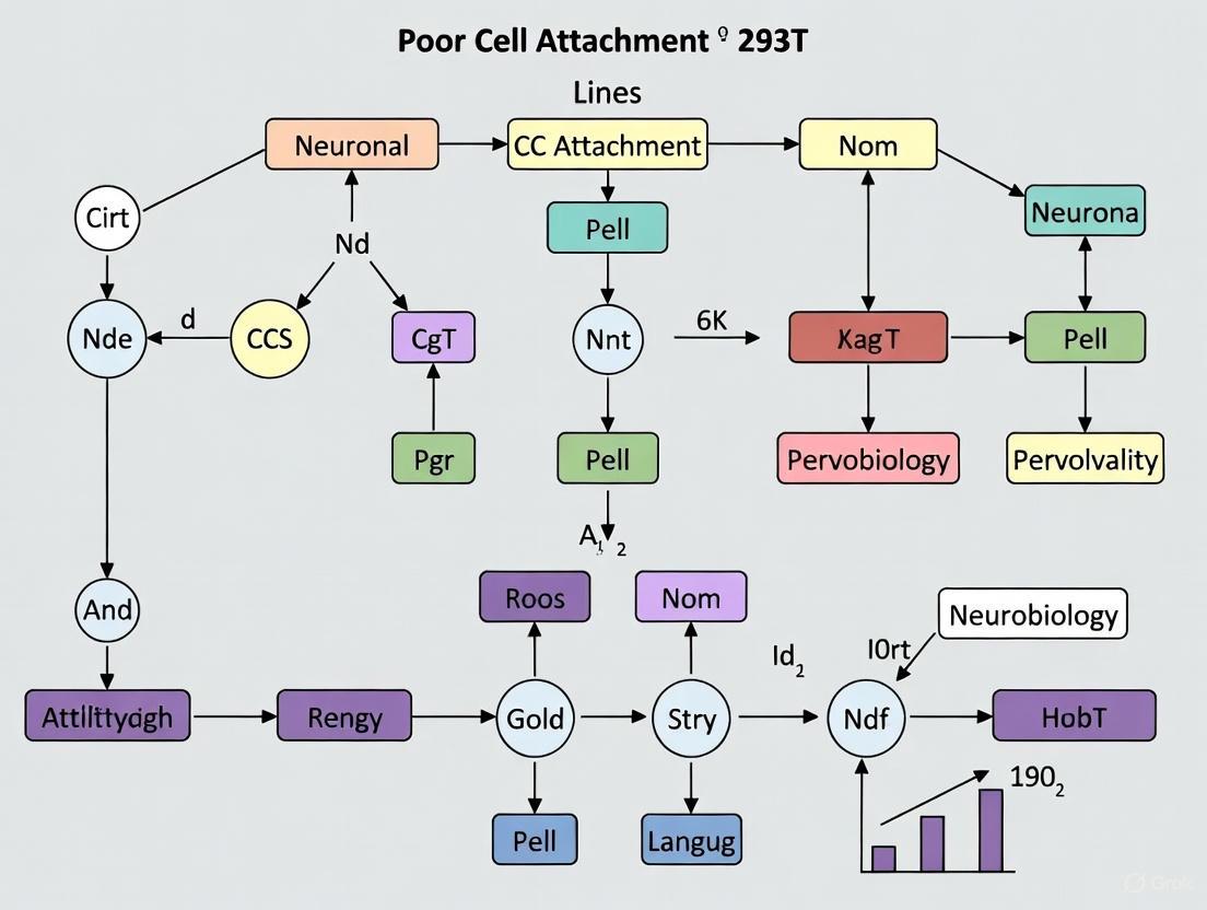
Diagram 1: HEK293 development and derivative lines.
Research Reagent Solutions
Table 3: Essential Materials for HEK293 Cell Culture and Experiments
| Reagent/Material | Function/Application | Technical Notes |
|---|---|---|
| High-glucose DMEM | Standard culture medium | Supplement with 10% FBS; may include antibiotics |
| Poly-D-Lysine | Surface coating for enhanced attachment | Particularly helpful for problematic attachment |
| Collagen-coated vessels | Alternative substrate for improved adhesion | Useful for assay plates requiring strong attachment |
| Serum-free medium | Suspension adaptation and specialized applications | Enables large-scale production in bioreactors |
| Calcium phosphate transfection reagents | High-efficiency nucleic acid delivery | HEK293 cells are particularly amenable to this method |
| Specific adenoviral vectors | Gene delivery and expression studies | HEK293 cells provide essential helper functions for adenoviral vector propagation [6] |
Frequently Asked Questions
Are HEK293 cells truly kidney cells?
While derived from human embryonic kidney tissue, subsequent analysis suggests HEK293 cells most closely resemble adrenal precursor cells with neuronal properties rather than typical kidney cells [1]. Transcriptome profiling shows closest resemblance to adrenal cells, which develop adjacent to kidneys and share neuronal characteristics [1]. This has important implications for experimental design and data interpretation.
What are the key differences between adherent and suspension variants?
Comparative genomic and transcriptomic analyses reveal significant differences between adherent (HEK293, 293E, 293T) and suspension (293-H, 293-F) derivatives [4]. Suspension-adapted lines show transcriptomic switching in cholesterol biosynthesis and differential expression of key genes (RARG, ID1, ZIC1, LOX, DHRS3) [4]. These molecular differences underlie their adaptation to different culture environments.
How does the SV40 large T antigen in HEK293T cells enhance protein production?
The SV40 large T antigen enables episomal replication of transfected plasmids containing the SV40 origin of replication [1]. This maintains high plasmid copy numbers within cells, dramatically increasing the yield of recombinant proteins or retroviral vectors produced from these cells [1] [2].
What biosafety considerations apply to working with HEK293 cells?
HEK293 cells require Biosafety Level 2 (BSL-2) containment as they contain integrated adenovirus 5 DNA sequences [7]. Primary hazards include potential exposure through droplet contact, mucous membrane exposure, or ingestion. Special considerations include the potential for recombination events between integrated viral sequences and exogenous viruses in experimental systems [7].
Diagram 2: Attachment issues causes and solutions.
Frequently Asked Questions (FAQs)
Q1: Why are my 293T cells not attaching properly after passaging or thawing?
A1: Poor attachment in 293T cells is a common issue rooted in their unique biology. Unlike many other cell lines, 293T cells possess a distinct and 'immature' actin cytoskeleton, which is a primary cause of their loose adherence [5]. Furthermore, these cells are exquisitely sensitive to temperature drops; exposure to temperatures below 30°C, even briefly, can cause significant detachment [5]. Patience is also key, as 293T cells can take several days to attach fully after thawing [5].
Q2: Does the genetic instability of 293T cells affect their adhesion properties?
A2: Yes. 293T cells are genetically unstable and possess a defective DNA mismatch repair mechanism, making them prone to genotypic and phenotypic drift over time [5]. Uncontrolled culture conditions, such as inconsistent subculturing, allowing over-confluence, or keeping cells in culture for extended periods, can exert selective pressures. This can lead to genetic changes that may manifest as altered cellular behavior, including changes in adhesion [5].
Q3: What is the molecular evidence for the 'immature' actin cytoskeleton in 293T cells?
A3: Research using Atomic Force Microscopy (AFM) has revealed unique mechanical properties in HEK293 cells. Unlike most cell types that soften when detached, HEK293 cells exhibit very low surface stiffness in their adherent state, which increases significantly after detachment [8]. This inverse mechanical behavior, driven by the actin cytoskeleton, is a defining characteristic that sets them apart from both normal stromal and cancer cells, justifying their classification into a distinct category with a specific actin organization [8].
Q4: Are 293T cells of neuronal or epithelial origin, and how does this relate to their markers?
A4: The origin of 293 cells has been debated. While they express some neuronal markers, studies confirm they retain several epithelial characteristics. They express epithelial markers such as E-cadherin, cytokeratins 5/8, desmoglein 2, occludin, and ZO-1 [9]. However, they also express mesenchymal markers like N-cadherin and vimentin, indicating a complex molecular profile [9].
Troubleshooting Guide: Poor Cell Attachment
| Issue | Primary Cause | Recommended Solution | Biological Basis |
|---|---|---|---|
| Slow attachment post-thaw | Innate cytoskeletal organization; Recovery from cryopreservation | Allow 2-4 days for attachment; Do not assume culture failure. Check viability. | Unique 'immature' actin cytoskeleton requires longer to reorganize and form stable attachments [5] [8]. |
| Spontaneous detachment | Temperature fluctuation | Use pre-warmed media and reagents; Minimize time outside incubator. | Actin cytoskeleton dynamics and cell adhesion are highly temperature-sensitive in 293Ts. Detachment occurs below 30°C [5]. |
| Weak adhesion on standard plates | Suboptimal surface charge/chemistry | Use coated surfaces (Poly-D-Lysine, Collagen) or specialty plastics (e.g., Corning CellBind) [5] [10]. | Coatings provide a more positively charged or ECM-rich surface, enhancing initial cell-substrate interaction [5] [10]. |
| Variable adhesion over long-term culture | Genetic instability and phenotypic drift | Maintain strict subculture regimes; Use low-passage cells from well-maintained master banks [5]. | A defective DNA mismatch repair system leads to genotypic drift, which can alter adhesion phenotypes over time [5]. |
Step-by-Step Diagnostic Protocol
Objective: To systematically identify the cause of poor 293T cell attachment and implement a corrective strategy.
Materials:
- Healthy, low-passage 293T cells
- Pre-warmed complete growth medium
- Poly-D-Lysine (PDL) or Collagen coating solution
- 6-well or 12-well culture plates (standard and coated)
- Phase-contrast microscope
- Cell viability stain (e.g., Trypan Blue)
Workflow:
Procedure:
Check Cell Viability:
- Harvest the problematic cell culture and mix a small aliquot with Trypan Blue stain (typically 1:1).
- Count the cells using a hemocytometer. A viability of >90% indicates that the detachment is likely not due to cell death but rather an adhesion or environmental problem [5]. If viability is low, investigate contamination or cryopreservation protocols.
Verify Culture Conditions:
- Ensure the incubator temperature is stable at 37°C.
- Always warm all media and reagents in a 37°C water bath before use. Do not leave cells at room temperature for extended periods during handling.
- If temperature is implicated, return the culture to the incubator and monitor for 24-48 hours. Cells may re-attach once returned to 37°C [5].
Test Substrate Enhancement:
- Prepare a culture plate coated with Poly-D-Lysine (PDL) or Collagen according to the manufacturer's instructions.
- Seed the 293T cells in parallel on the coated plate and a standard tissue culture plate.
- Observe adhesion over 24-48 hours. Significant improvement on the coated plate confirms that substrate optimization is required for your specific experimental setup [5] [10].
Assess Phenotypic Drift:
- If adhesion problems persist across multiple experiments with optimized conditions, consider phenotypic drift.
- Return to a low-passage, master working cell bank.
- Implement a strict sub-culture schedule to prevent over-confluence and minimize passaging. Regularly authenticate cells via STR profiling to monitor genotypic stability [5].
Key Signaling Pathways Regulating 293T Actin Dynamics
The adhesion and migration of 293T cells are regulated by complex signaling networks that converge on the actin cytoskeleton. The diagram below illustrates a key pathway involving PCTK3 and FAK/Rho signaling.
Pathway Explanation: Research shows that the kinase PCTK3 (CDK18) acts as a critical negative regulator of cell migration and adhesion in 293T cells. As illustrated, active PCTK3 suppresses Focal Adhesion Kinase (FAK) activity, which in turn keeps the RhoA/ROCK pathway in check [11]. This inhibition prevents the ROCK-mediated activation of LIM Kinase (LIMK) and Myosin Light Chain (MLC). The outcome is that the actin-severing protein Cofilin remains active, promoting F-actin turnover, and actomyosin contractility is low. Concurrently, PCTK3 promotes Rac1 activity, which is associated with lamellipodia formation. The net effect is a controlled balance that restrains excessive migration and stabilizes adhesion [11]. Knockdown of PCTK3 leads to hyperactivation of this pathway, resulting in increased actin polymerization, membrane blebbing, and loss of adhesion control.
The Scientist's Toolkit: Essential Research Reagents
This table lists key reagents used to study and manage the unique actin cytoskeleton of 293T cells, as cited in the literature.
| Research Reagent | Function / Application in 293T Research |
|---|---|
| Poly-D-Lysine (PDL) | Synthetic coating polymer that enhances initial cell attachment by providing a positive charge surface for cell membrane interaction [5]. |
| Cytochalasin D | A cell-permeable fungal toxin that inhibits actin polymerization by capping filament ends. Used experimentally to depolymerize the actin cytoskeleton and confirm its role in mechanical properties [8]. |
| Y27632 (ROCK Inhibitor) | A potent and selective inhibitor of Rho-associated kinase (ROCK). Used to investigate the role of the Rho/ROCK pathway in actin contractility and MLC phosphorylation in 293T cells [11]. |
| Collagen | An extracellular matrix (ECM) protein used to coat culture surfaces, providing a more natural substrate for integrin-mediated adhesion than plastic [5]. |
| Calyculin A | A potent inhibitor of protein phosphatases 1 and 2A. Used in research to induce hyperphosphorylation of cytoskeletal proteins and study actin cytoskeleton remodeling [8]. |
| ITO-MPS SAM-coated Substrate | A specialized scaffold with a self-assembled monolayer of 3-(mercaptopropyl) trimethoxysilane on indium tin oxide. Research shows it significantly enhances HEK293T adhesion and proliferation by promoting a favorable surface charge and metabolomic profile [10]. |
Impact of SV40 Large T Antigen on Cellular Adhesion Machinery
Research involving 293T neuronal cell lines is frequently hampered by a recurring and frustrating experimental issue: poor and unpredictable cell attachment. This problem directly impacts data reproducibility, cell viability, and the overall success of critical experiments. A growing body of evidence indicates that the expression of Simian Virus 40 (SV40) Large T Antigen, the very feature that makes 293T cells so valuable for high-yield protein production and viral packaging, is a primary contributor to this cellular adhesion instability [5] [12]. This technical support article, framed within a broader thesis on 293T neuronal research, explores the mechanistic link between SV40 Large T Antigen and the adhesion machinery, providing targeted troubleshooting guides and validated protocols to empower researchers to overcome these challenges.
Mechanisms: How SV40 Large T Antigen Disrupts Adhesion
The SV40 Large T Antigen is a multifunctional oncoprotein known for its ability to immortalize cells by targeting key tumor suppressors like p53 and pRb [13]. However, its impact extends to cellular processes critical for adhesion.
Direct Disruption of the Actin Cytoskeleton: The most significant factor is the direct alteration of the cell's structural scaffold. Unlike many common cancer cell lines or fibroblasts, 293T cells exhibit a unique and "immature" actin cytoskeleton [5]. Actin filaments are dynamic structures essential for cell attachment and spreading. The original derivation of the parental HEK293 line with adenovirus DNA, combined with the subsequent introduction of SV40 Large T Antigen, is believed to have primed these cells for cytoskeletal irregularities. Both viral proteins are known to reorganize the host cytoskeleton to facilitate viral replication, leaving a legacy of disrupted actin organization that manifests as weak adhesion [5].
Induction of Genomic Instability: SV40 Large T Antigen can breach genome integrity mechanisms, leading to DNA damage responses and chromosomal instability [14]. Furthermore, 293 cells possess an inherent defect in their DNA mismatch repair mechanism, making them particularly susceptible to genotypic and phenotypic drift over passages [5]. This instability can lead to unpredictable changes in the expression of proteins vital for adhesion and cytoskeletal integrity.
Temperature Sensitivity: A critical and often overlooked practical aspect is the profound temperature sensitivity of 293T cells. Research indicates that reducing the culture temperature to below 30°C can cause up to 60% of the monolayer to detach [5]. This is likely linked to the thermosensitive nature of the cellular structures and signaling pathways governing adhesion, which may be further destabilized by the presence of viral antigens.
Table 1: Molecular Mechanisms Linking SV40 Large T Antigen to Poor Adhesion
| Mechanism | Biological Consequence | Observed Phenotype in 293T Cells |
|---|---|---|
| Disrupted Actin Cytoskeleton [5] | Altered polymerization of actin microfilaments, the core scaffold for attachment. | "Immature" actin structure; loose, semi-adherent growth. |
| Genomic Instability [14] [5] | Uncontrolled genetic drift affects expression of adhesion-related proteins. | Unpredictable attachment behavior between passages; phenotypic variation. |
| Temperature Sensitivity [5] | Compromised integrity of adhesion complexes and cytoskeleton at sub-optimal temperatures. | Massive detachment upon minor temperature drops (e.g., during medium changes or imaging). |
The following diagram illustrates the interconnected pathways through which SV40 Large T Antigen impacts cellular adhesion:
Troubleshooting Guide & FAQs
This section provides direct, actionable solutions to common adhesion problems encountered with 293T cells.
Frequently Asked Questions
Q1: My 293T cells detach in large, jellyfish-like sheets, especially after transfection. Is this normal? Yes, this is a commonly reported phenomenon. Researchers often refer to these cells as "jellyfish cells" due to this specific behavior [15]. It is typically triggered by physical stress or the inherent weak adhesion. Ensuring gentle handling and using coated cultureware can mitigate this issue.
Q2: How long should it take for 293T cells to attach after thawing or passaging? Unlike many adherent lines, 293T cells can take several days to properly attach after thawing [5] [16]. Patience is critical. Do not assume the culture has failed if cells are not attached within 24 hours. Test viability if concerned, and allow time for recovery.
Q3: I need to run an assay at room temperature. How can I prevent my cells from detaching? This is a high-risk scenario. If the assay must run below 30°C, optimize the time window for data capture to be as short as possible. The most reliable solution is to use Poly-D-Lysine or collagen-coated plates to provide a stronger adhesive substrate that can withstand temperature fluctuations [5].
Step-by-Step Troubleshooting Protocol
The workflow below provides a systematic approach to diagnosing and resolving 293T attachment issues:
Table 2: Optimized Reagent Solutions for Improving 293T Cell Adhesion
| Reagent / Material | Function & Rationale | Protocol Note |
|---|---|---|
| Poly-D-Lysine (PDL) | Provides a positively charged surface that enhances attachment of negatively charged cell membranes. | Coat plates/flasks per manufacturer's instructions. Rinse before use. |
| Collagen | Mimics the natural extracellular matrix (ECM), providing integrin binding sites for strong adhesion. | Typically used as a thin coating on the culture surface. |
| CellBind (Corning) | A proprietary surface treatment that creates an optimal charge for cell attachment. | A ready-to-use alternative to manual coating procedures. |
| Pre-warmed Media/Reagents | Prevents temperature shock, which triggers immediate detachment. | Warm all reagents to 37°C before any contact with cells. |
| Hygromycin B | For 293TT cells; ensures selective pressure to maintain stable SV40 Large T Antigen expression [16]. | Use at 250 µg/ml in culture medium. |
The Scientist's Toolkit: Essential Research Reagents
Table 3: Key Reagents for Working with SV40 Large T Antigen and 293T Cells
| Reagent / Assay | Specific Function | Experimental Application |
|---|---|---|
| Lipofectamine 2000 | Lipid-based transfection reagent. | Achieving high transfection efficiency in 293T/293TT cells [16]. |
| qPCR & Nested PCR Assays | Detection of residual SV40 T-antigen DNA fragments. | Quality control and safety profiling of AAV vectors produced in 293T systems [17]. |
| EF1α Promoter Plasmids | Drives high-level recombinant protein expression in 293TT cells. | Superior to CMV promoter in 293TT cells due to copy-number responsive expression [16]. |
| Trypsin/EDTA | Proteolytic enzyme for cell dissociation. | Requires thorough (5-10 min) incubation for 293TT cells which adhere tightly to each other [16]. |
| Hygromycin B | Antibiotic selection. | Maintaining SV40 Large T Antigen expression in 293TT cell cultures [16]. |
The poor adhesion of 293T neuronal cell lines is not an isolated technical failure but a direct consequence of the SV40 Large T Antigen's profound impact on cellular architecture and stability. By understanding the underlying mechanisms—cytoskeletal disruption, genomic instability, and temperature sensitivity—researchers can move from frustration to strategic problem-solving. Implementing the recommended protocols, including strict temperature control, the use of coated surfaces, and gentle handling techniques, will significantly improve experimental reproducibility and success. Embracing these tailored practices ensures that the powerful advantages of 293T cells can be fully leveraged in advanced research and drug development.
Troubleshooting Guide: Addressing Common Experimental Issues
FAQ: Poor Cell Attachment in 293T Neuronal Research
Q: My 293T cells are detaching from the culture substrate during experiments. What could be causing this and how can I fix it?
A: Poor adhesion is a well-documented characteristic of HEK293-derived cells, including 293T lines. This issue stems from their unique biology and can be addressed through several validated methods.
Primary Cause: Unique Actin Cytoskeleton - Unlike many cancer cell lines, 293 cells possess an "immature" actin cytoskeleton that provides weaker structural support for adhesion [5]. This irregular cytoskeleton may be a residual effect of the original adenovirus transformation used to create the cell line [5].
Critical Factor: Temperature Sensitivity - 293 cells show remarkable temperature sensitivity. Reducing temperature to 30°C can cause up to 60% cell detachment from the monolayer [5]. Always use pre-warmed media and reagents, and minimize time outside the 37°C incubator.
Effective Solutions:
- Surface Coating: Use Poly-D-Lysine (PDL), collagen, or commercially treated plastics like CellBind to significantly improve attachment [5] [10].
- Novel Substrates: Recent research shows ITO-MPS SAM-coated substrates dramatically improve HEK293T adhesion and proliferation through metabolic pathway modulation [10].
- Patience with Recovery: After thawing or detachment, 293T cells may require several days to re-attach properly [5].
FAQ: Genetic Drift in Long-Term Culture
Q: I've noticed behavioral changes in my 293T cells after extended passaging. Could this be genetic drift?
A: Yes, 293 cells are notoriously genetically unstable. They possess a hypertriploid karyotype and a defective DNA mismatch repair mechanism, making them particularly prone to genotypic and phenotypic drift [5].
Contributing Factors:
- Passage Number: Extended culture periods without re-establishing banks exerts selective pressure [5].
- Culture Conditions: Over-confluency, inconsistent subculture regimes, and uncontrolled medium changes accelerate divergence [5].
- Inherent Instability: The original adenovirus transformation and subsequent genetic manipulations (e.g., SV40 Large T antigen in 293T) contribute to ongoing genomic evolution [5] [4].
Prevention Strategies:
Table 1: Chromosomal Instability Assessment in Cell Lines
| Parameter | Measurement Method | Typical Values in Aneuploid Lines | Research Implications |
|---|---|---|---|
| Modal Chromosome Number | G-banding karyotyping [18] | 52-86 chromosomes (human ovarian cancer lines) [18] | Defines ploidy category; hyperdiploid: 47-57, hypotriploid: 58-68, hypertriploid: 70-80 [18] |
| Aneuploid Score (AS) | Copy Number Variation (CNV) analysis [18] | 5-12 in ovarian cancer lines [18] | Higher scores indicate greater aneuploidy; correlates with CIN |
| Ploidy Value | ABSOLUTE algorithm/flow cytometry [18] | ~3.3 in whole-genome doubled cancers [18] | Distinguishes near-diploid (∼2.0) from polyploid populations |
| DNA Damage Foci | γH2AX immunofluorescence [19] | 34-54% of tetraploid cells show >10 foci vs. 5-9% diploid [19] | Marker of replication stress and DNA damage |
| Nuclear Area | Microscopy/Image analysis [19] | Increased in tetraploid cells [19] | Normalize DNA damage markers to nuclear size for accurate comparison |
Table 2: Research Reagent Solutions for Genetic Instability Studies
| Reagent/Material | Application | Function | Experimental Notes |
|---|---|---|---|
| Poly-D-Lysine (PDL) | Substrate coating [5] | Enhances cell attachment | Particularly effective for 293T cells [5] |
| ITO-MPS SAM-coated substrates | Advanced adhesion studies [10] | Promotes adhesion via metabolic reprogramming | Identified 16 adhesion-promoting metabolites [10] |
| APH (Aphidicolin) | DNA replication inhibition [19] | DNA polymerase inhibitor; studies replication stress | Low doses inhibit replication without direct DNA damage [19] |
| PHA-767491 | DNA replication studies [19] | Cdc7 kinase inhibitor; blocks replication initiation | Useful for probing replication dynamics in unstable lines [19] |
| CytoScan 750K Array | Karyotype analysis [20] | High-resolution CNV detection | Confirmed partial tetrasomy 8p in MBU-8 model [20] |
Experimental Protocols for Instability Assessment
Protocol 1: Assessing Ploidy Status via Karyotyping
Purpose: Determine chromosomal number and identify gross structural abnormalities in cell lines [18].
Methodology:
- Metaphase Arrest: Treat exponentially growing cells with colcemid (45 ng/mL) for 45-60 minutes [18].
- Hypotonic Treatment: Incubate cells in pre-warmed KCl solution (75 mM) for 30 minutes at 37°C [18].
- Fixation: Apply methanol-acetic acid (3:1) fixative with multiple changes [18].
- Slide Preparation: Drop cell suspension onto clean slides in a controlled humidity environment [18].
- G-banding:
Interpretation: Classify based on modal number: hyperdiploid (47-57), hypotriploid (58-68), hypertriploid (70-80) [18].
Protocol 2: DNA Replication Stress Analysis in Tetraploid Cells
Purpose: Evaluate replication-dependent DNA damage following whole-genome duplication events [19].
Methodology:
- Tetraploid Induction: Generate tetraploid cells through cytokinesis failure, mitotic slippage, or endoreplication [19].
- Cell Cycle Synchronization: Arrest cells in G1 phase using CDK4/6 or CDK2 inhibitors [19].
- DNA Damage Assessment:
- DNA Combing Analysis:
- Label DNA with nucleoside analogs (e.g., IdU, CIdU)
- Extract high molecular weight DNA and stretch on slides
- Analyze fork speed, inter-origin distance, and fork asymmetry [19]
Key Parameters: Tetraploid cells show increased fork speed, fork asymmetry, and under-/over-replicated regions [19].
Signaling Pathways and Experimental Workflows
Diagram 1: CIN mechanisms and cellular consequences. CIN arises from multiple molecular defects that generate genetic heterogeneity and phenotypic drift, impacting experimental reproducibility [21].
Diagram 2: Systematic troubleshooting for 293T adhesion issues. This workflow addresses the primary factors affecting 293T attachment with evidence-based solutions [5] [10].
Advanced Methodologies for Instability Research
Single-Cell DNA Sequencing for Karyotype Analysis
Application: Identify over-duplicated chromosomes and regional replication defects in unstable cell populations [19].
Workflow:
- Cell Preparation: Single-cell suspension of G2/M arrested cells to capture complete karyotypes [19].
- Library Preparation: Use microfluidic platforms for single-cell whole genome amplification [19].
- Sequencing: Shallow whole-genome sequencing to determine copy number variations [19].
- Analysis: Identify over- and under-replicated regions (e.g., 9n, 7n, 4n patterns in tetraploid backgrounds) [19].
Output: Reveals karyotypic heterogeneity within cell populations and identifies specific chromosomal regions prone to instability [19].
NMR Metabolomics for Adhesion Studies
Application: Understand metabolic changes associated with improved cellular adhesion in problematic lines like HEK293T [10].
Workflow:
- Sample Preparation: Culture cells on test substrates (e.g., ITO-MPS SAM) and collect conditioned media at 24h intervals up to 120h [10].
- NMR Analysis:
- Use 1H NMR spectroscopy for metabolic profiling
- Identify and quantify metabolites in spent media
- Compare against control substrates [10]
- Data Interpretation: Identify adhesion-promoting metabolites from the 26 typically detected compounds [10].
Validation: Correlate metabolic findings with MTT proliferation assays and confocal microscopy of cell morphology [10].
Frequently Asked Questions (FAQs)
Q1: Why are my 293T cells detaching from the culture vessel? The most common cause is exposure to temperatures below 30°C. HEK293 cells and their derivatives, including 293T cells, are highly temperature-sensitive and can detach if their environment cools even briefly below this threshold. This is due to their unique actin cytoskeleton, which differs from that of other common cell lines. If cells detach, test their viability, as they may not be dead and could re-attach after several days at 37°C [5].
Q2: What is the ideal temperature range for culturing 293T cells? For routine cell growth and attachment, maintain a constant temperature of 37°C. However, for enhanced recombinant protein expression post-transfection, a shift to a mild hypothermic condition (33°C) 24 hours after transfection can increase protein yield by approximately 1.5-fold without affecting protein properties [22]. Temperatures at or below 30°C should be strictly avoided as they severely compromise cell attachment [5].
Q3: My cells detached during an experiment. How can I prevent this in the future? Ensure all culture media and reagents are pre-warmed to 37°C before use. Avoid cooling flasks or plates during transfer to microscopes or plate readers. If your assay requires temperatures below 30°C, optimize the time window for data capture before detachment occurs, or use coated culture vessels to enhance initial attachment [5].
Troubleshooting Guide
Problem: Sudden and Widespread Cell Detachment
Possible Causes and Solutions:
Cause 1: Incubation Temperature Drop
Cause 2: Cool Reagents
- Solution: Always warm culture medium, PBS, and trypsin to 37°C in a water bath before adding them to cells. Do not use cold reagents directly from the refrigerator [23].
Cause 3: Physical Bumping or Agitation
Problem: Poor Initial Attachment After Thawing or Passaging
Possible Causes and Solutions:
- Cause 1: Inherent Cell Line Characteristics
- Solution: Be patient. Unlike many other cell lines, 293 cells can take several days to attach fully after thawing or passaging. Do not assume the culture has failed [5].
- Cause 2: Suboptimal Culture Vessel Surface
Quantitative Data on Temperature Effects
The table below summarizes the specific effects of temperature on 293 cells, based on experimental data.
Table 1: Effects of Temperature on HEK293/293S Cell Cultures
| Temperature | Effect on Cell Growth | Effect on Protein Expression | Effect on Cell Attachment |
|---|---|---|---|
| 37°C | Normal growth rate [22] | Baseline expression level [22] | Normal for adherent monolayer [5] |
| 33°C | Reduced growth rate [22] | ~1.5-fold higher expression of recombinant proteins (e.g., GFP, AMPA receptors) [22] | Maintained, provided it is not below the critical 30°C threshold [22] |
| 30°C or lower | Not specifically studied, but growth is expected to be further reduced | Not recommended; no enhancement observed below 33°C [22] | Up to ~60% loss of cells from the monolayer [5] |
Experimental Protocol: Enhancing Protein Yield via Temperature Shift
This protocol is adapted from a study demonstrating increased recombinant protein expression in HEK-293S cells [22].
Objective: To increase the yield of transiently expressed recombinant proteins by implementing a biphasic temperature culture system.
Key Materials:
- HEK-293S or 293T cells in logarithmic growth phase
- Standard cell culture reagents: DMEM, FBS, antibiotics, transfection reagent (e.g., calcium phosphate, Lipofectamine 2000)
- Two CO₂ incubators, calibrated and set to 37°C and 33°C, respectively
Workflow: The following diagram illustrates the biphasic temperature protocol for enhancing protein expression.
Procedure in Detail:
- Standard Culture and Transfection: Maintain and passage HEK293 cells at 37°C in a humidified incubator with 5-10% CO₂. Perform transient transfection when cells reach an appropriate density (e.g., 70-80% confluency) using your method of choice [22] [23].
- Post-Transfection Recovery: After adding the DNA-transfection reagent complexes, return the cells to the 37°C incubator for a minimum of 5 hours to allow for complex uptake [22].
- Temperature Shift for Enhanced Expression: Approximately 24 hours after transfection, transfer the culture to a pre-equilibrated incubator set to 33°C. The cells should remain at this temperature for the duration of the protein expression phase (e.g., 24-72 hours post-transfection) [22].
- Harvest: Harvest cells or culture supernatant for protein analysis as required.
Expected Outcome: Cultures shifted to 33°C post-transfection will show a significant increase (~1.5-fold) in recombinant protein yield compared to cultures maintained continuously at 37°C, as demonstrated with proteins like GFP and AMPA receptors [22].
The Scientist's Toolkit: Essential Reagents for 293 Cell Research
Table 2: Key Research Reagent Solutions
| Item | Function | Application Note |
|---|---|---|
| Poly-D-Lysine (PDL) | Coating agent that enhances cell attachment by providing a positively charged surface for cells to adhere to. | Crucial for improving attachment in temperature-sensitive assays or when using problematically adherent 293 cell lines [22] [5]. |
| DMEM + 10% FBS | Standard culture medium for routine growth and maintenance of adherent 293T cells. | The fetal bovine serum provides essential attachment and growth factors [22] [23]. |
| Geneticin (G418) | Antibiotic for selection of stable cell lines, such as 293FT/293T cells expressing resistance genes. | Used to maintain selective pressure on cells expressing the SV40 large T antigen and neomycin resistance gene [24]. |
| Lipofectamine 2000 | Lipid-based transfection reagent for efficient delivery of plasmid DNA into 293 cells. | High transfection efficiency makes it a common choice for transient protein expression or CRISPR editing [22] [23]. |
| Trypsin/EDTA | Enzyme solution used to dissociate adherent cells for passaging. | Avoid over-trypsinization; incubation for 1-2 minutes at 37°C is typically sufficient [23]. |
| Opti-MEM I Reduced Serum Medium | Serum-free medium used for diluting DNA and transfection reagents to form complexes. | Essential for achieving high efficiency during lipid-based transfection [22] [23]. |
Practical Strategies to Enhance 293T Cell Adhesion and Proliferation
Frequently Asked Questions (FAQs)
Q1: Why are my 293T cells not attaching properly, even after passaging? Poor attachment in 293T cells is a common issue rooted in their unique biology. Unlike many other cell lines, HEK293T cells have an "immature" actin cytoskeleton, which makes them naturally semi-adherent or loosely adherent [5]. Furthermore, these cells are highly sensitive to temperature drops; reducing the temperature to below 30°C can cause up to 60% of the cells to detach from the monolayer [5]. Other frequent causes include over-trypsinization, which damages cell surface adhesion proteins, and using an inappropriate seeding concentration [25].
Q2: What is the difference between Poly-D-Lysine (PDL) and Poly-L-Lysine (PLL), and which should I use? Both PDL and PLL are synthetic, positively charged polymers that enhance the electrostatic interaction between the negatively charged cell membrane and the culture surface [26]. The key difference is their resistance to cellular degradation. Some cells can digest the naturally occurring L-isomer (PLL). In these cases, the synthetic D-isomer (PDL) should be used because cells cannot break it down, ensuring the coating remains stable [26].
Q3: My neurons are forming large clumps instead of distributing evenly. How can I prevent this? Neuronal clumping is a common challenge. Research on human iPSC-derived neurons has shown that while single coatings like Matrigel or Laminin promote excellent neurite outgrowth, they often lead to large cell body clumps and bundle-like, straight neurites [27]. A highly effective solution is to use a double-coating strategy. Combining a primary layer of PDL with a secondary layer of Matrigel has been demonstrated to significantly reduce clumping, improve neuronal distribution, and enhance the density and branching of neurites [27].
Q4: Can I combine different coatings to improve cell attachment and function? Yes, double-coating protocols often yield superior results. A proven method is to first apply a poly-lysine (PDL or PLL) base layer, which provides a strong electrostatic foundation for attachment, followed by a secondary coating of an extracellular matrix protein like Laminin or Matrigel, which provides specific biological signals for cell spreading, differentiation, and maturation [27]. For instance, one study found that the PDL+Matrigel combination not only reduced clumping but also improved dendritic/axonal development and synaptic marker distribution in human neurons [27].
Troubleshooting Guide: Common Coating and Cell Attachment Issues
| Problem | Potential Causes | Recommended Solutions |
|---|---|---|
| Poor Cell Attachment | • Innate weak actin cytoskeleton of 293Ts [5]• Temperature falling below 30°C [5]• Over-trypsinization [25]• Inappropriate seeding concentration [25] | • Use pre-warmed media and reagents; avoid cooling [5].• Optimize trypsinization time and concentration [25].• Coat cultureware with PDL, collagen, or specialized plastics [5]. |
| Neuronal Clumping | • Single coatings (e.g., Laminin/Matrigel) promoting straight, bundle-like neurites and aggregation [27] | • Implement a double-coating protocol (e.g., PDL + Matrigel) [27]. |
| Uneven Coating | • Inconsistent solution application• Improper drying conditions | • Ensure the coating solution covers the surface evenly without bubbles.• Follow manufacturer instructions for incubation and allow to dry under sterile conditions. |
| Low Transfection Efficiency | • Poor cell health due to attachment issues• Cells detaching during the process | • Ensure robust attachment pre-transfection by using an optimized coating [10].• Use coatings that enhance cellular health and proliferation, such as ITO-MPS SAM for 293Ts [10]. |
Quantitative Coating Performance Data
Table 1: Comparison of Single-Coating Performance on Neuronal Morphology
This table summarizes the effects of different single coatings on the morphology of human iPSC-derived neurons, based on IncuCyte live-cell imaging analysis [27].
| Coating Type | Neurite Outgrowth Density | Branch Points | Cell Body Clumping | Notes |
|---|---|---|---|---|
| Poly-D-Lysine (PDL) | Low | Low | Low | Sparse neurite outgrowth; extensive cell debris observed. |
| Poly-L-Ornithine (PLO) | Low | Low | Low | Sparse neurite outgrowth; extensive cell debris observed. |
| Laminin | High | High | High | Produces abnormal, highly straight neurites and large clumps. |
| Matrigel | High | High | High | Produces abnormal, highly straight neurites and large clumps. |
Table 2: Efficacy of Double-Coating Strategies for Neuronal Culture
This table compares different double-coating conditions, demonstrating their ability to overcome the limitations of single coatings [27].
| Double-Coating Combination | Neurite Outgrowth | Reduction in Clumping | Neuronal Homogeneity/Purity | Key Findings |
|---|---|---|---|---|
| PDL + Matrigel | High | Significant | Enhanced | Most effective combination; improves synaptic marker distribution. |
| PDL + Laminin | High | Significant | Improved | Effective for reducing clumping. |
| PLO + Matrigel | High | Significant | Improved | Effective for reducing clumping. |
| PLO + Laminin | High | Significant | Improved | Effective for reducing clumping. |
Detailed Experimental Protocols
Protocol 1: Standard Poly-D-Lysine Coating for Coverslips
- Preparation: Prepare a sterile aqueous solution of Poly-D-Lysine at a recommended concentration (e.g., 0.1 mg/ml).
- Application: Place sterile glass coverslips in a culture dish. Add enough PDL solution to completely cover each coverslip.
- Incubation: Incubate at room temperature for a minimum of 1 hour, or according to the manufacturer's instructions. For enhanced attachment, incubation can be extended overnight at 4°C.
- Rinsing: After incubation, aspirate the PDL solution and rinse the coverslips three times with sterile distilled water to remove any unbound PDL.
- Drying and Sterilization: Allow the coverslips to air dry completely under a sterile hood.
- Storage: Coated coverslips can be stored sterile at 4°C for several weeks. Before use, ensure they are at room temperature.
Protocol 2: Double-Coating with PDL and Laminin/Matrigel for Neuronal Cultures
- Apply Base Layer: First, coat the culture vessel with Poly-D-Lysine by following the standard protocol above (Steps 1-5). Do not let the surface dry after the final rinse if proceeding immediately.
- Prepare ECM Solution: Thaw Matrigel or Laminin on ice and dilute it in cold, serum-free media or a recommended buffer (e.g., PBS) to the desired working concentration.
- Apply ECM Layer: Aspirate the final water rinse from the PDL-coated surface. Immediately add the cold, diluted Matrigel or Laminin solution to cover the surface.
- Incubate: Incub the culture vessel with the ECM solution for at least 2 hours at 37°C, or overnight at 4°C, to allow the proteins to bind to the PDL layer.
- Final Preparation: Before plating cells, aspirate the ECM solution. There is no need to rinse the surface. The coated vessel is now ready for cell seeding.
Signaling Pathways and Logical Workflows
Coating Selection Logic
Cell Attachment Mechanism
The Scientist's Toolkit: Essential Reagents for Coating Protocols
Table 3: Key Research Reagent Solutions for Cell Coating
| Reagent | Function / Explanation | Example Applications |
|---|---|---|
| Poly-D-Lysine (PDL) | A synthetic, positively charged polymer that binds to the negatively charged cell membrane, enhancing electrostatic attachment. Resists cellular degradation [26]. | General attachment for 293T, HEK293, and neuronal cell lines [26]. Often used as a base layer in double-coating. |
| Laminin | A natural extracellular matrix (ECM) glycoprotein that promotes adherence via specific binding domains for integrin receptors on cell surfaces [26]. | Supports neuronal differentiation, maturation, and adhesion of various cell types, including fibroblasts and epithelial cells [27] [26]. |
| Matrigel | A complex, reconstituted basement membrane extract containing ECM proteins like Laminin and Collagen. Provides a biologically active scaffold for cells [27]. | Promotes high-density neurite outgrowth in neuronal cultures; used in double-coating protocols to reduce clumping [27]. |
| Fibronectin | An ECM glycoprotein that promotes cell attachment via its central RGD (Arg-Gly-Asp) binding sequence, which is recognized by cell surface integrins [26]. | Adhesion of HEK293 cells, smooth muscle cells, endothelial cells, and fibroblasts [26]. |
| Collagen | A major structural ECM protein used as a coating to promote cell adherence and growth in culture. Type I is most common [26]. | Enhances adherence of epithelial cells, endothelial cells (HUVEC), HEK293, and CHO cell lines [26]. |
| 3-(mercaptopropyl) trimethoxysilane (MPS) | A chemical used to create a self-assembled monolayer (SAM) on conductive substrates like ITO, which can dramatically improve 293T cell adhesion and proliferation [10]. | A novel, high-performance substrate for 3D culture and organoid research involving HEK293T cells [10]. |
This technical support center provides troubleshooting guidance and best practices for researchers addressing the challenge of poor cell attachment in 293T cell lines, a common hurdle in neuronal and organoid research.
Frequently Asked Questions & Troubleshooting
Q1: Our HEK293T cells show poor adhesion and viability in 3D culture, hindering organoid development. What substrate modifications can improve this?
A1: Yes, functionalizing surfaces with specific self-assembled monolayers (SAMs) can significantly enhance HEK293T adhesion. A highly effective strategy involves using an Indium Tin Oxide (ITO) substrate coated with a SAM of 3-(mercaptopropyl) trimethoxysilane (MPS) [10] [28].
- Recommended Scaffold: ITO-MPS SAM-coated substrate.
- Evidence of Efficacy: Experiments using MTT assays demonstrated significantly improved HEK293T cell adhesion and proliferation on the ITO-MPS SAM scaffold compared to other surfaces [10] [28]. Confocal microscopy provided visual confirmation of this enhanced cellular environment [28].
- Mechanistic Insight: Nuclear Magnetic Resonance (NMR) metabolomic analysis of the cell culture media revealed that growth on the ITO-MPS SAM scaffold altered the metabolic profile, involving 26 identified metabolites, 16 of which are known promoters or modulators of cell adhesion [10] [28].
Q2: Besides ITO-MPS, what other chemical functionalizations can enhance cell-scaffold interactions?
A2: Surface functionalization is a broad field. The effectiveness can depend on the specific cell type and application. The table below summarizes common approaches cited in tissue engineering literature [29] [30].
| Functionalization Method | Key Characteristics | Primary Goal |
|---|---|---|
| SAMs with different end groups (e.g., -NH2 from APTES, -CH3 from ODT) [10] [28] | Alters surface charge, roughness, and hydrophobicity to influence cellular activity [10]. | To improve cell attachment and proliferation by modifying physicochemical substrate properties. |
| Immobilization of Bioactive Molecules (e.g., RGD peptides, fibronectin, laminin) [29] | Provides specific cell recognition sites that interact with cell integrin receptors [29]. | To directly promote integrin-mediated cell adhesion, proliferation, and differentiation. |
| Plasma Treatment (e.g., with reactive gases) [29] | Increases surface energy and creates new functional groups for further modification [29]. | To enhance surface wettability and tissue adhesion; a precursor step for further biofunctionalization. |
| Grafting of Macromolecules (e.g., Polyethylene Glycol - PEG) [29] | Can be used to reduce non-specific protein adsorption and cell adhesion, creating "non-fouling" surfaces [29]. | To prevent non-specific interactions or to create patterned surfaces where adhesion is spatially controlled. |
Q3: Why are HEK293T cells particularly prone to adhesion problems in complex culture systems?
A3: The HEK293 cell line, from which HEK293T is derived, exhibits a mixed phenotype that contributes to its weak adherence.
- Inherent Weak Adherence: HEK293 cells are historically known for loose adherence on standard culture substrates [10] [28].
- Complex Phenotype: These cells express markers of both epithelial cells (e.g., E-cadherin, cytokeratins) and mesenchymal cells (e.g., N-cadherin, vimentin), a phenotype that may impact stable cell-matrix interactions [9].
- Genomic Instability: Different HEK293 derivatives show significant genomic and transcriptomic divergence. Genes related to "cellular component organization, cell motility and cell adhesion" are frequently altered, which can affect how different sub-lines behave in culture [4].
Q4: How can we quantitatively assess the success of a new scaffold in improving cell adhesion?
A4: You can use a combination of direct cell assessment and material characterization techniques.
- Cell Proliferation/Viability: Perform MTT assays to quantitatively measure cell metabolic activity and proliferation, which is dependent on initial adhesion [10] [28] [31].
- Cell Morphology and Attachment: Use confocal microscopy or SEM imaging to visually confirm cell spreading, cytoskeletal organization, and overall cell morphology on the scaffold [28] [31].
- Protein Adsorption Strength: While more advanced, Molecular Dynamics (MD) Simulation can predict the adhesion energy between scaffold surfaces and key extracellular matrix proteins (e.g., fibronectin, laminin), providing a theoretical assessment of biocompatibility [31].
- Metabolomic Profiling: Techniques like NMR spectroscopy can analyze changes in the cell culture metabolome, offering insights into the biological pathways involved in improved adhesion [10] [28].
Experimental Protocols
Detailed Methodology: Preparing and Testing ITO-MPS SAM-Coated Substrates
This protocol is adapted from recent research on enhancing HEK293T cell adhesion [10] [28].
1. Substrate Preparation and SAM Formation
- Materials: ITO-coated glass slides, Toluene, Acetone, Ethanol (absolute), 3-(mercaptopropyl) trimethoxysilane (MPS), 3-(aminopropyl) triethoxysilane (APTES), 1-octadecanethiol (ODT), Nitrogen gas.
- Cleaning: Sonicate ITO glass slides sequentially in toluene, acetone, and ethanol for 5 minutes each, followed by sonication in deionized (DI) water for 30 minutes.
- Rinsing and Drying: Rinse the slides thoroughly with DI water and dry under a stream of Nitrogen (N₂) [10] [28].
- SAM Formation:
- For MPS-SAM (-SH end group): Immerse the clean ITO substrate in a 1 mM ethanolic solution of MPS for 12 hours.
- For APTES-SAM (-NH₂ end group): Immerse the clean ITO substrate in a 1 mM ethanolic solution of APTES for 12 hours.
- For ODT-SAM (-CH₃ end group): Immerse the clean ITO substrate in neat ODT liquid for 1 hour.
- Post-treatment: After immersion, rinse all substrates with ethanol and dry with N₂ gas. Sterilize all samples in 70% ethanol for 24 hours prior to cell seeding [10] [28].
2. Cell Seeding and Adhesion Assessment
- Cell Culture: Maintain HEK293T cells in standard culture conditions (e.g., DMEM with 10% FBS).
- Seeding: Seed cells onto the sterile, SAM-functionalized ITO substrates and control surfaces (e.g., plain glass or tissue culture plastic) at a desired density.
- MTT Assay for Proliferation:
- After a chosen incubation period (e.g., 24-120 hours), add MTT (3-[4,5-dimethylthiazol-2-yl]-2,5-diphenyltetrazolium bromide) solution to the culture media.
- Incubate for several hours to allow formazan crystal formation by viable, metabolically active cells.
- Dissolve the formed crystals with a solvent (e.g., DMSO) and measure the absorbance at a specific wavelength (typically 570 nm). Higher absorbance correlates with greater cell number and proliferation, indicating successful initial adhesion [10] [28].
- Confocal Microscopy:
Workflow: ITO-MPS SAM Scaffold Evaluation
The following diagram illustrates the key steps for preparing and evaluating the scaffold, from substrate functionalization to cellular and metabolomic analysis.
The Scientist's Toolkit: Research Reagent Solutions
| Essential Material | Function in Experiment |
|---|---|
| Indium Tin Oxide (ITO) coated glass slides | Serves as a transparent, conductive base substrate for self-assembled monolayer formation [10] [28]. |
| 3-(mercaptopropyl) trimethoxysilane (MPS) | Forms a self-assembled monolayer (SAM) on ITO, presenting reactive thiol (-SH) end groups that enhance HEK293T cell adhesion and proliferation [10] [28]. |
| 3-(aminopropyl) triethoxysilane (APTES) | Forms a SAM presenting amine (-NH₂) end groups; used for comparative analysis of surface chemistry effects [10] [28]. |
| 1-octadecanethiol (ODT) | Forms a SAM presenting methyl (-CH₃) terminal groups; used to study the effect of a hydrophobic surface on cell adhesion [10] [28]. |
| MTT Assay Kit | A colorimetric assay used to quantitatively measure cell proliferation, metabolic activity, and by extension, cell viability and adhesion [10] [28] [31]. |
Analysis of Adhesion-Related Pathways
Metabolomic analysis of HEK293T cells cultured on ITO-MPS SAM scaffolds revealed significant changes in the extracellular metabolic profile. The following diagram summarizes the proposed pathway through which the scaffold improves cell adhesion.
The HEK293T cell line is a cornerstone in biological research, prized for its high transfection efficiency and utility in protein production and viral vector development [10]. However, a significant and common challenge impeding its reliability, especially in neuronal and organoid research, is its inherently loose adherence [10] [5]. This phenotype is linked to a unique and "immature" actin cytoskeleton, a trait thought to stem from the cell line's original transformation with adenovirus 5 DNA [5]. Poor attachment can devastate experimental timelines and reproducibility, leading to cell death, unpredictable assay results, and failed differentiations. This guide provides targeted, evidence-based troubleshooting strategies to overcome these adhesion issues, ensuring robust and reliable 293T cell cultures.
FAQs and Troubleshooting Guides
Q1: Why are my HEK293T cells not attaching properly after thawing or passaging?
Several factors specific to HEK293T cells can cause poor attachment.
- Innate Biological Traits: HEK293T cells possess a distinctly different actin cytoskeleton compared to other common cell lines, which can be classified as "immature" actin. This fundamental biological difference directly impacts their adherence capabilities [5].
- Insufficient Recovery Time: Unlike many other adherent lines, 293T cells can take several days to attach following resuscitation from frozen. Patience is critical at this stage, and discarding cultures prematurely is a common mistake [5].
- Temperature Fluctuations: Adherence is highly temperature-sensitive. A drop in temperature below 30°C can cause up to 60% of cells to detach from the monolayer. Using pre-warmed media and reagents and avoiding prolonged exposure to room temperature during microscopy or plate reading is essential [5].
- Suboptimal Substrate: The physicochemical properties of the culture surface—such as charge, roughness, and hydrophobicity—greatly influence 293T cell adhesion. Standard tissue culture plastic may not provide sufficient anchorage [10].
Q2: What are the most effective surface coatings to improve 293T cell adhesion?
Coating your culture vessels can dramatically improve cell attachment. The choice of coating depends on your experimental needs. Research shows that modifying surfaces with specific chemical groups can create a more favorable environment.
Table: Comparison of Surface Coatings and Substrates for 293T Cells
| Coating/Substrate Type | Key Characteristics | Experimental Evidence | Key Considerations |
|---|---|---|---|
| Poly-D-Lysine (PDL) | Positively charged polymer that promotes cell attachment. | Anecdotally suggested and widely used in labs to resolve attachment issues [5]. | A standard, readily available solution for many adherent cell types. |
| Collagen | Extracellular matrix protein that provides natural binding sites. | Commonly used, though labs often have poor guidance on its optimization [10]. | Effectiveness can vary based on source and batch. |
| Specialized Treated Plastics (e.g., CellBind) | Proprietary surface treatments designed to enhance cell attachment. | Recommended as a solution to explore for specific processes like cell-based assays [5]. | Can be more expensive than standard tissue culture plastic. |
| Self-Assembled Monolayers (SAMs) on ITO | Engineered surfaces with defined end-groups (e.g., -SH, -NH₂). | ITO-MPS SAM-coated scaffolds showed significantly improved cell adhesion and proliferation in controlled studies [10] [28]. | A more advanced, research-oriented solution requiring specialized preparation. |
Q3: How does switching to serum-free media affect 293T cell adhesion and metabolism?
Adapting HEK293T cells to serum-free media is desirable for clinical applications and product purification but requires careful planning.
- Impact on Adhesion: Serum contains attachment factors. Its removal can initially reduce adhesion and viability. A successful transition requires a gradual adaptation process, progressively reducing serum concentration over about one month [32].
- Metabolic Shifts: Serum-free culture induces significant changes in the cellular metabolome. Studies show the largest metabolic differences are between adherent and suspension culture modes, followed by the medium condition (serum vs. serum-free). Notably, when cells are back-adapted to serum-supplemented medium, their metabolic profiles immediately reverse, highlighting the profound and dynamic effect of extracellular components [32].
- Adherent vs. Suspension: Interestingly, serum-free adherent cultures can achieve higher cell densities and growth rates than serum-free suspension cultures, underscoring that adherence can be maintained in defined media with proper protocols [32].
Experimental Protocols for Enhanced Adhesion
Protocol 1: Adapting HEK293T Cells to Serum-Free Media
This protocol outlines a stepwise adaptation to minimize cellular stress [32].
- Initiation: Begin with healthy, log-phase HEK293T cells cultured in your standard serum-supplemented control growth medium (CGM).
- Gradual Transition: Over several passages, progressively replace the CGM with your chosen serum-free medium (e.g., Freestyle 293 Expression Medium). A typical adaptation sequence might be:
- Steps 1 & 2: 50% CGM / 50% Serum-free medium
- Steps 3 & 4: 30% CGM / 70% Serum-free medium
- Steps 5 & 6: 100% Serum-free medium
- Monitoring: Maintain cells at 37°C with 5% CO₂. Subculture when confluence reaches 70%, and always prepare a backup from the previous adaptation step until viability in the new condition is stable (>80%).
- Maintenance: After at least 3-5 subcultures in 100% serum-free medium, cells are considered fully adapted. For suspension culture, add anti-clumping agents (e.g., 0.2% Anti-clumping agent, 1% Pluronic F-68) to the medium.
Protocol 2: Culturing on SAM-Modified ITO Substrates to Study Adhesion
This advanced protocol uses surface engineering to create an optimal substrate, with the ITO-MPS SAM scaffold showing particularly promising results [10] [28].
- Substrate Preparation:
- Clean ITO-coated glass slides by sonication in toluene, acetone, and ethanol (5 min each), followed by deionized water (30 min).
- Rinse with DI water and dry with N₂ gas.
- SAM Formation:
- For an ITO-MPS SAM (-SH end group), immerse the clean ITO substrates in a 1 mM ethanolic solution of 3-(mercaptopropyl) trimethoxysilane (MPS) for 12 hours.
- For other groups, use 1-octadecanethiol (ODT) for -CH₃ or 3-(aminopropyl) triethoxysilane (APTES) for -NH₂.
- Rinse modified substrates with ethanol and dry with N₂.
- Sterilization: Sterilize all SAM-coated substrates by immersing in 70% ethanol for 24 hours before seeding cells.
- Cell Seeding and Analysis: Seed HEK293T cells onto the sterilized substrates. Improved adhesion and proliferation can be confirmed using MTT assays and visualized via confocal microscopy [10].
The Scientist's Toolkit: Essential Reagents for 293T Culture
Table: Key Research Reagent Solutions for 293T Cell Culture
| Reagent Category | Specific Example | Function in 293T Culture |
|---|---|---|
| Surface Coatings | Poly-D-Lysine, Collagen, CellBind Plastic | Improves initial cell attachment and long-term adherence by providing a more favorable surface for the unique 293T actin cytoskeleton [5]. |
| Engineered Substrates | ITO-MPS SAM-coated substrate | Provides a transparent, conductive scaffold with specific chemical end-groups (e.g., -SH from MPS) that significantly enhance adhesion and proliferation, ideal for advanced studies [10]. |
| Serum-Free Media | Freestyle 293 Expression Medium | Chemically defined medium that facilitates scalable suspension culture and eliminates lot-to-lot variability and pathogen risks associated with fetal bovine serum (FBS) [32]. |
| Dissociation Reagents | Trypsin/EDTA, TrypLE Express, Cell Dissociation Buffer | Enzymatic and non-enzymatic agents used to detach adherent cells for subculturing. Gentle, non-enzymatic buffers help preserve cell surface proteins [33]. |
| Critical Supplements | Geneticin (G418), L-Glutamine, Anti-clumping Agent | Geneticin: Maintains selection pressure for 293FT cells expressing neomycin resistance [24]. L-Glutamine: Essential energy source. Anti-clumping agents: Prevent cell aggregation in suspension culture [32]. |
Visualizing the Adhesion Challenge and Solutions
The following diagram summarizes the core causes of poor adhesion in HEK293T cells and the corresponding solutions, providing a quick-reference diagnostic tool.
Research Reagent Solutions for 293T Cell Culture
The following table details essential reagents and materials specifically selected to support the growth and improve the attachment of 293T neuronal cell lines.
| Reagent/Material | Function & Importance for 293T Attachment |
|---|---|
| High-Quality Cell Culture Media | Provides essential nutrients, growth factors, and a buffered environment. Consistent, high-quality media is critical for maintaining cell health and promoting attachment [34]. |
| Fetal Bovine Serum (FBS) | A common supplement that provides a complex mixture of proteins, hormones, and attachment factors that are vital for 293T cell adhesion and proliferation. |
| Animal-Free Culture Supplements | Recombinant proteins and growth factors (e.g., cQrex portfolio ingredients like peptides and ket*o acids) can enhance productivity, nutrient stability, and control, offering a defined, consistent alternative to FBS for improving culture health [35]. |
| Cell Dissociation Reagents | Enzymatic (e.g., trypsin) or non-enzymatic solutions used during passaging to detach cells from the culture vessel without damaging surface receptors critical for re-attachment. |
| Extracellular Matrix (ECM) Coatings | Pre-coating culture surfaces with ECM proteins (e.g., poly-D-lysine, laminin, collagen) provides a physical and biochemical scaffold that significantly enhances the initial attachment and spreading of neuronal cell lines. |
| Cryopreservation Medium | A specialized medium containing a cryoprotectant like DMSO, which protects cells from ice crystal formation during the freeze-thaw cycle, preserving cell viability and post-thaw attachment capacity. |
Protocols for Optimal 293T Cell Health
Rapid Thawing and Plating Protocol
- Goal: To minimize the osmotic stress and ice crystal formation that occurs during thawing.
- Materials: Water bath (37°C), complete pre-warmed growth medium, centrifuge, culture vessel (pre-coated if required).
- Methodology:
- Remove the cryovial from liquid nitrogen storage.
- Immediately place it in a 37°C water bath and gently agitate until only a small ice crystal remains (≈1-2 minutes).
- Decontaminate the vial with 70% ethanol.
- Gently transfer the thawed cell suspension to a sterile centrifuge tube containing 10 mL of pre-warmed medium.
- Centrifuge at a low speed (e.g., 200 x g) for 5 minutes to pellet the cells and remove the DMSO-containing cryopreservant.
- Discard the supernatant and gently resuspend the cell pellet in fresh, pre-warmed complete medium.
- Seed the cells into an appropriately sized, pre-coated culture vessel.
Gentle Passaging for High Viability
- Goal: To subculture cells while maintaining high viability and integrity for subsequent re-attachment.
- Materials: Pre-warmed PBS (without Ca2+/Mg2+), pre-warmed dissociation reagent (e.g., Trypsin-EDTA), complete growth medium containing serum (to inactivate trypsin).
- Methodology:
- Aspirate the spent culture medium from the flask.
- Rinse the cell monolayer gently with PBS to remove any residual serum that would inhibit trypsin.
- Add a minimal, pre-calculated volume of trypsin to cover the monolayer.
- Incubate at 37°C for the shortest time necessary for the cells to detach (typically 1-3 minutes for 293T cells). Monitor under a microscope.
- Sharply tap the flask to dislodge cells. Neutralize the trypsin immediately by adding a volume of complete medium that is at least 2x the volume of trypsin used.
- Gently pipette the cell suspension to break up any clumps and transfer to a centrifuge tube.
- Centrifuge at 200 x g for 5 minutes. Resuspend the pellet in fresh medium and perform a cell count.
- Seed new culture vessels at the recommended seeding density.
Seeding for Maximum Attachment Efficiency
- Goal: To create an optimal environment for cells to adhere, spread, and resume proliferation quickly.
- Materials: Pre-coated culture vessels, single-cell suspension in complete growth medium, hemocytometer or automated cell counter.
- Methodology:
- Vessel Preparation: Pre-coat culture flasks/plates with an appropriate substrate (e.g., 0.1 mg/mL poly-D-lysine) for at least 30 minutes at 37°C. Aspirate the coating solution and allow to air dry in a sterile hood before use.
- Cell Counting: Accurately determine the cell concentration and viability using a hemocytometer and Trypan Blue exclusion.
- Dilution & Seeding: Dilute the cell suspension to the desired seeding concentration in a sufficient volume of complete, pre-warmed medium to ensure even distribution. For 293T cells, a common seeding density is between 0.5 - 1.0 x 10^5 cells/cm², which should be optimized for your specific application.
- Initial Incubation: Gently distribute the cell suspension evenly across the prepared vessel. Avoid swirling or shaking, which can cause cells to cluster. Place the vessel in a 37°C, 5% CO2 incubator and do not disturb for at least 16-24 hours to allow for initial attachment.
Workflow Diagram: Cell Culture Process
The following diagram visualizes the complete cell culture workflow, from thawing to experimental use, highlighting key decision points for troubleshooting attachment.
Troubleshooting Guide: Poor Cell Attachment
Problem: After thawing or passaging, cells are not attaching to the culture surface, remain rounded, or detach easily during medium changes.
FAQ 1: My 293T cells show poor viability and attachment after thawing. What are the primary causes?
- A: Post-thaw viability is highly sensitive to procedural stress. Key factors include:
- Slow Thawing: Ice crystal recrystallization damages cells. Ensure the thawing process is rapid.
- Improper Cryoprotectant Removal: DMSO is toxic to cells at 37°C. Failure to promptly dilute and centrifuge out the cryopreservation medium will reduce viability.
- Old or Improperly Stocked Cells: Using cells that have been in storage for an excessively long time or were incorrectly frozen can lead to inherently low recovery rates.
- Incorrect Seeding Density: Seeding too few cells can lead to poor paracrine signaling and survival; seeding too many can lead to rapid nutrient depletion and toxicity.
FAQ 2: I am using a recommended seeding density, but my cells still will not attach. What should I check?
- A: Focus on the quality of the substrate and the cell suspension.
- Verify Coating Efficacy: Ensure the extracellular matrix coating (e.g., poly-D-lysine) was prepared correctly, has not expired, and was applied to a sterile, clean surface. A poorly coated surface lacks the necessary ligands for cell adhesion.
- Check Medium and Supplements: Confirm that the culture medium is fresh, has the correct pH (7.2-7.4), and that all supplements, especially serum or attachment factor replacements, are present at the correct concentration and have not expired. Inconsistent or low-quality serum is a common culprit [34].
- Assess Cell Clumping: If cells are not a single-cell suspension at seeding, they will form clumps that attach poorly and have necrotic centers. Ensure the passaging procedure effectively generates a single-cell suspension by gentle pipetting after trypsin neutralization.
FAQ 3: My cells initially attach but then detach before reaching confluency. Why does this happen?
- A: This often points to issues with the culture environment or contamination.
- Physical Disturbance: Avoid moving or jostling the culture vessel for at least the first 16-24 hours after seeding to allow firm attachment.
- Microbial Contamination: Bacterial, fungal, or mycoplasma contamination can produce toxins and alter the pH of the medium, causing cells to detach. Check for cloudiness in the medium or a sudden shift in pH (color change) under a microscope.
- Over-trypsinization: Excessive exposure to trypsin during passaging can damage cell surface receptors and integrins required for attachment. Minimize the incubation time with trypsin and neutralize it promptly.
- Incorrect Incubator Conditions: Fluctuations in temperature (must be stable at 37°C) and CO2 levels (typically 5% for bicarbonate-buffered media) can stress cells and impair metabolism and attachment. Regularly calibrate your incubator.
Pre-warming Reagents and Maintaining Incubator Stability to Prevent Thermal Stress
Troubleshooting Guides
Guide 1: Troubleshooting Poor Cell Attachment in 293T Cells
Problem: 293T cells are detaching from the culture vessel or failing to attach after subculturing. Primary Cause: Thermal stress from reagents and an environment that are not maintained at a stable, optimal temperature.
| Observation | Probable Cause | Recommended Solution |
|---|---|---|
| Cells detach after routine handling or media change | Temperature of culture medium dropped below critical threshold during procedure | Always use pre-warmed media and solutions. Minimize time culture vessels are outside the incubator [5]. |
| Low cell viability and attachment after thawing | Slow or incomplete attachment due to cool reagents and temperature fluctuations during resuscitation | Use pre-warmed complete growth medium for resuspension. Be patient; 293 cells can take several days to attach post-thaw [5]. |
| Cells appear healthy but detach unpredictably | Incubator temperature instability or inaccurate calibration | Validate and regularly calibrate incubator temperature. Use a continuous monitoring system to track stability [36]. |
| Poor attachment in cell-based assays | Assay protocol or equipment (e.g., plate readers) exposes cells to temperatures <30°C | Optimize assay to shorten time outside incubator or use cultureware coated with Poly-D-Lysine or collagen to enhance attachment [5]. |
| Uneven attachment across the flask | Inconsistent pre-warming of media or failure to equilibrate reagents | Ensure media is fully and uniformly warmed in a temperature-controlled water bath (37°C) before use, and gently swirl the bottle [37] [38]. |
Guide 2: Troubleshooting Incubator and Temperature Monitoring Systems
Problem: Unable to maintain a stable, optimal temperature environment for 293T cell culture.
| Observation/Symptom | System Check | Resolution |
|---|---|---|
| Temperature alarms or fluctuations | Check door seals for tight closure and inspect for condensation or frost buildup. | Ensure the incubator door is properly closed after use. Have seals replaced if worn or damaged. |
| Slow recovery after door opening | Verify that the incubator is located in a draft-free area away from doors, windows, and air vents. | Relocate the incubator to a stable environment. Avoid opening the door unnecessarily. |
| Discrepancy between display and actual temperature | Calibrate the internal sensor against a traceable, certified reference thermometer. | Perform regular calibration as part of preventative maintenance. Adjust the setpoint based on calibration results. |
| Inability to track historical data | Confirm data logging is enabled on the monitoring system and check storage capacity. | Implement a continuous monitoring system (wired or wireless) for real-time data and audit trails [36]. |
Frequently Asked Questions (FAQs)
Q1: Why are 293T cells particularly sensitive to temperature changes? The 293 cell line and its derivatives, including 293T, possess a unique and "immature" actin cytoskeleton compared to other common cell lines [5]. This specific cellular architecture makes their attachment to the substrate highly dependent on temperature. Exposure to temperatures below 30°C can cause significant detachment from the culture surface [5].
Q2: What is the definitive temperature threshold to prevent 293T cell detachment? Temperatures below 30°C should be strictly avoided. Reducing the culture temperature to 30°C can result in the detachment of up to 60% of the cell monolayer [5]. The optimal incubation temperature is a stable 37°C with 5% CO₂ [39].
Q3: Besides temperature, what other factors can cause poor attachment in 293T cells?
- Genotypic Instability: 293 cells are inherently genetically unstable due to a defective DNA mismatch repair mechanism. Keeping cells in culture for extended periods or allowing them to become over-confluent can induce genotypic and phenotypic drift, potentially affecting attachment [5]. Use low-passage cells and maintain disciplined sub-culture regimes.
- Cultureware Substrate: These cells can be loosely adherent. If attachment problems persist despite temperature control, evaluate cultureware from different suppliers or use coatings like Poly-D-Lysine (PDL), collagen, or specialized treated plastics (e.g., CellBind) to improve cellular attachment [5].
Q4: What are the key features to look for in an incubator temperature monitoring system? For rigorous research, a monitoring system should provide:
- High accuracy and reliability to ensure data integrity [36].
- Real-time data acquisition and customizable alerts for immediate intervention [36].
- Robust data logging for compliance and troubleshooting [36].
- Wireless capability for flexibility and reduced installation complexity [36].
Q5: How long does it typically take for 293T cells to re-attach after a thermal stress event? If cells detach due to a temperature drop, do not assume the culture is lost. Upon returning the culture to a stable 37°C environment, it may take several days for the cells to re-attach. Check for cell viability and be patient [5].
Experimental Data and Protocols
Quantitative Impact of Temperature on Cell Attachment
The following data summarizes the critical relationship between temperature and 293 cell attachment, derived from experimental observations.
| Temperature | Impact on 293 Cell Attachment | Key Experimental Observation |
|---|---|---|
| 37°C | Optimal Attachment | Normal cell spreading, firm attachment, and healthy monolayer formation [39]. |
| 30°C | Significant Detachment | Can cause up to 60% loss of cells from the monolayer [5]. |
| <30°C | Severe Detachment | Near-complete failure of cells to attach or remain attached; not recommended [5]. |
| 28°C | Used for Protein Expression | Utilized post-transfection to enhance membrane localization of recombinant proteins; cells require re-plating at 37°C for stable attachment before recording [39]. |
Detailed Protocol: Pre-warming and Subculturing Adherent 293T Cells
This protocol is adapted from general adherent cell culture methods and specific 293T strategies to minimize thermal stress [37] [39].
Key Reagent Solutions:
- Complete Growth Medium: Pre-warmed to 37°C.
- Balanced Salt Solution (without calcium & magnesium): Pre-warmed to 37°C.
- Dissociation Reagent (e.g., Trypsin or TrypLE): Pre-warmed to 37°C.
- Phosphate Buffered Saline (PBS): Pre-warmed to 37°C.
Workflow:
Key Considerations:
- Pre-warming: All liquid reagents and empty culture vessels must be pre-warmed in a 37°C water bath for at least 30 minutes before use. Avoid using a hot plate or microwave [38].
- Work Quickly: Perform all steps in the laminar flow hood efficiently to minimize cooling. Work with one vessel at a time if possible.
- Microscope Check: Observe cells under a microscope to confirm ≥90% detachment before proceeding. Do not overtrypsinize [37].
- Stable Incubator: Ensure the incubator is calibrated and has recovered to 37°C before returning the culture.
The Scientist's Toolkit: Essential Materials
| Item | Function in Preventing Thermal Stress |
|---|---|
| Temperature-Controlled Water Bath | Provides a reliable and uniform method for pre-warming media and reagents to 37°C before they are introduced to the cells. |
| Calibrated Incubator | Maintains a constant temperature (37°C), humidity, and CO₂ level, providing a stable post-seeding environment critical for 293T cell attachment. |
| Continuous Temperature Monitor | Tracks incubator stability over time, providing data logs and alerts for any deviations that could compromise the culture [36]. |
| Pre-warmed Complete Growth Medium | Stops the action of trypsin and provides nutrients without subjecting cells to a thermal shock that can impede attachment and recovery [37]. |
| Poly-D-Lysine or Collagen Coating | Enhances cellular attachment to the substrate, providing a more resilient bond that can help mitigate minor temperature fluctuations [5]. |
Troubleshooting 293T Attachment Failure: A Step-by-Step Diagnostic Guide
Why are my HEK293T cells not adhering properly?
Poor cell adhesion in HEK293T cell lines is a common issue that can stall research progress. The HEK293T cell line is known to sometimes suffer from loose adherence, which limits its applications, especially in more complex systems like 3D cultures and organoids [10]. Proper adhesion is fundamental for promoting proliferation and the expression of cellular functionality [10].
The troubleshooting guide below will help you systematically diagnose and address the potential causes.
What are the proven solutions to improve HEK293T adhesion?
Research has identified several effective strategies to enhance HEK293T cell adhesion. The following table summarizes quantitative data on the performance of different substrate coatings, based on MTT assays and metabolomic analysis [10].
Table 1: Performance of Different Substrate Coatings for HEK293T Cells
| Substrate Type | Key Characteristics | Adhesion & Proliferation Performance | Key Metabolites Identified (NMR Analysis) |
|---|---|---|---|
| ITO-MPS SAM-coated | Scaffold with -SH end groups; transparent and conductive [10] | Most promising results; improved adhesion and proliferation [10] | 26 metabolites, including 16 promoters and modulators of adhesion [10] |
| SAM of APTES | Coating with -NH2 end groups [10] | Results reported, less promising than ITO-MPS [10] | Data not specified in study [10] |
| SAM of ODT | Coating with -CH3 end groups [10] | Results reported, less promising than ITO-MPS [10] | Data not specified in study [10] |
| Collagen | Traditional coating material [10] | Used in many labs, but often with poor guidance on optimization [10] | Data not specified in study [10] |
How do I implement the ITO-MPS SAM-coated substrate protocol?
This detailed methodology is adapted from a study that successfully enhanced HEK293T adhesion and proliferation [10].
Experimental Protocol: Preparing ITO-MPS SAM-coated Substrates
Substrate Cleaning:
- Use ITO-coated glass slides.
- Sonicate the slides sequentially in toluene, acetone, and ethanol, for 5 minutes in each solvent.
- Subsequently, sonicate in deionized (DI) water for 30 minutes.
- Rinse thoroughly with DI water and dry using a nitrogen (N₂) stream [10].
SAM Formation:
- Prepare a 1 mM ethanolic solution of 3-(mercaptopropyl) trimethoxysilane (MPS).
- Immerse the clean, dry ITO substrates in the MPS solution for 12 hours.
- After incubation, rinse the substrates with ethanol to remove any non-specifically bound molecules.
- Dry the substrates with a N₂ stream [10].
Sterilization:
- Sterilize the prepared ITO-MPS SAM-coated substrates by immersing them in 70% ethanol for 1 day prior to cell seeding [10].
The workflow for this protocol is outlined below.
What are the essential reagents for studying cell adhesion?
Table 2: Research Reagent Solutions for HEK293T Adhesion Studies
| Reagent / Material | Function / Application | Example / Note |
|---|---|---|
| ITO-coated glass slides | Serves as a transparent, conductive base substrate for functionalization [10] | Provides a platform for optical and electrochemical observation [10] |
| 3-(mercaptopropyl) trimethoxysilane (MPS) | Forms a self-assembled monolayer (SAM) with -SH end groups on ITO surfaces [10] | Key component of the most effective scaffold (ITO-MPS) in promoting adhesion [10] |
| 3-(aminopropyl) triethoxysilane (APTES) | Forms a SAM with -NH2 end groups on ITO surfaces [10] | Used for comparative substrate modification [10] |
| 1-octadecanethiol (ODT) | Forms a SAM with -CH3 end groups on ITO surfaces [10] | Used for comparative substrate modification [10] |
| MTT Assay Kit | Quantifies cell proliferation and metabolic activity [10] | Used to measure the success of adhesion and proliferation on different scaffolds [10] |
| Trypsin | Enzyme used to detach adherent cells for passaging or analysis [40] | Over-treatment can damage cells and affect subsequent re-adhesion; use brief, gentle treatment [40] |
| NMR Spectroscopy | Analytical technique for metabolomic analysis of cell culture media [10] | Can identify and quantify adhesion-promoting metabolites [10] |
FAQs: Core Principles of Trypsinization for 293T Cells
Q1: Why is correct trypsinization especially critical for 293T cells? A1: 293T cells are semi-adherent and possess a unique, "immature" actin cytoskeleton, making them more susceptible to damage from standard trypsinization protocols used for other cell lines [5]. Excessive trypsinization directly damages the adhesion proteins on the cell membrane that are essential for re-attachment [41].
Q2: What are the immediate signs of over-trypsinization? A2: The primary signs include:
- Poor Post-Seeding Attachment: Cells remain rounded and float in the medium instead of flattening and adhering to the substrate.
- Reduced Viability: A significant drop in cell viability, often below the recommended 90% for healthy cultures [33].
- Physical Damage: Cells may appear fragmented or produce an unusual amount of debris.
Q3: How can I minimize trypsinization time for 293T cells? A3: Several pre-treatment steps can reduce the required exposure:
- Rinse with PBS: Always wash the cell monolayer with a balanced salt solution without calcium and magnesium (e.g., DPBS) before adding trypsin. This removes serum and divalent cations that inhibit trypsin activity [33] [24].
- Pre-warm Reagents: Use pre-warmed trypsin and medium. Warming trypsin to 37°C increases its enzymatic efficiency, shortening the time needed for detachment [41].
Q4: What is the best method for neutralizing trypsin for 293T cells? A4: The most common and effective method is dilution with complete growth medium. The serum in the medium contains trypsin inhibitors that rapidly halt the enzymatic reaction. For a standard T-75 flask, adding 8-10 mL of complete medium to the detached cells in trypsin is sufficient [33] [24]. After dilution, the cell suspension should be centrifuged, the supernatant containing the trypsin discarded, and the cell pellet resuspended in fresh, pre-warmed complete medium for counting and seeding.
Troubleshooting Guide: Trypsinization Errors and Solutions
| Problem | Primary Cause | Impact on 293T Cells | Corrective Action |
|---|---|---|---|
| Poor post-trypsinization viability & attachment | Excessive Duration: Prolonged exposure to trypsin [41].High Concentration: Using a trypsin concentration that is too aggressive. | Damaged surface adhesion proteins, leading to apoptosis and failure to re-attach [41]. | Strictly limit trypsin contact time; monitor detachment under a microscope. Use standard concentrations (e.g., 0.05%-0.25%) and optimize downwards. |
| Cell Detachment is Incomplete | Insufficient Duration: Cells are not fully detached before neutralization.Inactive Trypsin: Using old or improperly stored trypsin.Incomplete Serum Removal: Serum left in the flask inhibits trypsin. | Forced pipetting of partially attached cells causes physical damage and clumping. | Ensure trypsin is fresh and fully covers the monolayer. Perform a thorough PBS wash before trypsinization. Gently tap the flask to aid detachment. |
| Clumping of Cells After Seeding | Over-trypsinization: Damaged cells release DNA, increasing viscosity and clumping.Inadequate Neutralization: Trypsin activity continues in the cell suspension.Insufficient Dispersion: Failing to pipette the cell suspension adequately after detachment. | Clumps lead to uneven growth, nutrient gradients, and unreliable experimental data. | Ensure complete neutralization with serum-containing medium. Resuspend the cell pellet thoroughly by pipetting 6-8 times after centrifugation [24]. |
Quantitative Data: Trypsinization Parameters
Table 1: General Guidelines for Enzymatic Dissociation. Note: Optimal conditions should be determined empirically for your specific 293T subline and culture conditions. [33]
| Parameter | Typical Range | Recommended Starting Point for 293T | Notes |
|---|---|---|---|
| Trypsin Concentration | 0.05% - 0.25% | 0.05% | Start with the lowest effective concentration to minimize damage. |
| Incubation Time | 2 - 15 minutes | 3 - 5 minutes at 37°C | Critical: Monitor visually. Process is complete when >90% of cells are rounded and detached. |
| Incubation Temperature | Room Temp - 37°C | 37°C | Higher temperature accelerates activity, reducing required time. |
| Seeding Density Post-Trypsinization | 2 - 5 x 10^4 cells/cm² | 2 - 5 x 10^4 cells/cm² | A sufficient seeding density promotes cell-cell interactions that aid re-attachment [24]. |
Table 2: Neutralization Solution Efficacy Comparison. [33]
| Neutralization Solution | Pros | Cons | Recommended for 293T |
|---|---|---|---|
| Complete Growth Medium (with serum) | Highly effective; provides immediate nutrients. | Introduces serum proteins. | Yes, standard method. |
| Soybean Trypsin Inhibitor (STI) | Chemically defined, serum-free. | Adds cost and extra step; requires removal. | For specific serum-free protocols. |
Experimental Protocol: Optimized Trypsinization for 293T Cells
Title: Step-by-Step Subculture of 293T Cells with Minimal Trypsinization Impact
Principle: This protocol is designed to minimize the duration and mechanical stress on 293T cells during subculturing, preserving surface proteins critical for rapid re-attachment. The workflow below summarizes the key steps and their role in ensuring cell health.
Materials:
- Cell Culture Flasks containing 293T cells at 80-90% confluency [24].
- Trypsin/EDTA solution (e.g., 0.05% or 0.25%), pre-warmed to 37°C.
- Dulbecco's Phosphate Buffered Saline (DPBS), without calcium and magnesium, pre-warmed.
- Complete Growth Medium: DMEM (high glucose) supplemented with 10% Fetal Bovine Serum (FBS), 0.1 mM NEAA, 2 mM L-Glutamine, 1 mM Sodium Pyruvate [24]. Pre-warmed to 37°C.
- Centrifuge Tubes.
- Hemocytometer or Automated Cell Counter.
- Water bath set to 37°C.
- Tissue culture incubator (37°C, 5-10% CO2).
Procedure:
- Preparation: Pre-warm all reagents (PBS, trypsin, complete medium) in a 37°C water bath. Label a new culture flask.
- Aspiration: Remove the spent culture medium from the flask of 293T cells by aspiration.
- Rinse: Add enough pre-warmed PBS (without Ca²⁺/Mg²⁺) to cover the cell monolayer (e.g., 10 mL for a T-75 flask). Gently rock the flask to wash the cells, then completely aspirate and discard the PBS. This step is critical for removing residual serum [33] [24].
- Trypsin Application: Add the minimal volume of pre-warmed trypsin solution needed to cover the monolayer (e.g., 2 mL for a T-75 flask). Gently rock the flask to ensure even coverage.
- Incubation: Immediately place the flask in a 37°C incubator for 3-5 minutes.
- Monitoring: Periodically view the cells under an inverted microscope. The cells are ready when approximately >90% have rounded up and are detaching. Do not leave cells in trypsin longer than necessary. If cells are difficult to detach, gently tapping the side of the flask can help.
- Neutralization: As soon as detachment is confirmed, add a volume of pre-warmed complete growth medium that is at least 2-4 times the volume of trypsin used (e.g., 8 mL for 2 mL of trypsin). Pipette the medium over the cell layer to thoroughly neutralize the trypsin and resuspend the cells.
- Centrifugation: Transfer the cell suspension to a centrifuge tube. Centrifuge at approximately 100-200 x g for 5-10 minutes to pellet the cells [33] [24].
- Resuspension: Carefully decant the supernatant. Resuspend the cell pellet in 2-5 mL of fresh, pre-warmed complete medium by pipetting up and down 6-8 times to achieve a single-cell suspension.
- Counting & Seeding: Count the cells using a hemocytometer or automated cell counter. Seed new culture flasks or plates at the recommended density of 2-5 x 10^4 viable cells/cm² [24].
The Scientist's Toolkit: Key Reagent Solutions
Table 3: Essential Reagents for 293T Cell Culture and Trypsinization. [33] [24]
| Reagent | Function | Application Note for 293T Cells |
|---|---|---|
| Trypsin/EDTA | Proteolytic enzyme that digests cell adhesion proteins. EDTA chelates calcium, further promoting dissociation. | Use the lowest effective concentration (e.g., 0.05%). Always neutralize promptly with serum-containing medium. |
| DPBS (without Ca2+/Mg2+) | A balanced salt solution used for washing cells. The absence of Ca2+/Mg2+ enhances trypsin activity. | Essential pre-rinse to remove serum inhibitors, leading to faster, more uniform trypsinization. |
| Complete Growth Medium with FBS | Provides nutrients, growth factors, and serum proteins (including trypsin inhibitors). | The standard solution for neutralizing trypsin activity after cell detachment. |
| Poly-L-Lysine | A synthetic cationic polymer that coats culture surfaces. | Enhances 293T cell attachment by increasing surface positivity, counteracting weak adhesion [39]. |
| Geneticin (G418) | An antibiotic that selects for neomycin-resistant cells. | Used to maintain selection pressure in 293FT cells (a 293T derivative) to ensure stable expression of the SV40 Large T antigen [24]. |
Optimizing Seeding Density to Promote Sufficient Cell-Cell Interaction
In the context of 293T neuronal cell line research, achieving robust cell-cell interaction is a common challenge, often complicated by the inherently weak adherence properties of HEK293T cells [10]. Proper cell-cell contact is crucial for replicating in vivo conditions, particularly in advanced applications like 3D culture and organoid systems [10]. Seeding density directly influences this cellular crosstalk, as it determines the frequency and quality of physical interactions between cells. This guide provides targeted troubleshooting and protocols to help researchers optimize this critical parameter.
FAQs and Troubleshooting Guides
What are the signs of suboptimal cell-cell interaction in my 293T cultures?
- Poor Network Formation: Cells fail to form the expected interconnected networks or synapses typical of neuronal models.
- Low Attachment Efficiency: Cells remain detached or float excessively after passaging [42] [43].
- Irregular Growth Patterns: Instead of uniform monolayers or organized structures, growth appears patchy and disorganized.
- Reduced Contractile Function: In engineered tissue models, this manifests as significantly lower maximum tetanic forces [44].
My 293T cells show poor attachment after seeding. What should I check first?
- Seeding Density Calculation: Verify your cell count and calculation. Low seeding density can result in cells being too sparse to initiate stable contacts [42].
- Surface Coating: Ensure culture vessels are properly coated with attachment factors like collagen I, Matrigel, or other extracellular matrix proteins [42] [43]. HEK293T cells have demonstrated improved adhesion and proliferation on specifically engineered substrates like ITO-MPS SAM-coated surfaces [10].
- Post-Seeding Handling: Avoid disturbing cultures immediately after seeding. Ensure plates are moved gently to the incubator without shaking [42].
- Cell Health and Viability: Perform a viability count prior to plating using trypan blue exclusion to ensure you're seeding healthy cells [42].
How does cell confluence affect differentiation and function in engineered models?
Achieving proper confluence before inducing differentiation is critical. Research with human skeletal muscle units demonstrates that cultures switched to differentiation media at 90-100% confluence produced constructs with significantly greater contractile forces and improved muscle structure compared to underconfluent or overconfluent cultures [44].
Seeding Density Recommendations for Various Applications
Table 1: Experimentally validated seeding densities for different research applications
| Application / Cell Type | Recommended Seeding Density | Key Findings | Source |
|---|---|---|---|
| Human skeletal muscle units (SMUs) | 1,000 - 5,000 cells/cm² | Lower densities (as low as 1,000 cells/cm²) showed no detrimental impact on muscle-like structure or contractile function. The highest density (25,000 cells/cm²) was detrimental to contractile function [44]. | [44] |
| General cell proliferation | Varies by cell type | A cellular automata model identified that parameters like cell-cell adhesion and contact inhibition significantly influence optimal density [45]. | [45] |
| hPSC culture (for comparison) | Not specified | To improve low attachment after plating, initial cell aggregate density can be increased 2-3 times [43]. | [43] |
Experimental Protocols
Protocol 1: Determining Optimal Seeding Density for Functional Outcomes
This protocol is adapted from skeletal muscle tissue engineering research, which provides a robust framework for optimizing seeding density to enhance cell-cell interactions and functional outcomes [44].
Materials:
- Cell culture plates
- Growth medium (e.g., Muscle Growth Medium - MGM)
- Differentiation medium (e.g., Muscle Differentiation Medium - MDM)
- Inverted microscope with camera
Procedure:
- Prepare Cell Suspension: Isolate and resuspend cells at known viability.
- Seed at Multiple Densities: Plate cells across a range of densities (e.g., 1,000; 2,500; 5,000; 10,000; and 25,000 cells/cm²) in growth medium.
- Monitor Confluence: Culture cells, feeding with growth medium. Track confluence daily via microscopy.
- Induce Differentiation: Once plates reach 90-100% confluence, switch to differentiation medium.
- Assess Functional Outcomes:
- Quantitative Measures: For contractile models, perform tetanic force measurements.
- Structural Analysis: Use immunohistochemical staining to examine tissue-like structure.
- Imaging: Use light microscopy to examine network formation, myotube formation, and hypertrophy.
Interpretation: The optimal density yields the best functional outcomes (e.g., highest contractile force) and structural maturity without overgrowth.
Protocol 2: Using a SAM Scaffold to Enhance HEK293T Adhesion
This protocol is based on recent research showing that 3-(mercaptopropyl) trimethoxysilane (MPS) self-assembled monolayers (SAMs) on indium tin oxide (ITO) substrates can significantly improve HEK293T cell adhesion and proliferation [10].
Materials:
- ITO-coated glass slides
- 3-(mercaptopropyl) trimethoxysilane (MPS)
- Toluene, acetone, and ethanol
- Nitrogen gas
- 70% ethanol for sterilization
Procedure:
- Clean Substrates: Sonicate ITO-glass slides sequentially in toluene, acetone, and ethanol for 5 minutes each, then in deionized water for 30 minutes.
- Rinse and Dry: Rinse slides with DI water and dry with nitrogen gas.
- Form SAM Coating: Immerse substrates in a 1 mM ethanolic solution of MPS for 12 hours.
- Rinse and Sterilize: Rinse with ethanol, dry with N₂, and sterilize in 70% ethanol for 24 hours before cell seeding.
Assessment:
- MTT Assay: Measure cell proliferation at 24-120 hour timepoints.
- Confocal Microscopy: Visually confirm enhanced cellular adhesion and spreading.
- NMR Metabolomics: Analyze metabolic changes in the media associated with improved adhesion.
The Scientist's Toolkit: Research Reagent Solutions
Table 2: Key reagents and materials for optimizing cell-cell interactions
| Reagent/Material | Function/Application | Example |
|---|---|---|
| Extracellular Matrix Coatings | Provides a physical substrate for cell attachment, influencing adhesion, morphology, and signaling. | Collagen I [42], Corning Matrigel [43], Vitronectin XF [43], Geltrex Matrix [42], Fibronectin. |
| Specialized Culture Media | Supplements provide necessary attachment factors and nutrients. | Williams' Medium E with Plating Supplements [42], mTeSR Plus for hPSCs [43]. |
| Surface-Functionalized Substrates | Engineered surfaces can drastically improve adhesion for challenging cells like HEK293T. | ITO-MPS SAM-coated substrates [10]. |
| Passaging/Dissociation Reagents | Non-enzymatic reagents help preserve cell-surface proteins critical for re-attachment and interaction. | ReLeSR [43], Gentle Cell Dissociation Reagent [43]. |
| Metabolomics Analysis Tools | To understand metabolic changes underlying improved adhesion and interaction. | NMR Spectroscopy [10]. |
Visual Guides
Diagram: Experimental Workflow for Seeding Density Optimization
Diagram: Cell-Cell Interaction Optimization Decision Tree
Why Do HEK293T Cells Detach So Easily?
HEK293T cells are inherently semi-adherent or loosely adherent [5]. This behavior is not necessarily a sign of poor health or technique but is rooted in their unique biology. Several key factors contribute to this tendency:
- Unique Cytoskeleton: Unlike many other cell lines, HEK293T cells possess an "immature" actin cytoskeleton. Actin filaments are crucial for strong cellular attachment, and this distinct architecture makes their adhesion fundamentally weaker [5].
- High Temperature Sensitivity: HEK293T cells are exquisitely sensitive to temperature drops. Reducing the culture temperature to below 30°C can cause up to 60% of the monolayer to detach. This can occur during routine handling, such as when moving flasks to a microscope [5].
- Genetic Instability: The HEK293T cell line has a defective DNA mismatch repair mechanism, making it prone to genotypic and phenotypic drift over passages. This can lead to unpredictable changes in cell behavior, including adhesion properties [5].
How to Assess Cell Viability After Detachment
When you find your cells floating, the first step is to determine if they are still alive. Do not assume the culture is lost [5].
- Visual Inspection: Detached HEK293T cells often form clumps that may be visible to the naked eye [46]. Under a microscope, you will see many cells in suspension.
- Viability Staining: The most reliable method is to use a viability stain, such as Trypan Blue. Sample the culture medium containing the detached cells and mix it with the dye. Live cells will exclude the dye and appear clear, while dead cells will take up the dye and appear blue. A high percentage of clear cells indicates a good chance of recovery.
A Step-by-Step Protocol for Recovery and Re-attachment
If viability is high, follow this detailed protocol to rescue your culture. The goal is to gently collect the cells, break up clumps, and re-seed them under optimal conditions [46].
Table 1: Recovery Protocol for Detached HEK293T Cells
| Step | Procedure | Key Details & Rationale |
|---|---|---|
| 1. Stabilize | Gently transfer the culture flask to a 37°C CO₂ incubator for 2-4 hours. | This allows the culture to stabilize. Do not leave it overnight before proceeding [46]. |
| 2. Collect | Transfer the entire medium (with detached cells) to a sterile centrifuge tube. Centrifuge at 1200 rpm for 3 minutes [46]. | Collects all cells, both adherent and detached, for a uniform reseeding. |
| 3. Wash | Resuspend the cell pellet in PBS by gently shaking the tube. Centrifuge again at 1200 rpm for 3 minutes [46]. | Avoid pipetting to resuspend, as this can damage cells. Gently shaking is less stressful [46]. |
| 4. Dissociate | Resuspend the pellet in ~1 mL of 0.25% trypsin. Gently shake and incubate at 37°C for 2 minutes [46]. | Brief trypsinization helps break apart cell clumps into single cells, which is critical for re-attachment. |
| 5. Neutralize | Add 5 mL of complete growth medium to stop the trypsin digestion. Now, gently pipette the suspension to ensure single cells [46]. | Pipetting at this step is necessary to achieve a single-cell suspension. Centrifuge again to pellet cells. |
| 6. Re-seed | Resuspend the final pellet in 6 mL of fresh, pre-warmed complete medium. Seed into a new flask at a higher density (e.g., 1:2 or 1:3 split ratio) [46]. | Using pre-warmed medium is critical. A higher seeding density aids recovery after transport-related stress [46]. |
| 7. Monitor | Check for cell dispersal after 24 hours. If large clumps persist, consider repeating the digestion. Change the medium after 48 hours to remove non-adherent debris [46]. | Be patient. Re-attachment can take several days [5]. |
Proactive Strategies to Improve Adherence
To prevent future detachment events, consider these optimized culture conditions:
- Strict Temperature Control: Always use pre-warmed media and reagents. Minimize the time cultures spend outside the 37°C incubator. If you must perform assays below 30°C, optimize the time window or use specialized surface coatings [5].
- Use Coated Surfaces:
- Standard Practice: Coat culture surfaces with Poly-D-Lysine (PDL), collagen, or use specially treated plastics like Corning CellBind to significantly improve initial cell attachment [5].
- Advanced Solutions: Recent research shows that modifying Indium Tin Oxide (ITO) substrates with a self-assembled monolayer (SAM) of 3-(mercaptopropyl) trimethoxysilane (MPS) creates a scaffold that greatly enhances HEK293T adhesion and proliferation, as confirmed by MTT assays and confocal microscopy [10] [47] [48].
- Manage Passage Practice: HEK293T cells are genetically unstable. Maintain strict control over passage numbers by using well-characterized master and working cell banks. Avoid keeping cells in culture for extended periods [5].
The Scientist's Toolkit: Key Reagents for HEK293T Culture
Table 2: Essential Reagents and Materials for HEK293T Cell Culture
| Item | Function | Application Note |
|---|---|---|
| Poly-D-Lysine (PDL) | Coats culture surfaces to enhance electrostatic interaction with the cell membrane, promoting attachment. | A standard and effective coating for improving HEK293T adherence to plastic and glass surfaces [5]. |
| Collagen | Provides a natural extracellular matrix (ECM) protein scaffold for cells to bind to via integrins. | Another common coating agent; however, optimization can be variable between labs [10]. |
| ITO-MPS SAM-coated Substrate | A conductive, modified surface that alters physicochemical properties to robustly enhance cell adhesion and proliferation. | An advanced, engineered substrate shown in recent studies to significantly improve HEK293T culture for 2D and 3D applications [10] [47]. |
| Trypsin/EDTA | Protease (trypsin) cleaves adhesion proteins; chelating agent (EDTA) binds calcium, disrupting cell-cell junctions. | Use for routine passaging and for dissociating clumps during recovery. Brief exposure is key to maintaining viability [46] [49]. |
Troubleshooting Common Questions (FAQs)
Q: My cells detached after I moved the flask to the microscope. Are they dead? A: Not necessarily. This is a classic sign of temperature-sensitive detachment. Check cell viability. If viability is high, simply return the flask to the 37°C incubator and wait; the cells may re-attach over several days [5].
Q: I've just resuscitated my frozen HEK293T cells, and they won't attach. What should I do? A: Patience is critical. HEK293T cells can take several days to attach after thawing, much longer than many other cell lines. Do not panic and change the medium prematurely. Ensure you are using pre-warmed media and consider using a coated flask for the first passage [5].
Q: Are there alternatives to enzymatic detachment for passaging? A: Yes. Emerging technologies focus on non-enzymatic detachment to preserve cell surface proteins and function. For example, a novel enzyme-free method uses alternating electrochemical current on a biocompatible polymer surface to achieve over 95% detachment efficiency while maintaining >90% cell viability. This is particularly promising for sensitive applications like cell therapy manufacturing [50] [49] [51].
Troubleshooting Guides
Guide 1: Identifying and Resolving Mycoplasma Contamination in 293T Cultures
Problem: My 293T cell cultures are showing poor cell attachment and slowed growth. What should I do?
Investigation and Solution: This problem is a classic sign of mycoplasma contamination. You should proceed with the following investigation and action plan.
Step 1: Confirm the Symptoms. Check for these additional signs specific to mycoplasma:
- Microscopy: Observe fixed cells under high magnification. While mycoplasma themselves are too small to see with standard light microscopy, you might notice subtle morphological changes or a general "unhealthy" appearance in your 293T cells [52].
- Medium Check: Unlike bacterial contamination, the culture medium typically remains clear with no obvious color change or turbidity, making detection by sight alone difficult [53].
- Growth Rate: Document a significantly slowed or completely halted proliferation rate compared to healthy cultures [54].
Step 2: Perform a Diagnostic Test. To conclusively confirm mycoplasma contamination, use a reliable detection method:
- PCR-Based Kits: These offer high sensitivity and specificity, with results available in a few hours. They are ideal for rapid screening [54] [55].
- DNA Fluorochrome Staining (e.g., Hoechst): This method stains all DNA. In contaminated cultures, you will see characteristic small, fluorescent spots (mycoplasma DNA) in the cytoplasm and surrounding the cell nuclei, which are absent in clean cultures [56] [55]. Note that degraded host cell DNA can cause false positives [57] [58].
- A New Colocalization Method: A recent (2025) advanced technique uses a combination of a DNA dye (Hoechst) and a membrane-specific fluorescent dye (WGA). Mycoplasma, which parasitizes the cell membrane, will show colocalization of the two stains. This method minimizes interference from cytoplasmic host DNA and improves accuracy over DNA staining alone [57] [58].
Step 3: Execute a Containment and Eradication Plan. Once contamination is confirmed:
- Immediately Quarantine the contaminated culture to prevent spread.
- Consider Discarding heavily contaminated cultures. Attempting to salvage them can cost more time and resources than starting fresh [53].
- If the culture is valuable, treat it with a specific anti-mycoplasma reagent. A common and effective choice is Plasmocin (at 25 µg/mL for two weeks), which has been shown to successfully eradicate mycoplasma and restore cell health in HEK-293 cells [56].
- Confirm Eradication: Re-test the culture at least one week after treatment concludes to ensure the mycoplasma has been fully eliminated [54].
Guide 2: Troubleshooting Poor Transfection Efficiency in 293T Cells
Problem: The transfection efficiency of my 293T cells has dropped dramatically. Could contamination be the cause?
Investigation and Solution: A sudden and significant drop in transfection efficiency is a highly specific indicator of mycoplasma contamination [56]. The following workflow outlines the cause and solution.
Supporting Quantitative Data: The link between mycoplasma and transfection failure is well-documented in HEK-293 cells. One study showed that mycoplasma-contaminated HEK-293 cells exhibited a drastic reduction in transfection efficiency:
| Transfected Plasmid | Expression in Contaminated Cells (vs. Clean Cells) | Citation |
|---|---|---|
| EGFP | Fluorescence area reduced to ~20% | [56] |
| Firefly Luciferase (Fluc) | Expression levels dropped to ~6% | [56] |
| Gaussia Luciferase (Gluc) | Expression levels dropped to ~5% | [56] |
- Step 1: Test for Mycoplasma. Use a PCR or fluorescence staining method as described in Guide 1 to confirm contamination.
- Step 2: Treat with Antibiotics. The most effective solution is to treat the culture with a proven anti-mycoplasma agent like Plasmocin. This treatment has been shown to fully restore transfection efficiency in contaminated HEK-293 cells [56].
- Step 3: Re-Test Transfection. After confirming mycoplasma eradication, re-perform your transfection experiment. You should observe a return to normal efficiency levels.
Frequently Asked Questions (FAQs)
FAQ 1: What exactly is Mycoplasma, and why is it such a common problem in cell culture, including for 293T cells?
Mycoplasma is a genus of very small (0.1–0.3 µm), wall-less bacteria [54] [55]. It is a pervasive problem, with contamination rates in labs ranging from 15% to 80% [56]. Its small size and lack of a cell wall make it difficult to detect under a standard microscope and allow it to pass through some sterilizing filters [55]. Furthermore, mycoplasma is resistant to common cell culture antibiotics like penicillin, which target cell wall synthesis [54] [52]. For 293T cells, mycoplasma attaches to the cell surface, competing for nutrients and disrupting normal cellular functions, which leads to issues like poor attachment, slowed growth, and aberrant experimental results [57] [56] [54].
FAQ 2: My cultures look clear, and the medium isn't cloudy. Can they still be contaminated with Mycoplasma?
Yes, absolutely. The absence of turbidity or dramatic color change in the culture medium is a hallmark of mycoplasma contamination and a primary reason it often goes unnoticed [53] [55]. Unlike most bacteria, mycoplasma does not cause visible cloudiness. It can grow to very high densities (10^7–10^8 organisms/mL) while only causing subtle changes in cell health, such as reduced growth rate or altered metabolism [52]. Therefore, you cannot rely on visual inspection alone to rule out mycoplasma.
FAQ 3: I've confirmed mycoplasma contamination. Should I try to supplement the media with L-Arginine to fix the issue?
Supplementing with L-Arginine is not a recommended or effective long-term strategy. While research shows that mycoplasma contamination depletes L-Arginine from the medium (and accumulates citrulline) and that adding high levels (e.g., 1.0 g/L) can lead to a minor, temporary improvement in transfection efficiency, this effect is minimal. One study showed that L-Arginine supplementation increased reporter gene expression by less than one-fold, which is far less effective than proper antibiotic treatment with Plasmocin [56]. The only reliable way to "fix" the issue is to eradicate the contaminant itself using specific anti-mycoplasma treatments.
FAQ 4: How can I prevent mycoplasma contamination from happening in the first place?
Prevention is multi-layered and relies on strict aseptic technique and rigorous lab practices:
- Master Aseptic Technique: Always work in a biosafety cabinet, use sterile equipment, and minimize talking or creating aerosols [59] [60] [55].
- Quarantine New Cell Lines: Treat all new incoming cell lines as potentially contaminated. Grow them separately from your main stock and test them for mycoplasma before integrating them into your workflow [53].
- Routine Screening: Implement a schedule for routine mycoplasma testing for all active cultures, ideally every 1-2 months [53].
- Use Quality Reagents: Source your fetal bovine serum (FBS) and other reagents from reputable suppliers, as these can be a source of contamination [53] [55].
- Avoid Antibiotic Overuse: Relying constantly on general antibiotics like penicillin/streptomycin can mask low-level contamination and promote the development of antibiotic-resistant strains [53].
Research Reagent Solutions
The following table lists key reagents used for the detection, treatment, and prevention of mycoplasma contamination.
| Reagent Name | Function / Application | Key Details |
|---|---|---|
| Hoechst 33258 | DNA fluorochrome staining for detection | Stains AT-rich DNA; reveals extra-nuclear fluorescent spots when mycoplasma is present [56] [55]. |
| Plasmocin | Antibiotic for treatment | Effective antibiotic regimen for eradicating mycoplasma from valuable cultures (e.g., 25 µg/mL for 2 weeks) [56]. |
| Mycoplasma Detection PCR Kit | Molecular detection | Highly sensitive and specific; provides rapid results for routine screening [56] [54] [55]. |
| Wheat Germ Agglutinin (WGA) | Membrane staining for advanced detection | Used in colocalization methods with Hoechst to pinpoint mycoplasma on the cell membrane, reducing false positives [57]. |
| BM-Cyclin (Ciprofloxacin) | Antibiotic for treatment | Another effective treatment option for attempting to cure mycoplasma infections [52]. |
| MycAway Spray | Surface decontamination / Prevention | Ready-to-use spray for disinfecting biosafety cabinets, incubators, and other work surfaces [53]. |
Experimental Protocol: Colocalization Detection Method
This protocol details a novel method for detecting mycoplasma, which improves accuracy by combining DNA and membrane staining to confirm mycoplasma is located on the host cell surface [57] [58].
Procedure:
- Cell Preparation: Grow your 293T cells on sterile glass coverslips in a culture dish until they are 60-70% confluent.
- Fixation: Remove the culture medium and wash the cells gently with phosphate-buffered saline (PBS). Fix the cells by incubating with a 4% paraformaldehyde solution in PBS for 15 minutes at room temperature.
- Permeabilization and Staining:
- Wash the fixed cells twice with PBS.
- Permeabilize the cells by incubating with 0.1% Triton X-100 in PBS for 10 minutes.
- Prepare a staining solution containing both Wheat Germ Agglutinin (WGA) conjugated to a red fluorophore (e.g., Alexa Fluor 594) to label the cell membrane and Hoechst 33258 to label DNA.
- Incubate the cells with this staining solution for 20-30 minutes in the dark.
- Mounting and Imaging: Wash the coverslip thoroughly with PBS to remove unbound dye. Mount the coverslip onto a microscope slide using an anti-fade mounting medium.
- Analysis: Observe the cells under a fluorescence microscope with appropriate filter sets.
- Positive Result for Mycoplasma: You will see bright blue spots (Hoechst-stained mycoplasma DNA) that are directly associated with the red-stained cell membrane (WGA). This colocalization is the key indicator.
- Negative Result: The blue Hoechst staining will be confined almost exclusively to the cell nucleus, with no significant extra-nuclear spots colocalizing with the membrane.
This method is superior to Hoechst staining alone because it differentiates true mycoplasma contamination from fluorescent artifacts caused by apoptotic bodies or other cytoplasmic DNA debris [57].
Validating Adhesion and Selecting the Right 293T Derivative for Your Research
FAQ: Frequently Asked Questions on Cell Adhesion Quantification
1. My MTT assay shows high absorbance, but my confocal images show low cell numbers. What is the cause of this discrepancy? This common issue often stems from chemical interference or the fundamental principle of the MTT assay. The MTT assay measures the metabolic activity of a cell population, not the absolute cell number. If your remaining cells are highly metabolically active, they can produce a strong formazan signal that does not accurately reflect actual cell adhesion rates. Furthermore, certain compounds, such as ascorbic acid or sulfhydryl-containing compounds, can non-enzymatically reduce MTT, leading to artificially high absorbance readings [61]. Always include control wells containing your test compounds and MTT reagent without cells to identify this type of interference.
2. What is the best way to normalize adhesion data from an MTT assay? For accurate quantification, always run parallel control wells. The most robust method is to include a "total cell" control, where cells are plated but not subjected to the adhesion assay's washing or inversion steps. The percentage of adhered cells can then be calculated using the formula: (Absorbance of Adhered Cells / Absorbance of Total Cells) × 100. This accounts for day-to-day variations in cell viability and MTT reduction efficiency [62].
3. My 293T cells show poor adhesion even on coated surfaces. What are the main culprits? The HEK293T cell line is notoriously known for its loose adherence [10]. Beyond the cell line's inherent properties, key factors to check are:
- Surface Coating: Standard collagen might not be sufficient. Research shows that surfaces functionalized with specific self-assembled monolayers (SAMs), like ITO-MPS, can significantly improve HEK293T adhesion and proliferation [10].
- Environmental Stress: Suboptimal conditions are a leading cause of attachment failure. Check for incubator temperature fluctuations, incorrect CO₂ levels, and contamination [63].
- Culture Medium: Inadequate nutrients or an inappropriate gas environment can prevent successful attachment. Ensure your medium is fresh and properly formulated [63].
4. When should I choose confocal microscopy over a simple colorimetric adhesion assay? The choice depends on your research question. Use a colorimetric MTT-based assay when you need a fast, quantitative readout of total adherent cell numbers in a high-throughput format [62]. Opt for confocal microscopy when you need to visualize the morphology of adhered cells, observe the structure and distribution of cell-cell adhesion complexes (like adherens junctions or desmosomes), or perform co-localization studies of specific adhesion proteins [64]. Confocal microscopy provides qualitative and spatial data that absorbance readings cannot.
Troubleshooting Guide: Poor Adhesion in 293T Neuronal Research
Problem: Inconsistent Adhesion Quantification Results
| Symptom | Possible Cause | Solution |
|---|---|---|
| High MTT signal but low cell count in images. | Chemical interference or high metabolic activity in few cells [61]. | Run compound-only controls; use an orthogonal viability assay (e.g., ATP-based luminescence). |
| High variability between technical replicates. | Inconsistent washing or cell seeding [62]. | Use automated plate washers; calibrate pipettes; ensure a homogeneous cell suspension before seeding. |
| Signal degradation after MTT solubilization. | Unstable solubilization solution [61]. | Use a recommended solubilization solution (e.g., SDS in DMF) and read plates within the specified time frame. |
| Low signal-to-noise ratio in MTT assay. | Insufficient MTT incubation or low cell seeding density. | Optimize MTT concentration and incubation time; increase cell number within the linear range of the assay [62]. |
Problem: Weak 293T Cell Attachment on Coated Surfaces
| Symptom | Possible Cause | Solution |
|---|---|---|
| Cells fail to attach and remain in suspension. | Inappropriate or degraded surface coating. | Test alternative coatings (e.g., poly-D-lysine, fibronectin, ITO-MPS SAM [10]); prepare fresh coating solutions. |
| Cells attach initially but detach after washing. | Environmental stress or cytotoxicity. | Verify incubator conditions (37°C, 5% CO₂); check medium for contamination; ensure test compounds are not cytotoxic [63]. |
| Irregular cell morphology and poor spreading. | Suboptimal culture medium or serum. | Use fresh, pre-warmed medium; test a different batch of fetal bovine serum (FBS); consider using specialized attachment factors [63]. |
Experimental Protocols for Key Adhesion Assays
Protocol 1: Gravitational Force-Based Cell-Cell Adhesion Assay with MTT Quantification
This protocol facilitates fast and reliable measurement of cell adhesion in a 96-well format, using gravity to separate non-adherent cells and MTT to quantify viable adherent cells [62].
Materials:
- Two 96-well cell culture plates (Plate A and Plate B)
- Adherent feeder cells (e.g., human Mesenchymal Stromal Cells - hMSC)
- Cell suspension to be tested (e.g., 293T cells)
- Appropriate cell culture medium
- MTT reagent (e.g., M2003, Sigma)
- Solubilization solution (e.g., DMSO or SDS in DMF)
- Microplate reader
Instructions:
- Preparing the Feeder Layer (Plate A): Seed adherent cells (e.g., hMSC) in a 96-well plate and grow until a confluent layer forms (typically overnight) [62].
- Adhesion Assay:
- Pre-warm culture medium to 37°C.
- Remove the medium from the adherent cells in Plate A.
- Add new culture medium containing the cell suspension to be tested (e.g., 75,000-150,000 Jurkat or 293T cells per well). Add any test compounds (e.g., blocking antibodies, cytokines). Ensure a total volume of 300-350 µl per well to prevent leakage during inversion [62].
- Incubate the plate at 37°C, 5% CO₂ for at least 1 hour to allow cell-cell contact.
- Separation of Non-Adherent Cells:
- Carefully turn Plate A upside down and place it over Plate B. Non-adherent cells will fall into Plate B due to gravitational force during an additional incubation period [62].
- Quantification of Adherent Cells:
- Return Plate A to its normal position. Add MTT reagent directly to the wells containing the adherent cells.
- Incubate for 1-4 hours at 37°C.
- Remove the medium and add the solubilization solution to dissolve the formed formazan crystals.
- Measure the absorbance at 540-570 nm using a microplate reader. The absorbance correlates with the number of viable adherent cells [62].
Protocol 2: Visualizing Adhesion Complexes with Confocal Microscopy
This protocol outlines the steps for staining and imaging key cell-cell adhesion complexes, such as adherens junctions and desmosomes, in fixed 293T cell cultures.
Materials:
- Cultured 293T cells on glass coverslips or in µ-Slide
- Phosphate Buffered Saline (PBS)
- Fixative (e.g., 4% Paraformaldehyde in PBS)
- Permeabilization buffer (e.g., 0.1% Triton X-100 in PBS)
- Blocking solution (e.g., 1-5% BSA in PBS)
- Primary antibodies (e.g., against E-Cadherin for adherens junctions, Desmoplakin for desmosomes)
- Fluorescently-labeled secondary antibodies
- Fluorescent phalloidin (for F-actin staining)
- DAPI (for nuclear staining)
- Mounting medium
- Confocal microscope
Instructions:
- Fixation: Aspirate the culture medium and wash cells gently with warm PBS. Add fixative for 15-20 minutes at room temperature.
- Permeabilization and Blocking: Wash cells with PBS. Apply permeabilization buffer for 5-10 minutes. Wash again and incubate with blocking solution for 30-60 minutes to reduce non-specific antibody binding.
- Antibody Staining:
- Incubate with primary antibody diluted in blocking solution for 1 hour at room temperature or overnight at 4°C.
- Wash thoroughly with PBS (3 x 5 minutes).
- Incubate with fluorescent secondary antibody and phalloidin (if needed) for 45-60 minutes at room temperature, protected from light.
- Nuclear Staining and Mounting: Wash with PBS. Incubate with DAPI for 5-10 minutes. Perform a final wash and mount the coverslip onto a glass slide using an anti-fade mounting medium.
- Imaging: Image the samples using a confocal microscope with appropriate laser lines and filters. Acquire Z-stacks to capture the 3D structure of the adhesion complexes [64].
Research Reagent Solutions for Cell Adhesion Studies
| Reagent | Function in Adhesion Research | Example Use Case |
|---|---|---|
| MTT (3-(4,5-dimethylthiazol-2-yl)-2,5-diphenyltetrazolium bromide) | Colorimetric reagent reduced by metabolically active cells to a purple formazan, used to quantify viable adherent cells [62] [65]. | Gravitational force adhesion assay [62]. |
| Self-Assembled Monolayers (SAMs) - ITO-MPS | Coated substrates that alter surface charge and hydrophobicity to dramatically improve adhesion and proliferation of weakly adherent cells like HEK293T [10]. | Creating a superior growth surface for 293T organoid or 3D culture systems [10]. |
| Poly-D-Lysine (PDL) | A synthetic polymer that coats culture surfaces, enhancing cell attachment by increasing positive charge and interaction with the negatively charged cell membrane [66]. | Standard coating protocol for improving initial attachment of neuronal and other sensitive cell lines. |
| CCK-8 / XTT / MTS | Alternative tetrazolium salts that yield a water-soluble formazan product, eliminating the need for a solubilization step and reducing toxicity compared to MTT [62] [61]. | Homogeneous, add-mix-measure cell viability and adhesion assays. |
| Antibodies (Anti-CD44, Anti-E-Cadherin) | Used to block specific adhesion molecules to study their function or to visualize adhesion complexes via immunofluorescence [62] [64]. | Functional blocking studies; confocal microscopy of adherens junctions [62]. |
Diagrams of Experimental Workflows and Signaling
Adhesion Assay Workflow
Adhesion Complex Visualization
Cell Adhesion Signaling Pathway
Within the context of a broader thesis on addressing the critical challenge of poor cell attachment in 293T neuronal cell line research, this technical support guide provides essential troubleshooting and methodological support. The inherent weak adherence of HEK293T cells severely limits their application in advanced in vitro models, such as organoids and 3D cultures, where robust cell-matrix interactions are essential [10] [28]. This resource details how Nuclear Magnetic Resonance (NMR) metabolomics can be leveraged to investigate and identify metabolic promoters of cell adhesion, offering researchers a pathway to optimize culture conditions and uncover the biochemical mechanisms driving improved attachment.
FAQs and Troubleshooting Guides
Experimental Design & Setup
Q1: Why are HEK293T cells a relevant model for studying cell adhesion in neuronal research?
Despite being a kidney-derived line, HEK293T cells are highly relevant for specialized neuronal and organoid research due to their superior transfection efficiency and protein expression capabilities, which are invaluable for genetic manipulation studies [10] [28]. They are often used in hybrid co-culture systems or engineered to express specific growth factors that promote neural differentiation and maturation [10]. However, their tendency for loose adherence is a major constraint [10] [28]. Successfully enhancing their adhesion is therefore a prerequisite for their effective use in constructing complex neuronal models.
Q2: What are the primary substrate modification strategies to improve HEK293T adhesion?
A prominent strategy involves functionalizing Indium Tin Oxide (ITO) substrates with Self-Assembled Monolayers (SAMs). These SAMs alter the surface's physicochemical properties—such as charge, roughness, and hydrophobicity—to enhance cell-matrix interactions [10].
Table: Common SAMs for Improving Cell Adhesion
| SAM Compound | End Group | Key Characteristics | Reported Efficacy for HEK293T |
|---|---|---|---|
| 3-(mercaptopropyl) trimethoxysilane (MPS) | -SH | Forms ITO-MPS SAM scaffold; promotes favorable metabolic changes | Most promising for adhesion and proliferation [10] |
| 3-(aminopropyl) triethoxysilane (APTES) | -NH₂ | Provides a positively charged surface | Tested, but less effective than MPS [10] |
| 1-octadecanethiol (ODT) | -CH₃ | Creates a hydrophobic surface | Tested, but less effective than MPS [10] |
NMR Metabolomics Workflow
Q3: What is the typical workflow for an NMR-based metabolomics study on cell adhesion?
A standard NMR metabolomics workflow involves several key stages, from sample collection to biological interpretation [67]. When applied to study cell adhesion, the culture media or cell extracts from different substrate conditions (e.g., SAM-coated vs. control) are compared.
Q4: What are the key advantages of using NMR over MS for this application?
NMR spectroscopy offers several unique benefits for metabolomics studies, making it particularly suitable for investigating cell adhesion:
- High Reproducibility and Quantitative Accuracy: NMR is highly quantitative, and a single internal standard is sufficient for the absolute quantitation of metabolites, providing robust data for comparing metabolic changes between experimental groups [67].
- Minimal Sample Preparation: It requires minimal sample pre-processing and is non-destructive, allowing the same sample to be used for subsequent analyses [67] [68].
- Identification of Unknowns: NMR is unparalleled in its ability to identify novel or unknown metabolites in complex mixtures without prior knowledge, which is crucial for discovering new adhesion promoters [67].
- Non-Invasive and Non-Destructive: The technique does not destroy the sample, allowing for longitudinal studies or further analysis with other methods [68].
Troubleshooting Common Issues
Q5: Our NMR spectra of cell culture media have a large water peak that obscures metabolite signals. How can this be mitigated?
The water signal is a common challenge. This is typically managed by using NMR pulse sequences specifically designed to suppress the strong water signal. Presaturation is a standard method employed during the 1D ¹H NMR experiment to minimize this interference and reveal the underlying metabolite peaks [67].
Q6: Our statistical model shows a clear separation between groups, but how do we identify the specific metabolites driving this change?
Once a separation is confirmed by multivariate statistics like OPLS-DA, you must proceed to metabolite identification. Follow this process:
- Inspect Loadings Plots: Use the loadings plot from your OPLS-DA model to pinpoint the specific spectral regions (chemical shifts) that contribute most to the group separation.
- Query Databases: Match the chemical shifts and peak multiplicity (e.g., singlet, doublet) from these regions against curated metabolite databases such as the Human Metabolome Database (HMDB) [69].
- Validate with 2D NMR: For definitive identification, especially for key drivers or unknown compounds, use 2D NMR experiments (e.g., ¹H-¹H COSY, ¹H-¹³C HSQC) on a pooled sample. These experiments provide correlated structural information that greatly increases confidence in assignment [70].
- Spike with Authentic Compounds: The gold standard for validation is to spike your sample with a pure, authentic standard of the suspected metabolite and observe if the corresponding NMR signals increase in intensity [69] [67].
Q7: Only a small number of metabolites are being identified in our NMR spectra. How can we improve metabolite coverage?
This is a common limitation when analyzing intact serum/plasma or complex media due to signal overlap or broadened lines from macromolecules [67]. To improve coverage:
- Employ 2D NMR: As noted in best-practice surveys, while 1D ¹H NMR is standard, only 36-45% of studies routinely use 2D NMR for validation and identification. Incorporating 2D experiments is crucial for expanding the number of confidently identified metabolites [70].
- Physically Remove Proteins: For samples like culture media supplemented with serum, consider using protein precipitation protocols (e.g., with organic solvents like methanol) to remove proteins. This sharpens metabolite signals and reveals many more low-concentration metabolites that were previously obscured [67].
Experimental Protocols
Objective: To create SAM-modified ITO substrates and culture HEK293T cells for adhesion studies.
Key Research Reagent Solutions:
| Reagent/Material | Function in the Experiment |
|---|---|
| ITO-coated glass slides | A transparent, conductive base substrate for SAM formation. |
| 3-(mercaptopropyl) trimethoxysilane (MPS) | Forms the ITO-MPS SAM scaffold with -SH end groups, identified as most effective. |
| Absolute Ethanol, Toluene, Acetone | Used for rigorous cleaning of ITO substrates to ensure uniform SAM formation. |
Methodology:
- Substrate Cleaning: Sonicate ITO-coated glass slides sequentially in toluene, acetone, and ethanol for 5 minutes each, followed by sonication in deionized water for 30 minutes.
- SAM Formation: Immerse the cleaned ITO substrates in a 1 mM ethanolic solution of MPS for 12 hours at room temperature.
- Rinsing and Drying: Rinse the SAM-functionalized substrates thoroughly with ethanol to remove physisorbed molecules and dry under a stream of nitrogen gas.
- Sterilization: Sterilize the prepared substrates by immersing in 70% ethanol for 24 hours prior to cell seeding.
- Cell Seeding and Culture: Seed HEK293T cells onto the functionalized substrates and culture for the desired period (e.g., up to 120 hours). Include control groups on unmodified surfaces.
Objective: To identify and quantify metabolites in the spent culture media that correlate with enhanced cell adhesion.
Methodology:
- Sample Collection: Collect cell culture media from HEK293T cells grown on SAM-modified and control substrates at designated time points.
- Sample Preparation: Centrifuge the media to remove any suspended cells or debris. Mix a precise volume of the supernatant (e.g., 500 µL) with an NMR buffer (e.g., 100 µL of D₂O containing 0.05% trimethylsilylpropanoic acid (TSP) as a chemical shift reference and quantification standard) [67].
- NMR Data Acquisition: Transfer the mixture to a standard NMR tube. Acquire 1D ¹H NMR spectra at 25°C using a standard presaturation pulse sequence (e.g., NOESYPR1D) for water suppression [67]. For metabolite identification, acquire 2D NMR spectra (e.g., ¹H-¹³C HSQC) on a pooled sample representing all experimental groups.
- Data Processing: Process the raw NMR data: apply Fourier transformation, phase and baseline correction, and reference the spectrum to TSP at 0.0 ppm.
Data Analysis and Interpretation
Key Metabolites Linked to Cell Adhesion
The application of NMR metabolomics to HEK293T cells cultured on ITO-MPS SAM scaffolds successfully identified a suite of metabolites associated with improved adhesion.
Table: Metabolites Identified via NMR in HEK293T Adhesion Study [10]
| Category | Number of Metabolites | Functional Significance |
|---|---|---|
| Adhesion Promoters and Modulators | 16 | Metabolites directly involved in facilitating or regulating cell-matrix adhesion pathways. |
| Other Identified Metabolites | 10 | Metabolites with other cellular functions, potentially indirectly supporting adhesion. |
| Total Metabolites Identified | 26 | The comprehensive metabolic profile providing insights into the biochemical state. |
Pathway Analysis and Visualization
The identified adhesion promoters are not isolated entities but function within interconnected biochemical networks. The metabolic shifts observed point towards increased energy production and biosynthesis to support the energetically costly process of adhesion and the formation of cellular structures.
This technical support center is designed to assist researchers working with HEK293 cell lines, with a specific focus on addressing the common challenge of poor cell attachment in 293T cell cultures. The 293T cell line, derived from parental HEK293 cells by transfection with the SV40 large T-antigen, is widely used in research and drug development for its high transfectivity and protein production capabilities [4]. However, its semi-adherent nature and unique cellular architecture often lead to attachment issues that can compromise experimental integrity and reproducibility. This guide provides targeted troubleshooting and FAQs to help scientists navigate these specific challenges.
Frequently Asked Questions (FAQs)
Q1: Why do my 293T cells detach so easily after passaging or during experiments?
A1: The 293 cell lineage is naturally semi-adherent or loosely adherent [5]. Several factors contribute to the attachment issues in 293T cells:
- Unique Actin Cytoskeleton: Unlike many other cell lines, 293 cells have an "immature" actin cytoskeleton, which is a core component of the cellular adhesion machinery [5]. This distinct architecture arose from the original transformation with sheared adenovirus 5 DNA, which is known to reorganize the host cell's cytoskeleton [5].
- Temperature Sensitivity: 293 cells are highly sensitive to temperature fluctuations. Reducing the culture temperature to below 30°C can cause up to 60% of the monolayer to detach [5]. This can occur if pre-warmed media are not used or if culture vessels are cooled during transfer to microscopes.
- Genetic Instability: HEK293 cells have a defective DNA mismatch repair mechanism, making them prone to genotypic and phenotypic drift over time, which can further affect adhesion properties if passage numbers are not carefully controlled [5].
Q2: What is the fundamental biological difference between adherent and suspension variants of HEK293?
A2: The transition from adherent to suspension growth involves significant changes at the genomic and metabolic levels. Omics studies comparing various HEK293 derivatives have shown:
- Transcriptomic Divergence: Hierarchical clustering clearly separates adherent (e.g., 293T, 293E) and suspension (e.g., 293-H, 293-F) cell lines based on their gene expression profiles [4].
- Metabolic Shifts: Key changes are associated with cellular component organization, cell motility, and cholesterol biosynthesis pathways [4]. Furthermore, suspension cells exhibit a significantly different intracellular metabolic profile, including a notably higher concentration of itaconate compared to their adherent counterparts [32].
- Serum-Free Adaptation: Adapting cells to serum-free conditions, a prerequisite for many suspension processes, also significantly impacts the cellular metabolome, with differences most pronounced between culture modes (adherent vs. suspension) [32].
Q3: My 293T cells have detached. Does this mean the culture is no longer viable?
A3: Not necessarily. Detachment does not automatically indicate cell death [5]. If cells detach, sample the culture and test for viability (e.g., using Trypan Blue exclusion). The cells may re-attach after being returned to a stable 37°C environment, though this can take several days. Patience is critical, as 293 cells can take several days to attach after resuscitation from frozen stocks [5].
Troubleshooting Guide: Poor Cell Attachment in 293T Cultures
| Symptom | Potential Cause | Recommended Solution |
|---|---|---|
| Cells detach shortly after seeding | Suboptimal growth surface [5] [71] | Switch plastic-ware vendors; use tissue culture-treated plastics designed for enhanced attachment (e.g., Corning CellBind); coat surfaces with Poly-D-Lysine (PDL) or collagen [5]. |
| Cells detach during media changes | Physical disruption from force of liquid [71] | Use a pipet controller that allows for slow, gentle media dispensing to minimize shear stress. |
| Cells fail to attach after thawing | Natural characteristic of the cell line [5] | Be patient. Do not discard the culture prematurely. Attachment can take several days. Ensure all reagents are pre-warmed and maintain a consistent 37°C environment [5]. |
| Random detachment during routine culture | Temperature drop below 30°C [5] | Strictly use pre-warmed media and reagents. Avoid exposing cultures to room temperature for extended periods during analysis. |
| Gradual loss of adherence over months | Genotypic drift due to high passage number [5] | Implement strict cell banking with controlled master and working banks. Control passage numbers and avoid keeping cells in continuous culture for extended periods. |
| Poor attachment in serum-free conditions | Lack of adhesion factors present in serum [32] | Follow a gradual adaptation protocol to serum-free media. For adherent serum-free culture, ensure the use of treated flasks and consider using specialized attachment factors [32]. |
Experimental Protocols for Adaptation and Analysis
Protocol 1: Adaptation of HEK293 Cells to Serum-Free Conditions
This protocol is adapted from established methodologies for transitioning adherent HEK293 cells to serum-free media, a common step before achieving suspension growth [32].
Key Materials:
- Cell Line: Adherent HEK293 cells (e.g., 293T).
- Media: Control Growth Medium (CGM: e.g., MEM + 10% FBS) and serum-free medium (e.g., Freestyle 293 Expression Medium).
- Reagents: Accutase, Anti-clumping agent (for suspension).
Workflow:
- Initiation: Start with healthy, exponentially growing adherent cells.
- Gradual Adaptation: Passage cells sequentially with medium mixtures containing progressively higher percentages of serum-free medium. A typical scheme might be:
- Steps 1-3: 50% CGM / 50% Serum-free
- Steps 4-5: 25% CGM / 75% Serum-free
- Step 6+: 100% Serum-free medium
- Monitoring: Passage cells when they reach 70-80% confluency. Always check viability; if it drops below 80%, return to the previous adaptation step.
- Confirmation: Cells are considered fully adapted after at least 3-5 successful passages in 100% serum-free medium.
- Transition to Suspension (Optional): For suspension culture, seed adapted adherent cells into non-treated flasks with serum-free medium supplemented with 0.2% anti-clumping agent. Incubate on an orbital shaker at 80-120 rpm [32].
Protocol 2: Using High-Content Screening to Analyze Cell Adhesion and Morphology
High-content screening (HCS) is a powerful tool for quantitatively assessing cell attachment, spreading, and cytoskeletal organization.
Key Materials:
- HCS Platform: e.g., Thermo Scientific CellInsight CX5 Platform [72].
- Microplates: Optically clear, tissue culture-treated microplates. For high magnification (40x+), use low-base Cyclic Olefin Copolymer (COC) or glass-bottom plates for optimal clarity [73].
- Staining Reagents:
- Nucleus: Hoechst 33342 or DAPI.
- Cytoskeleton: Alexa Fluor 488 or 568 phalloidin (stains F-actin).
- Focal Adhesions: Antibody against Vinculin or Paxillin.
Workflow:
- Cell Seeding: Seed 293T cells at an optimized density onto coated and uncoated HCS microplates.
- Staining: After an appropriate attachment period, fix cells and stain for the nucleus and cytoskeletal components.
- Automated Imaging: Use the HCS platform to automatically acquire images across multiple wells and sites with a 20x or 40x objective [72].
- Image Analysis: Use HCS software to quantify parameters such as:
- Cell Count: Total number of attached cells.
- Cell Spreading Area: Average area of individual cells.
- Cytoskeletal Organization: Intensity and distribution of F-actin staining.
- Morphological Indexes: e.g., Circularity (a measure of cell rounding vs. spreading).
The following tables consolidate key genomic and metabolic differences identified in the literature.
Table 1: Genomic and Growth Characteristics of HEK293 Derivatives [4]
| Cell Line | Type | Key Genetic Features | Growth & Adherence Notes |
|---|---|---|---|
| HEK293 (Parental) | Adherent | Original adenovirus 5 fragment integration on chr19; Most distant transcriptomic profile from progeny. | Original adherent line; forms the baseline for comparison. |
| 293T | Adherent | Expresses SV40 Large T-antigen; Genomic profile clusters with adherent derivatives. | Adherent; used for high-level transient protein expression. |
| 293E | Adherent | Expresses Epstein-Barr Virus Nuclear Antigen (EBNA1). | Adherent; used for stable protein expression. |
| 293-H | Suspension | Derived from a more adherent clone; transcriptome clusters with suspension lines. | Suspension adapted; can show strong adherence in plaque assays. |
| 293-F | Suspension | Clonal isolate adapted for serum-free suspension growth. | Grows in suspension; forms minimal clumps. |
Table 2: Metabolic Differences in Culture Modes [32]
| Metabolic Parameter | Adherent Culture | Suspension Culture | Significance |
|---|---|---|---|
| Intracellular Itaconate | Lower concentration | Significantly higher concentration | Suggests a major shift in the central carbon metabolism in suspension cells. |
| Metabolic Profile | Distinct profile | Distinct profile | Largest metabolic differences are between culture modes (adherent vs. suspension), rather than serum conditions. |
| Cholesterol Biosynthesis | -- | Gene expression switching identified [4] | Indicates reprogramming of key metabolic pathways during adaptation. |
The Scientist's Toolkit: Essential Research Reagents & Materials
| Item | Function & Application | Example Use Case |
|---|---|---|
| Poly-D-Lysine (PDL) | Coating solution that creates a positively charged surface to enhance attachment of weakly adherent cells like 293T [5]. | Coating culture plates or glass-bottom HCS microplates to improve 293T cell attachment and spreading for imaging assays [73]. |
| Serum-Free Medium | Chemically defined medium (e.g., Freestyle 293) that facilitates scalable suspension culture and eliminates lot-to-lot variability of FBS [32]. | Adapting 293 cells to suspension for bioproduction or viral vector packaging. |
| Anti-Clumping Agent | Additive for suspension culture that reduces cell aggregation, promoting single-cell suspension and improving growth and viability [32]. | Supplementing serum-free medium for 293-F or adapted 293S suspension cultures. |
| Tissue-Culture Treated Plastics | Surfaces (flasks, plates) that are negatively charged to make them hydrophilic, promoting cell attachment [71]. | Standard substrate for growing adherent 293T and HEK293 cells. |
| Non-Treated Plastics | Surfaces that are hydrophobic and prevent cell attachment. | Intentionally preventing attachment for suspension cultures or for specific cell types like monocytes [71]. |
| High-Content Screening Microplates | Optically optimized plates with ultra-flat, thin bottoms for high-resolution imaging. | Performing automated, quantitative analysis of cell adhesion, morphology, and cytoskeletal structure [73]. |
In research involving 293T neuronal cell lines, ensuring the genotypic and phenotypic consistency of your cells is not just a best practice—it is fundamental to achieving reliable and reproducible results. This is especially critical when investigating challenging phenomena like poor cell attachment, a common issue with 293 cells that can stem from their unique biology [5]. This guide will equip you with the knowledge to authenticate your cell lines through Short Tandem Repeat (STR) profiling and implement effective cell banking, thereby safeguarding your research from the costly consequences of misidentification and phenotypic drift.
FAQs on Cell Line Authentication
1. What is STR profiling and why is it non-negotiable for my 293T research?
STR profiling is a DNA fingerprinting technique that analyzes specific regions of the genome known as Short Tandem Repeats [74]. It is the gold standard for authenticating human cell lines. For 293T cells, which are known to be genetically unstable, regular STR profiling is crucial to confirm that the cells you are using are indeed 293T and have not been cross-contaminated by another, more aggressive cell line like HeLa [5] [75]. Using a misidentified cell line can invalidate years of research, with studies estimating that hundreds of millions of dollars have been wasted on publications using contaminated lines [75].
2. My 293T cells are not attaching properly. Could this be related to their genotype?
Yes, potentially. While 293T cells are naturally semi-adherent due to their unique "immature" actin cytoskeleton [5], a sudden or severe loss of adherence could indicate phenotypic drift. The 293 lineage is hypertriploid and has a defective DNA mismatch repair mechanism, making it prone to genotypic changes over time in culture [5] [76]. These genetic changes can manifest as altered phenotypic behaviors, including adhesion properties. STR profiling can help you rule out cross-contamination as the cause, allowing you to focus on other adhesion-specific troubleshooting.
3. When is the most critical time to perform STR profiling?
You should authenticate your cells at key stages of your research [74]:
- When you first receive a new cell line into your lab.
- Upon establishing a new working cell bank or master cell bank.
- Every 10 passages during continuous culture to monitor for genetic drift.
- At the start and end of a long or important experiment.
- Any time you have doubts about cell behavior or morphology.
4. What does STR profiling actually measure, and how do I interpret the results?
STR profiling analyzes the number of repeats at multiple specific DNA loci across the genome. The results are compared to a known reference profile. The similarity is calculated using algorithms like the Tanabe or Masters methods [75]. The table below summarizes the core algorithms used in tools like STRprofiler for comparing STR profiles.
| Algorithm Name | Formula | Primary Use |
|---|---|---|
| Tanabe (Sørenson-Dice) | 2 × (No. Shared Alleles) / (No. Query Alleles + No. Reference Alleles) |
General profile matching and relatedness [75]. |
| Masters (vs. Query) | (No. Shared Alleles) / (No. Query Alleles) |
Identifying potential contaminants in a sample [75]. |
| Masters (vs. Reference) | (No. Shared Alleles) / (No. Reference Alleles) |
Identifying potential contaminants in a sample [75]. |
A match score of 80% or higher is generally considered acceptable, though models with microsatellite instability may require additional validation [75]. The presence of three or more alleles at three or more loci is a red flag for potential sample mixing [75].
5. How does cell banking prevent issues with phenotypic consistency?
A structured cell banking system creates a snapshot of your cells at a specific, low passage number. By creating a Master Cell Bank (MCB) and deriving Working Cell Banks (WCBs) from it, you ensure that your experiments always start from a genetically consistent and well-characterized source. This practice limits the accumulation of genetic changes that occur with continuous passaging, which is a major cause of phenotypic drift, including changes in attachment, growth rate, and transfection efficiency [5].
Troubleshooting Guides
Guide 1: My 293T Cells Won't Attach
Poor adhesion in 293T cells can be due to several factors. Follow this logical pathway to diagnose and solve the problem.
Actionable Steps from the Diagram:
- Verify Temperature: 293T cells are exquisitely sensitive to temperature and can detach if it drops below 30°C [5]. Always use pre-warmed media and reagents, and minimize the time cultures spend outside the incubator.
- Optimize Substrate: The inherent adhesion of 293T cells can be improved. Consider using tissue culture plastic from different suppliers (e.g., Corning CellBind) or coating your surfaces with Poly-D-Lysine (PDL), collagen, or advanced substrates like ITO-MPS SAM-coated surfaces, which have been shown to significantly improve HEK293T adhesion and proliferation [10] [5].
- Authenticate Your Cells: If temperature and substrate are optimal, STR profiling is the critical next step. It will determine if you are working with the correct cell line or if the adhesion issue is a sign of the inherent genetic instability of the 293T line [5] [76].
Guide 2: Implementing a Cell Banking and Authentication Schedule
A proactive schedule for banking and authentication prevents problems before they derail your research. The following table provides a clear protocol.
| Activity | Recommended Frequency / Timing | Key Action Steps | Expected Outcome / Quality Control |
|---|---|---|---|
| STR Profiling | Upon cell line receipt; pre-banking; every 10 passages; at project end [74]. | 1. Culture cells. 2. Extract DNA. 3. Analyze core STR loci. 4. Compare to reference profile (e.g., in Cellosaurus). | ≥80% match to reference profile. No more than two alleles per locus in the original sample, unless due to genetic drift [75]. |
| Create Master Cell Bank (MCB) | Once, at the lowest possible passage after authentication. | 1. Expand authenticated culture. 2. Create multiple vials (e.g., 20-40) at a consistent cell density. 3. Cryopreserve. | A secure, genotypically homogeneous seed stock for all future work. |
| Create Working Cell Bank (WCB) | From one vial of the MCB. | 1. Thaw one MCB vial. 2. Expand for a limited number of passages. 3. Create multiple vials for routine experiments. | A convenient, low-passage source of cells that traces back to the authenticated MCB. |
| Routine Culture & Experimentation | Use vials from the WCB. Do not culture cells continuously for >2 months. | 1. Thaw a WCB vial. 2. Use for experiments for up to 10 passages. 3. Discard culture and thaw a new WCB vial. | Prevents genotypic and phenotypic drift by avoiding long-term culture [5]. |
The Scientist's Toolkit: Key Research Reagent Solutions
The following reagents and materials are essential for addressing 293T cell adhesion and ensuring identity.
| Reagent / Material | Function / Application | Key Notes |
|---|---|---|
| Poly-D-Lysine (PDL) | Coats tissue culture surfaces to enhance cellular attachment by increasing surface charge and promoting integrin binding. | A standard, widely-used solution for improving adhesion of difficult cell lines like 293T [5]. |
| Collagen | Extracellular matrix protein used as a coating to provide a more physiologically relevant substrate for cell attachment and spreading. | Another common coating; however, labs often have poor guidance on its optimization [10]. |
| ITO-MPS SAM-coated Substrate | A specialized conductive substrate functionalized with a self-assembled monolayer that has been shown to significantly improve HEK293T adhesion and proliferation [10]. | An advanced solution providing insights into adhesion pathways via associated metabolomic changes [10]. |
| STR Profiling Kit | A commercial kit containing primers to amplify core STR loci for DNA fingerprinting and cell line authentication. | Amplifies core loci (e.g., 8 core loci provides a 1 in 100 million discrimination rate); ATCC offers a service analyzing 17 STR loci plus Amelogenin [74]. |
| STRprofiler Software | A Python-based tool for high-throughput comparison of STR profiles against custom databases or Cellosaurus to authenticate cell lines and detect cross-contamination [75]. | Automates the comparison using Tanabe and Masters algorithms, flagging potential sample mixing for further investigation [75]. |
Experimental Protocols
Detailed Protocol 1: STR Profiling for Cell Line Authentication
This protocol outlines the steps for authenticating a human cell line using a commercial STR profiling service, such as the one offered by ATCC [74].
Key Materials:
- Log-phase cell culture.
- ATCC FTA Sample Collection Kit (or equivalent).
- Personal protective equipment.
Methodology:
- Cell Culture: Grow your 293T cells to approximately 70-80% confluency under standard conditions.
- Sample Collection:
- Harvest cells according to your standard protocol (e.g., trypsinization).
- Wash the cell pellet with phosphate-buffered saline (PBS).
- Resuspend the cells in a small volume of PBS to create a dense suspension.
- Spotting onto FTA Card:
- Pipette the recommended volume of cell suspension (as per the kit instructions) onto the designated circle of the FTA card.
- Allow the spot to air-dry completely at room temperature for several hours.
- Submission:
- Place the dried FTA card in the provided mailer envelope and send it to the service provider.
Data Analysis:
- The service provider will return an STR profile report listing the alleles detected at each locus.
- Use this profile with a tool like STRprofiler to compare it against the reference profile for HEK293T cells (available from repositories like Cellosaurus) [75].
- A match score of 80% or higher using the Tanabe algorithm generally indicates an authentic cell line [75].
Detailed Protocol 2: Establishing a Master Cell Bank
Key Materials:
- An authenticated, low-passage culture of 293T cells.
- Pre-warmed complete growth medium.
- Trypsin-EDTA solution.
- Freezing medium (e.g., growth medium with 10% DMSO).
- Cryogenic vials.
- Controlled-rate freezer or isopropanol freezing chamber.
Methodology:
- Cell Expansion: Expand the authenticated culture, ensuring it remains in the log phase of growth and does not become over-confluent.
- Harvesting: Harvest the cells when they are in their optimal growth phase. Perform a cell count to determine viability and concentration.
- Cryopreservation:
- Pellet the cells by centrifugation and resuspend them in ice-cold freezing medium at a high concentration (e.g., 1-5 x 10^6 cells/mL).
- Aliquot the cell suspension into cryogenic vials (e.g., 1 mL per vial).
- Freeze the vials using a controlled-rate freezer to ensure a cooling rate of approximately -1°C per minute. If unavailable, use an isopropanol chamber at -80°C for 24 hours before transferring to liquid nitrogen for long-term storage.
- Documentation: Label all vials clearly with the cell line name, passage number, date, and your initials. Maintain a detailed inventory log.
Quality Control:
- After the bank is established, thaw one vial to check for viability, attachment, and growth.
- STR profile the cells from this test thaw to confirm the genotype has been preserved through the banking process [74].
Core Concepts: Understanding 293T Cell Adhesion
Why are HEK293T cells considered loosely adherent, and why is this a critical issue for organoid research?
HEK293T cells are widely used in molecular biology and increasingly in organoid research due to their superior transfection efficiency and rapid proliferation [10] [28]. However, a significant limitation is their characteristic as loosely or semi-adherent cells [5]. This trait poses a major challenge for neuronal organoid formation, where robust cell-matrix interactions are essential for replicating complex 3D tissue architecture and function [10] [28].
The root causes of this poor adhesion are biological. HEK293T cells possess a unique and "immature" actin cytoskeleton compared to other common cell lines like cancer cells or fibroblasts [5]. The cytoskeleton, particularly actin filaments, is a dynamic core system responsible for cellular attachment and spreading through polymerization, interaction with the extracellular matrix (ECM), and involvement of integrins and cadherins [5]. Furthermore, the original immortalization of the parental HEK293 cell line with adenovirus 5 DNA, and the subsequent derivation of 293T cells with the SV40 large T antigen, may have contributed to this disrupted cytoskeletal organization [5]. This inherent biological constraint limits their use in advanced applications like 3D cultures and organoid systems [10].
Experimental Protocol: Enhancing Adhesion with SAM-Modified Substrates
The following protocol is adapted from a 2025 study investigating the adhesion of HEK293T cells on functionalized surfaces [10] [28].
A. Substrate Preparation and Functionalization
Objective: To create an Indium Tin Oxide (ITO) substrate coated with a Self-Assembled Monolayer (SAM) of 3-(mercaptopropyl) trimethoxysilane (MPS), which has been shown to significantly enhance HEK293T adhesion and proliferation [10].
Materials:
- ITO-coated glass slides
- Toluene, Acetone, Ethanol (absolute and 70%)
- Deionized (DI) Water
- 3-(mercaptopropyl) trimethoxysilane (MPS)
- Nitrogen (N₂) gas
Procedure:
- ITO Cleaning: Clean ITO-coated glass slides by sonication sequentially in toluene, acetone, and ethanol for 5 minutes each, followed by sonication in DI water for 30 minutes.
- Rinsing and Drying: Rinse the substrates thoroughly with DI water and dry using a stream of nitrogen gas.
- SAM Formation: Immerse the clean, dry ITO substrates in a 1 mM ethanolic solution of MPS for 12 hours.
- Final Rinse and Sterilization: After immersion, rinse the substrates with ethanol, dry with N₂, and sterilize them in 70% ethanol for 24 hours prior to cell seeding [10].
B. Cell Culture on ITO-MPS SAM Substrates
Procedure:
- Following sterilization, seed HEK293T cells onto the ITO-MPS SAM-coated substrates using standard cell culture techniques.
- Culture the cells for up to 120 hours, monitoring adhesion and proliferation.
- Critical Step - Temperature Control: Maintain a strict temperature of 37°C. HEK293T cells are highly sensitive to temperature fluctuations and can detach if the temperature drops below 30°C. Always use pre-warmed media and reagents, and minimize the time cultures spend outside the incubator [5].
C. Assessment of Adhesion and Proliferation
1. MTT Assay:
- Use the MTT (3-[4,5-dimethylthiazol-2-yl]-2,5-diphenyltetrazolium bromide) assay to quantitatively assess cell proliferation and metabolic activity, which indirectly confirms improved adhesion [10].
2. Confocal Microscopy:
- Use confocal microscopy to visually confirm the enhanced cellular environment, cell spreading, and monolayer formation on the ITO-MPS SAM scaffold compared to control substrates [10].
The experimental workflow for creating the enhanced-adhesion substrate and validating its performance is summarized below.
Performance Data and Technical Specifications
Table 1: Quantitative Comparison of HEK293T Cell Adhesion on Different SAM Substrates
This table summarizes the key findings from the MTT assay and metabolomic analysis, providing a clear comparison of the performance of different surface modifications [10].
| Substrate Type | SAM End Group | Cell Proliferation (Relative) | Key Metabolomic Findings | Recommended Use |
|---|---|---|---|---|
| ITO-MPS | -SH (Thiol) | Highest | 26 metabolites identified; 16 were promoters/modulators of adhesion | Optimal for robust adhesion in 2D/3D and organoid systems |
| ITO-APTES | -NH₂ (Amino) | Moderate | Data not specifically highlighted | Good alternative for general use |
| ITO-ODT | -CH₃ (Methyl) | Low | Data not specifically highlighted | Less effective for HEK293T |
| Standard Tissue Culture Plastic | N/A | Low (Baseline) | Baseline metabolomic profile | Not recommended without coating |
The Scientist's Toolkit: Essential Research Reagents
Table 2: Key Reagent Solutions for Enhanced 293T Adhesion
This table lists critical materials and their functions for successfully implementing this adhesion protocol.
| Reagent / Material | Function in the Protocol | Key Considerations |
|---|---|---|
| ITO-coated glass slides | Serves as a transparent, conductive base substrate for SAM formation. | Enables optical and electrochemical monitoring of cells [10]. |
| 3-(mercaptopropyl) trimethoxysilane (MPS) | Forms the SAM with -SH end groups that significantly promote HEK293T adhesion. | The ITO-MPS scaffold yielded the most promising results [10]. |
| Poly-D-Lysine (PDL) | A common alternative coating that promotes cell attachment by increasing surface positivity. | Often suggested in anecdotal evidence and troubleshooting guides for 293 cell attachment [5]. |
| Collagen | An extracellular matrix (ECM) protein used to coat surfaces and facilitate integrin-mediated attachment. | Frequently used but laboratories often have poor guidance on its optimization [10] [5]. |
| CellBind (Corning) Cultureware | A specifically treated surface chemistry on plastic cultureware designed to enhance cell attachment. | A commercial alternative if functionalized ITO slides are not feasible [5]. |
Troubleshooting Guide & Frequently Asked Questions (FAQs)
Q1: My 293T cells are detaching during routine culture or after an assay. Does this mean my culture has died? A: Not necessarily. HEK293T cells are notoriously temperature-sensitive. Detachment is often a reversible response to temperatures falling below 30°C [5]. Check the viability of the detached cells. If viable, re-incubate the culture flask at a strict 37°C and be patient, as re-attachment may take several days [5].
Q2: How long should I wait for 293T cells to attach after thawing a new vial? A: Unlike many other cell lines, 293T cells can take several days to attach after resuscitation from frozen. It is critical to have patience at this stage and avoid discarding the culture prematurely [5].
Q3: Beyond specialized substrates, what are the most critical factors for maintaining 293T adhesion? A: The two most critical factors are:
- Strict Temperature Control: Always use pre-warmed media and reagents. Avoid cooling flasks during transfer to microscopes or plate readers [5].
- High-Quality Cell Bank and Passage Control: HEK293 cells are genetically unstable due to a defective DNA mismatch repair mechanism. Avoid over-confluency, prolonged culture, and uncontrolled passage numbers to prevent genotypic and phenotypic drift that can alter adhesion [5].
Q4: The ITO-MPS protocol is complex for my lab. Are there simpler alternatives? A: Yes. Coating standard tissue culture plates with Poly-D-Lysine or collagen is a widely used and simpler alternative to improve 293T adhesion [5]. Alternatively, you can source commercially available cultureware like Corning CellBind which are specifically treated for difficult-to-attach cells [5].
The following flowchart provides a systematic approach to diagnosing and solving the most common adhesion problems.
Conclusion
Addressing poor attachment in 293T cells requires a multifaceted approach that combines a deep understanding of their unique biology with rigorous, optimized culture techniques. Success hinges on recognizing that 293T cells are inherently semi-adherent due to their distinct cytoskeleton and genetic background. By implementing tailored surface coatings, meticulously controlling environmental conditions—especially temperature—and employing systematic troubleshooting, researchers can significantly improve experimental outcomes. The future of 293T applications, particularly in complex neuronal organoid models and large-scale bioproduction, depends on these robust adhesion strategies. Ensuring genotypic stability through careful cell banking and validation will further enhance the reproducibility and reliability of research, solidifying the 293T cell line's critical role in advancing biomedical science and therapeutic development.
References
- 1. HEK 293 cells [https://en.wikipedia.org/wiki/HEK_293_cells]
- 2. What the HEK? A Beginner's Guide to HEK293 Cells [https://bitesizebio.com/45489/guide-to-hek293-cells/]
- 3. The Adenoviral E1B-55k Protein Present in HEK293 Cells ... [https://pmc.ncbi.nlm.nih.gov/articles/PMC8701144/]
- 4. Evolution from adherent to suspension: systems biology of ... [https://www.nature.com/articles/s41598-020-76137-8]
- 5. Sticky Issues with 293 Cells [https://www.culturecollections.org.uk/culture-collection-news/sticky-issues-with-293-cells/]
- 6. Adenovirus-mediated gene delivery: Potential applications for ... [https://pmc.ncbi.nlm.nih.gov/articles/PMC5609467/]
- 7. HEK 293 cell lines | Office of Research Assurances - Biosafety [https://biosafety.wsu.edu/hek-293-cell-lines/]
- 8. Distinct mechanical behavior of HEK293 cells in adherent ... [https://pmc.ncbi.nlm.nih.gov/articles/PMC4525692/]
- 9. 293 cells express both epithelial as well as mesenchymal cell ... [https://pmc.ncbi.nlm.nih.gov/articles/PMC4866952/]
- 10. Investigation of enhanced adhesion of HEK293T cells on ... [https://pmc.ncbi.nlm.nih.gov/articles/PMC12507750/]
- 11. PCTK3/CDK18 regulates cell migration and adhesion by ... [https://www.nature.com/articles/srep45545]
- 12. HEK293T vs. HEK293: Key Differences [https://www.cytion.com/us/Knowledge-Hub/Blog/HEK293T-vs.-HEK293-Key-Differences/]
- 13. SV40 large T antigen targets multiple cellular pathways to ... [https://pubmed.ncbi.nlm.nih.gov/16299533/]
- 14. Simian Virus 40 Large T Antigen Disrupts Genome Integrity ... [https://pmc.ncbi.nlm.nih.gov/articles/PMC2612341/]
- 15. HEK 293T cells - problems getting proper attachment. [http://www.protocol-online.org/biology-forums/posts/35750.html]
- 16. 293TT cells [https://ccrod.cancer.gov/confluence/display/LCOTF/293TT]
- 17. Assessment of Residual Full-Length SV40 Large T Antigen in ... [https://pubmed.ncbi.nlm.nih.gov/37171121/]
- 18. Ploidy Status of Ovarian Cancer Cell Lines and Their ... [https://www.mdpi.com/2218-273X/13/1/92]
- 19. Genetic instability from a single S phase after whole- ... [https://pmc.ncbi.nlm.nih.gov/articles/PMC8986533/]
- 20. Divergent leukaemia subclones as cellular models for ... [https://www.nature.com/articles/s41598-021-00623-w]
- 21. Chromosomal instability and its effect on cell lines - PMC [https://pmc.ncbi.nlm.nih.gov/articles/PMC10242654/]
- 22. Enhancing Protein Expression in HEK-293 Cells by Lowering ... [https://pmc.ncbi.nlm.nih.gov/articles/PMC4404257/]
- 23. Practical HEK293 Cell Culture & Gene-Editing Protocols [https://www.ubigene.us/application/hek293-cell-culture.html]
- 24. Growth and Maintenance of the 293FT Cell Line [https://www.thermofisher.com/us/en/home/references/protocols/cell-culture/transfection-protocol/growth-and-maintenance-of-the-293ft-cell-line.html]
- 25. Common cell culture problems and causes [https://www.bocsci.com/resources/common-cell-culture-problems-and-causes.html?srsltid=AfmBOoqVY1QnGL1VqB-cV8mpa9JCJ6ZHopPh6HDXYb7xyLtZquZwXWg_]
- 26. I'm Sticking With You: Four Coatings To Help Cells ... [https://bitesizebio.com/13452/im-sticking-with-you-four-coatings-to-help-cells-stick-to-microscopy-slides/]
- 27. Systematic Evaluation of Extracellular Coating Matrix on ... [https://www.mdpi.com/1422-0067/26/1/230]
- 28. Investigation of Enhanced Adhesion of HEK293T Cells on ... [https://www.frontiersin.org/article/10.3389/fbioe.2025.1652675]
- 29. Strategies for the chemical and biological functionalization ... [https://pmc.ncbi.nlm.nih.gov/articles/PMC4528590/]
- 30. Cell–scaffold interactions in tissue engineering for oral and ... [https://pmc.ncbi.nlm.nih.gov/articles/PMC9650009/]
- 31. A study on the cellular adhesion properties of a hybrid ... [https://www.nature.com/articles/s41598-025-01545-7]
- 32. The impact of serum-free culture on HEK293 cells [https://pmc.ncbi.nlm.nih.gov/articles/PMC9485887/]
- 33. Cell Culture Troubleshooting Guide [https://www.thermofisher.com/us/en/home/references/gibco-cell-culture-basics/cell-culture-troubleshooting/cell-culture-troubleshooting-guide]
- 34. Gibco Cell Culture Solutions | Thermo Fisher Scientific - US [https://www.thermofisher.com/us/en/home/brands/gibco.html]
- 35. 2025 Cell Culture Industry Trends Report: Long-Term ... [https://www.openpr.com/news/4183891/2025-cell-culture-industry-trends-report-long-term-outlook]
- 36. Incubator Temperature Monitoring 2025-2033 Trends [https://www.archivemarketresearch.com/reports/incubator-temperature-monitoring-437822]
- 37. Adherent Cell Culture Protocol [https://www.thermofisher.com/us/en/home/references/gibco-cell-culture-basics/cell-culture-protocols/subculturing-adherent-cells.html]
- 38. Cell Culture Guide - Techniques and Protocols [https://www.assaygenie.com/cell-culture-guide-techniques-and-protocols/?srsltid=AfmBOoqXOsGT-5xtMMsX2pKftycTAgd9GAxEG1DWVjVdsXnhEvTSyVZw]
- 39. Optimized Transfection Strategy for Expression and ... [https://pmc.ncbi.nlm.nih.gov/articles/PMC3182646/]
- 40. Accessible and accurate cytometry analysis of adherent ... [https://www.nature.com/articles/s41598-025-01957-5]
- 41. Common cell culture problems and causes [https://www.bocsci.com/resources/common-cell-culture-problems-and-causes.html?srsltid=AfmBOooI0i7nF2SME9aoB_g2SJzky0cDHarQeH3tAZbDmGjrWg8v7YeE]
- 42. Primary Cell Culture Support—Troubleshooting [https://www.thermofisher.com/us/en/home/technical-resources/technical-reference-library/cell-culture-support-center/primary-cell-culture-support/primary-cell-culture-support-troubleshooting.html]
- 43. Troubleshooting Guide for hPSC Culture [https://www.stemcell.com/troubleshooting-tips-for-hpsc-culture.html]
- 44. Impact of Cell Seeding Density and Cell Confluence on ... [https://pmc.ncbi.nlm.nih.gov/articles/PMC9131361/]
- 45. In silico characterization of cell–cell interactions using a ... [https://bmcresnotes.biomedcentral.com/articles/10.1186/s13104-017-2613-x]
- 46. Recovering from Detachment: Best Practices for HEK- 293 T Cells [https://www.linkedin.com/pulse/recovering-from-detachment-best-practices-hek-293t-cells-pricella-ji18e]
- 47. Frontiers | Investigation of enhanced adhesion of HEK 293 on... T cells [https://www.frontiersin.org/journals/bioengineering-and-biotechnology/articles/10.3389/fbioe.2025.1652675/full]
- 48. Investigation of enhanced adhesion of HEK 293 on... T cells [https://pubmed.ncbi.nlm.nih.gov/41079048/]
- 49. Cell detachment: A review of techniques, challenges, and ... [https://www.sciencedirect.com/science/article/abs/pii/S0079610725000173]
- 50. An improved way to detach cells from culture surfaces [https://news.mit.edu/2025/improved-way-detach-cells-culture-surfaces-1118]
- 51. Researchers present novel, enzyme-free strategy for ... [https://meche.mit.edu/news-media/researchers-present-novel-enzyme-free-strategy-detaching-cells-culture-surfaces]
- 52. Mycoplasma [https://unclineberger.org/tissueculture/contaminant/mycoplasmacontam/]
- 53. Troubleshooting Cell Culture Contamination: A Practical ... [https://www.yeasenbio.com/blogs/cell/troubleshooting-cell-culture-contamination-a-practical-solution-guide]
- 54. The Guide to Mycoplasma Contamination Treatment [https://www.procellsystem.com/resources/cell-culture-academy/the-guide-to-mycoplasma-contamination-treatment-2100]
- 55. How to Deal with Mycoplasma Contamination in Cell Culture [https://www.creative-biogene.com/blog/how-to-deal-with-mycoplasma-contamination-in-cell-culture]
- 56. Mycoplasma contamination-mediated attenuation of ... [https://pmc.ncbi.nlm.nih.gov/articles/PMC6885403/]
- 57. A new method for detecting mycoplasma in cell cultures ... [https://www.sciencedirect.com/science/article/pii/S2405580825002201]
- 58. A new method for detecting mycoplasma in cell cultures ... [https://pubmed.ncbi.nlm.nih.gov/40678803/]
- 59. Cell Culture Quality Control: Best Practices [https://cellculturecompany.com/cell-culture-quality-control-best-practices/]
- 60. How to Avoid Contamination in Cell Culture (Best Practices) [https://synapse.patsnap.com/article/how-to-avoid-contamination-in-cell-culture-best-practices]
- 61. Is Your MTT Assay the Right Choice? [https://www.promega.com/resources/pubhub/is-your-mtt-assay-really-the-best-choice/]
- 62. Innovative method for quantification of cell-cell adhesion in ... [https://pmc.ncbi.nlm.nih.gov/articles/PMC3210204/]
- 63. Poor Adherent Cell Attachment Troubleshooting [https://www.sigmaaldrich.com/US/en/technical-documents/technical-article/cell-culture-and-cell-culture-analysis/mammalian-cell-culture/poor-adherent-cell-attachment-troubleshooting?srsltid=AfmBOorY9y5S2VuaFaTKyPi7E0s9tPNlLDPzqoh8rtYS_7uyNspVzoPL]
- 64. Microscopic Visualization of Cell-Cell Adhesion Complexes at ... [https://pmc.ncbi.nlm.nih.gov/articles/PMC9065677/]
- 65. Simple colorimetric cell-cell adhesion assay using MTT ... [https://www.sciencedirect.com/science/article/pii/0022175993903182]
- 66. Interference and Artifacts in High-content Screening - NCBI - NIH [https://www.ncbi.nlm.nih.gov/books/NBK615088/]
- 67. NMR Based Metabolomics - PMC [https://pmc.ncbi.nlm.nih.gov/articles/PMC8816450/]
- 68. Nuclear magnetic resonance (NMR) | Metabolomics [https://www.ebi.ac.uk/training/online/courses/metabolomics-introduction/designing-a-metabolomics-study/nuclear-magnetic-resonance-nmr/]
- 69. A guide to the identification of metabolites in NMR-based ... [https://www.sciencedirect.com/science/article/pii/S2001037015300192]
- 70. Best Practices in NMR Metabolomics: Current State - PMC [https://pmc.ncbi.nlm.nih.gov/articles/PMC11999570/]
- 71. 8 Ways to Optimize Cell Cultures [https://vistalab.com/8-ways-to-optimize-cell-cultures/]
- 72. CellInsight CX5 High Content Screening (HCS) Platform [https://www.thermofisher.com/io/en/home/life-science/cell-analysis/cellular-imaging/high-content-screening/cellinsight-cx5.html]
- 73. High Content Screening (HCS) Microplates [https://www.corning.com/ru/ru/products/life-sciences/applications/drug-discovery-applications/high-content-screening.html]
- 74. Short Tandem Repeat (STR) Profiling Is Essential [https://www.atcc.org/blogs/2022/short-tandem-repeat-str-profiling-is-essential]
- 75. STRprofiler: efficient comparisons of short tandem repeat ... [https://pmc.ncbi.nlm.nih.gov/articles/PMC11634538/]
- 76. Genome dynamics of the human embryonic kidney 293 ... [https://www.nature.com/articles/ncomms5767]