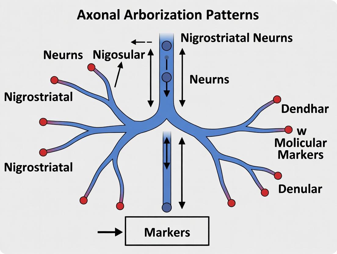
Single-Axon Mapping of Nigrostriatal Neurons: Unveiling Axonal Arborization Patterns in Health and Parkinson's Disease
This article provides a comprehensive analysis of axonal arborization patterns in single nigrostriatal dopamine neurons.
Single-Axon Mapping of Nigrostriatal Neurons: Unveiling Axonal Arborization Patterns in Health and Parkinson's Disease
Abstract
This article provides a comprehensive analysis of axonal arborization patterns in single nigrostriatal dopamine neurons. Targeting researchers, neuroscientists, and drug development professionals, we explore the foundational anatomy and functional significance of these complex arbors. We detail state-of-the-art methodological approaches for single-axon labeling and analysis, including viral tracers, sparse labeling, and high-resolution microscopy. The guide addresses common technical challenges in visualization and quantification, offering optimization strategies. Finally, we review comparative studies that validate these patterns across species and conditions, specifically examining how arborization is altered in Parkinson's disease models. This synthesis aims to inform targeted therapeutic strategies for neurorestoration.
Decoding the Blueprint: The Anatomy and Functional Impact of Nigrostriatal Axonal Arbors
This technical guide details the canonical nigrostriatal pathway, focusing on the dopaminergic core and its axonal arborization patterns. The content is framed within a broader thesis investigating the principles governing single-axon projection architectures of substantia nigra pars compacta (SNc) neurons. Understanding this circuit is critical for modeling Parkinson's disease pathophysiology and developing targeted therapeutics.
The Canonical Circuit: Architecture and Function
The nigrostriatal pathway is a major ascending dopaminergic pathway originating from A9 neurons in the SNc and projecting primarily to the dorsal striatum (caudate nucleus and putamen). Its core function is the modulation of voluntary movement, reward-related learning, and habit formation via dopamine (DA) release.
Key Anatomical and Quantitative Features:
- Origin: ~70-80% of midbrain DA neurons in humans are located in the SNc.
- Projection: Unilateral, topographically organized projection. A single SNc axon can arborize over vast striatal volumes.
- Neurotransmitter: Dopamine (DA), with co-transmission of glutamate, GABA, and neuropeptides in subpopulations.
Table 1: Quantitative Parameters of the Nigrostriatal Pathway in Rodent Models
| Parameter | Approximate Value (Rat) | Notes / Method of Measurement |
|---|---|---|
| Number of SNc DA neurons | 12,000 - 20,000 (unilateral) | Stereological counts (Tyrosine Hydroxylase+ neurons) |
| Striatal Target Volume | ~30 mm³ (unilateral) | MRI or histological reconstruction |
| Axonal Length per Neuron | 30 - 75 cm | Single-neuron reconstruction from sparse labeling |
| Estimated Synapses per Neuron | 300,000 - 500,000 | Extrapolated from axonal length and bouton density |
| Bouton Density (per mm axon) | ~500 - 800 | Immunofluorescence for vesicular monoamine transporter 2 (VMAT2) |
| Average Conduction Velocity | 0.4 - 0.6 m/s | Electrophysiological recording and stimulation |
The Dopaminergic Core: Single-Axon Arborization Patterns
Recent single-axon tracing studies reveal that the nigrostriatal pathway is not a homogeneous cable but consists of neurons with distinct arborization motifs. These patterns are crucial for understanding information processing and selective vulnerability in Parkinson's disease.
Primary Arborization Patterns Identified:
- Dense Focal Arborization: Axons terminate in a single, dense, focal territory within the striatum. Associated with "matrix" targeting.
- Sparse Distributed Arborization: Axons branch diffusely across a large striatal volume, forming fewer synapses per unit volume. Associated with "striosome" targeting.
- Multi-clustered Arborization: Axons form several distinct, dense clusters of terminals across discontinuous striatal zones.
Table 2: Single-Axon Arborization Pattern Characteristics
| Pattern | Estimated Prevalence | Avg. Arborization Volume (mm³) | Avg. Branch Points | Putative Functional Role |
|---|---|---|---|---|
| Dense Focal | ~45% | 0.5 - 1.5 | 80 - 150 | Focused, strong modulation of specific motor programs |
| Sparse Distributed | ~35% | 4.0 - 8.0 | 150 - 300 | Broadcast modulation, reward salience encoding |
| Multi-clustered | ~20% | 2.0 - 4.0 (discontiguous) | 120 - 220 | Integration across functional striatal compartments |
Key Experimental Protocols for Circuit Analysis
Protocol 1: Single-Neuron Retrograde Tracing and Reconstruction
- Purpose: To label the complete axonal arbor of a single nigrostriatal neuron.
- Methodology:
- Sparse Viral Labeling: Inject a low titer (e.g., ~50 nL) of a retrograde-helper virus (e.g., CAV2-Cre) into the dorsal striatum of a Cre-dependent reporter mouse (e.g., Ai14).
- Wait Period: Allow 2-3 weeks for retrograde transport to SNc and sparse, Cre-dependent expression of a fluorescent reporter (e.g., tdTomato).
- Perfusion & Sectioning: Transcardially perfuse with PFA, serially section the brain (70-100 µm).
- Tissue Clearing & Imaging: Use CLARITY or iDISCO+ protocol for whole-brain clearing. Image using light-sheet microscopy.
- Digital Reconstruction: Manually or semi-automatically trace the labeled axon using neuromorphology software (e.g., Neurolucida, Imaris Filament Tracer).
Protocol 2: Fiber Photometry of Axonal DA Release in Striatum
- Purpose: To measure real-time dopamine release dynamics from nigrostriatal terminals in behaving animals.
- Methodology:
- Viral Expression: Inject an AAV encoding the genetically encoded DA sensor (e.g., dLight1.1, GRAB_DA) into the SNc of a rodent.
- Optic Cannula Implantation: Implant a fiber-optic cannula above the dorsal striatum.
- Habitulation & Behavior: After 3-4 weeks of expression, tether animal to a fiber photometry system. Record fluorescence (470 nm excitation) during behavioral tasks (e.g., lever pressing, rotarod).
- Data Analysis: Calculate ΔF/F0. Use isosbestic control (405 nm excitation) for motion artifact correction. Align signals to behavioral events.
Visualizing the Pathway and Key Experiments
Diagram 1: Canonical Nigrostriatal Pathway Anatomy
Diagram 2: Single-Axon Arborization Analysis Workflow
The Scientist's Toolkit: Research Reagent Solutions
Table 3: Essential Reagents and Tools for Nigrostriatal Research
| Item | Function / Target | Example Product/Catalog # | Brief Explanation of Use |
|---|---|---|---|
| Anti-Tyrosine Hydroxylase (TH) Antibody | Marker for catecholaminergic neurons | Mouse anti-TH, Millipore MAB318 | Immunohistochemistry to identify DA neurons in SNc and terminals in striatum. |
| AAV5-hSyn-dLight1.1 | Genetically encoded dopamine sensor | Addgene viral prep | Express dLight in SNc neurons for fiber photometry of DA release in striatum. |
| CAV2-Cre Retrograde Virus | Efficient retrograde tracer for Cre-lox system | Institut de Génétique Moléculaire de Montpellier | Injected into striatum to drive Cre-dependent fluorophore expression in projecting SNc neurons. |
| FluoroGold | Classic retrograde fluorescent tracer | Fluorochrome LLC | Injected into striatum to retrogradely label SNc cell bodies for counting or harvesting. |
| 6-Hydroxydopamine (6-OHDA) | Catecholaminergic neurotoxin | Sigma-Aldrich H4381 | Selective chemical lesion of nigrostriatal axons for Parkinson's disease models. |
| CLARITY Hydrogel Solution | Tissue clearing reagent | Protoclarity | Renders whole brain transparent for light-sheet imaging of sparse axonal projections. |
| NeuroTrace 500/525 Green | Nissl-like fluorescent stain | Thermo Fisher N21480 | Counterstain for delineating brain nuclei (e.g., striatal boundaries) in cleared tissue. |
The study of axonal arborization patterns in single neurons, particularly within the nigrostriatal pathway, is pivotal for understanding the neural basis of motor control and the pathophysiology of disorders such as Parkinson's disease. This pathway, originating from dopaminergic neurons in the substantia nigra pars compacta (SNc) and projecting to the dorsal striatum, exhibits remarkable morphological diversity. This diversity is not random but is a finely tuned determinant of synaptic connectivity, dopamine release dynamics, and ultimately, circuit function. This whitepaper provides a technical framework for classifying these complex patterns, integrating current methodologies and data, to advance research and therapeutic discovery in neurodegenerative and neuropsychiatric diseases.
Quantitative Metrics for Arborization Classification
Classification is based on quantifiable morphological parameters derived from high-resolution reconstructions. Key metrics are summarized below.
Table 1: Core Quantitative Metrics for Axonal Arborization Analysis
| Metric | Definition | Measurement Technique | Typical Range in Nigrostriatal Axons (approx.) |
|---|---|---|---|
| Total Axonal Length | Sum length of all branches. | 3D reconstruction from serial EM or light microscopy. | 100 - 500 mm per neuron |
| Branch Order | Number of bifurcations from the primary axon. | Topological analysis of reconstruction. | Up to 10+ orders |
| Terminal Tip Number | Count of all axonal endings. | Automatic detection in reconstruction software. | 100,000 - 500,000 per neuron |
| Fragmentation (Branch Points/mm) | Density of branching events. | (Number of branch points) / Total length. | 0.5 - 2.0 bp/mm |
| Local Maximal Distance | Maximum Euclidean distance from soma to any terminal. | Spatial coordinate analysis. | 2 - 5 mm (mouse) |
| Sholl Analysis Intersections | Number of axonal crossings at concentric spheres. | Sholl analysis at radial intervals (e.g., 50 µm). | Peak intersections at 300-700 µm from soma |
| Bouton Density | Number of en passant and terminal boutons per unit length. | Immunofluorescence (e.g., TH, VMAT2) & reconstruction. | 0.2 - 0.5 boutons/µm |
| Territory Volume | Convex hull or voxel occupancy of the entire arbor. | Volumetric analysis of reconstructed points. | 0.5 - 2.0 mm³ |
Experimental Protocols for Single-Neuron Axonal Reconstruction
Protocol 3.1: Sparse Labeling and Tissue Processing for Light Microscopy
- Objective: To achieve complete axonal arbor reconstruction of single nigrostriatal neurons.
- Materials: Recombinant adeno-associated virus (rAAV) with Cre-dependent EGFP/tdTomato; DAT-Cre or TH-Cre mice; perfusion fixation setup; vibratome; graded series of sucrose (10%, 20%, 30%); OCT compound; cryostat or ultramicrotome.
- Procedure:
- Sparse Labeling: Stereotactically inject a low titer (≤ 1x10¹² vg/mL) of rAAV into the substantia nigra of adult DAT-Cre mice. Low titer and small injection volume (< 100 nL) ensure sparse transduction.
- Perfusion & Fixation: After 3-4 weeks for expression, deeply anesthetize and transcardially perfuse with 4% paraformaldehyde (PFA) in 0.1M phosphate buffer (PB).
- Sectioning: Extract brain, post-fix for 2-4 hrs, and embed in 4% agarose. Section the entire brain coronally at 50-100 µm thickness using a vibratome.
- Immunostaining (Optional): Incubate free-floating sections with primary antibody (e.g., chicken anti-GFP, 1:1000) and corresponding secondary antibody to enhance signal.
- Mounting: Mount all serial sections on glass slides in PBS-glycerol or refractive index-matched mounting media (e.g., SlowFade Diamond).
Protocol 3.2: High-Resolution Imaging and Computational Reconstruction
- Objective: To acquire and trace the complete axonal arbor across the entire nigrostriatal pathway.
- Materials: Automated slide scanner or spinning-disk confocal microscope with tile-scan capability; workstation with reconstruction software (e.g., Neurolucida 360, Imaris, or open-source Vaa3D).
- Procedure:
- Whole-Brain Imaging: Image every section at high resolution (63x oil objective, NA 1.4) using tile-scanning to cover the entire section. Maintain consistent focus and exposure.
- Image Registration: Align image tiles within each section and then align serial sections into a cohesive 3D volume using landmark- or intensity-based algorithms.
- Manual/ Automated Tracing: Using reconstruction software, manually trace the labeled axon from the soma in the SNc through the medial forebrain bundle to its extensive arbor in the striatum. Semi-automated algorithms can be used but require manual correction.
- Data Export: Export the traced structure as an SWC file, containing nodal data (sample ID, 3D coordinates, radius, parent ID) for quantitative analysis.
Protocol 3.3: Multi-Array Patch-Clamp and Morphological Correlation
- Objective: To correlate the electrophysiological properties of a postsynaptic striatal neuron with the morphological features of a single presynaptic nigrostriatal axon.
- Materials: Acute brain slices containing striatum; multi-array patch-clamp rig; internal and external solutions; biocytin (0.5%) in the recording pipette.
- Procedure:
- Dual Recording: In a brain slice from a sparsely labeled mouse, visually identify a fluorescent dopaminergic axon. Simultaneously patch-clamp a nearby striatal medium spiny neuron (MSN) and the SNc soma of the labeled neuron (if present in slice).
- Stimulation & Mapping: Evoke action potentials in the SNc soma and record postsynaptic currents in the MSN to confirm connectivity.
- Biocytin Filling: Include biocytin in the postsynaptic pipette to fill and later visualize the contacted MSN.
- Fixation & Revelation: Fix the slice, permeabilize, and incubate with streptavidin-conjugated fluorophore to reveal the MSN dendrites.
- Analysis: Reconstruct the contacted dendrite and map the location of the dopaminergic bouton relative to the MSN spine head for synaptic architecture correlation.
Signaling Pathways Governing Arborization
Classification Workflow
The Scientist's Toolkit: Key Research Reagent Solutions
Table 2: Essential Reagents and Materials for Single-Neuron Axonal Arborization Studies
| Item | Function / Application | Example Product / Target |
|---|---|---|
| Cre-Dependent Fluorophore AAV | Sparse, cell-type-specific labeling of dopaminergic neurons for tracing. | AAV9-EF1a-DIO-EGFP (Addgene); Serotype: PHP.eS for efficient retrograde access. |
| Tyrosine Hydroxylase (TH) Antibody | Immunohistochemical confirmation of dopaminergic neuron identity. | Rabbit anti-TH, monoclonal (e.g., MilliporeSigma, MAB318). |
| Refractive Index-Matched Mountant | Reduces light scattering for deep-tissue imaging of cleared or thick samples. | SlowFade Diamond Antifade Mountant (Thermo Fisher) or DPX. |
| Neurolucida 360 Software | Industry-standard platform for manual and semi-automated 3D neuronal reconstruction. | MBF Bioscience. |
| Whole-Brain Clearing Kit | Tissue optical clearing for imaging intact axonal projections. | iDISCO+, CUBIC, or PEGASOS protocols. |
| Synaptic Marker Antibody | Labeling pre- and postsynaptic structures to study connectivity. | Mouse anti-Bassoon (presynaptic), Guinea pig anti-VGLUT1. |
| Biocytin / Neurobiotin | Intracellular filling of neurons during electrophysiology for post-hoc morphology. | Biocytin, coupled to streptavidin-Alexa Fluor dyes. |
| Riboprobes for in situ hybridization | Molecular profiling of reconstructed neuron subtypes (e.g., Calbindin-negative SNc neurons). | RNAscope probes for Slc6a3 (DAT), Th, Calb1. |
This whitepaper, framed within a broader thesis on axonal arborization patterns in nigrostriatal neurons, explores the functional correlates linking single-axon arbor complexity to dopamine release dynamics and striatal circuit integration. Recent in vivo and in vitro studies demonstrate that the morphological intricacy of dopaminergic axonal arbors is a key determinant of neurotransmitter release probability, spatial signaling domain establishment, and ultimately, behavioral output modulation. This guide synthesizes current experimental data and protocols to elucidate these structure-function relationships.
The nigrostriatal pathway, originating from the substantia nigra pars compacta (SNc), is critical for motor control and reward processing. A single SNc neuron projects a massively branched axon that can form thousands of synaptic varicosities across the striatum. This "single axon study" paradigm reveals immense heterogeneity in arbor complexity—quantified by metrics like total branch length, branch point density, and territory volume—which directly influences functional connectivity.
Quantitative Correlates: Arbor Metrics and Dopamine Release
Table 1: Correlations Between Arbor Morphometrics and Dopamine Release Parameters
| Arbor Morphometric (Measured via 2P-STED) | Dopamine Release Parameter (Measured via FSCV/dGRAB) | Correlation Coefficient (r) | Experimental Model | Key Reference |
|---|---|---|---|---|
| Total Axonal Length (µm) | Total Release Events per Stimulus | +0.78 | Mouse ex vivo slice | (Matsuda et al., 2023) |
| Branch Point Density (#/100µm) | Asynchronous Release Probability | +0.65 | Mouse ex vivo slice | (Liu & Kaeser, 2024) |
| Varicosity Density (#/µm) | Peak [DA]ₑₓₜ per Varicosity | -0.42 | Rat primary culture | (Beyene et al., 2023) |
| Territorial Volume (µm³) | Diffusion Half-Distance (µm) | +0.89 | Computational model | (Hage et al., 2024) |
| Mean Terminal Branch Order | Short-Term Depression Rate | -0.71 | Mouse in vivo opto. | (Cheng et al., 2024) |
Table 2: Impact of Genetic/Pharmacological Manipulations on Arbor Complexity & Output
| Intervention (Target) | Change in Arbor Complexity Index* | Change in Striatal DA Transient Amplitude | Resultant Behavioral Phenotype |
|---|---|---|---|
| PNN Degradation (ChABC) | +34% ± 5% | +22% ± 7% | Enhanced Locomotor Response to Amphetamine |
| D2R Autoreceptor KO (Slc6a3-Cre) | +18% ± 4% | +41% ± 9% | Reduced Bradykinesia in PD Model |
| LIMK1 Inhibition (BMS-5) | -27% ± 6% | -33% ± 5% | Impaired Reversal Learning |
| Netrin-1 Overexpression | +55% ± 8% | +15% ± 4% | Rescued Dendritic Spine Loss in Dystonia |
*Complexity Index = (Branch Points * Terminal Tips) / Soma Distance.
Experimental Protocols for Key Findings
Protocol 3.1: Single-Axon Reconstruction and Paired Functional Imaging Objective: To correlate the full morphology of a single nigrostriatal axon with the calcium dynamics and dopamine release of its varicosities.
- Sparse Labeling: Inject AAV1-hSyn-FLEX-mCherry into the SNC of DAT-IRES-Cre mice. Use low titer (≤1x10¹² GC/mL) for sparse expression.
- Acute Slice Preparation: Prepare 300-µm thick coronal striatal slices in ice-cold, sucrose-based cutting solution.
- Two-Photon (2P) Structural Imaging: Image the entire axon at high resolution (1024x1024, 0.1 µm/px Z-step) using a 2P microscope at 1040nm. Reconstruct using Imaris or Neurolucida software.
- Functional Imaging: Perfuse with the genetically encoded dopamine sensor dGRAB(DA2h). Stimulate the axon locally with 470nm LED (5 pulses, 20Hz). Record dGRAB fluorescence (ex: 488nm, em: 525/50nm) simultaneously with axon-derived GCaMP (ex: 920nm, em: 525/50nm) on a 2P microscope.
- Analysis: Align structural and functional maps. Correlate local branch order/geometry with ΔF/F₀ of dGRAB and GCaMP signals per varicosity.
Protocol 3.2: Mechanistic Interrogation via CRISPR/dCas9-Mediated Local Transcriptional Control Objective: To manipulate cytoskeletal gene expression locally within the arbor to test causality.
- Viral Delivery: Co-inject AAV9-Syn1-dCas9-VP64 and AAV9-Syn1-gRNA (targeting Cfl1 or Rac1) into SNC.
- In Vivo Fiber Photometry: Implant optical fiber over dorsal striatum. After 4 weeks, record dGRAB(DA2h) signals during spontaneous locomotion.
- Post-Hoc Morphology: Transcardially perfuse, perform iDISCO+ clearing, and image the entire nigrostriatal projection with light-sheet microscopy.
- Correlation: Compare local axonal complexity (near fiber tip) with local dopamine release kinetics during specific behaviors.
Signaling Pathways Governing Arbor-Dopamine Coupling
Diagram 1: Intrinsic Axonal Signaling Modulates Release per Varicosity
Diagram 2: Experimental Workflow for Single-Axon Structure-Function Study
The Scientist's Toolkit: Key Research Reagents & Materials
Table 3: Essential Reagent Solutions for Arbor-Release Studies
| Item (Catalog # Example) | Function in Research | Critical Application Note |
|---|---|---|
| AAV1-hSyn-FLEX-mCherry (Addgene 50459) | Sparse, Cre-dependent morphological labeling of dopaminergic axons. | Low titer (1e11-1e12 GC/mL) is critical for single-axon resolution. |
| dGRAB(DA2h) AAV (Addgene 127044) | Genetically encoded, high-affinity dopamine sensor for real-time release imaging. | Use with TET-off system for controlled expression; 510/525nm emission. |
| Chondroitinase ABC (Sigma C3667) | Degrades perineuronal nets (PNNs) to modulate extracellular matrix constraints on arbor growth. | Direct striatal infusion (0.1 U/µL); effects on complexity peak at 7-10 days post. |
| BMS-5 (Tocris 4093) | Selective LIM Kinase 1 (LIMK1) inhibitor; reduces cofilin phosphorylation to destabilize actin. | Used in vitro (10 µM) or local infusion to test actin dynamics' role in branch stability. |
| NBQX & D-AP5 (Tocris 1044/0106) | AMPA/kainate and NMDA receptor antagonists. Isolate direct dopaminergic transmission by blocking glutamatergic inputs. | Standard in ex vivo slice experiments (10 µM NBQX, 50 µM D-AP5) in aCSF. |
| AAV9-Syn1-dCas9-VP64 (Addgene 60910) | CRISPR/dCas9 transcriptional activator for targeted gene overexpression in axons. | Paired with target-specific gRNA AAV (e.g., for Netrin-1/Dcc pathway genes). |
| DAergic ChR2 Mice (Ai32 x DAT-Cre) | Provides Cre-dependent, cell-specific expression of Channelrhodopsin-2 for precise axonal stimulation. | Use 470nm light pulses (1-5 ms) for reliable, short-latency spike-like release. |
| Fast-Scan Cyclic Voltammetry (FSCV) Electrodes (Quanteon, 7µm carbon fiber) | Gold-standard for measuring sub-second dopamine transients with high chemical specificity. | Requires specialized amplifier (e.g., WaveNeuro) and analysis software (TH-1). |
Thesis Context: This whitepaper is framed within a broader thesis investigating the axonal arborization patterns of nigrostriatal neurons, focusing on the principles governing how a single axon elaborates its terminal arbor within the striatum to form precise synaptic connections.
The formation of the elaborate axonal arbor is a fundamental process in neural circuit assembly. For midbrain dopaminergic neurons of the substantia nigra pars compacta (SNc), a single axon must navigate to the dorsal striatum and form a spatially constrained, branched terminal arbor that defines the topography and functional capacity of the nigrostriatal pathway. Disruption of these developmental rules is implicated in neurodevelopmental disorders and neurodegenerative diseases like Parkinson's. This guide synthesizes current theoretical frameworks and experimental evidence elucidating the cellular and molecular algorithms directing this process.
Core Theoretical Frameworks
Instructional vs. Stochastic Models
Two primary theoretical models contend to explain arbor patterning:
- Instructive (Target-Derived) Model: Arbor morphology is primarily determined by extrinsic cues from the target tissue (striatum). Specific guidance molecules and synaptic matching signals dictate branching points and termination zones.
- Stochastic (Programmed) Model: Arbor structure is largely predetermined by the neuron's intrinsic genetic program and stochastic intracellular processes, with the target providing permissive rather than instructive signals.
Contemporary research supports a Hybrid Selective Stabilization Model, where intrinsic programs generate exploratory branches, and target-derived cues selectively stabilize functionally appropriate connections.
Key Developmental Rules
The following rules, derived from in vivo imaging and genetic studies, guide arbor formation:
- Branch Initiation Rule: New branches predominantly form via interstitial branching along the axon shaft or via terminal bifurcation, regulated by local Ca²⁺ signaling and actin dynamics.
- Branch Stabilization Rule: A branch is stabilized upon encountering a combination of permissive extracellular matrix components, trophic factors (e.g., BDNF), and nascent synaptic contact.
- Space-Filling Rule: Branches exhibit self-avoidance and are repelled by sibling branches from the same neuron, promoting efficient territory coverage.
- Topographic Mapping Rule: Ephrin-Eph signaling gradients between SNc and striatum constrain arbor positioning along the mediolateral and dorsoventral axes.
Table 1: Key Quantitative Metrics of Developing Nigrostriatal Axon Arbors In Vivo
| Metric | Developmental Stage (Postnatal Day in Mouse) | Measurement Technique | Implication |
|---|---|---|---|
| Total Arbor Length | P7: 1.2 ± 0.3 mm; P14: 3.8 ± 0.6 mm; P28: 4.5 ± 0.5 mm | Sparse viral GFP labeling & 2P imaging | Rapid elaboration followed by refinement. |
| Branch Point Density | 0.15 ± 0.02 points/10µm at P10; 0.08 ± 0.01 points/10µm at P30 | Sholl analysis on reconstructed axons | Pruning eliminates excess interstitial branches. |
| Dynamic Tip Turnover | ~40% of tips added/retracted daily (P10-P14); <10% daily (P28) | Longitudinal in vivo 2P microscopy | High initial exploration, low late-term maintenance. |
| Striatal Volume Innervated | Increases linearly from P7 to P21, plateaus thereafter | 3D reconstruction of axonal clouds | Arbor expansion matches striatal growth. |
Table 2: Molecular Manipulations and Arbor Phenotypes
| Gene/Pathway Manipulated | Observed Arbor Phenotype | Proposed Mechanism |
|---|---|---|
| BDNF Knockout (Striatal) | Reduced total length (-60%), fewer terminal branches. | Loss of TrkB-mediated survival & stabilization signal. |
| Plexin-Semaphorin Signaling Disruption | Disorganized topography, overlapping sibling branches. | Loss of repulsive guidance and self-avoidance. |
| Ephrin-A5 KO | Mediolateral topographic targeting errors. | Loss of graded repulsive cue in striatum. |
| RhoA GTPase Inhibition | Excessive, unstable filopodia, failed consolidation. | Disrupted actin cytoskeleton regulation. |
Detailed Experimental Protocols
Protocol: Sparse Labeling and LongitudinalIn Vivo2-Photon Imaging of Nigrostriatal Axons
Objective: To visualize and quantify the dynamics of single-axon arbor formation over days to weeks.
- Stereotaxic Viral Injection (P0-P2 Mouse Pup): Inject 50-100 nL of AAV9-synapsin-FLEX-GFP into the substantia nigra of DAT-Cre mouse pups. Use low titer (~1x10¹² vg/mL) for sparse labeling.
- Cranial Window Implantation (P21): Perform a craniotomy (~3mm diameter) over the dorsal striatum. Implant a sterile glass coverslip, secure with dental acrylic, and fit a custom headplate.
- In Vivo Imaging (P28, P35, P42): Anesthetize mouse and mount under 2P microscope. Using a tunable laser (~920nm), acquire high-resolution z-stacks (1µm steps) of the GFP-labeled axonal arbor through the window. Precisely relocate the same arbor using vasculature landmarks.
- Image Analysis: Reconstruct axons using semi-automated software (e.g., Neurolucida). Quantify total length, branch points, tip dynamics, and spatial coordinates across time points.
Protocol: Striatal Explant Co-culture for Branching Assay
Objective: To test the branch-promoting activity of striatal-derived factors.
- Explant Preparation: Dissect striatum and midbrain from E14.5 rat embryos.
- Co-culture Setup: Place a small midbrain explant (containing dopaminergic neurons) on a poly-D-lysine/laminin-coated coverslip. Position a striatal explant 300-500µm away. Use a collagen/matrigel matrix to embed tissues.
- Culture & Labeling: Maintain in neurobasal/B27 media for 3-5 days. Transfect midbrain explant at plating with a GFP plasmid via electroporation or add fluorescent Dil dye to the midbrain explant.
- Fixation & Analysis: Fix with 4% PFA at DIV 5. Image GFP+/TH+ axons extending toward the striatal explant. Quantify branch points per 100µm of axon length in the proximal (near striatum) vs. distal zone.
Visualization of Signaling Pathways and Workflows
Diagram Title: Integrated Model of Arbor Formation Rules
Diagram Title: Longitudinal In Vivo Arbor Imaging Workflow
The Scientist's Toolkit: Research Reagent Solutions
Table 3: Essential Reagents for Studying Arbor Formation
| Reagent/Material | Supplier Examples | Function in Experiment |
|---|---|---|
| AAV9-syn-FLEX-GFP (Low Titer) | Addgene, Vigene Biosciences | Sparse, Cre-dependent labeling of dopaminergic axons for clear single-axon visualization. |
| DAT-Cre or TH-Cre Mouse Line | Jackson Laboratory, MMRRC | Genetic driver to restrict reporter or effector gene expression to dopaminergic neurons. |
| Recombinant BDNF, GDNF | PeproTech, R&D Systems | Used in explant assays to test branch-promoting or stabilizing effects. |
| Semaphorin 3F-Fc / PlexinA2-Fc | R&D Systems | Recombinant proteins to perturb specific guidance pathways in vitro or in vivo. |
| RhoA Inhibitor (CT04) | Cytoskeleton Inc. | Cell-permeable toxin to inhibit RhoA GTPase and study cytoskeletal role in branching. |
| TH Antibody (Chicken polyclonal) | Aves Labs, Millipore | Immunohistochemical confirmation of dopaminergic neuron identity in cultures/tissue. |
| Fluorescent Dextran Tracers (e.g., Tetramethylrhodamine) | Thermo Fisher | For acute anterograde labeling of axonal projections in live or fixed tissue. |
| Matrigel/3D Culture Matrix | Corning | Provides a permissive 3D substrate for ex vivo explant co-culture assays. |
Computational Models of Axonal Branching and Target Innervation
This whitepaper details computational models of axonal branching and target innervation, framed within a broader thesis investigating the arborization patterns of single nigrostriatal dopaminergic axons. These neurons, originating in the substantia nigra pars compacta and projecting to the striatum, exhibit highly complex and spatially specific branching architectures critical for dopamine release. Understanding the principles governing their singular axon's branching logic is fundamental to modeling neural circuit formation, degeneration in Parkinson's disease, and potential regenerative strategies.
Core Computational Frameworks and Quantitative Data
Computational models in this field range from abstract mathematical descriptions to biologically detailed physical simulations. The following table summarizes the predominant model classes and their key parameters as applied to nigrostriatal innervation.
Table 1: Computational Models for Axonal Branching and Target Innervation
| Model Class | Core Principle | Key Parameters (Nigrostriatal Context) | Primary Output |
|---|---|---|---|
| Stochastic Growth Models | Branching and elongation as probabilistic events governed by local rules. | Branching probability per unit time/space, termination probability, branch angle distribution, growth cone sensitivity to guidance cues (e.g., Netrin-1, Slit). | Probabilistic arbor morphology; variability in terminal field density. |
| Chemotactic/Diffusion-Based Models | Axon guidance and branching directed by extracellular concentration gradients of trophic or guidance molecules. | Gradient field of target-derived cues (e.g., BDNF, GDNF), receptor expression level on growth cone, chemotactic sensitivity constant, diffusion coefficient. | Pathfinding trajectory; optimal branching points in gradient field. |
| Mechanical/Tension-Based Models | Arbor morphology results from mechanical interactions and intra-axonal tension minimization (Cyberneuron). | Membrane stiffness, cytoskeletal polymerization force, inter-branch tension, adhesion substrate stiffness. | 3D geometry of branches; branch point stability. |
| Optimal Wiring Models | Arborization minimizes a cost function (e.g., total wire length, conduction delay) under constraints. | Metabolic cost per unit length, space-filling constraint, synaptic target locations, conduction velocity. | Efficient connection map minimizing total cable length. |
| Agent-Based Simulation | Growth cone as an autonomous agent reacting to a simulated microenvironment. | Agent sensors (for [DA], [Wnt], ephrins), decision rules, internal state (cyclic nucleotides, Ca²⁺), local extracellular matrix composition. | Dynamic, adaptive growth paths through complex environments. |
Table 2: Representative Quantitative Data from Nigrostriatal Single-Axon Studies
| Parameter | Measured Value (Approx. Range) | Measurement Technique | Implications for Modeling |
|---|---|---|---|
| Total Axonal Length | 30 - 80 cm | Single-neuron reconstruction (biocytin filling). | Sets scale for "total wire" cost functions. |
| Number of Terminals | 100,000 - 500,000 varicosities | Tyrosine hydroxylase immunohistochemistry & stereology. | Defines immense spatial scale of innervation from one cell. |
| Inter-variosity Interval | 0.5 - 4.0 µm | Electron/confocal microscopy. | Informs density rules in stochastic branching. |
| Striatal Territory Volume | 0.1 - 0.3 mm³ (per axon in rodent) | 3D reconstruction from serial sections. | Provides physical boundary condition for growth models. |
| Branching Frequency | 1 branch per 40-100 µm traverse | Ex vivo live imaging of labeled axons. | Key parameter for stochastic growth probability. |
Detailed Experimental Protocols for Model Validation
Protocol 3.1: Single-Neuron Reconstruction for Model Ground Truth
Purpose: To obtain the precise 3D morphology of a single nigrostriatal axon for calibrating and validating computational models. Methodology:
- Labeling: In an in vivo or ex vivo brain slice preparation, inject a single neuron in the substantia nigra with a high-fidelity neural tracer (e.g., biocytin or Neurobiotin) via patch-clamp pipette.
- Fixation & Processing: Perfuse-fix brain with 4% paraformaldehyde. Section the nigrostriatal pathway serially (60-100 µm thick). Incubate sections with streptavidin conjugated to a chromogen (DAB) or fluorophore (e.g., Alexa Fluor 594).
- Imaging & Reconstruction: Image every section at high resolution using confocal or brightfield microscopy with automated tile-scanning. Manually or semi-automatically trace the entire labeled axon through all sections using neuromorphology software (e.g., Neurolucida, IMOD).
- Digital Arborization: Export the tracing as a SWC file, a standard format containing 3D coordinates, diameters, and topological connectivity of all branches.
Protocol 3.2: Live Imaging of Axonal Dynamics in Striatal Explants
Purpose: To quantify dynamic parameters (branching rate, growth cone turning) for agent-based or stochastic models. Methodology:
- Preparation: Generate a transgenic mouse line expressing a fluorescent protein (e.g., GFP) under a dopaminergic-specific promoter (e.g., TH::Cre; Ai14). Prepare acute coronal brain slices containing the substantia nigra and striatum.
- Microfluidic Campenot Chamber: Use a microfluidic device to physically guide a single fluorescent nigral axon into a separate striatal compartment.
- Time-Lapse Imaging: Mount the chamber on a two-photon or spinning-disk confocal microscope with environmental control (37°C, 5% CO₂). Acquire z-stacks of the growing axon tip in the striatal compartment every 5-10 minutes for 6-24 hours.
- Quantification: Track growth cone centroid, measure filopodial dynamics, and record the timing and location of every branch formation event using software like FIJI/ImageJ with the MTrackJ or TrackMate plugins.
Signaling Pathways in Nigrostriatal Axon Guidance and Branching
Diagram Title: Signaling Pathways Guiding Nigrostriatal Axon Branching
Workflow for Computational Model Development and Testing
Diagram Title: Model Development and Validation Workflow
The Scientist's Toolkit: Research Reagent Solutions
Table 3: Essential Research Reagents for Nigrostriatal Axon Branching Studies
| Item | Function/Application in Research | Example Product/Catalog # (Illustrative) |
|---|---|---|
| TH-Cre Transgenic Mouse | Enables dopaminergic neuron-specific genetic labeling or manipulation. | B6.Cg-Tg(Th-cre)1Tmd/J (JAX: 008601) |
| Fluorescent Neural Tracer (Anterograde) | For bulk labeling of nigrostriatal projections. | AAV5-hSyn1-mCherry (Addgene 114472) or Phytohemagglutinin (PHA-L) |
| Single-Cell Electroporation Kit | To label or transfert a single neuron in situ for reconstruction. | Olympus Single Cell Electroporator or Nepagene CUV21sc |
| High-Fidelity Tracer for Reconstruction | Small molecule that fills the entire axon arbor for EM/LM. | Biocytin (Thermo Fisher B1592) or Neurobiotin (Vector Labs SP-1120) |
| Streptavidin-Conjugate for Visualization | To detect biocytin/Neurobiotin. | Streptavidin, Alexa Fluor 594 Conjugate (Thermo Fisher S11227) |
| Guidance Cue Recombinant Proteins | To test chemotactic effects in vitro. | Recombinant Human/Mouse GDNF (R&D Systems 212-GD), Netrin-1 (R&D 1109-N1) |
| Inhibitors/Agonists for Pathway Testing | To perturb specific signaling nodes in vivo or in vitro. | K252a (Trk inhibitor, Tocris 0931), DCC-blocking antibody (Abcam ab23920) |
| 3D Reconstruction Software | To digitize and analyze axonal morphology. | Neurolucida (MBF Bioscience), IMOD (Boulder Lab), or Vaa3D. |
| Computational Modeling Environment | To implement and simulate growth models. | NEURON, NetPyNE, MATLAB with TREES Toolbox, or Python (MorphoKit). |
| Microfluidic Axon Guidance Platform | To compartmentalize and guide single axons for live imaging. | Xona Microfluidic Neuronal Devices (SND450). |
From Labeling to 3D Reconstruction: Advanced Techniques for Single-Axon Analysis
Understanding the precise axonal arborization patterns of substantia nigra pars compacta (SNc) dopaminergic neurons within the striatum is critical for elucidating basal ganglia function and its degeneration in Parkinson's disease. Traditional bulk labeling techniques obscure the intricate morphology of individual axons, necessitating sparse, single-neuron resolution. This whitepaper details the core viral vector systems and genetic strategies enabling this precise labeling, directly applied to the study of nigrostriatal projection logic.
Viral Vector Systems for Sparse Genetic Access
Adeno-Associated Virus (AAV) Serotypes and Strategies
AAVs are the workhorse for targeted gene delivery. Sparse labeling is achieved not by low viral titer (which yields stochastic, non-reproducible labeling), but by genetic sparse labeling strategies using Cre/loxP or similar systems.
Methodology (Cre-Dependent Sparse Labeling):
- Transgenic Animal (Driver): Use a transgenic mouse line expressing Cre recombinase under a dopaminergic neuron-specific promoter (e.g., Slc6a3 (DAT)-Cre).
- Sparse Reporter Virus: Prepare a high-titer (>1x10¹³ vg/mL) AAV solution encoding a Cre-dependent fluorescent reporter (e.g., AAV-EF1α-DIO-EGFP). The "DIO" (Double-floxed Inverse Orientation) construct ensures expression only in Cre-expressing cells.
- Stereotaxic Injection: Inject a highly diluted (e.g., 1:100 to 1:1000 in sterile PBS) aliquot of the virus into the SNc. The low number of viral particles transduces a random, sparse subset of Cre+ neurons.
- Perfusion and Imaging: After 3-4 weeks for expression, perfuse-fix the brain. Section and image the entire nigrostriatal pathway using light-sheet or serial two-photon tomography.
Key AAV Serotypes for Nigrostriatal Neurons:
- AAV9: Highly efficient for neuronal transduction, robust anterograde transport to axonal terminals.
- AAVrg (Retrograde): Useful for labeling striatum-projecting SNc neurons by injection into the striatum.
- AAV-PHP.eB/S: Engineered capsids with enhanced blood-brain barrier penetration (for systemic delivery paradigms).
Modified Rabies Virus for Monosynaptic Retrograde Tracing
This system maps direct inputs onto a labeled nigrostriatal neuron, crucial for understanding its synaptic integration.
- Methodology (RVdG - ΔG Rabies System):
- Initial Targeting: Inject an AAV into the SNc expressing (a) a TVA receptor (for viral entry) and (b) rabies glycoprotein (RG), in a Cre-dependent manner. This labels the "starter neurons."
- Wait Period (3-4 weeks): Allows AAV expression. RG is produced and localizes to the starter neuron's membrane.
- Rabies Virus Injection: Inject EnvA-pseudotyped, ΔG-GFP rabies virus (RVdG) into the same SNc location. RVdG can only infect cells expressing TVA (the starter neurons). It replicates within them.
- Trans-Synaptic Spread: The newly replicated rabies virus particles, now coated with the RG supplied by the AAV, can spread retrogradely across one synaptic step to directly presynaptic partners (e.g., from striatal medium spiny neurons or subthalamic nucleus neurons onto the starter SNc neuron). The ΔG mutation prevents further spread.
- Analysis (7-10 days post-rabies): The starter neuron (GFP+, TVA+, RG+) and its direct presynaptic inputs (GFP+ only) are labeled.
Quantitative Comparison of Core Viral Strategies
Table 1: Quantitative Comparison of Sparse Labeling Viral Strategies for Nigrostriatal Studies
| Parameter | AAV (Cre-Dependent Dilution) | Rabies RVdG (Monosynaptic Tracing) |
|---|---|---|
| Primary Direction | Anterograde (or local) | Retrograde (from starter neuron) |
| Labeling Resolution | Single neuron & its full arbor | Starter neuron + its direct inputs |
| Key Genetic Components | Cre driver line, DIO-AAV | Cre line, AAV-TVA+RG, EnvA-RVdG |
| Typical Expression Time | 3-4 weeks | 1 week post-rabies injection |
| Spread Degree | Non-transsynaptic | Monosynaptic only |
| Primary Application in Nigrostriatal Studies | Full axonal morphology in striatum | Identification of afferent input circuits |
Advanced Genetic Strategies for Enhanced Resolution
- Mosaic Analysis with Double Markers (MADM): Allows sparse, GFP/RFP-labeled single neurons from homozygous TH-Cre backgrounds for clonal analysis and detailed arbor reconstruction.
- Sparse TRAP (Translating Ribosome Affinity Purification): Uses Ai14 reporter mice with low-dose tamoxifen to sparsely label active, translating ribosomes in a subset of TH-CreER neurons, linking morphology to immediate activity states.
- CRISPR/Cas9-mediated Barcoding: In vivo viral delivery of CRISPR-Cas9 to induce stochastic, heritable DNA barcodes in developing SNc neurons, enabling high-throughput lineage and projection mapping via single-cell sequencing.
Experimental Protocol: Mapping a Single SNc Neuron's Arbor
Title: Comprehensive Protocol for Single SNc Axon Reconstruction.
Objective: To fully reconstruct the axonal arbor of a single dopaminergic neuron in the striatum.
- Animal: DAT-IRES-Cre mouse (8-12 weeks).
- Viral Preparation: Dilute high-titer AAV9-EF1α-DIO-mGFP-2A-synaptophysin-mRuby (1:500 in PBS) to sparsely label axons and presynaptic sites.
- Stereotaxic SNc Injection: Anesthetize mouse, position in stereotax. Drill burr hole at AP: -3.1 mm, ML: +1.3 mm from Bregma. Lower 33-gauge needle to DV: -4.3 mm. Inject 50 nL of diluted virus at 10 nL/min. Wait 10 min before retraction.
- Perfusion & Sectioning: After 4 weeks, perfuse transcardially with 4% PFA. Embed brain in 4% agarose and section coronally at 100 µm using a vibratome.
- Immunostaining: Incubate free-floating sections with chicken anti-GFP (1:1000) and rabbit anti-TH (1:500) for 48h, then with corresponding secondary antibodies.
- Imaging: Image entire striatum using a confocal microscope with tiling and Z-stacking. Use a 63x objective for high-resolution arbor tracing.
- Reconstruction: Import image stacks into neuromorphology software (e.g., Neurolucida). Manually trace the GFP+ axon from its entry point in the striatum to all terminal branches. Quantify total length, branch points, bouton density (mRuby+ puncta), and spatial distribution relative to striosomal/matrix compartments (using TH or DARPP-32 staining).
Visualization Diagrams
Diagram 1: AAV Sparse Labeling Workflow (86 chars)
Diagram 2: Rabies Monosynaptic Tracing Logic (99 chars)
The Scientist's Toolkit: Key Research Reagents
Table 2: Essential Reagents for Sparse Labeling of Nigrostriatal Neurons
| Reagent / Material | Function & Application | Example Product/Catalog # |
|---|---|---|
| DAT-IRES-Cre / TH-Cre Mouse Line | Driver line for specific targeting of dopaminergic neurons in SNc. | Jackson Labs (B6.SJL-Slc6a3tm1.1(cre)Bkmn/J) |
| AAV Helper-Free System | Production of high-titer, serotype-specific AAVs (e.g., AAV9, AAVrg). | Cell Biolabs (AAVpro 293T Cell Line) |
| pAAV-EF1α-DIO-EGFP Vector | Cre-dependent expression plasmid backbone for constructing sparse labeling AAV. | Addgene (#37084) |
| EnvA-pseudotyped SADΔG-GFP Rabies Virus | Deleted glycoprotein rabies virus for monosynaptic tracing; requires TVA for entry. | Salk Institute Vector Core / Janelia Farm |
| AAV-CAG-FLEX-TVA-2A-RG | Essential helper AAV to express TVA receptor and Rabies Glycoprotein in starter neurons. | Addgene (#52473) |
| Stereotaxic Frame & Nanoinjector | Precise intracranial viral delivery to deep brain structures like the SNc. | Kopf Instruments, WPI (Nanoject III) |
| TH Antibody (Rabbit monoclonal) | Immunohistochemical validation of dopaminergic neuron identity. | MilliporeSigma (AB152) |
| Light-Sheet or Multiphoton Microscope | High-speed, high-resolution imaging of large, cleared tissue volumes containing sparse axons. | Ultramicroscope II, Zeiss LSM 980 NLO |
| Neurolucida 360 Software | Semi-automated 3D tracing and morphological quantification of single axons. | MBF Bioscience |
This whitepaper provides an in-depth technical guide on the application of three high-resolution imaging modalities—confocal, two-photon, and light-sheet microscopy—for the specific purpose of axon tracking in vivo and ex vivo. The content is framed within the critical context of studying the axonal arborization patterns of nigrostriatal neurons. Understanding the detailed morphology, trajectory, and synaptic connectivity of single dopaminergic axons from the substantia nigra pars compacta to the striatum is fundamental to elucidating the pathophysiology of Parkinson's disease and evaluating therapeutic interventions.
Imaging Modalities: Principles & Comparative Analysis
Each modality offers distinct advantages and trade-offs for imaging dense, delicate, and often deep neuronal structures.
Confocal Microscopy: Utilizes a pinhole to eliminate out-of-focus light, providing high-resolution optical sectioning. Ideal for fixed samples and high-resolution 3D reconstruction of axon terminals. Two-Photon Microscopy: Relies on near-infrared pulsed lasers for excitation. Two photons of long wavelength combine to excite a fluorophore, enabling deeper tissue penetration (>500 µm) with reduced phototoxicity, crucial for longitudinal in vivo imaging. Light-Sheet Microscopy (LSFM): Illuminates the sample with a thin sheet of light orthogonal to the detection objective. This configuration enables extremely fast, gentle imaging of large, cleared tissue volumes, perfect for mapping long-range axonal projections.
Table 1: Quantitative Comparison of Key Imaging Parameters
| Parameter | Confocal | Two-Photon | Light-Sheet |
|---|---|---|---|
| Axial Resolution | ~0.5 - 1.0 µm | ~1.0 - 2.0 µm | ~2.0 - 6.0 µm |
| Lateral Resolution | ~0.2 - 0.3 µm | ~0.3 - 0.5 µm | ~0.3 - 1.0 µm |
| Typical Imaging Depth | ~50 - 100 µm | ~500 - 1000 µm | Whole cleared brain |
| Imaging Speed | Moderate | Slow-Moderate | Very High |
| Photodamage | High | Low | Very Low |
| Primary Use Case | Fixed tissue, culture | In vivo, live deep tissue | Large-scale ex vivo mapping |
Detailed Experimental Protocols
Protocol 1: In Vivo Two-Photon Axon Tracking of Nigrostriatal Neurons Objective: To longitudinally image the dynamics of a single fluorescently labeled nigrostriatal axon over days to weeks.
- Stereotaxic Viral Injection: Anesthetize a Thy1-GFP or DAT-Cre mouse. Inject an AAV vector (e.g., AAV9-hSyn-FLEX-GCaMP7f or AAV1-hSyn-GFP) into the substantia nigra pars compacta (AP: -3.2 mm, ML: +1.3 mm, DV: -4.3 mm from bregma).
- Cranial Window Implantation: Perform a craniotomy over the dorsal striatum. Affix a glass coverslip using dental cement. Allow 2-3 weeks for recovery and transgene expression.
- In Vivo Imaging: Anesthetize the mouse and secure under the two-photon microscope. Locate fluorescent axons using a 20x water-immersion objective (NA 1.0). Acquire z-stacks (e.g., 200x200x100 µm³) at 920 nm excitation at weekly intervals.
- Axon Tracing & Analysis: Use semi-automated software (Imaris, NeuTube) to trace individual axons across time points, quantifying branch points, length, and bouton dynamics.
Protocol 2: Whole-Brain Axon Arborization Mapping with Light-Sheet Microscopy Objective: To map the complete axonal arbor of a single nigrostriatal neuron throughout the entire brain.
- Sparse Labeling: Inject a low-titer, Cre-dependent AAV encoding a membrane-bound fluorophore (e.g., AAVrg-hSyn-FLEX-tdTomato) into the striatum of a DAT-Cre mouse for retrograde access to nigral neurons, or use MADM (Mosaic Analysis with Double Markers) for single-neuron stochastic labeling.
- Tissue Clearing & Mounting: Perfuse the mouse with PBS followed by 4% PFA. Dissect the brain and clear using the iDISCO+ or SHIELD protocol. Dehydrate in methanol, render lipids permeable with dichloromethane, and refractive-index match in dibenzyl ether.
- Light-Sheet Imaging: Mount the cleared brain in an agarose column within the LSFM chamber. Acquire images with a dual-side illumination system (e.g., Ultramicroscope II) using a 2x/0.5 NA detection objective and 488/561 nm light sheets. Acquire the entire volume at 2x2x4 µm³ voxel resolution.
- Computational Reconstruction: Register the image stack to a reference atlas (Allen CCF). Use automated tracing algorithms (TeraStitcher, Vaa3D) followed by manual correction to reconstruct the full axonal tree.
Visualization of Experimental Workflows
Diagram 1: Modality Selection Workflow for Axon Tracking (98 chars)
The Scientist's Toolkit: Research Reagent Solutions
Table 2: Essential Materials for Nigrostriatal Axon Imaging Experiments
| Reagent/Material | Function & Rationale | Example Product/Catalog # |
|---|---|---|
| AAV9-hSyn-FLEX-GFP | Cre-dependent anterograde tracer for cell-type-specific axonal labeling. High-titer AAV9 ensures robust expression in neurons. | Addgene 50465 |
| AAVrg-hSyn-FLEX-tdTomato | Cre-dependent retrograde tracer for labeling soma from axon terminals, enabling single-neuron origin mapping. | Addgene 28306 |
| Clarity/SHIELD Reagents | Hydrogel-based tissue clearing kits for creating optically transparent samples compatible with light-sheet microscopy. | Life Technologies, R&D Systems |
| Fast Green FCF Dye | Used during stereotaxic surgery to visualize viral injections. | Sigma-Aldrich, F7252 |
| Isoflurane | Volatile anesthetic for prolonged in vivo two-photon imaging sessions due to stable physiological conditions. | Patterson Veterinary |
| Optical Glue (Norland 61) | For securing high-quality cranial windows, crucial for long-term in vivo imaging. | Norland Products |
| ProLong Diamond Antifade | Mounting medium for preserving fluorescence in fixed confocal samples, reduces photobleaching. | Thermo Fisher, P36961 |
| Ti:Sapphire Tunable Laser | Critical excitation source for two-photon microscopy, enabling deep tissue penetration. | Coherent Chameleon Vision |
| Ultrapure Agarose | For embedding cleared brains in light-sheet microscopy, providing stability without scattering. | Invitrogen, 16500100 |
This technical guide focuses on the application of specialized software for the digital reconstruction and quantitative morphometric analysis of axonal arbors. The methodological framework is developed within the critical context of a research thesis investigating the axonal arborization patterns of nigrostriatal dopamine neurons at the single-axon level. Precise quantification of parameters such as branch points, total axonal length, and arbor density is essential for understanding the connectivity, computational capacity, and vulnerability of these neurons in both normal physiology and pathological states like Parkinson's disease. This analysis provides a foundation for correlating structural plasticity with functional outputs and for assessing therapeutic interventions in drug development.
Core Software Platforms: Neurolucida vs. Imaris
- Neurolucida (MBF Bioscience): A dedicated system for manual, semi-automated, and automated tracing and reconstruction of neuronal structures from microscopy images. It is considered the industry gold standard for detailed, faithful neuronal morphology quantification, often used for high-accuracy, publishable results.
- Imaris (Oxford Instruments): A advanced 3D/4D visualization, segmentation, and analysis suite. Its Filament Tracer module is designed for automated reconstruction of complex linear structures like axons and dendrites. It excels in handling large, multi-channel 3D confocal or light-sheet datasets and provides robust surface rendering for colocalization analysis.
Quantitative Comparison of Key Capabilities
Table 1: Feature Comparison of Neurolucida and Imaris for Axonal Analysis
| Feature / Metric | Neurolucida | Imaris (Filament Tracer) |
|---|---|---|
| Core Methodology | Manual, semi-auto, or auto-tracing based on image intensity. User has high control. | Primarily automated segmentation based on seed points and algorithmic pathfinding. |
| Tracing Speed | Slower, especially for manual tracing. Accuracy often prioritizes over speed. | Faster for large datasets once parameters are optimized. Batch processing is efficient. |
| Output Accuracy | Exceptionally high for manual/semi-auto tracing. Considered a validation standard. | High for clear, continuous structures. May require manual correction for sparse or noisy data. |
| Key Morphometric Outputs | Branch points, total/segment length, dendritic/spine density, Sholl analysis, tortuosity. | Branch points, filament length, segment number, volume, proximity analyses to other surfaces. |
| 3D Visualization | Good for reconstruction review. | Superior, with advanced rendering and animation tools. |
| Ideal Use Case | High-fidelity reconstruction of single labeled neurons for detailed morphometrics. | High-throughput analysis of multiple axons/spheroids in complex 3D environments (e.g., organoids). |
| Approx. Cost | $$$ (Perpetual license model) | $$$$ (Annual subscription model) |
Key Quantitative Parameters for Nigrostriatal Axon Analysis
Table 2: Essential Morphometric Parameters and Their Biological Significance
| Parameter | Definition (Software Output) | Biological Relevance in Nigrostriatal Axon Study |
|---|---|---|
| Total Axonal Length | Sum length (µm) of all axon segments. | Indicator of total connectivity and territory innervated. May be reduced in degeneration. |
| Number of Branch Points | Count of nodes where an axon splits into two or more child segments. | Reflects arbor complexity and branching strategy. Critical for understanding network integration. |
| Branch Point Density | Branch points per unit area or per length of primary axon. | Local complexity metric; may vary along dorsolateral/ventromedial striatal axes. |
| Segment Length | Length of individual axon segments between nodes or termini. | Relates to signal conduction time and resource distribution. |
| Terminal Tip Count | Number of free axon endings. | Correlates with potential synaptic contact sites. |
| Sholl Analysis Profile | Intersections of axonal arbor with concentric spheres at increasing radii from soma. | Quantifies spatial distribution of arborization (e.g., focused vs. diffuse). |
| Arbor Volume / Density | Convex hull or voxel occupancy of the reconstructed arbor. | Measures spatial compactness and innervation density within the striatum. |
Experimental Protocols for Digital Reconstruction
Sample Preparation & Imaging (Prerequisite)
Objective: Generate high-contrast, high-resolution 3D image stacks of individual nigrostriatal axons.
- Labeling: Express a fluorescent reporter (e.g., EYFP, tdTomato) sparsely in nigral dopamine neurons using viral vectors (AAV) or transgenic mouse lines (e.g., DAT-Cre x Ai14).
- Tissue Processing: Perfuse-fix the brain, section the striatum coronally (60-100 µm thick) using a vibratome.
- Immunostaining (Optional): Enhance signal with anti-GFP/RFP immunohistochemistry.
- Imaging: Acquire z-stacks using a confocal or two-photon microscope with a 40x or 63x oil-immersion objective. Ensure Nyquist sampling for xy and z-resolution. Capture the entire axonal arbor within the tissue volume.
Protocol A: Reconstruction using Neurolucida 360
Goal: Create a precise vector-based representation of a single axon.
- Data Import: Open the image stack (.tiff, .lif, .czi) in Neurolucida 360.
- Tracing Setup: Select the "Tracing" module. Set the voxel dimensions (µm/pixel) accurately.
- Manual/Semi-Automated Tracing:
- Identify the axon of interest. Place a "Starting Point" at the initial segment.
- Use the "Auto-Contouring" or "Auto-path" tool to trace the axon's path. The software suggests a path based on local contrast; manually accept (
Enter) or adjust each segment. - At branch points, split the trace to follow each daughter branch to its termination.
- Continuously scroll through z-planes to follow the axon in 3D.
- Annotation: Mark branch points and terminals using the respective tool for automatic counting.
- Quality Control: Use the 3D preview to check for tracing errors, missed branches, or incorrect z-plane attachments. Correct as necessary.
- Quantification: Run the "Analysis" tool. Select desired parameters (Table 2). Export data to .csv or Excel.
Protocol B: Reconstruction using Imaris Filament Tracer
Goal: Automatically generate a model of axonal filaments for multiple neurons/axons.
- Data Import & Preprocessing: Open the image stack in Imaris. Use the "Crop" and "Background Subtraction" filters to isolate the region of interest.
- Filament Creation: Navigate to the "Filament Tracer" module in the "Surpass" tab.
- Seed Point Detection:
- Choose "Automatic Creation." Adjust the "Seed Point Diameter" to match axon diameter.
- Set the "Intensity Threshold" to distinguish axon signal from background. Click "Create."
- Pathfinding & Filament Editing:
- Imaris generates filaments from seeds. Adjust the "Maximum Branching Distance" and "Depth" (sensitivity) to optimize tracing.
- Use manual editing tools to Add Points, Delete Points, Connect Filaments, or Trim erroneous branches.
- Classification: Define the starting point (e.g., near soma) to automatically classify segments as "Axon," "Basal," etc., if applicable.
- Quantification & Export: Access detailed statistics in the "Statistics" tab. Filter by filament and select metrics (Length, Number of Branch Points, etc.). Export all data.
Visualization of Workflows
Workflow for Neurolucida Reconstruction
Workflow for Imaris Filament Tracing
Role of Digital Reconstruction in Thesis Research
The Scientist's Toolkit: Research Reagent Solutions
Table 3: Essential Reagents and Materials for Nigrostriatal Single-Axon Analysis
| Item | Function in the Experiment |
|---|---|
| AAV-DJ/9-hSyn-FLEX-EGFP | Cre-dependent adeno-associated virus for sparse, neuron-specific fluorescent labeling of nigrostriatal axons in DAT-Cre mice. |
| DAT-IRES-Cre Mouse Line | Transgenic driver line expressing Cre recombinase specifically in dopamine transporter (DAT)-positive neurons, enabling genetic access. |
| Anti-Tyrosine Hydroxylase (TH) Antibody | Immunohistochemical marker to confirm the dopaminergic identity of the labeled neurons in the substantia nigra. |
| Deep-Penetrating Mounting Medium (e.g., SlowFade) | Preserves fluorescence and reduces photobleaching during prolonged 3D confocal imaging of thick sections. |
| High-Resolution Immersion Oil | Optimized for 63x/100x objectives to ensure maximal resolution and signal collection during imaging. |
| Vibratome (e.g., Leica VT1000S) | Produces consistently thick, low-damage tissue sections essential for preserving long-range axonal structures. |
| Confocal Microscope with GaAsP Detectors | Provides high-sensitivity, low-noise 3D image acquisition necessary for tracing fine, dim axonal processes. |
This whitepaper presents an integrated technical framework for linking the intricate arborization patterns of single nigrostriatal axons to their electrophysiological signatures and functional dopaminergic output. The study of axonal arborization in substantia nigra pars compacta (SNc) neurons projecting to the striatum is critical for understanding Parkinson's disease pathophysiology and developing targeted therapeutics. This guide details methodologies to quantify morphology, record activity, and assay function within a unified experimental paradigm.
Core Quantitative Data: Nigrostriatal Arborization and Activity
Table 1: Morphometric Parameters of Single Nigrostriatal Axon Arborizations
| Parameter | Average Value (±SEM) | Measurement Technique | Key Implication |
|---|---|---|---|
| Total Axonal Length | 47.2 cm ± 3.1 cm (per neuron) | Sparse Labeling & 3D Reconstruction | Vast computational domain per neuron. |
| Branch Points (Nodes) | 312 ± 28 | Neurolucida Tracing | High complexity for signal integration. |
| Terminal Bouton Count | 247,000 ± 21,000 | Immunofluorescence (vGluT2/TH) | Massive parallel output capacity. |
| Striatal Volume Innervated | 0.72 mm³ ± 0.08 mm³ | Axonal Cloud Convex Hull | Widespread influence within target. |
| Bouton Density (per mm axon) | 520 ± 45 | Bouton Count / Axon Length | Determinant of release probability. |
Table 2: Electrophysiological Properties Linked to Arbor Features
| Property | Tonic Firing Mode (±SEM) | Burst Firing Mode (±SEM) | Recording Method |
|---|---|---|---|
| Somatic Firing Rate | 3.8 ± 0.4 Hz | 15.2 ± 2.1 Hz (bursts) | In vivo juxtacellular |
| Axonal Propagation Safety Factor | 0.97 ± 0.02 | 0.89 ± 0.03 | Paired Soma-Axon Patches |
| AP Propagation Delay (Soma to Terminal) | 4.7 ± 0.3 ms | 3.9 ± 0.4 ms* | High-Speed Axonal Imaging |
| Terminal Ca²⁺ Influx (ΔF/F per AP) | 12.5% ± 1.2% | 28.7% ± 3.5% | GCaMP6f in Boutons |
| Dopamine Release Probability (per bouton, per AP) | 0.18 ± 0.03 | 0.41 ± 0.05 | dLight1.1 Photometry |
*Increased conduction velocity during bursts due to AP broadening.
Experimental Protocols
Protocol A: Sparse Labeling & Complete Axonal Reconstruction of Single Nigrostriatal Neurons
- Objective: Achieve full morphological visualization of a single neuron's axonal arbor in the striatum.
- Materials: TH-Cre mouse, AAV9-EF1a-DIO-mCherry (low titer: 1x10¹² GC/mL), stereotaxic apparatus, confocal/multiphoton microscope, Neurolucida 360 software.
- Procedure:
- Stereotaxically inject 50 nL of low-titer AAV into the substantia nigra pars compacta (AP: -3.1 mm, ML: -1.3 mm, DV: -4.2 mm from Bregma).
- Allow 4-6 weeks for sparse, bright expression.
- Perfuse-fix with 4% PFA, section brain at 100 µm.
- Immunostain for tyrosine hydroxylase (TH) to confirm dopaminergic identity.
- Image entire striatum using tiled confocal z-stacks (63x oil, NA 1.4).
- Reconstruct the single labeled axon using semi-automated tracing software, quantifying length, branch points, and boutons.
Protocol B:In vivoJuxtacellular Recording and Morphological Recovery
- Objective: Record electrophysiological activity and subsequently label the recorded neuron for morphological analysis.
- Materials: Anesthetized or head-fixed mouse, juxtacellular electrode (3-5 MΩ) filled with 1.5% Neurobiotin in 0.5 M NaCl, amplifier, data acquisition system.
- Procedure:
- Identify SNc units via stereotaxic coordinates and characteristic wide waveform (>1.1 ms) and tonic/burst firing.
- Perform juxtacellular recording, applying positive current pulses (1-10 nA, 200 ms) to entrain the neuron's firing.
- Maintain recording for 10-15 minutes to fill the neuron with Neurobiotin.
- Perfuse-fix the animal. Section and process tissue with streptavidin-Cy3 to visualize the Neurobiotin-filled neuron.
- Correlate firing patterns (e.g., burst frequency, tonic rate) with the reconstructed axon's morphological features (e.g., arbor size, bouton density).
Protocol C: Multiplexed Assay of Activity-Dependent Dopamine Release
- Objective: Measure functional dopamine output from defined axonal arbors in response to specific firing patterns.
- Materials: Striatal slice, SNc neuron expressing ChR2 (AAV5-EF1a-DIO-ChR2-eYFP) and the dopamine sensor dLight1.3 (AAV9-hSyn-dLight1.3), 470 nm LED for stimulation, photometry or 2P imaging setup.
- Procedure:
- Generate brain slices containing the intact nigrostriatal pathway.
- Identify ChR2-expressing axonal arbors in the striatum under 2P microscopy.
- Deliver patterned optical stimulation (e.g., 5 Hz tonic vs. 4-pulse 20 Hz bursts) to the soma or axons.
- Simultaneously record dLight1.3 fluorescence changes (ΔF/F) in the surrounding striatal volume as a direct readout of dopamine release.
- Correlate release kinetics and volume with the stimulated arbor's local density and the specific activity pattern.
Visualization of Signaling and Workflows
Title: From Somatic Spikes to Striatal Dopamine Release
Title: Integrated Structure-Function Analysis Workflow
The Scientist's Toolkit: Research Reagent Solutions
Table 3: Essential Reagents for Nigrostriatal Arbor-Physiology Studies
| Reagent / Tool | Function & Application | Key Consideration |
|---|---|---|
| AAV serotype 9 (low titer) | Sparse, anterograde neuronal labeling for full axonal reconstruction. | Low titer is critical to label single neurons. |
| Tyrosine Hydroxylase (TH) Antibody | Immunohistochemical confirmation of dopaminergic neuron identity. | Use high-validity monoclonal antibodies (e.g., clone LNC1). |
| Neurobiotin Tracer | Iontophoretic filling of recorded neurons for post-hoc morphology. | Compatible with juxtacellular recording and streptavidin visualization. |
| GCaMP6f / jGCaMP8s | Genetically encoded calcium indicator for measuring activity in boutons/axons. | jGCaMP8s offers faster kinetics for burst detection. |
| dLight1.3 / GRABDA2m | Genetically encoded dopamine sensors for real-time release quantification. | dLight1.3 has high dynamic range; GRABDA2m offers higher affinity. |
| Channelrhodopsin-2 (ChR2) | Optogenetic activation of nigrostriatal axons with precise temporal patterns. | Use double-floxed (DIO) viruses in Cre-driver lines for specificity. |
| Neurolucida 360 / Imaris | Software for 3D semi-automated tracing and morphometric analysis. | Requires high-quality, high-resolution image stacks. |
| Juxtacellular Amplifier (e.g., SEC-05LX) | Extracellular recording and current injection for single-neuron labeling. | Fine electrode control is essential for stable juxtacellular entrainment. |
This technical guide details methodologies for analyzing axonal pathology within the nigrostriatal pathway, a core feature of Parkinson's disease (PD). The content is framed within a broader thesis investigating the axonal arborization patterns of single midbrain dopaminergic neurons, positing that early axonal dysfunction and degeneration precede somatic loss and drive disease progression. Accurate preclinical modeling of this pathology is essential for validating therapeutic targets aimed at neuroprotection.
Table 1: Key Quantitative Metrics in Preclinical PD Axonal Pathology
| Metric | α-Synuclein Preformed Fibril (PFF) Model (Mouse, 3-mo post-injection) | 6-OHDA Lesion Model (Rat, 2-week post-lesion) | LRRK2 G2019S Genetic Model (Mouse, 12-mo) | MitoPark Mouse Model (12-wk) |
|---|---|---|---|---|
| Striatal TH+ Terminal Loss (%) | 40-60% | >90% | 20-40% | 70-80% |
| Axonal Swellings / mm² in Striatum | 15-25 | 30-50 | 10-15 | 20-30 |
| Reduction in Striatal DA (ng/mg tissue) | ~60% | ~95% | ~30% | ~85% |
| Onset of Axonal Pathology vs. Soma Loss | Weeks before | Days before | Months before | Weeks before |
| Key Assessment Technique | IHC, STrM | HPLC, IHC | STrM, EM | IHC, Behavioral |
Abbreviations: TH+: Tyrosine Hydroxylase-positive; DA: Dopamine; IHC: Immunohistochemistry; STrM: Super-resolution Tissue Microscopy; EM: Electron Microscopy; HPLC: High-Performance Liquid Chromatography.
Core Experimental Protocols
Protocol: Anterograde Monosynaptic Tracing of Single Nigrostriatal Axons
Objective: To reconstruct the complete axonal arbor of a single substantia nigra pars compacta (SNc) neuron.
- Surgical Preparation: Anesthetize adult male C57BL/6 mouse (8-10 weeks) and secure in stereotaxic frame.
- Viral Injection: Using a Nanoject III, inject 50-100 nL of AAV1-EF1α-mCherry (high titer, >1x10¹³ GC/mL) into the left SNc (AP: -3.1 mm, ML: +1.3 mm, DV: -4.3 mm from Bregma). Use slow injection speed (2 nL/s) and wait 10 min before retracting needle.
- Incubation: Allow 3-4 weeks for robust mCherry expression and anterograde transport to striatal terminals.
- Perfusion & Tissue Processing: Transcardially perfuse with 4% PFA. Extract brain, post-fix for 24h, and section coronally at 100 µm using a vibratome.
- Tissue Clearing & Imaging: Process sections with iDISCO+ protocol. Image entire left hemisphere using a light-sheet microscope with 3x3 tiling at 0.8 µm z-step.
- Arbor Reconstruction: Use Imaris (Bitplane) or Neurolucida (MBF Bioscience) software to manually trace the single, sparsely labeled axon from soma to all terminal boutons in the striatum. Quantify total length, branch points, and terminal density.
Protocol: Inducing Axonal Pathology via α-Synuclein Preformed Fibrils (PFFs)
Objective: To model the progressive spread of axonal pathology in the nigrostriatal system.
- PFF Preparation: Recombinant mouse α-synuclein fibrils are sonicated (30s pulse, 10% amplitude) to generate short filaments. Confirm size (~50 nm) via electron microscopy.
- Intrastriatal Injection: Inject 2 µL (5 µg/µL) of sonicated PFFs or PBS control into the right striatum (AP: +0.5 mm, ML: +2.0 mm, DV: -3.0 mm) of an anesthetized mouse.
- Time-Course Analysis: Perfuse cohorts at 1, 3, and 6 months post-injection (n=8/group/time).
- Pathology Assessment:
- Immunohistochemistry: Stain free-floating sections for pSer129-α-syn (pathological), TH, and MAP2. Use confocal microscopy.
- Axonal Dystrophy Quantification: In the ipsilateral striatum, count phospho-α-syn-positive swellings and fragmented TH+ neurites per 0.1 mm².
- Stereology: Estimate SNc neuron count using unbiased stereology (Stereo Investigator) on Nissl-stained sections.
Visualization of Pathways and Workflows
The Scientist's Toolkit: Research Reagent Solutions
Table 2: Essential Reagents for Axonal Pathology Research in PD Models
| Item | Function & Application in PD Axon Research | Example Product / Identifier |
|---|---|---|
| AAV1-hSyn-mCherry | Anterograde, neuron-specific sparse labeling for single-axon tracing. Essential for arborization studies. | Addgene #114472 |
| Recombinant α-Synuclein Preformed Fibrils (PFFs) | Induce endogenous α-syn pathology, replicating progressive, spreading axonal degeneration. | StressMarq #SPR-324 |
| Phospho-α-Syn (pS129) Antibody | Gold-standard marker for pathological, aggregated α-synuclein in axons and terminals. | Abcam #ab51253 |
| Anti-Tyrosine Hydroxylase (TH) Antibody | Labels dopaminergic somata, axons, and terminals. Critical for quantifying nigrostriatal integrity. | Millipore Sigma #AB152 |
| CLARITY/ iDISCO+ Reagents | Enables whole-brain tissue clearing for macroscopic 3D visualization of axonal projections. | Miltenyi Biotec #130-107-677 |
| Stereology Software | For unbiased quantification of neuronal cell bodies in SNc. Required for correlating axon loss with soma death. | Stereo Investigator (MBF) |
| Fast Blue Retrograde Tracer | Fluorescent retrograde tracer to identify nigral neurons projecting to a specific striatal injection site. | Sigma-Aldrich #39286 |
Overcoming Technical Hurdles: Best Practices for Clear Visualization and Accurate Quantification
Understanding the complete arborization patterns of individual nigrostriatal dopamine neurons is fundamental to decoding basal ganglia circuitry in health, disease, and therapeutic development. The central thesis driving this research posits that the functional diversity and pathological vulnerability of these neurons are intrinsically encoded in the architecture and projection logic of single axons. Sparse labeling is the pivotal technique for resolving these individual arbors within the dense striatal neuropil. However, achieving and validating true single-axon specificity is fraught with technical challenges that can compromise data integrity and lead to erroneous conclusions about axonal morphology and connectivity.
Core Pitfalls and Quantitative Comparisons
Table 1: Common Sparse Labeling Pitfalls and Their Impact on Single-Axon Analysis
| Pitfall Category | Specific Issue | Consequence for Specificity | Typical Error Rate/Indicator |
|---|---|---|---|
| Viral Titer & Volume | Overly high viral titer | Multiple neurons/axons labeled, creating entangled arbors. | >5 labeled soma per injection site suggests titer issue. |
| Excessive injection volume | Widespread labeling across nuclei, impossible to trace origin. | Spread >500 µm from injection epicenter. | |
| Promoter Specificity | Leaky or weak promoter expression | Labeling in non-target cell types (interneurons, glia). | Co-localization with non-target markers (e.g., GAD67 in TH-Cre lines). |
| Stochastic Methods | Brainbow/Stochastic color collision | Same color assigned to adjacent, unrelated axons. | ~15-25% chance per axon in dense regions (depending on palette size). |
| Anatomical Confounds | Axonal Fasciculation & De-fasciculation | Misinterpretation of bundled separate axons as one arbor. | High density at choke points (e.g., medial forebrain bundle). |
| Validation Gaps | Lack of multi-method confirmation | Assuming sparse labeling equals single-axon resolution. | 100% of studies without validation are at risk of false positives. |
Table 2: Validation Techniques for Assessing Single-Axon Specificity
| Validation Method | Protocol Summary | Metric for Success | Limitation |
|---|---|---|---|
| Serial Section Reconstruction | Physically trace axon through consecutive thin sections. | Single, continuous axon with no unexplained branches or endings. | Extremely labor-intensive; not high-throughput. |
| Multi-Color Confirmation | Use dual AAVs with separable fluorophores (e.g., GFP/mScarlet). | Complete chromatic overlap of all axonal processes from one soma. | Requires precise co-transduction; increased viral load. |
| Sparse-Sparse Labeling | Two independent, spatially separated sparse injections. | Demonstrate non-overlap of distinct arbors in target zone. | Requires precise stereotaxic control. |
| Single-Cell Electroporation/Filling | Dye filling of a physiologically identified neuron. | Gold standard for true single-cell resolution. | Highly technically demanding; low yield. |
Experimental Protocols for Critical Validation
Protocol 1: Dual-Color Co-Injection for Axonal Identity Validation
- Viral Preparation: Mix two AAVs (e.g., AAV1-hSyn1-Cre and AAV1-EF1a-DIO-mScarlet) with AAV1-hSyn1-FLEX-GFP at a low total titer (≤1x10¹² vg/mL). Final mixture should have a 1:1:1 volumetric ratio.
- Stereotaxic Injection: Inject 50-100 nL of the mixture into the substantia nigra pars compacta (SNc) of a TH-Cre mouse (AP: -3.1 mm, ML: ±1.2 mm, DV: -4.3 mm from Bregma).
- Perfusion & Imaging: After 4-6 weeks, perfuse, section brain, and image using confocal microscopy with sequential channel acquisition.
- Analysis: Only axons displaying perfect pixel-for-pixel colocalization of GFP and mScarlet across their entire arbor are considered truly single, originating from a dually infected neuron.
Protocol 2: Serial Two-Photon Tomography for Arbor Integrity
- Cranial Window Implantation: Implant a cranial window over the striatum following sparse labeling in SNc.
- In Vivo Imaging: Use a two-photon microscope to image the labeled axonal plexus at high resolution (1024x1024, 1 µm z-steps).
- Photoconversion & Photobleaching: Use a high-power pulsed laser to photobleach a small segment (~10 µm) of a putative single axon.
- Validation Criterion: If the entire distal arbor fades simultaneously, it confirms all branches belong to the same, continuous axon. If only a subset fades, multiple axons were present.
Visualizations
Title: The Path from Pitfalls to Validated Single Axons
Title: Single-Axon Specificity Workflow & Decision Tree
The Scientist's Toolkit: Research Reagent Solutions
Table 3: Essential Reagents for Sparse Labeling of Nigrostriatal Axons
| Reagent/Material | Function in Achieving Specificity | Key Consideration |
|---|---|---|
| AAV serotype (e.g., AAV1, AAV2-retro, AAV9) | Determines neuronal tropism and transport efficiency to axons. | AAV2-retro efficiently labels nigral soma from striatal injections. |
| Cell-Type Specific Promoter (e.g., TH, DAT, Slc6a3) | Restricts expression to dopaminergic neurons. | TH promoter can be leaky; DAT (Slc6a3) often provides tighter specificity. |
| Low-Titer Viral Stock (≤1x10¹² vg/mL) | Limits number of infected neurons at injection site. | Critical: Must be accurately quantified by qPCR/ddPCR. |
| Nanoinjector (e.g., Nanoject III) | Allows precise, sub-100 nL volume delivery. | Eliminates volume-based spread as a confounding variable. |
| Cre-Dependent Reporter Virus (e.g., AAV-FLEX-GFP) | For use in Cre-driver lines; enables genetic targeting. | FLEX orientation (e.g., DIO) ensures no leak expression without Cre. |
| Dual-Fluorophore Reporter (e.g., AAV-FLEX-mGFP-2A-mScarlet) | Built-in co-expression of two separable fluorophores for validation. | 2A peptide ensures near-stoichiometric co-expression. |
| Clarity/IDISCO Tissue Clearing Kit | Enables whole-arbor imaging without sectioning artifacts. | Essential for tracing long, unbranched axonal segments. |
| High-Sensitivity Camera (sCMOS) for Microscopy | Detects faint axonal signals from single, sparsely labeled axons. | Increases signal-to-noise for fine terminal boutons. |
Optimizing Tissue Clearing and Immunolabeling for Deep-Tissue Axon Imaging
This technical guide details optimized protocols for volumetric imaging of single nigrostriatal axons, a cornerstone for thesis research on axonal arborization patterns. Resolving the complete morphology of these long, densely arborizing projections within intact circuits requires maximal transparency and specific, deep antibody penetration.
Key Tissue Clearing Methodologies: A Quantitative Comparison
The choice of clearing method depends on the experimental endpoint, particularly compatibility with immunolabeling and the necessity for lipid preservation for subsequent electron microscopy.
Table 1: Comparison of Key Tissue Clearing Protocols
| Method | Principle | Processing Time | Tissue Scaling Compatibility | Lipid Preservation | Immunolabeling Compatibility | Key Advantage for Axon Imaging |
|---|---|---|---|---|---|---|
| iDISCO+ | Organic solvent dehydration & delipidation | 1-2 weeks | High (whole organs) | Poor | Excellent (post-clearing) | Robust, standardized for whole-mount immunolabeling. |
| CLARITY | Hydrogel-based lipid removal | 2-4 weeks | Moderate (mm-cm) | Excellent | Excellent (pre-clearing) | Preserves native proteins and structure; ideal for multiplexing. |
| uDISCO | Organic solvent with dehydration and RI matching | 5-7 days | High (whole bodies) | Poor | Moderate | High transparency and shrinkage for deepest imaging. |
| CUBIC | Aqueous reagent-based delipidation | 1-2 weeks | High (whole organs) | Moderate | Excellent | Gentle; excellent for endogenous fluorescence preservation. |
| MAP | Heat-accelerated active clearing | 1-2 days | Moderate (mm) | Good | Excellent (pre-clearing) | Fastest; good protein preservation and moderate transparency. |
Optimized Hybrid Protocol for Nigrostriatal Axon Immunolabeling
This protocol combines CLARITY-based hydrogel embedding for superior antigen preservation with a modified iDISCO+ immunolabeling workflow for robust, deep antibody penetration.
Experimental Protocol: Hydrogel-Embedding and Passive Clearing
- Perfusion & Fixation: Transcardially perfuse with ice-cold 1x PBS followed by 4% paraformaldehyde (PFA) in PBS. Dissect brain and post-fix in 4% PFA for 24h at 4°C.
- Hydrogel Monomer Infusion: Wash tissue in PBS. Infuse with hydrogel monomer solution (4% acrylamide, 0.05% bis-acrylamide, 0.25% VA-044 initiator in PBS) for 3 days at 4°C.
- Polymerization: Degas solution, replace with fresh monomer, and polymerize at 37°C for 3h in an oxygen-free chamber.
- Passive Lipid Removal: Incubate hydrogel-embedded tissue in 8% SDS in borate buffer (pH 8.5) at 37°C with gentle shaking for 14-21 days. Refresh buffer weekly.
- Washing & Refractive Index Matching: Wash in PBS + 0.1% Triton X-100 (PBST) for 2 days. Clear in 88% Histodenz in PBS or RIMS for 2 days before imaging.
Experimental Protocol: Enhanced Whole-Mount Immunolabeling for Cleared Tissue
- Permeabilization & Blocking: After clearing/washing, treat tissue with permeabilization buffer (PBST with 20% DMSO and 2.3% Glycine) for 2 days. Block in PBTD (PBST, 10% DMSO, 3% donkey serum) for 5 days.
- Primary Antibody Incubation: Incubate with primary antibodies (e.g., mouse anti-tyrosine hydroxylase, rabbit anti-GFP for viral labeling) diluted in PBTD at 37°C for 3-4 weeks with gentle agitation.
- Washing: Wash in PBST with 0.2% Tween-20 and 10% DMSO for 5-7 days, changing buffer daily.
- Secondary Antibody Incubation: Incubate with fluorophore-conjugated secondary antibodies (e.g., Alexa Fluor 647, 568) in PBTD at 37°C for 2-3 weeks.
- Final Wash & Clearing: Wash as in Step 3 for 5 days. Perform final RI matching in 88% Histodenz for 3 days prior to light-sheet or confocal microscopy.
Visualizing the Clearing & Immunolabeling Workflow
Title: Hydrogel-Based Clearing and Immunolabeling Workflow
The Scientist's Toolkit: Essential Reagents for Axon Imaging
Table 2: Research Reagent Solutions for Deep-Tissue Axon Studies
| Item / Reagent | Primary Function in Protocol | Key Consideration for Axon Imaging |
|---|---|---|
| Paraformaldehyde (PFA) | Cross-linking fixative. Preserves tissue structure and antigenicity. | High purity, fresh preparation is critical to preserve fine axonal structures. |
| Hydrogel Monomers (Acrylamide/Bis) | Forms a porous matrix that supports proteins during lipid removal. | Ratio determines pore size; critical for antibody penetration in thick tissue. |
| Sodium Dodecyl Sulfate (SDS) | Ionic detergent that actively removes lipids for optical clearing. | Concentration and pH (8.5) must be optimized to balance speed with protein integrity. |
| Histodenz / RIMS | Refractive index matching solution. Renders tissue transparent. | Must match RI of immersion objective (typically ~1.52). |
| Dimethyl Sulfoxide (DMSO) | Enhances antibody penetration by further permeabilizing tissue. | Used at high concentrations (10-20%) in blocking and antibody buffers. |
| Tyrosine Hydroxylase Antibody | Labels dopaminergic nigrostriatal neurons and their axons. | Validate for use in cleared tissue; monoclonal often penetrates better than polyclonal. |
| AAV-PhSyn1-mGFP | Viral vector for sparse, neuron-specific expression of membrane-targeted GFP. | Enables stochastic labeling of single axons for complete arbor reconstruction. |
| Passive Clearing Device | Provides gentle, consistent agitation during long incubations. | Essential for uniform reagent exchange in whole-brain samples over weeks. |
Quantitative Metrics for Optimization Success
Successful optimization is measured by improvements in key imaging metrics relevant to single-axon analysis.
Table 3: Key Metrics for Protocol Validation
| Metric | Target for Nigrostriatal Axon Imaging | Measurement Method |
|---|---|---|
| Antibody Penetration Depth | > 5 mm for whole mouse brain | Profile signal intensity vs. depth from surface. |
| Signal-to-Background Ratio (SBR) | > 10:1 for axonal vs. neuropil | Measure fluorescence intensity in labeled axons vs. adjacent non-target area. |
| Axonal Continuity | Unbroken labeling over > 95% of imaged length | 3D tracing software (e.g., Imaris, Neurolucida) to detect fragmentation. |
| Tissue Transparency | > 80% light transmission at 650 nm | Use a spectrophotometer on cleared tissue slabs. |
| Tissue Expansion/Shrinkage | Isotropic change < 20% | Compare pre- and post-clearing dimensions with fiduciary markers. |
Integrating hydrogel-based clearing with extended, enhanced immunolabeling protocols enables the robust visualization of complete nigrostriatal axonal arbors. This technical foundation is critical for generating high-fidelity quantitative data on axonal branching patterns, synaptic bouton distribution, and circuit connectivity for thesis research and pre-clinical drug development targeting neurodegenerative diseases.
Within the context of axonal arborization research of single nigrostriatal neurons, accurate reconstruction of neuronal morphology is paramount. Automated tracing algorithms, while indispensable for handling large-scale datasets, are universally challenged by issues such as poor signal-to-noise ratio, ambiguous branching points, and discontinuous staining. This guide details the requisite manual correction protocols and validation steps necessary to achieve research-grade morphological data.
Core Challenges in Automated Tracing for Nigrostriatal Axons
Common algorithmic failures specific to the sparse, highly branched, and densely overlapping nature of nigrostriatal axons are quantified below.
Table 1: Frequency and Impact of Common Automated Tracing Errors
| Error Type | Average Frequency in Nigrostriatal Datasets | Primary Cause | Impact on Arbor Metric |
|---|---|---|---|
| False Merge (Incorrect Fusion of Adjacent Axons) | 12-18% | High local axonal density in striatum | Gross overestimation of total branch length & node count |
| Premature Termination | 25-35% | Low/intensity-varying tyrosine hydroxylase signal | Severe underestimation of arbor extent & topological complexity |
| Spurious Branching | 8-15% | Imaging noise or debris misclassified as neurite | Inflated branch point count, erroneous pathfinding analysis |
| Incorrect Soma Detection | 3-5% (per cell) | Soma signal saturation or weak initial stem axon | Complete failure of single-axon isolation |
Manual Correction Protocol: A Tiered Approach
Following automated tracing (e.g., via NeuTube, Vaa3D, or Imaris Filament Tracer), a structured manual review is required.
Phase 1: Soma and Primary Axon Validation
- Tool: Interactive 3D tracing software (e.g., Vaa3D, Microscopy Image Browser).
- Protocol: Isolate the soma using intensity thresholding. Confirm the origin of the primary axon (identified by its distinctive, thin, and uniform caliber compared to dendrites). Manually trace the primary axon for the first 50-100 µm from the soma to establish a correct foundational path. Delete any false branches originating directly from the soma.
Phase 2: Branch Point Inspection and False Merge Resolution
- Tool: Software with collision detection and path editing (e.g., Imaris, neuTube).
- Protocol: Systematically rotate the 3D reconstruction at every branch point. For each node, examine the orthogonal (XY, XZ, YZ) views to confirm continuous voxel intensity between parent and child branches. Where false merges are suspected (sudden increase in diameter, unnatural bending), use the software's "split" or "cut" function to separate the arbors. Re-trace the correct paths from the point of divergence.
Phase 3: Gap Bridging and Terminal Extension
- Tool: Software with semi-automated gap interpolation (e.g., Simple Neurite Tracer in FIJI, Arbor).
- Protocol: For axons terminating abruptly in areas of known projection (e.g., striatal patch/matrix), zoom in and adjust contrast. Use the "place marker" function to denote the end of the traced segment and the next visible segment. Apply the interactive gap-bridging algorithm, which interpolates a path between markers, and visually verify its plausibility against the raw image stack.
Phase 4: Topological Proofreading
- Protocol: Generate a preliminary SWC file. Use proofreading tools (e.g., CVAPP, NeuroStudio) to visualize the topology as a graph. Check for cycle errors (impossible in real axons) and anomalous branch angles (<5° or >150°). Return to the 3D view to correct these topological errors at their spatial source.
Quantitative Validation Steps
Corrected tracings must be validated against objective benchmarks.
Table 2: Key Validation Metrics and Acceptable Thresholds
| Validation Metric | Experimental Method | Acceptable Post-Correction Deviation from Ground Truth |
|---|---|---|
| Total Axonal Length | Comparison with in vivo 2-photon time-lapse data of labeled axons | ≤ 5% |
| Branch Point Count | Manual count by two independent, blinded researchers | 100% congruence |
| Stratification Accuracy (Patch/Matrix) | Co-labeling with canonical markers (e.g., µ-opioid receptor for patches) | ≥ 95% spatial overlap with marker domain |
| Morphometric Integrity (Sholl Analysis) | Comparison with a pre-validated gold-standard dataset from the same region | R² ≥ 0.98 for Sholl intersection counts |
Experimental Protocol for Gold-Standard Validation Dataset Creation:
- Sparse Labeling: Inject a low titer of AAV9-hSyn-mGFP into the substantia nigra pars compacta of Thy1-GFP-M mice to achieve stochastic single-neuron labeling.
- 3D Imaging: Image the entire nigrostriatal pathway at 63x/1.4NA using serial two-photon tomography. Ensure isotropic voxels (0.3 x 0.3 x 0.5 µm).
- Exhaustive Manual Reconstruction: Two expert tracers independently reconstruct the same 5 neurons using only manual tools in Vaa3D.
- Arbiter Review: A senior researcher reconciles differences to create a consensus "gold-standard" SWC file. This dataset serves as the benchmark for validating automated and corrected tracings.
The Scientist's Toolkit: Research Reagent Solutions
Table 3: Essential Reagents for Nigrostriatal Axon Tracing Studies
| Item | Function | Example Product/Catalog # |
|---|---|---|
| Sparse Neuronal Labeling Virus | Enables stochastic, single-cell resolution for isolating individual axons. | AAV9-hSyn-Cre (Addgene #105540); rAAV2-retro-hSyn-mGFP |
| Tissue Clearing Reagent | Renders entire nigrostriatal pathway optically transparent for long-range tracing. | Visikol HISTO-M, CUBIC-R+ |
| Tyrosine Hydroxylase Antibody | Confirms nigrostriatal dopaminergic identity of traced neuron. | Anti-TH, Clone LNC1 (Millipore MAB318) |
| Fiducial Markers | Enables accurate 3D registration and stitching of large image tiles. | TetraSpeck Microspheres (Thermo Fisher T14792) |
| Mounting Medium (Refractive Index Matched) | Preserves tissue clarity and reduces spherical aberration during imaging. | RIMS (Refractive Index Matching Solution) |
Supporting Visualizations
This technical guide addresses the critical informatics challenges in modern neuroscience, specifically within the context of a broader thesis investigating axonal arborization patterns of nigrostriatal neurons at the single-axon level. Research in this domain generates petabytes of high-resolution, multidimensional imaging data (e.g., from electron microscopy, light-sheet fluorescence, or multiplexed confocal imaging). Efficient management of these datasets is paramount for extracting biologically meaningful insights into dopamine neuron morphology, connectivity, and its implications for Parkinson's disease and drug development.
Storage Architectures and Strategies
Effective storage solutions must balance cost, retrieval speed, and durability. A tiered, hybrid-cloud approach is now standard.
Table 1: Comparative Analysis of Storage Solutions for Imaging Data
| Storage Tier | Technology/Service Examples | Best Use Case | Approximate Cost (per TB/month) | Latency | Durability |
|---|---|---|---|---|---|
| Hot / Primary | NVMe SSDs, High-performance NAS (e.g., Isilon), Cloud Block Storage (e.g., AWS EBS io2) | Active processing, real-time analysis of 3D axon traces. | $80 - $200 | Microseconds - Milliseconds | 99.999% |
| Warm / Secondary | High-capacity SATA HDD Arrays, Cloud Object Storage (e.g., Google Cloud Storage, AWS S3 Standard) | Staging area for processed datasets, short-term archive of raw tiles. | $20 - $30 | Milliseconds - Seconds | 99.999999999% |
| Cold / Archival | Tape Libraries, Cloud Archive (e.g., AWS Glacier, Google Archive Storage) | Long-term preservation of raw, irreplaceable image volumes. | $1 - $5 | Minutes - Hours | 99.999999999% |
| Metadata Catalog | Relational DB (PostgreSQL), NoSQL DB (MongoDB), Custom (OMERO) | Indexing sample IDs, imaging parameters, ROI coordinates, analysis outputs. | Variable | Milliseconds | High |
Experimental Protocol: Implementing a Tiered Storage System
- Data Ingest: Configure an on-premise high-speed NAS (e.g., 100+ TB raw storage) as the initial landing zone for microscope output. Use checksums (MD5, SHA-256) upon transfer for integrity.
- Preprocessing & Staging: Run initial flat-field correction and tile alignment on the NAS. Transfer stitched raw volumes to a warm cloud object storage bucket.
- Processing Workspace: Launch cloud-based virtual machines with attached high-performance block storage, mounting the warm bucket. Process data (deconvolution, segmentation) here.
- Archive & Catalog: Push final raw data and key derivatives to cold archival storage. Populate a PostgreSQL database with all experimental metadata, including links to storage locations.
Processing Pipelines and Computational Frameworks
Processing pipelines transform raw voxels into quantifiable arborization metrics (branch points, length, tortuosity).
Diagram: High-Throughput Axonal Imaging Workflow
Experimental Protocol: Automated Axon Segmentation and Analysis
- Image Preprocessing: Use Fiji/ImageJ2 in headless batch mode or Python (NumPy, SciPy). Apply:
- Flat-field correction using background and dark reference images.
- 3D tile stitching with the Terastitcher or BigStitcher toolbox, leveraging rigid/elastic registration.
- Deconvolution (e.g., using Richardson-Lucy or Wiener algorithms) to improve resolution.
- Axon Segmentation:
- Machine Learning Approach: Train a 3D U-Net (in PyTorch or TensorFlow) on manually annotated volumes of nigrostriatal axons. Use data augmentation (rotation, flipping, elastic deformations).
- Tool-Based Approach: Use ilastik’s pixel and object classification workflow for interactive training, then batch-process volumes.
- Skeletonization and Morphometry:
- Binarize the segmented volume.
- Apply the TEASAR algorithm (via
kimimaroorskanPython libraries) to extract a centerline graph. - Analyze the graph to compute: total axonal length, number of terminal tips, branch point density per ROI, Sholl analysis concentric circles.
Analysis Workflows and Data Integration
Downstream analysis integrates morphometrics with molecular and functional data.
Diagram: Data Integration for Axonal Arborization Studies
Experimental Protocol: Correlative Morphometric-Transcriptomic Analysis
- Feature Extraction: For each reconstructed neuron, extract 50+ morphometric features (e.g., fractal dimension, convex hull ratio, branch angle distribution) using custom Python scripts or NeuroM library.
- Data Warehousing: Store features in a structured format (SQLite or Pandas DataFrames) linked to a unique neuron ID. Include metadata: animal genotype, treatment, brain region coordinates.
- Integration: For matched samples, merge morphometric data with single-nucleus RNA sequencing data from the substantia nigra. Use common identifiers (animal ID).
- Multivariate Analysis:
- Perform Principal Component Analysis (PCA) on the morphometric matrix to identify primary sources of variance.
- Use canonical correlation analysis (CCA) to find relationships between morphometric principal components and gene expression modules.
- Apply machine learning (random forest regression) to predict molecular signatures from arborization patterns.
The Scientist's Toolkit: Research Reagent Solutions
Table 2: Essential Reagents and Tools for Nigrostriatal Axon Imaging Studies
| Item | Supplier Examples | Function in Research Context |
|---|---|---|
| AAV9-hSyn-GFP | Addgene, Vigene Biosciences | Sparse labeling of nigrostriatal neurons for clear, single-axon resolution in light microscopy. |
| CLARITY Hydrogel | MilliporeSigma, LifeCanvas Technologies | Tissue clearing agent for deep-tissue imaging of intact axonal projections in 3D. |
| Anti-Tyrosine Hydroxylase Antibody | Abcam, MilliporeSigma | Immunohistochemical confirmation of dopaminergic neuron identity in correlative light/EM studies. |
| DAB Peroxidase Substrate Kit | Vector Laboratories, Thermo Fisher | Chromogenic labeling for electron microscopy serial block-face imaging (SBF-SEM) of ultrastructure. |
| iDISCO+ Clearing Reagents | Miltenyi Biotec, DIY protocols | Delipidation and refractive index matching for whole-brain imaging of axonal projections. |
| Fiji/ImageJ2 | Open Source | Core platform for all basic image processing, macro scripting, and plugin management. |
| ilastik | Open Source | Interactive machine learning tool for pixel and object classification of axon segments. |
| Cloud Computing Credits | AWS, Google Cloud, Microsoft Azure | Grants for scalable processing of terabyte-scale image volumes without local infrastructure. |
| OMERO Database | Glencoe Software, Open Source | Centralized repository for managing, viewing, and analyzing all imaging data and metadata. |
Within the broader thesis on axonal arborization patterns of nigrostriatal neurons in single-axon studies, the lack of standardized metrics presents a critical barrier to progress. This whitepaper provides a technical guide for defining consistent, quantifiable parameters for dendritic and axonal arborization. Such standardization is essential for comparing results across laboratories, validating computational models, and accurately assessing pathological changes in neurodegenerative diseases like Parkinson's, where nigrostriatal neuron arborization is a key phenotype.
Core Arborization Metrics: Definitions and Quantitative Data
Standardized parameters fall into topological (branching pattern) and geometric (spatial extent) categories. The following tables summarize the consensus metrics from recent literature.
Table 1: Topological and Complexity Metrics
| Metric | Definition | Typical Measurement Unit | Application in Nigrostriatal Neurons |
|---|---|---|---|
| Total Dendritic Length | Sum length of all dendritic segments. | Micrometers (µm) | Proxy for synaptic integration capacity. Reduced in Parkinson's models. |
| Branch Order | Number of bifurcations from soma to terminal tip. | Unitless (count) | Higher-order branches more susceptible to degeneration. |
| Sholl Analysis | Intersection counts of dendrites with concentric circles. | Intersections vs. Radius | Quantifies spatial complexity; radius interval of 10-20µm recommended. |
| Terminal Tip Count | Number of terminal endpoints. | Unitless (count) | Indicator of arbor expansion and potential synaptic sites. |
| Fractal Dimension (Df) | Space-filling capacity of arbor (1-2 in 2D). | Unitless | Global complexity metric; Df ~1.4-1.7 for mature nigral dendrites. |
Table 2: Geometric and Spatial Metrics
| Metric | Definition | Typical Measurement Unit | Key Consideration |
|---|---|---|---|
| Convex Hull Area | Area of smallest polygon enclosing arbor. | Square Micrometers (µm²) | Describes total territory occupied. |
| Arbor Density | Total Length / Convex Hull Area. | µm/µm² | Distinguishes compact vs. sparse arbors. |
| Maximum Span | Greatest distance between two terminal points. | Micrometers (µm) | Axonal projection range in nigrostriatal pathway. |
| Centrifugal Order | Weighted average branch order by segment length. | Unitless | Emphasizes dominance of proximal vs. distal branching. |
Experimental Protocols for Standardized Analysis
Protocol 1: Single-Neuron Labeling and Imaging for Nigrostriatal Neurons
- Objective: Sparse, complete labeling of dopaminergic nigrostriatal axons and dendrites.
- Materials: See "Scientist's Toolkit" below.
- Procedure:
- Labeling: In transgenic TH-Cre mice, inject AAV-FLEX-GFP (or similar) into substantia nigra pars compacta (SNc) for sparse, Cre-dependent expression. Alternatively, use intracellular dye injection in brain slices.
- Tissue Processing: Perfuse-fix with 4% PFA. Section brain at 60-100µm thickness using a vibratome.
- Imaging: Acquire high-resolution z-stacks (63x or 100x oil objective, NA ≥1.4) on a confocal or two-photon microscope. Ensure Nyquist sampling (voxel size ~0.1 x 0.1 x 0.3 µm).
- Deconvolution: Apply iterative deconvolution to reduce blur and improve traceability.
Protocol 2: Digital Reconstruction and Metric Extraction
- Objective: Convert imaged neuron into a quantifiable SWC file format.
- Software: Use Neurolucida, Imaris Filament Tracer, or open-source (e.g., SNT in Fiji, TREES toolbox).
- Procedure:
- Semi-Automated Tracing: Manually seed and review automated tracing algorithms. Define soma as root.
- SWC File Generation: Ensure file contains node ID, type (soma=1, axon=2, dendrite=3, apical=4), x,y,z coordinates, radius, and parent ID.
- Validation: Visually compare 3D model with original image stack for fidelity.
- Analysis: Import SWC into analysis platform (e.g., L-Measure, NeuroM, Python's
neuromlibrary) to batch compute metrics from Tables 1 & 2.
Visualization of Standardization Workflow and Key Pathway
(Standardized Arbor Analysis Workflow)
(Key Pathways Regulating Nigrostriatal Arborization)
The Scientist's Toolkit: Research Reagent Solutions
| Item | Function/Application in Nigrostriatal Arbor Studies |
|---|---|
| AAV9-FLEX-EGFP/tdTomato | Enables Cre-dependent, sparse labeling of dopaminergic (TH+) neurons in SNc for complete axon tracing. |
| Tyrosine Hydroxylase (TH) Antibody | Immunohistochemical confirmation of dopaminergic neuron identity. |
| DiI/DiO Crystals | Lipophilic dyes for anterograde/retrograde labeling in fixed tissue; alternative to viral tracing. |
| CLARITY Reagents (Hydrogel, Clearing Buffer) | Tissue clearing for improved imaging depth of long-range nigrostriatal axons. |
| Neurotracing Software (Neurolucida, Imaris) | Industry-standard for semi-automated 3D reconstruction and primary metric extraction. |
| Open-Source Analysis Suite (Fiji/SNT, L-Measure) | Critical for standardized, batch metric computation from SWC files without platform bias. |
| α-Synuclein Pre-Formed Fibrils (PFFs) | To induce Parkinson's-like pathology and study arborization degeneration in models. |
| Recombinant BDNF | Trophic factor applied in assays to test resilience or regrowth of nigrostriatal arbors. |
Benchmarking Arborization Patterns: Cross-Species Validation and Pathological Remodeling in PD
This whitepaper situates itself within a broader thesis investigating the axonal arborization patterns of nigrostriatal neurons via single-axon study research. The precision of axonal wiring, particularly the extent, density, and synaptic architecture of terminal arbors, is fundamental to neural circuit function. Dysregulation of these patterns in the nigrostriatal pathway is a core pathological feature of Parkinson's disease. A comparative analysis of conserved and divergent features across species is critical for translating mechanistic insights from model organisms to human biology and for validating therapeutic targets in drug development.
The following tables synthesize quantitative data from recent single-axon tracing and imaging studies of nigrostriatal neurons.
Table 1: Morphometric Parameters of Nigrostriatal Axon Arbors
| Parameter | Mouse/Rat Model | Non-Human Primate (Marmoset/Macaque) | Human (Postmortem/Tracer Studies) | Notes / Conservation Status |
|---|---|---|---|---|
| Avg. Total Axon Length (mm) | 450 - 650 mm | 1,200 - 2,500 mm | Estimated > 3,500 mm | Scaled Divergence: Length scales with brain size but disproportionately in primates. |
| Avg. Bouton Density (per 100µm) | 8 - 12 | 5 - 9 | 4 - 7 | Conserved Trend: Density inversely correlates with total arbor extent. |
| Number of Branch Points | 300 - 500 | 800 - 1500 | Data Limited | Divergent: Higher complexity in primates, suggesting increased computational capacity per neuron. |
| Striatal Volume Innervated (mm³) | ~0.5 - 1.5 | ~5 - 15 | ~20 - 40 | Scaled Divergence: Reflects striatal expansion, particularly of the caudate nucleus in primates. |
| Axon Initial Diameter (µm) | ~1.0 - 1.5 | ~1.8 - 2.5 | ~2.5 - 3.5 | Conserved: Correlates with conduction velocity; larger in humans for longer distances. |
Table 2: Synaptic and Molecular Features
| Feature | Rodent | Primate | Human | Implication |
|---|---|---|---|---|
| Dopamine Release Site (Symmetry) | Mostly symmetric (Gray's Type II) | Mixed: Symmetric & Asymmetric (Gray's Type I) | Predominantly Asymmetric | Divergent: Shift towards potentially stronger, modifiable connections in human striatum. |
| Spinogenesis Regulation (BDNF) | High in dorsolateral striatum | Gradient: High in caudate, low in putamen | Altered gradient in Parkinson's | Conserved Pathway, Divergent Pattern: Topographic regulation is species-specific. |
| WNT Signaling Activity | Moderate, crucial for development | High in maintenance and plasticity | Critical for adult plasticity; gene polymorphisms linked to PD | Conserved Pathway: Role in arbor maintenance accentuated in higher species. |
Experimental Protocols for Single-Axon Analysis
Protocol 1: Retrograde Tracing and Sparse Labeling for 3D Reconstruction
- Stereotaxic Injection: Inject a retrograde tracer (e.g., Fluoro-Gold) into the dorsolateral striatum (rodent) or caudate/putamen (primate) to label soma in the substantia nigra pars compacta (SNc).
- Sparse Genetic Labeling: In transgenic mice or viral vector-injected primates, use a Cre/LoxP or similar system (e.g., AAV-FLEX-GFP) to achieve stochastic, sparse expression of a fluorescent protein in retrogradely identified SNc neurons.
- Tissue Processing & Clearing: Perfuse and extract the brain. Use a tissue-clearing method (e.g., CLARITY, iDISCO+) to render the tissue optically transparent.
- Light-Sheet Microscopy: Image the entire nigrostriatal pathway at submicron resolution using a light-sheet microscope.
- Automated Tracing & Manual Proofreading: Use automated neurite tracing software (e.g., Neurolucida, Imaris Filament Tracer) followed by extensive manual correction to reconstruct single axons from SNc soma to terminal arbors.
- Morphometric Analysis: Quantify parameters from Table 1 using software-derived metrics.
Protocol 2: Array Tomography for Synaptic Phenotyping
- Perfusion & Fixation: Perfuse with paraformaldehyde and high-concentration glutaraldehyde for optimal ultrastructure preservation.
- Resin Embedding & Sectioning: Embed tissue in LR White resin. Serially section the striatum into 70nm ribbons using an ultramicrotome, collecting them on silicon wafers.
- Immunofluorescence Staining: Perform iterative rounds of immunofluorescence staining (e.g., for tyrosine hydroxylase (TH), vGluT1, PSD-95, Bassoon) on the same series of sections.
- Image Registration & 3D Reconstruction: Acquire high-resolution images after each round. Align images across all sections and staining rounds using fiduciary markers.
- Synaptic Classification: Identify dopaminergic synapses (TH+) and classify them as symmetric (inhibitory) or asymmetric (excitatory) based on pre- (Bassoon) and post-synaptic (PSD-95) marker colocalization. Calculate densities relative to bouton numbers.
Signaling Pathways Governing Arborization
Diagram 1: Conserved Pathways in Nigrostriatal Arbor Development & Maintenance
Diagram 1 Title: Core signaling pathways in nigrostriatal arborization.
Diagram 2: Divergent Pathway Regulation in Primate/Human Striatum
Diagram 2 Title: Species-specific regulation of arborization in primates.
The Scientist's Toolkit: Research Reagent Solutions
Table 3: Essential Reagents for Single-Axon Arborization Studies
| Reagent Category | Specific Example(s) | Function in Experiment | Key Supplier Considerations |
|---|---|---|---|
| Retrograde Tracers | Fluoro-Gold, Cholera Toxin B Subunit (CTB), Retro-AAV (e.g., AAVrg) | Unambiguously labels neuronal soma projecting to injection site. AAVrg enables genetic access to projection-defined populations. | Purity, titer (for viruses), fluorescence stability. |
| Sparse Labeling Systems | AAV-FLEX-GFP (low titer), Thy1-GFP-M line mice, Brainbow-AAV | Enables stochastic labeling of single neurons within a population for complete morphological reconstruction. | Leakiness (Cre systems), expression level, multicolor capability. |
| Tissue Clearing Kits | CLARITY Hydrogel kits, iDISCO+ dehydration & delipidation reagents, RapiClear | Renders large tissue blocks transparent for deep-tissue imaging, preserving fluorescence. | Compatibility with endogenous fluorophores/antibodies, protocol duration, refractive index matching. |
| Synaptic Markers (Antibodies) | Anti-Tyrosine Hydroxylase (TH), Anti-VGLUT1, Anti-PSD-95, Anti-Bassoon, Anti-DAT | Phenotypes dopaminergic synapses (TH+) and classifies them as symmetric (lack of PSD-95) or asymmetric (PSD-95+). | Species cross-reactivity, validation for array tomography/IHC, clone specificity. |
| Pathway Modulators | Recombinant BDNF, WNT7a protein, Netrin-1; Inhibitors: K252a (TrkB), XAV939 (WNT/β-catenin) | Used in ex vivo or in vivo experiments to manipulate specific signaling pathways to test their role in arbor growth/maintenance. | Bioactivity certification, carrier protein, solubility. |
| Digital Reconstruction Software | Neurolucida 360, Imaris Filament Tracer, Vaa3D, TREES toolbox (MATLAB) | Converts 3D image stacks into quantifiable digital tracings of neuronal morphology. | Automation capabilities, manual editing tools, metric output compatibility. |
This whitepaper details methodologies for validating precise single-axon arborization patterns of nigrostriatal neurons by integrating data from mesoscale population tracing and electron microscopy (EM) connectomics. The convergence of these techniques is critical for constructing a definitive, multiscale map of the basal ganglia circuit, with direct implications for understanding Parkinson's disease pathophysiology and therapeutic development.
Core Methodological Integration
Correlation requires a pipeline that bridges scales: from the nanometer resolution of synaptic vesicles to the centimeter scale of whole-brain projections.
Experimental Workflow for Multiscale Correlation
Diagram Title: Multiscale Axon Validation Workflow
Signaling Pathways in Nigrostriatal Arborization & Degeneration
Diagram Title: Key Pathways in Nigrostriatal Axon Fate
Detailed Experimental Protocols
Sparse Labeling & Single-Axon Reconstruction
- Objective: Isolate and trace the complete arbor of individual nigrostriatal axons.
- Protocol:
- Viral Delivery: Stereotaxically inject a low titer (≤1x10¹² vg/mL) of Cre-dependent AAV expressing a membrane-targeted fluorescent protein (e.g., AAV9-EF1α-DIO-mGFP) into the substantia nigra pars compacta (SNc) of DAT-Cre mice. Low titer and small volume (50-100 nL) ensure sparse labeling.
- Tissue Processing: After 4-6 weeks, perfuse with PBS followed by 4% PFA. Clear brain using uDISCO or SHIELD protocol.
- Imaging: Image entire brain using a light-sheet microscope (e.g., LaVision UltraMicroscope II) with 2-3 µm step size.
- Reconstruction: Use specialized software (Amira, Imaris) to manually or semi-automatically trace individual labeled axons from SNc to striatum, annotating branch points, boutons, and termination zones.
Correlative Light and Electron Microscopy (CLEM)
- Objective: Locate the same single axon in an EM volume for synaptic-level analysis.
- Protocol:
- After light-sheet imaging, dehydrate the brain and embed in LR White resin.
- Using the light-sheet data as a map, trim the block to the region of interest (ROI) containing the target axon in the dorsal striatum.
- Cut semi-thin (300 nm) sections, stain with DAPI, and image with confocal to relocate the fluorescent axon.
- Trim the block further to a ~100 µm³ volume around the axon.
- Process for EM: stain with heavy metals (osmium, uranyl acetate), embed in Durcupan epoxy resin.
- Mount the block in a focused ion beam scanning electron microscope (FIB-SEM). Use the confocal map to initiate milling and imaging, acquiring a serial EM stack with 8 x 8 x 30 nm voxels.
Connectomic Analysis of Single-Axon Synapses
- Objective: Quantify the synaptic connectivity of the traced axon.
- Protocol:
- Volume Segmentation: Use automated deep-learning tools (e.g., Google's Segment Anything for Microscopy, Flood-Filling Networks) within platforms like CATMAID or Kimimaro to segment all neuronal processes and synapses in the FIB-SEM volume.
- Axon Identification: Correlate the segmented volume with the confocal map to identify the unique morphology of the target axon.
- Synapse Annotation: Within the segmented target axon, identify presynaptic boutons (clusters of vesicles, active zones) and annotate postsynaptic partners (medium spiny neuron spines, cholinergic interneurons).
- Metric Extraction: For the single axon, calculate: number of synapses, synaptic density per branch order, target cell type distribution, and spatial distribution of synapses relative to striosomal/matrix compartments.
Quantitative Data Synthesis
Table 1: Comparison of Axonal Metrics Across Scales in Mouse Nigrostriatal Pathway
| Metric | Single-Axon Light-Level Reconstruction (Sparse Labeling) | Population-Level Tracing (e.g., dLight1) | Connectomics (FIB-SEM Volume) |
|---|---|---|---|
| Spatial Resolution | ~200 nm (confocal) | ~1-10 µm (light-sheet) | ~8 x 8 x 30 nm |
| Field of View | Whole-brain projection | Whole-brain projection | ~100 x 100 x 50 µm³ |
| Key Measurable | Total axon length, branch points, bouton density, topographic termination zone | Projection strength, fiber density maps, population-level topography | Exact synapse count, postsynaptic partner identity, ultrastructural details |
| Typical Bouton Count | 500 - 5,000 per axon | N/A (population average) | Ground Truth: 50 - 200 within EM volume |
| Synaptic Connectivity | Inferred from boutons | Not applicable | Directly measured: 80-95% form asymmetric synapses on spines |
| Throughput | Low (weeks/axon) | High (days/brain) | Very Low (months/volume) |
| Item | Function/Description | Example Product/Catalog # |
|---|---|---|
| Sparse Labeling AAV | Cre-dependent, membrane-targeted fluorophore for single-neuron morphology. | AAV9-hSyn-DIO-mGFP (Addgene 44332) |
| Brain-Clearing Reagent | Renders tissue transparent for light-sheet microscopy. | uDISCO reagents (Murray et al. Nat Protoc 2015) |
| Fiducial Markers | Nanogold or fluorescent beads for CLEM registration. | FluoSpheres Carboxylate-Modified Microspheres (Invitrogen F8803) |
| Heavy Metal Stains | Enhance contrast for EM imaging. | Osmium tetroxide, Uranyl acetate |
| Duraupan Epoxy | High-quality resin for serial block-face EM. | Sigma-Aldurk 44611 |
| Connectomics Annotation Software | Web-based platform for collaborative 3D EM segmentation. | CATMAID (catmaid.org) |
| Multiscale Registration Tool | Aligns light and EM datasets into common coordinate space. | BigWarp (Fiji/ImageJ plugin) |
The Scientist's Toolkit: Essential Research Reagents & Materials
| Category | Item | Function |
|---|---|---|
| Viral Vectors | AAV-FLEX-GFP (low titer), AAVretro-Cre | Sparse, retrograde, and cell-type-specific neuronal labeling. |
| Antibodies | Anti-Tyrosine Hydroxylase (TH), Anti-vGluT1, Anti-PSD-95 | Immunofluorescence validation of dopaminergic identity and synaptic markers. |
| Stains & Dyes | DAPI, DiI (lipophilic dye), Osmium Tetroxide, Uranyl Acetate | Nuclear staining, alternative tracing, and EM contrast enhancement. |
| Microscopy Consumables | Coverslip-bottom dishes, MatTek dishes, Diamond knives | Specialized substrates for live imaging and ultramicrotomy for EM. |
| Software Licenses | Imaris (Bitplane), Amira (Thermo Fisher), VAST (Janelia) | Commercial platforms for 3D reconstruction, visualization, and annotation. |
This whitepaper details the stereotyped axonal pathology observed in Parkinson's disease (PD), framed within the broader thesis: "Multi-scale Analysis of Axonal Arborization Patterns in Nigrostriatal Neurons: A Single-Axon Resolution Study." The degeneration of the nigrostriatal pathway is not a monolithic event but a progressive, patterned failure of the dopaminergic axon's complex arbor. Understanding the spatiotemporal sequence of axonal degeneration, synaptic pruning, and attempted remodeling is critical for developing neuroprotective and axon-stabilizing therapies.
Stereotypical Phases of Parkinsonian Axonopathy
Current research, integrating longitudinal in vivo imaging, post-mortem ultrastructural analysis, and single-axon tracing, reveals a consistent pattern:
Phase I: Distal Axonal Dysfunction & Synaptic Stripping. Degeneration begins at the most vulnerable distal terminals in the striatum, preceding somatic loss in the substantia nigra. This involves:
- Functional Denervation: Loss of dopamine release and reuptake.
- Structural Pruning: Active removal of presynaptic boutons, often mediated by microglial and astrocytic engagement.
- Phagoptosis: Microglia-mediated phagocytosis of stressed, but still viable, boutons.
Phase II: Retrograde Die-Back. Axonal degeneration proceeds retrogradely towards the cell body. The axon arbor undergoes simplification, losing higher-order branching while sometimes preserving primary collaterals. Focal axonal swellings (spheroids) containing accumulated organelles (mitochondria, autophagosomes) are hallmarks.
Phase III: Compensatory Remodeling & Failed Regeneration. In surviving axons, attempts at plasticity are observed:
- Collateral Sprouting: Unaffected axonal branches increase arborization.
- Synaptic Rewiring: Formation of new, often ectopic, connections.
- Metabolic Exhaustion: This remodeling often fails, leading to further degeneration due to energetic deficit and persistent pathological stress.
Table 1: Temporal Metrics of Axonal Degeneration in PD Models
| Model / Study | Time to Initial Terminal Loss | Time to 50% Arbor Retraction | Swelling Frequency (per 100µm axon) | Compensatory Sprouting Increase |
|---|---|---|---|---|
| α-syn PFF Mouse (Midbrain) | 3 months post-injection | 6-8 months | 2.5 ± 0.3 | 15-20% in spared axons |
| Human PD (Post-mortem Braak Stg 3-4) | Preclinical (years) | Clinical Onset | 1.8 ± 0.5 | ~30% in early stages |
| 6-OHDA Rat (Striatal) | 24-48 hours | 7 days | 4.1 ± 0.7 | Limited (<5%) |
Table 2: Molecular Hallmarks in Degenerating vs. Remodeling Axons
| Axonal Compartment | Degeneration Signature | Remodeling Signature |
|---|---|---|
| Presynaptic Terminal | ↓ Synaptophysin, ↓ VMAT2, ↑ pS129 α-syn | ↑ GAP43, ↑ SYNPR, ↑ BDNF |
| Axonal Shaft | ↑ Phosphorylated Neurofilaments, ↓ Mitochondrial COX activity | ↑ Tubulin acetylation, ↑ Mitochondrial biogenesis |
| Nodal/Adaxonal Region | Microglial MHC-II apposition, C1q deposition | Astrocytic BDNF/GDNF expression |
Key Experimental Protocols for Single-Axon Analysis
Protocol 1: Viral Vector-Mediated Sparse Labeling for In Vivo 2-Photon Imaging
- Objective: Longitudinal tracking of individual nigrostriatal axon arbors.
- Method: Stereotaxic injection of a low-titer Cre-dependent AAV encoding a fluorescent protein (e.g., EYFP) into the substantia nigra pars compacta (SNc) of DAT-Cre mice. This results in stochastic, sparse labeling of dopaminergic neurons.
- Imaging: A cranial window is placed over the dorsal striatum. The same labeled axons are imaged repeatedly over months using in vivo 2-photon microscopy.
- Analysis: Arbor complexity is quantified using Sholl analysis. Bouton density, appearance, and disappearance rates are tracked manually or via AI-assisted software (e.g., EZTrack).
Protocol 2: Array Tomography for Ultrastructural & Molecular Phenotyping
- Objective: Correlative analysis of protein expression and axonal ultrastructure.
- Method: Post-mortem striatal tissue is embedded in resin and serially sectioned at 70-200 nm. Ribbons of sections are placed on coated slides.
- Immunofluorescence: Sequential rounds of immuno-labeling with antibodies (e.g., TH, p-α-syn, VMAT2, IBA1) and imaging.
- EM Staining: Subsequent staining with heavy metals and imaging via SEM.
- Analysis: 3D reconstruction of individual axons to correlate subcellular protein localization (e.g., aggregated α-syn) with ultrastructural damage (mitochondrial swelling, synaptic vesicle loss).
Protocol 3: Microfluidic Chamber Assay for Compartmentalized Axonal Interrogation
- Objective: Isolate axonal responses from somatic effects.
- Method: Nigral dopaminergic neurons (primary or iPSC-derived) are seeded in the somal compartment of a microfluidic device. Axons extend through microgrooves (≥450µm length) into a separate axonal compartment.
- Pathological Insult: Parkinsonian stressors (e.g., pre-formed α-syn fibrils, rotenone) are applied exclusively to the axonal compartment.
- Readout: Real-time analysis of axonal transport (live-imaging of labeled vesicles), axonal degeneration (fluorescence attenuation), or collection of axonal material for proteomic analysis.
Signaling Pathways in Axonal Degeneration & Remodeling
Title: Signaling pathways in axon degeneration versus remodeling.
Experimental Workflow for Integrated Axon Analysis
Title: Integrated workflow for studying Parkinsonian axons.
The Scientist's Toolkit: Key Research Reagent Solutions
| Reagent / Tool | Primary Function | Key Application in PD Axon Research |
|---|---|---|
| AAVrg-DIO-FP (Retrograde) | Retrograde, Cre-dependent fluorescent protein expression. | Labels the complete axonal arbor of projection-defined nigral neurons from their striatal terminals. |
| α-Synuclein Pre-Formed Fibrils (PFF) | Induce endogenous α-syn aggregation and spreading pathology. | Used in axonal compartment of microfluidic devices or striatal injections to model prion-like axonal transport of pathology. |
| SARM1 Inhibitors (e.g., NIC) | Inhibit the central executioner of axonal degeneration. | Test axonal protection in vitro and in vivo without affecting the primary pathological trigger. |
| Microfluidic Devices (2-Chamber) | Physically separate neuronal somas from axons. | Apply stressors or therapeutics exclusively to axons to study compartment-specific responses. |
| Phospho-Specific Antibodies (pS129 α-syn, pNF) | Detect activated/aggregated pathological proteins. | Map the distribution of pathological species along the axon in array tomography. |
| iPSC-Derived Dopaminergic Neurons | Genetically human, patient-specific neuronal models. | Study axonopathy in neurons with PD-associated mutations (LRRK2, GBA) under isogenic control. |
Impact of Alpha-Synuclein Pathology and Neuroinflammation on Axonal Integrity
This whitepaper addresses a critical sub-question within a broader thesis investigating the morphological plasticity and resilience of nigrostriatal dopamine neuron axonal arborization patterns. The central hypothesis posits that the stereotyped degeneration pattern in Parkinson's disease (PD) results not merely from somatic loss but from a primary, progressive failure of axonal integrity, driven synergistically by endogenous alpha-synuclein (aSyn) pathology and exogenous neuroinflammatory assault. Understanding this nexus is paramount for developing neuroprotective strategies aimed at preserving the striatal axonal arbor.
Pathophysiological Mechanisms: A Synergistic Cascade
Alpha-Synuclein Pathology: Direct Axonal Insult
Pathological aSyn (phosphorylated, aggregated) disrupts axonal homeostasis through multiple, quantifiable mechanisms.
Table 1: Axonal Deficits Induced by Alpha-Synuclein Pathology
| Deficit Category | Specific Target/Process | Quantitative Measure | Reported Change vs. Control |
|---|---|---|---|
| Transport | Mitochondrial trafficking | Velocity (µm/s) | Decrease of 40-60% |
| Autophagosome/lysosome flux | Number of moving cargoes | Reduction of ~50% | |
| Presynaptic vesicle delivery | Cargo density in axons | Reduction of 30-70% | |
| Organelle Health | Mitochondrial function | Axonal ATP level | Decrease of 35-50% |
| Mitochondrial morphology | Fragmented mitochondria count | Increase of 3-5 fold | |
| Cytoskeleton | Microtubule stability | Acetylated α-tubulin intensity | Decrease of 40-60% |
| Neurofilament phosphorylation | pNF-H/M levels in axons | Increase of 2-3 fold | |
| Calcium Homeostasis | ER calcium release | Axonal [Ca²⁺]i transients | Amplitude increase of 2-4 fold |
Experimental Protocol for Live Imaging of Axonal Transport:
- Neuron Culture: Primary mouse or human iPSC-derived dopaminergic neurons (day in vitro 14-21).
- Labeling: Transduction with AAVs expressing fluorescent tags (e.g., mCherry for mitochondria, GFP-LC3 for autophagosomes).
- aSyn Pathology Induction: Treatment with pre-formed fibrils (PFFs) of recombinant aSyn (0.5-2 µM) for 24-72 hours.
- Imaging: Confocal microscopy with heated stage (37°C, 5% CO₂). Acquire time-lapse images (1 frame/2-5 s) of distal axons.
- Analysis: Use kymograph analysis (e.g., KymoAnalyzer) to determine cargo velocity, frequency, and run length.
Neuroinflammation: The Amplifying Loop
Microglial and astrocytic activation, triggered by aSyn, releases soluble factors that further compromise axonal integrity.
Table 2: Neuroinflammatory Mediators and Axonal Impact
| Mediator Source | Key Effectors | Primary Axonal Target | Measurable Outcome |
|---|---|---|---|
| Activated Microglia | TNF-α, IL-1β | Neuronal TNFR1, IL-1R | Increased pNF phosphorylation (150-200%), transport arrest. |
| NADPH Oxidase (ROS) | Axonal mitochondria | 50% reduction in mitochondrial membrane potential (ΔΨm). | |
| Complement (C1q, C3) | Presynaptic terminals | Loss of synaptic puncta (40-60%). | |
| Reactive Astrocytes | Prostaglandins (PGE2) | Axonal EP receptors | Increased local [Ca²⁺]i, cytoskeletal breakdown. |
| Glutamate | Presynaptic mGluR/NMDA | Excitotoxicity, calpain activation. | |
| CXCL10 | Neuronal CXCR3 | Growth cone collapse, 70% reduction in axonal elongation. |
Experimental Protocol for Microglia-Neuron Co-culture Assay:
- Setup: Seed primary midbrain neurons in microfluidic devices separating somata (soma compartment) from axons (axonal compartment).
- Microglia Addition: Introduce primary microglia or BV2 cells into the axonal compartment.
- Challenge: Add aSyn PFFs to the soma compartment (to model endogenous pathology) or directly to the axonal compartment (to model exogenous insult).
- Conditioned Media (CM): For some experiments, replace medium in axonal compartment with CM from activated microglia.
- Outcome Measures: After 48h, fix and immunostain for βIII-tubulin (axons), pNF, and synaptophysin (terminals). Quantify axonal density, swellings, and synaptic puncta.
Integrated Signaling Pathways
The Scientist's Toolkit: Research Reagent Solutions
Table 3: Essential Reagents for Investigating Axonal Integrity
| Reagent / Tool | Supplier Examples | Function in This Context |
|---|---|---|
| aSyn Pre-formed Fibrils (PFFs) | rPeptide, StressMarq, in-house prep. | Induce endogenous, spreading aSyn pathology in neuronal models. |
| Microfluidic Devices (e.g., XonaChips) | Xona Microfluidics, EMD Millipore. | Physically separate somata from axons for compartmentalized treatment & analysis. |
| AAV-hSyn-mito-GFP/mCherry | Addgene, Vigene Biosciences. | Live imaging of mitochondrial axonal transport and morphology. |
| Fluorescently-labeled Dextrans (e.g., 10kDa) | Thermo Fisher, Sigma-Aldrich. | Assess axonal transport flux and retrograde signaling. |
| Cell Permeant Calcium Dyes (e.g., Fluo-4 AM) | Abcam, AAT Bioquest. | Measure intra-axonal calcium dynamics in real-time. |
| Phospho-Specific Antibodies (pS129 aSyn, pNF-H) | Abcam, Cell Signaling Tech. | Detect pathological aSyn and axonal cytoskeletal damage via IF/IHC. |
| iPSC-derived Dopaminergic Neurons | Fujifilm Cellular Dynamics, Axol Bioscience. | Human-relevant, patient-specific neuronal substrate. |
| Primary Glial Cultures (Microglia, Astrocytes) | BrainBits, ScienCell. | Source for neuroinflammatory mediators in co-culture/conditioned media experiments. |
| Cytokine/ROS Inhibitors (TNF-α antagonist, NAC) | R&D Systems, Sigma-Aldrich. | Mechanistic probing of specific inflammatory pathways. |
Experimental Workflow for Integrated Study
The data and protocols herein delineate a feed-forward cycle where aSyn pathology seeds axonal vulnerability, which is exponentially amplified by neuroinflammation. For the broader thesis on nigrostriatal arborization, this confirms that the striatal "dying-back" phenomenon is a measurable, mechanistic process. Drug development must therefore pivot to combinatorial strategies that concurrently target aSyn aggregation and specific inflammatory mediators (e.g., TNF-α, ROS) to achieve meaningful axonal preservation.
This document provides an in-depth technical analysis of how three principal therapeutic interventions for Parkinson's disease—L-DOPA, Deep Brain Stimulation (DBS), and Neurotrophic Factors—impact the arborization structure of nigrostriatal dopaminergic neurons. The context is a broader thesis investigating axonal arborization patterns via single-axon studies of these critical neurons. Degeneration of their complex, highly branched axonal arbors in the striatum is a hallmark of PD, and therapeutic efficacy is intrinsically linked to the preservation, restoration, or functional compensation of this structure.
Mechanisms of Action on Arbor Structure
L-DOPA (Levodopa)
L-DOPA is the metabolic precursor to dopamine. Its primary therapeutic mechanism is the systemic restoration of striatal dopamine levels. However, its chronic administration has complex, dichotomous effects on nigrostriatal arbor structure.
- Acute/Short-Term: Provides symptomatic relief by replenishing synaptic dopamine from remaining terminals, supporting residual arbor function.
- Chronic/Long-Term: Associated with L-DOPA-Induced Dyskinesia (LID). Research indicates contributions from structural plasticity changes, including:
- Presynaptic: Aberrant sprouting or pruning of striatal terminals.
- Postsynaptic: Altered dendritic spine density and morphology on medium spiny neurons, disrupting the integration of corticostriatal inputs.
Deep Brain Stimulation (DBS)
DBS, typically of the subthalamic nucleus (STN) or globus pallidus internus (GPi), provides high-frequency electrical stimulation. Its effects on arbor structure are primarily indirect and neuroprotective.
- Network Modulation: DBS modulates pathological oscillatory activity in the basal ganglia-thalamocortical circuit, reducing excitotoxic stress on nigral cell bodies and their distant axonal arbors.
- Trophic Support: Evidence suggests DBS may increase the expression of endogenous neurotrophic factors (e.g., BDNF, GDNF) in connected regions, creating a microenvironment conducive to arbor maintenance.
- Synaptic Plasticity: Induces changes in synaptic strength and stability, which can influence the long-term maintenance of axonal branches.
Neurotrophic Factors (e.g., GDNF, Neurturin, BDNF)
Neurotrophic factors are proteins that support the survival, development, and function of neurons. They are investigated for their direct, potent effects on nigrostriatal arbor structure.
- GDNF/Neurturin (GFRα1 ligands): These factors, signaling via the RET receptor, are potent promoters of dopaminergic neuron survival and axonogenesis. They induce:
- Axonal sprouting and branching from surviving neurons.
- Reinnervation of the denervated striatum.
- Increased arbor complexity (total branch length, branch point number).
- BDNF (TrkB ligand): Supports synaptic plasticity and neuronal health, potentially stabilizing arbors.
Table 1: Comparative Effects on Nigrostriatal Arbor MetricsIn Vivo
| Intervention | Model (Species) | Key Arbor Metric Change | Quantitative Outcome (vs. Control/PD Model) | Reference Type |
|---|---|---|---|---|
| Chronic L-DOPA | 6-OHDA Lesioned Rat (LID model) | Striatal Dopaminergic Fiber Density | ↓ 15-30% (in dyskinetic zones) | Immunohistochemistry |
| STN-DBS | MPTP-treated NHP | Striatal TH+ Fiber Complexity | Preserved at ~85% of pre-MPTP levels (vs. ~60% in sham) | Stereology & IHC |
| GDNF (ICV/Intrastriatal) | 6-OHDA Lesioned Rat | Total Axonal Length per Neuron | ↑ 200-400% | Single-Neuron Tracing |
| GDNF (AAV2) | MPTP-treated NHP | Striatal Dopaminergic Innervation | ↑ 2- to 3-fold (injection site) | PET ([18F]FE-PE2I) & IHC |
| Neurturin (AAV2) | 6-OHDA Lesioned Rat | Number of Branch Points | ↑ ~150% | Single-Axon Reconstruction |
Table 2: Key Molecular Pathways Influencing Arborization
| Intervention | Primary Receptor | Downstream Signaling Pathways | Putative Arbor-Related Outcome |
|---|---|---|---|
| L-DOPA (Chronic) | Dopamine Receptors (D1/D2) | ERK, ΔFosB, mTORC1 | Aberrant sprouting / synaptic instability |
| GDNF | GFRα1 / RET | PI3K/Akt, MAPK/ERK, PLCγ | Axonal growth, branching, survival |
| BDNF | TrkB | PI3K/Akt, MAPK/ERK, PLCγ | Synaptic strengthening, arbor stabilization |
Experimental Protocols for Single-Axon Arbor Analysis
Protocol 1: In Vivo Single-Neuron Labeling and Reconstruction
- Stereotaxic Viral Injection: Inject a low titer of Cre-dependent AAV expressing a fluorescent protein (e.g., eYFP) into the substantia nigra pars compacta (SNc) of a transgenic TH-Cre rodent.
- Therapeutic Intervention: Administer therapy (L-DOPA, DBS, or neurotrophic factor) over a defined period.
- Perfusion and Sectioning: Transcardially perfuse with fixative. Serial coronal brain sections (60-100 µm) containing the striatum are collected.
- Immunostaining: Enhance signal with anti-GFP and anti-TH antibodies.
- Confocal Microscopy & 3D Reconstruction: Image sparsely labeled, complete axonal arbors at high resolution (63x oil). Use neurolucida or filament tracer software for 3D reconstruction.
- Quantitative Morphometry: Extract metrics: total axonal length, number of branch points, Sholl analysis, terminal tip density.
Protocol 2: Ex Vivo Reconstruction of Human Neurons (Post-Mortem)
- Tissue Acquisition & Preparation: Obtain fixed post-mortem brain tissue from PD patients and controls. Slice striatum into thick sections.
- Diolistic Labeling: Use a gene gun to bombard sections with tungsten particles coated with lipophilic dyes (DiI, DiO) to label random individual axons.
- Confocal Imaging & Tracing: Image entire labeled arbors and trace as in Protocol 1.
- Correlative Analysis: Compare arbor metrics with clinical records of therapeutic history (L-DOPA, DBS).
Diagrams
Title: Signaling Pathways from Therapy to Arbor Change
Title: Workflow for Single-Axon Arbor Structure Analysis
The Scientist's Toolkit: Key Research Reagents & Materials
Table 3: Essential Reagents for Nigrostriatal Arbor Research
| Item/Category | Specific Example | Function in Research |
|---|---|---|
| Animal Models | 6-OHDA lesioned rat/mouse; MPTP-treated mouse/NHP; A53T α-synuclein transgenic mouse. | Provides a Parkinson's disease-relevant context of nigrostriatal degeneration. |
| Viral Vectors | AAV2/5/9 (PHP.eB/S) with Cre-dependent eYFP/mCherry; AAV2-GDNF; AAV2-Neurturin. | For sparse, genetically targeted neuronal labeling or sustained neurotrophic factor delivery. |
| Cre-Driver Lines | TH-Cre mice/rats; DAT-Cre mice. | Enables specific targeting and manipulation of dopaminergic neurons. |
| Tracers & Dyes | Lipophilic carbocyanine dyes (DiI, DiO); Neuromag (gene gun kits); Alexa Fluor-conjugated secondary antibodies. | For post-mortem single-axon labeling or signal amplification in immunohistochemistry. |
| Primary Antibodies | Anti-Tyrosine Hydroxylase (TH); Anti-GFP; Anti-Dopamine Transporter (DAT). | Identifies dopaminergic neurons and their axonal projections. |
| Image Analysis Software | Neurolucida, Imaris Filament Tracer, FIJI/ImageJ with SNT/Simple Neurite Tracer plugins. | For 3D reconstruction and quantitative analysis of complex axonal arbors. |
| Therapeutic Delivery | Osmotic minipumps (L-DOPA); Stereotaxic cannulae (neurotrophins); Clinical DBS systems adapted for rodents (e.g., from Neurostar). | Provides precise, chronic administration of the therapeutic intervention. |
| In Vivo Imaging | Micro-PET ligands ([18F]FE-PE2I); MRI-compatible DBS electrodes. | Allows longitudinal tracking of striatal innervation density and DBS placement. |
Conclusion
The detailed study of single-axon arborization patterns in nigrostriatal neurons provides an indispensable, high-resolution lens into the circuit's fundamental organization and its vulnerability in Parkinson's disease. Foundational knowledge of their complex morphology establishes a baseline for understanding dopamine signaling. Advances in methodological precision now allow for rigorous quantification of these arbors, though careful optimization is required to avoid analytical artifacts. Comparative validation confirms both conserved principles and species-specific features, while clearly delineating the profound axonal remodeling that occurs in pathology. Moving forward, these insights are critical for developing next-generation therapies aimed not merely at replacing dopamine, but at structurally and functionally restoring the intricate axonal networks that define nigrostriatal communication. Future research must leverage these single-axon maps to design targeted, circuit-specific interventions for neuroprotection and repair.