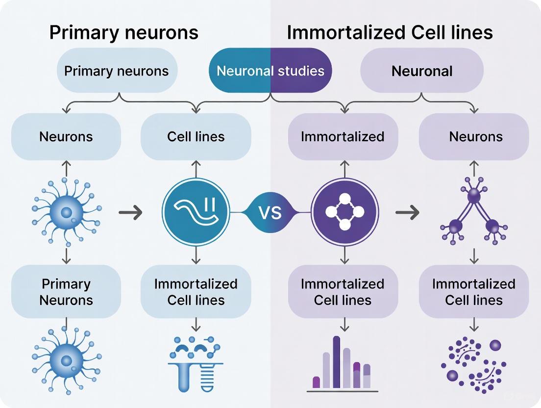
Primary Neurons vs. Immortalized Cell Lines: A Strategic Guide for Translational Neuroscience
This article provides a comprehensive comparison of primary neurons and immortalized neuronal cell lines for researchers and drug development professionals.
Primary Neurons vs. Immortalized Cell Lines: A Strategic Guide for Translational Neuroscience
Abstract
This article provides a comprehensive comparison of primary neurons and immortalized neuronal cell lines for researchers and drug development professionals. It covers the foundational biology and key characteristics of each model, details practical methodologies for their use in applications like toxicology and disease modeling, and offers solutions for common technical challenges. A critical validation framework is presented to guide model selection based on experimental goals, balancing physiological relevance with practicality. The synthesis aims to empower scientists to design more predictive and reproducible neuronal studies, ultimately enhancing the translational success of preclinical research.
Understanding the Core Biology: From Native Tissue to Immortalized Models
In vitro models are indispensable tools for advancing our understanding of neuronal function, disease mechanisms, and potential therapeutic interventions. The choice between using primary neurons and immortalized cell lines represents a fundamental decision that significantly influences the physiological relevance, reproducibility, and translational potential of research outcomes. Primary neurons are cells isolated directly from animal nervous tissue and maintained in culture, where they retain a more natural phenotype but do not proliferate [1]. In contrast, immortalized cell lines are populations of cells that, due to mutation or artificial manipulation, have evaded normal cellular senescence and can proliferate indefinitely in culture [2]. This technical guide provides an in-depth examination of these two model systems, comparing their characteristics, applications, and methodological considerations to inform researchers' experimental design within the broader context of neuronal studies.
Primary Neurons: The Gold Standard for Physiological Relevance
Definition and Fundamental Characteristics
Primary neurons are signaling cells isolated directly from animal brain tissue that maintain a more natural phenotype than immortalized cell lines, making them a superior neuronal cell culture model for many neuroscience experiments [1]. Unlike dividing cells, primary neurons are postmitotic, meaning they do not proliferate in culture [1]. These cells are typically isolated from specific brain regions such as the cortex or hippocampus of embryonic rats or mice, with common sources including day-18 Fisher 344 rat embryos, Sprague-Dawley rat embryos, or E-17 C57 BL/6 mice [1].
From a biological perspective, neurons are the primary components of the nervous system and are highly specialized for processing and transmitting cellular signals through electrical and chemical mechanisms [3]. They are electrically excitable cells that maintain voltage gradients across their membranes and generate action potentials when appropriately stimulated [3]. A typical neuron consists of a cell body (soma) containing the nucleus, dendrites that receive signals, and an axon that transmits signals to other cells [4] [3]. The soma contains specialized structures including Nissl bodies, which consist of rough endoplasmic reticulum and are involved in protein synthesis, reflecting the high metabolic activity of neurons [4] [3].
Key Structural and Functional Features
The enormous functional repertoire of neurons is reflected in their structural variation, particularly in their dendritic and axonal outgrowth patterns [4]. Mature neurons establish complex networks in culture, developing extensive axonal and dendritic branching that can be maintained for several weeks [5]. Primary neurons from rodent hippocampus and cortex can be maintained in serum-free media for up to four weeks, during which they differentiate and form functional synapses [1] [5].
When cultured in appropriate conditions such as Neurobasal Medium with B-27 Supplement, primary neurons exhibit high viability and purity with minimal glial cell growth [1]. These cultures demonstrate critical neuronal functions including calcium signaling in response to neurotransmitters within 7 days and neurite outgrowth by 14 days [1]. The morphological and functional development of these cultures makes them suitable for studying neuronal differentiation, synaptic connectivity, and network formation.
Immortalized Cell Lines: Practical Tools for High-Throughput Research
Definition and Generation Methods
Immortalized cell lines are populations of cells from multicellular organisms that would normally not proliferate indefinitely but, due to mutation, have evaded normal cellular senescence and can keep undergoing division [2]. These cells can be grown for prolonged periods in vitro, providing a consistent and renewable cell source [2]. There are several methods for generating immortalized cell lines, including isolation from naturally occurring cancers (e.g., HeLa cells from cervical cancer), introduction of viral genes that deregulate the cell cycle (e.g., adenovirus E1 gene in HEK 293 cells), artificial expression of key proteins required for immortality such as telomerase, and hybridoma technology for generating antibody-producing B cell lines [2].
It is important to distinguish immortalized cell lines from stem cells, which can also divide indefinitely but form a normal part of the development of a multicellular organism [2]. Many immortalized cell lines are the in vitro equivalent of cancerous cells, having undergone mutations that cause deregulation of the normal cell cycle controls, leading to uncontrolled proliferation [2].
Common Neuronal Cell Lines and Their Applications
In neuroscience research, several immortalized cell lines of neuronal origin are commonly used. The PC12 cell line, derived from a rat pheochromocytoma (adrenal gland tumor), can be induced to differentiate into a neuron-like phenotype in the presence of nerve growth factor (NGF) [6]. These cells can synthesize catecholamines, dopamine, and norepinephrine, and express enzymes involved in neurotransmitter production [6]. Neuroblastoma cell lines such as mouse Neuro-2a (N2a) and human SH-SY5Y are also widely used; they can be driven to differentiate by various stimuli and have been employed in electrophysiology and neurodevelopment studies [6].
However, these neuronal cell lines often exhibit abnormal traits not found in normal neurons. For example, PC12 cells produce an unusual combination of neurotransmitters (dopamine, norepinephrine, and acetylcholine) that no normal neuron produces in the same cell [6]. Similarly, SH-SY5Y cells exhibit immature neuronal features and typically fail to form functional synapses, limiting their utility for studying mature neuronal function [7].
Comparative Analysis: Key Differences and Experimental Considerations
Biological Relevance and Physiological Representation
The most significant distinction between primary neurons and immortalized cell lines lies in their biological relevance and ability to mimic in vivo physiology. Primary cells are generally considered the gold standard for physiological relevance as they are derived directly from living tissue and retain native cell morphology and physiological behaviors [7] [8]. They maintain a more natural phenotype than immortalized cell lines, making them better models for many neuroscience experiments [1].
In contrast, immortalized cell lines often originate from well-known tissue types but have undergone significant mutations to become immortal, which can substantially alter their biology [2]. Most neuronal immortalized cell culture models are derived from tumors and are frequently genomically abnormal [6]. Proteomic studies comparing cell lines to primary cells reveal dramatic differences, with cell lines often showing down-regulation of tissue-specific functions and up-regulation of proliferation-associated pathways [9]. For example, a quantitative proteomic comparison of hepatoma cell lines with primary hepatocytes found that cell lines were deficient in mitochondria, dramatically up-regulated cell cycle-associated functions, and largely shut down characteristic drug-metabolizing enzymes [9].
Practical Considerations: Reproducibility, Scalability, and Ease of Use
While primary neurons offer superior physiological relevance, immortalized cell lines provide significant practical advantages in terms of reproducibility, scalability, and ease of use. The table below summarizes the key comparative features of these two model systems:
Table 1: Comparative Features of Primary Neurons vs. Immortalized Cell Lines
| Feature | Primary Neurons | Immortalized Cell Lines |
|---|---|---|
| Biological Relevance | High - closer to native morphology and function [7] | Low - often non-physiological (e.g., cancer-derived) [7] |
| Reproducibility | Variable - donor-to-donor variability and technical challenges in isolation [7] | High - genetically identical populations, but prone to drift over passages [2] [6] |
| Scalability | Limited - low yield, difficult to expand [7] | High - easily scalable and can be expanded without limitation [2] [6] |
| Ease of Use | Technically complex, time-intensive, requires specialized skills [7] [5] | Simple to culture with robust growth characteristics [6] |
| Time to Assay | Several weeks post-dissection [5] | Can be assayed within 24-48 hours of thawing [7] |
| Cell Origin | Typically rodent-derived [1] [5] | Various sources, including human cancers [2] [6] |
| Cost Considerations | Higher - requires repeated isolation from animals [7] | Lower - continuous supply from existing stocks [6] |
Technical and Methodological Requirements
The methodological approaches for working with primary neurons versus immortalized cell lines differ significantly. Primary neuronal culture requires specialized technical expertise for brain dissection, tissue processing, and maintaining long-term cultures [5]. A standard protocol for culturing primary neurons from rat hippocampus and cortex involves multiple precise steps including extraction from E17-18 rat embryos, enzymatic digestion with papain, mechanical trituration, and plating in serum-free media with specific supplements [5]. These cultures can be maintained for several weeks without feeder cells and develop extensive axonal and dendritic branching [5].
In contrast, immortalized cell lines are generally easier to culture and maintain, requiring standard cell culture techniques without the need for specialized dissection skills [6]. They grow more robustly and do not require extraction from living animals, making them more accessible to laboratories without specialized neuroscience expertise [6]. However, they require careful monitoring for genetic drift and phenotypic changes that can occur with continuous passaging [2] [6].
Experimental Protocols and Methodologies
Protocol for Primary Cortical and Hippocampal Neuronal Cultures
The establishment of primary neuronal cultures requires meticulous technique and attention to detail. The following protocol, adapted from published methodologies, outlines the key steps for generating high-quality neuronal cultures from rodent embryonic brain tissue [5]:
Table 2: Key Reagent Solutions for Primary Neuronal Culture
| Reagent Solution | Composition | Function in Protocol |
|---|---|---|
| Preparation Medium | HBSS (Hank's Balanced Salt Solution), 1 mM sodium pyruvate, 10 mM HEPES [5] | Maintenance of tissue viability during dissection |
| Papain Solution | 0.5 mg papain, 10 μg DNase I in papain buffer (DL-Cysteine HCl, BSA, Glucose in PBS) [5] | Enzymatic digestion of extracellular matrix for tissue dissociation |
| Trituration Medium | Preparation medium with 10 μg DNase I [5] | Mechanical dissociation of tissue into single-cell suspension |
| Growing Medium | Neurobasal medium, 2% B-27 supplement, 1% L-glutamine, 1% penicillin-streptomycin [5] | Support of neuronal survival, growth, and differentiation while inhibiting glial proliferation |
| Poly-L-Lysine Coating | 1:10 dilution of stock Poly-L-Lysine in Milli-Q H2O [5] | Surface coating to promote neuronal adhesion |
Step-by-Step Methodology:
- Tissue Extraction: Sacrifice pregnant female rats at E17-18, remove embryos, and decapitate heads. Transfer to ice-cold PBS. Under sterile conditions and using a dissection microscope, remove brains and carefully separate cortex and hippocampus [5].
- Tissue Dissociation: Transfer dissected brain regions to pre-warmed papain solution and incubate for 10 minutes at 37°C. Remove papain solution and add trituration medium. Gently triturate tissue using a fire-polished glass Pasteur pipette until a single-cell suspension is achieved [5].
- Plating and Maintenance: Plate cells on poly-L-lysine coated culture vessels at desired density. Maintain cultures in growing medium at 37°C in a 5% CO₂ atmosphere. Perform partial medium changes twice weekly [5].
- Characterization and Validation: Assess neuronal purity and differentiation using immunocytochemistry for neuronal markers (e.g., MAP2, NeuN) and glial markers (e.g., GFAP) after 7-21 days in culture [5].
This protocol yields neuronal cultures that can be maintained for several weeks, developing extensive processes and functional synapses, suitable for a wide range of neurobiological applications [5].
Typical Workflow for Immortalized Neuronal Cell Lines
The workflow for culturing immortalized neuronal cell lines is generally more straightforward:
- Thawing and Expansion: Rapidly thaw cryopreserved cells and transfer to pre-warmed complete medium. Expand cells in standard culture conditions until sufficient numbers are obtained.
- Differentiation (if required): For cell lines like PC12 or SH-SY5Y that require differentiation to exhibit neuronal properties, apply appropriate differentiating agents (e.g., NGF for PC12 cells) for specified durations [6].
- Experimental Assays: Perform experiments during the appropriate growth or differentiation phase, typically within days rather than weeks as required for primary neurons.
Diagram 1: Experimental workflow comparison between primary neurons and immortalized cell lines
Applications in Neuronal Research and Drug Development
Optimal Use Cases for Each Model System
The choice between primary neurons and immortalized cell lines should be guided by the specific research question and experimental requirements. Primary neurons are particularly well-suited for:
- Studies of neuronal polarization, axon guidance, and dendritic spine formation due to their authentic morphological development [5].
- Synaptic function and plasticity research as they form functional synapses in culture [7].
- Electrophysiological investigations of native neuronal signaling properties [1].
- Neurotoxicity and neuroprotection studies where physiological relevance is critical for predictive value [7].
- Investigation of disease mechanisms using transgenic animal-derived neurons or human induced pluripotent stem cell (iPSC)-derived neurons [10].
Immortalized cell lines offer advantages for:
- High-throughput drug screening campaigns where scalability and reproducibility are paramount [6].
- Genetic manipulation studies requiring efficient transfection and clonal selection [6].
- Biochemical and molecular biology assays requiring large amounts of cellular material [6] [9].
- Preliminary mechanistic studies before validation in more complex systems [7].
- Production of recombinant proteins or viral vectors where scalable expansion is necessary [2].
Limitations and Challenges in Translational Research
Both model systems present significant limitations that researchers must consider when interpreting results and planning translational research pathways. The high failure rate of central nervous system (CNS)-targeted drug candidates (approximately 97% fail to reach market) reflects fundamental gaps in preclinical model predictivity [7]. Immortalized cell lines often fail to capture human-relevant phenotypes or mechanisms of action, particularly for complex CNS diseases [7].
Primary neurons from rodent sources, while more physiologically relevant than cell lines, still suffer from species mismatch that can undermine translational relevance [7]. Comparative transcriptomic studies have shown widespread differences in gene expression, regulation, and splicing between mouse and human tissues, creating significant limitations for modeling human-specific neurobiology [7].
Emerging Alternatives and Future Directions
The limitations of both primary neurons and immortalized cell lines have spurred the development of alternative models that aim to bridge the gap between physiological relevance and practical utility. Human induced pluripotent stem cell (iPSC)-derived neurons have emerged as a promising alternative, offering human-specific biology with renewable capacity [7] [10]. Unlike primary cells or immortalized cell lines, iPSCs can be renewed indefinitely and differentiated to somatic cell types, providing the potential for improved biological relevance and a closer human phenotype [7].
Novel technologies such as deterministic cell programming with opti-ox technology enable the consistent production of human iPSC-derived neurons with high batch-to-batch consistency, addressing the reproducibility challenges of both primary cells and traditional iPSC differentiation protocols [7]. These advanced models retain key advantages of both traditional systems while mitigating many of their limitations, offering researchers new opportunities to model human neurological diseases and develop more effective therapeutics.
The selection between primary neurons and immortalized cell lines represents a critical strategic decision in neuronal research, with significant implications for experimental outcomes and translational potential. Primary neurons offer superior physiological fidelity and are ideal for investigating fundamental neurobiological mechanisms where maintaining native neuronal properties is essential. Immortalized cell lines provide practical advantages in scalability, reproducibility, and ease of use, making them valuable tools for high-throughput applications and preliminary investigations. As the field advances, emerging technologies like human iPSC-derived neurons and deterministic programming approaches offer promising pathways to overcome the limitations of both traditional models. Researchers must carefully weigh the trade-offs between physiological relevance and practical considerations when selecting the most appropriate model system for their specific research objectives within the broader context of neuronal studies and drug development.
Primary neurons, isolated directly from neural tissue of animals or humans, serve as a cornerstone model in neuroscience research. These cells are distinguished from immortalized neuronal cell lines by their origin from living tissue rather than tumors or genetic immortalization. The use of primary neurons is framed within a critical debate regarding the most appropriate in vitro model for studying neuronal function, disease mechanisms, and therapeutic development. While immortalized cell lines like SH-SY5Y, PC12, and F-11 offer practical advantages of ease-of-use and scalability, they often fail to recapitulate the full physiological complexity of native neurons [11] [7] [12]. This whitepaper provides a comprehensive technical examination of primary neurons, detailing their sourcing methodologies, fundamental strengths, inherent limitations, and essential protocols to guide researchers in making informed model selection decisions for neuronal studies.
Sourcing and Isolation of Primary Neurons
The process of obtaining primary neurons involves careful tissue dissection, mechanical and enzymatic dissociation, and purification steps that vary based on the neural region of interest and developmental stage.
Regional and Developmental Considerations
Primary neuronal cultures can be established from various regions of the nervous system, each yielding populations with distinct characteristics. Common sources include the cortex, hippocampus, and dorsal root ganglia (DRG) [11] [12]. More specialized protocols also exist for regions such as the hindbrain, which contains diverse neuronal subtypes critical for vital functions like breathing and heart rate control [13]. The developmental stage at which neurons are harvested significantly impacts their properties and culture requirements. Most protocols utilize embryonic (E17-E19) or early postnatal tissue due to enhanced neuronal viability and greater resilience to the dissociation process compared to mature adult neurons [13] [14]. For instance, an optimized protocol for mouse fetal hindbrain neurons specifies embryonic day 17.5 (E17.5) as the optimal harvest time [13].
Core Isolation Methodology
The fundamental isolation process involves a carefully orchestrated sequence to maximize cell viability and purity:
- Dissection and Meninges Removal: Neural tissue is carefully dissected from the desired brain region, followed by meticulous removal of the protective meningeal layers to minimize contamination by non-neuronal cells [15].
- Enzymatic Digestion: The tissue is subjected to enzymatic digestion using agents such as trypsin to loosen the extracellular matrix and dissociate intercellular connections. The duration and concentration of enzymatic treatment must be precisely controlled to avoid damaging surface receptors and ion channels critical for neuronal function [13] [15] [14].
- Mechanical Dissociation: Following enzymatic treatment, the tissue is gently triturated using fire-polished glass Pasteur pipettes of decreasing diameters to create a single-cell suspension without causing excessive mechanical stress [13].
- Purification and Plating: The cell suspension is filtered through a cell strainer (e.g., 70 μm) to remove clumps and debris. Neurons are then purified from the mixed cell population using techniques such as immunocapture with magnetic beads (e.g., for negative selection of neurons) or Percoll gradient centrifugation [15]. Finally, cells are plated on substrates coated with poly-L-lysine or poly-D-lysine to promote adhesion, and maintained in specialized serum-free media formulations such as Neurobasal medium supplemented with B-27 to support neuronal health while suppressing glial proliferation [13] [14] [16].
Table 1: Key Solutions for Primary Neuron Isolation and Culture
| Solution/Reagent | Function | Example Composition |
|---|---|---|
| Digestion Medium | Tissue dissociation | Trypsin-EDTA (0.25%) + HEPES buffer [14] |
| Plating Medium | Initial cell adhesion and survival | MEM + 5% FBS + D-glucose + L-glutamine [14] |
| Maintenance Medium | Long-term culture support | Neurobasal Medium + B-27 Supplement + L-glutamine [14] [16] |
| Substrate Coating | Surface for neuron attachment | Poly-L-lysine in boric acid buffer [14] |
Diagram 1: Primary Neuron Isolation Workflow
Key Strengths of Primary Neurons
Superior Physiological Relevance
The principal advantage of primary neurons lies in their superior capacity to mimic in vivo neuronal biology compared to immortalized cell lines.
- Native Morphology and Network Formation: Primary neurons in culture develop distinct axons and dendrites, forming complex, synaptically connected networks that closely resemble neural circuitry in vivo [17] [16]. Immunofluorescence characterization demonstrates that these cultures can achieve purity above 90% and maintain typical neuronal morphology over extended periods (e.g., 14 days in culture) [16].
- Authentic Electrophysiological Properties: Mature primary neurons exhibit robust action potential generation and synaptic transmission capabilities [16]. This is critically important for studies of neuronal excitability, synaptic plasticity, and network dynamics. Advanced imaging techniques using genetically-encoded voltage indicators (GEVIs) like ASAP4.4-Kv have enabled high-resolution tracking of these fast electrical signals in primary sensory neurons, revealing previously unrecognized phenomena such as cell-to-cell electrical synchronization following tissue injury [18].
- Retention of Regional Identity and Heterogeneity: Primary cultures maintain the regional specificity and neurotransmitter diversity of their tissue source [13]. For example, hindbrain neurons produced in vitro continue to express a wide range of neurotransmitters, including acetylcholine, glutamate, GABA, glycine, and monoamines, which is essential for modeling region-specific functions and dysfunctions [13].
Direct Application in Disease Modeling and Screening
Primary neurons provide a critical platform for investigating disease mechanisms and evaluating therapeutic candidates.
- Neurodegenerative Disease Research: These cells are extensively used to model pathological processes in Alzheimer's disease and Parkinson's disease, allowing researchers to analyze mechanisms such as protein aggregation, synaptic dysfunction, and neuronal death [17] [16].
- Drug Screening and Neurotoxicity Assessment: Primary neuronal cultures serve as reliable systems for evaluating neuroprotective effects and neurotoxicity of candidate compounds, forming a crucial step in central nervous system drug development pipelines [16]. The high physiological relevance of primary neurons increases the predictive validity of these screens, potentially addressing the alarming 97% failure rate of CNS-targeted drug candidates in clinical trials [7].
- Studies of Neuronal Injury and Plasticity: Research utilizing primary neurons, particularly DRG neurons, has revealed remarkable dynamic transformations in sensory coding following tissue injury, including the emergence of synchronized electrical activity between adjacent neurons mediated by gap junctions—a phenomenon rarely observed in naïve animals [18].
Table 2: Quantitative Comparison of Neuronal Model Systems
| Characteristic | Primary Neurons | Immortalized Cell Lines | Human iPSC-Derived Neurons |
|---|---|---|---|
| Biological Relevance | High (native morphology/function) [7] | Low (often non-physiological, cancer-derived) [7] | Medium-High (human-specific, functional) [7] |
| Reproducibility | Low (high donor/harvest variability) [15] [7] | High (genetically identical) [7] | Medium (batch-to-batch variability in differentiation) [7] |
| Scalability | Low (limited yield, difficult to expand) [7] | High (easily scalable) [7] | Medium-High (improving with technologies like opti-ox) [7] |
| Time to Assay | Several weeks post-dissection [7] [14] | 24-48 hours post-thaw [7] | ~10 days post-thaw [7] |
| Species Origin | Typically rodent-derived [7] | Often non-human [7] | Human-derived [7] |
Inherent Limitations and Challenges
Despite their physiological advantages, primary neurons present significant practical challenges that researchers must carefully consider.
Technical and Practical Constraints
- Technically Demanding Isolation: The process of isolating primary neurons is complex and requires significant expertise. Factors such as tissue sourcing, precise enzymatic digestion timing, and procedural details dramatically impact outcomes, often resulting in low purity, inconsistent success rates, and reduced cell viability [15] [16]. The process is both time-consuming and expensive compared to simply thawing a vial of immortalized cells [15].
- Limited Lifespan and Expansion Potential: Unlike continuously dividing cell lines, mature primary neurons are post-mitotic and cannot be expanded through passaging [11] [15]. This fundamental limitation restricts the number of cells available for experiments and necessitates repeated isolations, introducing batch-to-batch variability [15] [7].
- Sensitivity to Culture Conditions: Primary neurons are highly sensitive to culture conditions, including medium composition, substrate coating, pH, and CO₂ levels [15]. Even with optimized systems, issues such as decreased survival rates, reduced purity over time, and poorly defined synaptic morphology frequently arise, negatively impacting experimental outcomes [16].
Source-Dependent Variability and Ethical Considerations
- Donor-to-Donor and Batch-to-Batch Variability: Each primary cell isolation produces a unique biological reagent with inherent variability in phenotype and function [15] [7]. This variability introduces experimental noise and complicates data interpretation, requiring extensive characterization of each batch and increasing the number of replicates needed for statistical power [15].
- Species-Specific Limitations: Most primary neurons are rodent-derived, carrying fundamental genetic and physiological differences from human biology that can undermine translational relevance [7]. Comparative transcriptomic studies have revealed widespread differences in gene expression, regulation, and splicing between mouse and human neural tissues [7].
- Ethical and Sourcing Constraints: The use of animal-derived tissues requires significant ethical considerations and protocol approvals, typically from an Institutional Animal Care and Use Committee [11]. Sourcing human brain tissue presents even greater ethical and practical challenges, limiting generalization of findings and forcing reliance on animal models that may not fully represent human conditions [15].
Essential Experimental Protocols
Detailed Protocol: Primary Cortical Neuron Culture and Transfection
This established protocol for culturing primary neurons from the mouse central nervous system (CNS) enables studies of neuronal function and gene manipulation [14].
Materials and Reagents:
- Calcium- and magnesium-free HBSS (CMF-HBSS)
- Digestion medium: 0.25% trypsin-EDTA + 10 mM HEPES
- DNase I solution (10 mg/mL)
- Neuronal plating medium: MEM + 0.6% D-glucose + 5% FBS + 2 mM L-glutamine
- Neuronal maintenance medium: Neurobasal medium + 1× B-27 + 0.5 mM L-glutamine
- Poly-L-lysine working solution (100 μg/mL in boric acid buffer)
Procedure:
- Substrate Coating: Coat culture vessels (e.g., 24-well plates with glass coverslips) with poly-L-lysine working solution. Incubate at 37°C for at least 1 hour, then rinse with sterile water before use.
- Tissue Dissociation: Dissect cortical tissue from E17.5 mouse embryos in CMF-HBSS. Transfer tissue to digestion medium and incubate at 37°C for 15 minutes. Remove digestion medium and add DNase I solution (10 μL/mL of CMF-HBSS). Mechanically dissociate tissue by trituration with fire-polished glass pipettes.
- Cell Seeding and Culture: Filter cell suspension through a 70 μm cell strainer. Centrifuge at 200 × g for 5 minutes. Resuspend pellet in neuronal plating medium, count cells, and seed at desired density (e.g., 50,000-100,000 cells/cm²) on coated vessels. After 4 hours, replace plating medium with neuronal maintenance medium.
- Transient Transfection (Two Methods):
- Electroporation (for freshly isolated neurons): Use the Mouse Neuron Nucleofector kit with freshly isolated neurons in suspension. Achieves up to 30% transfection efficiency but is unsuitable for adherent neurons with neurites.
- Cationic Lipid Transfection (for adherent neurons): Use Lipofectamine 2000 for neurons cultured for several days in vitro. Although efficiency is lower (1-2%), this method causes less physical stress and can yield higher transgene expression per transfected cell [14].
Diagram 2: Model Selection Decision Framework
The Scientist's Toolkit: Essential Research Reagents
Table 3: Key Reagent Solutions for Primary Neuron Research
| Reagent/Category | Specific Examples | Function in Neuron Culture |
|---|---|---|
| Basal Media | Neurobasal Medium, Neurobasal-A Medium, MEM | Nutrient foundation for culture; formulation varies by developmental stage (embryonic vs. mature) [14] [16] |
| Specialized Supplements | B-27 Supplement, CultureOne, N-2 Supplement | Serum-free formulations to support neuronal health and suppress glial proliferation [13] [14] |
| Adhesion Substrates | Poly-L-lysine, Poly-D-lysine, Laminin | Coating materials that promote neuronal attachment and neurite outgrowth [14] |
| Isolation Kits | Commercial Neuron Isolation Kits (e.g., Pricella) | Standardized reagent sets that simplify the isolation process and improve reproducibility [16] |
| Growth Factors | NGF, BDNF, GDNF, bFGF | Proteins that support neuronal survival, differentiation, and maturation in culture [12] |
Primary neurons remain an indispensable tool for neuroscience research, offering unparalleled physiological relevance that immortalized cell lines cannot match. Their capacity to form authentic synaptic networks, exhibit native electrophysiological properties, and retain regional specificity makes them particularly valuable for mechanistic studies of neuronal function, disease modeling, and preclinical drug evaluation. However, researchers must carefully weigh these strengths against significant practical limitations, including technical complexity, limited scalability, batch-to-batch variability, and species-specific differences. The decision to use primary neurons versus alternative models should be guided by specific research questions, technical capabilities, and translational goals. As technologies such as human iPSC-derived neurons continue to advance, the field moves toward models that combine the physiological relevance of primary neurons with the scalability and human relevance needed for modern drug discovery and mechanistic research.
The choice of an appropriate in vitro model is a fundamental consideration in neuroscience research, particularly for the study of neurodegenerative diseases like Parkinson's disease (PD). The central dilemma often involves balancing physiological relevance with practical experimental requirements. This whitepaper examines two extensively used immortalized human neuronal cell lines—SH-SY5Y and LUHMES—within the broader context of model selection for neuronal studies. We provide a technical analysis of their origins, characteristic utilities, documented limitations, and a critical challenge they both face: genetic drift. A clear understanding of these factors is essential for researchers and drug development professionals to design robust, reproducible, and physiologically relevant experiments.
Origins and Fundamental Characteristics
SH-SY5Y: A Human Neuroblastoma Model
The SH-SY5Y cell line is a subclone of the SK-N-SH cell line, which was originally isolated from a bone marrow biopsy of a four-year-old female with metastatic neuroblastoma [19] [20]. Through multiple rounds of subcloning, the SH-SY5Y line was established and first described in 1978 [19]. As a cancer-derived line, it is genetically female and possesses inherent genetic abnormalities, including an abnormal chromosome 1 (trisomy 1q) [19]. Phenotypically, SH-SY5Y cells are catecholaminergic, expressing both dopaminergic and adrenergic markers, such as tyrosine hydroxylase and dopamine-β-hydroxylase [19] [20]. This has underpinned their widespread use as a model for dopaminergic neurons in PD research, though they are not a pure dopaminergic population.
LUHMES: Conditionally Immortalized Dopaminergic Neurons
Lund Human Mesencephalic (LUHMES) cells are neural precursor cells derived from the ventral mesencephalon of an 8-week-old human fetus [21] [22]. They were conditionally immortalized using a tetracycline-regulated v-myc transgene [21]. This design allows for rapid and homogeneous differentiation into post-mitotic, dopaminergic neurons upon the downregulation of the v-myc gene, typically triggered by the addition of tetracycline or its analog [21] [22]. Unlike SH-SY5Y cells, LUHMES are not derived from a tumor and are considered to have a more defined and consistent dopaminergic phenotype upon differentiation.
Table 1: Core Characteristics of SH-SY5Y and LUHMES Cell Lines
| Characteristic | SH-SY5Y | LUHMES |
|---|---|---|
| Origin | Human neuroblastoma (bone marrow biopsy) [19] | Human fetal mesencephalon (8-week-old) [21] |
| Immortalization Method | Spontaneous (cancer-derived) | Conditional (v-myc transgene, Tet-Off) [21] [22] |
| Base Phenotype | Catecholaminergic (mixed dopaminergic/adrenergic) [19] [20] | Dopaminergic neuronal precursors [22] |
| Key Genetic Notes | Trisomy 1q; other cancer-associated mutations [19] | Conditionally immortalized; non-tumorigenic background [21] |
| Primary Differentiated Phenotype | Neuron-like, but immature; phenotype depends heavily on protocol [20] | Consistent, post-mitotic dopaminergic neurons [21] [23] |
Functional Utility in Neuronal Research
Differentiation and Phenotypic Maturity
A critical step in using these cell lines is their differentiation into a mature, neuron-like state.
- SH-SY5Y Differentiation: A wide variety of protocols exist, often using agents like retinoic acid (RA), brain-derived neurotrophic factor (BDNF), or phorbol esters (e.g., TPA) [19] [20]. This lack of standardization is a significant source of variability in the resulting cellular phenotype. The differentiated cells exhibit a characteristically dispersed morphology with well-developed neurites and smaller cell bodies [19]. However, the degree to which they recapitulate a genuine dopaminergic neuron remains inconsistent.
- LUHMES Differentiation: The process is more standardized and rapid, typically taking 4-5 days. It is initiated by the addition of tetracycline (to suppress v-myc), along with cAMP and glial cell line-derived neurotrophic factor (GDNF) [21]. This protocol yields a highly homogeneous population of cells that express high levels of neuronal and dopaminergic markers, such as tyrosine hydroxylase (TH), and exhibit electrophysiological activity [23] [22].
Comparative Performance in Toxicological and Disease Modeling
Both cell lines are used to model PD, often using neurotoxicants like MPP+ and 6-hydroxydopamine (6-OHDA). However, their responses differ markedly.
Table 2: Comparative Sensitivity to Parkinson's Disease-Relevant Insults
| Parameter | SH-SY5Y | LUHMES | Experimental Context |
|---|---|---|---|
| MPP+ LC50 | High micromolar range (resilient) [23] | Low micromolar range (3-5 µM; highly sensitive) [21] [23] | Differentiated cells, 48-72h exposure [21] [23] |
| 6-OHDA Sensitivity | Shows resilience [23] | Sensitive; associated with ATP depletion and elevated ROS [23] | Differentiated cells [23] |
| Dopaminergic Marker Expression | Inconsistent; often downregulated after differentiation [21] [20] | Consistent and high expression of TH and DAT [21] [23] | Post-differentiation [21] [23] [22] |
| General Neurotoxicant Sensitivity | Less sensitive to a panel of 32 known/suspected neurotoxicants [22] | Highly sensitive to most compounds in a panel of 32; more sensitive when differentiated [22] | Differentiated vs. undifferentiated state [22] |
The data indicate that differentiated LUHMES cells are significantly more sensitive to dopaminergic-specific toxicants and better maintain the key pathways relevant to PD, making them a more robust model for mechanistic toxicology studies [23] [22]. SH-SY5Y's resilience may be partly attributed to its cancer origin, which often involves upregulation of anti-apoptotic genes like BCL2 and BIRC5 (survivin) [22].
The Challenge of Genetic Drift
A major challenge with any immortalized cell line is genetic and phenotypic instability over time, a phenomenon known as genetic drift. This can lead to irreproducibility of results both within and between laboratories.
Documented Evidence of Drift
- SH-SY5Y: The loss of neuronal characteristics, such as noradrenaline uptake, with increasing passage number is a recognized issue. It is often recommended not to use these cells beyond passage 20 without verifying key characteristics [19]. Furthermore, a systematic review highlighted vast inconsistencies in culture conditions and differentiation protocols across studies, which compound the effects of genetic drift and make cross-study comparisons difficult [20].
- LUHMES: A striking example of functional drift was observed between two subpopulations (SP) of LUHMES cells maintained at different repositories (ATCC vs. the original provider, UKN). The "ATCC" subpopulation rapidly downregulated the dopamine transporter (DAT) and tyrosine hydroxylase (TH) after differentiation and tolerated up to 60 µM MPP+, whereas the "UKN" subpopulation maintained functional levels of these proteins and was sensitive to only 3-5 µM MPP+ [21]. Whole-genome sequencing revealed about 70 amino acid-changing single nucleotide variants (SNVs) between the subpopulations, but no single, obvious genetic cause for the phenotypic difference was identified, suggesting a polygenic mechanism [21].
Mechanisms and Oversight
Genetic drift can arise from several factors:
- De novo mutations during sub-culturing that provide a selective growth advantage [21].
- Heterogeneity of the starting culture, where pre-existing genetic subpopulations are selected for or against over time [21].
- Adaptation to specific culture conditions in different laboratories.
Detailed Experimental Protocols
SH-SY5Y Differentiation Protocol (Sample using Retinoic Acid)
This is a commonly cited method, though significant variations exist [19] [20].
- Cell Seeding: Plate SH-SY5Y cells at an appropriate density (e.g., 10,000-50,000 cells/cm²) in growth medium. Common growth media include DMEM or a 1:1 mixture of DMEM and Ham's F12, supplemented with 10% fetal bovine serum (FBS), 2 mM L-Glutamine, and 1% non-essential amino acids [19] [20].
- Retinoic Acid Treatment: 24 hours after plating, replace the growth medium with differentiation medium containing 10 µM all-trans retinoic acid (RA) in a low-serum (e.g., 1-2% FBS) or serum-free base medium.
- Medium Refreshment: Refresh the RA-containing differentiation medium every 2-3 days.
- Differentiation Duration: Maintain cells in RA for typically 3-7 days. Differentiated cells should exhibit a neuronal morphology with extended neurites and smaller cell bodies [19].
LUHMES Differentiation Protocol (Standardized Method)
The following protocol is adapted from established procedures [21] [22].
- Pre-differentiation (Day -1): Seed proliferating LUHMES cells in a T175 flask at a density of 8 million cells in proliferation medium (advanced DMEM/F12, 1x N2 supplement, 40 ng/ml FGF-2).
- Initiation of Differentiation (Day 0): Replace the proliferation medium with differentiation medium (advanced DMEM/F12, 1x N2 supplement, 2 µg/ml tetracycline, 1 mM dibutyryl-cAMP, and 2 ng/ml GDNF). The addition of tetracycline turns off the v-myc transgene, initiating cell cycle exit.
- Trypsinization and Re-plating (Day 2): Trypsinize the cells and seed them onto tissue culture dishes pre-coated with 50 µg/ml poly-L-ornithine and 1 µg/ml fibronectin. A recommended density is 1.5 x 10⁵ cells/cm². Use the same differentiation medium as in Step 2.
- Medium Exchange (Day 4): Exchange half or all of the differentiation medium for fresh medium.
- Fully Differentiated Neurons (Day 5+): Cells are typically fully differentiated into post-mitotic, dopaminergic neurons by day 5 or 6 and are ready for experimentation.
The Scientist's Toolkit: Essential Research Reagents
Table 3: Key Reagent Solutions for Cell Culture and Differentiation
| Reagent / Material | Function / Purpose | Example Usage |
|---|---|---|
| All-Trans Retinoic Acid (RA) | Induces neuronal differentiation; activates retinoic acid receptors leading to gene expression changes. | SH-SY5Y differentiation protocol [19]. |
| Tetracycline | Binds to Tet-responsive element; silences v-myc transgene in LUHMES, halting proliferation and triggering differentiation. | LUHMES differentiation protocol [21]. |
| Dibutyryl-cAMP (dbcAMP) | Cell-permeable cAMP analog; activates protein kinase A signaling pathways to promote neuronal maturation and survival. | LUHMES differentiation protocol [21]. |
| Glial Cell Line-Derived Neurotrophic Factor (GDNF) | Promotes survival and maturation of dopaminergic neurons; acts via RET receptor signaling. | LUHMES differentiation protocol [21]. |
| Poly-L-Ornithine / Laminin / Fibronectin | Substrate coating proteins; provide a adhesive surface that promotes neurite outgrowth and cell attachment. | Coating plates for LUHMES and SH-SY5Y differentiation [21]. |
| DMEM/F12 Medium | A common base medium providing essential nutrients, vitamins, and salts for cell growth. | Base for proliferation and differentiation media for both lines [19] [21]. |
| N-2 Supplement | A defined serum-free supplement containing insulin, transferrin, progesterone, selenite, and putrescine; supports survival of neuronal cells. | Used in LUHMES differentiation medium and some SH-SY5Y protocols [21]. |
The selection between SH-SY5Y and LUHMES cell lines, and indeed between immortalized lines and primary neurons, is a strategic decision with significant implications for research outcomes. SH-SY5Y cells offer the advantages of ease of use and familiarity but are hampered by phenotypic inconsistency, a non-purely dopaminergic background, and pronounced sensitivity to genetic drift and protocol variations. LUHMES cells, in contrast, provide a more physiologically relevant, consistent, and sensitive model for dopaminergic neuron biology and toxicology, albeit with a more defined and less flexible differentiation protocol.
The documented phenomenon of genetic drift in both lines, as starkly demonstrated by the LUHMES subpopulations, underscores a critical point: immortalized cells are dynamic entities, not static reagents. This necessitates rigorous cell culture practices, including:
- Using low passage number stocks.
- Maintaining detailed records of culture history.
- Periodically validating key phenotypic and functional characteristics (e.g., TH expression, MPP+ sensitivity).
- Where possible, using cells from a consistent and well-characterized source.
For research where high fidelity to human dopaminergic biology is paramount—such as in mechanistic studies of Parkinson's disease or screening for neurotoxicants—LUHMES cells present a superior in vitro model. However, for all immortalized lines, researchers must remain cognizant of their limitations, including their inability to fully replicate the complexity of primary neurons or the intact brain. The emerging field of human iPSC-derived neurons offers a promising path forward, potentially combining the scalability of cell lines with the physiological relevance of primary human cells [7].
In the field of neuroscience research and drug development, scientists face a fundamental dilemma: choosing between primary neurons isolated directly from living tissue and immortalized cell lines engineered for continuous proliferation. This decision represents a critical trade-off between physiological fidelity and experimental practicality that profoundly impacts research outcomes, reproducibility, and translational potential. Primary neurons, derived from embryonic or early postnatal brain regions, retain native cellular architecture, gene expression patterns, and functional characteristics that closely mirror the in vivo nervous system [24] [5]. In contrast, immortalized cell lines—typically generated from neuronal tumors or through genetic manipulation—offer unparalleled convenience, scalability, and standardization but often deviate significantly from physiological normality [6] [7].
The stakes for making an informed choice are substantial, particularly in central nervous system (CNS) drug discovery where attrition rates approach 97% for candidates entering Phase 1 clinical trials [7]. This staggering failure rate underscores a fundamental gap in preclinical model predictivity, with the choice between primary cells and cell lines representing a key contributing factor. As research advances toward more human-relevant models, understanding the precise nature of this trade-off becomes increasingly critical for generating meaningful, translatable data. This technical guide examines the core distinctions between these model systems, providing a structured framework for researchers to navigate this critical decision point in experimental design.
Fundamental Biological Differences Between Model Systems
Origin and Establishment
The biological genesis of primary neurons and immortalized cell lines establishes their fundamental differences in physiological relevance. Primary neuronal cultures are directly isolated from living neural tissues—typically embryonic or early postnatal hippocampus or cortex—through meticulous dissection and enzymatic dissociation [5]. These cells maintain their native genetic programming without artificial manipulation, providing a snapshot of neuronal function that closely resembles the in vivo state. The process involves extracting brains from E17-18 rat embryos, microdissecting specific regions like hippocampus and cortex, and digesting tissue with papain before mechanical trituration to create a single-cell suspension [5]. These non-dividing neurons are then cultured on coated surfaces with specialized media formulations that support their maturation into polarized cells with extensive axonal and dendritic arbors [24].
In stark contrast, immortalized cell lines undergo deliberate genetic manipulation to bypass natural cellular senescence mechanisms. Immortalization is achieved through various methods including introduction of viral oncogenes (SV40 T-antigen, HPV E6/E7 proteins), overexpression of human telomerase reverse transcriptase (hTERT), or manipulation of cell cycle regulators (c-MYC) [25] [26]. These interventions fundamentally alter cellular biology by disabling critical checkpoint pathways (p53/p16/pRb) and reactivating telomerase to maintain telomere length, thereby enabling unlimited proliferation [25]. Most neuronal cell lines, such as PC12, SH-SY5Y, and Neuro-2a, are derived from tumors—a origin that inherently shifts their biological priorities toward proliferation rather than specialized neuronal function [6].
Genetic and Proteomic Landscape
Comparative proteomic phenotyping reveals profound differences at the molecular level between cell lines and primary cells. A landmark study quantitatively comparing the hepatoma cell line Hepa1–6 with primary hepatocytes demonstrated an asymmetric distribution of approximately 4,063 proteins, with many proteins significantly down-regulated in the cell line [9]. Bioinformatic analysis revealed that cell lines dramatically up-regulate cell cycle-associated functions while largely shutting down tissue-specific metabolic enzymes [9]. This systematic shift in resource allocation creates a cellular identity centered on proliferation rather than physiological function.
At the genetic level, immortalized cells experience ongoing genetic drift—continuous evolution of genomes with repeated passage that further distances them from their original tissue source [8]. This instability is particularly pronounced in cell lines derived from late-stage cancers, which are notoriously vulnerable to phenotypic changes during continuous culture [8]. Primary cells, with their finite lifespan, maintain genomic and phenotypic stability throughout their culture period, preserving tissue-specific characteristics and providing more consistent, reliable models for studying neuronal function and dysfunction [8].
Table 1: Fundamental Biological Characteristics of Primary Neurons vs. Immortalized Cell Lines
| Characteristic | Primary Neurons | Immortalized Cell Lines |
|---|---|---|
| Origin | Direct isolation from neural tissue [5] | Tumors or genetic manipulation [6] |
| Genetic Status | Native, unmodified genome [8] | Genetically altered for infinite division [25] |
| Proliferation Capacity | Non-dividing, terminally differentiated [24] | Continuous division capability [6] |
| Key Manipulations | None beyond isolation | Introduction of immortalizing genes (SV40 T-antigen, hTERT, HPV E6/E7) [26] |
| Cellular Priority | Specialized neuronal function [24] | Proliferation and survival [9] |
| Genetic Stability | Stable throughout culture period [8] | Subject to genetic drift with passage [8] |
Quantitative Comparison of Functional Capabilities
Physiological and Functional Properties
The functional capabilities of primary neurons and immortalized cell lines differ dramatically, with profound implications for their utility in neuroscience research. Primary neurons develop electrically active synapses, elaborate dendritic arbors with spines, and establish functional neuronal networks that spontaneously fire action potentials [24]. Within weeks in culture, these cells form complex networks with appropriate neurotransmitter specification, receptor localization, and synaptic plasticity mechanisms that closely resemble their in vivo counterparts [5]. This robust recapitulation of native neuronal properties makes them invaluable for studying fundamental neurobiological processes.
Immortalized neuronal cell lines typically exhibit poor differentiation and lack many definitive neuronal features. Most fail to form functional synapses or develop mature myelin sheaths, significantly limiting their utility for studying network-level phenomena [24]. While some lines can be induced toward neuronal phenotypes—PC12 cells with nerve growth factor (NGF) or SH-SY5Y with retinoic acid—the resulting differentiation is often incomplete and unstable [24] [6]. These cells frequently display abnormal neurotransmitter combinations not found in normal neurons, such as simultaneously producing dopamine, norepinephrine, and acetylcholine within the same cell [6]. This pharmacological incongruity raises serious questions about their validity for neuropharmacological studies.
Experimental Practicalities
The practical considerations of working with these model systems reveal the inverse relationship between physiological relevance and experimental convenience. Primary neurons demand technically challenging isolation procedures requiring substantial skill and experience, with variable yields depending on dissection precision and animal age [5]. These cultures contain heterogeneous cell populations (mixed neurons and glia) that complicate experimental interpretation and provide limited cell quantities that challenge biochemical experiments [24]. Most significantly, their finite lifespan necessitates repeated isolations from animal tissue, creating substantial batch-to-batch variability and limiting long-term studies [8].
Immortalized cell lines offer compelling practical advantages with their unlimited expansion capability, providing abundant, consistent cellular material for high-throughput applications [6]. Their homogeneous populations enable standardization across laboratories and experiments, while their robust growth characteristics make them tolerant of variable culture conditions and suitable for large-scale screening campaigns [8]. However, these advantages come with significant caveats: cell lines are notoriously prone to cross-contamination (with HeLa cells being a particularly common contaminant) and require regular authentication to ensure identity and genetic stability over time [8].
Table 2: Functional Capabilities and Experimental Practicalities in Neuronal Model Systems
| Parameter | Primary Neurons | Immortalized Cell Lines |
|---|---|---|
| Synapse Formation | Functional, electrically active synapses [24] | Typically absent or non-functional [24] |
| Neurite Outgrowth | Extensive arbors with spines [5] | Often short, underdeveloped processes [24] |
| Network Activity | Spontaneous synchronized firing [24] | Limited or abnormal activity patterns |
| Differentiation Status | Fully mature neuronal phenotype [5] | Often immature, incomplete differentiation [24] |
| Culture Longevity | Weeks to months [24] | Indefinite with proper maintenance [6] |
| Scalability | Limited yield per isolation [8] | Essentially unlimited expansion [6] |
| Technical Difficulty | High (requires specialized expertise) [5] | Low (easy to culture and maintain) [6] |
| Reproducibility | Significant batch-to-batch variability [8] | High consistency when properly authenticated [6] |
| Throughput Capacity | Low to moderate | High, suitable for screening [27] |
Methodological Approaches and Experimental Protocols
Established Protocols for Primary Neuronal Culture
Robust methodology for culturing primary neurons requires meticulous attention to detail throughout the isolation and maintenance process. The following protocol for rat hippocampal and cortical neurons has been refined over decades to maximize viability and reproducibility [5]:
Tissue Dissection and Dissociation:
- Sacrifice pregnant female Wistar rats at E17-18 and remove embryos under sterile conditions.
- Decapitate embryos and transfer heads to ice-cold PBS for brain dissection.
- Under a stereomicroscope, carefully remove brains and isolate hippocampi and cortices in preparation medium.
- Transfer tissues to papain solution (0.5 mg papain, 10 μg DNase I in 5 ml PBS with DL-Cysteine and BSA) and incubate for 10 minutes at 37°C.
- Triturate tissues in DNase-containing preparation medium using fire-polished glass Pasteur pipettes.
- Filter cell suspension through mesh and concentrate by gentle centrifugation.
Plating and Maintenance:
- Plate cells on poly-L-lysine coated surfaces at desired densities (typically 50,000-100,000 cells/cm²).
- Use serum-free growing medium (Neurobasal supplemented with B27, L-glutamine, and penicillin-streptomycin).
- Maintain cultures at 37°C with 5% CO₂, with partial medium changes every 3-4 days.
- Employ antimitotics like 5-Fluoro-2'-deoxyuridine when necessary to suppress glial proliferation.
Under these optimized conditions, neurons begin extending processes within hours, establish polarized axons and dendrites by 4-7 days in vitro (DIV), and form functional synaptic connections by 14-21 DIV, developing extensive branching patterns characteristic of mature neurons [5].
Comparative Proteomic Phenotyping Protocol
To quantitatively assess functional preservation in cell lines compared to primary cells, researchers have developed sophisticated proteomic approaches. The SILAC (stable isotope labeling by amino acids in cell culture) method enables precise, mass spectrometry-based comparison of protein expression patterns [9]:
SILAC Labeling and Sample Preparation:
- Grow cell lines (e.g., Hepa1–6) in "light" (normal arginine/lysine) and "heavy" (13C6-arginine/13C2-lysine) media for at least 8 passages to ensure complete labeling.
- Isolate primary cells (e.g., hepatocytes) using standard procedures with appropriate controls.
- Mix equal protein amounts from primary cells and heavy-labeled cell lines.
- Precipitate proteins using methanol-chloroform extraction.
- Digest proteins with endoproteinase Lys-C followed by trypsin.
- Fractionate peptides using OFFGEL electrophoresis.
Mass Spectrometry and Data Analysis:
- Analyze fractions by LC-MS/MS on high-resolution instruments (LTQ-FT or LTQ-Orbitrap).
- Process raw spectra using MaxQuant software for peak detection, SILAC quantitation, and false discovery rate calculation.
- Search data against appropriate protein databases using Mascot with parameters set for full tryptic specificity, up to 3 missed cleavages, and fixed/variable modifications.
- Apply bioinformatic algorithms to extract functional phenotypes from differential protein distributions.
This powerful methodology revealed systematic functional differences, with cell lines showing mitochondrial deficiencies, metabolic pathway rearrangements, cell cycle up-regulation, and shutdown of tissue-specific functions like drug metabolism enzymes [9].
Decision Framework for Model Selection
The choice between primary neurons and immortalized cell lines should be guided by research objectives, technical constraints, and required biological relevance. The following decision framework integrates technical requirements with biological considerations:
Model Selection Decision Framework
Emerging Technologies and Future Directions
Advanced Culture Platforms and Methodologies
The historical dichotomy between primary cells and cell lines is being bridged by technological innovations that enhance both physiological relevance and experimental practicality. Live-cell imaging systems like the IncuCyte enable real-time, kinetic analysis of neurite outgrowth, network development, and compound effects without fixed-timepoint sampling [27]. These automated platforms maintain environmental control while capturing temporal dynamics of neuronal development, providing more comprehensive data than traditional endpoint assays [27]. Advanced microfluidic devices enable spatial and fluidic isolation of neuronal compartments, facilitating detailed studies of axonal transport, synapse formation, and network connectivity with unprecedented resolution [24].
Three-dimensional culture systems using scaffolds, hydrogels, and organoids better recapitulate the native tissue microenvironment, promoting more physiologically relevant cell-cell interactions and maturation states compared to traditional 2D monolayers [28]. These platforms support complex architectural organization and cellular diversity that more closely mimics in vivo conditions, addressing a critical limitation of conventional culture systems. Similarly, co-culture methodologies incorporating astrocytes, microglia, and other CNS cell types create more integrated models that capture the multicellular complexity of the nervous system [28].
Human Stem Cell-Derived Models
Human induced pluripotent stem cell (iPSC)-derived neurons represent a promising alternative that combines human genetic relevance with scalability. These systems can be differentiated into specific neuronal subtypes (cortical, dopaminergic, motor neurons) using either directed differentiation protocols or transcription-factor mediated programming [24]. New technologies like deterministic cell programming with opti-ox enable highly consistent production of human neurons with less than 2% gene expression variability across batches, addressing reproducibility concerns while maintaining human biological relevance [7].
These iPSC-derived models are particularly valuable for studying human-specific aspects of neuronal function, genetic neurological disorders, and for developing personalized therapeutic approaches. While not without challenges—including maturation limitations and protocol complexity—they offer a compelling middle ground between the physiological fidelity of primary neurons and the practical advantages of immortalized lines [7].
Essential Research Reagent Solutions
Successful neuronal culture requires careful selection of specialized reagents and substrates that support neuronal survival, maturation, and function. The following table details key solutions and their applications:
Table 3: Essential Research Reagents for Neuronal Cell Culture
| Reagent Category | Specific Examples | Function and Application |
|---|---|---|
| Dissociation Enzymes | Papain, Trypsin, DNase I | Tissue dissociation while preserving cell viability [5] |
| Surface Coatings | Poly-D-lysine, Poly-L-ornithine, Laminin | Promote neuronal attachment and process outgrowth [24] [5] |
| Basal Media | Neurobasal, DMEM, HBSS | Provide essential nutrients and salts [5] |
| Supplements | B27, N2, L-glutamine | Support neuronal survival and maturation [5] |
| Growth Factors | NGF, BDNF, GDNF, NT-3 | Promote neuronal differentiation and survival [6] |
| Metabolic Indicators | MTT, Alamar Blue, ATP lites | Assess cell viability and metabolic activity |
| Transfection Reagents | Lipofectamine, Viral vectors, Nucleofection | Enable genetic manipulation in post-mitotic neurons [24] |
| Fixation Agents | Paraformaldehyde, Glutaraldehyde | Preserve cellular structures for imaging [5] |
| Immunocytochemistry | Anti-MAP2, Anti-NeuN, Anti-GFAP | Identify neuronal and glial populations [5] |
| Live-Cell Probes | Calcium indicators, Mitochondrial dyes | Monitor dynamic physiological processes [27] |
The critical trade-off between physiological fidelity and experimental practicality in neuronal model selection requires thoughtful consideration of research goals, technical capabilities, and translational aspirations. Primary neurons remain the gold standard for physiological relevance, offering unmatched recapitulation of native neuronal properties including synapse formation, network activity, and appropriate pharmacological responses [24] [5]. Their utility is particularly evident in studies of synaptic function, network dynamics, and validation studies where biological accuracy is paramount. However, their technical demands, limited scalability, and donor variability present significant practical constraints [8].
Immortalized cell lines provide unparalleled experimental convenience for high-throughput screening, genetic manipulation, and biochemical assays requiring abundant material [6]. Their consistency and scalability make them valuable tools for early-stage discovery and mechanistic studies where physiological complexity can be temporarily sacrificed for practical advantages. However, their transformed nature, aberrant gene expression, and functional limitations necessitate cautious interpretation and validation in more physiological systems [9] [8].
The emerging generation of human stem cell-derived models and advanced culture platforms offers promising pathways to transcend this historical trade-off, providing increasingly human-relevant systems with improved reproducibility and scalability [7] [24]. As these technologies mature, they hold potential to bridge the gap between convenience and biological fidelity, potentially reducing the alarming attrition rates in CNS drug development [7]. Until then, the strategic selection and appropriate application of both primary neurons and immortalized cell lines—with clear understanding of their respective strengths and limitations—remains essential for generating robust, translatable neuroscience research.
Practical Guide: Isolation, Culture, and Key Research Applications
In the pursuit of understanding the central nervous system, researchers rely heavily on in vitro models to dissect neuronal function, development, and pathology. The choice of cellular model is pivotal, standing at the crossroads between physiological relevance and practical feasibility. While immortalized neuronal cell lines offer advantages in scalability and ease of use, they are often derived from tumors and lack key physiological characteristics of mature neurons, such as functional NMDA receptors and specific ion channels, which can limit their predictive power [7] [29]. Primary neurons, isolated directly from neural tissue, provide a superior model that retains native cell morphology, physiological signaling, and complex network behaviors essential for translational research [30] [15].
The use of primary neurons is fundamental for studies ranging from neurodevelopment and synaptic plasticity to mechanisms of neurodegenerative diseases like Alzheimer's and Parkinson's [30]. These cultures allow for experimental observation of neuron-neuron interactions, neuron-glial cell relationships, and synapse formation in a controlled environment [30]. Furthermore, they enable physiological evaluation of drug efficacy and toxicity, providing critical preclinical data on the safety and effectiveness of therapeutic compounds [30]. This technical guide provides a comprehensive framework for the isolation and culture of primary neurons, positioning these methods within the broader context of model selection for neuronal studies.
Primary Neurons vs. Immortalized Cell Lines: A Strategic Comparison
The decision between using primary neurons or immortalized cell lines involves careful consideration of their respective strengths and limitations. Primary neurons are isolated directly from animal or human nervous tissue and maintain much of their original in vivo characteristics, including appropriate expression of receptors, ion channels, and synaptic machinery [15]. This makes them exceptionally valuable for studies requiring high physiological relevance. However, they present significant challenges including limited lifespan, technical complexity in isolation, and inherent variability between preparations [7] [15].
In contrast, immortalized cell lines (such as SH-SY5Y, SK-N-SH, or Neuro-2a) are derived from tumors or genetically modified to bypass senescence. These models offer practical advantages including ease of culture, rapid proliferation, and suitability for high-throughput screening applications [7] [29]. The trade-off, however, is considerable: most are cancer-derived and optimized for proliferation rather than function, often exhibiting immature neuronal features, inconsistent expression of key ion channels and receptors, and failure to form functional synapses [7]. Studies have confirmed significant functional differences; for instance, PC12 cells lack functional NMDA receptors, and Neuro-2a cells show markedly reduced sensitivity to neurotoxins compared to primary neurons [29].
Table 1: Comprehensive Comparison of Neuronal Model Systems
| Feature | Primary Neurons | Immortalized Cell Lines | iPSC-Derived Neurons (e.g., ioCells) |
|---|---|---|---|
| Biological Relevance | Closer to native morphology and function [7] | Often non-physiological (e.g., cancer-derived) [7] | Human-specific and characterised for functionality [7] |
| Reproducibility | High donor-to-donor variability [7] | Reliable but prone to genetic drift and poor biological fidelity [7] [29] | High consistency (<2% gene expression variability) [7] |
| Scalability | Low yield, difficult to expand [7] | Easily scalable [7] | Consistent at scale (billions per manufacturing run) [7] |
| Ease of Use | Technically complex, time-intensive [7] [15] | Simple to culture [7] | Ready-to-use, no special handling required [7] |
| Time to Assay | Several weeks post-dissection [7] | Can be assayed within 24-48 hours of thawing [7] | Functional within ~10 days post-thaw [7] |
| Functional Synapses | Yes, form functional networks [31] [30] | Typically fail to form functional synapses [7] | Yes, designed to form mature synapses [7] |
| Cost & Accessibility | Moderate to high cost, requires animal tissue [15] | Low cost, commercially available [15] | High cost, commercially available [7] |
This comparison reveals a critical reality: the translational failure rate for CNS-targeted drug candidates approaches 97%, partly reflecting fundamental gaps in preclinical model predictivity [7]. While immortalized cell lines may suffice for preliminary screening, primary neurons often provide the necessary biological fidelity for later-stage validation where translational accuracy is essential. Emerging technologies like human-induced pluripotent stem cell (iPSC)-derived neurons offer promising alternatives that aim to balance human relevance with reproducibility and scalability [7].
The Scientist's Toolkit: Essential Reagents and Materials
Successful isolation and culture of primary neurons requires specific reagents and materials tailored to maintain neuronal health and viability. The following table catalogues essential components and their functions.
Table 2: Essential Research Reagent Solutions for Primary Neuron Isolation and Culture
| Reagent/Material | Function/Application | Examples & Notes |
|---|---|---|
| Enzymatic Dissociation Mix | Digests extracellular matrix to dissociate tissue into single cells. | Papain [32] [33] or trypsin [15] [34]; often combined with DNase I to digest released DNA [32]. |
| Basal Media | Base solution for dissection and culture. | HBSS (with or without Ca²⁺/Mg²⁺) [34], EBSS [32], or Dulbecco's PBS [31]. |
| Complete Culture Medium | Provides nutrients and signaling molecules for neuronal survival and growth. | Neurobasal Medium supplemented with B-27 [30] [33] [34] and GlutaMAX [31] [30]. |
| Growth Factors | Enhance neuronal survival and maturation. | Brain-Derived Neurotrophic Factor (BDNF) [31] or Nerve Growth Factor (NGF) [30]. |
| Coating Substrates | Provides adhesive surface for neuronal attachment. | Poly-D-Lysine [32] or Poly-L-Lysine [33] followed by Laminin [32]; critical for cell adhesion. |
| Serum Supplements | Provides growth factors, but use is often limited. | Fetal Bovine Serum (FBS) [31] [30]; many protocols use serum-free conditions to limit glial growth [34]. |
| Antibiotic-Antimycotic | Prevents bacterial and fungal contamination. | Penicillin-Streptomycin [31] [30] [34] or similar combinations. |
Detailed Protocols for Isolating Primary Neurons
Standardized Protocol for Cortical Neurons from Embryonic Rats
This established protocol for dissociating and culturing cortical neurons from embryonic rats (E17-E18) yields robust cultures suitable for a wide range of neurobiological applications [32].
Coating and Preparation of Cell Culture Plates
- Dilute Poly-D-Lysine to 50 µg/mL in sterile dH₂O and cover culture surface.
- Incubate plates for 1 hour at 37°C in a CO₂ incubator.
- Aspirate solution and wash wells three times with sterile dH₂O.
- Dilute Laminin to 10 µg/mL in sterile PBS and cover the poly-D-lysine-coated surfaces.
- Incubate overnight at 2-8°C.
- Aspirate Laminin immediately before plating cells and wash twice with sterile dH₂O [32].
Dissection of Rat Cortical Tissue
- Sacrifice pregnant dam using CO₂ asphyxiation and perform cesarean section to recover embryos.
- Decapitate embryos and place heads in cold PBS in a petri dish on ice.
- Using fine scissors, carefully cut through the skull from caudal to rostral, keeping cuts shallow to avoid damaging brain tissue.
- Remove whole brain from the head cavity and place in a new dish with cold PBS.
- Under a dissecting microscope, separate cerebral hemispheres along the median longitudinal fissure.
- Carefully peel off meninges using fine forceps, then identify and remove the hippocampus.
- Collect cortical tissue in cold PBS and cut into ~2 mm² pieces [32].
Dissociation and Culture
- Transfer tissue pieces to a 15 mL conical tube with Neuronal Base Media.
- Gently triturate tissue with a fire-polished Pasteur pipette until solution is homogenous (10-15 times).
- Centrifuge at 200 × g for 5 minutes at room temperature and decant supernatant.
- Resuspend cells in Complete Cortical Neuron Culture Media.
- Count cells using trypan blue exclusion and dilute to desired seeding density.
- Plate neurons on prepared culture plates and maintain in a 37°C, 5% CO₂ humidified incubator [32].
Advanced Protocol for Adult CNS Neurons
Traditional neuronal cultures predominantly use embryonic or early postnatal tissue. However, a groundbreaking protocol now enables the culture of mature adult central nervous system neurons (up to postnatal day 90 in mice), opening new avenues for studying adult neuronal physiology [31].
Critical Modifications for Adult Neurons
- Region-Specific Dissection: Process individual brain regions (motor cortex, hippocampus, striatum, cerebellum, or brainstem) as single 4-8 mm tissue blocks without further chopping to minimize trauma [31].
- Gentle Mechanical Dissociation: Use a gentle mechanical dissociator (e.g., GentleMACS Octo Dissociator) with very gentle agitation to gradually separate intricately interwoven neural components without excessive shear force [31].
- Enzymatic Digestion: Immerse tissue blocks in a solution containing papain and DNase, then incubate at 37°C for 30 minutes with gentle agitation [31].
- Density Gradient Centrifugation: Subject dissociated tissue to Percoll density gradient centrifugation at 3,000 × g to separate cells from debris [31].
- Add Neurotrophic Support: Incorporate 20 ng/mL of Brain-Derived Neurotrophic Factor (BDNF) as a survival factor for mature cortical neurons, replacing harsher steps like ammonium chloride lysis [31].
- Neuronal Enrichment: Use negative selection with biotinylated antibodies against non-neuronal cells (astrocytes, oligodendrocytes, microglia, endothelial cells) and magnetic separation columns to enrich for neurons [31].
Simultaneous Isolation of Multiple Primary Cell Types
For studies of neurovascular interactions or neuron-glia crosstalk, a novel protocol enables the simultaneous isolation of primary brain microvascular endothelial cells (BMECs) and neurons from individual newborn mice, eliminating inter-animal variability [33].
Key Workflow Steps
- Enzymatic Digestion: Process brain tissue using an optimized enzymatic digestion technique.
- Density Gradient Centrifugation: Use a bovine serum albumin (BSA) density gradient to simultaneously isolate neural tissue and microvascular segments from the same individual mouse.
- Parallel Processing: Filter and centrifuge neural tissue for primary cortical neuron culture on poly-L-lysine-coated plates, while subjecting microvascular segments to collagenase/dispase digestion and Percoll gradient centrifugation for BMEC culture on fibronectin-coated plates [33].
This innovative approach reduces animal use by 50% while doubling data yield per cohort, providing unprecedented fidelity for modeling neurovascular interactions in disease contexts [33].
Experimental Workflow: From Dissection to Functional Analysis
The following diagram illustrates the complete experimental workflow for primary neuron isolation and culture, integrating key decision points and procedures:
Diagram 1: Primary Neuron Isolation and Culture Workflow
Technical Challenges and Optimization Strategies
Isolating and maintaining primary neurons presents specific technical challenges that require careful optimization:
- Maximizing Cell Viability: Limit dissection time to 2-3 minutes per embryo, with total dissection time not exceeding one hour to maintain neuronal health [30]. Use pre-chilled solutions and work quickly on ice to minimize ischemia.
- Reducing Glial Contamination: Employ serum-free media conditions (e.g., Neurobasal with B-27) and consider additives like CultureOne to control astrocyte expansion without cytotoxic effects [34]. For higher purity, implement immunopanning techniques with cell-type-specific antibodies [31] [15].
- Ensuring Neuronal Maturation: Allow sufficient time in culture (10-14 days in vitro) for development of extensive axonal and dendritic branching, synaptogenesis, and establishment of functional networks [34].
- Maintaining Functional Properties: Validate neuronal function through patch-clamp recordings to confirm electrical excitability and immunofluorescence for synaptic markers to verify network maturity [34].
The isolation of primary neurons remains an indispensable methodology in neuroscience research, providing a physiologically relevant platform for studying neuronal function and dysfunction. While the protocols require significant technical skill and careful attention to detail, the resulting cultures offer superior biological fidelity compared to immortalized cell lines, particularly for studies requiring authentic neuronal signaling, synaptic connectivity, and network formation.
As the field evolves, emerging technologies like deterministic reprogramming of iPSCs (e.g., ioCells) offer promising alternatives that balance human relevance with reproducibility [7]. However, for many applications requiring mature neuronal phenotypes or specific regional identities, primary neurons remain the gold standard. By following the detailed protocols outlined in this guide and understanding the strategic position of primary neurons within the broader landscape of neuronal models, researchers can effectively leverage these powerful tools to advance our understanding of the nervous system and develop novel therapeutic strategies for neurological disorders.
Best Practices for Culturing and Maintaining Immortalized Neuronal Lines
The choice between primary neurons and immortalized cell lines is a fundamental consideration in neuroscience research, with each model offering distinct advantages and limitations. Primary neuronal cultures, derived directly from animal nervous tissue, are often considered the gold standard for physiological relevance as they retain native cell morphology, synaptic connectivity, and electrophysiological properties [24] [11]. However, they present significant practical challenges including limited proliferation capacity, cellular heterogeneity, technically demanding protocols, and the ethical concerns associated with using large numbers of laboratory animals [35] [7] [11].
Immortalized neuronal cell lines address several of these limitations through genetic manipulation that enables indefinite proliferation. These cells are typically derived from neuronal tumors or primary cells transformed with immortalizing genes [6]. They provide a theoretically infinite, genetically identical population of cells that grow robustly, require less technical skill to culture, and enable the extraction of large amounts of protein for biochemical assays [6]. This makes them particularly valuable for high-throughput screening applications, functional genomics, and standardized assays across laboratories [7] [24]. The major disadvantage, however, is that these cells cannot be considered "normal" – they may express unique gene patterns not found in vivo and often lack the mature phenotypic characteristics of fully differentiated neurons [6] [11].
Table 1: Fundamental Characteristics of Primary vs. Immortalized Neuronal Culture Systems
| Characteristic | Primary Neuronal Cultures | Immortalized Neuronal Cell Lines |
|---|---|---|
| Origin | Directly from nervous tissue (embryonic or early postnatal) [24] [36] | Neuronal tumors or genetically immortalized cells [6] |
| Proliferation | Limited or non-existent; postmitotic [11] | Continuous proliferation [6] |
| Physiological Relevance | High; retain native morphology and function [7] [24] | Variable; often incomplete differentiation [35] [24] |
| Reproducibility | Low; significant batch-to-batch and donor variability [7] | High; genetically homogeneous populations [6] |
| Technical Difficulty | High; requires precise dissection and culture conditions [7] [36] | Low; robust and easy to culture [7] [6] |
| Scalability | Limited; low yield difficult to expand [7] | High; easily scalable [7] |
| Time to Assay | Several weeks for mature networks [36] | Can often be assayed within 24-48 hours of differentiation [7] |
| Typical Applications | Studies requiring high physiological fidelity, synaptic function, disease modeling [24] [36] | High-throughput screening, mechanistic studies, protein production [7] [24] |
Core Principles of Immortalized Neuronal Cell Culture
Understanding Immortalization Methods and Their Implications
Immortalized cell lines are created through intentional genetic manipulation that enables cells to overcome the Hayflick limit (approximately 52 divisions for most somatic cells) [26]. The methods employed have significant implications for the resulting cell lines' characteristics and experimental applications:
- hTERT (catalytic subunit of telomerase): Ectopic expression maintains telomere length throughout multiple divisions, preventing cellular aging [26]. This approach may preserve other cellular functions better than oncogene-based methods.
- Viral oncogenes (SV40 T-antigen, HPV E6/E7): These proteins reduce the activity of tumor suppressor proteins (p53, Rb protein group) and may increase telomerase activity [26]. They typically induce robust immortalization but carry higher risks of malignant transformation.
- Conditional (reversible) immortalization: Emerging systems including c-MycERTAM (active only with 4-hydroxy tamoxifen), temperature-controlled SV40 T-antigen (active at 33-34°C), and Cre/LoxP-controlled systems allow researchers to "switch off" immortalization for differentiation studies [26].
The immortalization process itself can alter cellular properties. Studies have demonstrated that immortalized cells may show changes in differentiation potential, chromosomal abnormalities after extended passaging, and altered sensitivity to signaling molecules compared to their primary counterparts [26]. These potential alterations necessitate careful validation of results in primary cultures or animal models before extrapolating to physiological contexts [24] [6].
Common Immortalized Neuronal Cell Lines and Their Applications
Several well-characterized immortalized neuronal cell lines serve as workhorses in neuroscience research, each with distinct characteristics and optimal applications:
- PC12 (Rat pheochromocytoma): Derived from an adrenal gland tumor, these cells differentiate into a neuron-like phenotype in response to nerve growth factor (NGF) [6] [11]. They synthesize catecholamines, dopamine, and norepinephrine, making them valuable for studying neuronal differentiation, neurotransmitter release, and neurotoxicity [6].
- SH-SY5Y (Human neuroblastoma): A subclone of the SK-N-SH line, these cells can be differentiated using retinoic acid, phorbol esters, or dibutyryl cAMP to exhibit a more mature neuronal phenotype with long processes [11]. They express immature neuronal markers in their undifferentiated state and more mature markers (βIII-tubulin, synaptophysin, MAP2) after differentiation [11].
- Neuro-2a (N2a, Mouse neuroblastoma): Useful for studying pathways involved in neuronal differentiation, these cells can be driven to differentiate by cannabinoid and serotonin receptor stimulation [6].
- F-11 (Rat-mouse hybrid DRG line): Generated by fusing rat DRG neurons with mouse neuroblastoma cells, this line exhibits sensory neuron characteristics including response to capsaicin and expression of substance P [12]. They require differentiation with cAMP-elevating agents (db-cAMP, forskolin) to develop long neurites and appropriate receptor expression [12].
- ND7/23, 50B11, MED17.11: Additional DRG-derived lines with varying characteristics and usefulness for studying sensory neuron function and nociception [12].
Table 2: Characteristics and Differentiation Methods for Common Neuronal Cell Lines
| Cell Line | Origin | Differentiation Method | Key Characteristics | Common Applications |
|---|---|---|---|---|
| PC12 | Rat pheochromocytoma [6] | Nerve Growth Factor (NGF) [6] | Catecholaminergic, extends neurites, synthesizes dopamine and norepinephrine [6] | Neuronal differentiation, neurotransmitter release, neurotoxicity screening [6] |
| SH-SY5Y | Human neuroblastoma [11] | Retinoic acid, phorbol esters, dibutyryl cAMP [11] | Expresses βIII-tubulin, synaptophysin, MAP2 after differentiation [11] | Neurodegenerative disease modeling, neurodevelopment, neuropharmacology [11] |
| Neuro-2a (N2a) | Mouse neuroblastoma [6] | Cannabinoid/serotonin receptor stimulation, serum reduction [6] | Expresses tyrosine hydroxylase, choline acetylase [6] | Neurite outgrowth studies, electrophysiology, neurodevelopment [6] |
| F-11 | Rat-mouse DRG hybrid [12] | db-cAMP, forskolin [12] | Nociceptor-like, responds to capsaicin, expresses substance P [12] | Pain research, sensory neuron signaling, ion channel studies [12] |
| ND7/23 | DRG neuron x neuroblastoma hybrid [12] | Serum-free medium, growth factor withdrawal [12] | Sensory neuron markers, extends neurites [12] | Sensory biology, receptor characterization [12] |
Comprehensive Protocols for Culture Maintenance
Standard Culture Conditions and Medium Formulations
Successful maintenance of immortalized neuronal lines requires optimization of base media, supplements, and coating substrates. While specific requirements vary by cell line, several common principles apply:
Base Media and Supplements:
- Most immortalized neuronal lines are maintained in high-glucose DMEM or RPMI-1640 supplemented with 10% Fetal Bovine Serum (FBS) and 1% penicillin-streptomycin [35] [6]. Serum provides essential growth factors and promotes attachment and proliferation.
- For differentiation, serum concentration is typically reduced (to 1-2%) or eliminated and replaced with defined supplements such as N-2 or B-27 [37] [12]. These serum-free formulations support neuronal maturation while minimizing glial proliferation.
- Specific differentiation protocols often include additives such as retinoic acid (for SH-SY5Y), NGF (for PC12), or cAMP-elevating agents like dibutyryl cAMP or forskolin (for F-11 cells) [11] [12].
Substrate Coating: Immortalized neuronal lines require appropriately coated surfaces for attachment, survival, and neurite extension. Common coating protocols include:
- Poly-L-Lysine: Dilute to 100 μg/mL in sterile borate buffer (pH 8.4), add to coverslips or culture vessels, and incubate 12-16 hours at 37°C [37]. Remove and rinse 4× with sterile PBS before plating cells.
- Poly-D-Lysine: Similar protocol as poly-L-lysine, particularly favored for forebrain neurons [24].
- Laminin: Often used in combination with poly-lysine coatings to enhance neurite outgrowth [24].
Passaging Protocols: Unlike primary neurons, immortalized lines require regular passaging to maintain logarithmic growth. A standard protocol involves:
- Aspirate medium and rinse with sterile PBS (without calcium and magnesium)
- Add 0.25% trypsin-EDSA solution and incubate at 37°C for 2-5 minutes
- Neutralize trypsin with complete medium containing serum
- Centrifuge cell suspension at 300 × g for 5 minutes
- Resuspend pellet in fresh medium and plate at appropriate density
Differentiation Methodologies
The induction of a mature neuronal phenotype typically requires specific differentiation protocols that vary by cell line:
SH-SY5Y Differentiation Protocol:
- Plate cells at appropriate density (e.g., 1 × 10^4 cells/cm²) in complete medium
- After 24 hours, replace medium with differentiation medium containing 10 μM retinoic acid in serum-free medium [11]
- Culture for 3-5 days with medium changes every 2-3 days
- For enhanced differentiation, a sequential protocol with retinoic acid followed by brain-derived neurotrophic factor (BDNF) may be used
PC12 Differentiation Protocol:
- Plate cells at low density on collagen-coated dishes
- Replace growth medium with medium containing 50-100 ng/mL NGF [6]
- Maintain cultures for 5-7 days, changing medium every 2-3 days
- Differentiated cells will extend long, branching neurites and cease proliferation
F-11 Cell Differentiation Protocol:
- Plate cells at desired density
- Treat with 1 mM db-cAMP or 10 μM forskolin for 24-48 hours [12]
- Differentiated cells exhibit reduced proliferation and extended neurites
The following workflow diagram illustrates the key decision points in culturing and differentiating immortalized neuronal lines:
Quality Control and Characterization Techniques
Monitoring Cellular Health and Phenotypic Stability
Rigorous quality control is essential when working with immortalized neuronal lines due to potential phenotypic drift with extended passaging:
- Proliferation Rate Monitoring: Use standardized assays like Cell Counting Kit-8 (CCK-8) to measure growth rates every 24 hours over 7 days [35]. Sudden changes may indicate contamination, genetic drift, or culture problems.
- Morphological Assessment: Regularly document and compare cellular morphology, including cell body size, neurite extension, and branching patterns. Differentiated cells should exhibit extensive neurite networks.
- Passage Number Tracking: Strictly document passage numbers and establish limits for experimental use (typically passages 30-40 for many lines) [35]. Higher passages increase the risk of accumulated genetic abnormalities.
- Mycoplasma Testing: Conduct regular tests for mycoplasma contamination, which can alter cellular function without causing turbidity in media.
Characterization of Neuronal Markers and Function
Comprehensive validation of immortalized neuronal lines should include assessment of both structural and functional markers:
Immunocytochemical Markers:
- Neuronal Identity: βIII-tubulin (early neuronal marker), Microtubule-Associated Protein-2 (MAP2, mature neurons), Neuron-Specific Enolase (NSE), Neuronal Nuclear Protein (NeuN) [11]
- Synaptic Markers: Synaptophysin (presynaptic vesicles), PSD-95 (postsynaptic density) [37]
- Neurotransmitter Phenotype: Tyrosine hydroxylase (catecholaminergic neurons), glutamate decarboxylase (GABAergic neurons), choline acetyltransferase (cholinergic neurons)
Functional Assays:
- Calcium Imaging: Monitor intracellular calcium fluxes in response to depolarizing stimuli (e.g., KCl) or receptor-specific agonists [12]
- Electrophysiology: Patch clamp recording to verify the presence of voltage-gated sodium, potassium, and calcium channels [24] [12]
- Neurotransmitter Release: HPLC or ELISA-based detection of secreted neurotransmitters in response to depolarization
- Neurite Outgrowth Quantification: Automated imaging systems to measure total neurite length, branching points, and process complexity
Table 3: Essential Quality Control Checks for Immortalized Neuronal Lines
| Parameter | Assessment Method | Frequency | Acceptance Criteria |
|---|---|---|---|
| Proliferation Rate | CCK-8 assay, cell counting [35] | With each passage | Consistent doubling time (<20% variation) |
| Morphology | Phase-contrast microscopy, image analysis | Weekly | Characteristic neuronal morphology, minimal degeneration |
| Viability | Trypan blue exclusion, Live/Dead assay | With each passage | >90% viability |
| Mycoplasma | PCR-based detection, Hoechst staining | Monthly | Negative for contamination |
| Authentication | STR profiling, species verification | Annually | Matches reference profile |
| Differentiation Capacity | Neurite outgrowth measurement, marker expression | Every 10 passages | >60% cells extend neurites >2x cell body diameter |
| Functional Properties | Calcium imaging, electrophysiology | With new cell batch | Appropriate responses to depolarizing stimuli |
Troubleshooting Common Challenges
Addressing Technical Issues in Culture Maintenance
- Poor Attachment: Ensure proper coating substrate and concentration. Increase coating incubation time. Verify that culture vessels are tissue-culture treated. Check pH of coating solutions and culture medium.
- Slow Proliferation: Check serum quality and concentration. Verify incubation conditions (37°C, 5% CO₂). Test different seeding densities. Confirm absence of mycoplasma contamination.
- Spontaneous Differentiation: Increase serum concentration. Plate at lower density to reduce cell-cell contact. Avoid over-confluency by passaging more frequently.
- Loss of Differentiation Potential: Reduce passage number. Create new working stocks from earlier passages. Verify differentiation reagent activity and concentrations.
Limitations and Validation Strategies
A critical consideration when using immortalized neuronal lines is recognizing their inherent limitations compared to primary neurons:
- Incomplete Differentiation: Most immortalized lines exhibit poor differentiation and lack certain neuronal features such as definitive synapses and myelin sheath formation [24]. Validation using primary neural cultures is recommended [24].
- Altered Phenotype: Immortalization can change cellular properties, as demonstrated by studies showing that immortalized Müller glia cells showed similar but not identical neurogenic capacity compared to primary cells [35]. Some induced neuronal cells derived from primary MG expressed mature markers (HuC/D, Calbindin), while those from immortalized lines did not [35].
- Culture Condition Dependence: Responses can vary significantly with culture conditions. QMMuC-1 and ImM10 cells displayed variations in neuronal reprogramming efficiency between Neurobasal and DMEM/F12 media, while primary MG cells showed similar responses in both [35].
These limitations necessitate careful experimental design including:
- Corroboration with Primary Cultures: Key findings should be verified in primary neuronal cultures when possible [24] [6]
- Multiple Line Validation: Replicate critical experiments in more than one cell line
- Appropriate Assay Selection: Choose assays that match the specific strengths of immortalized lines (e.g., high-throughput screening rather than detailed synaptic physiology)
The Scientist's Toolkit: Essential Research Reagents
Table 4: Key Reagents for Immortalized Neuronal Cell Culture
| Reagent Category | Specific Examples | Function | Application Notes |
|---|---|---|---|
| Base Media | High-glucose DMEM, RPMI-1640, Neurobasal [35] [37] | Nutrient support for cell growth and maintenance | DMEM/RPMI for proliferation; Neurobasal for differentiation [35] |
| Serum/Supplements | Fetal Bovine Serum (FBS), B-27, N-2 [37] | Provides growth factors and hormones | Use 10% FBS for proliferation; serum-free with B-27/N-2 for differentiation [37] |
| Differentiation Agents | Retinoic acid, NGF, db-cAMP, forskolin [11] [12] | Induces neuronal maturation and cell cycle exit | Concentration and duration vary by cell line; optimize for each application |
| Coating Substrates | Poly-L-Lysine, Poly-D-Lysine, laminin [37] [24] | Promotes cell attachment and neurite outgrowth | Multiple coatings (e.g., poly-lysine + laminin) often enhance results |
| Antibiotics | Penicillin-Streptomycin, Gentamicin [35] [37] | Prevents bacterial contamination | Use at standard concentrations (1%); may omit for some applications |
| Dissociation Reagents | Trypsin-EDSA, Papain [36] | Detaches cells for passaging | Optimize concentration and timing to minimize damage |
| Characterization Antibodies | βIII-tubulin, MAP2, synaptophysin, NeuN [11] | Identifies neuronal markers and maturation state | Validate antibodies for specific cell lines and fixation methods |
Immortalized neuronal cell lines represent invaluable tools in neuroscience research when their properties and limitations are well understood. Their ease of culture, reproducibility, and scalability make them ideal for high-throughput applications, mechanistic studies, and protein production. However, researchers must remain cognizant that these cells do not fully recapitulate the complexity of primary neurons or in vivo systems. The immortalization process, culture conditions, and differentiation protocols significantly influence their phenotypic and functional characteristics. Successful implementation requires careful selection of appropriate cell lines, optimization of culture conditions, rigorous quality control, and validation of key findings in more physiologically relevant systems. When employed with these considerations in mind, immortalized neuronal lines continue to provide powerful experimental platforms that complement primary neuronal cultures and animal models in advancing our understanding of neuronal function and dysfunction.
In neurotoxicology and drug screening, the selection of an appropriate in vitro model is a pivotal decision that profoundly influences the predictive validity, translational relevance, and ultimate success of research outcomes. This choice is often framed as a trade-off between the physiological relevance of primary neurons and the practical scalability of immortalized cell lines [38] [7]. Primary neurons, isolated directly from animal or human tissue, retain native morphological, electrophysiological, and metabolic characteristics, offering a snapshot of in vivo complexity. In contrast, immortalized cell lines, often derived from tumors, provide a limitless, consistent, and easily manipulable population but frequently deviate from normal physiological function due to neoplastic transformation and adaptation to culture conditions [9] [15]. This guide provides an in-depth analysis of these model systems, equipping researchers with the data and protocols necessary to make informed decisions that align with their specific research objectives, whether for initial high-throughput compound screening or final, mechanistic validation studies.
Comparative Analysis of Neuronal Models
The functional differences between primary neurons and immortalized cell lines are substantial and stem from their fundamental biology. A quantitative proteomic study comparing the hepatoma cell line Hepa1–6 with primary hepatocytes revealed an asymmetric distribution of over 4,000 proteins, with the cell line showing significant down-regulation of mitochondria-associated proteins, metabolic pathways, and tissue-specific functions, while up-regulating cell cycle-associated functions [9]. Although this study focused on hepatic cells, the principle is directly applicable to neuronal models: immortalization favors proliferation at the expense of specialized cellular function.
The table below summarizes the core characteristics of each model system.
Table 1: Core Characteristics of Neuronal Models for Neurotoxicology
| Feature | Primary Neurons | Immortalized Cell Lines (e.g., SH-SY5Y) |
|---|---|---|
| Biological Origin | Directly isolated from nervous tissue (e.g., rodent cortex, hindbrain) [13] [15] | Derived from tumors or genetically immortalized cells [38] [7] |
| Genomic Profile | Diploid, physiologically normal [7] | Often aneuploid, with accumulated mutations [38] |
| Key Advantages | High physiological relevance; form functional synapses; native electrophysiology [13] [34] | Easy culture, scalability, and high reproducibility for screening [38] |
| Major Limitations | Limited lifespan, donor variability, technically challenging isolation [15] [7] | Deficient synaptic networks; immature neuronal features; poor predictive power [7] |
| Ideal Application | Final validation studies, mechanistic investigation of neurotoxicity [38] | Initial high-throughput compound screening [38] |
Functional Maturity and Neurotoxicology Endpoints
The functional maturity of a neuronal model directly determines which neurotoxicity endpoints can be reliably assessed.
Table 2: Functional Capacity for Key Neurotoxicity Endpoints
| Neurotoxicity Endpoint | Primary Neurons | Immortalized Cell Lines |
|---|---|---|
| Neurite Outgrowth | Extensive, complex branching observed in vitro [13] [34] | Often limited or aberrant outgrowth [7] |
| Synapse Formation | Mature synapses with pre- and post-synaptic markers [13] [34] | Typically fail to form functional synapses [7] |
| Electrophysiology | Robust action potentials and network activity [13] [34] | Lack consistent expression of key ion channels [7] |
| Metabolic Function | Preserve native metabolic pathways (e.g., glucose transport) [39] | Metabolic pathways rearranged [9] |
| Response to Injury | Recapitulate neuroinflammatory signaling [15] | Compromised, non-physiological stress responses |
Experimental Protocols for Model Systems
Protocol for Primary Mouse Fetal Hindbrain Neuronal Culture
This optimized protocol enables the reliable generation of primary hindbrain cultures, which contain diverse neuronal subtypes relevant to vital functions [13] [34].
Materials and Reagents:
- Solution 1: HBSS without Ca²⁺/Mg²⁺.
- Solution 2: HBSS with Ca²⁺/Mg²⁺, 10 mM HEPES, 1 mM sodium pyruvate.
- Digestion Enzyme: Trypsin 0.5% and EDTA 0.2%.
- Complete Culture Medium: Neurobasal Plus Medium, supplemented with B-27 Plus Supplement, L-glutamine, GlutaMax, and penicillin-streptomycin.
- Additive: CultureOne supplement (added at Day 3 in vitro to control astrocyte expansion) [13].
Methodology:
- Dissection: Isolate hindbrains from E17.5 mouse fetuses. Remove the cortex, cerebellum, and meninges carefully.
- Tissue Dissociation:
- Transfer hindbrains to Solution 1 and mechanically dissociate into 2–3 mm³ pieces.
- Add trypsin/EDTA solution and incubate for 15 minutes at 37°C.
- Loosen the tissue matrix by trituration using a fire-polished glass Pasteur pipette with a reduced diameter.
- Add Solution 2 to inactivate the trypsin.
- Plating and Culture:
- Allow large debris to settle, then collect the cell suspension.
- Plate cells on coated substrates at desired density.
- Maintain cultures in the complete NB27 medium. On the third day in vitro (DIV3), add CultureOne supplement to the medium.
- Neurons typically develop extensive axonal and dendritic branching by DIV10 and form mature, functional synapses amenable to patch-clamp recording and neurotoxicity assessment [13] [34].
Workflow for Immortalized Cell Line Culture and Screening
The general workflow for using cell lines like SH-SY5Y is less complex but requires validation for neurospecific endpoints.
Key Considerations:
- Differentiation: Many neuronal cell lines require differentiation protocols (e.g., using retinoic acid) to induce a more neuron-like phenotype, though this remains distinct from primary neurons [38].
- Validation: It is critical to validate that the pathway or phenotype being studied (e.g., specific receptor expression, ion channel function) is present and functional in the chosen cell line [38].
Table 3: Key Research Reagent Solutions for Neuronal Culture and Toxicology
| Reagent / Resource | Function / Application | Example |
|---|---|---|
| B-27 Supplement | Serum-free supplement designed to support the survival and growth of primary neurons. | B-27 Plus Supplement [13] |
| CultureOne | Chemically defined, serum-free supplement used to control the expansion of astrocytes in mixed primary cultures. | CultureOne Supplement [13] [34] |
| Neurobasal Medium | Optimized medium formulation for maintaining low astrocyte proliferation in primary neuronal cultures. | Neurobasal Plus Medium [13] |
| Extracellular Matrix Coatings | Provides a physiological substrate for cell attachment, neurite outgrowth, and overall polarization. | Poly-D-lysine, Laminin, Collagen |
| CRISPR Platforms | Enables precise genome editing for functional genomics screens, such as identifying genes that modulate neurogenesis or toxicity pathways. [39] | |
| AI Toxicity Prediction Databases | Provide large-scale chemical and bioactivity data for building in silico models to predict neurotoxic liabilities. [40] | TOXRIC, DrugBank, ChEMBL [40] |
Decision Framework and Emerging Technologies
The choice between model systems should be dictated by the research phase and the specific biological question. A tiered approach is often most effective: using immortalized lines for initial high-throughput screening and primary neurons for definitive validation of hits and mechanistic studies [38].
Emerging Models and Future Directions
- Induced Pluripotent Stem Cell (iPSC)-Derived Neurons: These human-origin cells represent a powerful bridge, offering the scalability of cell lines with the biological relevance of primary cells. They are particularly valuable for patient-specific disease modeling and human-specific neurotoxicology [38] [7].
- Zebrafish Models: Zebrafish larvae provide a unique in vivo platform for neurotoxicity screening. Their transparency allows for real-time imaging of neuronal integrity, and they exhibit quantifiable behaviors (e.g., locomotor activity, startle response) that serve as sensitive functional endpoints for high-throughput predictive toxicology [41].
- AI-Driven Toxicity Prediction: Artificial intelligence and machine learning models are increasingly used to predict neurotoxic potential by analyzing massive chemical structure and toxicity datasets. These in silico methods help prioritize compounds for experimental testing, improving efficiency [40].
There is no single "best" model for neurotoxicology and drug screening. The decision is a strategic one, balancing practical constraints against the required biological fidelity. Immortalized cell lines offer an indispensable tool for scalable discovery, whereas primary neurons provide the necessary physiological context for validation. The ongoing integration of advanced models like iPSC-derived neurons and complementary in vivo systems like zebrafish, augmented by AI-driven insights, is creating a more robust, predictive, and human-relevant framework for assessing neurotoxicity and ensuring the safety of new therapeutics.
In the quest to understand and treat complex neurological disorders, researchers are often confronted with a fundamental decision: whether to use primary neurons or immortalized cell lines. This choice is pivotal, as it directly influences the physiological relevance, reproducibility, and ultimately, the translational potential of the research. Primary cells, derived directly from living tissue, offer a model that closely mirrors the in vivo state but present significant practical challenges for large-scale studies. Immortalized cell lines, while offering robustness and scalability, often diverge from native biology due to their cancerous origins and genetic manipulations. More recently, advanced models such as induced pluripotent stem cell (iPSC)-derived neurons and 3D organoids have emerged as promising alternatives that aim to bridge this gap. This technical guide provides an in-depth comparison of these models, with a specific focus on their application in modeling neurodegeneration and neuroinflammation, and offers detailed methodologies for their implementation in disease-relevant research.
Model System Comparison: Primary Cells, Cell Lines, and Advanced Technologies
The selection of an appropriate in vitro model requires a careful evaluation of its strengths and limitations relative to the research objectives. The table below provides a quantitative comparison of the most widely used models in neurological research.
Table 1: Feature Comparison of Neuronal and Microglial Cell Models for Disease Modeling
| Feature | Primary Neurons (e.g., Rodent Cortex/Hippocampus) | Immortalized Neuronal Lines (e.g., SH-SY5Y, PC12) | Primary Microglia (Human/Mouse) | Immortalized Microglial Lines (e.g., HMC3, BV-2) | iPSC-Derived Models (Neurons & Microglia) |
|---|---|---|---|---|---|
| Biological Relevance | High; native morphology, synaptic activity, and gene expression [11] [24] | Low to Moderate; often cancer-derived, immature or non-physiological phenotypes [7] [24] | High; considered a gold standard for human microglia function [42] | Variable; HMC3 shown to be highly dissimilar to primary human microglia [42] | High; human-specific, can recapitulate disease phenotypes from patients [7] [43] |
| Key Strengths | Physiologically relevant synapses, ion channels, and neuronal circuitry [24] | Easily cultured, scalable, suitable for high-throughput screening [7] [27] | Retain key functions like phagocytosis and inflammatory response [42] | Convenient, consistent, and unlimited cell source [43] [44] | Human origin, potential for patient-specific modeling, renewable [7] [43] |
| Major Limitations | Finite lifespan, donor-to-donor variability, technically complex isolation [11] [24] | Poor predictive power, often lack definitive synapses and mature markers [7] [24] | Limited accessibility, low proliferative capacity, technically challenging isolation [42] | Genetic and functional drift; may lose primary cell characteristics [44] [42] | Time-consuming and costly differentiation protocols; potential batch-to-batch variability [7] [42] |
| Reproducibility | Low to Moderate; high batch-to-batch and donor variability [7] | High; genetically uniform populations [45] | Moderate; subject to donor variability [44] | High; genetically uniform populations [44] | Moderate to High; improved protocols (e.g., opti-ox) can achieve <2% gene expression variability [7] |
| Typical Time to Assay | Several weeks for mature networks [24] | Can be assayed within 24-48 hours; differentiation may require days [7] [11] | 3-7 days post-isolation [42] | Can be assayed within 24-48 hours [44] | Functional within ~10 days post-thaw (for pre-differentiated cells) [7] |
| Cost & Accessibility | High cost; requires ongoing animal or human tissue access [46] | Low cost; readily available from cell banks [45] | Very high cost; limited access to human brain tissue [42] | Low cost; readily available from cell banks [44] | High initial cost; becoming more accessible [43] |
Experimental Protocols for Key Applications
Protocol 1: Differentiating SH-SY5Y Cells for Neuronal Phenotyping
The SH-SY5Y human neuroblastoma cell line is widely used as a model for studying neurodegenerative mechanisms. However, its utility depends on effective differentiation from a proliferative neuroblast to a mature, neuron-like state.
Workflow Overview:
Detailed Methodology:
- Pre-coating: Coat culture surfaces with poly-D-lysine (50 µg/mL) or collagen to enhance neuronal attachment. Incubate for at least 1 hour at 37°C or overnight at 4°C, then wash twice with sterile water before seeding cells [11] [24].
- Seeding: Plate undifferentiated SH-SY5Y cells at a density of 1 x 10^4 to 5 x 10^4 cells per cm² in complete growth medium.
- Retinoic Acid Treatment: Once cells reach 50-60% confluence, replace the medium with differentiation medium containing 10 µM all-trans retinoic acid (RA) in serum-free medium. The reduction or removal of serum is critical to halt proliferation and induce differentiation [11].
- Maintenance: Replace the differentiation medium containing fresh RA every 2-3 days for 7-10 days. Observe under a microscope for the extension of long, branched neurites, a key morphological indicator of successful differentiation.
- Optional Trophic Factor Addition: For a more mature phenotype, a second-stage differentiation can be performed after 5-7 days of RA treatment by adding Brain-Derived Neurotrophic Factor (BDNF) at 50 ng/mL [11].
- Validation: Confirm neuronal differentiation via immunocytochemistry for neuronal markers such as βIII-tubulin (immature neuron), microtubule-associated protein 2 (MAP2, mature neuron), and synaptophysin (presynaptic terminals) [11].
Protocol 2: Establishing a Microglia-Containing Brain Organoid for Neuroinflammation Studies
The incorporation of microglia into 3D brain organoids is essential for accurately modeling neuroinflammation, as microglia are the brain's resident immune cells. This protocol outlines the co-culturing method.
Workflow Overview:
Detailed Methodology:
- Organoid Generation: Generate cerebral organoids from human iPSCs using established protocols, such as the Lancaster method, which involves embryoid body formation, neural induction, and Matrigel embedding, followed by differentiation in a spinning bioreactor for 30-60 days [43].
- Microglia Differentiation: In parallel, differentiate the same or a genetically matched iPSC line into microglia. This is typically achieved by directing cells through a yolk-sac-like hematopoietic progenitor stage using cytokines like IL-34, CSF-1, and TGF-β for 30-40 days to generate primitive microglia precursors (PMPs) [43] [42].
- Co-culture: Gently harvest the iPSC-derived microglia and seed them onto the mature brain organoids (typically after day 50-60). Allow the microglia to integrate and migrate into the organoid for 2-4 weeks. The organoids should be cultured in a medium that supports both neuronal and microglial health, often containing a mix of N2/B27 supplements and microglial cytokines like CSF-1 and IL-34 [43].
- Disease Modeling: To model neuroinflammation, challenge the microglia-containing organoids with disease-relevant stimuli. Examples include:
- Alzheimer's Disease: 1 µM synthetic Aβ42 oligomers or fibrils.
- Parkinson's Disease: Pre-formed fibrils of α-synuclein (0.5-2 µg/mL).
- General Neuroinflammation: 100 ng/mL Lipopolysaccharide (LPS).
- Functional Assays:
- Phagocytosis: Assess microglial function using pHrodo-labeled Aβ42 or synaptosomes. Primary human and iPSC-derived microglia show significantly higher phagocytic capacity compared to cell lines like HMC3 [42].
- Secretome Analysis: After 24-48 hours of stimulation, collect conditioned medium and analyze pro-inflammatory cytokines (e.g., IL-1β, TNF-α, IL-6) via ELISA or multiplex immunoassays. iPSC-derived microglia show robust and significant inflammatory secretions [42].
- Imaging: Fix organoids and perform immunohistochemistry for microglial markers (Iba1, TMEM119), neuronal markers (NeuN, MAP2), and phospho-Tau to assess neuropathological changes.
The Scientist's Toolkit: Essential Reagents and Materials
Table 2: Key Research Reagent Solutions for Neuronal Cell Culture and Disease Modeling
| Reagent/Category | Specific Examples | Function & Application Notes |
|---|---|---|
| Cell Culture Media & Supplements | Neurobasal, DMEM/F12, B-27 Supplement (minus Vitamin A for microglia), N-2 Supplement, GlutaMAX [42] [24] | Defined, serum-free media formulations that support the survival and maturation of primary neurons and iPSC-derived cells. B-27 is essential for neuronal health. |
| Growth & Differentiation Factors | Retinoic Acid (RA), Brain-Derived Neurotrophic Factor (BDNF), Nerve Growth Factor (NGF), Glial Cell Line-Derived Neurotrophic Factor (GDNF), Macrophage Colony-Stimulating Factor (M-CSF) [11] [24] | RA differentiates SH-SY5Y and NT2 cells. Neurotrophins (BDNF, NGF, GDNF) support neuronal survival and maturation. M-CSF is critical for microglial survival. |
| Extracellular Matrix (ECM) & Coatings | Poly-D-Lysine, Poly-L-Ornithine, Laminin, Matrigel [24] | Provide a adhesive substrate for neurons, which do not attach well to bare plastic. Essential for neurite outgrowth and long-term culture health. Matrigel is used for 3D organoid formation. |
| Key Cell Markers for Validation | Neurons: βIII-Tubulin, MAP2, NeuN, Synaptophysin. Microglia: Iba1, TMEM119, P2RY12. Astrocytes: GFAP, S100β [11] [42] [24] | Immunocytochemistry and flow cytometry markers used to confirm cell identity and purity. MAP2 and NeuN indicate mature neurons. TMEM119 is a highly specific human microglial marker. |
| Disease-Associated Inducers | Amyloid-beta (Aβ) Oligomers, Pre-formed α-Synuclein Fibrils, Lipopolysaccharide (LPS), Rotenone, 1-Methyl-4-phenylpyridinium (MPP+) [43] [47] | Agents used to model key pathological features of neurological diseases. Aβ and α-synuclein induce proteinopathy, while LPS triggers innate immune activation. |
The landscape of in vitro neuronal modeling is rapidly evolving. While the classic trade-off between the physiological relevance of primary cells and the practicality of immortalized lines remains, new technologies are actively bridging this divide. The emergence of standardized, human iPSC-derived cells, such as those produced with deterministic programming (e.g., opti-ox technology), promises a future of highly reproducible and biologically relevant models [7]. Furthermore, the development of complex 3D organoids that incorporate multiple cell types, including microglia, allows for the study of cell-cell interactions in a more physiologically relevant context, which is paramount for understanding neuroinflammation [43] [42]. For researchers today, the optimal strategy often involves a complementary approach: using immortalized lines for initial, high-throughput screening and primary or iPSC-derived models for downstream validation and deep mechanistic studies. This multi-tiered approach, leveraging the strengths of each model system, will accelerate the translation of basic research into effective therapies for neurodegenerative and neuroinflammatory diseases.
Solving Common Challenges: Variability, Viability, and Reproducibility
Addressing Batch-to-Batch Variability in Primary Cell Isolations
In the pursuit of modeling the complex biology of the nervous system, researchers are consistently faced with a critical choice: to use primary neurons or immortalized cell lines. Primary cells, isolated directly from neural tissue, maintain physiological functionality and structural integrity without the genetic modifications characteristic of immortalized lines, making them invaluable for studying cellular behavior, signaling pathways, and disease mechanisms in the central nervous system [15]. However, a significant obstacle persists in working with these biologically relevant models—batch-to-batch variability. This variation in tissue sources leads to inconsistency in phenotype and function, undermining experimental reproducibility and translational potential [15]. This technical guide examines the sources of this variability and provides detailed, actionable methodologies to mitigate its effects, enabling researchers to harness the full potential of primary neuronal cultures.
Primary Neurons vs. Immortalized Cell Lines: A Comparative Analysis
The decision between primary neurons and immortalized cell lines involves weighing physiological relevance against practical experimental needs. Immortalized cell lines, such as SH-SY5Y and SK-N-SH neuroblastomas, are widely used due to their ease of culture, rapid proliferation, and suitability for high-throughput assays [7]. However, these lines are often cancer-derived and optimized for proliferation, not function. They frequently exhibit immature neuronal features, fail to form functional synapses, and lack consistent expression of key ion channels and receptors, limiting their ability to replicate human-specific signaling pathways [7]. Furthermore, they are subject to genetic drift and phenotypic changes with continuous passage, compromising their reliability for translational research [8].
In contrast, primary neurons retain genomic and phenotypic stability, better preserving the in vivo characteristics of the tissue of origin [8]. They provide a more accurate model for studying neuron-specific processes, synaptic connectivity, and responses to pharmacological agents. The limitation, however, lies in their limited lifespan, technical culturing difficulty, and the inherent variability between isolations [15]. This batch-to-batch variation manifests as differences in cellular yield, viability, phenotypic marker expression, and functional responses, posing a significant challenge for reproducible research.
Table 1: Comparative Analysis of Cell Models for Neuronal Research
| Feature | Primary Neurons | Immortalized Cell Lines | iPSC-Derived Neurons (ioCells Example) |
|---|---|---|---|
| Biological Relevance | High; retain native morphology and function [8] | Low; often non-physiological (e.g., cancer-derived) [7] | High; human-specific and characterised for functionality [7] |
| Reproducibility | Low; high donor and batch variability [15] [7] | High initially, but prone to genetic drift [8] | High; <2% gene expression variability across lots [7] |
| Scalability | Low; finite lifespan, difficult to expand [15] | High; easily scalable [7] | High; consistent at scale (billions per run) [7] |
| Ease of Use | Low; technically complex, time-intensive [7] | High; simple to culture [7] | High; ready-to-use, no special handling [7] |
| Time to Assay | Several weeks post-dissection | Can be assayed within 24-48 hours | Functional within ~10 days post-thaw [7] |
| Human Origin | Typically rodent-derived [7] | Often non-human [7] | Yes; derived from human iPSCs [7] |
Understanding the multifaceted origins of variability is the first step toward its mitigation. Key factors include:
- Biological Source Factors: Age, gender, and species of the tissue donor introduce fundamental biological differences. Aged neurons have different characteristics and response capacities than embryonic or young cells [15]. Furthermore, significant transcriptomic differences between rodent and human cells can undermine translational relevance, a critical consideration when using rodent-derived primary cells [7].
- Isolation Procedure Variability: The multi-step process of primary cell isolation is a significant source of inconsistency. Enzymatic digestion, a core step, is sensitive to factors such as enzyme concentration, incubation time, and lot-to-lot variation in enzyme activity [48]. For instance, different lots of crude collagenase can have up to a two-fold difference in enzymatic activity, directly impacting yield and viability [48].
- Technical and Culture Artifacts: The in vitro environment exerts selective pressures. Minor fluctuations in pH, CO₂, substrate coating, and medium formulation can profoundly affect cell health and phenotype [15]. A common issue is the overgrowth of non-neuronal cells, such as fibroblasts, which can outcompete the primary neurons of interest if not adequately controlled during isolation or culture [49].
Strategies and Methodologies to Minimize Variability
Standardization of Isolation Protocols
Optimizing and rigorously adhering to a detailed isolation protocol is paramount. A systematic approach to protocol development is recommended:
- Literature Review and Preliminary Setup: Begin with established protocols for your specific tissue and cell type [48].
- Systematic Optimization: Empirically optimize the concentration of the primary enzyme (e.g., collagenase) before evaluating secondary enzymes (e.g., hyaluronidase, DNase) [48]. A suggested strategy is to vary the primary enzyme concentration by approximately 50% relative to the referenced procedure to find the optimal range [48].
- Quantitative Assessment: Use cell yield and viability as key metrics for comparing conditions. After establishing basic dissociation, evaluate specific application parameters like metabolic function or receptor binding capability [48].
The following diagram illustrates a generalized workflow for isolating multiple neural cell types from a single tissue sample, a method that maximizes yield and reduces the need for repeated isolations.
Advanced Separation and Culture Techniques
Employing precise separation methods is crucial for obtaining pure populations and reducing batch effects.
- Immunomagnetic Separation: This antibody-based method allows for the high-purity isolation of specific cell types from a heterogeneous mixture. A well-established tandem protocol for brain tissue involves sequentially isolating:
- Microglia using CD11b (ITGAM) magnetic beads.
- Astrocytes from the negative fraction using Astrocyte Cell Surface Antigen-2 (ACSA-2) beads.
- Neurons via negative selection by depleting the remaining non-neuronal cells with a biotin-antibody cocktail [15].
- Density Gradient Centrifugation: As an alternative to immunocapture, the Percoll gradient method uses density-based centrifugation to isolate specific cell types like microglia and astrocytes without expensive antibodies or enzymatic digestion, which can sometimes affect cell viability [15].
Robust Quality Control and Characterization
Implementing rigorous QC checks at isolation and throughout culture is non-negotiable for identifying and accounting for variability.
- Phenotypic Characterization: Confirm cell identity and purity using specific marker proteins both immediately after isolation and at key time points in culture. Key markers include:
- Functional Validation: Move beyond markers to assess relevant neuronal functions, such as electrophysiological activity, calcium signaling, or synaptic vesicle release, to ensure the cells are not only pure but also functionally competent.
Emerging Alternatives and Future Directions
While primary rodent neurons are a mainstay, new technologies are emerging to address the challenges of variability and human relevance.
- Human-Induced Pluripotent Stem Cell (iPSC)-Derived Models: iPSCs offer a potentially limitless source of human neurons. However, traditional directed differentiation protocols can be variable. New approaches, like deterministic cell programming (e.g., opti-ox technology), enable the precise, consistent, and large-scale production of human iPSC-derived neurons (e.g., ioCells) with minimal batch-to-batch gene expression variability [7].
- Advanced System Integration: The field is moving towards more complex in vitro models, such as 3D cultures and organ-on-a-chip systems (microphysiological systems). These platforms better recapitulate the in vivo microenvironment, including cell-cell and cell-matrix interactions, which can improve the predictive accuracy of drug metabolism, efficacy, and toxicity studies for the central nervous system [50].
The Scientist's Toolkit: Essential Reagents for Isolation
Table 2: Key Research Reagent Solutions for Primary Neural Cell Isolation
| Reagent / Material | Function / Purpose | Example Use in Protocol |
|---|---|---|
| Collagenase | Enzymatic digestion of intercellular proteins in tissue [49]. | Primary enzyme for tissue dissociation; concentration must be optimized (e.g., 0.05%-0.15%) [48]. |
| Hyaluronidase | Degrades hyaluronic acid in the extracellular matrix [49]. | Often used as a secondary enzyme with collagenase for improved dissociation [49]. |
| Trypsin-EDTA | Proteolytic enzyme for cell detachment and dissociation. | Used for further dissociation after initial digestion; incubation time is critical to maintain viability [15]. |
| Magnetic Beads (CD11b, ACSA-2) | Immunocapture of specific cell types via surface markers [15]. | Sequential isolation of microglia (CD11b+) and astrocytes (ACSA-2+) from a single-cell suspension [15]. |
| Percoll | Silica-based density gradient medium for cell separation [15]. | Density-based separation of microglia and astrocytes without antibodies [15]. |
| Cell Strainers/Filters | Removal of cell clumps and tissue debris post-digestion. | Filtering suspension through 75 μm membranes to obtain a single-cell suspension [49]. |
| Defined Culture Medium | Provides nutrients, growth factors, and maintains pH for cell survival. | Formulations are cell-type-specific and critical for maintaining health and phenotype [15]. |
Batch-to-batch variability remains an inherent challenge in primary cell isolations, but it is not insurmountable. By understanding its sources and implementing a rigorous strategy of protocol standardization, advanced separation techniques, and comprehensive quality control, researchers can significantly enhance the reproducibility and reliability of their work with primary neurons. The scientific community's growing focus on human relevance, coupled with advancements in iPSC and microphysiological systems, promises a future where researchers can access highly consistent, human-relevant neural models. This evolution will be crucial for improving the predictive power of in vitro studies and accelerating the development of novel therapeutics for neurological disorders.
Mitigating Genetic Drift and Phenotypic Instability in Cell Lines
The use of immortalized cell lines has been indispensable for decades in biological research, particularly in studies where primary neurons are difficult to obtain and maintain [28] [6]. However, a fundamental challenge persists: genetic drift and phenotypic instability that emerge with continuous passaging, potentially compromising experimental reproducibility and physiological relevance [26] [6] [8]. Immortalized cell lines are typically derived from tumors or through genetic manipulation that enables indefinite proliferation, bypassing the natural Hayflick limit—the finite number of divisions somatic cells can undergo before senescence [26] [51]. While this immortality provides practical advantages for scalable experiments, it comes at the cost of genomic and functional stability [8].
Within the context of neuronal research, where the fidelity of cellular phenotypes is paramount for modeling complex neurological functions and diseases, understanding and mitigating these instabilities becomes critically important. This technical guide examines the molecular origins of genetic drift and phenotypic shifts in cell lines, provides evidence-based quantification of these changes, and outlines systematic strategies to preserve cellular identity for robust and reproducible neuronal research.
Molecular Mechanisms Driving Instability in Cell Lines
Fundamental Causes of Genetic and Phenotypic Changes
Genetic drift and phenotypic instability in immortalized cell lines originate from several interconnected biological processes that distinct them from primary neurons.
- Telomere Maintenance and Replicative Senescence Bypass: Primary cells, including neurons, possess a limited replicative capacity governed by telomere shortening with each cell division. Immortalization techniques, such as the introduction of the telomerase (hTERT) gene, prevent this shortening, enabling unlimited division but disrupting natural cell cycle controls [26] [51].
- Oncogenic Transformation: Many immortalization methods utilize viral oncogenes (e.g., SV40 T-antigen, HPV E6/E7) or cellular proto-oncogenes (e.g., c-Myc). These genes inactivate tumor suppressor proteins like p53 and Rb, which can lead to uncontrolled proliferation and genomic instability [26].
- Selective Pressure in Culture: The in vitro environment applies selective pressure that favors fast-dividing variants, often at the expense of specialized functions. A comparative proteomic study demonstrated that hepatoma cell lines shift resources toward proliferation, drastically down-regulating liver-specific functions like drug metabolism [9].
- Accumulation of Mutations: The continuous cell division in immortalized lines increases the likelihood of random mutations. Over time, these mutations can accumulate and become fixed in the population, a phenomenon known as genetic drift [6] [8].
Comparative Analysis: Primary Neurons vs. Immortalized Cell Lines
The table below summarizes key differences that underscore the stability of primary neurons versus the inherent instability of immortalized cell lines.
Table 1: Characteristics of Primary Neurons vs. Immortalized Neuronal Cell Lines
| Characteristic | Primary Neurons | Immortalized Neuronal Cell Lines |
|---|---|---|
| Genetic Stability | High; finite divisions prevent major drift [8] | Low; continuous division leads to genetic drift [6] [8] |
| Phenotype | Stable, mature neuronal phenotype [15] | Often immature or de-differentiated; can express unusual neurotransmitter combinations [6] |
| Physiological Relevance | High; retain native morphology and function [28] [15] | Variable to low; may lack key ion channels, receptors, and synaptic functions [7] [52] |
| Lifespan in Culture | Limited (days to a few weeks) [15] | Essentially unlimited [6] |
| Key Advantages | Physiological relevance, functional synapses [28] | Scalability, ease of culture, genetic tractability [6] |
| Major Limitations | Donor variability, difficult isolation, short-lived [15] | Phenotypic instability, potential for misidentification [8] |
Quantitative Evidence of Functional Divergence
Proteomic and functional analyses provide concrete evidence of how immortalized cell lines diverge significantly from their primary cell counterparts.
A quantitative proteomic study comparing the mouse hepatoma cell line Hepa1–6 with primary hepatocytes quantified proteins across 4,063 identified genes. The analysis revealed an asymmetric distribution, with a majority of proteins being significantly down-regulated in the cell line. Bioinformatic analysis of this data painted a clear picture of functional decay [9]:
- Metabolic Deficiency: Hepa1–6 cells showed a profound deficiency in mitochondrial proteins, indicating a major re-arrangement of core metabolic pathways away from the highly specialized metabolism of primary liver cells.
- Proliferation Focus: The cells drastically up-regulated proteins associated with cell cycle functions, reflecting a shift of cellular resources toward proliferation.
- Loss of Specialized Function: The cell line largely shut down characteristic drug-metabolizing enzymes, a hallmark function of primary hepatocytes [9].
Table 2: Documented Phenotypic Changes in Immortalized Cell Lines
| Cell Type | Immortalization Method | Documented Phenotypic Change | Functional Consequence |
|---|---|---|---|
| Mesenchymal Stem Cells (MSCs) [26] | hTERT | Reduced adipogenic differentiation potential [26] | Limited utility for studying certain differentiation pathways |
| Neuronal Model (SH-SY5Y) [7] | Spontaneous (Cancer-derived) | Immature neuronal features; inconsistent expression of key ion channels and receptors; failure to form robust functional synapses [7] | Poor model for predicting human neuronal signaling and drug response |
| MSCs (ASC52Telo) [26] | hTERT | Impaired norepinephrine sensitivity due to basal Akt hyperphosphorylation [26] | Altered response to physiological signals |
Strategic Framework for Mitigating Instability
Proactive Culture Management Protocols
Implementing rigorous culture practices is the first line of defense against genetic drift and phenotypic instability.
- Strict Passage Number Monitoring: Maintain detailed records of population doublings. Establish a maximum passage number for your experiments and return to low-passage, authenticated frozen stocks frequently. Cell characteristics can change significantly after periods of continuous growth [6] [8].
- Regular Cellular Authentication: Utilize Short Tandem Repeat (STR) profiling to confirm cell line identity and check for cross-contamination. Journals and funding agencies increasingly require this validation [8].
- Functional Phenotype Validation: Regularly assess the expression of cell-type-specific markers and critical functions. For neuronal lines, this could include immunostaining for neuronal markers (e.g., MAP-2, β-III-tubulin) and functional assays for electrophysiological activity or neurotransmitter release [15] [52].
- Controlled Culture Conditions: Standardize media formulations, coating substrates (e.g., poly-lysine for neurons), and confluence levels at passaging. Environmental factors can exert selective pressures that drive phenotypic drift [28] [15].
Advanced Technological Solutions
Emerging technologies and refined models offer powerful tools to overcome the inherent limitations of traditional immortalized lines.
- Reversible Immortalization: This strategy uses systems where the immortalizing gene can be "switched off" (e.g., Cre/LoxP, Tet-On/Off, temperature-sensitive mutants like tsA58 SV40 T-antigen). This allows for large-scale expansion of cells followed by the restoration of a post-mitotic, differentiated state for experiments, thereby minimizing the impact of the immortalizing gene during functional assessment [26].
- Human Induced Pluripotent Stem Cell (iPSC)-Derived Neurons: These cells provide a renewable, human-specific alternative that bypasses many ethical concerns associated with primary human neurons. While not without their own challenges, such as differentiation variability, advanced programming technologies (e.g., opti-ox) are enabling the production of highly consistent and functionally validated human neurons [7].
- Standardized Quality Control Workflows: Develop and adhere to a standardized QC pipeline for cell lines. The diagram below outlines a comprehensive workflow for maintaining and validating cell lines.
The Scientist's Toolkit: Essential Reagents and Materials
The following table lists key reagents and their critical functions in the isolation of primary neurons and the culture and validation of neuronal cell lines, as derived from the experimental protocols in the search results.
Table 3: Research Reagent Solutions for Neuronal Cell Culture and Validation
| Reagent/Material | Function/Application | Example in Protocol |
|---|---|---|
| Poly-Lysine | Coating substrate for culture surfaces to promote neuronal adhesion and neurite outgrowth [28] | Coating of Petri dishes for 2D monolayer culture of primary neurons [28] |
| Trypsin | Proteolytic enzyme for tissue dissociation to create single-cell suspensions from brain tissue [15] [53] | Enzymatic digestion phase during isolation of primary brain cells [15] [53] |
| Magnetic Cell Sorting Kits | Isolation of specific cell types from a heterogeneous mixture using antibody-conjugated magnetic beads [15] | Tandem isolation of microglia (CD11b+), astrocytes (ACSA-2+), and neurons from the same mouse brain [15] |
| Nerve Growth Factor (NGF) | Differentiation factor that induces a neuronal phenotype in certain cell lines [6] | Differentiation of PC12 rat pheochromocytoma cells into neuron-like cells [6] |
| Defined Culture Medium | Provides essential nutrients, hormones, and growth factors tailored to support neuronal survival and function [15] | Formulation of specific medium for maintaining healthy and viable primary brain cell cultures [15] |
| Antibodies for Cell Markers | Identification and validation of cell identity and purity via immunostaining or flow cytometry [15] | Characterizing isolated cells using MAP-2 (neurons), GFAP (astrocytes), and IBA-1 (microglia) [15] |
Mitigating genetic drift and phenotypic instability in cell lines is not a single action but a continuous, disciplined practice integrated into the research workflow. The strategies outlined—from rigorous passage monitoring and cellular authentication to the adoption of advanced models like reversibly immortalized lines or human iPSC-derived neurons—provide a robust framework for enhancing the reliability of in vitro neuronal studies. As the field moves forward, the commitment to these practices will be paramount in bridging the gap between the convenience of immortalized cell lines and the physiological fidelity of primary neurons, ultimately strengthening the translational potential of neurological research.
Optimizing Culture Conditions for Long-Term Neuron Health and Function
The choice between primary neurons and immortalized cell lines represents a fundamental trade-off for neuroscientists, drug developers, and researchers studying neuronal function. Primary neurons, isolated directly from animal or human nervous tissue, provide superior physiological relevance but present significant challenges for long-term culture due to their limited lifespan and technical demands [7] [30] [54]. In contrast, immortalized cell lines offer practical advantages of unlimited expansion and experimental consistency but often lack key phenotypic characteristics of mature native neurons [26] [6] [12]. This technical guide examines evidence-based strategies to optimize culture conditions that maximize neuronal health and function for both model systems, framed within the context of selecting appropriate experimental tools for neuronal research.
Each model system serves distinct research objectives. Primary cultures closely mimic the in vivo environment, making them invaluable for physiological studies, disease modeling, and preclinical validation [30]. Their biological fidelity preserves complex intercellular interactions, neuron-glia relationships, and synapse formation essential for studying neuronal networks [30]. Immortalized neuronal cells, while less physiologically accurate, provide reproducible platforms for high-throughput screening, mechanistic studies, and investigations requiring large cell numbers [26] [51] [12]. Understanding the strengths and limitations of each system enables researchers to implement tailored culture conditions that maximize data quality and translational potential.
Fundamental Differences Between Primary Neurons and Immortalized Cell Lines
Origin and Characteristics
Primary neurons are isolated directly from nervous tissue of human donors or animals and undergo minimal manipulation to preserve original characteristics and functions [54]. These cells maintain normal diploid genomes and exhibit authentic neuronal properties but have a finite lifespan in culture, eventually undergoing senescence [51] [38]. Their isolation requires skilled dissection techniques and specialized protocols tailored to specific neural regions [30].
Immortalized cell lines are created through genetic manipulation that enables indefinite proliferation, either derived from tumors or intentionally immortalized using viral oncogenes, hTERT, or other immortalizing agents [26] [51] [6]. These cells represent homogeneous, genetically identical populations that grow robustly under standard culture conditions, but they often possess genomic alterations and may express unique gene patterns not found in native neurons [6] [38].
Table 1: Comparison of Primary Neurons vs. Immortalized Cell Lines
| Characteristic | Primary Neurons | Immortalized Cell Lines |
|---|---|---|
| Origin | Directly isolated from nervous tissue [54] | Tumors or intentional immortalization [6] [54] |
| Lifespan | Finite (undergo senescence) [51] | Unlimited proliferation [26] [51] |
| Physiological Relevance | High - closely resembles in vivo function [7] [30] | Variable - often non-physiological [7] [6] |
| Genetic Background | Normal diploid genome [38] | Often genomically abnormal/aneuploid [38] |
| Experimental Reproducibility | Lower due to donor variability [7] | Higher due to genetic uniformity [6] |
| Technical Difficulty | High - requires skilled dissection [30] | Low - easy to culture and maintain [6] [54] |
| Cost and Scalability | Low yield, difficult to scale [7] | Highly scalable, cost-effective [51] [12] |
| Typical Applications | Physiological studies, disease modeling, preclinical validation [30] [38] | High-throughput screening, mechanistic studies, initial investigations [26] [38] |
Common Neuronal Model Systems
Several well-characterized neuronal model systems have emerged as standards in neuroscience research:
Primary neuronal cultures are typically isolated from specific neuroanatomical regions including cortex, hippocampus, spinal cord, and dorsal root ganglia (DRG) [30]. Each region requires customized protocols for dissection, enzymatic dissociation, and culture conditions to optimize neuronal viability and purity [30]. For example, cortical neurons are typically isolated from rat embryos at embryonic days 17-18 (E17-E18), while hippocampal neurons are often obtained from postnatal days 1-2 (P1-P2) rats [30].
Common immortalized neuronal cell lines include:
- PC12 cells: Rat pheochromocytoma cells that differentiate into neuron-like cells in response to nerve growth factor (NGF); used for studying neuronal differentiation, neurotransmitter release, and neurotoxicity [6].
- SH-SY5Y cells: Human neuroblastoma cells that can be differentiated into neuron-like cells; employed in studies of neurodifferentiation, neurotoxicity, and neurodegenerative diseases [7] [6].
- Neuro-2a (N2a) cells: Mouse neuroblastoma cells used to study neuronal differentiation pathways [6].
- F-11 cells: DRG-derived hybrid cell line created by fusing rat DRG neurons with mouse neuroblastoma; used for pain research and sensory neuron studies [12].
- ND7/23 cells: Another DRG-derived line valuable for studying sensory neuron biology [12].
Critical Factors for Optimizing Long-Term Neuronal Culture
Physiological Oxygen Concentration
Traditional cell culture incubators maintain approximately 18-20% oxygen, but this represents hyperoxic conditions compared to physiological oxygen levels in nervous tissue, which range from 0.55% to 8% [55]. Culturing primary neurons at physiological oxygen concentrations (~5%) significantly improves neuronal health and reduces cellular stress [55].
Recent research demonstrates that sympathetic primary neurons cultured at 5% oxygen exhibit features consistent with reduced stress and diminished progression to full HSV-1 reactivation, despite minimal impacts on latency establishment [55]. This optimized oxygen environment better supports long-term studies of virus-cell interactions and improves the translational relevance of findings by more accurately replicating in vivo conditions [55].
Table 2: Optimized Culture Conditions for Long-Term Neuronal Health
| Parameter | Standard Conditions | Optimized Conditions | Impact on Neuronal Health |
|---|---|---|---|
| Oxygen Concentration | 18-20% (atmospheric) [55] | 5% (physiological) [55] | Reduces cellular stress, improves neuronal health and function [55] |
| Basal Medium | Varies by cell type | Neurobasal Plus for central neurons; F-12 for DRG neurons [30] | Supports long-term viability and reduces background excitation |
| Serum | Often 10% FBS | B-27 supplement instead of serum [30] | Redces glial proliferation, provides optimized neuronal support |
| Growth Factors | Often omitted | NGF (20 ng/mL for DRG neurons) [30] | Supports differentiation and maturation |
| Differentiation Agents | Varies | db-cAMP, forskolin, retinoic acid [12] | Promotes neuronal maturation and phenotype expression |
| Substrate | Poly-D-lysine or poly-L-lysine | Combination coating (e.g., PDL/laminin) [30] | Enhances neuronal attachment and neurite outgrowth |
Culture Media and Supplements
Serum-free formulations like Neurobasal Medium supplemented with B-27 have revolutionized long-term primary neuronal culture by supporting neuronal health while suppressing glial proliferation [30]. For DRG neurons, F-12 medium supplemented with nerve growth factor (NGF) at 20 ng/mL provides essential support for sensory neuron survival and function [30].
Region-specific optimization is critical for neuronal cultures. Cortical, hippocampal, and spinal cord neurons thrive in Neurobasal Plus medium with GlutaMAX and B-27 supplement, while DRG neurons require F-12 medium with fetal bovine serum and NGF [30]. These customized formulations address the unique metabolic requirements and signaling dependencies of distinct neuronal populations.
Differentiation and Phenotypic Maturation
Immortalized neuronal cell lines typically require differentiation protocols to express mature neuronal phenotypes. Most DRG-derived cell lines need exposure to growth factors (NGF, GDNF) or agents that increase intracellular cAMP (dibutyryl cAMP, forskolin) to differentiate into postmitotic neuronal phenotypes [12]. This differentiation is characterized by reduced cell division, neurite formation, changes in cell body morphology, and expression of molecules characteristic of adult sensory neurons [12].
For F-11 cells, differentiation with cAMP analogs like db-cAMP or adenylate cyclase stimulators such as forskolin promotes extensive neurite outgrowth and alters expression levels of receptors and channels toward a more mature neuronal phenotype [12]. Similarly, PC12 cells develop neuron-like morphology and electrical excitability in response to NGF [6].
Experimental Protocols for Enhanced Neuronal Culture
Protocol 1: Isolation and Culture of Primary Cortical Neurons
Materials:
- Embryonic Day 17-18 (E17-E18) rat embryos
- Cold Hanks' Balanced Salt Solution (HBSS)
- Neurobasal Plus Medium
- B-27 Supplement
- GlutaMAX
- Poly-D-lysine coating solution
- #5 Fine forceps, dissection scissors
Procedure:
- Prepare ice tray with cold HBSS in 100mm culture dish
- Sacrifice pregnant dam following approved ethical guidelines
- Isolate embryos and transfer to fresh dish with cold HBSS
- Position embryo prone; using fine forceps, remove skin and skull carefully to expose brain
- Remove meninges completely while preserving brain morphology
- Separate cerebral hemispheres and isolate cortical tissue
- Digest tissue with papain or trypsin followed by trituration
- Plate cells on poly-D-lysine coated surfaces at desired density
- Maintain in Neurobasal Plus medium with B-27 and GlutaMAX at 5% O₂, 37°C [30]
Critical Considerations:
- Limit dissection time to 2-3 minutes per embryo to maintain neuronal health
- Complete meninges removal is essential for neuronal purity but requires skill to avoid tissue damage
- Total dissection time should not exceed 1 hour for all embryos from one dam [30]
Protocol 2: Differentiation of Immortalized DRG Neuron Cell Lines
Materials:
- F-11, ND7/23, or 50B11 cells
- Appropriate maintenance medium (DMEM/F-12 or similar)
- Differentiation agents (db-cAMP, forskolin, NGF)
- Poly-D-lysine/laminin coated surfaces
Procedure for F-11 Cells:
- Culture cells in maintenance medium until 60-70% confluent
- Switch to differentiation medium containing db-cAMP (1mM) or forskolin (10µM)
- Maintain cells for 5-7 days, changing medium every 2-3 days
- Assess differentiation by neurite outgrowth and morphological changes
- For electrophysiological studies, allow 10-14 days for full maturation [12]
Assessment of Differentiation:
- Monitor reduction in proliferation rate
- Quantify neurite length and branching patterns
- Evaluate expression of neuronal markers (β-tubulin III, NF200)
- Assess functional properties (calcium imaging, electrophysiology) [12]
The Scientist's Toolkit: Essential Research Reagents
Table 3: Research Reagent Solutions for Neuronal Culture
| Reagent/Category | Specific Examples | Function and Application |
|---|---|---|
| Basal Media | Neurobasal Plus, F-12, DMEM/F-12 | Foundation for culture media; formulated for specific neuronal requirements [30] |
| Serum Supplements | B-27 Supplement, N-2 Supplement | Serum-free alternatives that support neuronal health while suppressing glial growth [30] |
| Growth Factors | NGF (Nerve Growth Factor), GDNF (Glial-Derived Neurotrophic Factor), BDNF (Brain-Derived Neurotrophic Factor) | Promote neuronal survival, differentiation, and maturation; essential for DRG neurons [30] [12] |
| Differentiation Agents | db-cAMP (dibutyryl cyclic AMP), forskolin, retinoic acid | Induce neuronal differentiation in immortalized cell lines; increase intracellular cAMP [12] |
| Attachment Substrates | Poly-D-lysine, poly-L-lysine, laminin, fibronectin | Enhance neuronal attachment to culture surfaces and promote neurite outgrowth [30] |
| Enzymes for Dissociation | Papain, trypsin-EDTA, collagenase | Digest extracellular matrix for tissue dissociation during primary neuron isolation [30] |
Decision Framework for Model Selection
The choice between primary neurons and immortalized cell lines should be guided by research objectives, technical capabilities, and translational requirements. The following decision pathway provides a systematic approach to model selection:
Optimizing culture conditions for long-term neuron health requires a nuanced understanding of the fundamental differences between primary neurons and immortalized cell lines. While primary neurons offer superior physiological relevance, they demand specialized techniques including physiological oxygen culture (5% O₂), region-specific media formulations, and precise dissection protocols to maintain health and functionality [55] [30]. Immortalized neuronal cell lines provide practical advantages for screening and mechanistic studies but require careful differentiation and phenotypic validation to ensure biological relevance [6] [12].
The emerging recognition that physiological oxygen tension dramatically improves neuronal health and reduces cellular stress represents a critical advancement for both model systems [55]. By implementing the optimized protocols, culture conditions, and decision frameworks outlined in this guide, researchers can select appropriate neuronal models and culture strategies that maximize experimental outcomes and translational potential for neuroscience research and drug development.
Strategies for Improving Experimental Reproducibility Across Both Models
In vitro neuronal models are fundamental tools for modern neuroscience research and drug development, with primary neurons and immortalized cell lines serving as the two most prevalent systems. Primary neurons, isolated directly from animal nervous tissue, are often considered the "gold standard" for physiological relevance as they retain native cell morphology and crucial physiological behaviors [7] [5]. In contrast, immortalized cell lines are genetically manipulated to proliferate indefinitely, offering practical advantages including unlimited expansion capacity, genetic consistency, and technical simplicity [26] [6]. However, both systems present significant challenges for experimental reproducibility, though the specific obstacles differ substantially between models. For primary neuronal cultures, the central challenges include donor-to-donor variability, technical complexity of isolation procedures, and limited scalability [7] [5]. Immortalized cell lines, while solving some practical limitations, introduce reproducibility concerns through genetic drift, phenotypic instability, and their fundamentally non-physiological nature as often cancer-derived systems [7] [26] [6]. This technical guide provides evidence-based strategies to enhance experimental reproducibility across both neuronal culture models, addressing their unique limitations while leveraging their respective strengths for more reliable, translatable research outcomes.
Table 1: Core Reproducibility Challenges in Neuronal Culture Models
| Challenge Area | Primary Neurons | Immortalized Cell Lines |
|---|---|---|
| Biological Fidelity | High physiological relevance but species mismatch concerns [7] | Often cancer-derived with non-physiological properties [7] [6] |
| Technical Variability | High donor-to-donor and batch-to-batch variability [7] | Genetic drift over passages; phenotypic instability [26] [6] |
| Protocol Standardization | Complex, skill-dependent isolation protocols [5] [34] | Simpler culture but differentiation protocols vary significantly [11] |
| Scalability | Limited yield; difficult to expand [7] | Easily scalable but may lose relevance at scale [7] |
| Characterization | Multiple neuronal subtypes in culture [34] | May express unusual combinations of neuronal markers [6] |
Foundational Principles for Reproducible Neuronal Cultures
Comprehensive Cell Line and Tissue Source Validation
Establishing reproducible neuronal cultures begins with meticulous validation of starting materials. For primary neurons, this includes strict control over embryonic developmental staging—typically E17-18 for rat cortical and hippocampal neurons [5] or E17.5 for mouse hindbrain neurons [34]. Precise anatomical dissection boundaries must be consistently maintained, as even minor variations in isolated brain regions introduce significant cellular heterogeneity. For immortalized lines, rigorous authentication and passage number tracking are essential, as phenotypic and genetic drift accelerates beyond recommended passages [6]. Both systems benefit from mycoplasma testing and contamination screening, with immortalized lines requiring additional verification against cross-contamination, particularly concerning aggressively growing lines like HeLa [6].
Systematic Environmental Control and Monitoring
Environmental factors profoundly impact neuronal phenotype and experimental outcomes. Standardized substrate coatings (poly-D-lysine, poly-L-ornithine, laminin) must be quality-controlled between batches, as coating consistency directly affects neuronal attachment, survival, and process outgrowth [24]. Serum-free, defined media formulations should be preferred where possible, as serum introduces significant batch variability; for primary neurons, Neurobasal medium with B-27 supplement has demonstrated excellent reproducibility [5] [34]. Critical incubation parameters (37°C, 5% CO₂, 95% humidity) require continuous monitoring with calibrated equipment, as neuronal health is exquisitely sensitive to minor environmental fluctuations. For specialized applications, more advanced environmental controls may be necessary, including precise oxygen tension regulation or mechanical stimulation systems [17].
Primary Neuron-Specific Reproducibility Strategies
Standardized Dissection and Isolation Protocols
The technical complexity of primary neuron isolation represents a major source of variability. Implementing highly detailed, step-by-step protocols with precise timing for each stage significantly improves consistency. For hippocampal and cortical neurons from E17-18 rat embryos, enzymatic digestion with papain (0.5 mg in 5 ml papain buffer) for exactly 10 minutes at 37°C, followed by mechanical trituration with fire-polished Pasteur pipettes of defined diameters (progressively reduced from 750μm to 675μm), yields highly reproducible cultures [5] [34]. Mechanical dissociation should employ consistent pipetting techniques—typically 10 up-and-down motions with each pipette type—to control for shear stress effects on cell viability. All solutions require precise formulation and pH adjustment (e.g., preparation medium at pH 7.2, Papain buffer with DL-Cysteine HCl, BSA, and glucose) with strict lot-to-lot quality control of all enzymatic components [5].
Culture Condition Optimization and Validation
Primary neurons demand carefully optimized culture conditions to maintain physiological properties while minimizing variability. Serum-free Neurobasal medium supplemented with B-27 has demonstrated excellent support for neuronal health while eliminating serum-associated batch effects [5]. For hindbrain neuronal cultures, the addition of CultureOne supplement at the third day in vitro effectively controls astrocyte expansion without requiring mitotic inhibitors, maintaining neuronal purity while supporting network development [34]. Regular medium changes (every 3-4 days) with strict pH and osmolarity monitoring are critical, as mature neuronal cultures are particularly vulnerable to metabolic byproduct accumulation. For long-term cultures (3+ weeks), minimal disturbance protocols with half-medium changes may improve survival while maintaining network integrity [24].
Table 2: Quality Control Metrics for Primary Neuronal Cultures
| QC Parameter | Assessment Method | Acceptance Criteria | Timeline |
|---|---|---|---|
| Cell Viability | Trypan Blue exclusion | >95% post-isolation [5] | Immediately post-isolation |
| Neuronal Purity | Immunofluorescence for NeuN, Map2 [5] [34] | >90% neuronal markers; <10% GFAP+ glia [34] | 3-7 days in vitro (DIV) |
| Network Maturation | Synaptic marker staining (Synapsin, PSD-95) [34] | Robust punctate staining by 10-14 DIV [34] | 10-14 DIV |
| Functional Validation | Patch-clamp electrophysiology [34] | Action potential generation; synaptic currents | 14-21 DIV |
| Morphological Development | Neurite outgrowth analysis | Extensive branching by 10 DIV [34] | 7-14 DIV |
The Scientist's Toolkit: Essential Reagents for Primary Neuronal Culture
Table 3: Key Research Reagent Solutions for Primary Neuronal Culture
| Reagent/Category | Specific Examples | Function & Importance | Protocol Specifications |
|---|---|---|---|
| Dissection Solutions | HBSS without Ca2+/Mg2+ [34]; Preparation medium (HBSS with sodium pyruvate, HEPES) [5] | Maintain ionic balance during dissection; support cell viability | Ice-cold throughout dissection; precise pH adjustment to 7.2 [5] |
| Digestive Enzymes | Papain (0.5mg/5ml) with DNase I [5]; Trypsin/EDTA [34] | Tissue dissociation while preserving surface receptors | Strict timing (10-15 min at 37°C); enzymatic inactivation with ovomucoid [5] |
| Basal Media | Neurobasal Plus Medium [34]; DMEM++ (with FBS) [5] | Nutritional support; formulation affects neuronal development | Serum-free for mature neurons; with serum initial plating when specified [5] [34] |
| Media Supplements | B-27 Plus Supplement [34]; CultureOne [34]; L-glutamine/GlutaMax [34] | Provide essential nutrients, antioxidants, and hormones | B-27 at manufacturer recommended concentration; CultureOne added at 3 DIV [34] |
| Substrate Coatings | Poly-D-lysine [24]; Poly-L-ornithine [24]; Laminin [24] | Promote neuronal attachment and process outgrowth | Quality control each batch; precise concentration and coating duration |
Immortalized Cell Line-Specific Reproducibility Strategies
Authentication, Passage Control, and Banking Practices
Immortalized neuronal cell lines require rigorous identity verification and passage management to prevent phenotypic drift and cross-contamination. Short tandem repeat (STR) profiling validates cell line identity, while regular mycoplasma testing prevents contamination that subtly alters neuronal function [6]. Implementing strict passage number limits—typically 20-30 passages from thawing for lines like SH-SY5Y or PC12—maintains phenotypic stability, as transcriptional profiles and differentiation capacity degrade beyond this range [26] [11]. Comprehensive cell banking practices with clearly documented master, working, and distribution tiers prevent genetic drift, with consistent thawing protocols (rapid thaw at 37°C, immediate dilution, and precise seeding densities) to maximize recovery consistency [6].
Standardized Differentiation and Characterization Protocols
Many immortalized neuronal lines require differentiation to express mature neuronal properties, and standardization of these protocols is essential for reproducibility. For SH-SY5Y cells, retinoic acid treatment (typically 10μM for 5-7 days) induces neuronal maturation, while PC12 cells respond to nerve growth factor (NGF) [11]. However, protocol variations significantly impact outcomes; establishing validated, laboratory-specific differentiation protocols with precise timing, concentration, and media formulation controls is essential [11]. Regular characterization of differentiation efficiency through morphological assessment (neurite outgrowth), immunocytochemistry (βIII-tubulin, MAP2, NeuN), and functional assays (calcium imaging, electrophysiology) provides quality control metrics [11]. For lines with heterogeneous differentiation responses, fluorescence-activated cell sorting (FACS) of successfully differentiated populations may improve experimental consistency.
Advanced Model Systems and Emerging Alternatives
Three-Dimensional and Co-culture Systems
Advanced culture platforms offer enhanced physiological relevance while introducing new reproducibility considerations. Three-dimensional neuronal cultures using hydrogel matrices (e.g., collagen, Matrigel, or synthetic peptides) better recapitulate native tissue architecture but require meticulous standardization of matrix composition, stiffness, and cell density [17]. Microfluidic platforms enable precise compartmentalization of neuronal processes and controlled microenvironments, but fluidic resistance, channel dimensions, and flow rates must be rigorously calibrated between devices [17]. Co-culture systems incorporating astrocytes, microglia, or other CNS cell types more accurately model neural interactions but demand careful control of cell ratio, seeding timing, and often specialized media formulations to support multiple cell types [17]. For all advanced systems, comprehensive characterization of the resulting models—including cellular organization, network activity, and transcriptomic profiles—establishes baseline metrics for cross-experiment comparison.
Stem Cell-Derived Neurons and Genetic Tools
Human induced pluripotent stem cell (iPSC)-derived neurons offer species-relevance and genetic fidelity while potentially addressing scalability limitations of primary neurons [7]. However, traditional directed differentiation approaches suffer from batch-to-batch variability and extended timelines. Novel technologies like deterministic reprogramming with opti-ox technology demonstrate significantly improved consistency, with reported <2% gene expression variability across manufacturing lots [7]. For both primary and immortalized systems, CRISPR/Cas9 gene editing enables precise genetic modification with isogenic controls, dramatically reducing genetic variability in disease modeling. Inducible expression systems (Tet-On/Tet-Off, Cre-ERT2) provide temporal control over gene expression or differentiation, allowing separation of proliferation and experimental phases [26] [34]. When implementing these advanced tools, careful validation of editing efficiency, off-target effects, and induction kinetics is essential for reproducible outcomes.
Table 4: Emerging Technologies for Enhanced Reproducibility
| Technology | Mechanism | Reproducibility Advantages | Implementation Considerations |
|---|---|---|---|
| Deterministic Cell Programming (opti-ox) | Precise transcription factor programming to define cell identity [7] | <2% gene expression variability across batches; consistent large-scale production [7] | Requires specialized technology access; initial characterization essential |
| CRISPR/Cas9 Gene Editing | Precise genetic modifications with isogenic controls | Reduces genetic background variability; creates precise disease models | Requires thorough off-target screening; clonal variation must be addressed |
| Inducible Expression Systems | Temporal control of gene expression (Cre-ERT2, Tet systems) [26] [34] | Separates proliferation from differentiation/experimental phases [26] | Optimization of inducer concentration and timing required for each line |
| 3D Bioprinting | Layer-by-layer deposition of cells and biomaterials | Precise control over 3D architecture; standardized tissue organization | Bioink consistency critical; printing parameters must be rigorously controlled |
Enhancing reproducibility across neuronal culture models requires systematic attention to both technical execution and quality assurance. For primary neuronal cultures, this means standardized dissection protocols, precise environmental control, and comprehensive functional validation. For immortalized lines, rigorous authentication, passage management, and differentiation standardization are paramount. Emerging technologies like deterministic reprogramming and advanced gene editing offer promising avenues for reducing biological variability while maintaining physiological relevance. Ultimately, selecting the appropriate model system requires balancing reproducibility needs with experimental questions—immortalized lines may suffice for high-throughput screening where scalability and genetic consistency are prioritized, while primary cultures remain valuable for physiological studies despite their technical complexity. By implementing the structured strategies outlined in this guide, researchers can significantly enhance the reliability and translational potential of their neuronal studies, regardless of their chosen model system.
Making the Strategic Choice: A Comparative Framework for Model Selection
The selection of an appropriate in vitro model is a critical foundational step in neuroscience research and drug discovery. The decision between using primary neurons—cells isolated directly from neural tissue—and immortalized cell lines—genetically altered cells that can divide indefinitely—carries significant implications for data relevance, experimental scalability, and resource allocation [7] [38]. Researchers are often caught between the superior biological relevance of primary cells and the practical scalability of immortalized lines. This whitepaper provides an in-depth technical comparison of these two model systems, focusing on their biological relevance, scalability, and cost-effectiveness within the context of neuronal studies. We present structured quantitative data, detailed experimental protocols, and practical toolkits to guide researchers, scientists, and drug development professionals in making evidence-based decisions for their specific research objectives and constraints. As the field evolves, new technologies such as induced pluripotent stem cell (iPSC)-derived neurons are emerging, but understanding the core trade-offs between traditional models remains essential for robust experimental design [7] [56].
Comprehensive Comparison Table: Primary Neurons vs. Immortalized Cell Lines
The following table synthesizes key characteristics of primary neurons and immortalized neuronal cell lines, providing a direct comparison across parameters critical for experimental planning and interpretation.
Table 1: Direct comparison of primary neurons and immortalized cell lines for neuronal studies.
| Feature | Primary Neurons | Immortalized Cell Lines (e.g., SH-SY5Y, SK-N-SH) |
|---|---|---|
| Biological Relevance & Origin | Isolated directly from animal (typically rodent) or human neural tissue; retain native morphology, synaptic activity, and physiological gene expression profiles [7] [28]. | Often cancer-derived (e.g., neuroblastomas); possess non-physiological, proliferative phenotypes and frequently exhibit immature neuronal features and inconsistent expression of key ion channels and receptors [7]. |
| Key Strengths | Closer to in vivo state, form functional synapses, suitable for studying complex neuronal signaling, connectivity, and disease mechanisms [7] [28]. | Easy to culture, rapid proliferation, genetically uniform, amenable to high-throughput assays and genetic manipulation [7] [57]. |
| Key Limitations | High donor-to-donor variability, limited lifespan and scalability, technically complex and time-intensive culture, species mismatch (if rodent-derived) [7] [58]. | Poor predictive power for human biology, often fail to translate to human tissue or in vivo models, can have aberrant genetic backgrounds [7] [38]. |
| Scalability & Reproducibility | Low yield, difficult to expand; high variability undermines experimental reproducibility [7]. | Easily scalable to billions of cells; high reproducibility for genetic studies but poor biological fidelity over time [7] [57]. |
| Time to Assay | Several weeks post-dissection [7]. | Can be assayed within 24-48 hours of thawing [7]. |
| Cost Considerations | High costs associated with animal use, skilled labor for isolation, and low yield [58]. | Cost-efficient for large-scale studies due to indefinite proliferation; lower per-experiment costs [57] [59]. |
| Typical Applications | Early-stage functional studies, disease modeling, mechanistic research where physiological relevance is paramount [28] [35]. | High-throughput drug screening, functional genomics, preliminary phenotypic screens, and viral vector production [7] [57] [59]. |
Experimental Protocols for Key Applications
Protocol for Isolation and Culture of Primary Mouse Müller Glia
This protocol, adapted from a 2025 study, details the isolation of primary Müller glia from postnatal mouse pups, a cell type with neurogenic potential [35].
1. Materials:
- Animals: Postnatal day 2 C57BL/6J mouse pups.
- Dissociation System: Papain Dissociation System (Worthington Biochemical, Cat. #LK003150).
- Culture Medium: High-glucose DMEM supplemented with 10% Fetal Bovine Serum (FBS) and 1% penicillin-streptomycin.
- Coating: 0.1% gelatin-coated culture dishes.
2. Method:
- Dissection and Dissociation: Dissect retinas from euthanized pups and incubate with activated Papain solution for 10 minutes in a 37°C water bath. Gently shake the tubes every 5 minutes.
- Reaction Arrest: Add ovomucoid and DNase I to the cell suspension to halt the papain activity.
- Centrifugation and Washing: Centrifuge the suspension at 400 g for 5 minutes at room temperature. Resuspend the cell pellet in the complete DMEM culture medium and centrifuge again at 300 g for 5 minutes.
- Plating and Expansion: Resuspend the final cell pellet in culture medium and seed onto 0.1% gelatin-coated dishes. The primary cells are typically ready for experimentation after two passages.
3. Key Considerations:
- Primary MG cells proliferate slowly and undergo senescence after four to eight passages, limiting scalability [35].
- This process requires the use of many postnatal pups, raising ethical considerations and costs [35].
Protocol for Small-Molecule-Induced Neuronal Reprogramming
This protocol is used to assess the neurogenic capacity of both primary and immortalized Müller glia cells, allowing for a direct functional comparison [35].
1. Materials:
- Test Cells: Primary Müller glia, QMMuC-1, or ImM10 immortalized cell lines.
- Induction Media: Two different base media, Neurobasal and DMEM/F12, are used to test culture condition dependence, both supplemented with a defined combination of small molecules.
2. Method:
- Seeding: Plate the cells at an appropriate density.
- Chemical Induction: Replace the standard growth medium with the small-molecule-containing induction media.
- Monitoring and Analysis: On day 7 post-induction, assess the cells for:
- Morphology: Changes from glial to neuron-like morphology.
- Reprogramming Efficiency: Percentage of cells expressing neuronal markers.
- Axon Outgrowth: Measurement of axon length.
- Marker Expression: Immunostaining for mature neuronal markers like HuC/D and Calbindin at later time points (e.g., day 14).
3. Key Findings from Comparative Studies:
- A subset of both primary and immortalized MG cells can be induced to form immature neuron-like cells.
- However, induced cells from immortalized lines (QMMuC-1, ImM10) showed variations in efficiency between the two culture media and failed to express mature neuronal markers (HuC/D, Calbindin), unlike primary MG-derived neurons [35]. This highlights a critical limitation in the biological relevance of the immortalized models for functional neurogenesis studies.
Workflow Visualization: Model Selection for Neuronal Studies
The following diagram illustrates the key decision-making workflow and logical relationships when choosing between primary neurons and immortalized cell lines.
The Scientist's Toolkit: Essential Research Reagents
Successful experimentation with neuronal cell models requires a suite of reliable reagents. The table below details key solutions used in the protocols and research discussed in this whitepaper.
Table 2: Key research reagent solutions for neuronal cell culture and experimentation.
| Reagent / Solution | Function / Application | Example Use-Case |
|---|---|---|
| Papain Dissociation System | Enzyme-based system for gentle dissociation of delicate neural tissues into viable single-cell suspensions. | Isolation of primary neurons or Müller glia from rodent retina or brain tissue [35]. |
| Poly-Lysine Coating | Synthetic polymer that enhances the attachment of neuronal cells to the culture vessel surface by increasing surface charge. | Coating culture dishes or coverslips prior to plating primary neurons to improve adherence and survival [28]. |
| Gibco StemFlex Medium | A complex, defined medium optimized for the robust growth and maintenance of induced pluripotent stem cells (iPSCs). | Culturing hiPSCs prior to differentiation into neurons for disease modeling or screening [56]. |
| DMEM/F12 & Neurobasal Media | Standard base media used for culturing a wide variety of mammalian cells, including immortalized lines and primary neurons. | Served as the base for chemical induction media in neuronal reprogramming studies of Müller glia [35]. |
| TrypLE Select Enzyme | A recombinant, animal-origin-free enzyme used to dissociate adherent cells into single cells for passaging or harvesting. | Gentle passaging of sensitive cell types like hiPSCs and primary cultures [56]. |
| Rho Kinase (ROCK) Inhibitor | A small molecule inhibitor that increases the survival of single dissociated cells, particularly stem cells, by reducing apoptosis. | Added to hiPSC culture medium for the first 24 hours after thawing or passaging to enhance cell viability [56]. |
| Cell Counting Kit-8 (CCK-8) | A colorimetric assay that uses a tetrazolium salt to measure the number of viable and proliferating cells in culture. | Determining proliferation rates of primary versus immortalized Müller glia cells over a 7-day period [35]. |
The choice between primary neurons and immortalized cell lines is not a matter of identifying a superior model, but of aligning the model's strengths and weaknesses with the specific goals of the research project. Primary neurons offer unparalleled physiological relevance for mechanistic studies and functional validation where fidelity to in vivo conditions is paramount. In contrast, immortalized cell lines provide the scalability, reproducibility, and cost-effectiveness required for high-throughput screening and early-stage discovery research [7] [38]. The direct comparison presented in this whitepaper underscores a fundamental trade-off: as scalability and practicality increase, biological relevance often decreases. This inverse relationship must be carefully managed. Researchers are encouraged to adopt a tiered strategy, using immortalized lines for initial, large-scale experiments and validating key findings in primary systems. Furthermore, the emergence of human iPSC-derived neurons represents a promising path forward, offering a potential bridge between scalability and human-specific biological relevance [7] [56]. By making an informed, context-dependent choice of model system, researchers in both academia and industry can optimize their resources and enhance the translational potential of their findings in neuroscience and drug development.
In vitro neuronal models are fundamental tools for neuroscience research, toxicology testing, and drug development. For decades, scientists have relied primarily on two established systems: primary neurons isolated directly from nervous tissue and immortalized cell lines capable of indefinite proliferation. Primary neurons, typically harvested from rodent brains, maintain native electrophysiological properties and synaptic connectivity but present significant practical challenges including limited lifespan, donor-to-donor variability, and difficult procurement [15] [60]. Immortalized neuronal lines, such as SH-SY5Y and LUHMES, offer convenience, scalability, and reproducibility but often derive from cancerous tissues and may exhibit altered biology that poorly reflects mature human neurons [52] [7].
Against this backdrop, induced pluripotent stem cell (iPSC)-derived neurons have emerged as a transformative alternative that bridges the gap between physiological relevance and practical application. By reprogramming adult somatic cells into a pluripotent state then differentiating them into specific neuronal subtypes, this technology enables generation of human neurons with patient-specific genetic backgrounds [61] [62]. The emergence of iPSC-derived neurons represents a paradigm shift in neuronal modeling, offering unprecedented opportunities to study neurological disorders, screen drug candidates, and develop personalized therapeutic strategies while addressing critical limitations of traditional model systems.
Comparative Analysis of Neuronal Models
Fundamental Characteristics and Technical Specifications
Table 1: Technical comparison of primary neurons, immortalized cell lines, and iPSC-derived neurons
| Characteristic | Primary Neurons | Immortalized Cell Lines | iPSC-Derived Neurons |
|---|---|---|---|
| Biological Relevance | High (native morphology and function) [60] | Low (often cancer-derived, proliferative status) [52] [7] | High (human-specific, characterized functionality) [7] [62] |
| Reproducibility | Low (high donor-to-donor variability) [15] | High (clonal homogeneity) [26] | Moderate to high (<2% gene expression variability with advanced protocols) [7] |
| Scalability | Low (limited yield, difficult to expand) [15] | High (easy to culture at scale) [26] | High (indefinite renewal of iPSCs) [61] |
| Lifespan | Finite (days to weeks in culture) [15] | Indefinite [26] | Indefinite (via iPSC renewal) [61] |
| Human Origin | Typically rodent-derived [7] | Often non-human or cancerous origin [52] | Yes (patient-specific) [61] [62] |
| Time to Assay | Several weeks post-dissection [15] | 24-48 hours post-thaw [7] | ~10 days post-thaw (for pre-differentiated neurons) [7] |
| Cost Considerations | High (isolation intensive) [15] | Low [52] | Moderate to high (differentiation protocols) [52] |
Functional and Phenotypic Assessment
Table 2: Functional capabilities across neuronal models
| Functional Capability | Primary Neurons | Immortalized Cell Lines | iPSC-Derived Neurons |
|---|---|---|---|
| Electrophysiological Activity | Mature action potentials, synaptic transmission [15] | Immature or aberrant activity [52] [7] | Mature action potentials, synaptic connections [62] |
| Synapse Formation | Extensive native synaptic networks [15] | Limited or absent [7] | Functional synapse formation demonstrated [63] |
| Disease Modeling Fidelity | Limited for human-specific diseases [15] | Poor (proliferation-related signaling distinct from post-mitotic neurons) [52] | High (recapitulate disease phenotypes) [63] [62] |
| Network Complexity | Native network organization [15] | Limited complexity [52] | Developing complex networks [63] |
| Predictive Validity for Drug Screening | Moderate (species differences) [60] | Low (high attrition rate in CNS drug development) [7] | High (successful pharmacological rescue demonstrated) [63] |
The iPSC Revolution: From Somatic Cells to Functional Neurons
Historical Development and Key Discoveries
The conceptual foundation for iPSC technology was established through pioneering nuclear transfer experiments by John Gurdon in the 1960s, demonstrating that differentiated cells retain the genetic information needed to generate an entire organism [61] [64]. This principle was further supported by cell fusion experiments in the 1970s showing that fusion between somatic cells and pluripotent embryonic carcinoma cells (ECCs) could reprogram somatic nuclei to a pluripotent state [64]. The isolation of embryonic stem cells (ESCs) from mouse blastocysts in 1981 and human ESCs in 1998 provided critical reference points for understanding pluripotency [61] [64].
The breakthrough discovery came in 2006-2007 when Shinya Yamanaka's team demonstrated that introducing four transcription factors (Oct4, Sox2, Klf4, and c-Myc, now known as Yamanaka factors) could reprogram mouse and human fibroblasts into induced pluripotent stem cells (iPSCs) [61] [62]. This revolutionary finding demonstrated that somatic cell fate could be reversed without nuclear transfer or cell fusion, earning Gurdon and Yamanaka the 2012 Nobel Prize in Physiology or Medicine. Subsequent research has refined reprogramming methods, including the development of non-integrating delivery systems and small molecule-based approaches to improve safety and efficiency [61].
Molecular Mechanisms of Cellular Reprogramming
The process of reprogramming somatic cells to iPSCs involves profound remodeling of the epigenetic landscape and gene expression profiles. During the early phase, somatic genes are silenced while early pluripotency-associated genes are activated through largely stochastic processes. The late phase involves more deterministic activation of core pluripotency factors and establishment of a stable self-renewing state [61]. This epigenetic resetting is crucial for granting iPSCs the capacity to differentiate into any cell type, including neurons.
Mesenchymal-to-epithelial transition (MET) represents a critical early event in reprogramming, particularly when starting with fibroblast populations [61]. The process also involves comprehensive metabolic rewiring, changes to proteostasis mechanisms, and reorganization of nuclear architecture. The resulting iPSCs closely resemble ESCs in their transcriptomic, epigenetic, and functional properties, though minor differences have been observed that may reflect technical variations or persistent epigenetic memory of the somatic cell origin [61] [64].
Experimental Framework: Generating and Validating iPSC-Derived Neurons
Core Protocol for iPSC Generation and Neuronal Differentiation
Table 3: Key reprogramming and differentiation methods
| Method Category | Specific Approaches | Key Features | Applications |
|---|---|---|---|
| Reprogramming Methods | Viral transduction (retrovirus, lentivirus) [62] | High efficiency, integration concerns | Research applications |
| Non-viral transfection (episomal vectors) [63] [62] | Lower efficiency, minimal integration | Clinical applications | |
| Sendai virus [62] | Non-integrating, high efficiency | Research and clinical applications | |
| Fully chemical reprogramming [61] | No genetic material, lower efficiency | Emerging research applications | |
| Neural Differentiation Strategies | Dual SMAD inhibition [62] | Efficient neural induction | General neuronal differentiation |
| Patterning with specific factors [62] | Generation of specific neuronal subtypes | Disease modeling, subtype studies | |
| Forced expression of transcription factors [65] | Direct programming to specific fates | Rapid, efficient subtype generation | |
| Maturation Platforms | Long-term culture (90+ days) [63] | Enhanced functional maturity | Disease modeling, electrophysiology |
| 3D organoid systems [65] [61] | Cell-cell interactions, tissue architecture | Development, complex disease modeling | |
| Co-culture with glial cells [65] | Enhanced synaptic development | Network studies, disease modeling |
Workflow for Generating and Characterizing iPSC-Derived Neurons
Diagram 1: iPSC-derived neuron workflow
Essential Quality Control Assessments
Rigorous characterization is essential to validate iPSC-derived neuronal cultures. Key quality metrics include:
Purity Assessment: Immunostaining for neuronal markers (MAP2, TUJ1) and motor neuron-specific markers (ChAT, HB9) with typical purity targets >90% [63]. Quantification of contaminating glial populations (GFAP+ astrocytes, CD11B+ microglia) should show minimal presence (<1%) [63].
Functional Maturation: Electrophysiological recordings demonstrating action potential generation and synaptic activity [62]. Calcium imaging showing coordinated network activity in mature cultures.
Molecular Profiling: RNA sequencing to verify expression of subtype-specific neuronal markers and absence of pluripotency genes [63]. Comparison to reference datasets from primary human neurons.
Morphological Analysis: Assessment of neurite outgrowth, branching complexity, and synapse formation through immunostaining for pre- and post-synaptic markers [63].
Research Reagent Solutions for iPSC-Derived Neuronal Studies
Table 4: Essential reagents and tools for iPSC-derived neuronal research
| Reagent Category | Specific Examples | Function | Application Notes |
|---|---|---|---|
| Reprogramming Factors | OCT4, SOX2, KLF4, c-MYC (OSKM) [61] [62] | Induction of pluripotency | Multiple delivery methods available |
| Neural Induction Agents | SMAD inhibitors (Noggin, SB431542) [62] | Promote neural lineage commitment | Dual SMAD inhibition standard approach |
| Patterning Factors | Retinoic acid, Sonic hedgehog, BDNF, GDNF [63] [62] | Specify neuronal subtypes | Concentration and timing critical |
| Cell Culture Matrices | Laminin, Poly-L-ornithine [63] | Support neuronal attachment and growth | Essential for long-term culture |
| Characterization Antibodies | MAP2, TUJ1, Synapsin, PSD95 [63] | Identify neuronal identity and synapses | Validate culture purity and maturity |
| Vital Reporters | HB9::GFP (motor neurons) [63] | Live monitoring of specific populations | Enable longitudinal studies |
| Functional Assay Kits | Calcium indicators, multielectrode arrays [63] | Assess functional activity | Verify electrophysiological maturity |
Signaling Pathways in Neuronal Differentiation and Maturation
Diagram 2: Neuronal differentiation signaling
The differentiation of iPSCs into mature neurons involves coordinated activation and inhibition of multiple signaling pathways. The process begins with downregulation of pluripotency factors (OCT4, SOX2, NANOG) followed by dual SMAD inhibition to direct cells toward neural ectoderm rather than mesoderm or endoderm fates [62]. Subsequent patterning factors including retinoic acid (RA), sonic hedgehog (SHH), and Wnt signaling establish anterior-posterior and dorso-ventral positional identities that determine neuronal subtypes [62]. Finally, maturation factors such as brain-derived neurotrophic factor (BDNF), glial cell line-derived neurotrophic factor (GDNF), and neurotrophin-3 (NT-3) promote neurite outgrowth, synaptic development, and functional maturation [63].
Application Case Study: Large-Scale Drug Screening in Sporadic ALS
A landmark 2025 study demonstrated the powerful application of iPSC-derived neurons in modeling sporadic amyotrophic lateral sclerosis (SALS) and conducting drug screening [63]. Researchers established an iPSC library from 100 SALS patients and 25 healthy controls, then differentiated these cells into spinal motor neurons using a optimized five-stage protocol. The resulting cultures showed 92.44% purity for motor neurons with minimal contamination from glial cells (<1% astrocytes, <0.05% microglia) [63].
Key findings from this comprehensive study included:
Disease Phenotype Recapitulation: SALS motor neurons exhibited significantly reduced survival and accelerated neurite degeneration compared to controls, mirroring key pathological hallmarks of ALS [63].
Clinical Correlation: The severity of in vitro neurodegeneration correlated with donor survival time, establishing face validity for the model [63].
Transcriptional Profiling: RNA sequencing revealed disease-specific expression patterns consistent with postmortem spinal cord tissues from ALS patients [63].
Drug Screening Platform: The platform was validated by correctly identifying the efficacy of riluzole (the only approved ALS drug that extends survival) and screening over 100 drugs previously tested in ALS clinical trials, 97% of which failed to mitigate neurodegeneration - reflecting clinical trial outcomes [63].
Combinatorial Therapy Discovery: The study identified a promising therapeutic combination of baricitinib, memantine, and riluzole that significantly increased SALS motor neuron survival across diverse genetic backgrounds [63].
This case study illustrates how iPSC-derived neuronal models can successfully recapitulate key disease features, serve as predictive platforms for therapeutic development, and identify novel treatment strategies for genetically complex neurological disorders.
iPSC-derived neurons represent a transformative technology that effectively bridges the critical gap between physiological relevance and practical utility in neuronal modeling. By combining the human-specific functionality of primary neurons with the scalability and genetic tractability of immortalized cell lines, this approach has already demonstrated significant value in disease modeling, drug screening, and therapeutic development. The capacity to generate patient-specific neurons that recapitulate disease pathology provides unprecedented opportunities for personalized medicine and understanding sporadic neurological disorders [65] [63] [62].
While challenges remain in standardization, maturation efficiency, and cost reduction, continued advancements in differentiation protocols, tissue engineering, and gene editing are rapidly addressing these limitations. The integration of iPSC-derived neurons with emerging technologies such as 3D organoid systems, optogenetics, and multi-omics approaches will further enhance their utility as physiologically relevant models. As the field progresses, iPSC-derived neurons are poised to become the premier platform for neurological research, ultimately accelerating the development of effective therapies for debilitating neurological conditions.
The selection of an appropriate in vitro model is a fundamental decision that profoundly influences the predictive power, reproducibility, and translational value of neuroscientific research. The central conflict often pits the high physiological relevance of primary neurons against the practical scalability and ease of immortalized cell lines [7] [24]. While primary neurons, isolated directly from animal or human tissue, retain native morphology, gene expression profiles, and synaptic functions, their use is hampered by technical complexity, donor-to-donor variability, and limited lifespan [7] [8]. Conversely, immortalized cell lines, which are genetically altered to divide indefinitely, offer homogeneity, ease of culture, and suitability for high-throughput screening but often at the cost of physiological accuracy, as many are cancer-derived and exhibit aberrant metabolic and signaling pathways [7] [26] [52].
This guide provides a structured framework for navigating this decision. By comparing key parameters in a detailed, tabular format and outlining context-specific experimental protocols, we aim to equip researchers with a practical "decision matrix" to align their model choice with specific research objectives, whether in fundamental mechanism exploration, drug discovery, or toxicology screening.
Model Comparison: A Detailed Feature Analysis
A critical step in model selection is a direct comparison of the inherent characteristics of each system. The table below summarizes the core advantages and limitations of primary neurons, immortalized cell lines, and the emerging alternative of human induced pluripotent stem cell (iPSC)-derived neurons.
Table 1: Comprehensive Comparison of Neuronal In Vitro Models
| Feature | Primary Neurons | Immortalized Cell Lines | iPSC-Derived Neurons |
|---|---|---|---|
| Biological Relevance | High; retain native morphology, polarity, and synaptic activity [24] | Low to Moderate; often cancer-derived, non-physiological, poor differentiation [7] [24] | High; human-specific, can model complex phenotypes and diseases [7] [52] |
| Reproducibility | Low; high donor-to-donor and batch-to-batch variability [7] | High; genetically homogeneous population [26] | Moderate; subject to protocol variability, but improved with new technologies [7] |
| Scalability | Low; limited cell yield, difficult to expand [7] | High; unlimited expansion potential [26] | High; renewable source, capable of large-scale production [7] |
| Ease of Use & Cost | Technically complex, time-intensive, and costly [7] [24] | Simple, low-cost, and easy to maintain [24] | Moderately complex; requires specialized differentiation protocols [42] |
| Time to Assay | Weeks (including dissection and maturation) [7] | Can be assayed within 24-48 hours [7] | Several weeks for full differentiation and maturation [42] |
| Genetic Manipulation | Challenging; low transfection efficiency, requires viral vectors [24] | Easy; highly amenable to transfection and genetic modification [26] [24] | Highly amenable; can be genetically engineered pre- or post-differentiation [24] |
| Key Limitations | Finite lifespan, heterogeneity, species mismatch (if rodent) [7] [28] | Genetic drift, poor fidelity to human biology, potential for contamination [7] [8] | Immaturity compared to adult neurons, protocol-dependent variability, cost [24] [52] |
The Decision Matrix: Aligning Model with Research Goals
The optimal choice is not universal but is dictated by the specific research context. The following matrix provides a guided framework for this decision-making process.
Table 2: Decision Matrix for Model Selection Based on Research Context
| Research Goal | Recommended Model | Rationale | Key Technical Considerations |
|---|---|---|---|
| High-Throughput Drug/Toxicant Screening | Immortalized Cell Lines (e.g., SH-SY5Y, LUHMES) [52] | Scalability, reproducibility, and cost-effectiveness are paramount for screening thousands of compounds. | Use differentiated lines where possible. Prioritize lines like LUHMES that show more mature neuronal properties [52]. |
| Mechanistic Studies of Synaptic Function & Neurodevelopment | Primary Neurons (rodent) [28] [24] | High physiological relevance is required to study complex processes like synapse formation, neurite outgrowth, and network activity. | Employ co-culture systems with glial cells or advanced 2D models like sandwich cultures to better mimic the native microenvironment [28]. |
| Disease Modeling (e.g., Parkinson's, Alzheimer's) | Human iPSC-Derived Neurons [24] [52] | Enables study of human-specific disease mechanisms using patient-derived cells, recapitulating key pathological features. | Ensure rigorous characterization of neuronal subtype and functional maturity. 3D organoid systems can add further complexity [24]. |
| Target Validation & Pathway Analysis | Immortalized Cell Lines [26] [24] | Ease of genetic manipulation allows for efficient overexpression or knockdown of targets to establish causal links. | Verify key findings in a more physiologically relevant model (e.g., primary cultures) before drawing final conclusions [24]. |
| Translational Research & Preclinical Validation | Co-Use of Primary Neurons and iPSC-Derived Neurons | Primary neurons provide a well-established benchmark, while human iPSC-derived neurons offer human-specific insights, de-risking translation. | Species differences with rodent primary cells can be a major confounder; human iPSC-derived models mitigate this risk [7] [52]. |
Experimental Protocols: Standardized Methodologies
Protocol for Culture of Primary Cortical Neurons
Source: Adapted from general neuronal culture methodologies [28] [24].
Principle: Isolate and maintain dissociated neurons from embryonic rodent brain tissue under defined conditions to support maturation and network formation.
Workflow Diagram: Primary Neuron Culture
Step-by-Step Procedure:
- Tissue Dissection: Dissect cerebral cortices from embryonic day 18 (E18) rats or postnatal day 0 (P0) mice in ice-cold, sterile dissection buffer [24].
- Dissociation: Incubate minced cortical tissue in a pre-warmed enzymatic solution (e.g., 2.5 U/mL papain in Hibernate A medium) for 15-20 minutes at 37°C [24] [42].
- Trituration: Gently triturate the tissue 10-15 times using a fire-polished Pasteur pipette of decreasing bore size to create a single-cell suspension [24].
- Plating: Resuspend the cell pellet in complete plating medium (e.g., Neurobasal-A medium supplemented with 2% B-27, 0.5 mM GlutaMAX, and 5% fetal bovine serum). Seed cells onto culture vessels pre-coated with poly-D-lysine (0.1 mg/mL) and laminin (2 µg/mL) at a density of 50,000-150,000 cells/cm² [24].
- Maintenance: After 4-24 hours, replace the plating medium with serum-free maintenance medium (omitting FBS). Subsequently, perform a 50% medium change every 3-4 days.
- Maturation: Cultures are typically ready for experimentation after 10-14 days in vitro (DIV), when robust synaptic connections have formed [24].
Protocol for Differentiation of SH-SY5Y Immortalized Line
Source: Based on standardized protocols for neuroblastoma differentiation [52].
Principle: Induce a post-mitotic, neuron-like state in human SH-SY5Y neuroblastoma cells using retinoic acid (RA), followed by brain-derived neurotrophic factor (BDNF) to enhance neuronal maturity.
Workflow Diagram: SH-SY5Y Differentiation
Step-by-Step Procedure:
- Seeding: Plate SH-SY5Y cells onto collagen-coated plates in standard growth medium (e.g., DMEM/F12 with 10% FBS) at a density that will reach ~50% confluence within 24 hours.
- Retinoic Acid Phase: Replace the growth medium with differentiation medium I: DMEM/F12 supplemented with 1% FBS and 10 µM all-trans-retinoic acid. Culture the cells for 3-5 days, with a full medium change every other day [52].
- BDNF Phase: Replace the RA-containing medium with differentiation medium II: DMEM/F12 with 1% FBS and 50 ng/mL brain-derived neurotrophic factor (BDNF). Continue culture for an additional 5-7 days, changing the medium every 2-3 days [52].
- Validation: Assess differentiation efficiency by measuring neurite outgrowth (e.g., average neurite length) and the expression of neuronal markers (e.g., βIII-tubulin, MAP2) via immunocytochemistry. A significant reduction in proliferation should be confirmed.
Advanced Models and Future Directions
The classic 2D monoculture dichotomy is being supplanted by more sophisticated systems that better recapitulate the in vivo environment.
- 3D Culture Systems: The transition from 2D to three-dimensional (3D) cultures using hydrogels, scaffolds, and bioprinting allows for the study of neuronal development, network formation, and cell-cell interactions within a more physiologically relevant spatial context [28]. These systems are particularly valuable for modeling tissue architecture and for neurodegenerative disease research where 3D pathology (e.g., plaque formation) is crucial [24].
- Co-culture Models: Simple neuronal monocultures are increasingly being replaced by co-culture systems. These include culturing neurons with astrocytes [28], microglia [42], and oligodendrocytes to study neuroinflammation, synaptic pruning, and neuroglial interactions. Methodologies range from direct mixing of cells to more advanced sandwich cultures and Transwell assays that allow for soluble factor exchange without direct contact [28].
- Microfluidic and Organ-on-a-Chip Models: Microfluidic devices enable the spatial separation of neuronal somas and axons, facilitating compartmentalized studies of axonal transport, regeneration, and synapse-specific functions [24]. These "lab-on-a-chip" platforms provide superior control over the cellular microenvironment and are powerful tools for high-content screening.
The Scientist's Toolkit: Essential Reagents and Materials
Table 3: Key Reagents for Neuronal Cell Culture and Experimentation
| Reagent/Material | Function | Example Use Case |
|---|---|---|
| Papain | Proteolytic enzyme for gentle tissue dissociation. | Isolation of primary neurons from solid brain tissue [24] [42]. |
| Poly-D-Lysine | Synthetic substrate for coating culture surfaces. | Promotes attachment and survival of primary neurons [24]. |
| Neurobasal Medium | Serum-free medium optimized for neuronal health. | Long-term maintenance of primary neuronal cultures, minimizing glial growth [24]. |
| B-27 Supplement | Defined serum-free supplement containing hormones and antioxidants. | Used with Neurobasal medium to support neuronal survival and reduce oxidative stress [24]. |
| Retinoic Acid (RA) | Vitamin A derivative that induces cell cycle exit and differentiation. | Differentiation of SH-SY5Y and other neuroblastoma cell lines [52]. |
| Brain-Derived Neurotrophic Factor (BDNF) | Key neurotrophin that supports neuronal survival and differentiation. | Enhancement of neuronal maturity in SH-SY5Y and iPSC-derived neuronal cultures [52]. |
| Opti-ox Technology | Deterministic cell programming for precise differentiation. | Production of highly consistent and scalable iPSC-derived neurons (ioCells) [7]. |
There is no single "best" model for neuronal research. The decision is a strategic trade-off between physiological relevance and practical experimental needs. Primary neurons remain the gold standard for studying fundamental neurobiology in a controlled yet physiologically meaningful setting. Immortalized cell lines are indispensable tools for high-throughput applications and genetic manipulation where scale and reproducibility are critical. The emergence of iPSC-derived technologies offers a powerful human-specific platform for disease modeling and translational research, bridging the gap between traditional models.
The most robust research strategy often involves a phased approach: using immortalized lines for initial discovery and screening, followed by validation in primary or iPSC-derived neurons to confirm physiological relevance. By applying the decision matrix and protocols outlined in this guide, researchers can make informed, context-driven choices that enhance the scientific rigor and impact of their work.
Central Nervous System (CNS) drug development represents one of the most challenging areas in pharmaceutical research, characterized by exceptionally high failure rates and lengthy development timelines. From 2002-2012, Alzheimer's disease drug development witnessed a 99.6% failure rate, while neuroprotective drugs for stroke have consistently failed in pivotal Phase III trials [66]. The overall probability of a drug candidate succeeding through all clinical phases is remarkably low, with industry analyses citing roughly 10-15% cumulative clinical success rates, meaning approximately 9 out of 10 candidates that enter trials ultimately fail [67]. This case study examines the multifaceted causes behind this attrition crisis, with particular focus on how the choice between primary neuronal cells and immortalized cell lines influences preclinical predictability and translational success.
The Scope of the CNS Attrition Problem
Quantitative Analysis of Clinical Failure Rates
Table 1: CNS Drug Development Timelines and Success Rates [68] [66] [67]
| Development Phase | Typical Duration | Attrition Rate | Primary Failure Causes |
|---|---|---|---|
| Discovery & Preclinical | 3-6 years | ~99% | Lack of efficacy in models, toxicity, poor ADME properties |
| Phase I Clinical Trials | Several months - 1 year | 30-40% | Human toxicity, intolerable side effects, poor PK/PD |
| Phase II Clinical Trials | 1-2 years | 60-70% | Inadequate efficacy in patients (40-50% of failures) |
| Phase III Clinical Trials | 2-4 years | 70-75% | Insufficient efficacy in larger trials, safety issues |
| Regulatory Review | 0.5-1 year | Variable | Insufficient evidence, manufacturing concerns |
| Total Timeline | 12-13 years average | ~90% overall failure |
The economic implications of these failure rates are staggering. The cost of developing a disease-modifying therapy for Alzheimer's disease, including the cost of failures, is estimated at $5.7 billion in the current environment [68]. This unsustainable attrition has led several major pharmaceutical companies to shutter CNS research divisions despite the growing unmet medical need and increasing prevalence of CNS disorders as populations age [66].
Unique Challenges in CNS Therapeutic Development
Multiple interconnected factors contribute to the high failure rates in CNS drug development:
Blood-Brain Barrier (BBB) Penetration: The BBB represents the most significant bottleneck, preventing >98% of small-molecule drugs and essentially 100% of large-molecule therapeutics from reaching their intended targets in the brain [66]. This biological barrier consists of specialized endothelial cells with tight junctions, pericytes, astrocytes, and other components of the neurovascular unit that tightly regulate CNS homeostasis.
Disease Complexity and Heterogeneity: CNS disorders typically involve multifactorial etiology and complex pathophysiology that remains incompletely understood [68]. Alzheimer's disease, for instance, involves numerous contributing factors including oxidative stress, neuroinflammation, energetic deficits, vascular damage, synaptic failure, axonal injury, tau pathology, and mitochondrial dysfunction [66].
Diagnostic and Monitoring Challenges: The slowly progressive nature of CNS diseases, coupled with the late emergence of clinical symptoms (after considerable brain change has occurred), complicates both diagnosis and the measurement of therapeutic response [68]. The most appropriate outcome measures for clinical trials have not been widely agreed upon, and recruiting appropriate trial participants remains time- and cost-intensive.
Preclinical Models: Critical Evaluation of Cellular Systems
The choice of cellular models in preclinical research fundamentally influences the predictive validity of experimental outcomes and represents a critical decision point in CNS drug development.
Primary Neuronal Cultures: Gold Standard with Limitations
Primary cells are derived directly from neural tissues and maintain more native morphology and physiological behaviors, making them desirable for studying cellular functions [27]. However, they present significant practical challenges:
Table 2: Comparative Analysis of CNS Cellular Models [7] [35] [27]
| Characteristic | Primary Neurons | Immortalized Cell Lines | iPSC-Derived Neurons |
|---|---|---|---|
| Biological Relevance | High - native morphology and function | Low - often non-physiological (e.g., cancer-derived) | Moderate-High - human-specific, characterized for functionality |
| Reproducibility | Low - high donor-to-donor variability | High - reliable but prone to genetic drift | Moderate - batch-to-batch variability in differentiation |
| Scalability | Low - limited yield, difficult to expand | High - easily scalable | Moderate - complex differentiation protocols |
| Species Context | Typically rodent-derived | Often non-human | Human-derived |
| Experimental Timeline | Several weeks post-dissection | Assay within 24-48 hours of thawing | Weeks to months for differentiation |
| Key Limitations | Technically complex, time-intensive, ethical concerns | Poor predictive power, often fail to translate to human tissue | Time-consuming, variable process, cost |
| Functional Validation | Native synaptic connections | Immature neuronal features, often lack functional synapses | Form functional synapses, patient-specific |
Primary Müller glia isolation protocols illustrate the technical complexity, requiring precise enzymatic digestion with papain and DNase, mechanical trituration, and sophisticated culture techniques [35]. Similar complexity exists for primary microglia isolation, where cultures must be meticulously maintained and purified through multiple washing steps [42]. These procedures demand significant technical expertise, and the resulting cells proliferate slowly and undergo early senescence after limited passages [35].
Immortalized Cell Lines: Practical Compromise with Physiological Gaps
Immortalized cell lines such as SH-SY5Y and SK-N-SH neuroblastomas offer practical advantages including ease of culture, rapid proliferation, and amenability to high-throughput assays [7]. However, their physiological limitations are substantial:
- Most are cancer-derived and optimized for proliferation rather than maintaining native neuronal functions
- They typically exhibit immature neuronal features and fail to form functional synapses
- They lack consistent expression of key ion channels and receptors, limiting their ability to replicate human-specific signaling pathways [7]
Comparative studies demonstrate these functional deficits. When examining Müller glia, immortalized QMMuC-1 and ImM10 cells displayed similar morphology and marker profiles as primary MG cells but showed significant variations in neuronal reprogramming efficiency and failed to express mature neuronal markers like HuC/D and Calbindin following chemical induction [35]. Similarly, in microglial studies, the immortalized HMC3 cell line demonstrated highly dissimilar characteristics to primary human microglia, instead displaying a phenotype resembling human pericotes, with markedly different secretome profiles and phagocytic capacity [42].
Emerging Models: Human iPSC-Derived Systems
Human induced pluripotent stem cell (iPSC)-derived models offer a promising alternative, potentially combining the human relevance of primary cells with improved scalability and reproducibility. Advanced technologies like deterministic cell programming with opti-ox technology enable production of consistent, well-characterized neuronal cells with <2% gene expression variability across lots [7]. In comparative microglia studies, iPSC-derived microglia most closely mirrored primary human microglia in marker expression, inflammatory responses, and phagocytic capacity [42].
Experimental Approaches and Methodologies
Advanced Cellular Characterization Techniques
Live-Cell Imaging Systems: Instruments like the IncuCyte have revolutionized real-time analysis of neuronal development, maturation, and conservation by continuously monitoring neurite outgrowth and network formation [27]. This approach provides significant advantages over traditional endpoint analyses:
- Enables continuous kinetic assessment of neurite outgrowth without introducing fixation artifacts
- Allows long-term observation of the same culture, reducing inter-sample variability
- Provides high-content data from a single experiment through automated image acquisition and analysis
- Permits dynamic monitoring of subtle cellular responses to therapeutic interventions
Standard protocols involve seeding cells in multi-well plates, treatment with test compounds, and automated image acquisition at regular intervals (typically every 2-6 hours) over periods ranging from days to weeks. The resulting data provides quantitative measures of neurite length, branching complexity, and network connectivity that serve as sensitive indicators of neuroprotective or neurotoxic effects [27].
Functional Phenotypic Screening: There is a growing shift toward phenotypic screening approaches that measure clinically relevant CNS phenotypes including neuroinflammation, oxidative stress, pathological protein aggregation, hyperexcitability, and neuroplasticity [69]. These platforms increasingly integrate patient-derived brain cells with higher-throughput models to balance physiological validity with scalability.
Blood-Brain Barrier Penetration Assessment
The development of sophisticated in vitro BBB models is critical for predicting CNS penetration early in the drug discovery process [66]. Modern approaches incorporate multiple cell types of the neurovascular unit (endothelial cells, pericytes, astrocytes) in physiologically relevant configurations that better replicate the in vivo BBB. These models facilitate:
- High-throughput screening of compound libraries for BBB permeability
- Mechanistic studies of BBB tight junctions, transporters, and enzymes
- Investigation of disease-specific BBB alterations in neurological disorders
- Testing strategies for enhanced CNS drug delivery
The Scientist's Toolkit: Essential Research Reagents
Table 3: Key Reagent Solutions for CNS Drug Discovery Research
| Reagent/Cell Type | Function in Research | Key Applications |
|---|---|---|
| Primary Neurons (rodent) | Gold standard for physiological relevance; native synaptic connections | Target validation, mechanistic studies, electrophysiology |
| Immortalized Cell Lines (SH-SY5Y, SK-N-SH) | High-throughput screening; cost-effective preliminary assessment | Initial compound screening, toxicity assessment, functional genomics |
| iPSC-Derived Neurons | Human-specific models with improved clinical relevance | Disease modeling, patient-specific drug response, complex phenotype studies |
| Primary Microglia | Native immune function in CNS; neuroinflammatory responses | Neuroinflammation studies, phagocytosis assays, cytokine profiling |
| BBB Co-culture Systems | Prediction of brain penetration; transport mechanism studies | Permeability screening, transporter interaction studies |
| Live-Cell Imaging Reagents | Real-time monitoring of neuronal morphology and function | Neurite outgrowth kinetics, network formation, cell viability |
Strategic Approaches to Reduce Attrition
Model Selection Framework
The choice between primary neurons, immortalized lines, and iPSC-derived models should be guided by specific research questions and development stages:
Integrated Approaches for Improved Translation
Functional Precision Medicine Paradigms: The emerging field of functional precision medicine (FPM) tests drug candidates directly on living patient-derived tissue samples, creating "preclinical trials" that may better predict clinical efficacy [70]. This approach:
- Addresses patient-to-patient heterogeneity through accrual of living tumor tissue
- Enables preclinical drug sensitivity testing across diverse genetic backgrounds
- Identifies non-responders and resistance mechanisms early in development
- Facilitates development of functional predictive biomarkers and companion diagnostics
Drug Repurposing Strategies: Repurposing approved drugs for CNS indications represents a promising approach to reduce development timelines and costs. While the traditional de novo drug discovery process requires 13-15 years and $2-3 billion, drug repurposing may require only 6.5 years and approximately $300 million on average [66]. However, recent analyses indicate that the clinical trial success rate for repurposed drugs is unexpectedly lower than that for all drugs in recent years, suggesting that this approach carries its own unique challenges [71].
Adaptive Clinical Trial Designs: Implementation of adaptive trial designs that allow modification based on accumulating data could reduce development times by months or even years [68]. Combined Phase 1/2 clinical trials and adaptive Phase 2/3 study designs enable more efficient dose selection and population enrichment, though these approaches require careful validation in the CNS context.
The crisis in CNS drug development demands fundamental changes in preclinical approaches, with particular attention to cellular model selection. While immortalized cell lines offer practical advantages for high-throughput screening, their physiological limitations contribute to the translational gap. Primary neurons provide higher biological fidelity but present challenges in scalability and reproducibility. Emerging technologies like human iPSC-derived models and deterministic programming approaches offer promising alternatives that balance relevance with consistency.
Future success will require:
- Strategic model selection aligned with specific research questions and development stages
- Integrated model systems that combine multiple cell types to better replicate CNS complexity
- Advanced functional characterization using live-cell imaging and multi-parameter assessment
- Human-specific systems to overcome species translation challenges
- Standardized validation frameworks to assess predictive capacity of preclinical models
As regulatory agencies formally recognize the growing misalignment between legacy model systems and translational needs, with the FDA beginning to endorse New Approach Methodologies (NAMs), the field is poised for transformation [7]. By critically evaluating and strategically implementing more physiologically relevant cellular models, the field may overcome the current attrition crisis and deliver the transformative therapies that patients with neurological disorders desperately need.
Conclusion
The choice between primary neurons and immortalized cell lines is not a matter of which is universally superior, but which is most appropriate for the specific research question and stage of investigation. Primary neurons offer unmatched physiological relevance for mechanistic studies but are hampered by practical constraints. Immortalized lines provide unparalleled scalability for screening but often lack the predictive power for complex human biology, contributing to the high failure rates in CNS drug development. The future of translational neuroscience lies in making strategic, context-dependent model choices and embracing validated human-specific models, such as consistently differentiated iPSC-derived neurons, which are now recognized by regulatory bodies as New Approach Methodologies (NAMs). By applying the comparative framework outlined here, researchers can optimize their experimental designs to generate more reliable, human-relevant data and accelerate the journey from basic discovery to clinical breakthrough.
References
- 1. Cryopreserved Primary Neurons from Cortex and ... [https://www.thermofisher.com/us/en/home/life-science/cell-culture/primary-cell-culture/neuronal-cell-culture/primary-cells.html]
- 2. Immortalised cell line [https://en.wikipedia.org/wiki/Immortalised_cell_line]
- 3. Neuron [https://en.wikipedia.org/wiki/Neuron]
- 4. Characteristics of the Neuron - Basic Neurochemistry - NCBI [https://www.ncbi.nlm.nih.gov/books/NBK28209/]
- 5. Culturing primary neurons from rat hippocampus and cortex [https://pmc.ncbi.nlm.nih.gov/articles/PMC7363313/]
- 6. Immortalised Cell Line - an overview [https://www.sciencedirect.com/topics/neuroscience/immortalised-cell-line]
- 7. Primary cells vs. cell lines: Is it not time for something better? [https://www.bit.bio/blog/primary-cells-vs-cell-lines]
- 8. Cell Lines vs. Primary Cells [https://www.stemcell.com/physiological-relevance-of-human-primary-cells-vs-cell-lines]
- 9. Comparative Proteomic Phenotyping of Cell Lines and ... [https://pmc.ncbi.nlm.nih.gov/articles/PMC2649808/]
- 10. Primary Neuronal Culture [https://www.stemcell.com/technical-resources/area-of-interest/neuroscience-research/primary-culture/scientific-posters.html]
- 11. General overview of neuronal cell culture - PMC [https://pmc.ncbi.nlm.nih.gov/articles/PMC4052554/]
- 12. Immortalized Dorsal Root Ganglion Neuron Cell Lines [https://www.frontiersin.org/journals/cellular-neuroscience/articles/10.3389/fncel.2020.00184/full]
- 13. Optimized Protocol for Culture of Mouse Fetal Hindbrain ... [https://pmc.ncbi.nlm.nih.gov/articles/PMC12153581/]
- 14. Primary Neuronal Culture and Transient Transfection [https://en.bio-protocol.org/en/bpdetail?id=5169&type=0]
- 15. Isolation of Primary Brain Cells: Challenges and Solutions [https://pmc.ncbi.nlm.nih.gov/articles/PMC12320896/]
- 16. From Challenges to Breakthroughs in Culturing Neurons [https://www.procellsystem.com/resources/cell-culture-academy/from-challenges-to-breakthroughs-in-culturing-neurons-2092]
- 17. Recent progresses in novel in vitro models of primary ... [https://www.frontiersin.org/journals/bioengineering-and-biotechnology/articles/10.3389/fbioe.2022.953031/full]
- 18. Imaging sensory transmission and neuronal plasticity in ... [https://pmc.ncbi.nlm.nih.gov/articles/PMC12246236/]
- 19. - SH - Wikipedia SY 5 Y [https://en.wikipedia.org/wiki/SH-SY5Y]
- 20. The SH - SY cell line in Parkinson’s disease research: a systematic... 5 Y [https://molecularneurodegeneration.biomedcentral.com/articles/10.1186/s13024-017-0149-0]
- 21. Major changes of cell function and toxicant sensitivity in ... [https://pmc.ncbi.nlm.nih.gov/articles/PMC6290691/]
- 22. Characterization of three human cell line models for high ... [https://pmc.ncbi.nlm.nih.gov/articles/PMC5094908/]
- 23. Robust In Vitro Models for Studying Parkinson's Disease? ... [https://pubmed.ncbi.nlm.nih.gov/39684831/]
- 24. Neuronal Cell Culture - an overview [https://www.sciencedirect.com/topics/neuroscience/neuronal-cell-culture]
- 25. Immortality of cell lines: challenges and advantages ... [https://pubmed.ncbi.nlm.nih.gov/23723166/]
- 26. Practical Use of Immortalized Cells in Medicine [https://pmc.ncbi.nlm.nih.gov/articles/PMC10454025/]
- 27. Real-Time Analysis of Neuronal Cell Cultures for CNS ... [https://www.mdpi.com/2076-3425/14/8/770]
- 28. Recent progresses in novel in vitro models of primary neurons [https://pmc.ncbi.nlm.nih.gov/articles/PMC9428288/]
- 29. Primary Neurons vs Neuronal Cell Lines [https://sciencellonline.com/en/blog/post/primary-neurons-vs-neuronal-cell-lines?srsltid=AfmBOopO8xS1ttbZa3_GlD_f-LXRXL0okYSdAzF4W-npGd6UK7Crhe-S]
- 30. Optimized Protocols for the Isolation and Culture of Primary ... [https://www.integrmed.org/journal/view.php?number=64]
- 31. Methods for culturing adult CNS neurons reveal a CNS ... [https://pmc.ncbi.nlm.nih.gov/articles/PMC9308166/]
- 32. Protocol for Culturing Rat Cortical Neurons [https://www.rndsystems.com/resources/protocols/protocol-culturing-rat-cortical-neurons]
- 33. Optimized protocols for the simultaneous isolation of ... [https://www.sciencedirect.com/science/article/abs/pii/S0165027025002122]
- 34. Primary Cell Cultures in Neurobiology: Optimized Protocol ... [https://www.mdpi.com/2073-4409/14/11/758]
- 35. Characterization and neurogenic responses of primary and ... [https://pmc.ncbi.nlm.nih.gov/articles/PMC12098385/]
- 36. Primary Neuron Cultures: Techniques, Applications, and ... [https://www.bmseed.com/news/unraveling-the-details-of-primary-neuron-cultures-techniques-applications-and-yields]
- 37. Protocol for the culturing of primary hippocampal mouse ... [https://pmc.ncbi.nlm.nih.gov/articles/PMC11017351/]
- 38. Stop Sabotaging Your Science With the Wrong Cell Type [https://www.synthego.com/blog/crispr-cell-type-model/]
- 39. Stanford Medicine study hints at ways to generate new ... [https://med.stanford.edu/news/all-news/2024/10/study-generating-neurons.html]
- 40. Artificial Intelligence-Driven Drug Toxicity Prediction [https://pmc.ncbi.nlm.nih.gov/articles/PMC12299075/]
- 41. Zebrafish neurotoxicity screening for drug safety [https://www.zeclinics.com/blog/zebrafish-neurotoxicity-screening]
- 42. In vitro models of microglia: a comparative study [https://www.nature.com/articles/s41598-025-99867-z]
- 43. A Comprehensive Review on Utilizing Human Brain ... [https://pmc.ncbi.nlm.nih.gov/articles/PMC11846768/]
- 44. Comprehensive review of macrophage models: primary cells ... [https://pmc.ncbi.nlm.nih.gov/articles/PMC12404969/]
- 45. Primary Cells vs. Immortalized Cell Lines: Key Differences [https://synapse.patsnap.com/article/primary-cells-vs-immortalized-cell-lines-key-differences]
- 46. From Donor to the Lab: A Fascinating Journey of Primary ... [https://www.frontiersin.org/journals/cell-and-developmental-biology/articles/10.3389/fcell.2021.711381/full]
- 47. Neuroinflammatory In Vitro Cell Culture Models and the ... [https://www.frontiersin.org/journals/pharmacology/articles/10.3389/fphar.2021.671734/full]
- 48. Optimization Techniques: Optimization Strategy [https://www.worthington-biochem.com/tools-resources/tissue-dissociation-guide/optimization-techniques-optimization-strategy]
- 49. optimising methods for primary cell culture isolation - PMC [https://pmc.ncbi.nlm.nih.gov/articles/PMC10785493/]
- 50. Novel emerging cell and organoid systems for the study of ... [https://www.sciencedirect.com/science/article/pii/S0090955625094978]
- 51. Cell Immortality: In Vitro Effective Techniques to Achieve and ... [https://pmc.ncbi.nlm.nih.gov/articles/PMC10971253/]
- 52. Immortalized neuronal lines versus human induced ... [https://www.sciencedirect.com/science/article/abs/pii/S2468748024000195]
- 53. Isolation of Primary Brain Cells: Challenges and Solutions [https://www.fortunejournals.com/articles/isolation-of-primary-brain-cells-challenges-and-solutions.html]
- 54. Primary vs Immortal Cell Lines [https://www.criver.com/eureka/ask-scientist-whats-difference-between-primary-and-immortalized-cells]
- 55. Physiological oxygen concentration during sympathetic ... [https://pubmed.ncbi.nlm.nih.gov/39526754]
- 56. A Simple Procedure for Creating Scalable Phenotypic ... [https://www.nature.com/articles/s41598-019-45265-1]
- 57. Immortalized Cell Line Market Growth 2025 Biologics ... [https://www.towardshealthcare.com/insights/immortalized-cell-line-market-sizing]
- 58. Primary Cell Culture Basics [https://www.sigmaaldrich.com/US/en/technical-documents/technical-article/cell-culture-and-cell-culture-analysis/primary-cell-culture/primary-cell-culture?srsltid=AfmBOorNvEaiINYShucnVQMPHfVz6A4pbTsgAG77AQ77LC_5lFnEPUWn]
- 59. Immortalized Cell Line Market Size, Share, Growth, Demand [https://www.consegicbusinessintelligence.com/immortalized-cell-line-market]
- 60. Unlocking the Power of Primary Cells: Advancements and ... [https://www.runtogen.com/blog/unlocking-the-power-of-primary-cells-advancements-and-applications-in-modern-research/]
- 61. Induced pluripotent stem cells (iPSCs) [https://www.nature.com/articles/s41392-024-01809-0]
- 62. Unlocking Neurogenetics with iPSC - Derived Neurons [https://www.numberanalytics.com/blog/ultimate-guide-ipsc-derived-neurons-neurogenetics]
- 63. Large-scale drug screening in iPSC-derived motor neurons ... [https://www.nature.com/articles/s41593-025-02118-7]
- 64. Induced pluripotency: history, mechanisms, and applications [https://pmc.ncbi.nlm.nih.gov/articles/PMC2956203/]
- 65. To recruit or to graft? Comparing the recruitment of resident... [https://pmc.ncbi.nlm.nih.gov/articles/PMC12133831/]
- 66. Drug Development for Central Nervous System Diseases ... [https://pmc.ncbi.nlm.nih.gov/articles/PMC7499354/]
- 67. Why Drug Development Takes Decades: Process & ... [https://intuitionlabs.ai/articles/drug-development-timeline-challenges]
- 68. Drug development in Alzheimer's disease: the path to 2025 [https://pmc.ncbi.nlm.nih.gov/articles/PMC5028936/]
- 69. Review Phenotypic approaches for CNS drugs [https://www.sciencedirect.com/science/article/pii/S0165614724001883]
- 70. Informing development of brain cancer therapies within " ... [https://www.sciencedirect.com/science/article/abs/pii/S0169409X25002212]
- 71. Dynamic clinical trial success rates for drugs in the 21st ... [https://www.nature.com/articles/s41467-025-64552-2]