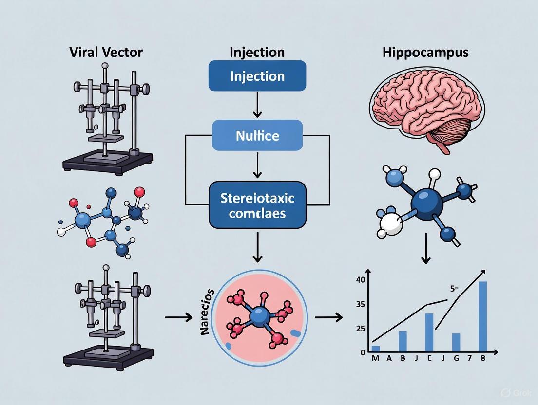
Precision Viral Vector Injection into the Hippocampus: A Guide to Stereotaxic Coordinates, Methods, and Applications
This article provides a comprehensive resource for researchers and drug development professionals on performing precise viral vector injections into the mouse hippocampus.
Precision Viral Vector Injection into the Hippocampus: A Guide to Stereotaxic Coordinates, Methods, and Applications
Abstract
This article provides a comprehensive resource for researchers and drug development professionals on performing precise viral vector injections into the mouse hippocampus. It covers foundational principles of hippocampal anatomy and viral vector selection, detailed stereotaxic methodologies for various developmental stages, advanced troubleshooting and optimization strategies, and rigorous validation techniques. By integrating established protocols with the latest innovations—such as electrophysiology-guided targeting and non-invasive focused ultrasound—this guide aims to enhance the accuracy, efficiency, and translational potential of hippocampal gene delivery for neuroscience research and therapeutic development.
Understanding Hippocampal Anatomy and Viral Vector Fundamentals for Gene Delivery
The hippocampus is a complex brain structure within the medial temporal lobe, fundamental for memory formation, spatial navigation, and associative learning [1] [2]. Its functional architecture is built upon a trisynaptic circuit that primarily involves the dentate gyrus (DG), CA3, and CA1 subfields [3] [2]. A precise understanding of these subregions' distinct roles is paramount for designing targeted experimental interventions, such as viral vector-mediated gene delivery, to study or treat neurological disorders.
This protocol details the methodologies for investigating and manipulating these subregions, framed within the context of stereotaxic viral vector injection. We provide a consolidated resource for researchers aiming to dissect the unique contributions of the dentate gyrus, CA3, and CA1 to hippocampal function, with a focus on quantitative approaches, reagent solutions, and practical experimental workflows.
Anatomical and Functional Profiles of Key Subregions
Distinct Roles in the Trisynaptic Circuit
The hippocampal trisynaptic circuit is a primarily unidirectional pathway that processes information through its key subregions. Table 1 summarizes the core anatomical and functional characteristics of these subregions.
Table 1: Anatomical and Functional Profile of Hippocampal Subregions
| Subregion | Principal Cell Type | Major Inputs | Major Outputs | Primary Functional Roles |
|---|---|---|---|---|
| Dentate Gyrus (DG) | Granule cells | Perforant path (from Entorhinal Cortex) [3] [2] | Mossy fibers to CA3 [3] [2] | Pattern Separation: Orthogonalizing similar inputs into distinct representations [4] [2]. |
| CA3 | Pyramidal cells | Mossy fibers from DG; Perforant path [3] | Schaffer collaterals to CA1; Recurrent collaterals to CA3 [3] | Pattern Completion: Auto-associative recall of memories from partial cues [4] [2]; Associative Memory [5]. |
| CA1 | Pyramidal cells | Schaffer collaterals from CA3 [3] [2] | To subiculum and entorhinal cortex [3] | Integration & Relay: Consolidates processed information for output to cortical areas [3]. |
The circuit initiates in the dentate gyrus, which receives multimodal sensory information from the entorhinal cortex via the perforant path [2]. Dentate granule cells project their distinctive mossy fibers to synapse onto the pyramidal neurons of CA3 [3] [2]. A key feature of the CA3 network is its extensive system of recurrent collaterals, which form excitatory connections among neighboring CA3 pyramidal cells. This architecture is theorized to function as an auto-associative network, enabling the storage and recall of memory patterns [3] [2]. CA3 neurons then project to CA1 via Schaffer collaterals [2]. CA1 serves as the primary output node of the hippocampus proper, sending integrated information to the subiculum and back to the entorhinal cortex for communication with the neocortex [3] [2].
Functional Specialization Evidence from Human Studies
Research combining high-resolution MRI with behavioral tasks has provided evidence for the distinct functional roles of these subregions in humans. Studies show that dentate gyrus volume predicts performance in pattern separation tasks, whereas CA3 volume predicts performance in object recognition memory, which relies on pattern completion [4]. Furthermore, aging has a variegated impact, with dentate gyrus volume decreasing with age and mediating age-related pattern separation deficits, while CA3 volume remains relatively age-independent [4].
Recent work on human hippocampal tissue has revealed unique microcircuit properties in CA3, which uses sparser synaptic connectivity than neocortical circuits. Combined with highly reliable and precise synaptic transmission, this sparse connectivity maximizes the associational power and memory storage capacity of the human CA3 network [5].
Figure 1: The Hippocampal Trisynaptic Circuit and Key Functional Roles. Information flows from the entorhinal cortex through the dentate gyrus, CA3, and CA1, with each subregion performing distinct computational functions.
Experimental Protocols for Targeted Hippocampal Manipulation
Protocol: Stereotaxic Viral Vector Injection into the Hippocampus
This protocol outlines the steps for targeted delivery of viral vectors to specific hippocampal subfields in the rodent brain, based on established methodologies [6].
Materials and Reagents
- Animal Model: Young adult male Sprague Dawley rats (7-8 weeks old) [6].
- Viral Vector: e.g., Ultra-purified AAV9 vectors for gene knockdown or expression (typical titer: >2 × 10¹³ GC/mL) [6].
- Anesthesia: Isoflurane (3% for induction, 1.0–1.5% for maintenance) [6].
- Analgesia: Buprenorphine hydrochloride (0.3 mg/kg, s.c.) [6].
- Stereotaxic Apparatus: Computer-guided robotic stereotaxic system with microinjection drive [6].
- Surgical Tools: Sterile Hamilton syringe, drill, and standard surgical kit.
Pre-operative Procedures
- Vector Preparation: Thaw viral vector on ice and briefly centrifuge before loading. Aspirate the required volume (e.g., 1 µL per site) into a sterile Hamilton syringe.
- Anesthesia and Analgesia: Induce anesthesia with isoflurane. Administer buprenorphine and normal saline subcutaneously for analgesia and hydration.
- Animal Positioning: Secure the animal in the stereotaxic frame. Apply artificial tears ointment to prevent corneal drying.
- Surgical Site Preparation: Shave the scalp, clean the skin sequentially with chlorhexidine and isopropyl alcohol, and make a mid-sagittal incision to expose the skull.
Stereotaxic Injection
- Coordinate Targeting: Identify Bregma and adjust the skull to a flat position. Use a rat brain atlas to determine coordinates for hippocampal subregions. Table 2 provides example coordinates from a recent study [6].
- Drilling and Injection: Drill holes at the calculated coordinates. Lower the syringe needle to the target depth. Infuse the vector slowly (e.g., 100 nL/min). Leave the needle in place for 5-10 minutes post-injection to prevent backflow before slowly retracting.
- Closure: Suture the incision and allow the animal to recover on a warm pad.
Table 2: Example Stereotaxic Coordinates for Rat Hippocampal Injection Sites [6]
| Hippocampal Region | Anterior-Posterior (AP from Bregma) | Medial-Lateral (ML) | Dorsal-Ventral (DV from skull surface) |
|---|---|---|---|
| Rostral Hippocampus | -2.8 mm | ±2.0 mm | -3.0 mm |
| Middle Hippocampus | -3.8 mm | ±3.0 mm | -3.0 mm |
| Caudal Hippocampus | -4.8 mm | ±4.2 mm | -4.0 mm |
Protocol: Validation of Functional Manipulation
Following vector delivery, functional outcomes can be assessed using behavioral and molecular analyses.
Behavioral Assay for Pattern Separation and Recognition
- Task: Use a behavioral pattern separation task that requires discriminating between highly similar objects (lures) [4].
- Procedure:
- Encoding Phase: Present objects for exploration.
- Retrieval Phase: Present identical objects, similar lures, and novel objects.
- Measurement: Record accuracy and response time for identifying similar lures (pattern separation) and identical objects (object recognition) [4].
- Expected Outcome: Successful dentate gyrus manipulation will specifically impair accuracy and increase response times for similar lures, while CA3 manipulation is predicted to affect object recognition memory [4].
Molecular Analysis of Signaling Pathways
- Western Blotting: Analyze hippocampal tissue lysates for changes in key synaptic proteins downstream of manipulation (e.g., Fyn, pNR2B, PSD95, total tau) [6].
- Proximity Ligation Assay (PLA): Detect and quantify protein-protein interactions (e.g., Fyn-tau complexes) in situ [6].
Figure 2: Experimental Workflow for Hippocampal Functional Manipulation. The process from viral vector preparation to final outcome assessment, highlighting key stages for validating subregion-specific effects.
The Scientist's Toolkit: Research Reagent Solutions
Successful investigation of hippocampal circuitry relies on a suite of specialized reagents and tools. Table 3 catalogs key solutions for experimental research in this domain.
Table 3: Research Reagent Solutions for Hippocampal Circuitry Studies
| Reagent / Tool | Function / Application | Example Use |
|---|---|---|
| AAV9 Vectors | In vivo gene delivery; high tropism for neurons and ability to cross the blood-brain barrier. | Neuronal-specific gene knockdown (e.g., CaMKII-promoter-driven Fyn-shRNA) [6]. |
| Fyn/SFK Inhibitors (e.g., Saracatinib) | Small molecule inhibition of Fyn kinase signaling. | Disease modification in epilepsy models; reduces hyperexcitability, neuroinflammation [6]. |
| Kainic Acid (KA) | Chemical convulsant acting on glutamate receptors. | Induction of status epilepticus (SE) to model temporal lobe epilepsy and study epileptogenesis [6]. |
| Stereotaxic Atlas | Digital or printed reference for precise brain targeting. | Determination of coordinates for hippocampal subregion injections [6]. |
| High-Resolution MRI | Non-invasive volumetric and functional imaging. | Manual segmentation of hippocampal subfields (CA1, CA3, DG) for correlation with memory performance [4]. |
The hippocampal subregions CA1, CA3, and the dentate gyrus form a highly specialized and interconnected circuit, each making unique contributions to memory processing. The experimental protocols and tools outlined here provide a framework for the precise manipulation and functional dissection of these subregions. Utilizing stereotaxic viral vector delivery, researchers can target specific molecular pathways within defined hippocampal areas to advance our understanding of their distinct roles in health and disease, thereby informing the development of targeted therapeutic strategies.
The selection of an appropriate viral vector is a critical first step in designing experiments for gene delivery into the hippocampus via stereotaxic injection. The ideal vector must align with the experimental goals, whether they require long-term stable expression, delivery of large genetic payloads, or transient manipulation of gene function. This application note provides a detailed comparison of four major viral vector systems—Adeno-Associated Virus (AAV), Lentivirus (LV), Helper-Dependent Adenovirus (HdAd), and Herpes Simplex Virus (HSV)—within the specific context of hippocampal research. We summarize their key characteristics in a structured table and provide foundational protocols for their use in stereotaxic procedures, framing this information within the rigorous demands of neuroscientific research and therapeutic development.
Vector Comparison Tables
To aid in initial vector selection, the core characteristics of each viral vector system are summarized in the table below.
Table 1: Core Characteristics of Viral Vectors for Hippocampal Research
| Feature | AAV | Lentivirus (LV) | Helper-Dependent Adenovirus (HdAd) | HSV-1 (Non-Replicative) |
|---|---|---|---|---|
| Packaging Capacity | ~4.7 kb [7] | ~8-9 kb [8] | ~36 kb [9] [10] | Up to ~40-50 kb [11] |
| Genomic Integration | Non-integrating (episomal) [7] | Integrating (stable) [7] | Non-integrating (episomal) [7] | Non-integrating (episomal, can establish latency) [12] |
| Expression Onset | 3-7 days [13] | 2-4 days | 1-2 days [10] | 1-3 days |
| Expression Duration | Long-term (months to years) [7] | Long-term (stable integration) [7] | Short-term (transient, weeks) [10] [7] | Long-term (months) in neurons [11] |
| Primary Cell Tropism (CNS) | Neurons (serotype-dependent) [14] | Neurons (dividing and non-dividing) [7] | Neurons and glia (broad) [9] | Neurons (highly neurotropic) [11] |
| Typical In Vivo Titer | 10¹² - 10¹³ GC/mL [13] | 10⁸ - 10⁹ IU/mL [15] | 10¹⁰ - 10¹² VP/mL | 10⁹ - 10¹⁰ PFU/mL |
| Immunogenicity | Low to moderate [14] | Moderate | High [10] [7] | Low (non-replicative vectors) [12] |
The following table outlines key experimental considerations for their application.
Table 2: Experimental Considerations for Hippocampal Injection
| Consideration | AAV | Lentivirus (LV) | Helper-Dependent Adenovirus (HdAd) | HSV-1 (Non-Replicative) |
|---|---|---|---|---|
| Key Advantage | High safety; long-term expression in neurons [7] [14] | Stable integration; large cargo capacity [8] [7] | Very large cargo capacity; high transduction efficiency [9] | Largest cargo capacity; highly neurotropic [11] |
| Primary Limitation | Small packaging capacity [7] | Risk of insertional mutagenesis [8] [7] | Strong immune response [10] [7] | Complex production; potential cytotoxicity variants [11] |
| Ideal for Hippocampal Studies Involving | Chronic gene replacement/silencing; circuit mapping | Stable expression in dividing progenitors; large transgenes | Delivery of large genetic constructs (e.g., CRISPR libraries); acute interventions | Delivery of multiple genes or large genomic fragments |
| Biosafety Level (BSL) | BSL-1 (typically) [13] | BSL-2 [13] | BSL-2 [13] | BSL-2 |
Essential Research Reagent Solutions
The table below lists key reagents and their functions for working with these viral vectors in a hippocampal injection paradigm.
Table 3: Key Research Reagents for Viral Vector Experiments
| Reagent / Material | Function / Application |
|---|---|
| Stereotaxic Instrument | Precise positioning of injection needle into defined hippocampal coordinates (e.g., DV: -3.0 mm, AP: -2.0 mm, ML: ±1.8 mm from Bregma in adult mice) [16]. |
| AAV Serotypes (e.g., AAV1, AAV9) | Determines cellular tropism (e.g., neuronal vs. glial) and efficiency of hippocampal transduction [14]. |
| VSV-G Pseudotyped Lentivirus | Provides a broad tropism for efficient transduction of central nervous system cells [15]. |
| HEK293T Cell Line | Standard production cell line for AAV, LV, and Ad vectors via transient transfection [15]. |
| U2OS-ICP4/ICP27 Cell Line | Complementing cell line used for the production of replication-defective HSV-1 vectors [11]. |
| Polybrene | Cationic polymer used to enhance lentiviral transduction efficiency in vitro by neutralizing charge repulsion. |
| Phosphate Buffered Saline (PBS) / Glycerol | Standard storage and dilution buffer for AAV and other viral vector stocks [13]. |
Experimental Protocol: Intracranial Stereotaxic Injection
This protocol outlines the core methodology for direct injection of viral vectors into the mouse hippocampus, based on established techniques [16].
Pre-injection Procedures
- Vector Preparation: Thaw viral aliquot on ice or at room temperature per manufacturer's instructions. Dilute if necessary in sterile PBS or artificial cerebrospinal fluid (aCSF) to the desired working titer. Keep on ice until use. Note: Avoid repeated freeze-thaw cycles [13].
- Stereotaxic Setup: Anesthetize the adult mouse and securely place it in the stereotaxic frame. Apply ophthalmic ointment to prevent corneal drying. Ensure the skull is level in all axes (anterior-posterior and medial-lateral).
- Coordinate Calculation: Identify Bregma and Lambda. Calculate the target coordinates for the hippocampal region (e.g., Dorsal Hippocampus: AP -2.0 mm, ML ±1.8 mm, DV -3.0 mm from Bregma).
- Dye Test for New Coordinates (Support Protocol): Before using the viral vector, perform a practice injection with a dye solution (e.g., Fast Green) to visually confirm the accuracy of your calculated coordinates and the flow of your injection system [16].
Surgical Injection
- Skull Exposure and Drilling: Make a midline incision to expose the skull. Gently drill a small craniotomy at the calculated AP and ML coordinates.
- Vector Loading: Load the prepared viral vector solution into a sterile glass syringe or a Hamilton syringe fitted with a pulled glass micropipette. Ensure the system is free of air bubbles.
- Injection: Slowly lower the injection needle to the target DV coordinate. Allow a 1-2 minute rest period for tissue settlement. Initiate injection using a micro-injection pump at a slow, controlled rate (e.g., 50-100 nL/minute). A typical injection volume for the mouse hippocampus is 500-1000 nL.
- Needle Withdrawal: After the full volume is delivered, leave the needle in place for an additional 5-10 minutes to allow for pressure dissipation and prevent backflow up the needle tract. Slowly retract the needle over 1-2 minutes.
Post-injection and Analysis
- Recovery and Post-op Care: Suture the incision and place the mouse in a clean, warm cage for recovery from anesthesia. Monitor until ambulatory. Administer post-operative analgesics as approved by the animal care protocol.
- Expression Timing: The time to peak transgene expression varies by vector. AAV typically requires 3-7 days [13], while Lentivirus requires 1-2 weeks for stable integration and expression.
- Validation: After an appropriate expression period, perfuse the animal and perform histology to verify the injection site and transgene expression (e.g., via fluorescence or immunohistochemistry).
Decision Pathway for Vector Selection
The following diagram illustrates the logical process for selecting the most suitable viral vector based on key experimental parameters.
Safety and Optimization Notes
- AAV Immunity: Pre-existing neutralizing antibodies can significantly reduce transduction efficiency. Screening animals or using less prevalent serotypes is recommended for in vivo work [14].
- Lentiviral Integration Risk: While integrating vectors provide long-term expression, they carry a theoretical risk of insertional mutagenesis. Consider non-integrating lentiviral vector designs for applications where this is a primary concern [8].
- Adenoviral Immunogenicity: The strong innate and adaptive immune response triggered by adenoviral vectors, including HdAd, can lead to rapid clearance of transduced cells and transient expression. This property may be harnessed for vaccine development but is a major limitation for most gene therapy applications [10] [7].
- HSV Batch Safety: For HSV-1 vectors, stringent batch quality control is essential. Whole-genome sequencing is recommended to identify and exclude syncytial variants with mutations (e.g., in UL27/gB) that can induce hyperexcitability and cytotoxicity in transduced neurons [11].
Within the rapidly advancing field of gene therapy, the selection of an appropriate viral vector is a critical determinant for experimental and therapeutic success. This is particularly true for sophisticated applications such as stereotaxic injections into the hippocampus, where precision is paramount. Adeno-associated virus (AAV) vectors have emerged as a leading delivery platform due to their favorable safety profile, including non-pathogenicity and low immunogenicity, as well as their capacity for long-term transgene expression [17]. A comprehensive understanding of three fundamental vector properties—packaging capacity, tropism, and transgene expression kinetics—is essential for researchers and drug development professionals designing gene therapy experiments and products. This application note details these core properties within the context of hippocampal research, providing structured data, validated protocols, and essential resource guides to inform vector selection and experimental design.
Core Vector Properties and Quantitative Data
AAV Packaging Capacity and Strategies
The packaging capacity of AAV is a primary consideration during transgene design. The fundamental limit for AAV vectors is approximately 4.7 kilobases (kb) of single-stranded DNA, a constraint dictated by the physical size of the viral capsid [18] [17]. This space must accommodate the entire expression cassette, including the transgene, promoter, enhancers, and polyadenylation signal, alongside the essential Inverted Terminal Repeat (ITR) sequences, which themselves occupy about 300 base pairs [18].
Table 1: AAV Packaging Capacity and Serotype Profile
| Property | Specification | Notes and Implications |
|---|---|---|
| General Packaging Capacity | ~4.7 kb [18] [17] | Includes ITRs, promoter, transgene, and polyA signal. |
| ITR Sequence Length | ~300 bp [18] | Reduces available space for the transgene expression cassette. |
| Capacity by Serotype | Consistently 4.7 kb across common serotypes (AAV1, 2, 5, 6, 8, 9, DJ, rh10, Anc80) [18] | Packaging limit is independent of serotype selection. |
| Dual Vector Approaches | Trans-splicing and Cre-lox Recombination [18] | Strategies to deliver larger transgenes by splitting them across two separate AAV particles. |
When a transgene exceeds the 4.7 kb limit, several strategies can be employed. Researchers can opt for a minigene approach, using a shortened version of the gene that retains essential functional domains. Alternatively, swapping regulatory elements for more compact versions (e.g., minimal promoters) or performing codon optimization can reduce sequence length. For larger genetic payloads, a dual AAV system is a viable, though less efficient, strategy. The two leading methods are the trans-splicing approach, where two vectors carry parts of the transgene that are spliced together post-transcriptionally, and the Cre-lox recombination approach, which uses Cre recombinase to reconstruct the full transgene from two "split" vectors within the target cell [18].
Tropism and Serotype Selection
Tropism, or the specificity of a viral serotype for particular cells and tissues, is a cornerstone of targeted gene delivery. Natural AAV serotypes exhibit distinct tissue preferences based on their interactions with cell surface receptors, cellular uptake mechanisms, and intracellular trafficking pathways [17]. This inherent tropism can be harnessed to target specific cell populations within the hippocampus, such as neurons or astrocytes.
Table 2: AAV Serotype Tropism and Promoter Specificity for CNS Targeting
| Serotype | Documented CNS Tropism | Common CNS Promoters | Target Cell Type |
|---|---|---|---|
| AAV2 | Neurons (efficient axonal transport) [19] | CAG, hSyn, CamKII | Neurons (widespread or specific subtypes) |
| AAV8 | Hippocampal dentate granule cells [20] | CAG (pan-mammalian), hSyn (neuron-specific) | Neurons |
| AAV9 | Widespread CNS transduction; crosses blood-brain barrier [21] [22] | hSyn, CamKII, CAG | Neurons |
| AAVrh10 | Efficient CNS transduction | Specific promoters | Neurons |
The choice of promoter is equally critical for cell-type specificity. While the CAG promoter drives strong, ubiquitous expression, cell-specific promoters like the human synapsin (hSyn) promoter and the CamKII promoter provide targeted expression in neurons, enabling precise mechanistic studies [22] [20]. For example, co-injection of AAV8 with a CAG promoter and AAV9 with an hSyn promoter has been successfully used to express different biosensors in hippocampal granule cells [20].
Transgene Expression Kinetics
The kinetics of transgene expression—the time course of its onset and durability—varies significantly based on the promoter and the fate of the vector genome post-injection. Intraparenchymal delivery of AAV to the mouse striatum reveals distinct temporal patterns:
- CAG Promoter: Drives rapid expression, typically peaking within 3 weeks post-injection before stabilizing to lower, sustained levels [22].
- hSyn Promoter: Exhibits a slower onset, with protein expression increasing gradually to reach a maximum at 3 months post-injection. Contrary to some earlier reports, expression driven by hSyn shows long-term durability without silencing at least up to 6 months, with mRNA levels continuing to rise at this late time point [22].
The underlying mechanism for long-term expression involves the processing of the single-stranded AAV genome within the nucleus. The linear vector genome is converted into stable, double-stranded circular episomes and concatemers, which are the predominant form associated with persistent transgene expression [22]. This genome conversion is a dynamic process that continues for months after a single administration.
Figure 1: AAV Transgene Expression Pathway. The diagram illustrates the intracellular journey of the AAV vector from injection to durable transgene expression, highlighting the key steps of genome processing and promoter-specific kinetics.
Detailed Experimental Protocol: Hippocampal AAV Delivery
This protocol details the steps for intracranial injection of AAV vectors into the juvenile mouse hippocampus for the expression of fluorescent biosensors, a technique critical for studying brain metabolism and function [20].
Materials and Reagents
- Experimental Animals: C57BL/6NCrl mice at postnatal day 1 or 2 (P1-P2).
- Viral Vectors: AAV vectors of desired serotype (e.g., AAV8, AAV9) and promoter (e.g., CAG, hSyn), titer range 10^10-10^14 gc/ml, stored at -80°C.
- Micropipettes: 10 µl glass capillary tubes (e.g., Wiretrol II).
- Stereotaxic Instrument: Digital stereotaxic instrument with a microsyringe pump and controller (e.g., World Precision Instruments UMP3 pump).
- Pipette Puller: Flaming/Brown type (e.g., Sutter Instrument P-97).
- Stereo Zoom Microscope: For visual control during procedures.
- Surgical Supplies: Dumont #5 forceps, 30-gauge needles, 70% isopropyl alcohol wipes, surgical tape, sterile saline (0.9% NaCl).
Procedure
A. Micropipette Preparation
- Pull a 10 µl glass capillary tube using a micropipette puller to create two micropipettes with a total length of 4.0-4.5 cm and a taper length of ~0.5 cm.
- Use fine forceps to carefully break the tip to a final outer diameter of 10-25 µm. Verify the dimensions under a microscope with a reticle.
B. Viral Mix Preparation
- Thaw aliquots of AAV vectors on ice.
- Mix viral preparations at the desired ratio (e.g., 1:1 for co-expression of two biosensors) in a sterile tube and let it equilibrate on ice for 10 minutes. Dilute with sterile saline if necessary.
C. Micropipette Loading
- Fill a micropipette with mineral oil using a 3 ml syringe fitted with a MicroFil 28-gauge needle.
- Mount the micropipette onto the holder of the stereotaxic instrument connected to the microinjector.
- Eject 500 nl of mineral oil to confirm the tip is open and the system is functioning.
- Pipette the viral mix onto a piece of Parafilm. Using the stereotaxic instrument and microscope, lower the micropipette tip into the viral suspension to draw it up by capillary action.
D. Stereotaxic Hippocampal Injection
- Anesthetize the P1-P2 pup on a cooled ice block.
- Secure the head in the stereotaxic apparatus.
- Identify the injection site using lambda and bregma sutures as landmarks. The target coordinates for the hippocampus should be determined based on a validated brain atlas for juvenile mice.
- Carefully lower the loaded micropipette to the calculated dorsoventral coordinate.
- Infuse the viral vector (e.g., 100-200 nL per site) at a slow, constant rate (e.g., 1-2 nL/sec) to minimize tissue damage and reflux.
- Leave the pipette in place for 1-2 minutes post-injection before slow withdrawal.
- Allow the pup to recover on a warm heating pad before returning it to the home cage.
E. Expression and Analysis
- Allow 2-4 weeks for robust transgene expression before analysis. Note that kinetics are promoter-dependent, with hSyn potentially requiring up to 3 months for maximal expression [22].
- Analyze expression using techniques such as fluorescence microscopy on acute brain slices or in vivo imaging.
Figure 2: Hippocampal AAV Injection Workflow. The experimental flowchart from vector preparation to final analysis, emphasizing the critical incubation period for transgene expression.
The Scientist's Toolkit: Research Reagent Solutions
Table 3: Essential Materials and Reagents for Hippocampal AAV Delivery
| Item | Function/Application | Example Product/Catalog Number |
|---|---|---|
| AAV Vectors | Delivery of genetic material (biosensors, effectors). | AAV8.CAG.Peredox (Addgene #73807); AAV9.hSyn.RCaMP1h [20] |
| Glass Micropipettes | Precise intracranial delivery of viral suspension. | Wiretrol II disposable micropipets (5-000-2005) [20] |
| Micropipette Puller | Fabrication of fine-tipped injection pipettes. | Sutter Instrument P-97 Flaming/Brown puller [20] |
| Stereotaxic Instrument & Pump | Accurate targeting and infusion into deep brain structures. | Digital stereotaxic instrument with microsyringe pump (e.g., WPI UMP3) [20] |
| Stereo Zoom Microscope | Visual confirmation of pipette tip size and loading procedure. | World Precision Instruments PZMIV-BS [20] |
The strategic selection and application of AAV vectors, grounded in a deep understanding of their packaging capacity, serotype tropism, and expression kinetics, are fundamental to the success of gene therapy research in the hippocampus. The quantitative data, detailed protocols, and reagent guidance provided in this application note serve as a foundational resource for researchers embarking on stereotaxic gene delivery experiments. By carefully considering the trade-offs between promoter strength, cell specificity, and expression timing, scientists can optimize their experimental designs to answer complex neurological questions and advance the development of next-generation gene therapies for CNS disorders. As the field progresses, continued optimization of vector engineering and delivery techniques will further enhance the precision and efficacy of hippocampal gene targeting.
Stereotaxic surgery is an indispensable technique in modern neuroscience, enabling precise navigation to specific brain regions for interventions such as viral vector injection, drug delivery, and electrode implantation. The technique is based on a three-dimensional Cartesian coordinate system (mediolateral, anteroposterior, and dorsoventral axes) that uses cranial landmarks as reference points [23]. For hippocampal targeting, particularly in the context of gene therapy research for conditions like Alzheimer's disease and frontotemporal dementia, precision is paramount [24] [25]. The success of these approaches hinges on accurately defining the coordinate system origin through skull landmarks—primarily Bregma and Lambda—and translating these coordinates to target locations using standardized brain atlases [23] [26]. This protocol details the established methods for defining stereotaxic coordinates, with specific application to viral vector injection into the hippocampal formation.
Anatomical Landmarks and Coordinate Systems
Defining Bregma and Lambda
The adult mouse skull features several sutures (joints between bones) that are visible under dissection microscopy. The two most critical for stereotaxic alignment are:
- Bregma: The point of intersection between the coronal suture (between frontal and parietal bones) and the sagittal suture (along the midline between left and right parietal bones) [23] [27].
- Lambda: The connection point of the sagittal suture and the lambdoid suture (between parietal and occipital bones) [23] [27].
These landmarks are not merely abstract points; they serve as the fundamental reference for the entire stereotaxic coordinate system. The Bregma is most frequently used as the origin point (0,0,0) for calculating all other target coordinates [23].
Stereotaxic Coordinate Conventions
All stereotaxic coordinate systems follow a right-handed Cartesian coordinate system [27]. The conventions for a Bregma-based absolute coordinate system are detailed in the table below.
Table 1: Stereotaxic Bregma-Based Absolute Coordinate System Conventions
| Axis | Description | Positive Direction | Negative Direction |
|---|---|---|---|
| AP (Anterior-Posterior) | Primary reference axis | Anterior to Bregma | Posterior to Bregma |
| ML (Medial-Lateral) | Perpendicular to AP axis | Right of midline | Left of midline |
| DV (Dorsal-Ventral) | Perpendicular to AP/ML plane | Ventral (downward) from Bregma | Dorsal (upward) from Bregma |
The alignment of the skull in the stereotaxic apparatus is critical. The Lambda landmark is essential for ensuring the skull is perfectly leveled in the horizontal plane along the anteroposterior axis. The skull is considered level when the dorsal vertical readings at Bregma and Lambda are identical [23].
Following the establishment of the coordinate system using skull landmarks, researchers must consult a brain atlas to determine the precise coordinates of their target structure. Several atlases provide detailed anatomical context.
Table 2: Key Mouse Brain Atlases for Stereotaxic Targeting
| Atlas Name | Key Features | Stereotaxic Reference | Access |
|---|---|---|---|
| Paxinos & Franklin's Mouse Brain | The most widely used atlas; based on Nissl and AChE staining of 40 µm sections [23]. | Bregma | Commercial |
| Allen Mouse Brain Common Coordinate Framework (CCF) | High-resolution 3D reference atlas; incorporates multimodal data (gene expression, connectivity) [23]. | An integrated framework that can be aligned to stereotaxic coordinates [27]. | Open Access [28] |
| Waxholm Space Rat Brain Atlas | Detailed 3D volumetric atlas of the rat brain; includes MR/DTI data and bregma/lambda positions [29]. | Bregma and Lambda for coordinate conversion [29]. | Open Access [29] |
It is vital to note that discrepancies can exist between different atlases and that factors such as animal strain, body weight, age, and sex can cause variations in craniometric parameters and brain volume [23]. Therefore, pilot studies and histological verification are recommended to optimize coordinates for specific experimental conditions.
Workflow for Defining Hippocampal Coordinates
The process of moving from a surgical preparation to a defined hippocampal injection coordinate involves a systematic workflow.
Diagram 1: Workflow for defining and using stereotaxic coordinates for hippocampal targeting.
Application in Viral Vector Research for Hippocampal Targeting
The precise definition of stereotaxic coordinates is a cornerstone for advanced viral vector applications in the hippocampus, enabling cell-type-specific manipulation and gene therapy.
Enhancer-Driven Gene Expression (EDGE) for Cell-Type Specificity
While traditional promoters like CaMKIIα drive expression broadly across hippocampal excitatory neurons, newer enhancer-AAV (Enhancer-Driven Gene Expression) approaches achieve superior cell-type specificity [30] [31]. The methodology involves identifying neuron-type-specific regulatory transcriptional sequences (enhancers) and packaging them into AAV vectors to limit transgene expression to a defined population [31].
For example, the enhancer AAV.3x(core)mscRE4 has been shown to drive highly selective expression in dentate gyrus granule cells, with negligible off-target expression in neighboring CA3 pyramidal cells or hilar mossy cells [30]. This level of specificity is crucial for dissecting the unique roles of hippocampal subregions in memory and disease.
Gene Therapy Delivery for Neurological Disorders
Stereotaxic delivery of AAV vectors expressing therapeutic genes is a promising strategy for neurodegenerative diseases. For instance:
- AD Treatment: Hippocampal delivery of an AAV vector expressing Brain-Derived Neurotrophic Factor (BDNF) via a novel serotype (AAVT42) mitigated neuronal degeneration and rescued cognitive impairments in three different Alzheimer's disease mouse models [24].
- FTD Treatment: Intrathalamic delivery of AAV vector expressing progranulin (AVB-101) achieved widespread cortical biodistribution and reversed pathology in a frontotemporal dementia model, demonstrating how precise stereotaxic injection into a connected node can influence a broader network [25].
The logical relationship between coordinate precision and successful experimental outcomes in these applications is summarized below.
Diagram 2: Relationship between stereotaxic precision, enhancer-AAV technology, and successful experimental outcomes in hippocampal research.
Research Reagent Solutions for Hippocampal Targeting
The following table details key materials and reagents essential for performing stereotaxic surgery and viral vector injection in the hippocampus.
Table 3: Essential Research Reagents and Materials for Hippocampal Stereotaxic Injection
| Item | Function/Application | Examples / Notes |
|---|---|---|
| Stereotaxic Apparatus | Precise 3D navigation and head fixation for rodents. | Commercially available from Kopf Instruments, RWD Life Science, Harvard Apparatus [23]. |
| Viral Vectors (AAV) | Delivery of genetic material (transgenes, Cre, opsins, etc.). | Serotypes: PHP.eB, AAV9, AAVT42 [30] [24]. Payloads: Enhancer-AAVs (e.g., AAV.3x(core)mscRE4.YFP) for cell-type specificity [30] [31]. |
| Brain Atlases | Provide anatomical maps and reference coordinates for targeting. | Paxinos & Franklin's Mouse Brain Atlas; Allen Mouse Brain CCF; Waxholm Space Rat Atlas [23] [28] [29]. |
| Anesthesia System | Surgical anesthesia and maintenance during the procedure. | Often integrated with stereotaxic mask (e.g., isoflurane) [23]. |
| Microsyringe & Micropump | Precise volume control for viral vector infusion. | Hamilton syringes; nano/picopumps for small volumes (e.g., 150 nL) [30]. |
Detailed Protocol: Defining Coordinates and Injecting the Dorsal Hippocampus
This protocol outlines the key steps for targeting the mouse dorsal hippocampus, a common site for memory-related interventions.
Animal Preparation and Skull Leveling
- Anesthetize the mouse and securely place it in the stereotaxic apparatus using ear bars and a nose clamp.
- Ensure stable anesthesia throughout the procedure using an integrated inhalator mask.
- Make a midline incision on the scalp to expose the skull.
- Clear the skull surface of tissue and moisture to clearly visualize the Bregma and Lambda sutures.
- Level the skull: Lower a sterile needle attached to the micromanipulator onto Bregma and zero the dorsoventral (DV) reading. Move the needle to Lambda and check the DV reading. Adjust the angle of the head until the DV readings at Bregma and Lambda are identical, ensuring a level skull in the anteroposterior plane [23].
Setting the Origin and Calculating Target Coordinates
- Set the origin: Position the needle tip precisely on Bregma. Set the anteroposterior (AP), mediolateral (ML), and dorsoventral (DV) coordinates of the stereotaxic apparatus to zero (0,0,0) [23] [27].
- Consult the atlas: Refer to a chosen brain atlas, such as Paxinos and Franklin's, to obtain the approximate stereotaxic coordinates for the dorsal hippocampus. Example coordinates from Bregma might be: AP: -2.0 mm, ML: ±1.5 mm, DV: -1.8 mm.
- Navigate to the target: Using the micromanipulator, move the injection needle to the calculated AP and ML coordinates.
- Mark the skull at the target ML coordinates and perform a small craniotomy using a dental drill.
Viral Vector Injection and Recovery
- Lower the injection needle to the target DV coordinate at a controlled speed.
- Infuse the viral vector (e.g., AAV.3x(core)mscRE4.YFP, titer ~1.90E+13 vg/mL) at a slow, constant rate (e.g., 100 nL/min for a total volume of 150-200 nL) [30].
- Wait 5-10 minutes after infusion to allow for pressure dissipation and prevent backflow up the injection tract.
- Slowly retract the injection needle.
- Suture the incision and provide appropriate post-operative care and analgesia until the animal fully recovers.
Post-Injection Validation
- Allow adequate expression time: For AAVs, typically 2-4 weeks are required for robust transgene expression.
- Perfuse and fix the animal, then section the brain.
- Verify injection placement histologically through fluorescence microscopy (if reporting a fluorophore) or immunohistochemistry.
- Document the precise injection sites and any variations from the intended target for accurate data interpretation.
Ethical Considerations and Animal Welfare in Invasive Neurosurgical Procedures
Ethical Framework and Justification
Adherence to a rigorous ethical framework is mandatory for research involving invasive neurosurgical procedures on nonhuman animals. This framework ensures that the acquisition of scientific knowledge is balanced with the highest standards of animal welfare.
Table 1: Core Ethical Principles for Animal Research (The 3Rs)
| Ethical Principle | Description | Application to Hippocampal Surgery |
|---|---|---|
| Replacement | Use non-animal alternatives when possible [32]. | Use computational models or in vitro systems for preliminary studies before in vivo work. |
| Reduction | Use the minimum number of animals to obtain valid results [32]. | Employ optimal experimental design and statistical power analysis to minimize animal numbers. |
| Refinement | Modify procedures to minimize or eliminate pain and distress [32]. | Use advanced anesthetics, analgesics, and aseptic surgical technique. |
The primary ethical mandate is that research must be undertaken with a clear scientific purpose, justified by the expectation of significantly increasing knowledge or benefiting health [32]. Furthermore, all procedures must be reviewed and approved by an Institutional Animal Care and Use Committee (IACUC) before initiation [32]. The psychologist should monitor the research and the subjects' welfare throughout the course of an investigation to ensure continued justification for the research [32].
Pre-Operative Protocols
Personnel and Training
All personnel must receive explicit instruction in experimental methods, care, maintenance, and handling of the species being studied [32]. Activities must not exceed an individual's competencies, training, and experience [32].
Animal Acquisition and Housing
Laboratory animals must be acquired lawfully [32]. Housing facilities must meet or exceed current regulations and guidelines to provide healthful conditions [32].
Anesthesia and Analgesia
A Ketamine/Xylazine solution (e.g., 2 ml of 50 mg/ml Ketamine, 0.01 g Xylazine in 10 ml physiological saline) can be administered intraperitoneally to induce anesthesia, with depth confirmed by a tail pinch reflex test [33]. Pre-operative analgesia (e.g., Buprenorphine) must be administered.
Stereotaxic Viral Vector Injection Protocol
This protocol details a refined method for accurate viral vector injection into the mouse hippocampal CA1 pyramidal cell layer, utilizing simultaneous electrophysiological monitoring to enhance precision [33].
Surgical Preparation
- Animals: 4- to 6-week-old C57/BL6J mice.
- Pre-treatment: Administer 20% mannitol (0.03 ml/BW, i.p.) 30 minutes pre-anesthesia for brain decompression [33].
- Anesthesia: Induce with Ketamine/Xylazine solution (i.p.), then secure the head in a stereotaxic apparatus (e.g., Narishige) [33].
- Preparation: Incise the skin, create 2-3 bur holes corresponding to hippocampal injection sites using an electrical drill, and expose the cortical surface, keeping it moist with physiological saline [33].
Injection System Setup and Targeting
- Microinjection Electrode: Pull a glass pipette (1 mm outer diameter) to a tip diameter of ~25 μm. Fill with viral vector solution and connect via a polyethylene tube to a microsyringe pump [33].
- Electrophysiological Monitoring: Integrate a wire into the pipette to connect to a differential amplifier and EEG analysis system. This allows for real-time monitoring of theta oscillations (4-8 Hz), which are largest in the hippocampal CA1 layer [33].
- Coordinate-Based Targeting: Initial coordinates based on a mouse brain atlas (e.g., AP: -2.18 mm from bregma; ML: ±1.6 mm) [33].
- Precision Placement: Set the first needle position at the cortical surface as zero. Insert the needle while recording EEG and theta oscillation data at each 0.05-0.1 mm of depth. The pipette tip is reliably positioned at the target based on the integrated values of the theta oscillation [33].
Injection and Recovery
- Injection: After positioning the tip at the target location, expel the virus solution at a rate of 0.2 µl/min for eight minutes using a microsyringe pump [33].
- Post-operative Care: Monitor animals until fully recovered from anesthesia. Provide post-operative monitoring and care, which must include the use of analgesics and antibiotics, to minimize discomfort, prevent infection, and promote recovery from the procedure [32].
Flowchart: Stereotaxic injection workflow with ethical monitoring.
Peri- and Post-Operative Care
Table 2: Summary of Surgical and Post-Operative Ethical Requirements
| Procedure | Ethical Requirement | Reference |
|---|---|---|
| Survival Surgery | Must be performed using aseptic technique and anesthesia. | [32] |
| Anesthesia | Conducted under direct supervision of a trained, competent person. | [32] |
| Analgesia | Post-operative monitoring must include analgesics to minimize pain. | [32] |
| Multiple Surgeries | Generally not permitted; must be specifically justified and approved by IACUC. | [32] |
| Severe Distress | Animals must be euthanized immediately if severe, unalleviated distress occurs. | [32] |
Experimental Endpoints and Euthanasia
When euthanasia is appropriate, it must be accomplished in a humane manner, appropriate for the species and age to ensure immediate death, in accordance with the latest AVMA Guidelines [32].
The Scientist's Toolkit: Research Reagent Solutions
Table 3: Essential Materials for Stereotaxic Hippocampal Injections
| Item | Function | Example/Specification |
|---|---|---|
| Stereotaxic Apparatus | Precise head fixation and coordinate-based navigation. | Narishige, etc. [33] |
| Microinjection Pipette | Delivery of viral vectors to target brain region. | Glass, 1 mm OD, pulled to ~25 μm tip [33] |
| Microsyringe Pump | Controlled, slow injection of small volumes. | Programmable pump (e.g., LMS), 0.2 μl/min rate [33] |
| Viral Vectors | Introduction of exogenous genes (e.g., for optogenetics). | Herpes simplex virus, Lentivirus [33] |
| Electrophysiology System | Real-time monitoring for precise targeting. | Amplifier, A/D converter (e.g., PowerLab) [33] |
| Anesthetic Agents | Induction and maintenance of surgical anesthesia. | Ketamine/Xylazine solution [33] |
| Analgesics | Management of post-operative pain. | Buprenorphine, etc. [32] |
Diagram: Ethical refinement via precision targeting.
Step-by-Step Stereotaxic Injection Protocols from Neonatal to Adult Models
This document details the essential procedures for the surgical setup for stereotaxic viral vector injection into the hippocampus, a cornerstone technique for in vivo neuroepigenetic editing and neuromodulator monitoring [34] [35]. The reproducibility and success of such experiments, which are vital for investigating neural circuits and animal behavior, depend critically on a robust aseptic technique to prevent infection, precise anesthesia to ensure animal well-being, and accurate stereotaxic instrumentation to reliably target deep brain structures [36] [34]. The protocols herein are framed within the context of manipulating and monitoring neuronal activity in the hippocampus, providing a standardized foundation for high-quality neuroscientific research.
Anesthesia and Pre-operative Preparation
Anesthetic Regimens
A variety of anesthetic regimens are suitable for rodent stereotaxic surgery. The choice depends on experimental needs, such as the anticipated duration of the procedure.
Table 1: Common Anesthetic Agents for Rodent Stereotaxic Surgery
| Anesthetic Agent | Typical Dosage and Route | Key Considerations |
|---|---|---|
| Isoflurane (Inhalant) | Induction: 3-4%; Maintenance: 1-2% in oxygen [36] | Allows for rapid control over depth of anesthesia. Requires a vaporizer and scavenging system. |
| Ketamine/Xylazine (Injectable) | 100 mg/kg Ketamine + 5 mg/kg Xylazine, intraperitoneal (IP) [34] | Provides a stable plane of anesthesia for shorter procedures. |
| Pre-operative Analgesia | Buprenorphine (e.g., 3.25 mg/kg subcutaneous) [36] | Administered pre-emptively to manage post-surgical pain. Essential for animal welfare. |
Pre-operative Setup and Animal Preparation
Once anesthetized, the animal must be prepared for surgery. Confirmation of a surgical plane of anesthesia is a critical first step, indicated by a lack of response to a hind paw pinch [34]. The animal is then secured in a stereotaxic instrument on a heating pad to maintain body temperature at 37°C throughout the procedure [36] [34]. Key preparatory steps include:
- Eye Care: Application of sterile ocular lubricant to prevent corneal drying [34].
- Fur Removal and Skin Asepsis: Shaving the scalp and sequentially cleaning the skin with alcohol prep wipes and an antiseptic such as Betadine [34].
- Incision: A midline incision is made to expose the skull, which may then be cleaned with sterile saline to visualize Bregma and Lambda, the key anatomical landmarks for coordinate determination [34].
Stereotaxic Instrumentation and Targeting
Instrument Setup and Skull Leveling
The dual-animal stereotaxic instrument must be positioned in a clean workspace, with all surgical tools sterilized prior to use, for example, in a bead sterilizer [34]. After securing the animal's head using ear bars and an incisor adapter, the skull must be leveled. This is achieved by measuring the z-coordinates at Bregma and Lambda and adjusting the head position until these coordinates are equal, ensuring a flat skull position [34].
Coordinate Calculation and Injection Setup
With the skull leveled, the tip of the injection syringe is positioned on Bregma, and its x, y, and z coordinates are recorded. The target coordinates for the hippocampus, obtained from a stereotaxic atlas, are subtracted from the Bregma coordinates to determine the final injection site [34]. A small craniotomy is drilled at the calculated location using a dental drill with a fine burr (e.g., 0.6 mm), taking care not to damage the underlying brain tissue [34].
For viral injection, a Hamilton syringe is prepared by flushing with acetone followed by sterile PBS to ensure patency [34]. The syringe is then loaded, often by drawing a small air bubble followed by the viral solution (e.g., 0.5-1 µL), to create a visible separation from the PBS in the syringe barrel [34].
Diagram 1: Stereotaxic viral injection workflow.
Aseptic and Sterile Technique
Principles and Definitions
Aseptic technique is a strict set of procedures to prevent contamination by pathogens, which is paramount for survival surgery to ensure animal recovery and valid experimental results [37]. Key terms include:
- Clean Technique: Aims to reduce the overall number of germs. Clean items, like boxed gloves, are free from dirt but not sterile [37].
- Aseptic/Sterile Technique: Aims to eliminate germs completely. "Aseptic" often describes the procedures, while "sterile" describes the instruments and environment [37].
Core Elements of Aseptic Practice
The four key elements of aseptic technique are tool and patient preparation, barriers, contact guidelines, and environmental controls [37].
Table 2: Core Elements of Aseptic Technique in Stereotaxic Surgery
| Element | Application in Stereotaxic Surgery |
|---|---|
| Tool & Patient Prep | Surgical instruments sterilized (e.g., autoclave, bead sterilizer). Animal's scalp disinfected with Betadine and alcohol [34] [37]. |
| Barriers | Surgeon wears sterile gloves, mask, and gown. Sterile drapes create a sterile field around the surgical site. |
| Contact Guidelines | Sterile personnel and items only contact other sterile items. Non-sterile items (e.g., unsterilized manipulators) are not touched with sterile gloves. |
| Environmental Controls | Surgery is performed in a dedicated, clean area. Doors are kept closed to minimize air currents [37]. |
Appropriate Hand Hygiene
Hand hygiene is the single most important practice for reducing infection transmission [38]. The "Five Moments for Hand Hygiene" dictate that hands must be cleaned:
- Immediately before touching the animal (pre-anesthesia).
- Before performing an aseptic task (e.g., loading the viral vector).
- Before moving from a soiled to a clean body site on the animal.
- After touching the animal or its immediate environment.
- After contact with potentially contaminated surfaces [38].
For hand hygiene, an alcohol-based hand rub is preferred unless hands are visibly soiled, in which case washing with soap and water for at least 20 seconds is required [38].
Diagram 2: Foundational pillars of aseptic technique.
Experimental Protocol: Viral Vector Injection and Optical Fiber Implantation
This protocol integrates the principles above for a specific application: injecting a viral sensor and implanting an optical fiber in the mouse hippocampus for in vivo photometry [36] [35].
Materials and Reagents
Table 3: Research Reagent Solutions for Hippocampal Viral Delivery
| Category | Item | Function/Application |
|---|---|---|
| Anesthesia & Analgesia | Isoflurane, Ketamine/Xylazine, Buprenorphine SR | Induce and maintain anesthesia; provide pre-emptive and post-operative pain relief [36] [34]. |
| Viral Vectors | AAV9-hSyn-GRAB*ACh3.0, AAV5-CAG-dlight1.3b, AAV9-hSyn-FLEX-iGluSnFR | Genetically encoded sensors for monitoring neurotransmitters (ACh, DA, Glu) [36]. |
| Surgical Supplies | Hamilton Syringe (5 µL, 33-gauge needle), Dental Drill (0.6 mm burr), Stereotaxic Instrument | Precise delivery of nanoliter volumes of virus; creating a craniotomy; stabilizing the animal's head [36] [34]. |
| Skull Adhesion & Sealants | Metabond, Kwik-Sil | Permanently secure implants (optical fiber, head plate) to the skull; seal the craniotomy [36]. |
| Post-operative Care | Meloxicam, Saline (0.9%) | Administered subcutaneously for post-surgical anti-inflammation and hydration [36]. |
Step-by-Step Procedure
- Anesthesia and Preparation: Induce anesthesia with isoflurane (3-4%) and maintain at 1-2%. Administer buprenorphine extended-release (3.25 mg/kg, subcutaneous) for analgesia. Secure the mouse in the stereotaxic frame on a heating pad [36].
- Incision and Skull Leveling: Shave and disinfect the scalp. Make a midline incision and retract the skin. Level the skull as described in Section 3.1 [34].
- Viral Injection:
- Calculate coordinates for the dorsal hippocampus (e.g., from Bregma: AP -2.0 mm, ML ±1.5 mm, DV -1.8 mm).
- Drill a craniotomy at the target AP and ML coordinates.
- Load the purified AAV (e.g., AAV9-hSyn-GRAB*ACh3.0) into a Hamilton syringe [34].
- Lower the syringe to the target DV coordinate at a slow, controlled rate.
- Inject the virus (e.g., 200-300 nL) at a slow rate (e.g., 100 nL/min) [36].
- Wait 5-10 minutes after injection to allow for diffusion before slowly retracting the syringe [34].
- Optical Fiber Implantation (if required for the experiment):
- Securing the Implant and Closure:
- Post-operative Recovery and Care:
- Place the animal in a clean, warm cage until fully ambulatory.
- Administer post-operative injections of meloxicam (5 mg/kg) and 1 mL saline subcutaneously daily for 3-4 days [36].
- Monitor the animal closely for signs of distress or infection. Allow a minimum of two weeks for recovery and full viral expression before commencing behavioral experiments or data collection [36] [34].
A meticulous surgical setup is non-negotiable for the success and reproducibility of stereotaxic viral vector injections. By rigorously adhering to the detailed protocols for anesthesia, stereotaxic instrumentation, and aseptic technique outlined in this document, researchers can ensure animal welfare, minimize experimental variables, and reliably perform sophisticated neural manipulations and recordings in the hippocampus. This foundational work enables high-quality research into brain function and the mechanisms underlying neurological disorders.
Targeting specific hippocampal subregions via stereotaxic surgery is a cornerstone technique in neuroscience, crucial for investigating learning, memory, and emotional processing, as well as for developing gene therapies for neurological disorders. The hippocampus, with its distinct dorsal (posterior in primates) and ventral (anterior in primates) functional domains, presents a significant challenge for precise intervention. The dorsal hippocampus is primarily implicated in spatial learning and memory, while the ventral hippocampus regulates emotional and motivational behaviors [39]. This application note provides a standardized framework for calculating coordinates and executing stereotaxic injections to target these subregions in mice and rats, framed within the context of advanced viral vector research. We integrate the latest anatomical atlases, detailed protocols, and essential quality control measures for viral vectors to enhance experimental reproducibility and translational potential.
Anatomical Foundations and Coordinate Systems
Precise targeting begins with a foundational understanding of hippocampal anatomy and the choice of a reliable reference atlas. Traditional two-dimensional atlases, while useful, are limited by sectioning intervals of hundreds of micrometers, which can hinder accurate three-dimensional reconstruction and boundary determination [40].
The advent of high-resolution, whole-brain datasets has revolutionized this field. The Stereotaxic Topographic Atlas of the Mouse brain (STAM), for instance, provides a three-dimensional Nissl-stained dataset with an isotropic 1-μm resolution [40]. This allows for the observation of continuous anatomical changes and the precise determination of where specific structures begin and end along any axis. For viral vector studies, such precision is indispensable for ensuring that the transduction occurs in the intended cellular population.
A critical principle in stereotaxic surgery is the use of a stable coordinate system. The STAM atlas, for example, defines its spatial coordinate system based on both cranial and intracranial reference points, known as datum marks [40]. Consistency in referencing these points, such as Bregma, is vital for minimizing variability between subjects and experimental sessions.
Stereotaxic Coordinate Tables
The following tables provide standard stereotaxic coordinates for targeting the dorsal and ventral hippocampus in adult mice and rats. All coordinates are given in millimeters relative to Bregma, with the skull surface as the depth reference.
Table 1: Stereotaxic Coordinates for Mouse Hippocampal Subregions
| Species / Structure | Anterior-Posterior (AP) | Medial-Lateral (ML) | Dorsal-Ventral (DV) |
|---|---|---|---|
| Mouse - Dorsal Hippocampus | -1.8 to -2.0 | ±1.0 to ±1.8 | -1.8 to -2.2 [41] [42] |
| Mouse - Ventral Hippocampus (CA1v) | -3.0 to -3.3 | ±2.8 to ±3.2 | -3.5 to -4.2 [39] |
Table 2: Stereotaxic Coordinates for Rat Hippocampal Subregions
| Species / Structure | Anterior-Posterior (AP) | Medial-Lateral (ML) | Dorsal-Ventral (DV) |
|---|---|---|---|
| Rat - Dorsal Hippocampus | -3.6 to -4.0 | ±2.0 to ±2.5 | -3.0 to -3.5 |
| Rat - Ventral Hippocampus | -5.0 to -5.3 | ±4.5 to ±5.0 | -6.5 to -7.5 |
Note: These coordinates are representative starting points. Always validate and adjust coordinates based on your specific animal strain, age, sex, and the reference atlas used in your laboratory.
Detailed Experimental Protocol for Viral Vector Injection
The following protocol details the key steps for a stereotaxic injection into the mouse hippocampus, adaptable for rats with adjustments to coordinates and injection volumes [41] [43] [42].
Pre-surgical Preparations
- Viral Vector Preparation: Thaw the viral vector (e.g., AAV) aliquot on ice. Briefly centrifuge before use to collect contents at the bottom of the tube. Avoid repeated freeze-thaw cycles [43].
- Animal Preparation: Anesthetize the mouse using an approved method, such as isoflurane (5% for induction, 1-2% for maintenance) or a ketamine/xylazine mixture (e.g., 100 mg/kg and 10 mg/kg, respectively, administered intraperitoneally). Apply a lubricating ophthalmic ointment to prevent corneal drying.
- Stereotaxic Setup: Secure the animal in the stereotaxic frame using ear bars and a nose clamp. Ensure the skull is level by confirming equal DV coordinates at Bregma and Lambda.
Surgical Procedure and Injection
- Craniotomy: Make a midline scalp incision and clean the skull surface. Identify Bregma and use it as the zero point. Drill a small craniotomy (∼0.5 mm diameter) at the calculated AP and ML coordinates for your target.
- Vector Injection: Load a sterile glass micropipette (tip diameter ∼10-50 μm) or a 33-gauge Hamilton needle with the viral vector. Lower the needle slowly to the target DV coordinate.
- Infusion: Initiate the infusion using a nano-liter injector. A typical injection volume for the mouse hippocampus is 50-100 nL per site [41] [43]. Infuse at a slow, controlled rate (e.g., 10-20 nL/min) to minimize tissue damage and backflow along the needle track.
- Needle Withdrawal: After the infusion is complete, leave the needle in place for an additional 5-10 minutes before slowly retracting it. This allows for adequate diffusion of the vector away from the injection site.
- Closure: Suture the scalp and administer post-operative analgesia (e.g., Buprenorphine, 0.05-0.1 mg/kg). Monitor the animal until it fully recovers from anesthesia.
The Scientist's Toolkit: Essential Materials and Reagents
Table 3: Research Reagent Solutions for Hippocampal Stereotaxic Surgery
| Item | Function/Application | Example/Specification |
|---|---|---|
| Adeno-Associated Virus (AAV) | Gene delivery vehicle; offers long-term transgene expression and high tropism for neurons. | Serotypes like AAV9 are commonly used (e.g., AAV9-CaMKII-ArchT-GFP) [43]. |
| Kainic Acid (KA) | Glutamate receptor agonist; used to chemically induce seizures and model temporal lobe epilepsy. | Kainic Acid Monohydrate; administered via stereotaxic injection [42]. |
| Isoflurane | Inhalable general anesthetic for surgical procedures. | Typically used at 1-3% concentration in oxygen [43] [42]. |
| Buprenorphine | Opioid analgesic for post-operative pain management. | Administered at 0.05-0.1 mg/kg [42]. |
| Digital PCR (dPCR) | Quality control for viral vectors; used to quantify vector genome titer and assess genome integrity. | Enables multiplex assays targeting specific regions like ITR, promoter, and poly-A tail [44]. |
| Fluoro-Gold | Retrograde tracer used for neural circuit mapping. | Used in a 4% solution for pressure injection [45]. |
| Biotinylated Dextran Amine (BDA) | Anterograde tracer used for mapping axonal projections. | 5%, 3,000 MW, lysine fixable; injected into the spinal cord or brain [45]. |
Viral Vector Considerations for Hippocampal Targeting
The efficacy of gene therapy interventions heavily relies on the quality and properties of the viral vector.
- Vector Genome Integrity: A critical quality attribute is the proportion of intact vector genomes. Digital PCR (dPCR) has emerged as a powerful tool for quantifying genome integrity, often revealing that only 20-40% of genomes in a preparation are fully intact, despite capsids appearing "full" in other assays [44]. This integrity directly correlates with potency, making it a key parameter to monitor during production [44].
- Capsid Serotype and Promoter Selection: The choice of AAV serotype (e.g., AAV1, AAV2, AAV5, AAV9) and a cell-type-specific promoter (e.g., CaMKIIα for excitatory neurons) determines the efficiency and specificity of transduction in the hippocampus [46] [43].
- Production and Purification: The production process itself can impact integrity. GC-rich promoter regions can form secondary structures that hinder replication, and the method used for capsid lysis (e.g., temperature vs. Proteinase K) can also affect the final product [44]. Orthogonal analytical techniques like analytical ultracentrifugation (AUC) and Mass Photometry are recommended for assessing full-to-empty capsid ratios [44].
Workflow and Pathway Diagrams
The following diagrams outline the core experimental workflow and the logical relationship between vector integrity and experimental success.
Diagram 1: Stereotaxic Viral Vector Injection Workflow. This flowchart summarizes the key stages of a stereotaxic injection experiment, from initial planning to final analysis, highlighting the critical pre-surgical step of viral vector quality control (QC).
Diagram 2: Impact of Viral Vector Integrity on Experimental Outcomes. This causal loop diagram illustrates how the integrity of the viral vector genome directly influences functional titer, the efficiency of in vivo transduction, and ultimately, the reliability and interpretation of experimental results.
Application Note: Optimizing Micropipettes for Hippocampal Injection
The Science and Art of Pipette Pulling
The creation of glass micropipettes for stereotaxic injection is a critical step that combines scientific understanding with practical artistry. The glass transition—the transformation from a brittle to a soft, viscous state upon heating—is central to this process. Achieving tips of consistent shape and size requires careful control of environmental factors and the physical parameters of the puller [47].
Key Pulling Parameters and Their Effects: The table below summarizes how adjustments to key parameters on a programmable pipette puller (e.g., WPI's PUL-1000) influence the final pipette geometry. These guidelines allow researchers to fine-tune their instruments based on the specific glass capillary type and laboratory conditions [47].
| Parameter | Effect of Increase | Effect of Decrease |
|---|---|---|
| Heat | Longer Taper | Shorter Taper |
| Force | Smaller Tips, Longer Taper | Larger Tips, Shorter Taper |
| Distance | Smaller Tips | Larger Tips |
| Delay | Shorter Taper | Longer Taper |
Critical Factors Influencing Pull Quality:
- Filament Characteristics: Platinum/Iridium filaments slowly oxidize over time, changing their heating properties and eventually burning out, which necessitates periodic replacement [47].
- Glass Type: Different glass capillaries (e.g., Borosilicate, Quartz) have different softening points (melting point temperatures), which can even vary between manufacturing lots. The puller program must be adjusted accordingly [47].
- Environmental Conditions: Room temperature and humidity affect heat transfer via convection and can alter pull outcomes even with an identical program. Allowing the filament and its holders to cool between pulls is essential for consistency [47].
Viral Vector Solution Preparation
The preparation of the viral vector solution is crucial for injection viability and transfection efficiency. Key considerations include the choice of the viral vector and ensuring the solution's integrity.
- Vector Selection: Adeno-associated viruses (AAVs) are commonly used for gene delivery to the hippocampus. Different serotypes offer varying tropisms; for example, a newly engineered serotype, AAVT42, has demonstrated superior neuronal tropism in the mouse central nervous system compared to AAV9 [24].
- Solution Handling: Viral vectors should be diluted in sterile, non-cytotoxic buffers such as phosphate-buffered saline (PBS) to the appropriate titer. The solution must be kept on ice and protected from light to maintain viral potency. Centrifugation at high speed (e.g., >13,000 rpm for 1-2 minutes) is recommended immediately before loading into the pipette to pellet any particulate matter that could cause clogging [6].
Experimental Protocols & Workflows
Protocol: Hippocampal-Targeted Viral Vector Injection in Rodents
This protocol details the procedure for bilateral hippocampal injection of AAV vectors in rats, as derived from current literature [6].
I. Pre-Surgical Preparation
- Animals: Use young adult male Sprague Dawley rats (7–8 weeks old). House individually with a 12-hour light/dark cycle and ad libitum access to food and water.
- Anesthesia: Induce anesthesia with 3% isoflurane and maintain with 1.0–1.5% isoflurane throughout the stereotaxic procedure.
- Analgesia: Administer Buprenorphine hydrochloride (0.3 mg/kg, subcutaneously) immediately after anesthesia induction.
- Viral Vector: Prepare the AAV vector (e.g., AAV9-CaMKII-GFP or AAV9-CaMKII-Fyn-shRNA) at a typical titer of >1x10¹³ genome copies (GC)/mL. Keep on ice and protected from light [6].
II. Stereotaxic Surgery and Injection
- Secure the anesthetized animal in a stereotaxic frame with a heating pad to maintain body temperature.
- Shave the scalp, make a mid-sagittal incision, and clean the exposed skull with chlorhexidine and isopropyl alcohol.
- Level the skull such that Bregma and Lambda are on the same horizontal plane.
- Identify Bregma and calculate the target coordinates for the hippocampus. For bilateral injections in rats, the following four-site coordinate set (relative to Bregma) can be used to ensure maximal transduction [6]:
- Site 1 (Rostral): Anteroposterior (AP) -3.0 mm, Mediolateral (ML) ±2.0 mm, Dorsoventral (DV) -3.0 mm
- Site 2 (Middle): AP -4.5 mm, ML ±3.0 mm, DV -4.5 mm
- Site 3 (Caudal): AP -5.5 mm, ML ±4.5 mm, DV -5.0 mm
- Site 4 (Caudal): AP -5.5 mm, ML ±5.0 mm, DV -6.0 mm
- Drill small craniotomies at each calculated coordinate.
- Load ~1 µL of the ultra-purified viral vector solution (~10¹⁰ GC) into a sterile Hamilton syringe or a pulled glass micropipette connected to a microinjection system [6].
- Lower the injection needle slowly to the target DV coordinate.
- Initiate infusion at a controlled flow rate. Recent findings emphasize that flow rate is a critical determinant of delivery efficiency. For a model system involving mesenteric arteries, a slower flow rate of 0.83 cm/s resulted in significantly higher levels of microRNA-126 delivery compared to faster rates (e.g., 1.47 cm/s and 1.89 cm/s) across various ultrasound conditions [48]. This principle translates to intracranial injections, where slower infusion rates (e.g., 50-100 nL/min) are generally recommended to minimize tissue damage and backflow along the injection tract.
- After completing the infusion, leave the needle in place for 5-10 minutes to allow for pressure equilibration and solution diffusion into the tissue.
- Slowly retract the needle and repeat the process for all remaining injection sites.
- Close the surgical wound with sutures or wound clips and monitor the animal until it fully recovers from anesthesia.
The following table consolidates key quantitative parameters from the literature for hippocampal viral vector delivery.
| Parameter | Typical Range / Value | Application Note / Context |
|---|---|---|
| Pipette Tip Size | Customizable via puller settings | Smaller tips for precise targeting; larger tips for higher volume/distribution [47]. |
| Viral Titer | >1x10¹³ GC/mL (Ultra-purified AAV) | High titer is required for effective neuronal transduction [6]. |
| Injection Volume (Rat) | 1 µL per site | Used for AAV-shRNA delivery across 4 hippocampal sites [6]. |
| Flow Rate | 50-100 nL/min (recommended) | Slower flow rates (e.g., equivalent to 0.83 cm/s in a model system) enhance gene delivery efficiency and minimize tissue damage/backflow [48]. |
| Needle Dwell Time | 5-10 minutes post-infusion | Critical for pressure dissipation and reducing reflux up the injection tract. |
The Scientist's Toolkit: Essential Research Reagents and Materials
| Item | Function / Description |
|---|---|
| AAV Vector (e.g., AAV9, AAVT42) | Engineered adeno-associated virus serving as the gene delivery vehicle. Serotype choice (e.g., AAV9 vs. novel AAVT42) affects neuronal tropism and transduction efficiency in the hippocampus [24]. |
| scramble-shRNA Control AAV | A control vector encoding a non-targeting shRNA sequence; essential for attributing phenotypic effects to the specific gene knockdown in the experiment [6]. |
| Kainic Acid | A potent glutamate receptor agonist used in rodent models to chemically induce Status Epilepticus (SE) for studies on epileptogenesis, often following viral vector injection [6]. |
| Diazepam | A benzodiazepine administered post-procedure to terminate or moderate seizure activity in seizure models, ensuring animal welfare [6]. |
| Isoflurane | Volatile inhalant anesthetic for induction and maintenance of surgical-plane anesthesia during stereotaxic procedures [6]. |
| Buprenorphine | Opioid analgesic administered pre-emptively and post-operatively for pain management in accordance with animal welfare protocols [6]. |
| Phosphate-Buffered Saline (PBS) | Isotonic, pH-balanced buffer used for diluting viral vectors to the desired titer and for trans-cardiac perfusion [6]. |
Signaling Pathways and Experimental Workflows
Fyn Kinase Signaling in Epileptogenesis
Hippocampal Viral Injection Experimental Workflow
Within viral vector-based research targeting the hippocampus, the choice of injection protocol is critically dependent on the age of the subject. This document outlines two principal methodologies: intracerebroventricular (ICV) injection for neonatal mice and direct parenchymal injection for adults. These age-specific approaches are foundational for studies in neurodevelopment, circuit mapping, and therapeutic drug development, as they accommodate fundamental differences in skull integrity, brain size, and cerebrospinal fluid dynamics [49] [50].
The table below summarizes the core parameters for the two primary injection protocols, highlighting the strategic differences tailored to the developmental stage of the mouse model.
Table 1: Key Comparative Parameters for Neonatal ICV and Adult Parenchymal Injections
| Parameter | Neonatal ICV Injection | Adult Direct Parenchymal Injection |
|---|---|---|
| Developmental Stage | Postnatal Day 0 (P0), within 3-6 hours of birth [50] | Juvenile (e.g., P14-P21) and Adult mice (e.g., >P60) [51] |
| Primary Advantage | Widespread, global transgene expression throughout the brain [49] [50] | Focal, region-specific transduction (e.g., hippocampal subregions) [51] |
| Injection Volume | Up to 2 µL per ventricle (total 4 µL) [49] | Typically 30 nL to 500 nL, depending on the target region [51] |
| Viral Titer | Serial dilutions (e.g., 10^8 to 10^10 viral particles/hemisphere) can control transduction density [49] | High titer stocks (e.g., 3 × 10^12 vp/mL diluted to 3 × 10^11 vp/mL) [51] |
| Surgical Approach | Free-hand or stereotaxic; no craniotomy required [49] | Stereotaxic surgery requiring craniotomy via micro-drill burr (0.7 mm) [51] |
| Anesthesia | Hypothermia (ice-induced) [49] | Injectable (e.g., Ketamine/Xylazine) or inhaled anesthesia (e.g., Isoflurane) [51] [50] |
| Key Viral Vectors | Adeno-associated virus (e.g., AAV8, AAV9, AAV-PHP.eB) [49] [50] | Helper-dependent Adenoviral Vectors (HdAd), AAV, Lentivirus [51] |
| Expression Onset & Duration | Within days; can persist for the animal's lifetime (up to one year) [49] [50] | Rapid onset with HdAd; stable, long-term expression [51] |
Detailed Experimental Protocols
Protocol 1: Intracerebroventricular (ICV) Injection in Neonatal Mice
This protocol is designed for widespread transgene delivery in the neonatal mouse brain, leveraging the immature ependymal lining for efficient dissemination [49] [50].
Pre-procedural Preparations
- Viral Solution: Thaw AAV aliquot on ice. Dilute the virus in ice-cold 1x PBS. Add a visible tracer like 0.05% trypan blue to monitor injection success [49].
- Pups: Ensure pups are within 3-6 hours of birth and have visible milk spots. Place them on a warming pad until the moment of injection [50].
- Equipment: Load a 10 µL syringe with a 32-gauge needle with 5 µL of the prepared viral solution [49].
Injection Procedure
- Anesthesia: Transfer a single pup from the warming pad to a pre-cooled metal plate on ice. Induce hypothermia anesthesia for 2-3 minutes, confirmed by a lack of response to a gentle toe pinch [49].
- Landmarking: Wipe the pup's head with 70% ethanol. Visually identify the cranial sutures (bregma and lambda). The injection site is approximately 0.8-1 mm lateral to the sagittal suture, halfway between lambda and bregma [49].
- Injection: Hold the syringe perpendicular to the skull surface. Insert the needle at the marked site to a depth of approximately 3 mm, where a slight decrease in resistance indicates entry into the lateral ventricle. Slowly inject a maximum of 2 µL of the viral solution, watching for the spread of trypan blue in the ventricle. Slowly withdraw the needle [49].
- Contralateral Injection: Allow the first injection site to close before repeating the procedure on the opposite hemisphere [49].
- Recovery: Return the pup to the warming pad until fully active, then reunite it with the mother [49].
Protocol 2: Direct Parenchymal Injection in Adult Mice
This protocol enables precise, region-specific gene delivery, such as to the hippocampus, using stereotaxic instrumentation [51] [35].
Pre-procedural Preparations
- Viral Solution: Prepare the injection solution by mixing the viral stock (e.g., HdAd at 3 × 10^12 vp/mL) with 20% mannitol and storage buffer to a final concentration of 3 × 10^11 vp/mL [51].
- Animal Anesthesia: Anesthetize the mouse using an appropriate regimen (e.g., Ketamine/Xylazine cocktail) and secure it in a stereotaxic frame. Maintain body temperature with a heating pad. Administer a local analgesic like Bupivacaine at the incision site [51] [50].
- Stereotaxic Coordination: Shave the scalp, clean with povidone-iodine and ethanol. Make a midline incision to expose the skull. Identify and mark Bregma. Calculate the precise anteroposterior (AP) and mediolateral (ML) coordinates for the target hippocampal region (e.g., Dorsal Hippocampus: AP -2.0 mm, ML ±1.5 mm from Bregma). Mark the skull and perform a craniotomy with a 0.7 mm micro drill burr [51].
Injection Procedure
- Needle Placement: Load a fine glass capillary or a Hamilton syringe with the viral solution. Lower the needle vertically to the target dorsoventral (DV) coordinate (e.g., DV -1.8 mm from the brain surface) at a controlled speed [51] [35].
- Virus Infusion: Initiate the injection using a nanoinjector. A typical volume for hippocampal injection is 30-500 nL, infused at a slow, constant rate (e.g., 50 nL/min) to minimize tissue damage and backflow [51].
- Needle Withdrawal: After infusion, leave the needle in place for 5-10 minutes to allow for pressure equilibration and viral diffusion. Subsequently, slowly retract the needle from the brain [51].
- Post-operative Care: Suture the wound and administer a post-operative analgesic (e.g., Meloxicam-SR). Place the animal in a clean cage on a heating pad until it fully recovers from anesthesia [51].
Visualization of Workflows
The following diagrams, generated with Graphviz, illustrate the logical flow and key decision points for each protocol.
Neonatal ICV Injection Workflow
Adult Parenchymal Injection Workflow
The Scientist's Toolkit: Essential Research Reagents and Materials
Successful execution of these protocols relies on specialized reagents and equipment. The following table lists key materials.
Table 2: Essential Research Reagent Solutions and Materials
| Item | Function/Application | Example Specifications / Notes |
|---|---|---|
| Adeno-associated Virus (AAV) | Efficient neuronal transduction; serotypes (AAV8, AAV9) preferred for neonates [49] [50]. | Titer: 10¹² - 10¹³ GC/mL; can be diluted for controlled expression density [49]. |
| Helper-dependent Adenoviral (HdAd) Vectors | Large packaging capacity (up to 37 kb); rapid, stable expression in adults [51]. | Ideal for large genes (e.g., CaV2.1 α1 subunit); titer: ~3 × 10¹² vp/mL [51]. |
| Stereotaxic Instrument | Precise positioning for adult and neonatal (adapted) injections [51] [50]. | Includes manipulator arm, neonatal stage, and ear bars [49] [50]. |
| Microinjection Syringe | Delivery of viral solution into brain tissue. | Hamilton syringe (e.g., 700 series) or glass capillaries; 32G needle for neonates [51] [49]. |
| Nanoliter Injector / Infusion Pump | Controlled, slow infusion of viral volumes to minimize tissue damage [51]. | Enables precise control of injection rate (e.g., 50 nL/min) [51]. |
| Storage Buffer | Maintains viral stability and integrity during preparation and injection [51]. | Composition: 250 mM Sucrose, 10 mM HEPES, 1 mM MgCl₂, pH 7.4 [51]. |
| Trypan Blue Dye (0.05%) | Visual tracer added to viral solution to confirm successful intraventricular delivery in neonates [49]. | Provides immediate visual feedback on injection accuracy [49]. |
Precise targeting of the hippocampal CA1 pyramidal layer is a critical requirement for studies investigating synaptic plasticity, learning, and memory. Traditional stereotaxic injection techniques rely on anatomical coordinates derived from brain atlases, but these methods face significant limitations due to individual anatomical variability, brain swelling, and the challenging anatomy of the CA1 pyramidal layer, which is only approximately 0.1 mm thick [33]. Even minor miscalculations can result in off-target deliveries, compromising experimental validity and leading to wasted resources.
This Application Note details an advanced targeting methodology that overcomes these limitations by using real-time theta oscillation (4-8 Hz) monitoring to guide placement within the CA1 pyramidal layer. By leveraging a well-characterized electrophysiological signature of this region, researchers can achieve unprecedented accuracy in the delivery of viral vectors, chemicals, and other biological materials for studies in molecular neuroscience and drug development [33].
Theta Oscillations as a Physiological Landmark
Theta oscillations are rhythmic 4-8 Hz fluctuations in the local field potential that are prominently associated with hippocampal-dependent functions such as spatial navigation and memory formation [33] [52]. Within the hippocampus, these oscillations are not uniform; their power is greatest in the stratum lacunosum-moleculare of CA1 and is controlled by cholinergic inputs from the medial septum [33].
Recent research in humans has further refined our understanding, suggesting the existence of functionally distinct high-theta (~8 Hz) and low-theta (~3 Hz) oscillations, with the higher frequency oscillations being more prominent in the posterior hippocampus and correlated with movement speed during spatial navigation [53]. This functional-anatomical relationship underscores the value of theta as a precise localizing signal. The methodology described herein utilizes the integrated value of these theta oscillations as a quantitative, real-time readout to accurately position the injection pipette within the thin pyramidal cell layer [33].
Table 1: Characteristics of Hippocampal Theta Oscillations Across Species
| Species | Frequency Range | Key Behavioral Context | Notable Features |
|---|---|---|---|
| Mouse/Rat | 4-8 Hz [33] | Locomotion, REM sleep [52] | Persistent during movement; largest amplitude in CA1 SL-M [33] |
| Human | 2-14 Hz [53] | Spatial navigation, memory [52] [53] | Occurs in short bouts (~400 ms); higher power during fast movement [52] |
| Human (High-Theta) | ~8 Hz [53] | Spatial navigation | Prevalent in posterior hippocampus; frequency correlates with movement speed [53] |
| Human (Low-Theta) | ~3 Hz [53] | Non-spatial cognitive processes | More prevalent in anterior hippocampus; frequency not speed-dependent [53] |
Materials and Reagent Solutions
Table 2: Essential Research Reagents and Equipment
| Item | Specification/Function | Example/Note |
|---|---|---|
| Stereotaxic Apparatus | Head fixation and precise coordinate positioning | Narishige, David Kopf Instruments |
| Microinjection Pipette | Glass capillary pulled to ~25 μm tip diameter [33] | 1 mm outer diameter; A-M System |
| Micropipette Puller | Creates consistent, fine-tipped injection pipettes | Sutter Instruments P-97/1000 |
| Microsyringe Pump | Programmed, slow injection of solution (e.g., 0.2 μl/min) [33] | LMS, World Precision Instruments |
| Recording Wire | Conducts EEG signal from pipette; Ag/AgCl or copper [33] | Copper wire is a cost-effective alternative |
| Differential Amplifier | Amplifies theta signal (10,000x gain) [33] | Cygnus Technology |
| Data Acquisition System | Analog-to-digital conversion and analysis of EEG | PowerLab (AD Instruments) with PC |
| Viral Vector | Gene delivery; common choices include HSV, LV, AAV [33] [54] | Herpes Simplex Virus (HSV), Lentivirus (LV) [33] |
| Anesthetic | Surgical anesthesia for in vivo procedures | Ketamine/Xylazine mixture [33] |
Detailed Experimental Protocol
Step 1: Preparation of the Microinjection Electrode and Circuit
- Pipette Pulling: Use a multi-step program on an automatic puller to create a glass micropipette with a final tip diameter of approximately 25 μm [33].
- Pipette Filling: Carefully backfill the pipette with your solution of interest (e.g., viral vector, dye, fluorescent microspheres), ensuring no air bubbles are present.
- Electrode Assembly: Insert a sterile Ag/AgCl or copper wire into the tapered end of the pipette to make contact with the solution. This wire will serve as the recording electrode.
- Pressure Tubing Connection: Connect a polyethylene tube to the back of the pipette, sealing the joint with a flame. Connect the other end of the tube to a microsyringe filled with liquid paraffin, which will be used to apply pressure for injection.
- Circuit Connection: Connect the recording wire to the positive input of a differential amplifier and ground the negative input to the stereotaxic frame's ear bar [33].
Step 2: Animal Preparation and Surgery
- Pre-anesthesia: Administer an intraperitoneal injection of 20% mannitol (0.03 ml per gram of body weight) for brain decompression approximately 30 minutes before the main anesthetic [33].
- Anesthesia: Induce surgical anesthesia with an intraperitoneal injection of a Ketamine/Xylazine solution. Continuously monitor the depth of anesthesia via tail pinch reflex.
- Stereotaxic Fixation: Secure the animal's head in the stereotaxic apparatus. Make a midline skin incision to expose the skull and clean the surface.
- Craniotomy: Using predefined coordinates (e.g., AP: -2.18 mm from bregma, ML: ±1.6 mm from sagittal suture for mouse dorsal hippocampus), drill 2-3 small bur holes through the skull. Keep the exposed cortical surface moist with physiological saline [33].
Step 3: Theta-Monitored Pipette Insertion and Targeting
- Signal Calibration: Lower the pipette tip until it just touches the cortical surface. Use the EEG detection at this point to set the depth to zero.
- Insertion and Monitoring: Advance the pipette into the brain in small increments (0.05-0.1 mm). At each step, record the EEG and perform a real-time power analysis of the 4-8 Hz theta band [33].
- Target Identification: As the pipette approaches the hippocampal layers, the power of the theta oscillation will increase noticeably. The target is the point of maximum integrated theta power within the pyramidal cell layer. This electrophysiological signature provides a more reliable target than a predetermined depth coordinate.
Diagram 1: Workflow for theta-monitored stereotaxic injection.
Step 4: Microinjection and Post-Procedural Handling
- Controlled Injection: Once the target is confirmed, initiate the injection using a microsyringe pump. A slow, constant infusion rate of 0.2 μl/min for 1-8 minutes (depending on the solution and volume) is recommended to maximize infection efficiency and minimize tissue damage [33].
- Pipette Retraction: After the injection is complete, wait 5-10 minutes to allow for pressure dissipation before slowly retracting the pipette.
- Post-operative Care: Suture the skin incision and provide appropriate post-operative analgesia and monitoring until the animal fully recovers from anesthesia.
- Histological Validation: After an appropriate survival period, perfuse the animal and section the brain. Confirm the injection site using dye visualization, fluorescent microspheres, or immunohistochemistry for the transgene (e.g., GluA1) [33].
Validation and Performance Data
This theta-guided method has been quantitatively validated against traditional coordinate-based approaches. In one study, targeted injections of dye or lentiviral vectors were performed, with sites subsequently confirmed histologically.
Table 3: Comparison of Targeting Methods for CA1 Pyramidal Layer Injection
| Targeting Method | Number of Injections | Success Rate | Key Advantages | Key Limitations |
|---|---|---|---|---|
| Theta Oscillation Monitoring | Not explicitly stated | High (precise to 0.1 mm layer) [33] | Compensates for individual variability; Real-time feedback | Requires specialized EEG equipment; More complex setup |
| Standard Atlas Coordinates | 10 [33] | Variable/Moderate | Simple; Fast; No extra equipment needed | Inaccurate with anatomical variability or brain swelling [33] |
| Presumptive Coordinates (Atlas) | 20 [33] | Variable/Moderate | Simple; Fast | Inaccurate with anatomical variability or brain swelling [33] |
The data demonstrates that the theta-guided method reliably achieves precise placement within the CA1 pyramidal layer, a level of accuracy that is difficult to guarantee with conventional methods that are susceptible to individual anatomical differences [33].
Integration with Viral Vector Technology
The precise delivery of viral vectors (e.g., HSV, LV, AAV) is fundamental for manipulating gene expression in specific neuronal populations. This targeting protocol is perfectly suited for this application, ensuring that high-value viral vector solutions are delivered with maximum efficiency.
Key considerations for viral vector use include:
- Serotype Selection: Choose a vector with appropriate tropism for hippocampal neurons (e.g., AAVs are commonly used for their high neuron specificity and safety profile) [54].
- Biosafety: All work with viral vectors must adhere to NIH and institutional biosafety guidelines. For example, AAVs typically require BSL-1 containment, while lentiviruses require BSL-2 practices [54].
- Injection Parameters: Slow infusion rates (as used in this protocol) are critical for achieving adequate transduction while minimizing tissue damage and backflow along the injection tract [33].
Advanced Applications and Future Directions
The integration of real-time physiological monitoring with stereotaxic surgery opens up several advanced research avenues:
- Cell-Type-Specific Manipulations: Combining this precise targeting with Cre-dependent viral vector systems allows for functional studies of specific CA1 pyramidal neuron subpopulations, which are known to be heterogeneous in their birthdate, connectivity, and function [55] [56].
- Circuit Mapping: This method can be used to deliver tracers, such as monosynaptic rabies virus, to map direct synaptic inputs to specific CA1 cell types with high spatial accuracy [55].
- Behavioral Neurophysiology: Theta-guided implants of optrodes or tetrodes can be performed to record from or manipulate identified pyramidal cells during spatial navigation and memory tasks, linking cellular activity to behavior.
Diagram 2: Simplified connectivity of the hippocampal CA1 circuit, highlighting key theta-related inputs. Note: NRe primarily targets interneurons, not pyramidal cells [57].
The integration of real-time theta oscillation monitoring into stereotaxic surgery represents a significant technological advancement for targeting the hippocampal CA1 region. This protocol provides researchers with a robust, reliable, and validated method to overcome the limitations of traditional coordinate-based targeting. By ensuring precise delivery of viral vectors and other reagents, this technique enhances the validity of experimental outcomes, reduces animal use, and accelerates research into the molecular and circuit mechanisms of hippocampal function in health and disease.
This document provides detailed Application Notes and Protocols for two pivotal therapeutic strategies in neuroscience: the delivery of Brain-Derived Neurotrophic Factor (BDNF) for Alzheimer's disease (AD) models and chemogenetic receptors for seizure control. These methodologies are framed within a broader thesis research context involving stereotaxic viral vector injection into the hippocampus, a critical technique for region-specific genetic manipulation. The content is structured to equip researchers, scientists, and drug development professionals with standardized protocols, quantitative data summaries, and visual workflows to facilitate the replication and advancement of these innovative approaches. The focus on hippocampal targeting underscores its central role in the pathophysiology of both AD and temporal lobe epilepsy, making it a prime target for experimental therapeutic intervention.
Case Study 1: Delivering BDNF for Alzheimer's Disease Models
Background and Rationale
Brain-Derived Neurotrophic Factor (BDNF) is a key protein in the central nervous system that supports neurogenesis, synaptic plasticity, neuronal survival, and cognitive function [58] [59]. In Alzheimer's disease, BDNF signaling is significantly impaired, with post-mortem studies showing reduced BDNF levels in the hippocampus and cortex of AD patients, a deterioration that correlates with synaptic loss and cognitive decline [58] [59]. The BDNF pathway involves the conversion of a precursor protein (proBDNF) to its mature form (mBDNF). proBDNF primarily binds to the p75 neurotrophin receptor (p75NTR), promoting neuronal apoptosis and long-term depression (LTD), while mBDNF binds to the tropomyosin receptor kinase B (TrkB), activating downstream pathways that support neuronal survival, dendritic growth, and synaptic plasticity, such as the PI3K/Akt and ERK/CREB pathways [58] [59]. Restoring BDNF signaling has emerged as a promising disease-modifying strategy for AD, aiming to counteract the synaptic dysfunction that underlies memory impairments [59].
Key Experimental Findings
Recent pre-clinical studies utilizing advanced viral vector technology have demonstrated the therapeutic potential of BDNF gene delivery.
- AAVT42-mediated BDNF delivery: A newly engineered adeno-associated virus (AAV) serotype, AAVT42, showed superior neuronal tropism in the central nervous system compared to AAV9. Hippocampus-targeted delivery of BDNF via AAVT42 in three AD mouse models (APP/PS1, rTg4510, and 3 × Tg) resulted in:
- Mitigation of neuronal degeneration or loss.
- Alleviation of cognitive impairment in behavioral tests.
- No significant effect on amyloid-β deposition or tau phosphorylation, suggesting its benefits are independent of core AD pathologies [24].
- Transcriptomic analysis: In the 3 × Tg mouse model, BDNF overexpression orchestrated the up-regulation of genes associated with neuronal structural organization and synaptic transmission (e.g., Npy, Crh, Tac1) and the down-regulation of genes like Bone Morphogenetic Proteins (Bmps) [24].
- BDNF Signaling Pathway: The diagram below illustrates the molecular cascade activated by BDNF/TrkB binding, which underlies its neuroprotective effects [58] [59].
Table 1: Summary of Key Outcomes from AAVT42-BDNF Gene Therapy in AD Mouse Models
| Parameter | APP/PS1 Model | rTg4510 Model | 3 × Tg Model | Notes |
|---|---|---|---|---|
| AAV Serotype | AAVT42 | AAVT42 | AAVT42 | Superior neuronal tropism vs. AAV9 [24] |
| Neuronal Loss | Mitigated | Mitigated | Mitigated | [24] |
| Cognitive Impairment | Alleviated | Alleviated | Alleviated | Rescued in behavioral tests [24] |
| Aβ Deposition | No effect | No effect | No effect | Therapy independent of amyloid pathology [24] |
| Tau Phosphorylation | No effect | No effect | No effect | Therapy independent of tau pathology [24] |
| Key Gene Changes | N/A | N/A | ↑ Npy, Crh, Tac1; ↓ Bmps | Transcriptomic analysis [24] |
Detailed Protocol: Hippocampal BDNF Gene Delivery via Stereotaxic AAV Injection
This protocol details the intracerebral injection of AAV-BDNF into the mouse hippocampus using stereotaxic surgery [24] [60].
I. Pre-Surgical Preparation
- Viral Vector: Utilize an AAV vector (e.g., AAVT42) encoding the BDNF gene under a neuron-specific promoter (e.g., hSyn). Aliquot and store vectors at -80°C.
- Animals: Adult AD model mice (e.g., APP/PS1, rTg4510, 3 × Tg) and wild-type controls. House under standard conditions.
- Anesthesia: Prepare Ketamine/Xylazine mixture (e.g., 100 mg/kg and 10 mg/kg, respectively) or use isoflurane (3-5% for induction, 1-2% for maintenance).
- Stereotaxic Apparatus: Calibrate the stereotaxic frame and ensure all components are sterile.
- Other Reagents: Betadine, 70% ethanol, sterile saline, ophthalmic ointment, analgesic (e.g., Buprenorphine, 0.1 mg/kg), and post-operative recovery equipment.
II. Surgical Procedure
- Anesthetize the mouse and secure it in the stereotaxic frame using ear bars. Apply ophthalmic ointment.
- Shave the scalp and disinfect the surgical area alternately with betadine and 70% ethanol three times.
- Make a midline incision on the scalp (~1.5 cm) to expose the skull.
- Level the skull such that Bregma and Lambda are in the same horizontal plane.
- Calculate coordinates for the hippocampus relative to Bregma. For example, for the dorsal hippocampus: Anteroposterior = -2.0 mm, Mediolateral = ±1.5 mm, Dorsoventral = -1.8 mm.
- Drill a small burr hole carefully at the calculated coordinates.
- Load the viral vector into a Hamilton syringe (e.g., 10 µL) fitted with a glass micropipette or a fine-gauge needle. Typical titer: 1x10^12 - 1x10^13 vg/mL.
- Slowly lower the needle to the target dorsoventral coordinate.
- Infuse the virus at a slow, controlled rate (e.g., 100 nL per minute). A typical total volume is 2-3 µL per site.
- Wait 5-10 minutes after infusion to allow for pressure dissipation before slowly retracting the needle.
- Suture the incision and administer an analgesic.
III. Post-Surgical Care and Validation
- Recovery: Monitor animals on a heating pad until fully awake and mobile. Continue analgesic administration for 48-72 hours post-surgery.
- Expression Period: Allow 3-6 weeks for robust transgene expression.
- Validation:
- Behavioral Testing: Assess cognitive improvement using Morris Water Maze, Novel Object Recognition, or Fear Conditioning tests.
- Histology: Perfuse and section the brain. Confirm BDNF expression and localization via immunohistochemistry. Assess neuronal survival (e.g., with NeuN staining) and synaptic density (e.g., with PSD-95 or synaptophysin staining).
- Molecular Analysis: Perform RNA sequencing or PCR on hippocampal tissue to analyze transcriptomic changes, as described in the key findings [24].
Case Study 2: Delivering Chemogenetic Receptors for Seizure Control
Background and Rationale
Chemogenetics is a technique that involves engineering synthetic receptors to be selectively activated by synthetic ligands, allowing for precise control of neuronal activity in defined cell populations [61] [62]. This approach is particularly appealing for treating neurological disorders like epilepsy, where current pharmacotheracies often lack specificity and efficacy, leading to dose-limiting side effects and a high rate of treatment resistance [61] [62]. In contrast, chemogenetic strategies enable cell-type and pathway-specific modulation, offering the potential to suppress seizure activity in hyperexcitable circuits while sparing normal brain function [61].
Two primary chemogenetic platforms are:
- DREADDs (Designer Receptors Exclusively Activated by Designer Drugs): Engineered G-protein-coupled receptors (e.g., Gi-coupled hM4Di) that modulate neuronal firing through intracellular signaling cascades when activated by ligands like clozapine-N-oxide (CNO) or JHU37160 [61] [63].
- Engineered Ligand-Gated Ion Channels (eLGICs): Such as BARNI (Bradanicline- and Acetylcholine-activated Receptor for Neuronal Inhibition), a channel formed by fusing a modified α7 nicotinic acetylcholine receptor ligand-binding domain to the chloride-permeable pore of the α1 glycine receptor. Activation by the agonist bradanicline directly opens chloride channels, hyperpolarizing and inhibiting neurons [62].
Key Experimental Findings
Chemogenetic approaches have demonstrated significant success in controlling seizures in pre-clinical models.
- DREADD-mediated seizure suppression: In the intrahippocampal kainic acid (IHKA) mouse model of temporal lobe epilepsy, expression of the inhibitory hM4Di DREADD in excitatory hippocampal neurons and activation with clozapine (0.1 mg/kg) or JHU37160 (0.1 mg/kg) led to:
- Effective suppression of spontaneous seizures.
- Effects lasting up to 34 hours (clozapine) and 26 hours (JHU37160) after a single injection.
- Reduction in the duration of seizures that did occur.
- Superior performance in seizure suppression compared to the standard anti-seizure medication levetiracetam [63].
- BARNI-mediated seizure control: In mice, hippocampal expression of the BARNI channel and its activation by bradanicline (100 mg/kg, i.p.):
- Increased the threshold for electrically evoked seizures, making it harder to trigger a seizure.
- Decreased the frequency of spontaneous seizures in chronically epileptic mice.
- Suppressed neuronal excitability in hippocampal slices, with effects sustained for over 20 minutes [62].
- Chemogenetic Seizure Control Workflow: The diagram below outlines the experimental workflow from viral delivery to seizure assessment [62] [63].
Table 2: Summary of Chemogenetic Approaches in Preclinical Seizure Models
| Parameter | DREADD (hM4Di) [63] | BARNI Channel [62] |
|---|---|---|
| Model | Intrahippocampal Kainic Acid (IHKA) Mouse | Acute/Chronic Seizure Models (Mice) |
| Target Cell/Region | Excitatory Hippocampal Neurons | Hippocampal Neurons |
| Agonist & Dose | Clozapine (0.1 mg/kg) or JHU37160 (0.1 mg/kg) | Bradanicline (100 mg/kg, i.p.) |
| Efficacy - Seizure Frequency | Suppressed spontaneous seizures | Decreased spontaneous seizure frequency |
| Efficacy - Seizure Threshold | Not Reported | Increased threshold for evoked seizures |
| Duration of Effect | Up to 34 h (Clozapine), 26 h (JHU37160) | Several hours (plasma/brain concentration >EC50) |
| Comparison to Standard ASD | Outperformed Levetiracetam | Not directly compared to standard ASDs |
Detailed Protocol: Hippocampal Delivery and Validation of Chemogenetic Receptors
This protocol outlines the procedure for expressing and testing chemogenetic receptors in the hippocampus for seizure control [62] [63].
I. Viral Vector Delivery
- Steps 1-8 of the Surgical Procedure in Section 2.4 are identical for this application.
- Viral Vectors:
- For DREADDs: AAV carrying hM4Di under the CamKIIα promoter (for excitatory neurons) or hSyn promoter (pan-neuronal).
- For BARNI: AAV carrying the BARNI gene under a pan-neuronal promoter (e.g., hSyn).
- Injection Parameters: Coordinate selection, infusion rate, and volume are similar to the BDNF protocol.
II. Electrode Implantation for Seizure Monitoring (Optional but Recommended) Following the viral injection, and during the same surgical session, implant electrodes for EEG recording and/or seizure induction.
- Implant intracranial EEG electrodes (e.g., in the hippocampus and/or cortex) and a ground/reference screw (e.g., over the cerebellum).
- Secure the electrodes and the injection cannula to the skull using dental acrylic.
- Allow for recovery and transgene expression (3-6 weeks).
III. Agonist Administration and Seizure Monitoring
- Agonist Preparation:
- Clozapine-N-oxide (CNO), JHU37160, or Clozapine for DREADDs. Prepare in sterile saline or DMSO/saline mixture.
- Bradanicline for BARNI. Prepare in an appropriate vehicle.
- Systemic Administration: Administer agonist via intraperitoneal (i.p.) injection at the determined dose.
- Seizure Monitoring and Assessment:
- Spontaneous Seizures: For chronic models like IHKA, record continuous video-EEG before and after agonist administration to quantify seizure frequency, duration, and severity.
- Evoked Seizures: In acute models, after agonist administration, evoke seizures via electrical stimulation (e.g., of the perforant path) or chemical convulsants. Measure the threshold for seizure onset and seizure duration.
- Data Analysis: Compare seizure metrics (frequency, duration, threshold) during the agonist "ON" period to the baseline "OFF" period.
The Scientist's Toolkit: Essential Research Reagents and Materials
Table 3: Key Reagent Solutions for Hippocampal-Targeted Gene Therapy Studies
| Reagent / Material | Function / Application | Examples / Notes |
|---|---|---|
| Adeno-Associated Viral (AAV) Vectors | In vivo gene delivery to neurons. | AAVT42: For superior neuronal tropism (CNS) [24]. AAV9: Common serotype for CNS delivery [64]. Use titers of 10¹²–10¹³ vg/mL. |
| Chemogenetic Actuators | Precision control of neuronal activity. | hM4Di (DREADD): Gi-coupled inhibitory receptor [63]. BARNI: Inhibitory ion channel activated by bradanicline [62]. |
| Chemogenetic Agonists | Activate engineered chemogenetic receptors. | Clozapine, JHU37160: For DREADDs [63]. Bradanicline: For BARNI channels; has clinical-stage safety data [62]. |
| Stereotaxic Frame | Precise targeting of brain regions for injection. | Includes micromanipulators for accurate coordinate targeting (e.g., from David Kopf Instruments or Stoelting) [60]. |
| Focused Ultrasound (FUS) | Non-invasive, site-specific BBB opening for systemic AAV delivery. | Enables targeted AAV entry from blood; can be combined with engineered AAVs for enhanced specificity [64]. |
Solving Common Pitfalls and Enhancing Precision in Hippocampal Injections
Application Notes for Stereotaxic Surgery
This document provides detailed protocols and application notes for managing key surgical challenges—brain swelling, skull imperfections, and bleeding—within the context of advanced neuroscience research involving viral vector injection into hippocampal stereotaxic coordinates. These protocols are designed to ensure experimental reproducibility, maximize animal welfare, and protect valuable viral vector constructs.
Addressing Brain Swelling (Cerebral Edema)
Brain swelling poses a significant risk during intracranial injections, as it can distort neuroanatomy, compromise targeting accuracy, and increase intracranial pressure (ICP), potentially leading to tissue damage.
Quantitative Data on Swelling-Related Complications: The following table summarizes common complications associated with elevated intracranial pressure and brain swelling, as observed in clinical decompressive craniectomies, which inform our experimental mitigation strategies. [65]
| Complication | Description | Impact on Research |
|---|---|---|
| External Cerebral Herniation | Protrusion of brain tissue through a surgical opening. [65] | Disrupts stereotaxic targeting; causes mechanical compression and tissue necrosis. [65] |
| Syndrome of the Trephined | Neurological deficit due to a sunken scalp flap and altered CSF dynamics. [65] | Can cause cognitive and motor deficits in animal models, confounding behavioral study results. [65] |
| Subdural Hygroma/Hydrocephalus | CSF accumulation due to altered pressure gradients and absorption. [65] | Creates fluid-filled voids that distort brain anatomy and dilute viral vectors. [65] |
Experimental Protocol: Managing Intraoperative Brain Swelling
- Objective: To preemptively mitigate and manage cerebral edema during stereotaxic surgery to preserve targeting accuracy and tissue viability.
- Materials:
- Anesthesia machine with ventilator
- Mannitol (20%) or hypertonic saline
- Steroid (e.g., Dexamethasone)
- Animal ventilator for controlled respiration
- Procedure:
- Preoperative Preparation: Administer a single intraperitoneal (IP) dose of dexamethasone (e.g., 2 mg/kg) 1-2 hours before surgery to reduce peri-inflammatory edema.
- Anesthesia & Ventilation: Ensure proper anesthetic depth to prevent pain-induced hypertension. Maintain the animal on a ventilator to keep PaCO2 within 35-45 mmHg; mild hyperventilation to 30-35 mmHg can be used transiently for acute ICP reduction by causing cerebral vasoconstriction. [65]
- Head Positioning: Secure the animal in the stereotaxic frame with the head positioned to facilitate venous drainage, avoiding neck flexion that can impede jugular flow.
- Pharmacological Intervention: If signs of swelling are observed after craniotomy (e.g., tense dura, parenchymal protrusion), administer mannitol (0.5-1 g/kg) via slow IV or IP injection over 10-20 minutes.
- CSF Drainage (Advanced): For persistent severe swelling, consider inserting a pre-pulled glass micropipette into the lateral ventricle to drain a small volume of CSF, thereby reducing total intracranial volume. [65]
- Closure: After the injection procedure, consider a delayed closure or a larger cranioplasty if a significant bone flap was removed, to accommodate swelling in the immediate post-op period.
Managing Skull Imperfections and Achieving Stable Stereotaxis
A stable and precise cranial opening is fundamental for accurate viral vector delivery. Imperfections can lead to vector leakage, tissue damage, and failed transduction.
Quantitative Data on Bone Margin Planning: Data from computer-assisted jaw surgery provides a framework for precision in cranial operations, emphasizing the importance of accounting for discrepancies between planned and actual bone margins. [66]
| Factor | Planned Margin vs. Pathological Finding | Recommended "Leeway" Distance |
|---|---|---|
| Overall Correlation | Strong correlation (r_s=0.74, P<0.01), but with variance (SD=6.26 mm). [66] | - |
| Malignant Tumor Context | Higher level of discrepancy (SD=7.44 mm). [66] | Add 15 mm safety margin to planned osteotomy. [66] |
| Benign Lesion Context | Lower level of discrepancy (SD=4.40 mm). [66] | Add 9 mm safety margin to planned osteotomy. [66] |
| Maxilla vs. Mandible | No significant correlation in maxilla cases (P=0.16). [66] | Exercise extra caution and consider larger margins for maxilla/non-uniform bone structures. [66] |
Experimental Protocol: Precision Craniotomy for Stereotaxic Injection
- Objective: To create a clean, precise, and correctly sized cranial window for viral vector injection, minimizing meningeal trauma and brain exposure.
- Materials:
- High-speed drill with < 0.5 mm burr
- Stereotaxic drill press or guide
- Saline irrigation
- Bone wax
- 3D-printed stereotaxic guide (optional for high-throughput studies)
- Procedure:
- Planning: Based on stereotaxic coordinates, mark the center of the craniotomy on the exposed skull. The diameter of the intended opening should be just large enough for the injection needle, typically 1-2 mm.
- Drilling Technique:
- Use a high-speed drill with a fine burr. Hold the drill perpendicular to the skull surface or use a stereotaxic drill guide for perfect alignment.
- Apply gentle, intermittent pressure and continuously irrigate with saline to prevent thermal injury to the underlying cortex.
- Drill until the bone becomes translucent and the underlying dura is visible. Do not penetrate the inner cortical layer.
- Bone Flap Removal: Use a fine forceps or hypodermic needle to gently lift the circular bone flap. If the flap is adherent, carefully dissect it from the dura with a micro-blade.
- Hemostasis: Control any bleeding from bone edges with a minimal application of bone wax.
- Dural Incision: Perform a small, clean incision in the dura using a sterile 27-30G needle or a dural micro-knife. This prevents the injection needle from deflecting off the tough dural membrane.
Controlling Intraoperative Bleeding
Bleeding can obscure the surgical field, cause local tissue damage, and potentially allow viral vectors to enter the circulation.
Quantitative Data on Hemorrhagic Complications: Hemorrhage is a recognized early complication of intracranial procedures, often exacerbated by sudden decompression or underlying coagulopathies. [65]
| Type of Hemorrhage | Common Cause | Mitigation Strategy |
|---|---|---|
| Epidural/Subdural Hematoma | Sudden decompression, dural vessel injury, or coagulopathy. [65] | Pre-op review of antithrombotic meds, controlled decompression, meticulous hemostasis. [65] |
| Parenchymal Hematoma | Expansion of pre-existing contusion or injury during surgery. [65] | Avoid traversing major vasculature with injection track; use small-gauge needles. [65] |
| Superficial Cortical Bleeding | Disruption of pial vessels during dural opening or needle insertion. | Topical application of hemostatic agents; gentle pressure. |
Experimental Protocol: Hemostasis During Stereotaxic Procedures
- Objective: To achieve and maintain a bloodless surgical field from skin incision to closure.
- Materials:
- Bipolar cautery
- Absorbable gelatin sponge (e.g., Gelfoam)
- Hemostatic matrix (e.g., Surgiflo)
- Sterile cotton-tipped applicators
- Procedure:
- Scalp Incision: Infuse local anesthetic with epinephrine along the incision line to promote vasoconstriction. Use a scalpel for a single, clean cut rather than multiple ragged incisions. Use bipolar cautery for immediate hemostasis of scalp vessels.
- Skull Surface: Control bleeding from skull emissary veins with bone wax.
- Dural and Cortical Bleeding:
- For minor oozing from the dura, apply a small piece of gelatin sponge soaked in saline.
- For more significant dural vessel bleeding, use precise bipolar cautery at a low setting.
- For cortical surface bleeding after dural incision, apply a small piece of hemostatic matrix or gelatin sponge with gentle pressure using a cotton applicator. Do not suction the brain tissue directly.
- Deep Parenchymal Bleeding: If bleeding occurs along the needle track, leave the needle in place for several minutes to act as a tamponade. Upon slow withdrawal, be prepared to place a small pledget of hemostatic agent at the cortical entry point.
The Scientist's Toolkit: Research Reagent Solutions
| Item | Function in Protocol |
|---|---|
| Mannitol (20%) | Osmotic diuretic used to reduce brain volume and intracranial pressure by drawing water out of the brain parenchyma. [65] |
| Dexamethasone | Corticosteroid used to reduce peri-inflammatory edema around the injection site and surgical trauma. [65] |
| Absorbable Gelatin Sponge | Provides a physical matrix for platelet aggregation, used for topical hemostasis at the bone edge or on the cortical surface. [65] |
| Bone Wax | Non-absorbable emulsion used to mechanically seal bleeding vessels within the bone marrow of the skull. |
| Hemostatic Matrix | Flowable gelatin-thrombin mixture that conforms to irregular surfaces for control of parenchymal bleeding. |
| Viral Vector (e.g., AAV) | Genetic payload carrier for delivering transgenes (e.g., opsins, reporters, modulators) to specific hippocampal cell populations. |
Surgical Challenge Management Workflows
Diagram 1: Brain Swelling Management
Diagram 2: Integrated Surgical Challenge Workflow
Within the field of neuroscience, the efficacy of viral vector-mediated gene delivery is a cornerstone for probing neural circuits, modeling diseases, and developing therapeutic strategies. This application note provides a detailed framework for optimizing viral transduction, with a specific focus on stereotaxic injections targeting the hippocampus. The successful delivery and expression of a transgene hinge on a triad of critical factors: achieving a high-quality viral titer, selecting a cell-appropriate promoter, and executing a precise delivery protocol. We will dissect each of these components, providing quantitative data, standardized protocols, and visual guides to equip researchers with the tools to enhance the efficiency and specificity of their experiments within the context of a broader thesis on hippocampal research.
Promoter Selection for Cell-Type Specific Expression
The promoter is a primary determinant of both the strength and specificity of transgene expression. Selecting the right promoter is crucial for targeting specific cell populations in the hippocampus, such as excitatory neurons in the CA1 or dentate gyrus regions.
Table 1: Comparison of Promoters for Transgene Expression in the Brain
| Promoter | Expression Profile | Relative Strength in Hippocampus | Key Characteristics |
|---|---|---|---|
| sCAG | Ubiquitous (neurons, astrocytes, oligodendrocytes) | Very High | Strong, sustained expression; large size may limit payload capacity [67]. |
| hCMV | Ubiquitous (neurons, glia) | High | Very strong initial expression; may be susceptible to silencing over time [67]. |
| hSyn (Human Synapsin) | Neuron-Specific | High | Restricts expression to neurons; excellent for general neuronal targeting [68] [67]. |
| CaMKIIα | Excitatory Neuron-Specific | High (in Ca1/DG) | Confers specific expression in excitatory neurons (e.g., pyramidal cells); ideal for pathway-specific studies [68] [69]. |
| mPGK | Ubiquitous | Moderate | Weaker than sCAG/hCMV; drives expression in neurons and oligodendrocytes [67]. |
Research demonstrates that the human synapsin (hSyn) and CaMKIIα promoters are particularly effective for neuronal transduction in the hippocampus. A direct comparison showed that both hSyn and CaMKIIα drive robust GFP expression in the brain comparable to the ubiquitous CAG promoter, but with significantly less off-target expression in peripheral tissues like the liver after systemic delivery, highlighting their specificity [68]. Furthermore, when injected into the sensorimotor cortex, AAV1 with the hSyn promoter resulted in strong, neuron-specific expression, including in layer V projection neurons [67].
Viral Vector Systems and Titer Optimization
Choosing a Viral Vector
The choice of viral vector dictates the longevity of expression and the efficiency of transducing different cell types.
Table 2: Common Viral Vector Systems for Neuroscience Research
| Vector | Genome | Integration | Primary Cell Targets in CNS | Advantages | Disadvantages |
|---|---|---|---|---|---|
| AAV (Adeno-Associated Virus) | ssDNA | Non-integrating (episomal) | Neurons, Astrocytes (serotype-dependent) | Low immunogenicity, high neuron tropism, long-term expression [70] [60]. | Limited payload capacity (~4.7 kb) [70]. |
| Lentivirus (LV) | RNA | Integrating (stable) | Dividing and non-dividing cells, including neurons [71]. | Larger payload capacity, stable long-term expression. | Higher risk of insertional mutagenesis; more complex biosafety requirements [70]. |
| Gamma-retrovirus (γRV) | RNA | Integrating (stable) | Dividing cells only | - | Poor tropism for innate immune cells like NK cells [70]. |
AAV is the most widely used vector for neuroscience applications due to its safety and efficacy. Serotypes such as AAV1, AAV8, and AAV9, including the engineered capsid PHP.eB, show efficient transduction of hippocampal neurons [68] [72] [67].
Critical Parameters for Titer and Efficiency
Optimizing viral titer and transduction conditions is essential for achieving high expression without cytotoxicity.
Table 3: Critical Process Parameters for Viral Transduction Optimization
| Parameter | Definition | Optimization Consideration | Impact on CQAs |
|---|---|---|---|
| Multiplicity of Infection (MOI) | Ratio of infectious viral particles to target cells. | Must be titrated for each cell type and viral prep. High MOI can increase efficiency but risk toxicity and high VCN [70]. | Directly impacts Transduction Efficiency and Vector Copy Number (VCN) [70]. |
| Vector Copy Number (VCN) | Average number of viral integrations per cell. | For clinical applications, VCN is typically maintained below 5 copies/cell [70]. | Critical for safety (genotoxic risk) and efficacy [70]. |
| Titer | Concentration of viral vector (vg/mL). | High titer (e.g., >1x10¹³ vg/mL) is often needed for in vivo transduction [68] [69]. | High titer can lead to neurotoxicity and neuroinflammation if too high [69]. |
| Transduction Enhancers | Reagents that facilitate viral entry (e.g., polybrene). | Can boost efficiency but may have cell toxicity [70]. | Improves Transduction Efficiency [70]. |
| Cell Pre-activation | Stimulating target cells (e.g., T-cells) before transduction. | Upregulates viral receptor expression [70]. | Significantly enhances Transduction Efficiency [70]. |
A key study revealed that the level of transgene expression is directly influenced by the injected viral titer. However, high levels of DREADD expression in hippocampal neurons, achieved with high AAV titers, induced significant neuronal loss and neuroinflammation, while lower, non-toxic titers still produced functional neuromodulation [69]. This underscores the necessity of titer titration.
Recent findings also show that packaging lentivirus under mild hypoxic conditions (10% O₂) can increase viral titers and transduction efficiency by approximately 10%. Furthermore, inhibiting HIF-1α signaling in target cells with compounds like PX-478 can enhance viral entry, and combining these strategies synergistically improved transduction efficiency by 20% [71].
Stereotaxic Injection Protocol for Hippocampal Delivery
The following is a detailed protocol for the intracranial injection of AAV into the mouse hippocampus, adapted from established methods [72] [73].
Materials and Reagents
- Experimental Animals: C57BL/6 mice (e.g., 4-6 weeks old for in vivo injection).
- Viral Vectors: High-titer AAV (e.g., AAV8 or AAV9, >1x10¹³ vg/mL) with desired promoter (e.g., hSyn, CaMKIIα) and transgene.
- Anesthetics: Ketamine/Xylazine solution or isoflurane.
- Stereotaxic Instrument: Digital stereotaxic instrument with a micromanipulator.
- Microsyringe Pump: For controlled injection flow (e.g., World Precision Instruments UMP3).
- Injection Pipettes: Glass micropipettes (e.g., 1 mm outer diameter, pulled to a tip diameter of 10-25 μm).
- Surgical Tools: Scalpel, forceps, drill, sutures.
Pre-Surgical Preparation
- Viral Preparation: Thaw viral aliquots on ice. Centrifuge briefly before loading to remove debris.
- Pipette Pulling and Loading:
- Pull glass capillaries to a final tip diameter of 10-25 μm [72].
- Carefully break the tip with forceps under a microscope.
- Back-fill the pipette with mineral oil using a syringe with a MicroFil needle.
- Mount the pipette on the holder and expel a small amount of oil to ensure the tip is patent.
- Front-load 4-6 µL of viral suspension by placing a drop on Parafilm and using the stereotaxic instrument to lower the pipette tip into the solution [72].
- Animal Anesthesia and Positioning:
- Anesthetize the mouse and secure its head in the stereotaxic frame using ear bars.
- Apply ophthalmic ointment to prevent corneal drying.
- Shave the scalp and disinfect the surgical area with alternating betadine and 70% ethanol wipes.
Surgical Procedure and Injection
- Craniotomy:
- Make a midline incision on the scalp to expose the skull.
- Gently scrape the skull clean with a scalpel blade or cotton swab.
- Identify Bregma and Lambda landmarks. Level the skull so the height difference between these points is <0.05 mm.
- Using the stereotaxic coordinates, mark the injection site(s) for the hippocampus (e.g., for CA1: AP -2.0 mm, ML ±1.5 mm from Bregma). Drill a small burr hole at each mark.
- Hippocampal Targeting and Injection:
- Lower the loaded pipette to Bregma and zero the coordinates.
- Move the pipette to the first injection site and slowly lower it to the dorsal-ventral (DV) coordinate for the hippocampal layer (e.g., DV -1.3 mm from the brain surface for CA1).
- Optional Precision Technique: For utmost accuracy in CA1, monitor local field potentials. A surge in theta oscillation (4-8 Hz) indicates entry into the hippocampal pyramidal cell layer [73]. Stop descent and proceed with injection.
- Initiate injection using the microsyringe pump. A typical injection volume is 500 nL per site, infused at a slow rate of 100 nL/min [72].
- After infusion is complete, leave the pipette in place for 5-10 minutes to allow for pressure dissemination and prevent backflow.
- Slowly retract the pipette (e.g., over 1-2 minutes).
- Post-Injection and Recovery:
- Repeat for any additional injection sites.
- Suture the scalp and administer a post-operative analgesic (e.g., Buprenorphine).
- Place the animal in a clean, warm cage and monitor until it fully recovers from anesthesia.
The Scientist's Toolkit: Essential Research Reagents
Table 4: Key Research Reagents for Viral Transduction Experiments
| Reagent / Material | Function / Application | Example |
|---|---|---|
| AAV, Serotype 8 or 9 | Viral vector for in vivo neuronal transduction; broad tropism and high efficiency [72]. | AAV9.hSyn.GFP (Penn Vector Core) |
| PHP.eB Capsid Variant | Engineered AAV9 capsid with enhanced blood-brain barrier penetration for systemic delivery [68]. | PHP.eB-CAG-GFP |
| hM4D(Gi) DREADD | Chemogenetic tool for inhibitory neuromodulation; used to validate functional transgene expression [69]. | AAV2/7-CaMKIIα-hM4D(Gi) |
| PX-478 | HIF-1α inhibitor; pretreatment enhances lentiviral transduction efficiency in target cells [71]. | - |
| Polybrene | Cationic polymer; enhances viral infection by neutralizing charge repulsions, but can be cytotoxic [70]. | - |
Workflow and Signaling Pathways
Experimental Workflow for Optimized Hippocampal Transduction
Strategy for Enhancing Transduction Efficiency
The precise delivery of viral vectors to the hippocampus represents a critical challenge in neuroscience research and therapeutic development. Off-target transduction can compromise experimental results and pose significant safety risks in clinical applications. This Application Note provides a comprehensive framework of techniques to limit vector spread and minimize damage to adjacent structures during stereotaxic injections, with specific focus on hippocampal targeting. The protocols synthesize advanced vector engineering, surgical precision, and innovative delivery strategies to achieve maximal target specificity while preserving the integrity of surrounding neural architecture. Within the context of a broader thesis on viral vector injection into hippocampal stereotaxic coordinates, these methodologies provide essential controls for ensuring data validity and translational potential.
Viral Vector Selection and Engineering for Hippocampal Specificity
Table 1: Comparative Properties of Viral Vectors for Hippocampal Targeting
| Vector Type | Packaging Capacity | Tropism for Hippocampal Cells | Immunogenicity | Expression Onset/Duration | Key Advantages | Primary Limitations |
|---|---|---|---|---|---|---|
| AAV2 | <5 kb | Primarily neurons after direct parenchymal injection [74] | Low [74] | Moderate onset, stable long-term expression [74] | Well-characterized, extensive safety profile | Limited diffusion from injection site, pre-existing antibodies in ~50% of humans [74] |
| AAV5 | <5 kb | Enhanced transduction in striatum and cerebellum [74] | Low [74] | Moderate onset, stable long-term expression [74] | Improved transduction efficiency in certain brain regions | Not hippocampus-specific |
| AAV8 | <5 kb | Crosses BBB, widespread CNS delivery [74] | Low [74] | Moderate onset, stable long-term expression [74] | Systemic administration possible | Peripheral off-target effects [75] |
| AAV9 | <5 kb | Crosses BBB, neonatal neurons and adult astrocytes [74] | Low [74] | Moderate onset, stable long-term expression [74] | Efficient CNS targeting via intravenous delivery [75] | Robust peripheral transduction, pre-existing immunity concerns [75] |
| AAVrh.8 | <5 kb | Widespread brain transduction [75] | Low [74] | Moderate onset, stable long-term expression [74] | Superior brain specificity with fewer peripheral targets than other AAVs [75] | Limited commercial availability |
| Lentivirus | 9-10 kb | Dividing and non-dividing cells including neurons [74] | Low [74] | Slow onset, stable long-term expression [74] | Large cargo capacity, integrates into host genome | Random integration risk, lower titer than AAV |
| Adenovirus | 26-45 kb | Broad, including neuronal cells [74] | High [74] | Rapid onset, transient (2 weeks-several months) [74] | High transduction efficiency, large capacity | Significant inflammatory response, short expression |
| HSV-1 | 40-50 kb | Strong neuronal tropism [11] | High [11] | Rapid onset, long-term possible | Extremely large payload capacity | Cytopathic effects, requires careful mutation screening [11] |
Advanced Vector Engineering Strategies
Promoter Selection for Cell-Type Specificity: The choice of promoter represents a primary determinant of cellular targeting. While ubiquitous promoters like CMV, CAG, and EF1α drive strong expression across multiple cell types, their lack of specificity results in significant off-target transduction. For hippocampal applications, consider neuron-specific promoters (hSyn, CaMKIIα) or astrocyte-specific promoters (GFAP) to restrict expression to desired cellular populations. The CaMKIIα promoter provides particularly selective expression in excitatory hippocampal neurons.
MicroRNA-Mediated Detargeting: This sophisticated approach leverages endogenous microRNA expression patterns to suppress transgene expression in off-target cells. For example, incorporating target sequences for miR142-3p (enriched in hematopoietic cells) effectively suppresses transgene expression in peripheral blood mononuclear cells, addressing a significant safety concern in clinical applications [76]. The implementation involves cloning tandem repeats of miRNA target sequences into the 3' untranslated region of the transgene expression cassette, enabling post-transcriptional regulation in specific cell types.
Optimized Vector Backbones: Recent advances in vector design have demonstrated that reintroducing transcription factor binding sites (e.g., Sp1 and NF-κB) into AAV expression cassettes significantly enhances packaging efficiency and functional titer [77]. These optimized backbones improve transduction efficiency without increasing viral load, thereby reducing the required dose and potential off-target effects.
Stereotaxic Surgical Protocol for Precise Hippocampal Delivery
Preoperative Planning
Animal Preparation: Utilize male C57BL/6J mice aged 8-12 weeks for all experiments. House animals under a standard 12-h light/dark cycle with ad libitum access to food and water. For experiments involving inducible systems, implement doxycycline-containing diet (40 mg/kg food) one week prior to surgery [78]. House mice in groups of five per cage, except when behavioral testing requires individual housing.
Surgical Instrument Preparation: Sterilize all surgical instruments (forceps, scalpel, small scissors, hemostats, and sutures) using an autoclave or appropriate cold sterilization method. Arrange sterile surgical tools on one side of the stereotaxic frame for easy access during the procedure. Maintain strict aseptic technique throughout the surgical process to minimize infection risk [78].
Viral Vector Preparation: Thaw viral aliquots on ice immediately before surgery. For combination approaches (e.g., Tet-Off systems), mix AAV2/9-c-Fos-tTA with TRE-hM3Dq or TRE-hM4Di at a 1:1 ratio [78]. Maintain mixed viruses on ice throughout the injection procedure. Use high-titer preparations (approximately 1 × 10^13 viral particles per mL) to minimize injection volumes [78].
Surgical Procedure
Animal Positioning and Anesthesia: Anesthetize the mouse using isoflurane (4% for induction, 1.5-2% for maintenance) or injectable anesthetics (Avertin, 250 mg/kg i.p.). Secure the animal in the stereotaxic apparatus using ear bars. Apply ophthalmic ointment to prevent corneal drying. Administer analgesic (Meloxicam, 5 mg/kg s.c.) prior to incision.
Skull Exposure and Coordinate Determination: Shave the scalp and disinfect with alternating betadine and ethanol scrubs. Make a midline incision (~1.5 cm) to expose the skull. Gently clear fascia and dry the skull surface. Identify bregma and lambda landmarks and adjust the head position to ensure skull flatness (difference between bregma and lambda ≤ 0.05 mm). Mark hippocampal coordinates relative to bregma: Anteroposterior -2.0 mm, Mediolateral ±1.5 mm, Dorsoventral -1.8 mm [78].
Craniotomy and Vector Injection: Drill a small craniotomy (~0.5 mm diameter) at the marked coordinates. Load the viral suspension into a Hamilton syringe fitted with a glass micropipette (tip diameter 50-100 μm). Lower the needle slowly to the target depth at a rate of 0.1 mm/s. Allow the brain to settle for 2 minutes before initiating injection.
Injection Parameters for Hippocampal Delivery:
- Injection Volume: 200-500 nL per site
- Injection Rate: 50-100 nL/min using a microprocessor-controlled pump
- Post-injection Diffusion Time: 10 minutes before needle withdrawal
- Withdrawal Rate: 0.1 mm/min with periodic pauses
Multiple Injection Strategy: For complete hippocampal coverage, implement a multi-track approach with 2-3 injections along the dorsoventral axis (e.g., -1.4 mm, -1.8 mm, -2.2 mm from brain surface). Limit each track to 200-300 nL to minimize tissue damage and reflux along the injection tract.
Postoperative Care and Validation
Animal Recovery: Following needle withdrawal, suture the incision and administer saline (1 mL s.c.) for hydration. Place the animal in a clean, warm cage with monitoring until fully ambulatory. Continue analgesic administration (Meloxicam) for 48 hours post-surgery.
Histological Verification: After appropriate expression period (2-4 weeks for AAV), transcardially perfuse animals with 4% paraformaldehyde. Section brains at 40-50 μm thickness using a cryostat or vibratome. Process sections for immunohistochemistry using antibodies against the transgene product (e.g., mCherry for hM3Dq/hM4Di [78]) and neuronal markers (NeuN, GFAP) to assess targeting specificity and potential off-target effects.
Visualization of Targeting Strategies
Figure 1: Multimodal Strategy for Minimizing Off-Target Effects
Research Reagent Solutions for Hippocampal Targeting
Table 2: Essential Reagents for Precise Hippocampal Targeting
| Reagent Category | Specific Examples | Function/Application | Implementation Notes |
|---|---|---|---|
| Viral Vectors | AAV2/9-c-Fos-tTA, TRE-hM3Dq-mCherry, TRE-hM4Di-mCherry [78] | Chemogenetic manipulation of neural circuits | Combine at 1:1 ratio; titer ~1×10^13 vp/mL [78] |
| Inducible Systems | Tet-Off (TetR + TRE) [78] | Temporal control of transgene expression | Doxycycline diet (40 mg/kg) for suppression [78] |
| Cell-Type Specific Promoters | CaMKIIα (excitatory neurons), hSyn (pan-neuronal), GFAP (astrocytes) | Cellular targeting within hippocampus | CaMKIIα provides ~90% specificity for excitatory neurons |
| miRNA Detargeting Sequences | miR142-3p target sequences [76] | Suppress transgene in hematopoietic cells | Quadruple repeats in 3' UTR provide maximal suppression [76] |
| Surgical Anesthetics | Isoflurane, Avertin (250 mg/kg) [78] | Maintain stable anesthesia during procedure | Isoflurane allows faster recovery and titration |
| Analgesics | Meloxicam (5 mg/kg) [78] | Post-operative pain management | Administer pre-op and for 48 hours post-op |
| Stereotaxic Equipment | Hamilton syringe, glass micropipettes (50-100 μm) | Precise vector delivery | Microprocessor-controlled pumps for consistent flow rates |
| Histological Validation | Anti-mCherry, Anti-NeuN, Anti-GFAP antibodies [78] | Assess targeting accuracy and off-target effects | Combine with DAPI for anatomical reference |
Discussion and Technical Considerations
Integration with Broader Hippocampal Research
The methodologies described herein provide essential controls for thesis research involving hippocampal stereotaxic injections. When contextualizing these techniques within a broader research framework, consider how off-target mitigation strengthens experimental validity in circuit mapping, behavior studies, and therapeutic development. Specific attention should be paid to the compatibility of detargeting strategies with experimental endpoints—for instance, miRNA approaches may interfere with certain molecular analyses.
Troubleshooting Common Challenges
Vector Reflux Along Injection Tract: This common issue can be minimized through slow injection rates (50 nL/min), extended diffusion times (10 minutes), and stepped needle withdrawal with pauses. Additionally, bevelled needles can be oriented to direct flow away from sensitive structures.
Inflammatory Responses: Despite AAV's low immunogenicity, certain serotypes may elicit host responses, particularly at high doses [74]. Pre-screening animals for neutralizing antibodies, using purified vector preparations, and employing immunosuppressive regimens (e.g., dexamethasone) in susceptible models can mitigate these effects.
Batch Variability in Vector Production: Rigorous quality control including whole-genome sequencing is essential, as demonstrated by HSV-1 studies identifying mutations in UL27 that confer fusogenic capacity and neuronal hyperexcitability [11]. Similar principles apply to AAV preparations where capsid integrity directly influences tropism.
Future Directions
Emerging technologies including circuit-specific promoters, novel AAV capsids with enhanced hippocampal tropism, and inducible systems with improved kinetics will further refine spatial and temporal precision. The integration of real-time MRI guidance with stereotaxic systems represents a promising avenue for visual confirmation of delivery accuracy in large animal models and clinical applications.
The blood-brain barrier (BBB) represents the most significant challenge for pharmaceutical treatment of brain diseases, preventing the passage of over 98% of potential therapeutic agents from the bloodstream into the brain parenchyma [79]. This highly selective barrier, formed by specialized endothelial cells connected through tight junctions, strictly regulates molecular transport to maintain brain homeostasis but consequently blocks most neurologically active drugs [80] [81]. Focused ultrasound (FUS) combined with intravascular microbubbles has emerged as a revolutionary non-invasive method for achieving transient, localized, and reproducible BBB opening, enabling targeted drug delivery to specific brain regions without surgical intervention [80] [79].
The integration of this technology with viral vector gene delivery approaches represents a paradigm shift in neuroscience research and therapeutic development. Traditional intracranial injection methods for viral vector delivery, while effective, involve invasive surgical procedures with inherent risks including damage to healthy tissue, scarring, and complications from anesthesia [82] [83]. FUS-mediated BBB opening offers a complementary approach that can enhance the delivery of systemically administered viral vectors to targeted brain regions such as the hippocampus, potentially improving transduction efficiency while reducing procedural risks [83]. This application note details the methodology, parameters, and safety considerations for implementing FUS with microbubbles in preclinical research, with particular emphasis on its integration with viral vector-based gene delivery to the hippocampus.
Mechanism of Action and Biological Principles
Blood-Brain Barrier Structure and Function
The BBB is a complex cellular structure composed primarily of brain endothelial cells that form continuous tight junctions, significantly limiting paracellular transport [80]. These tight junctions comprise transmembrane proteins including claudins, occludins, and junctional adhesion molecules (JAM), which are anchored to intracellular protein networks such as zonula occludens-1 (ZO-1) [80]. Additionally, brain endothelial cells exhibit reduced fenestrations and transport vesicles compared to peripheral endothelium, further restricting transcellular passage of molecules [80]. The neurovascular unit, consisting of astrocytes, pericytes, microglia, neurons, and the basal lamina, provides crucial support and regulation to the BBB [80] [79]. This sophisticated structure effectively excludes most molecules larger than 400 Da, with efflux transporters such as p-glycoprotein actively removing substances that manage to cross the endothelial membrane [80] [81].
FUS and Microbubble Interaction Mechanism
The fundamental principle underlying FUS-mediated BBB opening involves the interaction between ultrasound energy and intravenously administered microbubbles within the cerebral vasculature. When circulating microbubbles pass through the ultrasound focal zone, they undergo volumetric oscillations (cavitation) in response to the acoustic pressure waves [83]. These mechanical oscillations exert localized forces on the vascular endothelium, temporarily disrupting the tight junctions and activating enhanced transcellular transport mechanisms without causing permanent damage to the blood vessels or surrounding brain tissue [79] [84].
The cavitation behavior can be categorized into two primary regimes: stable cavitation and inertial cavitation. Stable cavitation involves symmetric, low-amplitude bubble oscillations at lower acoustic pressures, generating mechanical stresses that reversibly open the BBB [83] [84]. Inertial cavitation occurs at higher acoustic pressures and is characterized by violent bubble expansion and collapse, which can cause vascular damage and hemorrhage if not properly controlled [83]. Advanced FUS systems incorporate real-time acoustic feedback controllers to maintain stable cavitation and prevent transition to inertial cavitation, significantly enhancing treatment safety [79] [83]. The mechanical effects of microbubble oscillation lead to temporary disassembly of tight junction proteins and stimulation of vesicular transport activity, creating transient openings in the BBB that permit therapeutic agents, including viral vectors, to enter the brain parenchyma [84] [81].
Experimental Parameters and Optimization
Critical Ultrasound Parameters
Successful BBB opening using FUS requires careful optimization of multiple ultrasound parameters to achieve consistent, safe, and effective results. The acoustic pressure (mechanical index) must be maintained within an optimal window—sufficient to induce stable microbubble cavitation but below the threshold for inertial cavitation and vascular damage [83] [81]. Frequency selection balances penetration depth and focal spot size, with lower frequencies (0.2-1.5 MHz) providing better skull penetration but larger focal regions [79]. Pulse characteristics, including burst length, pulse repetition frequency, and duty cycle, significantly influence the efficiency of BBB opening and the extent of bioeffects [79] [85]. Recent advances in pulse sequencing have demonstrated that Rapid Short-Pulse (RaSP) sequences (e.g., 5 μs pulses at 1.25 kHz) can achieve effective BBB opening with reduced extravasation of endogenous proteins and preserved dendritic spine integrity compared to traditional longer pulse sequences [85].
Table 1: Optimal Ultrasound Parameters for Preclinical BBB Opening
| Parameter | Typical Range | Impact on BBB Opening | Safety Considerations |
|---|---|---|---|
| Frequency | 0.2-1.5 MHz [79] | Lower frequencies provide better skull penetration but larger focal spot size | Higher frequencies allow more precise targeting but require higher pressure |
| Peak-Negative Pressure | 0.3-0.7 MPa [85] [81] | Directly correlates with extent of BBB opening | Pressures >0.7 MPa increase risk of hemorrhage and tissue damage |
| Burst Length | 5 μs - 20 ms [79] [85] | Longer pulses increase BBB permeability but also increase bioeffects | RaSP sequences (5 μs) reduce protein extravasation and immune response |
| Pulse Repetition Frequency | 0.5-10 Hz [79] [86] | Affects microbubble replenishment in treatment area | Lower PRF allows adequate microbubble reperfusion between pulses |
| Duty Cycle | 0.1-20% [86] [81] | Higher duty cycles increase energy deposition | Lower duty cycles reduce thermal effects and tissue heating |
| Treatment Duration | 30-120 seconds [86] | Longer treatments increase opening volume | Multiple shorter sessions may improve safety profile |
Microbubble Parameters and Formulations
Microbubbles serve as the central mediators of FUS-induced BBB opening, concentrating the ultrasound energy and transferring mechanical forces to the vascular endothelium. These gas-filled spheres (typically 1-10 μm in diameter) consist of lipid, protein, or polymer shells encapsulating perfluorocarbon or sulfur hexafluoride gases [83]. Commercial microbubble formulations including Definity, Optison, and SonoVue are FDA-approved for clinical use as ultrasound contrast agents and have been extensively utilized in preclinical BBB opening studies [83] [81]. The size distribution, concentration, and shell properties of microbubbles significantly influence the efficiency and safety of BBB opening, with optimal parameters varying based on the specific experimental setup and therapeutic objectives [83] [81].
Table 2: Microbubble Formulations and Parameters for BBB Opening
| Parameter | Optimal Range | Impact on BBB Opening | Clinical Formulations |
|---|---|---|---|
| Diameter | 1-8 μm [83] [81] | Larger microbubbles generate stronger cavitation but may obstruct capillaries | Definity (1.1-3.3 μm), Optison (3.0-4.5 μm), SonoVue (2.5 μm) |
| Concentration | 10^7-10^9 bubbles/kg [83] [81] | Higher concentrations increase BBB permeability but raise safety concerns | Definity (1.2×10^10/mL), Optison (8×10^8/mL), SonoVue (2×10^8/mL) |
| Shell Composition | Lipid, Albumin, Polymer [83] | Affects stability, acoustic response, and biodistribution | Lipid shells (Definity, SonoVue), Albumin shells (Optison) |
| Gas Core | C3F8, SF6 [83] [81] | Determines stability and persistence in circulation | C3F8 (Optison, Definity), SF6 (SonoVue) |
| Dosing | 10-100 μL/kg [86] [81] | Dose-dependent increase in BBB opening volume | Typical human doses: 0.01-0.1 mL/kg for Definity, 0.006-0.1 mL/kg for Optison |
| Injection Timing | 10-60 seconds before sonication [86] | Ensures adequate microbubble concentration in target vasculature | Bolus injection followed by saline flush standard protocol |
Quantitative Relationships and Safety Thresholds
The efficacy and safety of FUS-mediated BBB opening are governed by quantifiable relationships between ultrasound parameters, microbubble characteristics, and biological effects. The mechanical index (MI), defined as the peak-negative pressure divided by the square root of frequency, provides a standardized parameter for comparing acoustic exposures across different systems [83]. For safe BBB opening in preclinical models, the MI should typically be maintained between 0.3-0.7 MPa·MHz⁻¹/² [83] [81]. The microbubble volume dose (MVD), calculated as the total volume of gas injected per body weight, has been proposed as a unified parameter to normalize dosing across different microbubble formulations [83]. Studies have demonstrated a strong correlation between acoustic emissions detected during sonication (harmonic and broadband signals) and the extent of BBB opening, enabling real-time feedback control to optimize treatment safety and efficacy [83] [84].
Integrated Protocol for Hippocampal BBB Opening with Viral Vector Delivery
This comprehensive protocol outlines the standardized procedure for targeted BBB opening in the hippocampal region to enhance delivery of systemically administered adeno-associated viral (AAV) vectors for gene therapy applications.
Pre-Procedure Preparation
Equipment and Reagents:
- Focused ultrasound system with image guidance (MRI or ultrasound)
- Stereotaxic frame for precise head positioning
- Microbubble contrast agent (Definity, Optison, or SonoVue)
- Adeno-associated viral vector preparation (aliquoted, titer ≥ 1×10¹² vg/mL)
- Anesthesia system (isoflurane vaporizer with induction chamber)
- Physiological monitoring equipment (body temperature, respiratory rate)
- Hair removal cream (for small animals) or clippers
- Ultrasound coupling gel (degassed)
Animal Preparation:
- Anesthetize the animal using 3-4% isoflurane in oxygen for induction, maintained at 1.5-2% during procedures.
- Secure the animal in a stereotaxic frame using ear bars and bite bar to ensure stable head positioning.
- Apply eye lubricant to prevent corneal drying during anesthesia.
- Remove hair from the scalp using depilatory cream or clippers, followed by thorough cleaning of the skin.
- Maintain body temperature at 37°C using a feedback-controlled heating pad throughout the procedure.
Target Localization:
- For MRI-guided FUS: Acquire T2-weighted anatomical images to identify the hippocampal region. For mice, standard stereotaxic coordinates relative to bregma are approximately -2.0 mm posterior, ±1.8 mm lateral, and -1.8 mm ventral [86].
- For neuronavigation-guided FUS: Register the animal's head to a pre-acquired MRI dataset using fiducial markers or surface matching.
- Position the FUS transducer to target the desired hippocampal subregions (CA1, CA3, or dentate gyrus) based on experimental requirements.
FUS Procedure and Microbubble Administration
Microbubble Preparation and Administration:
- Prepare microbubbles according to manufacturer instructions. For Definity, activate via vialmix for 45 seconds; for Optison, gently agitate to resuspend particles.
- Calculate the appropriate microbubble dose based on animal weight (typically 10-20 μL/kg for Definity, 50-100 μL/kg for Optison) [83] [81].
- Dilute the microbubble suspension in sterile saline to a total injection volume of 100-200 μL for mice or 1-2 mL for larger animals.
- Establish intravenous access via tail vein (rodents) or ear vein (rabbits) using a 27-30G catheter.
FUS Sonication Protocol:
- Set FUS parameters to safe operating ranges: frequency = 1.0-1.5 MHz, mechanical index = 0.4-0.6, burst length = 5-20 ms, pulse repetition frequency = 1-5 Hz, duty cycle = 1-5% [79] [85].
- Administer microbubble suspension as a bolus injection followed by 50-100 μL saline flush.
- Initiate FUS sonication 10-30 seconds after microbubble injection when circulation peak concentration is expected.
- Apply sonication for 60-120 seconds total duration, divided into multiple 30-second sessions with 30-second intervals to minimize thermal effects [86].
- Monitor acoustic emissions in real-time using a passive cavitation detector to ensure stable cavitation regime and adjust acoustic power if inertial cavitation signals are detected.
Viral Vector Administration and Post-Procedure Care
AAV Vector Delivery:
- Systemically administer AAV vectors via intravenous injection either immediately before FUS sonication or during the procedure, depending on vector pharmacokinetics.
- For optimal hippocampal transduction, utilize AAV serotypes with demonstrated CNS tropism (AAV1, AAV2, AAV5, AAV9, or AAVrh.10) [60].
- Consider using cell-type specific promoters (CaMKIIα for excitatory neurons, GFAP for astrocytes, SYN1 for pan-neuronal expression) to target specific hippocampal cell populations.
- Standard AAV doses for systemic administration following FUS typically range from 1×10¹¹ to 1×10¹³ vector genomes per animal, depending on the specific serotype and promoter combination.
Post-Procedure Monitoring and Analysis:
- Acquire T1-weighted contrast-enhanced MRI images 10-30 minutes after gadolinium-based contrast agent injection (0.1-0.3 mmol/kg) to confirm successful BBB opening [86] [84].
- Monitor animals continuously until fully recovered from anesthesia, then return to home cages with appropriate post-operative care.
- Assess behavioral outcomes and potential neurological deficits using standardized test batteries (open field, rotarod, Morris water maze for hippocampal function).
- Allow appropriate expression time for AAV transgenes (typically 2-4 weeks) before conducting functional or histological analyses.
- Perfuse animals for histological assessment of hippocampal transduction efficiency, including immunohistochemistry for target proteins, assessment of inflammatory responses, and evaluation of potential tissue damage.
Safety Assessment and Efficacy Validation
Multimodal Imaging for Safety and Efficacy Monitoring
Comprehensive assessment of FUS-mediated BBB opening requires multimodal imaging approaches to evaluate both the efficacy of barrier disruption and potential safety concerns. Magnetic resonance imaging (MRI) serves as the primary modality for targeting and immediate assessment, with T1-weighted contrast-enhanced sequences providing qualitative confirmation of BBB opening through gadolinium extravasation [86] [84]. Quantitative T1 mapping techniques enable precise measurement of contrast agent concentration within the targeted hippocampal region, allowing for objective comparison between treatment sessions [84]. Dynamic contrast-enhanced (DCE) MRI can track the pharmacokinetics of BBB opening and closure, providing temporal information about barrier integrity recovery [84]. Additional MRI sequences, including T2-weighted and T2*-weighted imaging, detect potential edema and microhemorrhages, respectively [84]. Positron emission tomography (PET) with specific radiotracers offers complementary information about neuroinflammation (e.g., using [11C]PK11195 for microglial activation) and metabolic changes following FUS treatment [84].
Table 3: Safety and Efficacy Assessment Methods for FUS-Induced BBB Opening
| Assessment Method | Parameters Measured | Timeline | Safety/Efficacy Indicators |
|---|---|---|---|
| T1w Contrast-Enhanced MRI | Gadolinium extravasation volume and intensity [86] [84] | Immediate (30 min post-FUS) and 24h for closure confirmation | Hyperintense signal indicates BBB opening; resolution indicates closure |
| T1 Mapping | Quantitative contrast agent concentration [84] | Pre-FUS and multiple timepoints post-FUS (0, 6, 24h) | Provides absolute concentration measurements for kinetics analysis |
| T2*/SWI MRI | Microhemorrhage detection [84] | 24-48 hours post-FUS | Hypointense spots indicate vascular damage; should be minimal in safe procedures |
| Passive Cavitation Detection | Harmonic and broadband emissions during sonication [83] [84] | Real-time during FUS procedure | Stable cavitation (harmonics) indicates safe operation; inertial cavitation (broadband) indicates potential damage |
| Immunohistochemistry | Tight junction proteins (claudin-5, ZO-1), neuronal markers, glial activation [80] [85] | Terminal (days to weeks post-FUS | Integrity of neurovascular unit, neuronal health, inflammatory response |
| Behavioral Testing | Hippocampal-dependent memory tasks [86] | 1-4 weeks post-FUS | Preservation of cognitive function indicates procedure safety |
Histological and Molecular Validation
Comprehensive histological analysis provides essential validation of both the safety and efficacy of FUS-mediated BBB opening for hippocampal gene delivery. Immunohistochemical staining for tight junction proteins (claudin-5, ZO-1, occludin) should demonstrate rapid recovery within 6-24 hours post-sonication, confirming the transient nature of BBB disruption [80]. Assessment of neuronal integrity using markers such as NeuN and MAP2, combined with evaluation of apoptotic markers (cleaved caspase-3), verifies the absence of significant neuronal damage [85]. Analysis of microglial (Iba1) and astrocytic (GFAP) activation reveals the extent of neuroinflammatory response, which should be minimal with optimized FUS parameters [85]. For evaluation of AAV transduction efficiency, visualization of the transgene product (e.g., GFP fluorescence) combined with cell-type specific markers quantifies hippocampal cell transduction rates and specificity [82] [60]. Recent studies implementing RaSP sequences have demonstrated significant reductions in albumin and immunoglobulin extravasation, elimination of T-cell infiltration, and preserved dendritic spine integrity compared to conventional pulse sequences, highlighting the importance of parameter optimization for enhanced safety profiles [85].
Research Reagent Solutions
Table 4: Essential Research Reagents for FUS-Mediated BBB Opening Studies
| Reagent Category | Specific Examples | Function | Application Notes |
|---|---|---|---|
| Microbubble Contrast Agents | Definity (lipid/C3F8), Optison (albumin/C3F8), SonoVue (lipid/SF6) [83] [81] | Mediate ultrasound energy transfer to vasculature | Definity offers small size (1.1-3.3 μm); Optison provides consistent performance; choose based on size distribution and shell properties |
| AAV Vector Serotypes | AAV1, AAV2, AAV5, AAV9, AAVrh.10 [60] | Gene delivery to hippocampal cells | AAV9 crosses BBB more efficiently; AAV2 offers well-characterized profile; serotype selection depends on target cell type |
| Promoters for CNS Expression | Synapsin (SYN1), CaMKIIα, GFAP, CAG, CMV [82] [60] | Cell-type specific transgene expression | SYN1 for pan-neuronal; CaMKIIα for excitatory neurons; GFAP for astrocytes; CAG/CMV for strong ubiquitous expression |
| MRI Contrast Agents | Gadoteridol, Gadodiamide, Gadobutrol (0.5-1.0 M) [86] [84] | Visualization and quantification of BBB opening | Small molecular weight (~500 Da) agents demonstrate BBB permeability; use for immediate efficacy assessment |
| Histological Markers | Claudin-5, ZO-1 (tight junctions); Iba1 (microglia); GFAP (astrocytes); NeuN (neurons) [80] [85] | Safety assessment and structural evaluation | Combine multiple markers for comprehensive safety profile; tight junction stains should show recovery within 24h |
| Animal Model Systems | Wild-type mice/rats, Alzheimer's models (APP/PS1), Parkinson's models [86] | Disease modeling and therapeutic testing | APP/PS1 mice useful for evaluating FUS in amyloid pathology; ensure appropriate age-matched controls |
Focused ultrasound combined with microbubbles represents a transformative technology for non-invasive BBB opening that significantly enhances the potential for viral vector-mediated gene delivery to the hippocampus. This integrated approach enables targeted, efficient, and safe delivery of AAV vectors to specific brain regions without the inherent risks associated with invasive surgical procedures. The parameter optimization, safety monitoring protocols, and validation methods outlined in this application note provide researchers with a comprehensive framework for implementing this technology in preclinical studies. As FUS systems continue to advance with improved targeting capabilities, real-time feedback control, and optimized pulse sequences, this technology holds tremendous promise for accelerating the development of novel gene therapies for neurological disorders including Alzheimer's disease, epilepsy, and other conditions with significant hippocampal involvement. The non-invasive nature of FUS-mediated BBB opening further facilitates repeated treatments, enabling chronic therapeutic regimens that may be necessary for effective management of progressive neurodegenerative diseases.
Within the context of viral vector injection into hippocampal stereotaxic coordinates, the selection of an optimal adeno-associated virus (AAV) capsid is a critical determinant of experimental and therapeutic success. Adeno-associated virus (AAV) has emerged as a premier delivery vehicle for gene therapy due to its minimal pathogenicity and ability to establish long-term gene expression [17]. The broad array of naturally occurring and engineered AAV serotypes enables preferential transduction of different cell types within the nervous system, yet selecting the appropriate capsid variant from the extensive toolkit remains challenging [87]. This application note provides a comprehensive guide to leveraging engineered AAV capsids for enhanced neuronal targeting while minimizing off-target transduction, with particular emphasis on hippocampal applications. The ability to precisely target neuronal populations while reducing peripheral transduction is especially valuable for both basic neuroscience research and clinical applications in neurological disorders, where specificity and safety are paramount considerations.
AAV Biology and Capsid Engineering Strategies
Fundamental AAV Biology
AAV is a non-enveloped virus with an icosahedral capsid composed of VP1, VP2, and VP3 proteins in a 1:1:10 ratio, containing a single-stranded DNA genome of approximately 4.7 kb [17]. The viral genome is flanked by inverted terminal repeats (ITRs) that serve as origins of replication and packaging signals [17]. Naturally occurring AAV serotypes differ primarily in the variable regions of their capsid sequences, particularly in VP3, which determine their tissue tropism by interacting with distinct cell surface receptors [87] [17]. These receptor interactions include sialic acid for AAV1, 4, 5, and 6; heparan sulfate proteoglycan for AAV2; the laminin receptor for AAV8; and galactose for AAV9 [87].
Capsid Engineering Approaches
Multiple strategies have been developed to engineer AAV capsids with enhanced properties for neuronal targeting:
- Peptide Insertion: Introducing novel receptor-binding peptides onto the capsid surface to alter native tropism [87]
- Directed Evolution: Screening shuffled AAV capsid libraries in vivo to identify variants with desired tropism [87] [46] [88]
- Rational Design: Using structural information for site-directed mutagenesis to enhance transduction efficiency or reduce immune recognition [87] [17]
- Hybrid Capsids: Combining capsid proteins from different serotypes to create vectors with blended properties [87]
- Chemical Modification: Covalently attaching receptor-binding moieties to the viral capsid [87]
The following workflow illustrates the typical process for developing and applying engineered AAV capsids for hippocampal research:
AAV Serotype Tropism in the Central Nervous System
Cellular Tropism of Natural Serotypes
Different AAV serotypes exhibit distinct cellular preferences within the nervous system, which can be leveraged for specific experimental goals:
Table 1: Cellular Tropism of AAV Serotypes in the Nervous System
| Serotype | Neuronal Tropism | Glial Tropism | Transduction Efficiency | Spread from Injection Site | Primary Applications |
|---|---|---|---|---|---|
| AAV1 | Strong | Weak (some astrocytes) | High | Widespread | Widespread neuronal transduction |
| AAV2 | Moderate | Weak (some astrocytes) | Low to moderate | Limited | Small nucleus targeting |
| AAV4 | Minimal | Strong (astrocytes, ependymal) | Low to moderate | Limited | Ependymal cell targeting |
| AAV5 | Weak in neurons | Strong (astrocytes) | Moderate | Moderate | Astrocyte transduction |
| AAV6 | Strong | Weak | High | Moderate | Spinal motor neurons |
| AAV7 | Strong | Weak | High | Widespread | CSF-mediated delivery |
| AAV8 | Strong | Moderate (some oligodendrocytes) | High | Widespread | DRG neurons |
| AAV9 | Strong | Moderate | High | Widespread | Widespread neuronal transduction |
| AAVrh.10 | Strong | Weak | High | Widespread | Widespread neuronal transduction |
| AAVrh.43 | Weak | Strong (astrocytes) | Moderate | Moderate | Astroglial targeting |
Following direct intraparenchymal brain injection, most AAV serotypes (1, 2, 5, 7, 8, 9, and rh.10) exhibit strong neuronal tropism, with gene expression primarily colocalizing with neuronal markers [87]. Notable exceptions include AAV4 and AAVrh.43, which demonstrate preferential transduction of astrocytes [87]. The strength and distribution of neuronal transduction varies considerably among serotypes, with AAV1, AAV9, and AAVrh.10 mediating the most robust and widespread neuronal expression [87].
In vitro studies using primary cultured neurons reveal similar tropism differences. AAV1, AAV6, and AAV7 demonstrate the strongest neuronal tropism, with over 75% of transduced cells representing neurons [87]. AAV5 exhibits preferential glial tropism in vitro, with rare colocalization with the neuronal marker NeuN [87]. These in vitro findings provide valuable guidance for selecting serotypes for specific cell culture applications.
Engineered Capsids for Enhanced Neuronal Targeting
Recent engineering efforts have produced novel AAV variants with improved neuronal targeting capabilities:
AAV-MG1.2: Originally proposed as microglia-targeting, this capsid actually directs specific expression in forebrain excitatory neurons, including hippocampal pyramidal cells, in both mice and rats [89]. This capsid exhibits the unique property of labeling a sub-layer of CA1 pyramidal neurons at lower titers, enabling targeted neural circuit tracing of excitatory neurons [89].
AAV-X1 Family: Engineered from AAV9 through peptide insertion and directed evolution, these variants specifically target brain endothelial cells in mice when administered systemically [88]. While not directly neuronal, these capsids enable genetic engineering of the blood-brain barrier, potentially creating a "biofactory" for neuroactive proteins [88].
AAV-PHP.eB and PHP.V1: These engineered variants exhibit enhanced CNS transduction following systemic administration, with PHP.V1 showing improved endothelial tropism while maintaining neuronal transduction capability [88].
Table 2: Performance Characteristics of Engineered AAV Capsids
| Capsid | Parent Serotype | Key Features | Neuronal Transduction Efficiency | Specificity | Peripheral Detargeting |
|---|---|---|---|---|---|
| AAV-MG1.2 | Not specified | Targets excitatory neurons in hippocampus and cortex | High in excitatory neurons | Very high for excitatory neurons | Moderate |
| AAV-X1 | AAV9 | Endothelial-specific in mice | Minimal (endothelial-specific) | ~95% endothelial in mice | Low (similar liver transduction to AAV9) |
| AAV-X1.1 | AAV-X1 | Enhanced endothelial targeting | Minimal (endothelial-specific) | ~85% endothelial in mice | Improved liver detargeting |
| AAV-PHP.V1 | AAV9 | Enhanced CNS transduction | Moderate | ~40% endothelial, also neurons/astrocytes | Moderate |
| AAV-BR1 | AAV9 | Endothelial targeting | Low to moderate | ~60% endothelial, also neurons | Moderate |
Strategies for Reducing Peripheral Transduction
Capsid Engineering Approaches
Minimizing off-target transduction, particularly in peripheral organs like the liver, is crucial for enhancing the safety profile of AAV-based gene therapies. Several strategies have been developed to achieve this goal:
Receptor-Binding Modifications: Engineering capsid mutations that alter binding to ubiquitously expressed receptors while maintaining or enhancing binding to neuronal receptors [87] [88].
Liver-Detargeting Peptides: Incorporating specific peptide motifs that reduce hepatic uptake while preserving CNS transduction [88].
Capsid Shuffling: Recombining genomic sequences from different serotypes to create novel variants with desired detargeting properties [87] [46].
Molecular Detargeting Strategies
miRNA-Mediated Detargeting: Incorporating microRNA target sites into the expression cassette enables post-transcriptional silencing in off-target tissues [88]. For example, including miR-122 target sites specifically reduces transgene expression in hepatocytes while maintaining expression in the CNS [88].
Cell-Type-Specific Promoters: Using promoters that are active primarily in neuronal populations further enhances specificity, particularly when combined with tropism-enhanced capsids [17].
Administration Route Optimization
The route of administration significantly impacts both target transduction and peripheral off-targeting:
Intracranial Stereotaxic Injection: Direct delivery to the hippocampus or other brain regions maximizes local transduction while minimizing peripheral exposure [87] [16] [90].
Cerebrospinal Fluid Delivery: Intrathecal or intracerebroventricular administration can provide broader CNS coverage while reducing peripheral transduction compared to systemic delivery [87] [91].
Intravenous Immunoglobulin Pre-treatment: Administration of intravenous immunoglobulin (IVIg) before intrathecal AAV delivery significantly reduces transduction in liver and other peripheral organs without compromising CNS transduction in mice and non-human primates [91].
The following diagram illustrates the key strategies for reducing peripheral transduction while maintaining central nervous system targeting:
Protocols for Hippocampal Delivery and Evaluation
Intracranial Stereotaxic Injection Protocol
Stereotaxic injection directly into the hippocampus enables precise regional targeting while minimizing peripheral transduction [16] [90]. The following protocol is adapted from established methods for AAV delivery in mice:
Materials and Reagents:
- AAV vector (appropriate titer and serotype)
- Stereotaxic apparatus with mouse adapter
- Injection pump (e.g., Hamilton syringe or nanoinjector)
- 10-μL Hamilton syringe
- Anesthesia system (isoflurane/oxygen)
- Surgical tools (scalpel, forceps, scissors)
- Drill with fine bit (e.g., 0.5 mm)
- Sterile sutures or wound clips
- Analgesics (buprenorphine or equivalent)
- Eye ointment
- Betadine solution and 70% ethanol
Pre-operative Preparation:
- Thaw AAV vector on ice and briefly centrifuge before loading.
- Set up stereotaxic apparatus and calibrate injection pump.
- Determine hippocampal coordinates based on mouse brain atlas (e.g., from bregma: AP -2.0 mm, ML ±1.8 mm, DV -1.8 mm for dorsal hippocampus).
- Anesthetize mouse with 2-4% isoflurane and maintain at 1-2% during surgery.
- Secure mouse in stereotaxic frame with ear bars and nose cone.
- Apply lubricating ointment to eyes to prevent drying.
- Shave head and disinfect with betadine followed by 70% ethanol (repeat 3 times).
- Administer pre-operative analgesic.
Surgical Procedure:
- Make a midline incision through the skin to expose the skull.
- Gently clear connective tissue from the skull surface.
- Identify bregma and lambda landmarks, adjusting head position until height difference is minimal.
- Calculate target coordinates relative to bregma.
- Drill a small burr hole through the skull at the target coordinates.
- Slowly lower the injection needle to the target depth in the hippocampus.
- Infuse the AAV vector at a controlled rate (e.g., 100 nL/min for a total volume of 500-1000 nL).
- After completion, wait 5-10 minutes before slowly retracting the needle to prevent reflux.
- Repeat for contralateral hippocampus if required.
- Close the incision with sutures or wound clips.
- Administer post-operative analgesic and monitor recovery.
Post-operative Care:
- House mouse individually until fully recovered from anesthesia.
- Monitor for signs of distress or infection daily.
- Allow 2-4 weeks for optimal transgene expression before analysis.
Evaluation of Transduction Efficiency and Specificity
Tissue Processing and Sectioning:
- Perfuse transcardially with PBS followed by 4% PFA.
- Dissect brain and post-fix in 4% PFA for 4-6 hours at 4°C.
- Cryoprotect in 30% sucrose until sunk.
- Embed in OCT compound and section on cryostat (20-40 μm thickness).
Immunohistochemical Analysis:
- Perform antigen retrieval if required.
- Block sections in 5% normal serum with 0.3% Triton X-100.
- Incubate with primary antibodies (e.g., NeuN for neurons, GFAP for astrocytes, Iba1 for microglia).
- Incubate with fluorophore-conjugated secondary antibodies.
- Counterstain with DAPI and mount with antifade medium.
Quantification and Imaging:
- Image sections using confocal or epifluorescence microscopy.
- Quantify transduced cells in hippocampus and peripheral organs (liver, spleen).
- Calculate transduction efficiency as percentage of target cells expressing transgene.
- Determine specificity as percentage of transduced cells that are of the desired cell type.
- Assess spread from injection site by measuring distribution of transgene expression.
The Scientist's Toolkit: Essential Research Reagents
Table 3: Key Research Reagent Solutions for AAV-Based Neuronal Targeting
| Reagent/Category | Specific Examples | Function/Application | Considerations |
|---|---|---|---|
| AAV Serotypes | AAV1, AAV6, AAV9, AAVrh.10 | Strong neuronal tropism for hippocampal targeting | AAV1 provides high efficiency; AAV9 widespread distribution |
| Engineered Capsids | AAV-MG1.2, AAV-X1, AAV-PHP.V1 | Enhanced cell-type specificity | AAV-MG1.2 targets excitatory neurons; verify expected tropism in your system |
| Promoters | Synapsin, CaMKIIα, hSyn, CAG | Cell-type specific or strong ubiquitous expression | Synapsin for pan-neuronal; CaMKIIα for excitatory neurons |
| Reporters | eGFP, mCherry, tdTomato | Visualization of transduced cells | tdTomato is bright for neuronal tracing; eGFP for general purpose |
| Cre-Dependent Systems | DIO (Double-floxed Inverse Orientation) FLEX | Cell-specific expression in Cre driver lines | Essential for intersectional targeting strategies |
| Stereotaxic Equipment | Hamilton syringes, Nanoinjectors | Precise intracranial delivery | Calibrate injection speed and volume for hippocampal regions |
| Validation Antibodies | NeuN, GFAP, Iba1, MAP2 | Identification of transduced cell types | Use for immunohistochemical verification of tropism |
| Detection Reagents | Fluorophore-conjugated secondaries | Visualization of primary antibodies | Select appropriate species reactivity and filter sets |
Engineered AAV capsids represent powerful tools for achieving enhanced neuronal tropism with reduced peripheral transduction in hippocampal research. The strategic selection of natural serotypes or engineered variants, combined with optimized delivery methods and detargeting strategies, enables unprecedented precision in neuronal targeting. Researchers should consider both the cellular specificity and transduction efficiency of available capsids when designing experiments involving hippocampal stereotaxic injections. Continued development of AAV engineering technologies promises even greater targeting precision and safety profiles for both basic neuroscience research and therapeutic applications in neurological disorders. By leveraging the protocols and guidelines presented in this application note, researchers can maximize experimental outcomes while minimizing confounding off-target effects.
Techniques for Verifying Injection Accuracy and Comparing Viral Vector Efficacy
In modern neuroscience, particularly in research employing viral vector injections into the hippocampus, a significant challenge lies in correlating the in vivo functional activity of neurons with their precise molecular and structural identity. While techniques like stereotactic viral delivery enable robust transgene expression in specific hippocampal subregions (e.g., CA1), and in vivo calcium imaging captures the dynamic activity of these neurons during behavior, these methods often lack the resolution to determine the specific cellular phenotypes involved. Post-hoc histological analysis bridges this critical gap. It allows researchers to take neurons recorded during a behavioral paradigm and, after fixation and sectioning of the brain, identify their neurochemical makeup, morphological class, or connectivity. This process transforms a population of anonymous, active cells into a defined set of players with known identities, dramatically enriching the interpretation of functional data. This Application Note details the protocols for combining stereotactic viral injection with post-hoc histological techniques, framing them within the essential context of a research pipeline aimed at dissecting hippocampal circuit function.
Experimental Workflow & Key Protocols
The complete experimental pipeline, from initial surgery to final analysis, involves a sequence of critical steps outlined in the diagram below.
Protocol 1: Stereotactic Injection into Hippocampal CA1
This protocol is adapted from established methods for injecting miRNA-expressing lentiviruses [92] and is equally applicable for delivering fluorescent reporters, sensors, or Cre-dependent constructs.
Materials:
- Concentrated viral vector (e.g., Lentivirus, AAV; ≥1×10⁹ infectious particles/mL)
- Anesthetized mouse (e.g., using Ketamine/Xylazine mixture, 80-200 mg/kg and 7-20 mg/kg, respectively)
- Stereotactic apparatus
- Microinjection pump with a fine-tipped glass syringe or pipette
- Surgical tools (scalpel, clamps, drill)
Procedure:
- Animal Preparation: Anesthetize the mouse and secure it in the stereotactic frame. Ensure the skull is level by confirming equal dorsal/ventral coordinates at bregma and lambda.
- Skull Exposure and Targeting: Make a mid-sagittal incision (~1.5 cm) and retract the skin. Gently clean the skull. Zero the stereotactic instrument at the bregma. Move the syringe to the target coordinates relative to bregma: Anterior/Posterior: -2.0 mm; Medial/Lateral: ±1.8 mm; Dorsal/Ventral: -1.5 mm [92].
- Drilling and Injection: Drill a small craniotomy at the marked location. Slowly lower the syringe to the Dorsal/Ventral coordinate of -1.5 mm. Set the injection pump to a slow flow rate (0.02 µL/min) to allow for proper diffusion and prevent backflow. Infuse the desired volume (e.g., 0.5 µL). After infusion, wait 5-10 minutes before slowly retracting the syringe to minimize reflux.
- Post-operative Care: Suture the wound and provide analgesia (e.g., Rimadyl, 5-10 mg). Place the animal on a heating pad for recovery and monitor until it regains consciousness.
Protocol 2: In Vivo Calcium Imaging in Freely Moving Mice
This protocol leverages miniaturized microscopes (miniscopes) to record neuronal activity in animals unencumbered by tethers [93].
Materials:
- Mouse expressing a genetically encoded calcium indicator (e.g., GCaMP) in hippocampal neurons.
- Miniaturized epifluorescence microscope (miniscope) with a microendoscopic lens.
- Surgical supplies for lens and baseplate implantation.
Procedure:
- Lens Implantation: In a separate surgery following viral injection, implant a gradient-index (GRIN) lens or microendoscopic lens above the transfected region of the hippocampus.
- Baseplate Attachment: After a recovery period, attach a baseplate to the skull, allowing for secure and repeatable mounting of the miniscope.
- Habitiation: Habituate the animal to wearing the miniscope in the experimental context.
- Data Acquisition: Mount the miniscope and record calcium-dependent fluorescence while the animal is engaged in a behavioral task (e.g., spatial navigation in a Morris Water Maze). The acquired video files represent the dynamic activity of neuronal ensembles.
Protocol 3: Post-hoc Immunohistochemistry and Cell Matching
This protocol details the steps to fix the brain, identify imaged neurons histologically, and match them to the in vivo recordings [94] [93].
Materials:
- Phosphate-Buffered Saline (PBS) and 4% Paraformaldehyde (PFA)
- Cryostat or vibratome
- Primary antibodies (e.g., against Parvalbumin, Calretinin, Calbindin, GFP)
- Fluorescently conjugated secondary antibodies
- Mounting medium with DAPI
- Confocal microscope
Procedure:
- Perfusion and Fixation: At the experiment's conclusion, deeply anesthetize the mouse and transcardially perfuse with PBS followed by ice-cold 4% PFA. Extract the brain and post-fix in 4% PFA for 12-24 hours.
- Sectioning: Cut coronal sections (30-100 µm thick) containing the hippocampus using a vibratome or cryostat.
- Immunostaining: Process free-floating sections for immunohistochemistry. Incubate with primary antibodies (e.g., mouse anti-Parvalbumin, rabbit anti-GFP) for 24-48 hours at 4°C, followed by appropriate secondary antibodies (e.g., Alexa Fluor 568 anti-mouse, Alexa Fluor 488 anti-rabbit). Counterstain with DAPI to visualize cell nuclei.
- High-Resolution Imaging: Acquire high-resolution z-stack images of the hippocampal region using a confocal microscope. Include the unique pattern of blood vessels in your images, as they serve as critical landmarks for alignment.
- Image Co-registration and Cell Matching: Use specialized software to align the in vivo functional image stacks (from the miniscope) with the ex vivo histological stacks (from the confocal). This process involves:
- Landmark-based alignment: Using the blood vessel pattern as a fiducial marker to warp the in vivo images onto the histological reference frame [93].
- Cell matching: Identifying the same neurons across the two imaging sessions. In ideal conditions, this can be achieved for a high percentage of cells (e.g., >95% in some cortical studies using two-photon microscopy [94] and 43-89% in hippocampal studies using miniscopes [93]).
The following tables summarize key quantitative metrics and outcomes from the described methodologies.
Table 1: Stereotactic Injection Parameters for Hippocampal CA1
| Parameter | Value | Specification / Rationale |
|---|---|---|
| Anterior/Posterior | -2.0 mm | Relative to Bregma [92] |
| Medial/Lateral | ±1.8 mm | Relative to Bregma [92] |
| Dorsal/Ventral | -1.5 mm | Relative to brain surface [92] |
| Injection Volume | 0.2 - 0.5 µL | Typical range for localized expression |
| Injection Speed | 0.02 µL/min | Slow infusion to prevent backflow [92] |
| Post-injection Wait | 5 - 10 min | Allows diffusion from syringe tip |
Table 2: Post-hoc Cell Matching Efficiency Across Studies
| Brain Region | Imaging Method | Matching Efficiency | Key Factor for Success |
|---|---|---|---|
| Neocortex (L2/3) | Two-photon microscopy | >95% [94] | Stable preparation; high-resolution in vivo imaging |
| Hippocampus | Miniaturized microscope (Miniscope) | 60% (Range: 43-89%) [93] | Use of blood vessels as fiducial landmarks for 2D-to-3D alignment |
| Neocortex | Two-photon microscopy & post hoc IHC | High (Method evaluated) [94] | Compatibility with synthetic (OGB-1) and genetic (YC3.60) indicators |
The Scientist's Toolkit: Essential Research Reagents & Materials
Successful execution of this integrated approach relies on a suite of specialized reagents and tools.
Table 3: Key Research Reagent Solutions
| Item | Function / Application | Example(s) |
|---|---|---|
| Viral Vectors | Delivery of genetic material (e.g., sensors, reporters, modulators). | Adeno-associated virus (AAV) [94], Lentivirus [92] |
| Calcium Indicators | Reporting neuronal activity via changes in fluorescence. | Genetically encoded: YC3.60 [94], GCaMP; Synthetic: OGB-1 AM [94] |
| Fixative | Preserves tissue structure and immobilizes antigens for analysis. | 4% Paraformaldehyde (PFA) [94] |
| Primary Antibodies | Bind specifically to target proteins for histological identification. | Anti-Parvalbumin, Anti-Calretinin, Anti-Calbindin [94], Anti-GFP |
| Secondary Antibodies | Conjugated to fluorophores, they detect and visualize primary antibodies. | Alexa Fluor 488, 568, or 647 conjugates |
| Mounting Medium | Preserves fluorescence and allows for high-resolution imaging. | Medium with DAPI for nuclear counterstaining |
Advanced Technique: Biochemical Tagging of Cellular Activity
A powerful emerging alternative to optical imaging for capturing activity history is the use of biochemical tagging systems. The diagram and description below detail one such innovative tool.
Ca2+-activated split-TurboID (CaST) is a novel enzyme-catalyzed system that rapidly and biochemically tags neurons experiencing elevated intracellular calcium levels [95]. Unlike fluorescent sensors, CaST does not require light for activation, making it ideal for non-invasive use in deep brain structures like the hippocampus and in freely behaving animals.
Mechanism of Action:
- Design: The system consists of two fragments of the split-TurboID enzyme. One fragment is fused to a variant of the M13 peptide, and the other is fused to Calmodulin (CaM). At baseline calcium levels, the enzyme remains split and inactive.
- Activation: Upon neuronal activation and a rise in cytosolic Ca²⁺, CaM binds calcium and subsequently interacts with the M13 peptide. This binding reconstitutes the active TurboID enzyme.
- Tagging: If the exogenous molecule biotin is supplied systemically during this activation window, the reconstituted TurboID rapidly biotinylates nearby proteins within minutes.
- Post-hoc Detection: This biotinylation acts as a permanent, biochemical "tag" on neurons that were active during the biotin injection window. The tag can be detected immediately after the experiment using standard immunohistochemistry with streptavidin conjugates, allowing for the correlation of activity history with molecular identity [95].
This method offers a stable, non-optical readout of cellular activity history, providing a complementary approach to traditional calcium imaging.
Within the context of viral vector injection into the hippocampus using stereotaxic coordinates, accurately quantifying transduction efficiency is paramount for evaluating experimental outcomes and therapeutic potential. Transduction efficiency refers to the success with which a viral vector delivers its genetic cargo into target cells, leading to transgene expression [96]. This protocol details three cornerstone methodologies—microscopy, quantitative polymerase chain reaction (qPCR), and immunoblotting—for providing complementary quantitative and qualitative data on this efficiency. These techniques are critical for assessing the performance of viral vectors, such as Adeno-Associated Viruses (AAVs), which are commonly used in neuroscience research for their relative safety and neuronal tropism [96] [64].
Microscopy for Qualitative and Quantitative Assessment
Fluorescence microscopy is a direct method to visualize and quantify transgene expression, particularly when the transgene is a fluorescent protein like GFP.
Experimental Protocol
Workflow Overview: Tissue Preparation → Image Acquisition → Image Analysis.
Tissue Preparation:
- Perfusion and Fixation: Following the experimental timeframe (e.g., 2-4 weeks post-injection for AAVs [64]), deeply anesthetize the animal and perform transcardial perfusion with ice-cold phosphate-buffered saline (PBS) followed by 4% paraformaldehyde (PFA).
- Brain Sectioning: Extract the brain, post-fix in 4% PFA overnight, and cryoprotect in 30% sucrose. Section the hippocampus coronally (30-40 μm thickness) using a cryostat or vibratome.
- Immunohistochemistry (Optional, for amplification): If the transgene is not fluorescent or requires signal amplification, incubate free-floating sections with a primary antibody against the transgene product (e.g., anti-GFP), followed by a fluorescently conjugated secondary antibody. Counterstain with DAPI to label all cell nuclei.
Image Acquisition:
- Acquire high-resolution, multi-channel images of the hippocampal injection site using a confocal or epifluorescence microscope.
- Use consistent microscope settings (exposure time, laser power, gain) across all samples within an experiment.
- Include control sections from non-injected or empty-vector injected animals to determine background autofluorescence.
Image Analysis:
- Quantification of Transduction Area: Use image analysis software (e.g., ImageJ/Fiji) to threshold the fluorescent signal and calculate the percentage of the hippocampal area that is transduced.
- Cell Counting: Manually or using automated cell-counting algorithms, count the number of fluorescent-positive cells within a defined region of interest (ROI), such as the dentate gyrus or CA1. This can be expressed as the total number of transduced cells or as a percentage of all DAPI-positive cells in the ROI.
Data Interpretation
Microscopy provides spatial context, allowing researchers to confirm that transduction is localized to the intended hippocampal subregions [35]. It is excellent for assessing cellular tropism (e.g., neuronal vs. glial transduction) but can be semi-quantitative. Accuracy depends on sample preparation and image analysis parameters.
qPCR for Quantifying Viral Genome Copy Number
qPCR provides a highly sensitive and quantitative measure of the physical presence of the viral vector genome within a tissue, which is a prerequisite for transgene expression.
Experimental Protocol
Workflow Overview: DNA Extraction → qPCR Reaction → Data Analysis.
DNA Extraction from Hippocampal Tissue:
- Dissect the hippocampus ipsilateral to the injection site and rapidly freeze on dry ice. Homogenize the tissue.
- Extract total DNA using a commercial kit designed for tissues, ensuring high purity (A260/A280 ratio ~1.8).
- Accurately quantify the DNA concentration using a spectrophotometer.
qPCR Reaction Setup:
- Primer Design: Design two sets of primers and probes:
- Transgene-specific: Targets a sequence unique to the viral vector genome.
- Reference gene-specific: Targets a single-copy endogenous gene (e.g., mRpL1 for mice, RPP30 for humans) for normalization.
- Standard Curve Preparation: Create a serial dilution of a plasmid containing the transgene sequence with a known copy number. This curve is essential for absolute quantification.
- Run qPCR: Set up reactions in triplicate for each sample, standard, and no-template control (NTC). Use a master mix containing DNA polymerase, dNTPs, and fluorescent probe.
- Primer Design: Design two sets of primers and probes:
Data Analysis:
- The qPCR software generates a Cycle Threshold (Ct) value for each reaction.
- Using the standard curve, interpolate the copy number of the transgene in each sample.
- Normalize the transgene copy number to the copy number of the reference gene in the same sample. The result is often expressed as vector genomes per diploid cell or as normalized copies per microgram of total DNA.
Data Interpretation
qPCR is highly quantitative and can detect very low levels of viral DNA [97]. It is a direct measure of viral entry and genome persistence but does not confirm that the transgene is being transcribed or translated into a functional protein.
Immunoblotting for Quantifying Transgene Protein Expression
Immunoblotting (Western blotting) assesses the final functional output of transduction: the level of protein encoded by the transgene.
Experimental Protocol
Workflow Overview: Protein Extraction → Gel Electrophoresis → Transfer → Immunodetection.
Protein Extraction from Hippocampal Tissue:
- Homogenize the freshly dissected or frozen hippocampal tissue in RIPA buffer containing protease and phosphatase inhibitors.
- Centrifuge the homogenate at high speed to pellet insoluble debris.
- Transfer the supernatant and quantify total protein concentration using an assay like BCA or Bradford.
Gel Electrophoresis and Transfer:
- Mix equal amounts of total protein (e.g., 20-30 μg) with loading dye, denature by heating, and load onto a polyacrylamide gel.
- Separate proteins by molecular weight via SDS-PAGE.
- Electrophoretically transfer the proteins from the gel onto a nitrocellulose or PVDF membrane.
Immunodetection:
- Blocking: Incubate the membrane in a blocking solution (e.g., 5% non-fat milk) to prevent non-specific antibody binding.
- Primary Antibody Incubation: Incubate the membrane with a primary antibody specific for the transgene protein and a loading control protein (e.g., β-Actin or GAPDH) overnight at 4°C.
- Secondary Antibody Incubation: Wash the membrane and incubate with an enzyme-conjugated (e.g., HRP) secondary antibody.
- Signal Detection: Apply a chemiluminescent substrate to the membrane and capture the signal using a digital imager.
Densitometric Analysis:
- Use image analysis software to measure the band intensity for the transgene protein and the loading control.
- Normalize the transgene band intensity to the loading control band intensity for each sample.
- Express the data as a fold-change relative to a control sample (e.g., from a sham-injected animal).
Data Interpretation
Immunoblotting confirms that the transgene is not only present but also successfully translated into a protein of the correct size. It provides a quantitative measure of protein expression levels but lacks the spatial resolution of microscopy.
The following table provides a consolidated comparison of the three core techniques to guide method selection.
Table 1: Comparison of Methods for Quantifying Transduction Efficiency
| Feature | Microscopy | qPCR | Immunoblotting |
|---|---|---|---|
| What is Measured | Presence and location of transgene protein (directly or via antibody) [64] | Copy number of viral vector genome in tissue [97] | Abundance of transgene-encoded protein |
| Quantitative Nature | Semi-quantitative to quantitative (with rigorous controls) | Highly quantitative | Semi-quantitative |
| Spatial Context | Yes (cellular and subregional) | No (bulk tissue analysis) | No (bulk tissue analysis) |
| Key Output Metrics | Transduced area (%), number of transduced cells, cellular tropism | Vector genomes per diploid cell, normalized copies/μg DNA | Normalized band intensity (fold-change) |
| Sensitivity | Moderate (depends on expression level and signal amplification) | Very High (can detect single copies) | Moderate to High |
| Throughput | Low to Moderate | High | Moderate |
| Primary Application | Confirming injection site specificity and assessing cellular tropism [35] | Precisely quantifying viral load and biodistribution [97] | Verifying protein expression level and size |
The Scientist's Toolkit: Essential Research Reagents
Successful quantification of transduction efficiency relies on a suite of critical reagents.
Table 2: Key Research Reagent Solutions for Transduction Analysis
| Item | Function/Benefit |
|---|---|
| High-Titer AAV Preparations | Essential for efficient transduction. Serotypes like AAV9 or engineered variants (e.g., AAV-PHP.eB) can offer enhanced neuronal tropism or crossing of the blood-brain barrier [64]. |
| Stereotaxic Frame & Microinjection System | Enables precise delivery of viral vectors to specific hippocampal coordinates (e.g., Anteroposterior, Mediolateral, Dorsoventral) [35]. |
| Cre-Dependent Expression System | Allows cell-type-specific transgene expression. AAVs with a floxed transgene are only expressed in Cre-expressing cells, vital for targeting specific neuronal populations [64]. |
| Validated Primary Antibodies | Critical for immunohistochemistry and immunoblotting. Specificity must be confirmed for accurate detection of the transgene product (e.g., anti-GFP, anti-mCherry). |
| Fluorescent Secondary Antibodies | Used to detect primary antibodies in microscopy. Conjugates with different emission spectra allow for multi-color imaging. |
| qPCR Master Mix with Probe | Provides the necessary enzymes, buffers, and fluorescent chemistry for sensitive and specific quantification of viral genomes and reference genes. |
| Single-Copy Reference Gene Assay | Enables accurate normalization in qPCR experiments, allowing calculation of vector genomes per diploid cell [97]. |
| Chemiluminescent Substrate | For immunoblotting, provides the signal for detecting the protein of interest. High-sensitivity substrates are available for low-abundance proteins. |
Application Notes
This document provides detailed application notes and protocols for the functional validation of neuromodulatory interventions, with a specific focus on studies involving viral vector injection into the hippocampus. The methodologies outlined herein are designed to provide researchers with a robust framework for assessing the functional impact on behavior, electrophysiology, and neuronal excitability.
The Role of Functional Assays in Validation
Within the framework of clinical variant interpretation, "well-established" functional studies are critical evidence for establishing gene-disease relationships [98]. The key attributes of a reliable functional assay include that it must be analytically sound and reflect the biological environment of the gene product [98]. For neuroscientific research, this translates to using assays that accurately capture the complex functions of neural circuits, such as learning, memory, and seizure susceptibility, in a manner that is relevant to the disease mechanism being studied.
Seizure Threshold as a Metric of Cortical Excitability
The seizure threshold is a fundamental parameter for characterizing seizure models and neuronal excitability. It can be quantified as the amount of a convulsant drug required to induce a seizure, the minimal electrical stimulus charge needed to trigger a seizure, or the time necessary for a seizure to occur [99]. An intervention that lowers the seizure threshold is considered to have a proconvulsant effect, whereas one that increases the seizure threshold demonstrates an anticonvulsant effect [99].
Table 1: Methods for Determining Seizure Threshold
| Method | Measured Parameter | Proconvulsant Effect | Anticonvulsant Effect |
|---|---|---|---|
| Chemical Convulsant | Dose (e.g., mg/kg) required to induce seizure | Lower dose required | Higher dose required |
| Electrical Stimulation | Minimal charge (mC) or current (µA) required to induce seizure | Lower charge/current required | Higher charge/current required |
| Timed Infusion | Latency (seconds/minutes) to seizure onset | Shorter latency | Longer latency |
Experimental evidence demonstrates the utility of this approach. A study investigating Vagus Nerve Stimulation (VNS) found that 1 hour of VNS significantly increased the motor seizure threshold (MST) in rats from a baseline of 1072 µA to 1420 µA following cortical electrical stimulation, indicating a modulation of cortical excitability [100].
Validating Viral Vector Delivery to the Hippocampus
A critical prerequisite for functional validation is the accurate delivery of genetic material to the target brain structure. A stereotaxic injection system that incorporates simultaneous theta oscillation (4-8 Hz) monitoring significantly improves the precision of targeting the mouse hippocampal CA1 pyramidal cell layer [33]. This method uses the amplitude of the theta rhythm as a real-time electrophysiological landmark to guide the injection pipette to the correct depth, overcoming the limitations of relying on skull landmarks alone, especially when working with animals of varying sizes [33].
Diagram 1: Workflow for Hippocampal Injection
Detailed Experimental Protocols
Protocol 1: Stereotaxic Injection of Viral Vectors into the Mouse Hippocampal CA1 Region
This protocol details the procedure for accurate gene delivery to the hippocampus using adeno-associated viral (rAAV) vectors, a common tool for genetic manipulation of the nervous system [60].
I. Materials and Reagents
- Pulled Glass Pipette: 1 mm outer diameter, pulled to a tip diameter of ~25 µm [33].
- Viral Vector Solution: e.g., rAAV encoding the gene of interest [33] [60].
- Microsyringe Pump: For programmed injections at a constant rate [33].
- Stereotaxic Apparatus [33].
- Anesthetics: Ketamine/Xylazine solution [33].
- EEG Recording System: Differential amplifier, analog-to-digital converter, and analysis software [33].
II. Procedure
- Preparation: Induce anesthesia in a 4-6 week-old mouse (e.g., C57/BL6J) with an intraperitoneal injection of Ketamine/Xylazine. Confirm deep anesthesia by the absence of a tail-pinch reflex. Secure the head in the stereotaxic frame.
- Surgery: Make a skin incision to expose the skull. Use an electrical drill to create a small bur hole at the target coordinates relative to Bregma (e.g., AP: -2.18 mm, ML: ±1.6 mm) [33].
- Pipette Insertion and Theta Monitoring:
- Fill the glass pipette with the viral vector solution.
- Insert the pipette into the brain. Use the EEG recording system to monitor the local field potential as the pipette is advanced in 0.05-0.1 mm steps.
- The integrative value of the 4-8 Hz theta oscillation will be largest upon reaching the hippocampal CA1 pyramidal cell layer. Set this depth to zero for the final injection coordinate [33].
- Injection: Once positioned, initiate the microsyringe pump to expel the virus solution at a slow, constant rate (e.g., 0.2 µl/min for 8 minutes) to maximize infection efficiency and minimize tissue damage [33].
- Closure and Recovery: After the injection is complete, wait 1-2 minutes before slowly retracting the pipette. Suture the skin and allow the animal to recover on a heating pad. Post-operative analgesia should be provided as per institutional guidelines.
Protocol 2: Modulating and Measuring the Motor Seizure Threshold
This protocol describes a method to assess the effect of a neuromodulatory intervention, such as vagus nerve stimulation, on cortical excitability.
I. Materials and Reagents
- Cortical Stimulation Electrodes: Placed bilaterally on the motor cortex [100].
- Seizure Induction System: Electrical stimulator capable of delivering controlled currents.
- Vagus Nerve Stimulation (VNS) Setup: Cuff electrode implanted around the left vagus nerve [100].
II. Procedure
- Baseline Motor Seizure Threshold (MST) Determination:
- Application of Modulatory Intervention:
- Deliver the intervention to be tested (e.g., 1 hour of VNS with standard stimulation parameters) [100].
- Post-Intervention MST Assessment:
- Immediately after the intervention, re-determine the MST using the same procedure as in the baseline step.
- Data Analysis:
- Compare the pre- and post-intervention MST values. A statistically significant increase in the MST indicates that the intervention has an anticovulsant effect by reducing cortical excitability [100].
Diagram 2: Seizure Threshold Modulation
Protocol 3: Behavioral Analysis of Mouse Behavior
Behavioral assays are a cornerstone of functional validation. The selection of the correct observational method is critical for efficient data collection and for yielding conclusive results pertinent to the research question [101].
Essential Considerations:
- Ethogram: Utilize a predefined, operationalized list of behaviors (an ethogram) arranged by their adaptive meaning to the animal. This ensures consistency in scoring across different observers and sessions [101].
- Assay Selection: Choose behavioral tests that directly address your hypothesis. Common tests include the open field test for general activity and anxiety-like behavior, the rotarod for motor coordination and learning, and various nose-poking or maze tasks for assessing learning and memory [102].
- Validation: Properly designed and validated behavioral studies are essential. Poorly designed studies can lead to wasted effort and misleading data [101].
The Scientist's Toolkit: Research Reagent Solutions
Table 2: Essential Materials for Functional Validation in Neuroscience
| Item | Function/Application | Example Usage |
|---|---|---|
| Adeno-associated Viral (rAAV) Vectors | Gene delivery to the nervous system for spatiotemporal genetic manipulation. | Overexpression or knockdown of a target gene in hippocampal neurons [33] [60]. |
| Stereotaxic Injection System | Precise navigation and delivery of reagents to specific brain coordinates. | Targeting the hippocampal CA1 region [33]. |
| Theta Oscillation Monitoring | Real-time electrophysiological landmark for accurate targeting of hippocampal layers. | Guiding injection pipette to the CA1 pyramidal cell layer [33]. |
| Motor Seizure Threshold (MST) | A quantitative measure of neuronal excitability and cortical function. | Evaluating the anticonvulsant effect of Vagus Nerve Stimulation (VNS) [100]. |
| Behavioral Ethogram | A standardized catalog of species-typical behaviors for consistent observation and scoring. | Providing a foundation for designing and interpreting mouse behavioral assays [101]. |
Within the context of a broader thesis on viral vector injection into hippocampus stereotaxic coordinates research, this document provides a critical evaluation of specific adeno-associated virus (AAV) serotypes for hippocampal neuron transduction. Adeno-associated virus (AAV) has become a pivotal tool in gene therapy, offering a safe and efficient platform for long-term transgene expression in the central nervous system (CNS) [46]. The hippocampus, a structure critical for learning and memory, is a key target for gene therapy interventions in neurodegenerative diseases such as Alzheimer's disease (AD). However, selecting the appropriate viral vector is paramount for achieving sufficient transduction efficiency and therapeutic effect while minimizing invasiveness and immune responses [103]. This analysis focuses on a comparative assessment of AAV9 and the engineered capsid AAVT42, and acknowledges the current information gap regarding Helper-dependent Adenovirus (HdAd) for hippocampal applications. We present structured quantitative data, detailed protocols for in vivo experimentation, and essential toolkits to facilitate translational research.
Vector Characteristics and Comparative Analysis
Profiled Serotypes: AAV9 and AAVT42
AAV9 is a well-characterized serotype known for its broad tropism and ability to cross the blood-brain barrier (BBB) when administered intravenously at high doses [103]. Its utility in direct intraparenchymal injections is also well-established, providing efficient neuronal transduction in localized brain regions [22].
AAVT42 is a novel capsid generated through directed evolution from a shuffled library of AAV1-4 and AAV6-9 cap genes. It has been specifically selected for its enhanced neuronal tropism, which has been characterized in both mice and non-human primates [104] [105]. This engineered capsid represents a step towards more targeted CNS delivery.
Table 1: Comparative Profile of AAV Serotypes for Hippocampal Transduction
| Feature | AAV9 | AAVT42 | HdAd |
|---|---|---|---|
| Capsid Type | Natural serotype | Engineered via directed evolution [105] | Information unavailable in search results |
| Primary Tropism | Broad, with BBB-crossing ability [103] | Neuronal [104] [105] | To be determined |
| Packaging Capacity | ~4.7-6 kb [103] | ~4.7-6 kb (assumed, shared with AAV) | To be determined |
| Hippocampal Transduction Efficiency (Mouse) | Well-established [22] | High, demonstrated in AD models [104] [105] | To be determined |
| Key Advantage | Well-studied; systemic CNS delivery possible | Optimized for neuronal targeting in evolved hosts | To be determined |
| Noted Application | General CNS gene transfer; striatal injections [22] | BDNF gene therapy in AD mouse models [104] [105] | To be determined |
Quantitative Data from Preclinical Studies
Quantitative data from recent studies provides critical insights into the performance of these vectors in a hippocampal context, particularly for AAVT42.
Table 2: Quantitative Data from Hippocampal AAV Studies
| Parameter | AAVT42-BDNF (in APP/PS1 mice) [104] [105] | AAV9-CAG-GFP (in Striatum) [22] | Notes & Context |
|---|---|---|---|
| Injection Volume | 1 µL per site (bilateral) | 2 µL per hemisphere | Hippocampal injections typically use smaller volumes. |
| Viral Titer | 4 × 10^12 genome copies/mL | 6 × 10^9 vg/µL (total 1.2 × 10^10 vg/hemisphere) | Dosage is critical for efficacy and safety. |
| Expression Onset | Not explicitly stated | Detectable protein at 10 days; peaks at 3 weeks (CAG promoter) [22] | Kinetics are promoter and serotype-dependent. |
| Expression Durability | Rescued cognitive impairment at 4-5 months post-injection [105] | Stable protein expression observed up to 6 months [22] | AAV is known for long-term expression. |
| Key Transcriptomic Findings | Upregulation of Npy, Crh, Tac1; downregulation of neurodegenerative pathways [104] | N/A | Demonstrates mechanistic impact of BDNF gene therapy. |
Experimental Protocols for Hippocampal Delivery
Direct intraparenchymal injection via stereotaxic surgery is a primary method for precise hippocampal targeting, allowing for high local transduction while minimizing systemic exposure [103]. The following protocol details this procedure for AAV delivery in rodent models.
Stereotaxic Intrahippocampal Injection of AAV Vectors
This protocol is adapted from methodologies used in recent studies with AAVT42 and AAV9 [106] [104] [105].
Materials & Reagents
- Viral Vector: AAV9- or AAVT42-based construct (e.g., AAVT42-CMV-BDNF at 4x10^12 gc/mL) [105].
- Animals: Adult mice (e.g., C57BL/6J or transgenic models like APP/PS1).
- Anesthesia: Ketamine/Xylazine mixture (e.g., 100 mg/kg Ketamine, 10 mg/kg Xylazine) or Isoflurane (3-4% for induction, 1-2% for maintenance) [36] [22].
- Analgesia: Buprenorphine SR (3.25 mg/kg, subcutaneous) [36].
- Stereotaxic Apparatus with a micro-injector (e.g., 10 µL Hamilton syringe or pulled glass pipette) and syringe pump.
- Surgical Supplies: Scalpel, drill, sutures, tissue adhesive.
Pre-operative Procedures
- Anesthesia: Induce and maintain anesthesia, placing the mouse in a stereotaxic frame on a heating pad.
- Analgesia: Administer buprenorphine extended-release subcutaneously for pre-emptive analgesia.
- Surgical Site Preparation: Perform a midline scalp incision and clean the exposed skull. Ensure the skull is level.
Stereotaxic Injection
- Coordinate Identification: Identify Bregma and calculate the target coordinates for the dorsal hippocampus. Common coordinates from Bregma are [106] [105]:
- Anteroposterior (AP): -1.94 mm
- Mediolateral (ML): ±1.50 mm
- Dorsoventral (DV): -1.80 mm
- Craniotomy: Drill a small burr hole at the calculated coordinates.
- Vector Infusion:
- Needle Withdrawal: After infusion, leave the needle in place for an additional 5-10 minutes before slowly retracting it [106] [105].
Post-operative Care
- Suture the scalp and administer a post-operative injection of Meloxicam (5 mg/kg) and 1 mL of saline subcutaneously for 4 days to ensure hydration and pain management [36].
- Recovery: House mice individually with a heating pad until fully ambulatory. Allow a minimum of 2-3 weeks for robust transgene expression before subsequent experiments [36].
Diagram 1: Stereotaxic injection workflow for precise hippocampal targeting.
The Scientist's Toolkit: Essential Research Reagents
Successful execution of hippocampal transduction studies requires a suite of reliable reagents and tools. The following table catalogs key solutions referenced in recent literature.
Table 3: Research Reagent Solutions for Hippocampal Transduction Studies
| Reagent / Resource | Function / Application | Example Use Case | Source |
|---|---|---|---|
| AAV9-hSyn-GFP | Drives neuron-specific expression of GFP; useful for mapping and efficiency studies. | Characterizing transduction kinetics and durability in striatum [22]. | Commercially available (e.g., Vigene, Addgene). |
| AAVT42-CMV-BDNF | Delivers BDNF transgene for neuroprotection under a strong ubiquitous promoter. | Rescuing neuronal function in Alzheimer's disease mouse models [104] [105]. | Custom production via directed evolution [105]. |
| PHP.eB Capsid | AAV capsid variant with enhanced blood-brain barrier penetrance after systemic administration. | Enables non-invasive brain-wide transduction for enhancer AAV screening [107]. | Commercially available (e.g., Addgene). |
| pAAV-hSyn-FLEX-TeLC | Cre-dependent expression of Tetanus Toxin Light Chain for synaptic blockade. | Suppressing acetylcholine release from specific interneuron populations [36]. | Addgene (#135391). |
| GRAB-ACh3.0 (AAV9) | Genetically encoded sensor for monitoring acetylcholine dynamics in vivo. | Monitoring neurotransmitter release in the striatum during behavior [36]. | WZ Biosciences. |
Mechanism of Action and Transcriptomic Analysis
Understanding the intracellular journey of AAV and the consequent transcriptomic changes is crucial for interpreting experimental outcomes and designing therapeutic strategies.
Diagram 2: AAV transduction mechanism and downstream transcriptomic effects, based on BDNF gene therapy study.
The mechanism begins with cellular entry and genome processing. After intracellular trafficking and nuclear entry, the single-stranded AAV genome is converted into double-stranded DNA, which subsequently forms stable circular episomes and concatemers that are responsible for long-term transgene expression [22]. The choice of promoter (e.g., CMV, hSyn) then governs cell-specific transcription.
For therapeutic transgenes like BDNF, the translated protein activates downstream signaling cascades (e.g., TrkB receptor signaling), leading to widespread transcriptomic modulation. As demonstrated with AAVT42-BDNF, this includes:
- Upregulation of key genes such as Npy, Crh, and Tac1, which are implicated in shared neuroprotective mechanisms across different Alzheimer's disease models [104] [105].
- Downregulation of pathways related to neurodegenerative disorders, metabolism, and biosynthesis [104].
This transcriptomic reprogramming ultimately contributes to the observed functional outcomes, including enhanced neuronal survival, improved synaptic function, and the rescue of learning and memory deficits in preclinical models [105].
This application note provides a structured comparison of AAV9 and AAVT42 serotypes, underscoring the trade-offs between the well-characterized profile of AAV9 and the optimized neuronal targeting of the engineered AAVT42 capsid. The detailed protocols and reagent toolkit offer a practical foundation for researchers embarking on hippocampal gene delivery studies. The search results confirm the therapeutic relevance of these vectors, particularly AAVT42-mediated BDNF delivery for Alzheimer's disease, but also highlight a complete lack of contemporary data on HdAd vectors in this specific context. Future work should focus on head-to-head in vivo comparisons of these serotypes within the hippocampus, further engineering of capsids for even greater specificity, and the translation of these optimized delivery strategies toward clinical applications for neurological disorders.
The preclinical assessment of potential Alzheimer's disease (AD) therapeutics relies heavily on accurately measuring the rescue of cognitive deficits and neuronal degeneration in mouse models. The successful translation of findings from these models to human clinical trials requires rigorous, standardized protocols that can reliably detect meaningful phenotypic improvements [108]. This Application Note provides a detailed framework for assessing therapeutic outcomes in AD mouse models, with particular emphasis on viral vector-mediated interventions targeting the hippocampus, a brain region critically involved in memory and early vulnerability in AD.
The cardinal neuropathological features of AD include amyloid-β (Aβ) plaques and neurofibrillary tangles composed of hyperphosphorylated tau, which lead to synaptic dysfunction, neuronal death, and ultimately, cognitive decline [109] [110]. While transgenic mouse models have been instrumental in advancing our understanding of AD mechanisms, they predominantly model familial AD (FAD) through overexpression of mutant human genes (APP, PSEN1) and often fail to fully recapitulate the robust neurodegeneration and complex etiology of sporadic AD [108]. Newer generations of models, including knock-in (KI) approaches and those incorporating risk genes like APOE4 and TREM2, are being developed to more accurately model the disease [109] [110]. A critical challenge remains the very high failure rate in clinical trials (~99.6%), underscoring the importance of thorough preclinical phenotyping and the use of multiple models to evaluate potential therapies [108].
Established Alzheimer's Mouse Models for Therapeutic Testing
Selecting an appropriate mouse model is the first critical step in therapeutic assessment. The choice should be guided by the specific research question, the therapeutic target (amyloid, tau, neuroinflammation), and the desired pathological and behavioral readouts.
Table 1: Characteristics of Common Alzheimer's Disease Mouse Models
| Model Name | Genetic Manipulation | Key Pathological Features | Onset of Cognitive Deficits | Strengths | Limitations |
|---|---|---|---|---|---|
| 5xFAD | Transgenic: hAPP (Swedish, Florida, London) & hPSEN1 (M146L, L286V) | Amyloid plaques at ~2 mo; intraneuronal Aβ; gliosis; synaptic loss; neuron loss in layer 5 [109] [111] | Age-dependent memory deficits [111] | Rapid, aggressive amyloid pathology; neuronal loss | No neurofibrillary tangles; motor phenotype [109] [111] |
| APP/PS1 | Transgenic: hAPP (Swedish) & hPSEN1 (deltaE9) | Aβ deposits at ~4 mo; plaques in hippocampus/cortex at ~9 mo; synaptic dysfunction [109] | Memory deficits at ~6 mo; spatial navigation deficits at ~12 mo [109] | Well-characterized amyloid pathology; robust plaques | Lacks significant tau pathology [109] |
| 3xTg-AD | Transgenic: hAPP (Swedish), hMAPT (P301L), & PSEN1 (M146V) knock-in | Aβ deposits at ~6 mo; tau pathology at ~12 mo; synaptic dysfunction [111] | Cognitive impairment by ~4 mo, prior to plaques/tangles [111] | Develops both plaques and tangles; progressive pathology | Pathology integrated at single locus may not fully reflect human AD progression |
| Tau P301S (Line PS19) | Transgenic: hMAPT (P301S) | Tau seeding at ~1.5 mo; NFTs at ~6 mo; neurodegeneration at ~9 mo [109] | Memory/learning impairment at ~3 mo [109] | Pure tauopathy model; robust neurodegeneration | No amyloid pathology; shortened lifespan [109] |
| Tg2576 | Transgenic: hAPP (Swedish) | Dense plaques at ~7-8 mo; widespread deposits at ~11-13 mo [109] [110] | Impaired spatial/working memory at ~9-12 mo [109] | Modest model for amyloid pathogenesis; well-studied | Lacks tau pathology and widespread neuronal loss [109] |
Quantifying Therapeutic Efficacy: A Case Study on HSPC Transplantation
A recent study demonstrates a comprehensive approach to evaluating a novel therapeutic intervention—wild-type (WT) hematopoietic stem and progenitor cell (HSPC) transplantation—in the aggressive 5xFAD mouse model [112]. This multi-faceted assessment provides a template for measuring rescue across behavioral, pathological, and molecular domains.
Experimental Intervention and Workflow
The intervention involved a single systemic transplantation of WT HSPCs into 5xFAD mice. The comprehensive analysis of therapeutic outcomes was performed across multiple timelines to capture different aspects of rescue, from molecular changes to functional recovery.
Quantitative Assessment of Therapeutic Rescue
The efficacy of WT HSPC transplantation was evaluated through a battery of behavioral, pathological, and molecular analyses, with key quantitative outcomes summarized below.
Table 2: Quantitative Outcomes of WT HSPC Transplantation in 5xFAD Mice [112]
| Assessment Domain | Specific Metric | Outcome in Treated 5xFAD vs. Untreated 5xFAD |
|---|---|---|
| Cognitive Function | Memory loss and neurocognitive impairment | Complete prevention of deficits |
| Amyloid Pathology | Aβ plaque density in hippocampus and cortex | Significant decrease |
| Neuroinflammation | Microgliosis and inflammatory markers | Significantly reduced |
| Transcriptomic Signature | Disease-associated microglia (DAM) gene expression | Significant decrease in cortex |
| Vascular Integrity | Blood-brain barrier integrity | Partially preserved |
| Synaptic Function | Long-term potentiation (LTP) at hippocampal synapses | Rescued to control levels |
This multi-level analysis demonstrated that WT HSPC transplantation not only prevented the development of cognitive deficits but also modified underlying AD-related pathology, including reduced amyloid plaque density, decreased neuroinflammation, and a shift in microglial and endothelial transcriptomic profiles away from disease-associated states [112].
Detailed Protocol: Viral Vector Delivery to the Mouse Hippocampus
Viral vector-mediated gene delivery is a powerful approach for targeted intervention in specific brain regions. The following protocol details stereotaxic injection into the mouse hippocampus, a key region for memory processing and AD pathology.
Pre-Surgical Preparation
- Animals: Use AD model mice (e.g., 5xFAD, APP/PS1) and wild-type controls at the age when pathology is developing (e.g., 2-4 months for 5xFAD). Ensure appropriate group randomization and blinding.
- Anesthesia: Prepare ketamine/xylazine mixture (100 mg/kg ketamine and 5-10 mg/kg xylazine in sterile saline) [113] or isoflurane (3-4% for induction, 1-2% for maintenance) [36].
- Analgesia: Administer buprenorphine extended-release (3.25 mg/kg, subcutaneous) pre-operatively [36].
- Viral Vectors: Thaw aliquots of purified virus (e.g., AAV, HSV, lentivirus) on ice. For AAV serotypes with retrograde functionality (e.g., AAV2-retro, AAV11), note their enhanced capability for targeting projection neurons through axon terminal uptake [114] [115]. Typical titers range from 10¹² to 10¹³ genome copies/mL.
- Equipment: Sterile stereotaxic instrument (e.g., Kopf Model 902), heating pad, Hamilton syringes (5-10 μL) with small-gauge needles (33 gauge), dental drill with 0.6 mm burr, surgical tools (scalpel, forceps, scissors) [113].
Stereotaxic Surgery Procedure
- Anesthesia and Positioning: Induce anesthesia and secure the mouse in the stereotaxic apparatus using ear bars and an incisor adapter. Apply ocular lubricant to protect the eyes. Maintain body temperature at 37°C throughout the procedure [113] [36].
- Skin Incision and Skull Exposure: Shave the scalp, disinfect with alternating betadine and 70% ethanol scrubs. Make a midline incision (~1-1.5 cm) and retract the skin using surgical clips or sutures. Gently clean the skull surface with sterile saline to visualize bregma and lambda landmarks [113].
- Skull Leveling: Measure the dorsal-ventral (DV) coordinates at bregma and lambda. Adjust the position of the head in the stereotaxic instrument until these coordinates are equal, ensuring a level skull plane [113].
- Coordinate Calculation and Craniotomy: Identify the target coordinates for the hippocampal region (e.g., for dorsal hippocampus: AP -2.0 mm, ML ±1.5 mm from bregma; DV -1.8 mm from brain surface). Mark the injection site and perform a small craniotomy using a dental drill, taking care not to damage the dura [113] [36].
- Viral Injection: Draw up the viral solution into the Hamilton syringe, introducing a small air bubble to separate the virus from the sterile PBS in the syringe. Slowly lower the needle to the target DV coordinate. Infuse the virus at a slow, controlled rate (e.g., 100 nL/min for a total volume of 0.5-1 μL) using a microprocessor-controlled pump. After completion, wait 5-10 minutes to allow for diffusion before slowly retracting the needle [113] [36].
- Closure and Recovery: Close the incision with surgical sutures or tissue adhesive. Apply topical antibiotic ointment. Administer local anesthetic (bupivacaine) subcutaneously near the wound. Place the animal in a clean, warm cage until fully recovered from anesthesia. Administer post-operative analgesics (e.g., meloxicam, 5 mg/kg subcutaneous) for 48-72 hours [113] [36].
Post-Injection Timeline and Validation
- Expression Time: Allow adequate time for transgene expression: 2-3 weeks for AAVs, 2-3 days for HSV with peak expression at 7-10 days [113].
- Histological Validation: Perfuse mice transcardially with PBS followed by 4% PFA. Prepare frozen or paraffin sections. Confirm injection site and transgene expression using immunohistochemistry for markers such as GFP (for reporter constructs) and target proteins (e.g., Aβ, p-tau).
- Functional Validation: Assess target engagement through Western blot, RNA analysis, or functional assays as appropriate for the transgene.
The Scientist's Toolkit: Key Research Reagents
Table 3: Essential Research Reagents for AD Model Interventions
| Reagent / Tool | Function / Application | Examples / Notes |
|---|---|---|
| AAV Vectors | Gene delivery for overexpression or knockdown; circuit mapping | AAV2-retro, AAV11 for efficient retrograde targeting [114] [115]; Serotypes 1, 2, 5, 8, 9 for regional transduction [114] |
| Rabies Viral Vectors | Monosynaptic retrograde tracing for circuit connectivity | CVS-N2c strain: higher efficiency, less toxicity than SAD B19 [116]; Requires G-deleted (ΔG) system for monosynaptic restriction |
| Genetically Encoded Sensors | Monitoring neurotransmitter release and neuronal activity in vivo | GRAB-ACh3.0 (ACh); dLight1.3b (DA); iGluSnFR (glutamate) [36] |
| Cre-Dependent Vectors | Cell-type specific targeting in Cre-driver lines | FLEX, DIO designs for expression in specific neuronal populations [36] |
| Optogenetic Tools | Neuronal activation or inhibition with light | Channelrhodopsin (ChR2) for activation; Halorhodopsin (NpHR) for inhibition |
| Chemical Indicators | Histological staining for pathology | Thioflavin-S (amyloid plaques), X-34 (amyloid), AT8 antibody (phospho-tau) |
Analysis of Therapeutic Outcomes
Behavioral and Cognitive Assessment
- Morris Water Maze: Assess spatial learning and memory. Measure escape latency, path length, time spent in target quadrant, and platform crossings during probe trial. Test over 4-6 days with a probe trial on day 5 or 6.
- Y-Maze: Evaluate spatial working memory using spontaneous alternation percentage. Single session of 5-8 minutes in a three-arm maze.
- Contextual Fear Conditioning: Assess hippocampal-dependent associative learning. Measure freezing behavior in response to context (hippocampal-dependent) and tone (amygdala-dependent). Training followed by testing at 1 hour and 24 hours.
- Novel Object Recognition: Test recognition memory. Measure discrimination index between novel and familiar objects. Habituation, training, and testing sessions over 3 days.
Pathological and Molecular Analysis
- Amyloid Burden Quantification: Perform immunohistochemistry using antibodies against Aβ (e.g., 6E10, 4G8) or thioflavin-S staining. Quantify plaque number, size, and percentage area covered in hippocampal and cortical regions using image analysis software (e.g., ImageJ).
- Tau Pathology Assessment: Immunostaining with phospho-tau antibodies (e.g., AT8, AT100). Quantify immunoreactivity in hippocampal subregions (CA1, CA3, dentate gyrus) and cortex.
- Synaptic Density Measurement: Immunofluorescence for pre-synaptic (synaptophysin, bassoon) and post-synaptic (PSD-95) markers. Quantify puncta density and colocalization in stratum radiatum of CA1.
- Neuroinflammation Profiling: Immunostaining for microglia (Iba1) and astrocytes (GFAP). Analyze morphology, cell density, and activation state. Measure cytokine levels (IL-1β, TNF-α) via ELISA or multiplex assays.
- Neuronal Loss Assessment: Stereological counting of neurons in hippocampal subfields (e.g., CA1) using NeuN staining and unbiased stereology (optical fractionator).
- Transcriptomic Analysis: RNA sequencing of microdissected hippocampal tissue to identify gene expression changes in pathways related to neuroinflammation, synaptic function, and neurodegeneration.
Robust assessment of therapeutic outcomes in Alzheimer's mouse models requires a multi-dimensional approach that integrates behavioral, pathological, and molecular analyses. The protocols outlined in this Application Note provide a standardized framework for evaluating potential AD therapies, with particular emphasis on viral vector-mediated interventions targeting the hippocampus. As the field moves toward more sophisticated models that better recapitulate the complexity of human AD, including those incorporating sporadic AD risk genes and multiple pathologies, these rigorous assessment criteria will be essential for improving the translatability of preclinical findings to successful clinical interventions.
Conclusion
Precise viral vector injection into the hippocampus using stereotaxic coordinates is a powerful, evolving methodology that bridges basic neuroscience and therapeutic development. The integration of refined surgical protocols with novel targeting technologies—such as electrophysiological guidance and non-invasive focused ultrasound—significantly enhances the specificity and translational potential of this approach. Future directions will likely focus on the clinical translation of these techniques, the development of next-generation engineered viral capsids with superior targeting capabilities, and the combination of gene delivery with real-time functional monitoring. These advancements promise to unlock new strategies for treating a wide array of neurological and neuropsychiatric disorders rooted in hippocampal dysfunction.
References
- 1. Neuroanatomy, Hippocampus - StatPearls - NCBI Bookshelf [https://www.ncbi.nlm.nih.gov/books/NBK482171/]
- 2. Hippocampus - an overview | ScienceDirect Topics [https://www.sciencedirect.com/topics/neuroscience/hippocampus]
- 3. Hippocampal subfields [https://en.wikipedia.org/wiki/Hippocampal_subfields]
- 4. The impact of ageing reveals distinct roles for human ... [https://www.nature.com/articles/s41598-017-13853-8]
- 5. Human hippocampal CA3 uses specific functional ... [https://www.sciencedirect.com/science/article/pii/S0092867424013382]
- 6. The Effects of Neuronal Fyn Knockdown in the Hippocampus ... [https://pmc.ncbi.nlm.nih.gov/articles/PMC12110385/]
- 7. Viral Vectors Compared: AAV vs. Lentivirus vs. Adenovirus [https://synapse.patsnap.com/article/viral-vectors-compared-aav-vs-lentivirus-vs-adenovirus]
- 8. Reducing Risk in Lentiviral-Based Gene Therapy with ... [https://www.isctglobal.org/telegrafthub/blogs/isct-head-office1/2025/08/11/reducing-risk-in-lentiviral-based-gene-therapy-wit]
- 9. Viral vector platforms within the gene therapy landscape - PMC [https://pmc.ncbi.nlm.nih.gov/articles/PMC7868676/]
- 10. Review Four decades of adenovirus gene transfer vectors [https://www.sciencedirect.com/science/article/pii/S1525001625002710]
- 11. Genetic mutations in HSV-1 replication-defective vectors [https://www.nature.com/articles/s41434-025-00566-1]
- 12. Safety of Non-Replicative and Oncolytic Replication-Selective ... [https://pmc.ncbi.nlm.nih.gov/articles/PMC11329358/]
- 13. AAV Product Manual & MSDS [https://www.vectorbiolabs.com/resources/product-manuals/]
- 14. Emerging Technologies Tackling Adeno-Associated Viruses ... [https://pmc.ncbi.nlm.nih.gov/articles/PMC12655054/]
- 15. Challenges in lentiviral vector production: retro- ... [https://pmc.ncbi.nlm.nih.gov/articles/PMC12159608/]
- 16. Techniques for Intracranial Stereotaxic Injections of Adeno- ... [https://pubmed.ncbi.nlm.nih.gov/30395418/]
- 17. Adeno-associated virus as a delivery vector for gene ... [https://www.nature.com/articles/s41392-024-01780-w]
- 18. AAV Packaging Capacity Resources [https://bioinnovatise.com/articles/aav-packaging-capacity/]
- 19. Guided delivery of adeno-associated viral vectors into the ... [https://pmc.ncbi.nlm.nih.gov/articles/PMC3279613/]
- 20. Delivery of AAV for Expression of Fluorescent Biosensors ... [https://en.bio-protocol.org/en/bpdetail?id=4259&type=0]
- 21. AAV Gene Therapy Market Size to Hit USD ... [https://www.precedenceresearch.com/aav-gene-therapy-market]
- 22. Kinetics and durability of transgene expression after ... [https://www.frontiersin.org/journals/neurology/articles/10.3389/fneur.2022.1051559/full]
- 23. Where do you measure the Bregma for rodent stereotaxic ... [https://pmc.ncbi.nlm.nih.gov/articles/PMC10776314/]
- 24. Hippocampus-targeted BDNF gene therapy to rescue ... [https://www.sciencedirect.com/science/article/pii/S2352304225001382]
- 25. Intrathalamic delivery of adeno-associated viral vector ... [https://pubmed.ncbi.nlm.nih.gov/41311476/]
- 26. Stereotaxic coordinate system [https://bio-protocol.org/exchange/minidetail?id=1851905&type=30]
- 27. Coordinates systems [https://support.brainstem.org/datamodel/schemas/coordinates/]
- 28. Reference Atlas :: Allen Brain Atlas: Mouse Brain [https://mouse.brain-map.org/static/atlas]
- 29. Rat Brain Atlas - Tools [https://www.ebrains.eu/tools/rat-brain]
- 30. An enhancer-AAV approach selectively targeting dentate ... [https://pmc.ncbi.nlm.nih.gov/articles/PMC10831952/]
- 31. Generation of an enhancer-driven gene expression viral ... [https://www.frontiersin.org/journals/neuroscience/articles/10.3389/fnins.2024.1274174/full]
- 32. Committee on Animal Research and Ethics (CARE) [https://www.apa.org/science/leadership/care/guidelines]
- 33. Selective Injection System into Hippocampus CA1 via Monitored... [https://pmc.ncbi.nlm.nih.gov/articles/PMC3865149/]
- 34. Stereotaxic Surgery and Viral Delivery of Zinc-Finger ... [https://pmc.ncbi.nlm.nih.gov/articles/PMC6372096/]
- 35. Protocol to study adenosine release in the adult mouse ... [https://www.sciencedirect.com/science/article/pii/S2666166725004022]
- 36. Stereotaxic injections of viral vectors and chronic optical ... [https://www.protocols.io/view/stereotaxic-injections-of-viral-vectors-and-chroni-d6h69b9e.html]
- 37. Aseptic Technique: What It Is & What To Know [https://my.clevelandclinic.org/health/treatments/aseptic-technique]
- 38. Chapter 4 Aseptic Technique - Nursing Skills - NCBI - NIH [https://www.ncbi.nlm.nih.gov/books/NBK593203/]
- 39. Ventral hippocampus neurons encode meal-related memory [https://pmc.ncbi.nlm.nih.gov/articles/PMC12159138/]
- 40. A mouse brain stereotaxic topographic atlas with isotropic ... [https://www.nature.com/articles/s41586-025-09211-8]
- 41. Protocol for interfering peptide injection into adult mouse ... [https://www.sciencedirect.com/science/article/pii/S2666166721003865]
- 42. A Standardized Protocol for Stereotaxic Intrahippocampal ... [https://www.frontiersin.org/journals/neuroscience/articles/10.3389/fnins.2017.00160/full]
- 43. Protocol for preparing mouse hippocampal slices for ex ... [https://pmc.ncbi.nlm.nih.gov/articles/PMC11937662/]
- 44. A Q&A on Viral Vector Integrity Analysis: Advancements in ... [https://www.isctglobal.org/telegrafthub/blogs/francheska-juliano/2025/10/08/a-qa-on-viral-vector-integrity-analysis-advancemen]
- 45. A quantitative analysis of spinothalamic tract neurons in adult ... [https://pmc.ncbi.nlm.nih.gov/articles/PMC2996724/]
- 46. AAV vector development, back to the future [https://www.sciencedirect.com/science/article/pii/S1525001625002734]
- 47. Science and Art of Pulling Micropipettes [https://www.wpiinc.com/blog/post/Science-and-Art-of-Pulling-Micropipettes?srsltid=AfmBOooaCBElyw_MSfGzcGEkW-3lVb4Uwi6zGICUBRA1UYA7G2VWUIz5]
- 48. Flow rate modulates focused ultrasound-mediated vascular ... [https://pmc.ncbi.nlm.nih.gov/articles/PMC11754011/]
- 49. Intracerebroventricular Viral Injection of the Neonatal Mouse ... [https://pmc.ncbi.nlm.nih.gov/articles/PMC4199253/]
- 50. Stable, neuron-specific gene expression in the mouse brain [https://jbioleng.biomedcentral.com/articles/10.1186/s13036-023-00400-5]
- 51. Stereotactic Delivery of Helper-dependent Adenoviral Viral ... [https://pmc.ncbi.nlm.nih.gov/articles/PMC10450731/]
- 52. Theta Oscillations in the Human Medial Temporal Lobe ... [https://pmc.ncbi.nlm.nih.gov/articles/PMC5937848/]
- 53. Functionally distinct high and low theta oscillations in the ... [https://www.nature.com/articles/s41467-020-15670-6]
- 54. Viral Vector Systems for Gene Therapy - PubMed Central - NIH [https://pmc.ncbi.nlm.nih.gov/articles/PMC9134621/]
- 55. Cell-Type-Specific Circuit Connectivity of Hippocampal ... [https://www.sciencedirect.com/science/article/pii/S2211124714001442]
- 56. CA1 pyramidal cell diversity is rooted in the time of ... [https://elifesciences.org/articles/69270]
- 57. Hippocampal pyramidal cells of the CA1 region are not a ... [https://journals.plos.org/plosbiology/article?id=10.1371/journal.pbio.3003419]
- 58. Unlocking Alzheimer's Disease: The Role of BDNF Signaling ... [https://pmc.ncbi.nlm.nih.gov/articles/PMC12084274/]
- 59. Brain-derived neurotrophic factor in Alzheimer's disease and ... [https://translationalneurodegeneration.biomedcentral.com/articles/10.1186/s40035-022-00279-0]
- 60. A brief guide for gene delivery to the brain using adeno- ... [https://www.sciencedirect.com/science/article/pii/S1016847825000135]
- 61. Seizing Control of Neuronal Activity: Chemogenetic ... [https://pmc.ncbi.nlm.nih.gov/articles/PMC9549225/]
- 62. Acetylcholine receptor based chemogenetics engineered ... [https://www.nature.com/articles/s41467-024-44853-8]
- 63. Chemogenetic Seizure Control with Clozapine and the ... [https://www.sciencedirect.com/science/article/pii/S1878747923001721]
- 64. Engineering viral vectors for acoustically targeted gene ... [https://www.nature.com/articles/s41467-024-48974-y]
- 65. Complications Following Decompressive Craniectomy - NCBI [https://www.ncbi.nlm.nih.gov/books/NBK617537/]
- 66. A quantitative of bone resection margin distances in... comparison [https://pmc.ncbi.nlm.nih.gov/articles/PMC10793803/]
- 67. Optimization of adeno-associated viral vector-mediated ... [https://www.nature.com/articles/s41434-020-0169-1]
- 68. Toward Development of Neuron Specific Transduction After ... [https://pmc.ncbi.nlm.nih.gov/articles/PMC8426581/]
- 69. Level of hM4D(Gi) DREADD Expression Determines Inhibitory... | eNeuro [https://www.eneuro.org/content/8/6/ENEURO.0105-21.2021]
- 70. Optimizing viral transduction in immune cell therapy ... [https://pmc.ncbi.nlm.nih.gov/articles/PMC12046913/]
- 71. Hypoxia and HIF-1 inhibition enhance lentiviral transduction ... [https://bmcbiotechnol.biomedcentral.com/articles/10.1186/s12896-025-00969-3]
- 72. Delivery of AAV for Expression of Fluorescent Biosensors in Juvenile... [https://bio-protocol.org/en/bpdetail?id=4259&type=0]
- 73. Selective Injection System into Hippocampus CA1 via... | PLOS One [https://journals.plos.org/plosone/article?id=10.1371/journal.pone.0083129]
- 74. Viral vectors in neurodegenerative diseases - PubMed Central [https://pmc.ncbi.nlm.nih.gov/articles/PMC12218768/]
- 75. Routes of administration for adeno-associated viruses ... [https://www.frontiersin.org/journals/molecular-neuroscience/articles/10.3389/fnmol.2022.988914/full]
- 76. Brain-targeted delivery of neuroprotective survival gene ... [https://translational-medicine.biomedcentral.com/articles/10.1186/s12967-023-04816-x]
- 77. The therapeutic implications of all-in-one AAV-delivered ... [https://www.nature.com/articles/s41467-024-50515-6]
- 78. Protocol for labeling and manipulating “home”-activated ... [https://pmc.ncbi.nlm.nih.gov/articles/PMC11981720/]
- 79. Drug delivery across the blood-brain barrier using focused ... [https://pmc.ncbi.nlm.nih.gov/articles/PMC4388327/]
- 80. Focused ultrasound-mediated drug delivery through the blood-brain barrier - PMC [https://pmc.ncbi.nlm.nih.gov/articles/PMC4702264/]
- 81. Ultrasound-Induced Blood-Brain Barrier Opening - PMC [https://pmc.ncbi.nlm.nih.gov/articles/PMC4038976/]
- 82. Intracranial Injection of Adeno-associated Viral Vectors [https://pmc.ncbi.nlm.nih.gov/articles/PMC3159586/]
- 83. State-of-the-art of microbubble-assisted blood-brain barrier disruption - PMC [https://pmc.ncbi.nlm.nih.gov/articles/PMC6134932/]
- 84. Imaging strategies to assess efficacy and safety of ... [https://www.nature.com/articles/s44384-025-00024-7]
- 85. Biological effects of rapid short pulses of focused ... [https://www.sciencedirect.com/science/article/pii/S0168365925002664]
- 86. Noninvasive and Transient Blood-Brain Barrier Opening in the ... [https://pmc.ncbi.nlm.nih.gov/articles/PMC3919133/]
- 87. Controlling AAV Tropism in the Nervous System with Natural ... [https://pmc.ncbi.nlm.nih.gov/articles/PMC4993104/]
- 88. Functional gene delivery to and across brain vasculature of ... [https://www.nature.com/articles/s41467-023-38582-7]
- 89. An AAV capsid proposed as microglia-targeting directs ... [https://www.sciencedirect.com/science/article/pii/S2667237525000906]
- 90. Intracranial Stereotactic Injection of Adeno-Associated Viral ... [https://www.creative-biogene.com/support/intracranial-stereotactic-injection-of-adeno-associated-viral-vectors-protocol.html]
- 91. Intravenous immunoglobulin prevents peripheral liver ... [https://pubmed.ncbi.nlm.nih.gov/36320416/]
- 92. Stereotactic Injection of MicroRNA-expressing Lentiviruses to ... [https://pmc.ncbi.nlm.nih.gov/articles/PMC3727175/]
- 93. Ca2+ imaging of neurons in freely moving rats with ... [https://www.sciencedirect.com/science/article/pii/S0165027020301886]
- 94. Post hoc immunostaining of GABAergic neuronal subtypes ... [https://pmc.ncbi.nlm.nih.gov/articles/PMC3261390/]
- 95. Rapid, biochemical tagging of cellular activity history in vivo [https://www.nature.com/articles/s41592-024-02375-7]
- 96. Transfection types, methods and strategies: a technical review [https://pmc.ncbi.nlm.nih.gov/articles/PMC8067914/]
- 97. A novel approach to quantitate biodistribution and ... [https://www.sciencedirect.com/science/article/pii/S2329050124001426]
- 98. Comparative analysis of functional assay evidence use by ... [https://pmc.ncbi.nlm.nih.gov/articles/PMC6884856/]
- 99. Seizure Threshold - an overview [https://www.sciencedirect.com/topics/medicine-and-dentistry/seizure-threshold]
- 100. Modulation of seizure threshold by vagus nerve stimulation ... [https://pubmed.ncbi.nlm.nih.gov/20003088/]
- 101. Methods and Protocols | Mouse Behavior Ethogram [https://med.stanford.edu/mousebehavior/methods-and-protocols.html]
- 102. Behavioral analyses in mice [https://www.protocols.io/view/behavioral-analyses-in-mice-rm7vzj234lx1/v1]
- 103. Adeno-associated virus vector delivery to the brain [https://pmc.ncbi.nlm.nih.gov/articles/PMC11892011/]
- 104. BDNF gene therapy rescues neuronal function via unique ... [https://pubmed.ncbi.nlm.nih.gov/40599204/]
- 105. BDNF gene therapy rescues neuronal function via unique and ... [https://pmc.ncbi.nlm.nih.gov/articles/PMC12212124/]
- 106. Stereotaxic injection of the AAV vectors [https://bio-protocol.org/exchange/minidetail?id=3756771&type=30]
- 107. Enhancer AAV toolbox for accessing and perturbing striatal ... [https://pmc.ncbi.nlm.nih.gov/articles/PMC11463465/]
- 108. Alzheimer's Disease: Experimental Models and Reality - PMC [https://pmc.ncbi.nlm.nih.gov/articles/PMC5253109/]
- 109. Updates on mouse models of Alzheimer's disease - PMC [https://pmc.ncbi.nlm.nih.gov/articles/PMC10926682/]
- 110. Updates on mouse models of Alzheimer's disease [https://molecularneurodegeneration.biomedcentral.com/articles/10.1186/s13024-024-00712-0]
- 111. Alzheimer's Disease Research Models [https://www.alzforum.org/research-models/alzheimers-disease]
- 112. Rescue of Alzheimer's disease phenotype in a mouse ... [https://www.sciencedirect.com/science/article/pii/S2211124723009671]
- 113. Viral Expression of Epigenome Editing Tools in Rodent Brain ... [https://pmc.ncbi.nlm.nih.gov/articles/PMC5963503/]
- 114. Viral vectors for neuronal cell type-specific visualization and ... [https://pmc.ncbi.nlm.nih.gov/articles/PMC7484153/]
- 115. AAV11 enables efficient retrograde targeting of projection ... [https://www.nature.com/articles/s41467-023-39554-7]
- 116. Fast, high-throughput production of improved rabies viral ... [https://elifesciences.org/articles/79848]