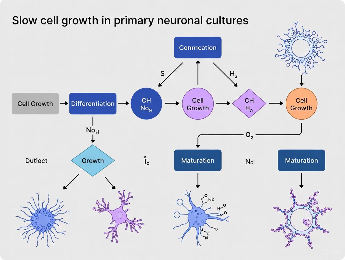
Optimizing Primary Neuronal Cultures: Strategies to Overcome Slow Growth and Enhance Model Validity
This article provides a comprehensive guide for researchers and drug development professionals tackling the pervasive challenge of slow cell growth in primary neuronal cultures.
Optimizing Primary Neuronal Cultures: Strategies to Overcome Slow Growth and Enhance Model Validity
Abstract
This article provides a comprehensive guide for researchers and drug development professionals tackling the pervasive challenge of slow cell growth in primary neuronal cultures. It synthesizes foundational knowledge on the molecular and metabolic underpinnings of neuronal maturation with robust, region-specific methodological protocols. The content explores advanced techniques such as 3D modeling and CRISPR screening to improve growth outcomes, offers systematic troubleshooting for common pitfalls like low viability and poor synaptogenesis, and establishes functional validation benchmarks using electrophysiology and other assays. By integrating foundational science with practical optimization, this resource aims to enhance the reproducibility, physiological relevance, and predictive power of in vitro neuronal models for basic research and preclinical testing.
Understanding the Roots of Slow Growth: Neuronal Energetics, Aging, and Regional Specificity
Frequently Asked Questions (FAQs)
Q1: Why is there slow growth or increased cell death in my primary neuronal cultures? Neurons rely on oxidative phosphorylation (OXPHOS) for energy and must shut off aerobic glycolysis to survive and differentiate. Using non-physiological, high-glucose culture conditions can prevent this essential metabolic transition, leading to energetic stress and cell death [1]. Constitutive expression of glycolytic enzymes like HK2 and LDHA, which occurs in high glucose, is directly toxic to neurons [1].
Q2: My neurons are surviving, but their electrical activity or synaptic function is impaired. Could the culture conditions be the cause? Yes. The weak glycolytic metabolism of neurons is not a limitation but a physiological requirement for maintaining proper mitochondrial function, redox balance, and cognitive function. Artificially boosting glycolysis in neurons through high glucose levels or genetic means leads to mitochondrial complex I disassembly, bioenergetic deficiency, and oxidative stress, which can severely compromise neuronal function [2].
Q3: How do glucose levels affect other brain cells, like astrocytes, in my co-cultures? Astrocytes respond differently to glucose availability. While neurons require low glycolysis, chronic glucose starvation can trigger pro-inflammatory responses in primary cortical astrocytes. Long-term culture in low glucose (e.g., 2 mM) can shift astrocytes toward a pro-inflammatory A1-like phenotype, which may alter the culture environment and impact neuronal health through neuroinflammatory mechanisms [3] [4].
Q4: What is a common mistake when thawing and plating primary neural cells that affects their metabolism? A common error is centrifuging cells immediately after thawing. The damage from centrifugation can be harsher for primary cells than the residual cryoprotectant (DMSO). Diluting the DMSO by following recommended seeding densities and changing the media the next day is often sufficient. Using overly harsh trypsin during subculturing can also damage cells and impair their recovery and subsequent metabolic function [5].
Troubleshooting Guide: Metabolic Issues in Neuronal Cultures
Problem: Poor Neuronal Survival and Differentiation
| Possible Cause | Evidence/Symptom | Recommended Action |
|---|---|---|
| Failure to switch from glycolysis to OXPHOS | Cell death upon differentiation; high expression of HK2 and LDHA. | Ensure culture conditions support metabolic transition; avoid constitutive high-glucose media that maintains aerobic glycolysis [1]. |
| Inappropriate glucose concentration | General poor health; metabolic stress. | Use physiological glucose levels (e.g., 2-5 mM) instead of standard high-glucose DMEM (25 mM) to mimic the brain environment [3] [2]. |
| Improper handling during thawing and plating | Low viability post-thaw; cells fail to attach. | Thaw cells quickly; do not centrifuge to remove DMSO; plate immediately at recommended densities; use pre-rinsed materials and pre-warmed medium [6]. |
Problem: Altered Neuronal Function (e.g., Electrophysiology, Signaling)
| Possible Cause | Evidence/Symptom | Recommended Action |
|---|---|---|
| Artificially enhanced neuronal glycolysis | Mitochondrial ROS stress; impaired complex I function; reduced pentose phosphate pathway flux. | Stabilize the low-glycolytic phenotype of neurons by ensuring culture conditions do not artificially boost PFKFB3 activity [2]. |
| Disruption of NAD+/NADPH homeostasis | Increased oxidative stress; reduced glutathione (GSH) levels. | Monitor redox balance; consider that forcing glycolysis consumes NAD+ and can impair the neuroprotective PPP [2]. |
Key Metabolic Pathways and Experimental Profiles
Metabolic Transition During Neuronal Differentiation
The shift from proliferating neural progenitor cells (NPCs) to post-mitotic neurons involves a fundamental metabolic reprogramming from aerobic glycolysis to oxidative phosphorylation. The following diagram outlines the key regulators and enzymes involved in this critical transition.
Survival Signaling Under Glucose Starvation
Under conditions of glucose stress, cells activate conserved signaling pathways to reprogram metabolism and ensure survival. The mTORC1-4EBP1/2 axis plays a critical role in this adaptive response.
Table: Metabolic Reprogramming During Neuronal Differentiation from NPCs to Neurons [1]
| Metabolic Enzyme / Regulator | Change in Differentiated Neurons | Functional Consequence |
|---|---|---|
| HK2 (Hexokinase 2) | Dramatically decreased | Shuts off first step of aerobic glycolysis; essential for survival. |
| LDHA (Lactate Dehydrogenase A) | Dramatically decreased | Reduces lactate production; shifts from glycolytic metabolism. |
| PKM Splicing (Pyruvate Kinase) | Shift from PKM2 to PKM1 isoform | Promotes oxidative metabolism over glycolytic flux. |
| c-MYC / N-MYC | Protein levels decrease | Removes transcriptional activation of HK2 and LDHA genes. |
| PGC-1α | Significantly increased | Sustains transcription of metabolic and mitochondrial genes. |
| ERRγ | Significantly increased | Works with PGC-1α to maintain OXPHOS capacity. |
Table: Consequences of Artificially Enhancing Neuronal Glycolysis [2]
| Parameter | Change in Glycolytic (Pfkfb3-expressing) Neurons | Impact on Neuronal Health |
|---|---|---|
| Glycolytic Flux | Increased | Disrupts normal bioenergetic balance. |
| Pentose Phosphate Pathway (PPP) Flux | Decreased | Reduces NADPH production, impairing antioxidant defense. |
| Mitochondrial ROS | Increased | Causes oxidative stress and mitochondrial damage. |
| Reduced Glutathione (GSH) | Decreased | Compromises cellular ability to neutralize ROS. |
| Mitochondrial Complex I | Disassembled | Impairs OXPHOS, leading to bioenergetic deficiency. |
| In Vivo Outcome | Cognitive decline | Ultimately compromises higher-order brain function. |
The Scientist's Toolkit: Key Research Reagents
Table: Essential Reagents for Studying Neuronal Metabolism
| Reagent / Tool | Function / Application | Example Use Case |
|---|---|---|
| APC/C-CDH1 Activity | Maintains low PFKFB3 via degradation | Preserving the natural "hypoglycolytic" state of neurons [2]. |
| 4EBP1/2 (Active) | Inhibits cap-dependent translation | Promoting cell survival during glucose starvation by reprogramming fatty acid metabolism [7]. |
| mTORC1 Inhibitors (e.g., Rapamycin, Ku-0063794) | Modulates mTORC1 signaling | Investigating the role of mTORC1 inhibition in metabolic stress response [7]. |
| B-27 Supplement | Serum-free neuronal culture supplement | Supporting long-term survival of primary neurons; ensure correct version and fresh preparation [6]. |
| Physiological Glucose Media | Culture medium with ~2-5 mM glucose | Mimicking brain interstitial fluid glucose levels to avoid non-physiological metabolic stress [3] [2]. |
| ROCK Inhibitor (Y-27632) | Reduces apoptosis in primary cells | Improving viability after thawing or plating sensitive primary neural cells [6]. |
| Poly-L-Ornithine / Coating Matrix | Substrate for cell attachment | Promoting adhesion of primary neurons and astrocytes; critical when using serum-free supplements [6] [3]. |
Troubleshooting Guides
Troubleshooting Slow Growth in Primary Neuronal Cultures
Problem: My primary neuronal cultures are exhibiting slow or stalled growth.
Possible Causes & Solutions:
| Possible Cause | Recommendation |
|---|---|
| Improper Thawing Technique | Thaw cells rapidly (less than 2 minutes at 37°C). Primary neurons are extremely fragile upon recovery; avoid centrifugation. [6] |
| Low Seeding Density | Perform a viability count prior to plating and follow the recommended seeding density. Incorrect density can heavily impact growth patterns. [6] [8] |
| Poor Cell Attachment | Ensure culture vessels are properly coated with an appropriate matrix (e.g., poly-D-lysine, laminin). Shorten the interval between removing the coating solution and adding cells to prevent drying. [6] |
| Sub-optimal Culture Medium | Use fresh, correct medium formulation. For B-27 supplemented medium, ensure it is not expired, was thawed properly, and is used within its stability period (2 weeks at 4°C). [6] |
| Static Electricity | In low-humidity environments, static can disrupt attachment of cells in plastic vessels. Wipe the outside of the vessel or use an antistatic device. [9] |
| Incubation Issues | Minimize temperature fluctuations by reducing how often the incubator is opened. Ensure humidification to prevent evaporation, which affects growth rates. [9] |
| Senescent Cell Population | This is normal in cultures from aged donors. A population of old, large cells that no longer proliferate will be present. Younger, small cells should continue to grow. [6] |
Troubleshooting Experimental Models of Neuronal Aging
Problem: My model of aged neurons is not showing expected molecular hallmarks.
Possible Causes & Solutions:
| Possible Cause | Recommendation |
|---|---|
| Inappropriate Aging Model | To retain aging hallmarks, consider using direct transdifferentiation of aged human fibroblasts into neurons, as bypassing the iPSC stage prevents the reversal of aging-associated markers. [10] |
| Failure to Verify Aging Markers | Confirm the biological age of your neuronal model. Use bisulfite sequencing of CpG methylation to estimate biological age and check for elevated expression of senescence markers like p16INK4A. [10] |
| Lack of Chronic Stress Phenotype | Aged neurons exhibit chronic cellular stress. Verify the mislocalization of splicing proteins (e.g., TDP-43, SNRNP70) from the nucleus to the cytoplasm as a key hallmark. [10] [11] |
| Inadequate Stress Challenge Test | Test neuronal resilience by applying an acute oxidative stressor (e.g., sodium arsenite). Aged neurons will show a significantly prolonged recovery time and a failure to properly form and resolve stress granules. [10] |
Frequently Asked Questions (FAQs)
Q1: Why should I consider cellular age in my neurodegeneration research, even when studying mutation-driven diseases? Aging is the most prominent risk factor for nearly all neurodegenerative diseases. Even in mutation-driven pathologies, symptom onset is typically later in life, indicating that aging processes predispose neurons to poor resiliency and exacerbate the impact of pathological mutations. [10] [11]
Q2: What is the link between TDP-43 mislocalization and aging in neurons? In aged neurons, the RNA-binding protein TDP-43 mislocalizes from the nucleus to the cytoplasm. This is an aging-specific phenomenon that leads to widespread alternative splicing defects. Since TDP-43 aggregation is implicated in >95% of ALS and >50% of Alzheimer's cases, this age-related mislocalization may be a critical initiating factor in pathology. [10] [11] [12]
Q3: My primary neurons are not attaching properly in my 96-well plate. What could be wrong? It is likely that the coating matrix dried out because the time between removing the coating solution and adding the cells was too long. The coating matrix loses its attachment ability when dry. Work with only a few wells at a time to shorten this interval. [6]
Q4: How does chronic cellular stress in aged neurons differ from an acute stress response? Aged neurons suffer from a baseline of chronic cellular stress that disrupts normal coping mechanisms. This includes malfunctioning ubiquitylation machinery and poor HSP90α chaperone activity within stress granules. Consequently, when faced with a new, acute stress event, aged neurons cannot effectively recruit splicing proteins to stress granules and fail to mount a proper protective response. [10] [11]
Q5: What are some key molecular readouts to confirm an "aged neuron" phenotype in my model? Key hallmarks to verify include:
- Mislocalized Splicing Proteins: Cytoplasmic accumulation of proteins like TDP-43, SNRNP70, and PRPF8. [10]
- Chronic Stress Signatures: Impaired stress granule resolution and depleted HSP90α from granules. [10]
- Dysregulated Splicing: Appearance of cryptic exons in known TDP-43 target genes like STMN2 and UNC13A. [10]
- Aging Markers: Elevated levels of p16INK4A and specific CpG methylation patterns. [10]
Experimental Data & Protocols
Key Quantitative Findings from Aging Neuron Research
Table 1: Proteomic Changes in Aged Human Neurons
| Measurement | Finding in Aged (Tdiff) Neurons | Significance / Associated Pathway |
|---|---|---|
| RNA-Binding Proteins (RBPs) | Broadly depleted [10] | Destabilization of RNA metabolism and splicing |
| Spliceosome Components | Most de-enriched RBP metabolic pathway [10] | Leads to widespread alternative splicing errors |
| Oxidative Phosphorylation Proteins | Highly retained / Upregulated [10] | Aged neurons may prioritize metabolic pathways over RNA homeostasis |
| TDP-43 Interactome | Enriched with spliceosome proteins; De-enriched with stress granule components (e.g., G3BP2, Caprin1) [10] | Reflects mislocalization and altered function in aging |
Table 2: Characteristics of the Neuronal Stress Response in Aging
| Characteristic | Young Neurons | Aged Neurons |
|---|---|---|
| Basal TDP-43 Localization | Nuclear [10] | Mislocalized to cytoplasm, forming foci [10] |
| Response to Acute Stress | Rapid recovery; proper stress granule formation [11] | Prolonged recovery; failure to make stress-responsive proteins [10] [11] |
| Stress Granule Composition | Normal recruitment of TDP-43 and spliceosome components [10] | Depleted of HSP90α chaperone; chronic retention [10] |
| Splicing Fidelity Under Stress | Maintained [10] | Disrupted; increased cryptic exon inclusion [10] |
Detailed Experimental Protocol: Analyzing Splicing Protein Mislocalization
Method: Immunofluorescence and quantification of nuclear vs. cytoplasmic localization in neuronal models. [10]
Cell Preparation:
- Generate aged neurons via transdifferentiation from human fibroblasts of aged healthy donors. Use iPSC-derived neurons from the same donor as an isogenic young control.
- Plate neurons on coated glass coverslips until desired maturity.
Fixation and Staining:
- Fix cells with 4% paraformaldehyde for 15 minutes at room temperature.
- Permeabilize and block with 0.1% Triton X-100 and 5% normal serum in PBS for 1 hour.
- Incubate with primary antibodies overnight at 4°C. Key targets include:
- TDP-43: A dementia- and ALS-associated protein central to the phenotype.
- SNRNP70 (U1 snRNP component): A core spliceosome protein.
- PRPF8 (tri-snRNP component): Another core spliceosome protein.
- Map2 (Microtubule-Associated Protein 2): A neuronal-specific marker to identify neuronal soma and processes.
- The next day, wash and incubate with appropriate fluorescent secondary antibodies for 1 hour at room temperature.
Imaging and Analysis:
- Image using super-resolution or high-confocal microscopy.
- Use the Map2 channel to define the neuronal soma and a marker like DAPI to define the nucleus.
- Quantify the mean fluorescence intensity of the splicing protein (e.g., TDP-43) in the nuclear and cytoplasmic compartments for each neuron.
- Calculate a nuclear-to-cytoplasmic (N/C) ratio. A significant decrease in this ratio in transdifferentiated (aged) neurons compared to iPSC-derived (young) controls indicates mislocalization.
Signaling Pathways and Workflows
Diagram Title: Molecular Pathway from Neuronal Aging to Reduced Resiliency
Diagram Title: Experimental Workflow for Studying Neuronal Aging
The Scientist's Toolkit: Research Reagent Solutions
Table 3: Essential Reagents for Investigating Aging-Associated Neuronal Decline
| Reagent / Material | Function in the Context of Neuronal Aging Research |
|---|---|
| Primary Human Fibroblasts (Aged Donors) | Starting material for transdifferentiation; provides a model that retains in vivo aging hallmarks like DNA methylation patterns. [10] |
| Neuronal Transcription Factors | Used in lentiviral vectors for the direct transdifferentiation of fibroblasts into neurons, bypassing the pluripotent state. [10] |
| B-27 Supplement | A critical serum-free supplement for the long-term health and maintenance of primary neurons. Proper handling and freshness are essential. [6] |
| Coating Matrix (e.g., Poly-D-Lysine, Laminin) | Provides a surface for neuronal attachment and neurite outgrowth, which is critical for healthy culture and preventing anoikis. [6] |
| Antibodies for Splicing Proteins (TDP-43, SNRNP70, PRPF8) | Key tools for immunofluorescence experiments to quantify the mislocalization of these proteins from the nucleus to the cytoplasm. [10] |
| Sodium Arsenite | An oxidative stressor used in acute stress tests to challenge neurons and evaluate the resilience of the stress response pathway. [10] |
| Antibodies for Stress Granule Markers (G3BP1/2, Caprin1) | Used to visualize and analyze stress granule formation, composition, and resolution in response to stress. [10] |
| HSP90α Inhibitors/Modulators | Research tools to probe the specific role of the HSP90α chaperone in stress granule dynamics and the chronic stress state. [10] [11] |
FAQs & Troubleshooting for Slow Neuronal Growth
Q1: My cortical neurons are not forming sufficient synaptic connections after 7 days in culture (DIV7). What could be wrong? A: Cortical neurons require precise neurotrophic support and substrate coating. Ensure you are using a poly-D-lysine (PDL) coating at a sufficient concentration (see Table 1). A common issue is the degradation of Brain-Derived Neurotrophic Factor (BDNF) in the culture medium. Supplement with fresh BDNF (20-50 ng/mL) every 48-72 hours.
Q2: Hippocampal neurons from my P0 pups are showing poor axonal outgrowth. How can I improve this? A: Hippocampal neurons are exquisitely dependent on the Wnt signaling pathway for axonal specification and growth. Your culture may lack essential Wnt components. Supplementing with Wnt-3a (25 ng/mL) or using a feeder layer of astrocytes can provide the necessary signaling environment. Also, verify the osmolarity of your dissection medium, as hippocampal neurons are particularly sensitive to osmotic stress.
Q3: My hindbrain (cerebellar) cultures have excessive glial proliferation, overwhelming the neurons. How can I suppress this? A: Hindbrain cultures, especially from the cerebellum, contain a high density of granule neuron precursors that require specific mitogens for proliferation, but this can also lead to glial overgrowth. Use a defined, serum-free medium to inhibit glial division. The addition of cytosine β-D-arabinofuranoside (Ara-C, 2-5 µM) at DIV 2-4 can be applied to selectively inhibit dividing glial cells without harming post-mitotic neurons.
Q4: Spinal cord motoneurons consistently show low viability after plating. What are the critical survival factors I might be missing? A: Spinal motoneurons have an absolute requirement for a combination of trophic factors that are not always present in standard neuronal media. You must provide a cocktail including:
- Glial Cell Line-Derived Neurotrophic Factor (GDNF) at 10 ng/mL
- Ciliary Neurotrophic Factor (CNTF) at 10 ng/mL
- Neurotrophin-3 (NT-3) at 10 ng/mL The absence of any one of these can lead to rapid apoptosis. Also, ensure your substrate is a combination of PDL and laminin (see Table 1).
Table 1: Optimized Coating and Media Formulations for Primary Neuronal Cultures
| Neural Region | Recommended Coating | Coating Concentration | Critical Soluble Factors & Concentrations | Optimal Seeding Density |
|---|---|---|---|---|
| Cortex | Poly-D-Lysine (PDL) | 50-100 µg/mL | BDNF (20-50 ng/mL), NT-3 (10 ng/mL) | 50,000 - 100,000 cells/cm² |
| Hippocampus | PDL | 100 µg/mL | Wnt-3a (25 ng/mL), BDNF (50 ng/mL) | 75,000 - 150,000 cells/cm² |
| Hindbrain (Cerebellar) | PDL + Laminin | PDL (50 µg/mL) + Laminin (5 µg/mL) | Sonic Hedgehog (SHH, 25 ng/mL), BDNF (25 ng/mL) | 100,000 - 200,000 cells/cm² |
| Spinal Cord | PDL + Laminin | PDL (50 µg/mL) + Laminin (10 µg/mL) | GDNF (10 ng/mL), CNTF (10 ng/mL), NT-3 (10 ng/mL) | 25,000 - 50,000 cells/cm² |
Experimental Protocols
Protocol 1: Optimized Coating Procedure for Adherence and Growth
- Prepare a sterile solution of Poly-D-Lysine (PDL) in borate buffer (pH 8.4) or sterile water at the desired concentration (see Table 1).
- Add enough PDL solution to cover the culture surface (e.g., 0.5 mL for a 24-well plate).
- Incubate at 37°C for a minimum of 1 hour or at room temperature overnight.
- Aspirate the PDL solution and rinse the surface 3 times with sterile, cell-culture grade water.
- (For laminin coating) Dilute laminin in cold, serum-free medium (e.g., Neurobasal) to the desired concentration. Add to the PDL-coated, rinsed surface.
- Incubate with laminin solution for at least 2 hours at 37°C.
- Aspirate the laminin solution immediately before plating the dissociated neurons. Do not allow the surface to dry.
Protocol 2: Trophic Factor Supplementation for Spinal Motoneuron Survival
- Following dissociation of spinal cord tissue and motoneuron enrichment (e.g., via density gradient centrifugation), resuspend the cell pellet in pre-warmed, complete motoneuron culture medium.
- Prepare a 1000X stock solution of the trophic factor cocktail (GDNF, CNTF, NT-3) in a carrier solution like PBS with 0.1% BSA.
- Add the stock solution to the cell suspension to achieve the final working concentrations (10 ng/mL each).
- Plate the cells onto the PDL/laminin-coated cultureware.
- Perform a 50% medium change every 3-4 days, replenishing the trophic factors at their full original concentration with each change.
Signaling Pathways & Workflows
Diagram: Wnt Pathway in Axonal Growth
Diagram: Primary Neuron Culture Workflow
The Scientist's Toolkit
Table 2: Essential Research Reagent Solutions
| Reagent | Function/Benefit |
|---|---|
| Poly-D-Lysine (PDL) | Synthetic polymer that provides a positively charged surface for neuronal attachment. |
| Laminin | Extracellular matrix protein critical for axon guidance and motoneuron survival; often used with PDL. |
| Neurobasal Medium | Serum-free medium optimized for long-term survival of primary neurons, minimizing glial growth. |
| B-27 Supplement | A defined serum-free supplement providing hormones, antioxidants, and proteins essential for neuronal health. |
| Brain-Derived Neurotrophic Factor (BDNF) | Key neurotrophin supporting survival and differentiation of cortical and hippocampal neurons. |
| Glial Cell Line-Derived Neurotrophic Factor (GDNF) | Critical trophic factor for the survival of midbrain dopaminergic and spinal motoneurons. |
| Cytosine β-D-arabinofuranoside (Ara-C) | Antimitotic agent used to suppress the proliferation of non-neuronal cells (e.g., glia) in culture. |
| Papain | Proteolytic enzyme used for gentle tissue dissociation to isolate viable primary neurons. |
Technical Support Center: Troubleshooting Guides and FAQs
Frequently Asked Questions (FAQs)
FAQ 1: What are the key morphological indicators of a healthy primary neuron culture? A healthy primary cortical or hippocampal neuron culture should show neurons adhering to the well surface within one hour after seeding. Within the first two days, neurons should have extended minor processes and show signs of axon outgrowth. By four days, dendritic outgrowth should be visible, and by one week, the culture should start forming a mature network. Healthy cultures can typically be maintained beyond 3 weeks [13].
FAQ 2: How can I control glial overgrowth in my primary neuronal cultures without harming the neurons? Glial overgrowth is a common challenge. Using serum-free media like Neurobasal, optimized for neurons, and supplemented with B27, helps control glial proliferation [13]. If further inhibition is necessary, cytosine arabinoside (AraC) is an established method but should be used at low concentrations and only when essential due to reported off-target neurotoxic effects [13].
FAQ 3: My hepatocytes are showing low attachment efficiency. What could be the cause? Low attachment can result from several factors [6]:
- Insufficient attachment time: Allow more time for cells to attach before proceeding.
- Dried coating substrate: Ensure the coating matrix on plates does not dry out; shorten the interval between removing the coating solution and adding cells.
- Incorrect substratum: Use appropriate coated plates (e.g., Gibco Collagen I-Coated Plates).
- Improper thawing technique: Thaw cells rapidly (<2 mins at 37°C) and use recommended thawing medium.
FAQ 4: What does the presence of large, flat cells in my culture indicate? The appearance of large, flat cells in adult keratinocyte cultures can be a normal population of senescent cells that no longer proliferate. Younger, smaller cells should continue to proliferate. As a culture ages, the proportion of these large cells increases until growth stops [6].
Troubleshooting Common Experimental Issues
Problem: Poor Early Neuron Health Post-Dissection
- Potential Cause: Cell damage during the dissection or dissociation process [13].
- Recommendations:
- Animal Age: For rat cultures, prefer embryonic stages (E17-19) over postnatal, as neurons are more resilient to shearing [13].
- Dissociation Enzyme: Consider using papain instead of trypsin, as trypsin can cause RNA degradation. For cortical neurons, mechanical trituration alone may be sufficient [13].
- Technique: Perform mechanical trituration gently and avoid bubbles to prevent shearing. Allow neurons to rest after dissociation before seeding [13].
Problem: Sub-optimal Monolayer Confluency in Hepatocyte Cultures
- Potential Causes and Recommendations [6]:
| Potential Cause | Recommendation |
|---|---|
| Seeding density too low | Check the lot-specific specification sheet for the appropriate density. |
| Insufficient cell dispersion | Disperse cells evenly by moving the plate slowly in a figure-eight and back-and-forth pattern in the incubator. |
| Low attachment efficiency | Refer to troubleshooting guides for low attachment. |
Problem: Astrocytes Failing to Adhere Properly in a 96-well Plate
- Potential Cause 1: The matrix coating the well dried out because the time between removal of the coating solution and cell addition was too long [6].
- Solution: Shorten the time interval and work with only a few wells at a time.
- Potential Cause 2: Cells formed clumps during the slow dispensing process [6].
- Solution: Resuspend the cell mixture thoroughly before dispensing into the plate.
Astrocyte Phenotypes in Neuroinflammation and Disease
Astrocytes can adopt different, context-specific functional states in response to injury or disease. Two well-characterized reactive phenotypes are A1 (neurotoxic/pro-inflammatory) and A2 (neuroprotective/anti-inflammatory) [14].
Table: Characteristics of A1 and A2 Astrocyte Phenotypes
| Feature | A1 Phenotype (Neurotoxic) | A2 Phenotype (Neuroprotective) |
|---|---|---|
| Primary Inducers | Activated microglia releasing IL-1α, TNF-α, and C1q [14]. | Not specified in search results, but generally associated with anti-inflammatory signals. |
| Key Markers | Not specified. | S100A10 protein [14]. |
| General Function | Harmful; releases soluble toxins that destroy neurons and oligodendrocytes, induces synaptic dysfunction [14]. | Beneficial; upregulates neurotrophic factors and anti-inflammatory cytokines, supports neuronal survival and synaptogenesis [14]. |
| Role in Disease | Contributes to neurodegeneration in diseases like Alzheimer's, Parkinson's, and following spinal cord injury [14]. | Promotes repair and limits inflammation; its induction is an early adaptive response to acute neurotoxic injury [14]. |
Experimental Protocol: Primary Astrocyte Isolation and Culture from Rat Cerebral Cortex
This protocol is adapted for researching astrocyte functions and their interactions with neurons [15].
1. Specimen Sampling (Using 1-3 day old Sprague-Dawley rats)
- Decapitate the pup and make a midline incision in the skull to remove the intact brain. Place it in pre-cooled PBS.
- Use micro-scissors and micro-forceps to dissect the cerebral cortex.
- Under a dissecting microscope, carefully peel off the meninges from the cortical tissue (or roll the tissue on sterile filter paper to remove them).
- Collect the processed tissue and cut it into 1-3 mm³ pieces.
2. Tissue Processing and Digestion
- Add 3-5 times the tissue volume of a digestion enzyme. Incubate at 37°C with shaking in a water bath for 10 minutes.
- Add FBS to stop the digestion. Pipette repeatedly until the tissue is dispersed.
- Filter the cell suspension sequentially through 100-mesh and 200-mesh cell strainers to remove large tissue fragments and microvascular tissue.
- Collect the filtrate and centrifuge at 1,200 rpm for 5 minutes to obtain a cell pellet.
- Resuspend the cell pellet in a complete medium and seed it in T25 flasks at a density of 5-10×10⁵ cells per flask.
3. Cell Isolation and Purification
- The initial culture is a mixed population (approx. 48% astrocytes, 11% microglia, 40% neurons) [15].
- After 24 hours, replace the medium to remove some neuronal cells.
- Purification Method 1 (Passaging): Digest and passage the cells. Microglia are difficult to digest and do not proliferate well, so multiple passages will enrich for astrocytes [15].
- Purification Method 2 (Shaking): Seal the culture flask and place it on a shaker at 200 rpm for 48 hours. This removes floating microglia, leaving behind adherent, high-purity astrocytes [15].
4. Cell Identification
- Identify astrocytes via immunofluorescence staining for the characteristic marker GFAP (Glial Fibrillary Acidic Protein). A purity of up to 90% can be achieved [15].
- Cultured astrocytes should exhibit elongated processes with extensive branching, small cell bodies, and a star-shaped structure forming a network [15].
The Scientist's Toolkit: Key Research Reagent Solutions
Table: Essential Reagents for Astrocyte and Primary Neuron Research
| Item | Function / Application | Key Notes |
|---|---|---|
| Neurobasal Medium | A serum-free medium optimized for the long-term culture of primary neurons, supports neuronal health while minimizing glial growth [13]. | Should be used with B27 supplement. |
| B27 Supplement | A serum-free supplement providing hormones, nutrients, and antioxidants crucial for neuronal survival and health [13]. | Check expiration date. Supplemented medium is stable for only 2 weeks at 4°C [6]. |
| Poly-D-Lysine (PDL) | A positively charged polymer used to coat culture surfaces, enabling the adhesion of primary neurons and astrocytes [13]. | More resistant to enzymatic degradation than Poly-L-Lysine (PLL) [13]. |
| Cytosine Arabinoside (AraC) | A chemical used to inhibit the proliferation of glial cells in primary neuronal cultures, helping to maintain neuronal purity [13]. | Use at low concentrations and only when necessary due to potential neurotoxic effects [13]. |
| GFAP Antibody | Glial Fibrillary Acidic Protein antibody; used in immunofluorescence to identify and assess the purity and activation status of astrocytes [15]. | Expression level is positively correlated with astrocyte activation [15]. |
| Papain | A protease enzyme used as a gentler alternative to trypsin for dissociating neural tissue during primary cell isolation, helping to preserve cell health [13]. | Can help reduce RNA degradation and improve early neuron health compared to trypsin [13]. |
| GLAST / GLT-1 Transporters | Functional markers for astrocytes; these glutamate transporters are primarily responsible for clearing glutamate from the synaptic cleft, preventing excitotoxicity [15]. | Indispensable for studying astrocyte role in neuronal metabolism and synaptic regulation [15]. |
Proven Protocols for Enhanced Neuronal Viability, Yield, and Maturation
This technical support center is designed to assist researchers in overcoming the prevalent challenge of slow cell growth and poor viability in primary neuronal cultures. Within the context of optimizing neuronal yield and health for drug discovery and basic research, the following troubleshooting guides and detailed protocols address common pitfalls encountered during the dissociation and culture of cortex, hippocampus, and hindbrain neurons.
Troubleshooting Guides & FAQs
Q1: My neuronal yield is consistently low after tissue dissociation. What are the primary causes? A: Low yield typically stems from issues during the enzymatic and mechanical dissociation steps. The most common factors are summarized below.
| Factor | Optimal Condition/Value | Common Pitfall | Impact on Yield |
|---|---|---|---|
| Enzyme Concentration | 15-20 U/mL Papain | Excessive concentration (>25 U/mL) | Induces proteolytic damage to surface receptors, reducing viability. |
| Digestion Time | 20-30 mins at 37°C | Prolonged time (>45 mins) | Leads to oxidative stress and apoptosis. |
| Trituration Pipette Bore | Fire-polished, ~1.0-1.5 mm | Using a narrow, unpolished tip | Causes excessive shear stress and physical cell lysis. |
| DNase Concentration | 50-100 µg/mL | Omitting DNase | Viscous DNA from lysed cells traps live cells, reducing recovery. |
Q2: I observe significant glial contamination in my cultures after 5-7 days. How can I suppress this? A: Glial proliferation can outcompete and hinder neuronal network development. The key is to use antimitotics at the correct developmental window.
| Reagent | Recommended Concentration & Timing | Function | Note |
|---|---|---|---|
| Cytosine β-D-arabinofuranoside (Ara-C) | 1-5 µM, added at DIV 3-4 | Inhibits DNA synthesis, selectively killing dividing glial cells. | Adding at plating (DIV 0) can be toxic to neurons. |
| 5-Fluoro-2'-deoxyuridine (FDU) | 10-20 µM, added at DIV 3-4 | Similar mechanism to Ara-C; inhibits thymidylate synthase. | Often used in combination with uridine. |
Q3: My hippocampal neurons are not forming robust synaptic connections. What culture conditions are critical? A: Synaptogenesis requires precise neurotrophic support and a permissive substrate. Key parameters are quantified below.
| Parameter | Cortex | Hippocampus | Hindbrain |
|---|---|---|---|
| Optimal Seeding Density | 150-200k cells/cm² | 75-100k cells/cm² | 50-75k cells/cm² |
| Critical Neurotrophic Factor | BDNF (25-50 ng/mL) | BDNF (25-50 ng/mL) | GDNF (10-25 ng/mL) |
| Poly-D-Lysine Coating | 50-100 µg/mL | 100 µg/mL | 50 µg/mL |
Experimental Protocols
Protocol 1: Optimized Enzymatic Dissociation for High Viability
- Dissection & Collection: Rapidly dissect brain regions in ice-cold, calcium-free HBSS supplemented with 10 mM HEPES and 1 mM kynurenic acid (to block excitotoxicity).
- Enzymatic Digestion: Incubate tissue pieces in 15-20 U/mL papain solution, dissolved in HBSS-HEPES, for 20-30 minutes at 37°C in a humidified CO₂ incubator. Gently agitate every 10 minutes.
- Enzyme Quenching: Carefully remove the papain solution and replace with a quenching medium (Neurobasal-A containing 10% FBS and 1% BSA).
- Mechanical Trituration: Gently triturate the tissue 10-15 times using a fire-polished Pasteur pipette of decreasing bore sizes. Allow large pieces to settle for 2-3 minutes between triturations.
- Cell Collection & Plating: Pool the supernatant containing dissociated cells. Centrifuge at 150-200 x g for 5 minutes. Resuspend the pellet in complete neuronal culture medium (e.g., Neurobasal-A + B-27 + GlutaMAX) and plate on pre-coated surfaces at the densities specified in the table above.
Protocol 2: Glial Suppression Protocol
- At Day In Vitro (DIV) 3-4, perform a half-medium change with fresh, pre-warmed neuronal culture medium.
- Add a stock solution of Ara-C directly to the culture medium to achieve a final working concentration of 2.5 µM.
- Return cultures to the incubator for 48-72 hours.
- Perform a full medium change with Ara-C-free neuronal culture medium to remove the antimitotic and any cellular debris.
Visualizations
Diagram 1: Neuron Culture Workflow
Diagram 2: Key Signaling for Neuronal Health
The Scientist's Toolkit
| Research Reagent | Function & Explanation |
|---|---|
| Papain | Proteolytic enzyme used to gently break down the extracellular matrix, freeing individual neurons from the tissue with minimal damage. |
| Neurobasal-A Medium | A specially formulated, serum-free medium designed to support the long-term survival of primary neurons while minimizing glial growth. |
| B-27 Supplement | A defined, serum-free supplement containing hormones, antioxidants, and proteins essential for neuronal survival and growth. |
| Poly-D-Lysine | A synthetic polymer that coats culture surfaces, providing a positively charged substrate that enhances neuronal attachment. |
| Cytosine β-D-arabinofuranoside (Ara-C) | An antimitotic agent used to selectively inhibit the proliferation of non-neuronal cells (e.g., astrocytes, microglia) in culture. |
| Kynurenic Acid | A broad-spectrum glutamate receptor antagonist added during dissection and dissociation to protect neurons from excitotoxicity-induced death. |
Troubleshooting Guides & FAQs
Q1: My primary neuronal cultures are exhibiting slow growth and poor neurite outgrowth despite using B-27. What could be the cause? A1: This is often due to non-physiological glucose levels. Standard media (e.g., DMEM) contains 25 mM glucose, while the brain's extracellular fluid is ~2.5-5 mM. Hyperglycemic conditions induce oxidative stress, impairing neuronal development. Transition to a basal medium like Neurobasal-A and optimize glucose to a physiological range (3-5 mM). Ensure B-27 is freshly added and has not undergone multiple freeze-thaw cycles.
Q2: How do I choose between B-27 and CultureOne supplements? A2: The choice depends on your specific neuronal population and research goals. B-27 is a widely used, robust supplement for general neuronal health and synapse formation. CultureOne is a more defined alternative that can reduce batch-to-batch variability. For a direct comparison, refer to Table 1.
Q3: What is the recommended protocol for testing glucose and supplement combinations? A3: Follow the experimental workflow below to systematically test conditions and identify the optimal media formulation for your specific culture system.
Q4: My cells are dying after media change. Is my supplement mixture toxic? A4: Toxicity can occur from improper supplement preparation or using degraded antioxidants in the supplements. Always thaw supplements quickly at 37°C, aliquot to avoid repeated freeze-thaws, and pre-warm media to 37°C before adding to cells. Ensure your glucose stock solution is sterile and at the correct pH.
Experimental Protocol: Media Optimization for Primary Neuronal Cultures
Objective: To determine the optimal combination of physiological glucose and critical supplements (B-27, CultureOne) for enhancing cell viability and neurite outgrowth in primary cortical neurons.
Materials:
- Primary cortical neurons (E18 rat)
- Neurobasal-A Base Medium
- B-27 Supplement (50X)
- CultureOne Supplement (100X)
- D-Glucose solution
- Phosphate Buffered Saline (PBS)
- Poly-D-Lysine coated plates
Method:
- Prepare Media Formulations: Create the following media variants in Neurobasal-A, sterilizing by filtration (0.22 µm).
- Condition A: 5 mM Glucose + 1X B-27
- Condition B: 25 mM Glucose + 1X B-27
- Condition C: 5 mM Glucose + 1X CultureOne
- Condition D: 25 mM Glucose + 1X CultureOne
- Condition E (Control): 25 mM Glucose (No supplement)
Cell Plating: Plate dissociated cortical neurons at a density of 50,000 cells/cm² in poly-D-lysine coated 24-well plates. Culture all wells initially in a standard maintenance medium for 4 hours to allow attachment.
Media Application: After the 4-hour attachment period, carefully aspirate the initial medium and replace it with the 500 µL of the respective test media (Conditions A-E).
Maintenance: Maintain cultures at 37°C and 5% CO₂. Perform a 50% media exchange with the respective fresh test media every 3 days.
Analysis (Day 7 In Vitro):
- Viability Assay: Use a Calcein-AM/EthD-1 live/dead assay kit. Calculate the percentage of viable cells from 5 random fields per well.
- Neurite Outgrowth: Fix cells and immunostain for β-III-Tubulin. Acquire images and quantify the average neurite length per neuron using automated image analysis software (e.g., ImageJ with NeuriteTracer plugin).
Data Presentation
Table 1: Comparison of Media Supplement Formulations
| Feature | B-27 Supplement | CultureOne Supplement |
|---|---|---|
| Composition | Complex, serum-free formulation with antioxidants, hormones, and proteins. | Defined, concentrated mixture of essential components including transferrin, insulin, and lipids. |
| Primary Use | Long-term survival and growth of primary neurons; supports synapse formation. | General supplement for primary cells; can reduce batch variability in neuronal cultures. |
| Key Advantage | Extensive validation in neuroscience research; robust performance. | High definition reduces unknown variables; potential for more consistent results. |
| Consideration | Potential batch-to-batch variability due to complex composition. | May require culture-specific optimization as it is less specialized for neurons. |
Table 2: Quantitative Outcomes of Media Optimization (Representative Data)
| Culture Condition | Cell Viability (%) | Average Neurite Length (µm) |
|---|---|---|
| A: 5 mM Glu + B-27 | 92.5 ± 3.1 | 145.2 ± 12.8 |
| B: 25 mM Glu + B-27 | 78.3 ± 5.6 | 98.7 ± 10.4 |
| C: 5 mM Glu + CultureOne | 88.1 ± 4.2 | 120.5 ± 11.9 |
| D: 25 mM Glu + CultureOne | 75.8 ± 6.0 | 85.3 ± 9.7 |
| E: 25 mM Glu (No Suppl.) | 45.2 ± 8.4 | 35.6 ± 8.1 |
Visualizations
Media Optimization Workflow
Glucose & Supplement Impact on Growth
The Scientist's Toolkit
| Research Reagent | Function & Rationale |
|---|---|
| Neurobasal-A Medium | A optimized basal medium designed specifically for the long-term survival of primary neurons, with minimal glial growth. |
| B-27 Supplement | A serum-free supplement providing hormones, antioxidants, and proteins crucial for neuronal health and reducing oxidative stress. |
| CultureOne Supplement | A concentrated, defined supplement mix intended to support a variety of primary cells, offering an alternative to reduce batch variability. |
| D-Glucose | The primary energy source for neurons. Using a physiologically relevant concentration (3-5 mM) is critical to mimic the brain microenvironment. |
| Poly-D-Lysine | A synthetic polymer used to coat culture surfaces, providing a positive charge that enhances the attachment of neuronal cells. |
Technical Support Center
Troubleshooting Guide & FAQ
This support center addresses common challenges in establishing and maintaining advanced 3D neural co-cultures, framed within the thesis of overcoming the limitations of slow growth and functionality in primary neuronal cultures.
Section 1: General Setup & Culture Health
Q: My 3D neural co-culture shows poor cell viability after the first week. What could be the cause?
- A: Poor initial viability is often linked to suboptimal hydrogel encapsulation or nutrient diffusion issues. Ensure your hydrogel (e.g., Matrigel) is properly thawed and mixed with cells on ice to prevent premature polymerization. After plating, confirm that the gel has fully set before adding culture medium. Inadequate gas exchange can also be a factor; ensure your culture vessels allow for sufficient CO2 diffusion and avoid overfilling wells with medium.
Q: The neurite outgrowth in my 3D model is less extensive than expected. How can I improve this?
- A: Suboptimal neurite outgrowth can stem from several factors. First, verify the stiffness of your hydrogel matrix; a modulus between 0.5-1 kPa is often ideal for neurons. Second, ensure your differentiation and maturation protocol includes a full complement of neurotrophic factors. Refer to Table 1 for recommended concentrations. Finally, confirm the health and purity of your initial iPSC-derived neural progenitor cells (NPCs), as low-quality starters will compromise downstream complexity.
Section 2: Co-culture System & Functional Maturation
Q: My astrocytes in the co-culture system are not adopting a mature, stellate morphology. What should I check?
- A: Immature astrocyte morphology is frequently due to insufficient maturation time or the absence of key soluble factors. After co-culture initiation, a maturation period of 4-6 weeks is typically required. The inclusion of CNTF (Ciliary Neurotrophic Factor) and db-cAMP in the maturation medium is crucial for driving astrocytic maturity. See the Experimental Protocol for detailed steps.
Q: How can I confirm functional neuronal activity and synaptic connectivity in my 3D model?
- A: Functional maturity is best confirmed through a combination of assays.
- Calcium Imaging: Use fluorescent dyes (e.g., Fluo-4 AM) to detect spontaneous calcium oscillations, indicating active neuronal firing.
- Multi-Electrode Array (MEA): Record extracellular field potentials to demonstrate network-level bursting and synchronized activity.
- Immunocytochemistry: Stain for pre-synaptic (e.g., Synapsin-1) and post-synaptic (e.g., PSD-95) markers to visualize physical synapses.
- A: Functional maturity is best confirmed through a combination of assays.
Section 3: Data Reproducibility & Assay Troubleshooting
- Q: I am observing high variability in my functional assay readouts (e.g., MEA spikes) between different culture batches. How can I improve reproducibility?
- A: Batch-to-batch variability is a common challenge. Standardize your starting population by using a consistent and well-characterized iPSC line. Implement rigorous quality control for NPCs, ensuring a high percentage (>90%) express Pax6 or Sox1. Furthermore, maintain strict consistency in hydrogel preparation and cell seeding density across all batches. Table 2 provides quantitative benchmarks for key maturation markers to help you assess your culture's progress.
Experimental Protocols
Protocol 1: Generation of 3D Human iPSC-Derived Neural Co-cultures [citation:3, citation:9]
Objective: To establish a physiologically relevant 3D neural model containing neurons and astrocytes derived from human iPSCs.
Materials:
- Human iPSC-derived Neural Progenitor Cells (NPCs)
- Matrigel, Growth Factor Reduced (or similar hydrogel)
- Neural Basal Medium
- Neurotrophic Factor Cocktail (BDNF, GDNF, NT-3)
- Astrocyte Maturation Factors (CNTF, db-cAMP)
- Low-adhesion 96-well U-bottom plates
Methodology:
- Preparation: Thaw Matrigel on ice overnight. Pre-cool pipette tips and tubes.
- Cell Harvest: Gently dissociate your NPC cultures into a single-cell suspension. Count and centrifuge cells.
- Encapsulation: Resuspend the NPC pellet in ice-cold Matrigel at a density of 10-15 million cells/mL. Gently mix to avoid air bubbles.
- Droplet Formation: Pipette 20-30 µL droplets of the cell-Matrigel suspension into the wells of the low-adhesion plate.
- Polymerization: Incubate the plate at 37°C for 30 minutes to allow the hydrogel droplets to fully polymerize.
- Feeding: Carefully overlay each droplet with pre-warmed Neural Basal Medium supplemented with neurotrophic factors (see Table 1).
- Maturation: Culture the 3D constructs for 4-6 weeks, with half-medium changes every 2-3 days. After 2 weeks, add astrocyte maturation factors (CNTF, db-cAMP) to the medium to promote glial differentiation and co-culture establishment.
Protocol 2: Functional Validation via Calcium Imaging
Objective: To assess spontaneous neuronal activity in 3D neural co-cultures.
Materials:
- Mature 3D neural co-cultures
- Calcium-sensitive dye (e.g., Fluo-4 AM)
- Live-cell imaging compatible HEPES-buffered solution
- Confocal or spinning-disk microscope with environmental chamber
Methodology:
- Dye Loading: Incubate 3D cultures in a solution containing 2-5 µM Fluo-4 AM for 45-60 minutes at 37°C.
- Wash & Recovery: Replace the dye solution with fresh, pre-warmed imaging buffer. Allow a 15-20 minute recovery period for de-esterification of the dye.
- Image Acquisition: Mount the culture on a microscope stage maintained at 37°C. Acquire time-lapse images (e.g., 2-4 frames per second) for 5-10 minutes.
- Analysis: Use image analysis software (e.g., ImageJ/FIJI with plug-ins) to identify active neurons and quantify fluorescence intensity (ΔF/F0) over time to detect calcium transients.
Quantitative Data Summary
Table 1: Key Neurotrophic Factors for 3D Neural Co-culture Maturation
| Factor | Function | Recommended Concentration | Reference |
|---|---|---|---|
| BDNF | Promotes neuronal survival, differentiation, and synaptic plasticity. | 20 ng/mL | |
| GDNF | Supports survival of dopaminergic and other neuronal subtypes. | 10 ng/mL | |
| NT-3 | Encourages differentiation and outgrowth of specific neuronal populations. | 10 ng/mL | |
| CNTF | Drives astrocyte maturation in co-culture systems. | 10 ng/mL |
Table 2: Benchmarking Maturation Markers in 3D vs. 2D Cultures
| Parameter | 2D Monoculture (Neurons) | 3D Co-culture (Neurons & Astrocytes) | Measurement Timepoint |
|---|---|---|---|
| Synapsin-1 Puncta Density | ~150 puncta/100 µm² | ~400 puncta/100 µm² | Day 40-50 |
| Mean Network Burst Rate (MEA) | 0.5 ± 0.2 bursts/min | 2.1 ± 0.5 bursts/min | Day 50-60 |
| % Cells with Ca²⁺ Transients | 25% ± 5% | 65% ± 8% | Day 45 |
Visualizations
Title: 3D Neural Co-culture Workflow
Title: Neurotrophic Signaling Pathway
The Scientist's Toolkit
Table 3: Essential Research Reagents for 3D Neural Co-cultures
| Item | Function | Example |
|---|---|---|
| Synthetic Hydrogel | Provides a defined, xeno-free 3D scaffold with tunable mechanical properties. | PEG-based hydrogels |
| Natural Hydrogel | Provides a biologically active, basement membrane-like environment for cell growth. | Matrigel, Collagen I |
| Neurotrophic Factors | Cocktail of proteins essential for neuronal survival, differentiation, and synapse formation. | BDNF, GDNF, NT-3 |
| Astrocyte Maturation Factors | Compounds that drive the differentiation of NPCs into mature, functional astrocytes. | CNTF, db-cAMP |
| Calcium-Sensitive Dyes | Cell-permeable fluorescent dyes used to visualize and quantify neuronal activity. | Fluo-4 AM, Cal-520 AM |
| Low-Adhesion Plates | Prevents 3D constructs from adhering to the plate, maintaining their spherical structure. | U-bottom spheroid plates |
Troubleshooting Guides
Low Transfection Efficiency in Primary Neurons
Problem: Low delivery efficiency of CRISPR/Cas9 components into primary neuronal cultures.
Potential Causes and Solutions:
| Cause | Solution | Reference |
|---|---|---|
| Suboptimal electroporation parameters | Use high-voltage, low-capacitance settings (e.g., 500 V, 50 µF) to target granule cells, or lower voltage, higher capacitance (e.g., 220 V, 975 µF) for astrocytes. Optimize parameters for your specific neuronal subtype. | [16] |
| Low cell viability post-transfection | Use specialized nucleofection systems (e.g., Amaxa P3 Primary Cell 4D-Nucleofector) with neuron-specific kits. Ensure proper handling: fast thawing, gentle trituration with wide-bore tips, and no centrifugation of extremely fragile neurons. | [6] [17] |
| Inefficient delivery method | Choose electroporation over chemical transfection for pre-seeding genetic manipulation. Electroporation allows for higher throughput and is more suitable for screening applications. | [18] [17] |
Irregular or Incomplete Protein Knockout
Problem: Persistent protein expression detected after CRISPR-mediated knockout attempts in neurons.
Potential Causes and Solutions:
| Cause | Solution | Reference |
|---|---|---|
| Slow protein turnover in post-mitotic cells | Allow sufficient time for protein depletion. Knockdown may lag >4 days post-transfection. Plan experimental endpoints accordingly, considering the half-life of the target protein. | [18] |
| Inefficient sgRNA design | Design 3-4 sgRNAs per gene to mitigate performance variability. Use bioinformatic tools (e.g., Benchling) to select sgRNAs with high on-target scores. Target exons common to all prominent protein isoforms, especially early exons. | [19] [20] [18] |
| Alternative protein isoforms | Design sgRNAs to target an exon present in all major isoforms of the transcript to prevent expression of truncated or alternative proteins. | [19] |
Poor Neuronal Health and Viability After Transfection
Problem: Neurons show poor viability, unhealthy morphology, or slow growth after electroporation and CRISPR editing.
Potential Causes and Solutions:
| Cause | Solution | Reference |
|---|---|---|
| Incorrect culture conditions | Use specialized neuronal medium (e.g., Neurobasal plus) with appropriate supplements (B-27, GlutaMAX). Ensure freshness of B-27 supplement, as it is stable for only 2 weeks at 4°C. Avoid improper thawing/refreezing. | [6] [21] |
| Improper substrate coating | Ensure culture vessels are properly coated with poly-L-lysine or other adhesion matrices. Do not allow the coating solution to dry out before adding cells, as this drastically reduces attachment. | [6] [17] |
| Excessive cellular stress | For complex phenotypic analyses like neurite outgrowth, consider a replating step at 2 Days In Vitro (DIV). This resets neuronal morphology and allows for clearer observation of CRISPR-induced growth phenotypes. | [18] [17] |
High Variability in Screening Results
Problem: High batch-to-batch variability or inconsistent results in high-content screening data.
Potential Causes and Solutions:
| Cause | Solution | Reference |
|---|---|---|
| Batch-to-batch variation in primary cells | Characterize each batch of isolated primary neurons. Source tissue from animals of consistent age, gender, and species to minimize biological variability. | [22] |
| Insufficient sgRNA library coverage | In pooled screens, ensure deep sequencing coverage (recommended depth of at least 200x per sample) to reliably detect changes in sgRNA abundance. | [20] |
| Inconsistent selection pressure | If no significant gene enrichment is found, it may be due to insufficient selection pressure. Optimize and standardize the selection conditions (e.g., drug concentration, duration of treatment) applied during the screen. | [20] |
Frequently Asked Questions (FAQs)
CRISPR Design and Validation
Q1: Why do different sgRNAs targeting the same gene show variable performance in my neuronal screen? The editing efficiency of each sgRNA is influenced by its intrinsic sequence properties and chromatin accessibility. It is recommended to design and use 3-4 sgRNAs per gene to ensure robust and reliable knockout, mitigating the impact of poor performance from any single sgRNA [20].
Q2: How can I confirm successful protein knockout in primary neurons? Validation should occur at multiple levels:
- Genomic Level: Use Sanger sequencing of the target region and analyze the results with tools like ICE (Inference of CRISPR Edits) to confirm the presence of insertion/deletion mutations [19].
- Protein Level: Perform immunocytochemistry or western blotting to assess protein depletion. Allow sufficient time (often 4-6 days) for existing protein to turnover post-transfection [19] [18].
Q3: What is a key consideration when designing sgRNAs for knockout studies? Beware of alternative splicing and multiple protein isoforms. Your sgRNA should be designed to target an exon that is present in all prominent isoforms of your target gene. This prevents the persistent expression of a functional, truncated protein that could confound your results [19].
Experimental Workflow and Timing
Q4: How long does it take for CRISPR to knock out a protein in primary neurons? Protein knockdown in primary neurons is not immediate. One study showed that targeted proteins became undetectable by immunohistochemistry in over 80% of transfected cells, but this effect lagged at least four days behind transfection. The exact timing depends on the half-life of the specific target protein [18].
Q5: How can I study early neurite outgrowth phenotypes if protein knockout is slow? A combination of electroporation and replating is an effective strategy. Neurons are transfected before initial seeding and then replated at 2 Days In Vitro (DIV). This resets the neurons into an undifferentiated stage, allowing you to observe the full course of differentiation and outgrowth after the knockout has taken effect [18] [17].
Q6: My CRISPR screen did not show significant gene enrichment. What could be wrong? The absence of significant hits is often due to insufficient selection pressure rather than statistical errors. Try increasing the selection pressure (e.g., higher drug concentration) or extending the duration of the screen to allow for clearer enrichment or depletion of sgRNAs [20].
Data Analysis
Q7: How should I prioritize candidate genes from my CRISPR screen data? Two common approaches are:
- RRA Score Ranking: The Robust Rank Aggregation (RRA) algorithm provides a composite score and rank. Genes with higher ranks are more likely to be true hits.
- LFC and p-value: Combining log-fold change (LFC) and p-value thresholds allows for explicit cutoffs but may include more false positives. It is generally recommended to prioritize using the RRA rank-based method [20].
Q8: What are the most commonly used tools for analyzing CRISPR screen data? MAGeCK (Model-based Analysis of Genome-wide CRISPR-Cas9 Knockout) is one of the most widely used tools. It incorporates both the RRA algorithm for single-condition comparisons and the MLE (Maximum Likelihood Estimation) algorithm for modeling multi-condition experiments [20].
Experimental Protocols
Key Workflow for CRISPR Screening in Primary Neurons
The diagram below illustrates the core workflow for conducting a high-content CRISPR screen in primary neurons, integrating electroporation and a replating step to study phenotypes like neurite outgrowth.
Detailed Protocol: Electroporation and Replating of Primary Hippocampal Neurons
This protocol is adapted from established methodologies for genetic manipulation and subsequent phenotypic analysis of primary neurons [18] [17].
1. Neuron Isolation and Electroporation
- Dissection: Isolate hippocampi from embryonic (E18.5) mice. Remove meninges carefully to reduce non-neuronal cell contamination [17] [21].
- Dissociation: Incubate tissue in pre-warmed TrypLE Express or papain (e.g., 6 min at 37°C). Triturate gently in Hibernate E medium supplemented with B-27 and GlutaMAX to create a single-cell suspension [18] [17].
- Electroporation: Use a nucleofector system (e.g., Lonza 4D-Nucleofector with P3 Primary Cell Kit). Transfect 250,000 neurons with 3 µg of plasmid DNA (e.g., pX330 vector expressing both Cas9 and your sgRNA). Plate transfected neurons in nucleofection medium and replace with fresh Neurobasal Plus complete medium after 5 hours [17].
2. Replating for Morphological Analysis (at DIV2)
- Prepare Coated Plates: Coat cover slips with 0.1 mg/mL poly-L-lysine overnight.
- Detach Neurons: Collect and save 350 µL of conditioned medium per well. Aspirate the remaining medium and add 500 µL prewarmed TrypLE Express per well. Incubate for 15 min in a humidified incubator at 37°C.
- Resuspend and Replate: Gently rinse the well bottom with the TrypLE solution to detach neurons. Add 500 µL prewarmed NB+ medium to stop the reaction. Transfer the cell suspension to a tube and centrifuge at 7000 rpm for 5 min. Resuspend the pellet in 500 µL conditioned medium and plate onto the prepared cover slips [17].
3. Phenotyping and Data Collection
- After replating, neurons will re-extend neurites. Fix and immunostain cells at desired time points (e.g., 1-2 days post-replating).
- Use automated high-content imaging systems to capture neuronal morphology.
- Quantify phenotypes such as neurite length, branching, or soma size using appropriate software.
The Scientist's Toolkit: Essential Reagents and Materials
The following table lists key reagents and materials crucial for successfully performing CRISPR screening in primary neurons.
| Item | Function/Application | Example/Specification |
|---|---|---|
| pX330 Plasmid | All-in-one vector expressing both S. pyogenes Cas9 and a custom sgRNA. | Addgene #42230 [18] |
| Neurobasal Plus Medium | A optimized medium formulation for supporting the long-term survival and health of primary neurons. | Often supplemented with B-27 and GlutaMAX [6] [21] |
| B-27 Supplement | A serum-free supplement essential for neuron survival and growth. Critical note: Prepared medium is stable for 2 weeks at 4°C. | [6] |
| Poly-L-Lysine | A substrate coating used to promote neuronal attachment to culture vessels. | Typical coating concentration: 0.1 mg/mL [17] |
| TrypLE Express | An animal-origin-free enzyme for gentle dissociation of neural tissue and for detaching neurons during replating. | [17] |
| 4D-Nucleofector System | An electroporation device designed for high-efficiency transfection of sensitive primary cells, including neurons. | With X kit L [17] |
| MAGeCK Software | A widely used bioinformatic tool for the statistical analysis of genome-wide CRISPR screen data. | [20] |
Solving Common Pitfalls: A Systematic Guide to Improving Neuronal Health and Growth Kinetics
Troubleshooting Guides
FAQ: Primary Neuron Viability and Adhesion
1. My primary neurons are detaching during long-term culture. What can I do?
Poor long-term adhesion, particularly on glass surfaces used for imaging and electrophysiology, is a common challenge. This is often due to insufficient surface charge or inappropriate extracellular matrix (ECM) coating.
- Solution: Implement a charged amine-based plasma polymer coating, such as Diaminopropane (DAP), on your glass surfaces. This positive-charged treatment significantly enhances long-term cell adherence [23].
- Protocol: After applying the DAP coating, add a laminin-based coating on top. The combination of DAP and laminin has been shown to optimally support the maturation of fundamental ion channel properties and synaptic activity in human neurons over extended periods [23].
- Additional Tip: Ensure you are using pre-warmed, complete growth medium and the correct seeding density. Avoid centrifuging primary neurons after thawing, as they are extremely fragile [6].
2. I am switching to a serum-free, chemically-defined medium, and my cells are not attaching well. How can I improve this?
The transition from serum-containing to serum-free (chemically-defined) medium can disrupt adhesion because serum contains undefined attachment factors. A systematic adaptation protocol and defined attachment substrates are key [24].
- Solution: Use a gradual adaptation (GA) method instead of direct adaptation. This involves incrementally increasing the proportion of CD medium while decreasing the serum-containing medium over several passages to minimize cellular stress [24].
- Coating Requirement: In CD medium, you must use a coating matrix, as there are no attachment factors in the supplements. Fibronectin has been shown to substantially improve cell attachment and viability during CD medium adaptation, outperforming laminin and collagen IV in some cell types [6] [24].
- Protocol: Coat your culture vessels with a defined ECM protein like fibronectin before seeding cells. Follow a decision-flow procedure to objectively monitor cell health and confluence during the adaptation process [24].
3. My neural cell culture has low viability after thawing. What are the critical steps I might be missing?
Primary neural cells are very fragile and require careful handling during recovery from cryopreservation.
- Thawing Technique: Thaw cells rapidly (less than 2 minutes) in a 37°C water bath. Do not expose cells to air during the thawing process [6].
- Handling: After thawing, transfer cells to a pre-rinsed tube and slowly add pre-warmed complete growth medium in a drop-wise manner. Adding the full volume of medium at once can cause osmotic shock, decreasing viability. Use wide-bore pipette tips to avoid rough handling [6].
- Centrifugation: For primary neurons, do not centrifuge the cells after thawing, as they are extremely fragile. For other cell types like hepatocytes, ensure you use the correct centrifugation speed and time (e.g., 100 x g for 10 minutes for human hepatocytes) [6].
4. I see inconsistent results between batches of my primary brain cell isolations. How can I improve consistency?
Batch-to-batch variation is a recognized challenge with primary cell isolations, often due to phenotypic differences between tissue sources [22].
- Characterization: Perform a phenotypic characterization of each batch to identify and account for inconsistencies [22].
- Standardized Coating:DAP-treated glass has been shown to reduce technical variability of human neuronal models, leading to more reliable and reproducible experimental outcomes [23].
- Consider Source Factors: Age, gender, and species of the tissue source can significantly affect cell characteristics. It is recommended to use a proper sample size determined by power analysis and to account for these factors in experimental design [22].
Table 1: Comparison of Surface Coating Strategies for Improving Cell Adhesion
| Coating Type | Key Advantage | Optimal Use Case | Evidence of Efficacy |
|---|---|---|---|
| Diaminopropane (DAP) Plasma Polymer | Enhances long-term adhesion on glass; reduces technical variability. | Long-term cultures, imaging, patch-clamping, and optogenetics. | Optimally supports maturation of ion channels and synaptic activity in human neurons [23]. |
| Fibronectin | Superior for cell attachment and viability during serum-free adaptation. | Transitioning sensitive adherent cells to chemically-defined medium. | Outperformed laminin and collagen IV in supporting HUVEC adaptation to CD medium [24]. |
| Laminin on DAP | Combines structural support with enhanced surface charge. | Long-term neuronal cultures requiring electrophysiological maturation. | The combination was found to be optimal for reducing detachment in long-term studies [23]. |
Table 2: Comparison of Medium Adaptation Methods
| Adaptation Method | Description | Advantage | Disadvantage |
|---|---|---|---|
| Gradual Adaptation (GA) | Incrementally increasing the proportion of CD medium over multiple passages [24]. | Minimizes cellular stress; allows cells to acclimate to new conditions. | Requires more time and passages; uses more reagents. |
| Direct Adaptation (DA) | Immediate transfer of cells to 100% CD medium [24]. | Fastest method. | High risk of growth inhibition, poor attachment, and culture failure. |
Experimental Protocols
Detailed Protocol: Adaptation to Chemically-Defined (CD) Medium
This protocol is adapted from methods used for endothelial cells and can be applied to other sensitive adherent cell types with optimization [24].
Materials:
- Basal CD medium formulation (e.g., DMEM/F12)
- Required growth factors and supplements (see Table 3)
- Serum-containing (SC) control medium
- Coating material (e.g., Fibronectin)
- Cell culture vessels
Procedure:
- Preparation: Recover cells from cryopreservation and culture them in SC medium for at least two passages to ensure full recovery.
- Coating: Coat culture vessels with a defined ECM protein like fibronectin.
- Gradual Adaptation:
- Passage 1: Seed cells in a mixture of 25% CD medium and 75% SC medium.
- Monitoring: Exchange the medium every 48 hours. Use an AI-based image analysis or visual inspection to track confluence and cell health.
- Passage 2: Once cells reach suitable confluence (e.g., ~80%), passage them into a new vessel with a higher ratio of CD medium (e.g., 50% CD / 50% SC).
- Progression: Continue increasing the CD medium proportion with each passage (e.g., to 75% CD) until the cells are thriving in 100% CD medium.
- Validation: Confirm successful adaptation by comparing growth rates, morphology, and viability to cells maintained in SC medium.
Detailed Protocol: Application of Diaminopropane (DAP) Coating
This protocol is based on research demonstrating enhanced long-term adherence of human brain cells [23].
Application:
- Use on glass coverslips or plates intended for long-term culture, live imaging, patch-clamping, or optogenetics.
Procedure:
- Obtain glass surfaces treated with a thin-film of diaminopropane plasma polymer.
- Following the DAP treatment, coat the surfaces with laminin to provide a bioactive substrate.
- Seed cells as usual. The charged amine-based surface of the DAP coating promotes robust cell attachment.
Experimental Workflow and Signaling Pathways
Diagram: Workflow for Serum-Free Medium Adaptation
The Scientist's Toolkit
Table 3: Essential Research Reagents for Enhanced Adhesion and Serum-Free Culture
| Reagent / Material | Function | Key Consideration |
|---|---|---|
| Diaminopropane (DAP) | Charged amine-based plasma polymer coating that drastically improves long-term cell adhesion on glass [23]. | Ideal for electrophysiology and imaging applications; often used with a laminin overlay. |
| Fibronectin | A defined extracellular matrix (ECM) protein that provides a scaffold for cell attachment, particularly critical in serum-free conditions [24]. | Outperformed other ECM proteins like laminin and collagen IV during medium adaptation in one study [24]. |
| Laminin | A key ECM protein that provides structural and bioactive support for neuronal cells. | The combination of laminin on a DAP-coated surface was optimal for neuronal maturation [23]. |
| B-27 Supplement | A serum-free supplement formulated to support the growth and health of primary neurons and neural stem cells. | Check expiration date and ensure it is not thawed/refrozen excessively. The supplemented medium is stable for only 2 weeks at 4°C [6]. |
| Chemically-Defined Basal Medium (e.g., DMEM/F12) | A transparent, animal-component-free base medium that supports reproducible cell culture. | Must be supplemented with specific growth factors, hormones, and attachments factors since it lacks serum [24]. |
| ROCK Inhibitor (Y-27632) | A small molecule that reduces apoptosis in single-cell dissociations of sensitive cells, like stem cells. | Can be used during passaging to prevent extensive cell death and improve plating efficiency [6]. |
A fundamental challenge in primary neuronal culture is managing the coexistence of neurons and glial cells. While glial cells provide essential trophic support, their rapid proliferation often leads to overgrowth, ultimately overwhelming the post-mitotic neurons and compromising experimental outcomes [13] [25]. The most established method to control glial proliferation involves using cytostatic drugs like cytosine β-D-arabinofuranoside (AraC) or 5-fluoro-2'-deoxyuridine (FUdR) [25]. However, these cytotoxic agents are not neuron-specific and can cause unintended neurotoxic effects, potentially altering the very biological processes under investigation [25]. This guide outlines strategic, non-cytotoxic alternatives to achieve high-purity neuronal cultures, preserving the intrinsic properties of neurons for more reliable and physiologically relevant results.
Strategic Approaches for Glial Control
The following strategies focus on prevention and mechanical separation rather than chemical elimination.
| Approach | Mechanism | Key Benefit |
|---|---|---|
| Use of Embryonic Tissue | Harvests neurons at a developmental stage with fewer progenitor glial cells. | Reduces the initial glial population at the source. [13] [26] |
| Optimized Serum-Free Medium | Uses media formulations that do not support glial proliferation (e.g., Neurobasal/B-27). | Selectively creates an unfavorable environment for glial growth. [13] [26] |
| Mechanical Separation | Exploits differential adhesion rates between neurons and glia. | Allows for physical separation of a neuron-enriched population. [27] |
| Glial Feeder Layer | Cultures neurons on a pre-established, mitotically inactivated layer of glial cells. | Provides trophic support without risk of overgrowth. [13] |
Non-Cytotoxic Troubleshooting FAQs
Q1: My neuronal cultures are consistently overrun by astrocytes after 7 days in vitro (DIV). I do not want to use AraC. What is my first consideration?
A: The most impactful step is source tissue selection. Using embryonic tissue (rat E16-E18) is highly recommended, as prenatal brains possess more undifferentiated neurons and a significantly lower density of glial progenitor cells compared to postnatal tissue. This reduces the initial contaminating glial population from the outset [13] [26].
Q2: How can my culture medium composition help minimize glial overgrowth?
A: The choice of medium is critical. You must use serum-free media, such as Neurobasal medium supplemented with B-27. Serum-containing media (e.g., those with Fetal Bovine Serum) actively promote the proliferation of glial cells like astrocytes. Serum-free formulations are specifically designed to support neuronal health while creating a suboptimal environment for glial division [13] [26]. Always prepare medium fresh from frozen supplement stocks and avoid using medium that has been stored for more than two weeks [6].
Q3: Is there a way to physically separate neurons from glial cells during the culture setup?
A: Yes, a differential plating or pre-plating step can be highly effective. After dissociating the neural tissue, plate the cell suspension onto an uncoated culture dish for 1-2 hours. Glial cells, particularly astrocytes, adhere to the plastic much faster than neurons. After this short incubation, you can collect the neuron-enriched supernatant and transfer it to your properly coated culture vessel, leaving a significant portion of glia behind [27].
Q4: My neurons require glial-derived trophic support for long-term survival, but the glia eventually take over. How can I resolve this?
A: Consider using a glial feeder layer. Culture glial cells separately, and once they form a confluent layer, treat them with a mitotic inhibitor (e.g., Mitomycin C) to permanently halt their division. You can then plate your primary neurons on top of this inactivated feeder layer. This system provides the necessary physiological support from glia without the risk of overgrowth [13].
Detailed Experimental Protocols
Protocol 1: Primary Hippocampal Neuron Culture from Embryonic Rat Using Serum-Free Conditions
This protocol is optimized for high neuronal yield and minimal glial contamination.
Workflow Overview:
Materials & Reagents:
- Animals: Timed-pregnant Sprague-Dawley rat, embryonic day 18 (E18) [13].
- Dissection Solution: Ice-cold Hanks' Balanced Salt Solution (HBSS) with glucose [21].
- Enzyme: Papain solution (recommended as a gentler alternative to trypsin) [13].
- Coating Substrate: Poly-D-lysine (PDL), molecular weight > 30,000 [13] [26].
- Culture Medium: Neurobasal medium supplemented with B-27, GlutaMAX, and optional penicillin/streptomycin [13].
Step-by-Step Procedure:
- Coating: Coat culture plates or coverslips with PDL (e.g., 0.1 mg/mL) for at least 1 hour at 37°C or overnight at room temperature. Remove the solution and wash thoroughly 2-3 times with sterile water before use. Incomplete washing can be toxic to neurons [26].
- Dissection: Sacrifice the dam according to approved ethical guidelines. Quickly dissect the embryos and remove the brains into ice-cold HBSS. Isolate the hippocampi under a microscope, carefully removing the meninges to reduce glial contamination [21].
- Tissue Dissociation: Incubate the hippocampi in pre-warmed papain solution for 15-20 minutes at 37°C. Gently triturate the tissue 10-15 times using a fire-polished Pasteur pipette. Avoid creating bubbles, as the surface tension can shear and damage cells [13].
- (Optional) Pre-plating: Transfer the cell suspension to an uncoated cell culture dish and incubate for 1-1.5 hours in a CO₂ incubator. Then, collect the supernatant, which is now enriched with neurons, and proceed to counting [27].
- Plating: Centrifuge the cell suspension at a low speed (e.g., 180 x g for 5 min). Resuspend the pellet in complete Neurobasal/B-27 medium. Count cells and plate at the recommended density for your application (e.g., 25,000 - 120,000 cells/cm² for cortical/hippocampal neurons) [13].
- Maintenance: Perform half-medium changes every 3-4 days with fresh Neurobasal/B-27 medium. Minimize agitation and disturbance to the cultures to support healthy network formation [13] [26].
Protocol 2: Establishing a Glial Feeder Layer for Neuronal Support
This protocol describes the creation of a mitotically inactivated glial bed for co-culture.
Workflow Overview:
Procedure Summary:
- Culture glial cells (e.g., from a postnatal rodent cortex) in a standard serum-containing medium until they reach full confluence.
- Treat the confluent glial monolayer with Mitomycin C (typically 10 µg/mL for 2-4 hours) to permanently arrest cell division.
- Wash the glial feeder layer extensively with PBS or culture medium to ensure all traces of the mitotic inhibitor are removed.
- Plate the dissociated primary neurons, prepared as in Protocol 1, directly onto the inactivated glial feeder layer in neuronal serum-free medium [13].
The Scientist's Toolkit: Essential Reagents for Glial Control
The following table lists key reagents that are fundamental to implementing the non-cytotoxic strategies described above.
Research Reagent Solutions
| Reagent | Function in Glial Control | Key Considerations |
|---|---|---|
| Poly-D-Lysine (PDL) | Coating substrate that promotes neuronal adhesion. Essential for serum-free cultures. | More resistant to proteolytic degradation than Poly-L-Lysine (PLL), leading to more stable adhesion. [13] [26] |
| Neurobasal Medium | A serum-free medium optimized for neuronal survival. | Formulated to support low background glial proliferation. Must be used with supplements. [13] |
| B-27 Supplement | A defined serum-free supplement containing hormones, antioxidants, and other nutrients vital for neurons. | Critical for neuronal health in serum-free conditions. Check expiration, and use supplemented medium within two weeks. [13] [6] |
| Papain | Protease used for gentle enzymatic dissociation of neural tissue. | Can be gentler than trypsin and may help maintain neuronal health, improving yield. [13] |
Quantitative Comparison of Cytostatic Agents
While this guide focuses on non-cytotoxic methods, understanding the limitations of common cytostatics is valuable. The table below summarizes data from a systematic comparison, highlighting why researchers seek alternatives.
| Treatment | Concentration | Key Effect on Neurons | Key Effect on Glia | Resulting Neuron: Astrocyte Ratio |
|---|---|---|---|---|
| AraC | 1 - 5 µM | Documented neurotoxic effects at higher concentrations. | Inhibits proliferation but effectiveness is limited at low, non-toxic doses. | Lower than FUdR, cannot achieve high neuron enrichment in postnatal cultures. |
| FUdR | 4 - 75 µM | No adverse effects on voltage-gated Na+ currents observed at effective concentrations. | Potent inhibition of proliferation; higher potency than AraC. | Up to 10:1 in postnatal cultures (P0-2). |
| Untreated Control | N/A | Healthy survival. | Active proliferation, leading to overgrowth. | Very low, culture becomes glia-dominated. |
Core Concepts: Neurotrophic Factors and Neuronal Maturation
What are the key neurotrophic factors involved in neuronal maturation, and what are their primary functions? Neurotrophic factors are proteins that support the development, survival, and plasticity of neurons. In the context of neuronal maturation, several key players have been identified, each with distinct roles [28]. Their functions extend from promoting neurite outgrowth to regulating synaptic plasticity, which is essential for functional neuronal networks.
The table below summarizes the primary neurotrophic factors relevant to accelerating neuronal maturity:
Table 1: Key Neurotrophic Factors and Their Roles in Neuronal Maturation
| Neurotrophic Factor | Primary Receptors | Core Functions in Maturation |
|---|---|---|
| Brain-Derived Neurotrophic Factor (BDNF) | TrkB, p75NTR | Promotes synaptic plasticity, neurite outgrowth, and neuronal survival; enhances GABAergic maturation and neurotransmitter release [29] [30] [28]. |
| Glial Cell Line-Derived Neurotrophic Factor (GDNF) | RET, GFRα1 | Supports survival and neurite outgrowth of dopaminergic and motor neurons; often required in combination with other factors like BDNF for specific neuronal populations [28] [31]. |
| Nerve Growth Factor (NGF) | TrkA, p75NTR | Primarily supports survival and neurite outgrowth of peripheral sympathetic, sensory, and forebrain cholinergic neurons [32]. |
Why is BDNF considered particularly important for functional maturity? BDNF is a cornerstone of activity-dependent plasticity. It not only supports neuronal survival but is critically involved in shaping the functional properties of neural circuits [30]. Its actions include:
- Enhancing Inhibitory Synapses: Long-term BDNF treatment selectively promotes the maturation of GABAergic neurons, including increasing GABA release, enlarging soma size, and upregulating GABA-synthesizing enzymes and receptors. This is crucial for maintaining excitatory/inhibitory balance [29].
- Promoting Structural Growth: BDNF facilitates dendritic arborization and axonal growth, which are fundamental for establishing neuronal connectivity [30].
- Modulating Synaptic Vesicles: BDNF upregulates the expression of synaptic vesicle-associated proteins, thereby increasing the resting pool of synaptic vesicles and potentiiating synaptic transmission [29].
Troubleshooting Guide: FAQs on Neurotrophic Factor Supplementation
FAQ 1: My primary neuronal cultures are maturing too slowly. What supplementation strategy can I use to accelerate functional maturity? Slow maturation is a common challenge, particularly with human neurons. A multi-targeted pharmacological approach has shown significant promise. A cocktail of compounds known as GENtoniK can be applied to drive a broad spectrum of maturity phenotypes [33].
- Composition of GENtoniK Cocktail:
- GSK2879552: An inhibitor of Lysine-Specific Demethylase 1 (LSD1/KDM1A), an epigenetic regulator.
- EPZ-5676: An inhibitor of Disruptor of Telomerase-like 1 (DOT1L), a histone methyltransferase.
- N-methyl-d-aspartate (NMDA): An agonist of NMDA-type glutamate receptors to activate calcium-dependent transcription.
- Bay K 8644: An agonist of L-Type Calcium Channels (LTCCs), also to activate calcium-dependent transcription.
- Rationale: This combination simultaneously targets chromatin remodeling (via LSD1 and DOT1L inhibition) and calcium-dependent transcription (via NMDA and LTCC activation), which are key cell-intrinsic pathways that control the timing of maturation [33].
- Typical Treatment Protocol: Treat cortical neuron cultures for 7 days (e.g., from Day in vitro (DIV) 7 to DIV 14). Conduct maturity assessments after a subsequent 7-day period in compound-free medium (e.g., at DIV 21) to evaluate long-lasting effects [33].
FAQ 2: How do I quantify the maturity of my neurons after treatment? A multi-phenotypic assay that combines morphological, structural, and functional readouts is recommended for a comprehensive assessment. The table below outlines key parameters and how to measure them.
Table 2: Multi-Phenotypic Assay for Assessing Neuronal Maturity
| Maturity Domain | Specific Parameter | Measurement Technique |
|---|---|---|
| Morphological & Structural | Dendritic Arborization | Automated tracing of MAP2-immunostained neurons to quantify total neurite length and branching [33]. |
| Nuclear Morphology | Analysis of DAPI staining to measure increases in nuclear size and roundness [33]. | |
| Synaptic Density | Immunocytochemistry for pre- and post-synaptic markers (e.g., PSD-95, Synapsin) or imaging and analysis of dendritic spines [34]. | |
| Functional | Synaptic Activity & Excitability | Measurement of KCl-induced Immediate Early Gene (IEG) expression (FOS, EGR-1) via immunostaining [33]. |
| Synaptic Transmission | Electrophysiological recordings (patch-clamp) of miniature excitatory postsynaptic currents (mEPSCs) [34] [33]. | |
| Receptor Expression | Surface immunostaining for glutamate receptors (e.g., AMPA receptor subunit GluA2) [34]. |
FAQ 3: I am working with dopaminergic neurons. Are there any special considerations for neurotrophic factor supplementation? Yes, dopaminergic neurons have specific trophic requirements. Evidence suggests that for certain populations of dopaminergic primary sensory neurons, both BDNF and GDNF are required simultaneously for survival in vivo [31]. Relying on a single factor may yield suboptimal results. Furthermore, boosting BDNF levels has been linked to increased dopamine release in the striatum, which is highly relevant for modeling and treating Parkinson's disease [35].
FAQ 4: Can I use exercise as a model to understand endogenous neurotrophic factor mechanisms? Absolutely. Exercise is a potent, natural inducer of BDNF. Studies in rodents have shown that:
- Running for 30 days increased BDNF levels by nearly 60% and boosted dopamine release by 40% in the dorsal striatum [35].
- This BDNF boost is essential for the sustained increase in dopamine release, as the effect was abolished when BDNF was artificially reduced [35]. This model is highly useful for understanding the long-term, functional consequences of enhanced BDNF signaling on brain health and circuit function.
Detailed Experimental Protocols
Protocol: Characterizing Synaptic Growth in Primary Cortical Neurons This protocol outlines the steps for assessing the effects of neurotrophic factors or other compounds on synaptic growth, combining functional and morphological analyses [34].
A. Culture Preparation and Treatment
- Culture Preparation: Isolate cortical neurons from P0 mouse pups. Plate cells on poly-D-lysine (PDL)-coated coverslips at a density of 6x10⁴ cells/mL in Neurobasal-A medium supplemented with B-27 and GlutaMAX.
- Maintenance: At DIV4, add AraC (1 μg/mL) for 12 hours to inhibit glial cell proliferation. Replace 250 μL of medium with fresh, pre-warmed medium every 3 days.
- Treatment: Apply the neurotrophic factor or maturation cocktail (e.g., GENtoniK) at the desired stage (e.g., DIV 7-14 for a 7-day treatment). For chronic BDNF treatment, a common concentration is 50 ng/mL [29].
B. Functional Assessment via Electrophysiology
- Solutions: Prepare extracellular fluid (ECF) containing (in mM): 120 NaCl, 3.0 KCl, 2.0 CaCl₂, 1.2 MgCl₂, 25 HEPES, 25 D-glucose, pH 7.35. Prepare intracellular solution for whole-cell patch-clamp recording.
- Recording: At DIV13-16, perform whole-cell voltage-clamp recordings on treated neurons. To record miniature excitatory postsynaptic currents (mEPSCs), add tetrodotoxin (TTX, 1 μM) and picrotoxin (100 μM) to the ECF to block action potentials and inhibitory currents, respectively.
- Analysis: Analyze the frequency and amplitude of mEPSCs using appropriate software (e.g., Clampfit). Increased mEPSC frequency suggests an increase in functional synapse number.
C. Morphological Assessment via Immunocytochemistry and Spine Imaging
- Fixation and Staining: At DIV15-16, fix neurons and perform immunocytochemistry. For spine analysis, transfer cultures with a plasmid expressing a fluorescent protein like GFP to fill the neuronal morphology.
- Imaging: Obtain high-resolution z-stack images of secondary or tertiary dendritic segments using a confocal microscope (e.g., 63x oil objective).
- Analysis: Use image analysis software (e.g., ImageJ, Neurolucida) to classify and count dendritic spines (e.g., mushroom, stubby, thin) and quantify spine density.
The Scientist's Toolkit: Research Reagent Solutions
Table 3: Essential Reagents for Neurotrophic Factor Research
| Reagent / Tool | Function / Application | Example |
|---|---|---|
| Recombinant BDNF | To supplement cultures directly to study the effects of enhanced BDNF signaling on maturation, survival, and plasticity. | Human or rat recombinant BDNF protein [29]. |
| TrkB Receptor Agonists/Antagonists | To pharmacologically activate or inhibit the primary BDNF receptor, allowing for mechanistic studies of the BDNF/TrkB pathway. | Agonist: 7,8-Dihydroxyflavone; Antagonist: ANA-12. |
| GENtoniK Cocktail | A combination of small molecules to accelerate neuronal maturation by targeting epigenetic and activity-dependent pathways. | GSK2879552 (LSD1i), EPZ-5676 (DOT1Li), NMDA, Bay K 8644 (LTCC agonist) [33]. |
| AAV-BDNF Vector | For gene therapy approaches to enable long-term, endogenous production of BDNF in specific cell populations. | Used in clinical trials for Parkinson's disease [28]. |
| BDNF ELISA Kit | To quantitatively measure BDNF protein levels in cell culture supernatants, tissue homogenates, or other samples. | Commercial kits from R&D Systems, Promega, etc. [36] |
| Anti-BDNF Antibody | For immunohistochemistry or Western blotting to visualize the localization and expression of BDNF protein. | Multiple commercial sources available. |
| Anti-GAD65/67 Antibody | A marker for GABAergic neurons, used to assess BDNF-induced maturation of inhibitory synapses [29]. | Antibody AB1511 (Chemicon) [29]. |
Signaling Pathways and Experimental Workflows
BDNF Signaling in Neuronal Maturation
Diagram 1: BDNF/TrkB Signaling Cascade. Mature BDNF binding to TrkB receptor initiates several downstream pathways (PLCγ, PI3K, MAPK) that promote neuronal survival, synaptic plasticity, and structural maturation. In contrast, proBDNF binding to p75NTR can promote apoptosis [36] [30].
Workflow for Maturation Compound Screening
Diagram 2: Neuronal Maturity Screening Workflow. This high-content screening assay assesses multiple maturity parameters after transient compound treatment and a withdrawal period to identify triggers of long-lasting maturation [33].
Troubleshooting Guide: FAQs on Metabolism and Cell Health
Q1: My primary neurons are showing slow growth and poor network formation. Could this be related to their energy metabolism?
Yes, this is a common manifestation of an energetic deficit. Primary neurons primarily rely on oxidative metabolism for their high energy demands [37]. However, under suboptimal culture conditions, cells can become hypoxic, forcing a shift to the less efficient glycolytic metabolism [38]. This shift does not generate enough ATP to support energetically expensive processes like neurite outgrowth and synaptic formation, leading to the slow growth you observe. Ensuring proper oxygenation and using a culture medium optimized for neuronal oxidative metabolism is crucial [13].
Q2: I suspect my static culture dishes are creating a hypoxic environment. What evidence supports this, and what is the solution?
A classic study on primary renal tubules, which have high metabolic demands similar to neurons, directly demonstrated this issue. Researchers found that cells in stationary (DISH) cultures exhibited a rapid decline in oxidative metabolism and ATP content, accompanied by increased lactate production—a clear sign of glycolytic dominance [38]. This occurred because static conditions allow oxygen to deplete in the culture medium. The study showed that transferring these cultures to shaking (SHAKE) conditions restored oxidative metabolism and function, confirming that reoxygenation reverses the deficit [38]. For neuronal cultures, ensuring medium volume is not excessive and using culture flasks with permeable gas membranes can improve oxygen availability.
Q3: Why would a cell use less efficient glycolysis if oxidative metabolism produces more ATP?
While oxidative metabolism is more efficient in ATP yield per glucose molecule, glycolysis generates ATP at a much faster rate [39]. For rapidly proliferating cells, this speed can be advantageous. Furthermore, glycolysis provides metabolic intermediates for biosynthetic pathways, such as the pentose phosphate pathway for generating nucleotides and antioxidants, which are essential for building biomass [39] [40]. In the context of pathology, the "Warburg effect" (aerobic glycolysis) is a hallmark of cancer, but similar metabolic shifts can occur in non-transformed cells under stress [39].
Q4: How can I experimentally assess the glycolytic and oxidative balance in my cell cultures?
Real-time metabolic analysis can be performed using an extracellular flux analyzer, such as the Seahorse XF Analyzer. This instrument simultaneously measures the Oxygen Consumption Rate (OCR), an indicator of oxidative phosphorylation, and the Extracellular Acidification Rate (ECAR), largely a report of glycolytic lactate production [41]. By sequentially injecting modulators of metabolism (e.g., oligomycin, FCCP, rotenone), you can generate a detailed profile of the cell's metabolic function and capacity, allowing you to pinpoint the nature of an energetic deficit [41].
Quantitative Data on Metabolic Function
Table 1: Key Metabolic Parameters in Different Culture Conditions (from renal proximal tubule cultures) [38]
| Metabolic Parameter | SHAKE Cultures (Oxidative) | DISH Cultures (Glycolytic) | Measurement Notes |
|---|---|---|---|
| Oxygen Consumption | Fully preserved | Continuous decline over 6 hours | Indicator of oxidative metabolism |
| ATP Content | Maintained | Significantly decreased | Core energy currency |
| Lactate Production | Low | Stimulated (detectable within 1 hour) | Indicator of glycolytic flux |
| Estimated Medium O₂ | Normoxic (~20%) | Hypoxic (1-3%) | Consequence of static culture |
| Functional Capacity | High (transport preserved) | Low | Linked to energy availability |
Table 2: Optimized Plating Densities for Rat Primary Neurons [13]
| Neuron Type | Experiment Type | Recommended Density (cells/cm²) |
|---|---|---|
| Cortical | Biochemistry | 120,000 |
| Cortical | Histology | 25,000 - 60,000 |
| Hippocampal | Biochemistry | 60,000 |
| Hippocampal | Histology | 25,000 - 60,000 |
Experimental Protocols
This protocol allows for the direct functional measurement of glycolytic and oxidative metabolism in living cells.
Key Reagents:
- XF Base Medium: A minimal, buffered medium for assay execution.
- Glucose: Primary glycolytic substrate.
- L-glutamine: Key mitochondrial substrate.
- Oligomycin: ATP synthase inhibitor; used to calculate ATP-linked respiration and glycolytic capacity.
- FCCP: Mitochondrial uncoupler; used to measure maximum respiratory capacity.
- Rotenone/Antimycin A: Complex I and III inhibitors; used to shut down mitochondrial respiration and measure non-mitochondrial oxygen consumption.
Workflow:
- Cell Seeding: Seed cells in a specialized XF microplate at an optimized density and culture for the desired time.
- Assay Preparation: ~24 hours later, replace growth medium with XF Base Medium (supplemented with 1-2 mM L-glutamine). Incubate cells in a non-CO₂ incubator for 45-60 minutes.
- Baseline Measurement: Load the cell plate into the analyzer to obtain baseline OCR and ECAR readings.
- Drug Injections: Sequentially inject the following compounds while continuously measuring OCR and ECAR:
- Port A: Glucose (to assess glycolytic response).
- Port B: Oligomycin (to probe ATP coupling).
- Port C: FCCP (to uncouple mitochondria and measure reserve capacity).
- Port D: Rotenone & Antimycin A (to inhibit mitochondrial function).
- Data Analysis: Calculate key parameters from the resulting kinetic graph, including basal respiration, ATP-linked respiration, proton leak, maximal respiration, and glycolytic reserve.
This protocol is designed to maximize neuronal viability and health from the start, preventing energetic deficits.
Key Reagents:
- Neurobasal Medium: A serum-free medium optimized for neuronal health, supporting oxidative metabolism and minimizing glial overgrowth [13].
- B-27 Supplement: Provides essential hormones, antioxidants, and other factors for long-term neuronal survival.
- Poly-D-Lysine (PDL): A positively charged coating substrate that promotes neuronal adhesion.
- Papain: A gentle enzyme for tissue dissociation, preferred over trypsin to reduce damage.
Workflow:
- Dissection: Isolate cortices from E17-E18 rat embryos in cold, oxygenated HBSS. Limit dissection time to 2-3 minutes per embryo to maintain viability [21].
- Tissue Dissociation: Incubate cortical tissue in papain solution. Use gentle, mechanical trituration with a fire-polished Pasteur pipette to create a single-cell suspension, avoiding bubbles.
- Coating: Coat culture vessels with Poly-D-Lysine solution for at least 1 hour prior to plating to ensure proper attachment.
- Plating: Plate cells at the recommended high density (see Table 2) in complete Neurobasal/B-27 medium.
- Maintenance: Perform half-medium changes every 3-7 days with fresh, pre-warmed Neurobasal/B-27 medium to maintain nutrient levels and avoid waste buildup.
The Scientist's Toolkit: Essential Research Reagents
Table 3: Key Reagents for Metabolic Research in Neuronal Cultures
| Reagent / Tool | Function / Purpose | Specific Example / Note |
|---|---|---|
| Neurobasal Medium | Serum-free medium optimized for neuronal culture; supports oxidative metabolism and minimizes glial growth. | Often used with B-27 supplement [13]. |
| B-27 Supplement | Provides a defined set of proteins, hormones, and antioxidants crucial for long-term neuronal health. | Prepare medium fresh; supplemented medium is stable for ~2 weeks at 4°C [13]. |
| Extracellular Flux Analyzer | Instrument for real-time, simultaneous measurement of OCR (oxidation) and ECAR (glycolysis) in live cells. | e.g., Seahorse XF Analyzer [41]. |
| Poly-D-Lysine (PDL) | Coating substrate for culture vessels; provides a positively charged surface for strong neuronal adhesion. | More resistant to proteolysis than Poly-L-Lysine [13]. |
| Papain | Protease for enzymatic dissociation of neural tissue; gentler on cells than trypsin, improving viability. | Helps preserve surface proteins [13]. |
| Oligomycin | ATP synthase inhibitor. In metabolic assays, it reveals the proportion of mitochondrial respiration used to make ATP. | Part of the Mitochondrial Stress Test kit [41]. |
| FCCP | Mitochondrial uncoupler. In metabolic assays, it collapses the proton gradient to reveal the maximum respiratory capacity of the cell. | Part of the Mitochondrial Stress Test kit [41]. |
| Cytosine Arabinoside (AraC) | Antimitotic agent used to inhibit the proliferation of glial cells in neuronal cultures, preserving neuronal purity. | Use at low concentrations due to potential neurotoxic side effects [13]. |
Benchmarking Success: Functional Assays and Comparative Analysis of Culture Models
Technical Support Center
Troubleshooting Guides & FAQs
This guide addresses common experimental challenges when validating neuronal network maturity, providing solutions to help you achieve reliable and reproducible data.
Frequently Asked Questions
My primary neuronal cultures are showing slow or no growth. What could be the cause? Slow growth in primary neuronal cultures can stem from several issues related to technique, environment, or reagents [9].
- Cause & Recommendation:
- Improper Seeding Density: Check literature or product specifications for the appropriate seeding density for your cell type. A count performed before plating is crucial [6].
- Poor Coating or Cell Attachment: Ensure culture vessels are properly coated with attachment factors like poly-L-lysine or Matrigel [6] [42]. Dried coating matrix can severely impact attachment [6].
- Sub-optimal Culture Medium: Use fresh, high-quality media formulated for neuronal cells (e.g., Neurobasal medium supplemented with B-27). Always check expiration dates and avoid repeated thawing of supplements [6] [42].
- Incorrect Incubation Conditions: Maintain a stable, humidified environment at 37°C with 5% CO₂. Minimize incubator door openings to prevent temperature and pH fluctuations [9].
- Rough Handling During Passaging: Primary neurons, especially after cryopreservation, are extremely fragile. Mix cells slowly and use wide-bore pipette tips to prevent shear stress. Avoid centrifugation unless absolutely necessary [6].
- Cause & Recommendation:
I'm observing low cell viability after thawing my cryopreserved neurons. How can I improve this?
- Rapid Thawing: Thaw cells quickly (less than 2 minutes) in a 37°C water bath [6].
- Gentle Handling: Upon thawing, transfer cells to a pre-rinsed tube and add pre-warmed medium in a drop-wise fashion to avoid osmotic shock. Do not add the full volume of medium at once [6] [43].
- Proper Plating: Plate the cells immediately after thawing and counting. Do not leave them in suspension for extended periods [6].
The spontaneous activity in my cultures is weak or inconsistent. How can I enhance network activity?
- Ensure Sufficient Maturity: Neuronal networks require time to form synaptic connections. For mouse cortical neurons, robust activity is typically measured between DIV 14-16 [44], while human induced neurons (iNs) may require DIV 35-40 [44]. Cerebral organoids can take over 100 days to develop synchronized bursting [45].
- Optimize the Imaging/Recording Buffer: For Ca²⁺-imaging, a modified Tyrode's solution with elevated potassium (e.g., 8 mM KCl) and calcium (e.g., 4 mM CaCl₂) can enhance neuronal excitability and improve signal detection [44].
- Co-culture with Glial Cells: A supporting layer of mouse glia can provide essential factors for neuronal survival, dendritic arborization, and synapse formation, leading to more robust network activity in human iN cultures [44].
- Confirm Reagent Quality: Use fresh, high-quality supplements. For example, B-27 supplemented medium is stable for only two weeks at 4°C [6].
My calcium imaging signals are dim. What can I do to improve the signal-to-noise ratio?
- Check GCaMP Expression: Ensure your viral transduction or transfection of the GCaMP indicator is efficient. For primary neurons, transducing at the time of plating can improve expression [43].
- Avoid Photobleaching and Phototoxicity: Use minimal light intensity and exposure time. Long-term recording can cause photobleaching and damage cells [44].
- Use an Antifade Mountant: For fixed cells, using antifade reagents like SlowFade Diamond or ProLong Diamond can increase photostability and reduce initial fluorescence quenching [43].
The background noise in my MEA recordings is high. How can I reduce it?
- Electrical Shielding: Ensure the recording setup is properly grounded and shielded from external electrical interference.
- Data Filtering: Apply appropriate bandpass filters during data analysis. A common practice is to filter raw voltage traces between 1.5–3 kHz for spike detection [42].
- Background Suppressor: For certain fluorescent indicators, using a background suppressor reagent can help reduce non-specific signal [43].
Experimental Protocols for Network Validation
Detailed Methodology: Calcium Imaging for Network Activity
This protocol enables scalable analysis of spontaneous network activity in cultured human and mouse neurons without external stimulation [44].
Cell Culture:
- Human Neurons: Generate Ngn2-induced human neurons from ES cells via lentiviral delivery. Co-culture them with a mouse glial feeder layer on Matrigel-coated coverslips in a 24-well plate. Maintain in Neurobasal medium supplemented with B-27 and perform imaging at DIV 35-40 [44].
- Mouse Neurons: Isolate cortical primary neurons from P0-P1 mice. Seed on Matrigel-coated coverslips and maintain in Neurobasal medium. Infect with lentiviruses expressing GCaMP6m (and Cre if needed) around DIV 3. Perform imaging at DIV 14-16 [44].
Sample Preparation for Imaging:
Image Acquisition:
Data Analysis:
- Use automated algorithms (e.g., in Matlab) to quantify parameters like:
- Synchronous firing rate: Measures network-level activity.
- Ca²⁺-spike amplitude and frequency: Measures single-neuron dynamics [44].
- Use automated algorithms (e.g., in Matlab) to quantify parameters like:
Detailed Methodology: Multi-Electrode Array (MEA) Recording
This protocol is adapted for recording from dissociated cortical cultures and 3D cerebral organoids [42] [45].
Sample Preparation:
- Dissociated Cortical Cultures: Plate dissociated motor cortex cells from P0-P1 mice directly on poly-L-lysine coated HD-MEA chips at a density of ~40,000 cells per chip. Maintain in neurobasal-based culture media, with recordings typically performed from DIV 12 to 18 [42].
- Cerebral Organoids (COs): Transfer a mature CO (≥ 3 months) onto the MEA probe. A thin film of culture medium should be maintained to ensure conductivity while preventing short-circuiting [45].
Data Acquisition:
- Place the MEA chip on the recording apparatus and allow it to equilibrate.
- Record extracellular voltage signals for 3–5 minutes. For HD-MEAs, a sampling rate of 20 kHz is typical [42].
- Maintain a humidified atmosphere with 5% CO₂ at 37°C during recording if possible, or return cultures to the incubator promptly after short sessions [42].
Data Analysis:
- Spike Detection: Bandpass filter raw traces (e.g., 1.5–3 kHz) and use a detection algorithm (e.g., Precise Timing Spike Detection (PTSD)) with a threshold set to 10 times the standard deviation [42].
- Spike Sorting: Employ algorithms like K-means clustering and principal component analysis to isolate single units [42].
- Burst and Network Burst Detection:
Table 1: Key Parameters for Network Maturity from Literature
| Parameter | Calcium Imaging (Mouse Cortical Neurons) | HD-MEA (Mouse Cortical Neurons) | MEA (Cerebral Organoids) |
|---|---|---|---|
| Culture Duration | DIV 14-16 [44] | DIV 12-18 [42] | >100 days [45] |
| Synchronous Burst Rate | Quantifiable [44] | Detectable [42] | Emerges around day 120 [45] |
| Spike Rate | N/A | Reported [42] | Increases over time, e.g., from day 64 to 99 [45] |
| Network Burst Duration | N/A | Reported [42] | ~985 ± 152 ms (at day 161) [45] |
| Key Analysis Tools | Custom Matlab algorithms [44] | Brainwave software, K-means clustering [42] | Custom MEA analysis software [45] |
Table 2: Common Reagent Formulations
| Reagent | Function | Example Composition |
|---|---|---|
| Ca²⁺-imaging Buffer [44] | Enhances neuronal excitability and Ca²⁺ influx for robust fluorescence signals. | 25 mM HEPES, 140 mM NaCl, 8 mM KCl, 1 mM MgCl₂, 10 mM glucose, 4 mM CaCl₂, 10 μM glycine, pH 7.2–7.4. |
| Neuronal Culture Medium [42] | Supports long-term survival and maturation of primary neurons. | Neurobasal-A medium, 0.5% B-27 supplement, 1 mM L-glutamine, 35 mM glucose, penicillin/streptomycin. |
| Coating Solution [42] | Promotes neuronal attachment and neurite outgrowth. | Poly-L-lysine (0.1 µg/ml). |
The Scientist's Toolkit: Essential Materials
Table 3: Research Reagent Solutions
| Item | Function | Example/Brand |
|---|---|---|
| GCaMP Ca²⁺ Indicator | Genetically encoded sensor for visualizing intracellular calcium flux, a proxy for neuronal activation. | AAV-hSyn-GCaMP6m [44] |
| B-27 Supplement | Serum-free supplement crucial for the long-term survival and health of primary neurons. | Gibco B-27 Supplement [44] [6] |
| Poly-L-Lysine | Coating molecule for culture surfaces to enhance attachment of neurons. | Sigma-Aldrich [42] |
| Neurobasal Medium | A optimized medium for the maintenance of low-density neurons in culture. | ThermoFisher Scientific [44] [42] |
| BrainPhys Medium | A medium formulated to support neuronal synaptic function; used for maturation of cerebral organoids [45]. | StemCell Technologies [45] |
| Matrigel | Complex basement membrane matrix used for coating surfaces to support complex cell growth and differentiation. | Corning [44] |
Experimental Workflow Visualization
Experimental Workflow with Key Checks
Key Signaling in Network Maturation
Pathway to Network Maturity
Troubleshooting Guides
Frequently Asked Questions
My primary neuronal culture shows poor synapse formation after 14 DIV. What could be the cause? Poor synapse formation can often be traced to suboptimal culture conditions or reagent issues. Ensure you are using the correct B-27 Supplement, as using an expired lot, improper storage (thawed supplement should not be kept at room temperature for more than 30 minutes), or using supplemented medium that is older than two weeks can severely impact neuronal health and synaptogenesis [6]. Furthermore, confirm that your culture has reached an appropriate maturation stage, as synaptic connections become better established around 14 days in vitro (DIV) [46].
I am getting inconsistent results when quantifying synaptophysin puncta. How can I improve the reliability? Inconsistent quantification of synaptic markers like synaptophysin is a common challenge. Relying solely on intensity threshold-based segmentation can be confounded by spurious signals and high background. To improve reliability, consider adopting a segmentation-independent image analysis method, such as calculating the auto-correlation function (ACF), which uses a sliding window Pearson correlation to provide a more objective measure of punctate staining performance and reduce bias [46]. Additionally, using a combination of a pre- and postsynaptic marker (e.g., synaptophysin and MAP2) and analyzing their colocalization via the cross-correlation function (CCF) can provide a more specific readout for mature synapses [46].
After thawing my cryopreserved primary neurons, I observe low cell viability and poor attachment. What should I do? Low viability and attachment are frequently linked to the thawing and initial plating process. Primary neurons are extremely fragile. Ensure fast thawing (<2 minutes at 37°C) and do not centrifuge the cells after thawing, as the damage from centrifugation can be harsher than the effect of residual DMSO in the culture media. Use a pre-warmed, specialized neuronal growth medium and pre-rinse all materials with this medium, not PBS or HBSS, as the lack of protein can reduce viability. Plate the cells immediately after counting at the recommended seeding density [6] [5].
The expression of my neurotypic proteins (like MAP2 and Synapsin) is low in my cerebellar granule cell (CGC) cultures. Is this a cultural or experimental issue? The ontogenetic expression of neurotypic proteins, including MAP2 and Synapsin I, is a biomarker for neuronal development in vitro. Reduced expression can indicate compromised cellular health or inhibited development. This can be caused by chemical perturbation, such as exposure to inhibitors of key signaling pathways like MAPK [47]. It is also critical to ensure your CGC cultures are grown in the required depolarizing conditions (e.g., 25 mM KCl) for survival and maturation [47].
Troubleshooting Common Experimental Issues
The table below summarizes common problems, their potential causes, and recommended solutions.
| Problem | Potential Cause | Recommendation |
|---|---|---|
| Poor Cell Health Post-Thaw | Improper thawing technique; Rough handling | Thaw cells quickly (<2 mins at 37°C). Use wide-bore pipette tips and mix slowly [6] [5]. |
| Low Cell Attachment | Dried coating matrix; Incorrect medium | Shorten time between removal of coating solution and cell seeding. Use matrix-coated plates and specialized neuronal medium, not PBS [6]. |
| High Background in Immunostaining | Antibody specificity; Suboptimal protocol | Validate antibody performance using segmentation-independent analysis (ACF) [46]. |
| Weak Staining for Synaptic Markers | Antibody sensitivity; Culture immaturity | Select antibodies with high labeling performance (ACF amplitude >0.75). Ensure cultures are matured to at least 14 DIV [46] [47]. |
| High Variability in Synapse Counts | Over-reliance on single markers; Segmentation errors | Use a colocalization readout (e.g., pre- and postsynaptic markers). Implement segmentation-independent CCF analysis or a proximity ligation assay (PLA) for higher sensitivity [46]. |
| Inhibited Neurite Outgrowth | Chemical inhibition; Suboptimal culture conditions | Verify test compounds are not inhibiting key pathways (e.g., MAPK). Use recommended seeding density and specialty media formulated for optimal growth [5] [47]. |
Experimental Protocols
Protocol 1: Quantifying Neurite Outgrowth Using Automated Image Analysis
This protocol is adapted from methods used to assess cerebellar granule cell (CGC) development, where quantitative analysis of neurite outgrowth was performed using an automated image acquisition and analysis system [47].
Key Materials:
- Primary neuronal cultures (e.g., Cerebellar Granule Cells)
- Culture medium (e.g., DMEM with 25 mM KCl for CGCs)
- Fixative (e.g., 4% Paraformaldehyde)
- Permeabilization buffer (e.g., 0.1% Triton X-100)
- Blocking solution (e.g., 5% normal serum)
- Primary antibody: Anti-MAP2 (rabbit polyclonal)
- Fluorescently-labeled secondary antibody
- High-content or confocal microscope
Methodology:
- Culture and Treat Cells: Plate primary neurons at an appropriate density and allow them to develop in vitro. Treat with compounds as required.
- Fix and Immunostain: On the day of analysis (e.g., DIV 2, 5, or 7), fix cultures and perform immunocytochemistry for a neuronal morphology marker like MAP2, a dendrite-specific protein [47].
- Image Acquisition: Using an automated microscope, acquire high-resolution images of multiple random fields per well. Ensure parameters (exposure, gain) are consistent across all samples.
- Automated Analysis: Use image analysis software (e.g., ImageJ with neurite tracing plugin or commercial high-content analysis software) to perform the following:
- Cell Body Identification: Identify and count individual cell bodies based on MAP2 staining.
- Neurite Detection: Apply algorithms to detect neurites emanating from cell bodies.
- Quantification: Measure key parameters such as total neurite length per cell, number of neurites per cell, and number of branching points.
This method allows for sensitive detection of changes in neurite outgrowth, which may not be apparent from qualitative observation alone [47].
Protocol 2: Segmentation-Independent Synapse Quantification
This protocol describes a method to overcome the limitations of traditional threshold-based segmentation for quantifying synaptic puncta, using auto-correlation and cross-correlation analysis [46].
Key Materials:
- Primary cortical or hippocampal cultures (e.g., 14-28 DIV)
- Fixative
- Blocking solution
- Primary antibodies: Presynaptic marker (e.g., Synaptophysin, Synapsin) and postsynaptic marker (e.g., PSD95, Homer)
- Fluorescently-labeled secondary antibodies
- Confocal microscope
Methodology:
- Culture and Immunostaining: Grow neuronal cultures to a mature stage (14 DIV or later). Fix and co-stain with well-validated pre- and postsynaptic antibodies [46].
- Image Acquisition: Acquire high-resolution confocal images from both fluorescence channels. Ensure minimal bleed-through between channels.
- Auto-Correlation Function (ACF) Analysis:
- This analysis is performed on a single channel to evaluate the quality of the punctate staining.
- For each image, the analysis automatically determines the ACF as a function of a lateral shift using a sliding window Pearson correlation.
- The ACF amplitude serves as a proxy for the local signal-to-background ratio. An amplitude >0.75 indicates good staining.
- The Full Width at Half Maximum (FWHM) reports on the average spot size. A value <11 pixels (calibrated to your system) is typically expected for synaptic puncta.
- Use this to screen antibodies for optimal labeling performance before synapse quantification.
- Cross-Correlation Function (CCF) Analysis:
- This analysis quantifies the colocalization between the pre- and postsynaptic markers.
- The CCF performs a sliding window Pearson correlation analysis between the two fluorescence channels.
- The CCF amplitude reports on the degree of colocalization, which is a readout for mature synapses.
- The FWHM of the CCF reports on the combined size of the paired markers.
This segmentation-independent approach increases the sensitivity and objectivity of synapse density quantification in neuronal cultures [46].
Signaling Pathways and Experimental Workflows
Diagram: Workflow for Synapse Analysis & Quantification
Diagram: Key Signaling in Neurodevelopment & Toxicity
The Scientist's Toolkit: Research Reagent Solutions
The table below lists essential materials and their functions for successfully conducting experiments in neurite outgrowth and synapse formation.
| Research Reagent | Function & Application |
|---|---|
| Anti-MAP2 Antibody | Immunostaining marker for dendrites and neuronal cell bodies; used for qualitative and quantitative analysis of neurite outgrowth and neuronal morphology [47]. |
| Anti-Synapsin I / Synaptophysin Antibody | Immunostaining markers for presynaptic terminals; used to visualize and quantify synapse density and formation [46] [47]. |
| PSD95 Antibody | A key postsynaptic scaffold protein at excitatory synapses; used in colocalization studies with presynaptic markers to identify mature synapses [46]. |
| B-27 Supplement | Serum-free supplement essential for the long-term survival and health of primary neurons in culture. Critical for promoting synaptogenesis [6]. |
| GAP-43 Antibody | Immunoblotting or immunostaining marker for axonal growth cones; a biomarker for active neurite outgrowth and neuronal development [47]. |
| Poly-D-Lysine / Coating Matrix | Coating substrate for culture vessels to facilitate attachment and survival of primary neuronal cells [6]. |
Technical Support Center: Troubleshooting Slow Growth in Neuronal Models
This technical support center provides targeted guidance for researchers grappling with the challenge of slow cell growth, a common hurdle in primary neuronal culture research. The following FAQs, protocols, and toolkits are designed to help you diagnose and address issues across different neuronal models.
FAQs & Troubleshooting Guides
Q1: My primary rodent neurons are exhibiting slow neurite outgrowth and low viability after 5 days in culture. What are the primary culprits? A1: Slow growth in primary neurons is frequently attributed to the initial plating conditions and media health.
- Cause 1: Suboptimal Coating. Inadequate or inconsistent coating with poly-D-lysine (PDL) or laminin fails to provide the necessary adhesive substrate.
- Solution: Prepare fresh PDL solution for each use. Ensure the coating covers the entire culture surface and incubate for the recommended time (typically 1-24 hours at 37°C or room temperature). Rinse thoroughly with sterile water before plating to remove any unbound toxic monomers.
- Cause 2: Low Initial Cell Viability. The dissociation process during isolation is harsh. Plating a high percentage of non-viable cells secretes toxins and inhibits healthy growth.
- Solution: Always perform a live/dead cell count (e.g., using Trypan Blue) immediately before plating. Aim for >95% viability. If viability is low, use a density gradient centrifugation step during isolation to purify live cells.
- Cause 3: Excitotoxicity. An imbalance in glutamatergic signaling can lead to excitotoxic cell death.
- Solution: Include 2-5 µM MK-801 (a non-competitive NMDA receptor antagonist) in the plating media for the first 24 hours only to prevent initial excitotoxicity.
Q2: My iPSC-derived neurons are maturing too slowly, failing to express mature neuronal markers (e.g., MAP2, Synapsin) even after 6 weeks. How can I accelerate functional maturation? A2: Slow maturation is a key limitation of 2D iPSC-neuronal cultures.
- Cause 1: Inefficient Differentiation Protocol. Variability in the initial neural induction step can lead to heterogeneous cultures with many progenitor cells.
- Solution: Standardize the protocol using well-defined small molecule inhibitors (e.g., SMAD inhibitors). Quantify the percentage of PAX6+ neural progenitor cells (NPCs) at the end of the induction phase; it should be >85% before proceeding to terminal differentiation.
- Protocol: A standard dual-SMAD inhibition protocol is detailed below.
- Cause 2: Lack of Trophic Support and Co-culture.
- Solution: Supplement media with key neurotrophins (BDNF, GDNF, NT-3 at 10-20 ng/mL each). Consider co-culturing with primary rodent astrocytes (in a transwell system to prevent overgrowth) to provide physiological cues.
Q3: The core of my 3D neuronal spheroids appears necrotic after 3 weeks in culture. How can I improve nutrient penetration and viability? A3: Necrosis is a classic sign of diffusion limitations in dense 3D structures.
- Cause 1: Spheroid Diameter Exceeds Diffusion Limit.
- Solution: Control the initial seeding cell number to form spheroids with a diameter consistently below 500 µm. For larger spheroids, use specialized bioreactors or spinner flasks that provide mild agitation to enhance medium exchange at the surface.
- Cause 2: Inadequate Medium Perfusion in Static Culture.
- Solution: Increase the frequency of medium changes (e.g., 50% medium change every 2 days instead of every 3-4 days). Use specialized ultra-low attachment plates with a round bottom to ensure spheroids remain centered and bathed in medium.
Quantitative Model Comparison
Table 1: Key Growth and Maturation Metrics Across Neuronal Models
| Metric | Primary Rodent Neurons (Cortical) | iPSC-Derived Neurons (Cortical) | 3D Neuronal Spheroids |
|---|---|---|---|
| Time to Neurite Outgrowth | 2-4 hours | 7-14 days | 7-14 days (within spheroid) |
| Time to Functional Synapses | 5-7 days | 4-8 weeks | 4-8 weeks |
| Spontaneous Activity (MEA) | 7-10 days | 6-10 weeks | 6-10 weeks |
| Typical Experiment Duration | 10-21 days | 8-12 weeks | 8-20+ weeks |
| Relative Neuronal Yield | High (from one animal) | Virtually unlimited | Moderate per spheroid |
| Key Growth Challenge | Initial viability, excitotoxicity | Slow maturation, heterogeneity | Core necrosis, diffusion limits |
Experimental Protocols
Protocol 1: Primary Cortical Neuron Isolation and Plating (P0-P1 Rat Pups)
- Reagents: HBSS (Ca2+/Mg2+-free), Papain solution, DNase I, Ovomucoid inhibitor, Plating Media (Neurobasal-A, B-27, GlutaMAX, Pen/Strep).
- Steps:
- Dissect cortices in ice-cold HBSS.
- Incubate tissue in pre-warmed Papain/DNase solution (20 mg/mL Papain, 0.005% DNase) for 20-30 min at 37°C.
- Triturate gently 10-15 times with a fire-polished Pasteur pipette in Ovomucoid inhibitor solution to stop the reaction.
- Centrifuge cell suspension at 150 x g for 5 min.
- Resuspend pellet in Plating Media + 5% FBS. Filter through a 40 µm cell strainer.
- Count cells using Trypan Blue exclusion. Plate at desired density (e.g., 50,000-150,000 cells/cm²) on PDL-coated plates.
- After 4-6 hours, replace media with serum-free Plating Media to inhibit glial growth.
Protocol 2: iPSC to Cortical Neuron Differentiation via Dual-SMAD Inhibition
- Reagents: mTeSR1, Accutase, Matrigel, N2/B27 supplements, Small Molecules (SB431542, LDN193189, CHIR99021, Retinoic Acid).
- Steps:
- Neural Induction (Days 1-10): Plate iPSCs as single cells. The next day, switch to N2/B27 media with 10 µM SB431542 (TGF-β inhibitor) and 100 nM LDN193189 (BMP inhibitor). Change media every other day.
- Neural Progenitor Expansion (Days 11-20): Passage cells using Accutase and plate on Matrigel. Maintain in N2/B27 media with 3 µM CHIR99021 (GSK-3β inhibitor).
- Terminal Differentiation (Days 21+): Plate NPCs at high density. Switch to N2/B27 media supplemented with 20 ng/mL BDNF and 1 µM Retinoic Acid. Change media twice weekly for 4-8 weeks.
Visualizations
Diagram 1: iPSC to Neuron Differentiation Workflow
Diagram 2: Key Signaling Pathways in Neuronal Maturation
The Scientist's Toolkit
Table 2: Essential Reagents for Neuronal Culture and Functional Assays
| Reagent | Function & Application |
|---|---|
| Poly-D-Lysine (PDL) | Synthetic polymer coating for culture surfaces to promote neuronal adhesion. |
| B-27 Supplement | Serum-free supplement for long-term maintenance of primary neurons and iPSC-neurons. |
| Neurotrophins (BDNF, GDNF) | Recombinant proteins that promote neuronal survival, differentiation, and synaptogenesis. |
| LDN193189 | Small molecule inhibitor of BMP signaling; critical for neural induction from iPSCs. |
| MK-801 | Non-competitive NMDA receptor antagonist; used briefly post-plating to prevent excitotoxicity. |
| Papain | Proteolytic enzyme for gentle tissue dissociation during primary neuron isolation. |
| Accutase | Cell detachment solution for passaging iPSCs and neural progenitors while maintaining viability. |
Technical Support Center: Troubleshooting Primary Neuronal Cultures
Frequently Asked Questions
1. My primary neurons are not adhering properly to the culture surface. What could be wrong? Primary neurons require a positively charged coating substrate to adhere effectively. If cells are failing to attach, the most common causes are:
- Incorrect or degraded coating: The poly-D-lysine (PDL) or poly-L-lysine (PLL) solution may be old or improperly prepared. PDL is more resistant to enzymatic degradation than PLL and is generally preferred [13]. Ensure your working solution is fresh.
- Dried coating substrate: If the interval between removing the coating solution and adding cells is too long, the substrate can dry out, causing cells to lose attachment ability. Work quickly with only a few wells at a time [6].
- Missing attachment factors: If you are using Animal Origin–Free (AOF) supplements, there are no inherent attachment factors. You must use a supplemental Coating Matrix Kit for cells to adhere [6].
2. What are the signs of a healthy primary neuron culture, and how can I achieve them? A healthy culture should show the following developmental milestones [13]:
- Neurons adhere to the surface within one hour of seeding.
- Within the first two days, cells extend minor processes and show signs of axon outgrowth.
- By day four, dendritic outgrowth should be visible.
- By one week, neurons start forming a mature network and can be maintained beyond three weeks. To achieve this, use serum-free conditions with Neurobasal medium supplemented with B27 and L-glutamine or GlutaMAX. Establish the correct plating density and perform half medium changes every 3-7 days [13] [48].
3. How can I control glial cell overgrowth in my neuronal cultures? Glial cells provide trophic support but can overgrow the neurons. Several methods can control this:
- Use of cytostatics: Cytosine arabinoside (AraC) is an established method to inhibit glial proliferation. However, it has reported off-target neurotoxic effects and should be used only when necessary at low concentrations [13].
- Optimized medium: Serum-free media like Neurobasal with B27 supplement support long-term neuronal cultures with minimal glial growth, reducing the need for a glial feeder layer [13].
- Animal age: Using primary neurons from embryonic stages (E17-19 in rats) results in a lower initial density of glial cells [13] [48].
4. My cell viability is low after thawing cryopreserved cells. How can I improve this? Low viability after thawing is often related to technique. Key points include [6]:
- Thaw quickly: Thaw vials at 37°C for less than 2 minutes.
- Proper medium: Use a specialized thawing medium like HTM Medium to remove the cryoprotectant.
- Gentle handling: Mix cells slowly and use wide-bore pipette tips to avoid shearing fragile cells.
- Proper centrifugation: Use the correct speed and time (e.g., 100 x g for 10 min at room temperature for human hepatocytes).
- Plate immediately: Count and plate cells immediately after preparation; do not let them sit.
5. Why is my monolayer confluency sub-optimal, showing holes or debris? This indicates dying cells and can be caused by [6]:
- Incorrect seeding density: A density that is too low or too high. Check the lot-specific specification sheet for the recommended density.
- Improper dispersion during plating: Disperse cells evenly by moving the plate slowly in a figure-eight and back-and-forth pattern in the incubator.
- Toxicity of test compound: The test compound itself may be toxic. Review its concentration and solvent.
- Old or sub-optimal culture medium: Use fresh medium prepared weekly with newly diluted supplements.
Research Reagent Solutions
Table: Essential Reagents for Primary Neuronal Culture and Toxicology
| Reagent/Material | Function/Application | Key Details |
|---|---|---|
| Neurobasal Medium | Serum-free medium optimized for neuronal culture [13]. | Supports long-term growth of neurons with minimal glial cell proliferation. |
| B-27 Supplement | Serum-free supplement providing hormones, antioxidants, and proteins [13] [48]. | Critical for neuron survival; check expiration and avoid multiple freeze-thaw cycles. |
| Poly-D-Lysine (PDL) | Coating substrate for culture surfaces [13]. | Positively charged polymer promoting neuron adhesion; more protease-resistant than PLL. |
| Papain | Protease for gentle enzymatic dissociation of tissue [48]. | Preferred over trypsin for tissue dissociation as it causes less RNA degradation [13]. |
| Cytosine Arabinoside (AraC) | Antimitotic agent to inhibit glial cell proliferation [13]. | Use at low concentrations due to potential neurotoxic side effects. |
| 3D Culture Scaffolds | Provides structural support for 3D tumor cultures [49]. | Includes hydrogel scaffolds (e.g., Matrigel) to mimic the in vivo cellular microenvironment. |
Experimental Protocols
Detailed Protocol: Culturing Primary Neurons from Rat Hippocampus and Cortex [48]
Materials and Solutions:
- Preparation Medium: HBSS, 1 mM sodium pyruvate, 10 mM HEPES, pH 7.2.
- Papain Solution: 0.5 mg papain, 10 µg DNase I in 5 ml Papain Buffer (containing DL-Cysteine HCl, BSA, and Glucose in PBS).
- Trituration Medium: 10 µg DNase I in 10 ml Preparation Medium.
- Growing Medium: Neurobasal medium, 2% B27 supplement, 1% L-glutamine, 1% penicillin–streptomycin.
- Coating Solution: Poly-L-Lysine or Poly-D-Lysine, diluted in Milli-Q water.
Extraction and Cell Preparation:
- Dissection: Terminally anesthetize a pregnant female Wistar rat (E17–18). Remove embryos and decapitate heads. Transfer to ice-cold PBS.
- Brain Dissection: Under a stereo microscope, dissect out the cortices and hippocampi in ice-cold preparation medium.
- Digestion: Transfer tissues to a pre-warmed papain solution. Incubate for 10 minutes at 37°C.
- Trituration: Remove excess papain and add 3 ml of trituration medium. Gently triturate the tissue with a fire-polished glass Pasteur pipette approximately ten times to create a single-cell suspension.
- Plating: Count cells and plate at the recommended density (e.g., for hippocampal neurons: 60,000/cm² for biochemistry, 25,000-60,000/cm² for histology) [13] on PDL-coated plates or coverslips.
- Maintenance: Culture cells in a humidified incubator at 37°C with 5% CO₂. Perform half-medium changes every 3-4 days with fresh, pre-warmed growing medium.
Quantitative Data in Preclinical Toxicology
Table: Performance Metrics of Preclinical Models in Toxicology and Drug Screening
| Model System | Key Predictive Strengths | Limitations / Challenges | Reported Performance / Context |
|---|---|---|---|
| 2D Cell Cultures | Cost-effective, high-throughput, simple [49]. | Fails to replicate complex tumor physiology and in vivo drug effects [49]. | N/A |
| Animal Models (Mice, Rats, Dogs) | Predicts safe starting dose and qualitative toxicities in humans for some drug classes (e.g., platinum anticancer drugs) [50]. | Ethical concerns, expensive, time-consuming, limited predictive value for human disease [49]. | All species (mice, rats, dogs) had value in predicting human toxicity for platinum compounds [50]. |
| 3D Tumor Cultures & Organoids | Better simulation of in vivo physiology, cost-effective for drug screening, maintains patient-specific tumor features [49]. | Technically challenging to establish and standardize; not all cell types may be represented. | Bridges the gap between 2D cultures and animal models; used in drug resistance studies and personalized therapy [49]. |
| Primary Neuron Cultures | Genetically stable, similar to post-mitotic neuronal cells, considered a gold standard [13] [48]. | Do not divide, require fresh dissection, substantial skill needed [48]. | Reproducible cultures can be maintained for over 3 weeks, developing extensive branching [48]. |
Workflow and Relationship Diagrams
Establishing Predictive Preclinical Models
Troubleshooting Slow Growth in Cultures
Conclusion
Overcoming slow growth in primary neuronal cultures requires a multifaceted approach that addresses underlying metabolic, regional, and age-related biological constraints. By adopting physiologically relevant culture conditions, such as lower glucose media, and implementing region-specific optimized protocols, researchers can significantly enhance neuronal health, yield, and functional maturation. The integration of advanced models like 3D cultures and human iPSC-derived systems, validated through rigorous electrophysiological and morphological benchmarking, provides more predictive platforms for neuroscience research and neurotoxicity screening. Future directions should focus on further refining the glial-neuronal interplay in vitro, standardizing functional readouts across laboratories, and leveraging genetic tools like CRISPR to dissect the molecular pathways governing neuronal growth and resilience, ultimately bridging the gap between in vitro models and in vivo brain function for improved therapeutic development.
References
- 1. Metabolic reprogramming during neuronal differentiation ... [https://pmc.ncbi.nlm.nih.gov/articles/PMC4963198/]
- 2. Weak neuronal glycolysis sustains cognition and ... [https://www.nature.com/articles/s42255-024-01049-0]
- 3. Long-Term Glucose Starvation Induces Inflammatory ... [https://pmc.ncbi.nlm.nih.gov/articles/PMC8585803/]
- 4. Long-Term Glucose Starvation Induces Inflammatory ... [https://pubmed.ncbi.nlm.nih.gov/33580474/]
- 5. Tech tips for primary cell culture - common errors [https://bioscience.lonza.com/lonza_bs/CH/en/tech-tips-for-primary-cell-culture-common-errors]
- 6. Primary Cell Culture Support—Troubleshooting [https://www.thermofisher.com/us/en/home/technical-resources/technical-reference-library/cell-culture-support-center/primary-cell-culture-support/primary-cell-culture-support-troubleshooting.html]
- 7. mTORC1 regulates cell survival under glucose starvation ... [https://www.nature.com/articles/s41467-024-48386-y]
- 8. Poor Cell Growth Troubleshooting [https://www.sigmaaldrich.com/US/en/technical-documents/technical-article/cell-culture-and-cell-culture-analysis/mammalian-cell-culture/poor-cell-growth?srsltid=AfmBOoqmoXMFE3CClvltVzNbBYyJWJ1wFNO_VL_r6FhVPVBIxCllfhIi]
- 9. What's Killing My Cell Cultures? Troubleshooting ... [https://www.corning.com/worldwide/en/products/life-sciences/resources/stories/at-the-bench/what-is-killing-my-cell-cultures-troubleshooting-cell-growth-issues.html]
- 10. causes mislocalization of Neuronal and... aging splicing proteins [https://pmc.ncbi.nlm.nih.gov/articles/PMC12148940/]
- 11. Diminished Stress Response in Aging Linked to... Neurons [https://www.genengnews.com/topics/omics/diminished-stress-response-in-aging-neurons-linked-to-neurodegeneration/]
- 12. Drives Neuronal Mislocalization, Aging Splicing Protein Stress [https://scienmag.com/neuronal-aging-drives-splicing-protein-mislocalization-stress/]
- 13. Tips for Keeping Primary Neuron Cultures Healthy [https://dendrotek.ca/blogs/posts/tips-for-keeping-primary-neuron-cultures-healthy]
- 14. Astrocyte in Neurological Disease: Pathogenesis and Therapy [https://pmc.ncbi.nlm.nih.gov/articles/PMC12271643/]
- 15. Essential Techniques for Primary Cell Isolation [https://www.pri-cella.com/resources/cell-culture-academy/decoding-astrocytes-essential-techniques-for-primary-cell-isolation-2029]
- 16. Electroporation of primary neural cultures: a simple method ... [https://pubmed.ncbi.nlm.nih.gov/12483315/]
- 17. Neuron Replating, a Powerful and Versatile Approach to ... [https://www.eneuro.org/content/8/3/ENEURO.0536-20.2021]
- 18. The application of CRISPR technology to high content ... [https://pmc.ncbi.nlm.nih.gov/articles/PMC5393931/]
- 19. Irregular Protein Expression After CRISPR Edits [https://www.synthego.com/blog/crispr-irregular-protein-expression/]
- 20. CRISPR Screen Analysis: Expert Troubleshooting Q&A [https://www.ubigene.us/application/crispr-screening-data-analytics-faq.html]
- 21. Optimized Protocols for the Isolation and Culture of Primary ... [https://www.integrmed.org/journal/view.php?number=64]
- 22. Isolation of Primary Brain Cells: Challenges and Solutions [https://pmc.ncbi.nlm.nih.gov/articles/PMC12320896/]
- 23. Article Long-term adherence of human brain cells in vitro is ... [https://www.sciencedirect.com/science/article/pii/S2213671122000571]
- 24. Chemically-defined medium formulation and adaptation ... [https://www.nature.com/articles/s41598-025-19226-w]
- 25. Adjusting the neuron to astrocyte ratio with cytostatics in ... [https://pmc.ncbi.nlm.nih.gov/articles/PMC8906639/]
- 26. 7 Tips to successfully culture primary rodent neurons [https://www.ptglab.com/news/blog/7-tips-to-successfully-culture-primary-rodent-neurons/?srsltid=AfmBOornM8wzYKDK3bju66mh1xN3cZV0PDa8-27eigMh_zOK3dcWA067]
- 27. Optimization of cerebellar Purkinje neuron cultures and ... [https://pmc.ncbi.nlm.nih.gov/articles/PMC6699752/]
- 28. GDNF and other growth factors [https://www.parkinsons.org.uk/research/research-blog/research-explained/gdnf-and-other-growth-factors]
- 29. Brain-Derived Neurotrophic Factor Promotes the ... [https://pmc.ncbi.nlm.nih.gov/articles/PMC6757965/]
- 30. Brain-Derived Neurotrophic Factor and the Development of ... [https://pmc.ncbi.nlm.nih.gov/articles/PMC2893579/]
- 31. (PDF) Brain-Derived Neurotrophic and Glial Cell Line-Derived... Factor [https://www.academia.edu/30639334/Brain_Derived_Neurotrophic_Factor_and_Glial_Cell_Line_Derived_Neurotrophic_Factor_Are_Required_Simultaneously_for_Survival_of_Dopaminergic_Primary_Sensory_Neurons_In_Vivo]
- 32. Nerve Growth Factor: A Focus on Neuroscience and Therapy [https://pmc.ncbi.nlm.nih.gov/articles/PMC4812798/]
- 33. Combined small-molecule treatment accelerates ... [https://www.nature.com/articles/s41587-023-02031-z]
- 34. Characterizing neurotrophic -induced synaptic growth in... factor [https://pmc.ncbi.nlm.nih.gov/articles/PMC8783201/]
- 35. Boost in Nerve Growth Protein Helps Explain Why Running ... [https://nyulangone.org/news/boost-nerve-growth-protein-helps-explain-why-running-supports-brain-health]
- 36. The effect of dietary supplementation on brain-derived ... [https://pmc.ncbi.nlm.nih.gov/articles/PMC6426252/]
- 37. Glycolytic Metabolism, Brain Resilience, and Alzheimer's ... [https://pmc.ncbi.nlm.nih.gov/articles/PMC8113697/]
- 38. and Glycolytic in primary renal proximal tubule... oxidative metabolism [https://pubmed.ncbi.nlm.nih.gov/2764094/]
- 39. Oxidative and Glycolytic Metabolism - PubMed Central - NIH [https://pmc.ncbi.nlm.nih.gov/articles/PMC12345780/]
- 40. Balancing the cellular budget: Lessons in metabolism from ... [https://www.sciencedirect.com/science/article/abs/pii/S0303264725001819]
- 41. Rapid Analysis of Glycolytic and Oxidative Substrate... | PLOS One [https://journals.plos.org/plosone/article?id=10.1371/journal.pone.0109916]
- 42. High-density multielectrode arrays bring cellular resolution ... [https://www.nature.com/articles/s41598-024-83883-6]
- 43. Neuroscience Support—Troubleshooting [https://www.thermofisher.com/us/en/home/technical-resources/technical-reference-library/cell-analysis-support-center/neuroscience-support/neuroscience-support-troubleshooting.html]
- 44. A simple Ca2+-imaging approach to neural network analyses ... [https://pmc.ncbi.nlm.nih.gov/articles/PMC7853247/]
- 45. Electrophysiological Maturation of Cerebral Organoids ... [https://pmc.ncbi.nlm.nih.gov/articles/PMC7562943/]
- 46. Systematic Quantification of Synapses in Primary Neuronal ... [https://pmc.ncbi.nlm.nih.gov/articles/PMC7516133/]
- 47. Protein biomarkers associated with growth and ... [https://www.sciencedirect.com/science/article/abs/pii/S0300483X08002369]
- 48. Culturing primary neurons from rat hippocampus and cortex [https://pmc.ncbi.nlm.nih.gov/articles/PMC7363313/]
- 49. 3D tumor cultures for drug resistance and screening development in... [https://molecular-cancer.biomedcentral.com/articles/10.1186/s12943-025-02281-2]
- 50. [PDF] Predictive of value toxicology... | Semantic Scholar preclinical [https://www.semanticscholar.org/paper/Predictive-value-of-preclinical-toxicology-studies-Clark-Andrews/4b522af674e7c7062479715ba44fd3aa8d9d158b]