Optimized Protocol for Primary Cortical Neuron Isolation and Culture: Enhanced Yield, Purity, and Functional Maturation
This article provides a comprehensive guide for researchers and drug development professionals on the optimized isolation and culture of primary cortical neurons.
Optimized Protocol for Primary Cortical Neuron Isolation and Culture: Enhanced Yield, Purity, and Functional Maturation
Abstract
This article provides a comprehensive guide for researchers and drug development professionals on the optimized isolation and culture of primary cortical neurons. It covers the foundational principles of primary neuronal cultures and their superiority over immortalized cell lines for physiological relevance. A detailed, step-by-step methodological protocol is presented, incorporating refinements in enzymatic dissociation, substrate coating, and serum-free culture conditions to maximize neuronal yield and viability. The content includes a dedicated troubleshooting section addressing common challenges such as low cell adhesion and glial contamination, alongside validation techniques using immunocytochemistry and functional assays. By comparing existing methods and highlighting a novel approach for the simultaneous isolation of multiple cell types, this resource aims to establish robust, reproducible, and highly pure cortical neuron cultures essential for advanced neuroscience research and preclinical drug screening.
Why Primary Cortical Neurons? Foundations for Physiologically Relevant Models
The Critical Advantages of Primary Neurons Over Immortalized Cell Lines
Primary neurons, isolated directly from neural tissue, provide a physiologically relevant model that closely mimics the in vivo microenvironment, making them indispensable for rigorous neuroscience research and drug development. In contrast to immortalized cell lines, which are often cancer-derived and genetically altered, primary neurons retain native cellular morphology, electrophysiological properties, and appropriate synaptic signaling pathways. This application note delineates the critical advantages of primary neuronal cultures, presents optimized isolation protocols, and provides practical guidance for researchers seeking to implement these gold-standard models in their investigative workflows.
The selection of an appropriate cellular model is a foundational decision in experimental neuroscience, directly influencing the translational potential of research findings. While immortalized neuronal cell lines offer practical benefits such as ease of culture and scalability, their limitations in replicating complex human biology are increasingly apparent. Primary neurons, directly isolated from animal or human nervous tissue, maintain the characteristic morphology, gene expression profiles, and functional properties of their in vivo counterparts [1]. These cells undergo authentic synaptogenesis and develop functional networks in culture, providing a critical window into normal neurodevelopment and disease pathophysiology.
The translational gap in neuroscience is starkly evidenced by the exceptionally high failure rates of central nervous system (CNS)-targeted drug candidates, with approximately 97% failing to progress from Phase 1 clinical trials to market approval [2]. This attrition reflects a fundamental disconnect between preclinical models and human biology, a gap exacerbated by reliance on immortalized lines that often fail to capture human-relevant phenotypes and mechanisms of action.
Comparative Analysis: Primary Neurons vs. Immortalized Cell Lines
Fundamental Biological Differences
Immortalized cell lines, such as SH-SY5Y and SK-N-SH neuroblastomas, are typically derived from cancerous tissue and genetically altered for indefinite proliferation. This transformation comes at the expense of biological fidelity: these models often exhibit immature neuronal features, fail to form functional synapses, and demonstrate inconsistent expression of key ion channels and receptors essential for neuronal signaling [2]. Their optimized proliferation characteristics directly conflict with the post-mitotic, differentiated state of mature neurons.
Primary neuronal cultures, in contrast, are isolated from specific neuroanatomical regions (e.g., cortex, hippocampus, spinal cord, dorsal root ganglia) and maintain region-specific properties. These cells display characteristic somatic morphology with extensive neurite arborization, form functional excitatory and inhibitory synapses, and exhibit appropriate electrophysiological responses to stimuli [1] [3]. Their synaptic connectivity and neuron-glia interactions more accurately reflect the complex cellular relationships found in intact nervous tissue.
Quantitative Comparison of Key Characteristics
Table 1: Comprehensive Comparison of Primary Neurons vs. Immortalized Cell Lines
| Characteristic | Primary Neurons | Immortalized Cell Lines |
|---|---|---|
| Biological Relevance | High; retains native morphology and function [2] | Low; often non-physiological (e.g., cancer-derived) [2] |
| Reproducibility | Moderate (donor-to-donor variability) [2] | High (genetic uniformity) but prone to drift [2] |
| Scalability | Low yield, difficult to expand [2] | Easily scalable [2] |
| Experimental Timeline | Several weeks post-dissection [1] | Can be assayed within 24-48 hours of thawing [2] |
| Species Origin | Typically rodent-derived [2] | Often human, but with transformed genotype [2] |
| Functional Synapses | Yes; form mature, functional networks [4] | Typically deficient or immature [2] |
| Regional Specificity | High (cortex, hippocampus, DRG, etc.) [1] | Low; often lack regional identity [2] |
| Electrophysiological Properties | Native-like responses; excitable membranes [4] | Often aberrant or inconsistent [2] |
| Genetic Profile | Unmodified; native expression [1] | Modified; often cancerous origin [5] |
| Typical Applications | Disease modeling, mechanistic studies, validation [1] | Preliminary screening, functional genomics [2] |
Optimized Protocols for Primary Neuron Isolation and Culture
Region-Specific Isolation from Rat Nervous System
Davaa et al. (2025) established optimized protocols for isolating primary neurons from distinct regions of the rat nervous system, enabling researchers to access specialized neuronal populations with preserved regional characteristics [1].
Materials and Reagents:
- Hanks' Balanced Salt Solution (HBSS), cold
- Neurobasal Plus Medium
- B-27 Supplement
- GlutaMAX Supplement
- Poly-L-lysine-coated plates
- Papain or trypsin for enzymatic dissociation
- #5 fine forceps for microdissection
Step-by-Step Protocol for Cortical Neuron Isolation:
Tissue Dissection:
- Sacrifice E17-E18 pregnant rat and extract embryos
- Place embryos in 100-mm culture dish filled with cold HBSS on ice
- Using fine forceps, remove skin and skull carefully to expose brain
- Isolate cerebral hemispheres and remove meninges completely
- Separate cortical tissue from other brain regions
- Limit dissection time to 2-3 minutes per embryo to maintain viability
Tissue Dissociation:
- Collect cortical tissues in 15-mL tube containing cold HBSS
- Incubate with papain (20 U/mL) for 30 minutes at 37°C
- Triturate tissue gently using fire-polished glass Pasteur pipette
- Pass cell suspension through 70-μm cell strainer
- Centrifuge at 300 × g for 5 minutes
Plating and Culture:
- Resuspend cells in Neuronal Culture Medium (Neurobasal Plus with B-27 and GlutaMAX)
- Plate onto poly-L-lysine-coated plates at desired density (50,000-100,000 cells/cm²)
- Maintain at 37°C with 5% CO₂
- Perform half-medium changes every 3-4 days
This protocol yields robust cortical cultures with high neuronal purity, suitable for electrophysiology, immunocytochemistry, and molecular analyses within 7-14 days in vitro [1].
Simultaneous Isolation of Multiple Cell Types from Single Animals
Zhou et al. (2025) developed an innovative enzymatic digestion/BSA density gradient technique that enables simultaneous isolation of primary brain microvascular endothelial cells (BMECs) and cortical neurons from individual neonatal mice [3] [6]. This approach eliminates inter-animal variability in neurovascular unit studies and allows paired analysis of neurovascular crosstalk within identical genetic/physiological contexts.
Key Advantages:
- Eliminates genetic confounders by using cells from same animal
- Reduces processing time by 40-60% compared to conventional methods
- Yields higher purity for both cell types
- Enables study of cell-cell interactions in syngeneic systems
The resulting primary cortical neurons display characteristic morphology with extensive neurite arborization, demonstrate heightened sensitivity to oxygen-glucose deprivation, and maintain functional neurotransmitter secretion capabilities [6].
Optimized Live-Cell Imaging Conditions for Primary Neurons
Maintaining neuronal health during long-term imaging requires careful optimization of culture conditions. Recent research has identified critical factors for preserving viability during extended observation periods:
Culture Media Comparison:
- Brainphys Imaging Medium (BPI) with SM1 system demonstrates superior performance for maintaining neuron viability, outgrowth, and self-organization during live imaging
- Neurobasal Medium with B-27 shows reduced cell survival under phototoxic conditions
- BPI medium contains light-protective compounds and antioxidants that mitigate phototoxicity [7]
Extracellular Matrix Optimization:
- Combination of Poly-D-Lysine with laminin provides optimal anchorage and bioactive cues
- Human-derived laminin (particularly LN511) promotes morphological and functional maturation
- Laminin isoforms synergize with culture media to support neuronal health [7]
Seeding Density Considerations:
- Higher densities (2×10⁵ cells/cm²) foster somata clustering and paracrine support
- Lower densities (1×10⁵ cells/cm²) increase vulnerability to phototoxic damage
- Density selection should align with experimental requirements [7]
The Scientist's Toolkit: Essential Research Reagent Solutions
Table 2: Critical Reagents for Primary Neuron Culture and Analysis
| Reagent/Category | Specific Examples | Function & Application |
|---|---|---|
| Basal Media | Neurobasal Plus, Brainphys Imaging | Provides nutritional foundation; BPI offers photoprotection for imaging [7] |
| Supplements | B-27, GlutaMAX, N-2 | Supports neuronal survival, reduces glial proliferation [1] |
| Enzymes | Papain, Trypsin, Collagenase/Dispase | Tissue dissociation; papain preferred for neuronal viability [1] [6] |
| Extracellular Matrix | Poly-L-lysine, Laminin (murine/human) | Surface coating for cell adhesion; laminin promotes maturation [7] |
| Growth Factors | NGF, BDNF, GDNF, bFGF | Enhances survival, neurite outgrowth, and differentiation |
| Assessment Kits | PrestoBlue, LIVE/DEAD, ELISA | Viability assessment, cytotoxicity testing, protein quantification |
| Immunostaining | β-III-tubulin, MAP2, NeuN, Synapsin | Neuronal identification, morphology, and synaptic analysis |
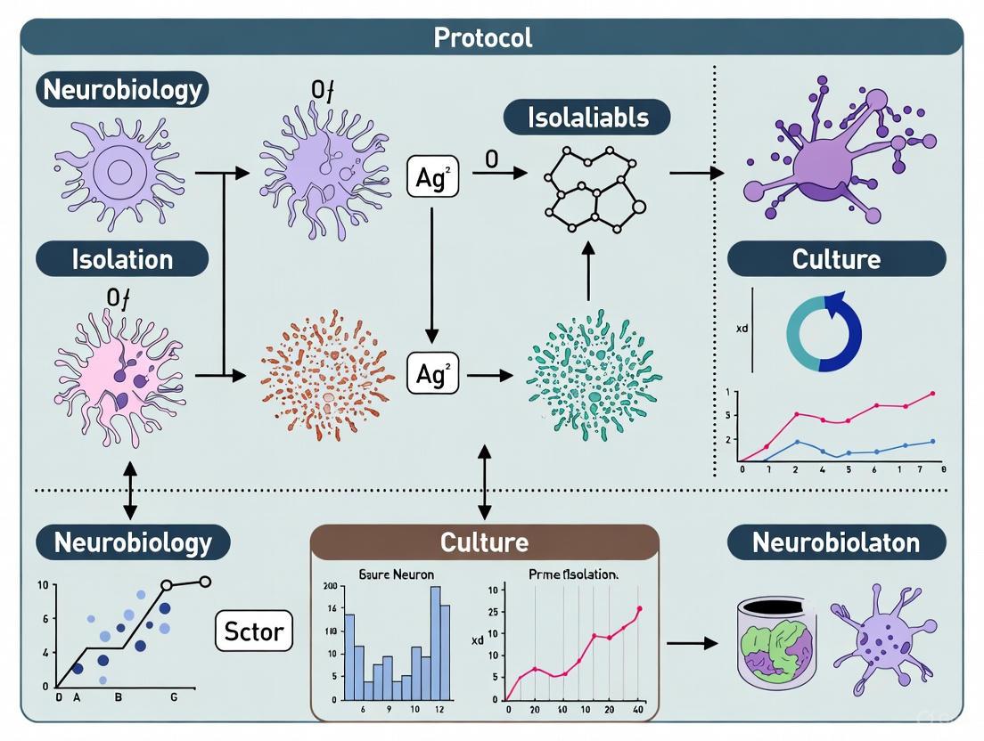
Experimental Workflow: From Isolation to Functional Analysis
The following diagram illustrates the complete experimental pathway for primary neuron culture and application:
Primary neurons remain an indispensable tool for neuroscience research, offering unparalleled physiological relevance that immortalized cell lines cannot match. While their culture requires specialized expertise and careful protocol implementation, the biological fidelity they provide is essential for meaningful investigation of neural function, disease mechanisms, and therapeutic development. The optimized methodologies presented in this application note empower researchers to overcome technical challenges and leverage the full potential of primary neuronal cultures in their research programs. As the field advances, emerging technologies such as human iPSC-derived neurons offer promising alternatives, but primary neurons from well-established protocols continue to provide the gold standard for physiological relevance in vitro.
Key Applications in Neuroscience Research and Drug Development
Primary neuronal cultures, particularly those derived from the cortex, are indispensable tools in modern neuroscience. These cultures provide a physiologically relevant in vitro system that closely mimics the in vivo environment, making them ideal for investigating neuronal function, development, and pathology [1]. The isolation and culture of primary neurons from specific regions of the nervous system represent fundamental techniques that enable researchers to explore distinct neural populations and their roles in health and disease. This application note details optimized protocols for primary cortical neuron isolation and culture, framing them within the context of their crucial applications in mechanistic studies and drug development pipelines. The reliability of these models hinges on standardized, reproducible methods that ensure high neuronal viability, purity, and functional maturation, thereby enhancing the translational value of the data generated.
Key Applications of Primary Neuronal Cultures
Primary neuronal cultures serve as versatile platforms across multiple domains of neuroscience research and pharmaceutical development. Their applications span from disease modeling to high-throughput compound screening.
Table 1: Key Applications of Primary Neuronal Cultures in Research and Drug Development
| Application Domain | Specific Use Cases | Relevance |
|---|---|---|
| Disease Modeling | Modeling neurodegenerative diseases (Alzheimer's, Parkinson's, ALS) [1] [8]; Neurodevelopmental disorder studies | Recapitulates disease-specific pathophysiology and cellular vulnerability [9]. |
| Drug Discovery & Screening | Target validation; Efficacy testing of candidate compounds; Toxicity assessments [1] [8]; High-Throughput Screening (HTS) [9] | Provides human-relevant, physiologically contextual data for preclinical verification [1]. |
| Mechanistic Studies | Neuron-neuron interactions; Synapse formation and function; Neuron-glial cell relationships [1] [4]; Intracellular signaling | Enables direct observation and manipulation of cellular processes. |
| Alternative Models | Chicken embryo models for Alzheimer's disease [10]; iPSC-derived motor neurons for ALS [8] | Offers accessible, human-genetics-based models that bypass certain ethical and technical constraints. |
Disease Modeling and Drug Screening
Primary cortical cultures are extensively used to model human neurodegenerative disorders. They allow for the exploration of pathological mechanisms and the evaluation of potential therapeutic strategies in a controlled environment [1]. The value of these models has been further amplified by the advent of induced pluripotent stem cell (iPSC) technologies. Large-scale iPSC libraries derived from patients with sporadic Amyotrophic Lateral Sclerosis (ALS), for instance, have enabled population-wide phenotypic screening and the identification of promising therapeutic combinations, such as baricitinib, memantine, and riluzole [8]. This approach successfully recapitulated key disease features like reduced neuronal survival and neurite degeneration, validating its use for preclinical testing.
Optimized Protocol for Primary Cortical Neuron Isolation and Culture
This section provides a detailed methodology for the isolation and culture of primary cortical neurons from embryonic rats, optimized for high neuronal yield and purity [1].
Materials and Reagents
Table 2: Essential Reagents and Materials for Cortical Neuron Culture
| Item | Specification/Function | Notes |
|---|---|---|
| Animals | Pregnant Sprague-Dawley rats (E17–E18) [1] | Timing is critical for neuronal viability. |
| Dissection Solution | Cold Hanks’ Balanced Salt Solution (HBSS) [1] | Maintained on ice to preserve tissue health. |
| Enzymatic Dissociation | Trypsin 0.25% in PBS [11] | Loosens tissue matrix; concentration and timing require optimization. |
| Culture Medium | Neurobasal Plus Medium, supplemented with B-27, GlutaMAX, and Penicillin/Streptomycin (P/S) [1] [4] | Serum-free formulation supports neuronal growth and suppresses glial proliferation. |
| Coating Substrate | Poly-L-lysine [11] | Promotes neuronal adhesion to the culture vessel. |
Step-by-Step Procedure
1. Coating of Culture Surfaces:
- Prepare a sterile working solution of poly-L-lysine.
- Coat the surface of culture plates/dishes and incubate for the recommended duration.
- Aspirate the coating solution and rinse thoroughly with sterile water before allowing the surfaces to air dry completely in a biosafety cabinet [1].
2. Dissection and Tissue Isolation:
- Euthanize a pregnant rat (E17–E18) following approved institutional animal ethics guidelines.
- Aseptically remove embryos and place them in a chilled culture dish containing HBSS on ice.
- Under a dissecting microscope, immobilize an embryo's head and carefully remove the skin and skull using fine forceps (#5) to expose the brain.
- Isolate the whole brain and carefully remove the meninges to improve neuronal purity.
- Separate the cerebral cortices from the rest of the brain, collecting them in a tube containing cold HBSS.
- Critical Note: Limit dissection time to 2–3 minutes per embryo to maintain neuronal health. The entire dissection process for a litter should not exceed 1 hour [1].
3. Tissue Dissociation:
- Mechanically dissociate the pooled cortical tissues into smaller pieces.
- Incubate the tissue with a pre-warmed enzymatic solution (e.g., trypsin) at 37°C for 15 minutes to loosen the extracellular matrix [11].
- Neutralize the enzyme activity by adding a culture medium containing serum or a specific inhibitor.
- Triturate the tissue gently using fire-polished glass Pasteur pipettes of decreasing bore size to achieve a single-cell suspension without excessive mechanical stress [1] [4].
- Pass the cell suspension through a 70-μm cell strainer to remove any remaining aggregates [11].
4. Plating and Maintenance:
- Centrifuge the cell suspension at a low speed, resuspend the pellet in complete neuronal culture medium, and perform a cell count.
- Plate the cells at the desired density onto the pre-coated culture vessels.
- Maintain cultures in a humidified incubator at 37°C with 5% CO₂.
- Around the third or fourth day in vitro (DIV), add an antimitotic agent (e.g., CultureOne supplement [4]) to curb the proliferation of non-neuronal cells like astrocytes.
- Perform a partial medium change every 3-4 days to replenish nutrients.
The Scientist's Toolkit: Essential Research Reagent Solutions
The success of primary neuronal culture is highly dependent on the consistent use of high-quality, functionally appropriate reagents.
Table 3: Key Research Reagent Solutions for Primary Neuronal Culture
| Reagent | Function | Application Notes |
|---|---|---|
| Neurobasal Plus Medium | A optimized, serum-free basal medium designed to support the long-term survival and growth of primary neurons [1] [4]. | Superior to DMEM for neuronal cultures; minimizes background excitation and glial overgrowth. |
| B-27 Supplement | A defined, serum-free supplement containing hormones, antioxidants, and other neuronal survival factors [1] [4]. | Crucial for enhancing neuronal survival and promoting neurite outgrowth. |
| GlutaMAX Supplement | A stable dipeptide (L-alanyl-L-glutamine) that replaces L-glutamine, preventing the accumulation of toxic ammonia in the medium [1]. | Ensures a consistent supply of glutamine for neurotransmitter synthesis and energy production. |
| Poly-L-Lysine | A synthetic, positively charged polymer that coats the culture surface, facilitating the attachment of negatively charged neuronal cell membranes [11]. | Essential for ensuring high cell adherence; must be thoroughly rinsed before use. |
| Nerve Growth Factor (NGF) | A neurotrophic factor critical for the survival, development, and maintenance of specific neuronal populations, such as DRG neurons [1]. | Region-specific requirement; not typically needed for standard cortical cultures. |
| CultureOne Supplement | A chemically defined supplement used to suppress the proliferation of fibroblasts and other dividing cells, such as astrocytes [4]. | Added after neurons have adhered (e.g., DIV3) to maintain a neuron-enriched culture. |
The isolation and culture of primary cortical neurons remain a cornerstone technique in neuroscience. The optimized protocols detailed herein, which emphasize region-specific dissection, refined dissociation techniques, and customized culture conditions, are pivotal for generating robust and reproducible in vitro models [1]. These models are instrumental in advancing our understanding of neuronal biology and pathology. Furthermore, the integration of primary cultures with emerging technologies like large-scale iPSC screening and 3D brain organoids [8] [9] is paving the way for more personalized and human-relevant approaches in drug discovery. By providing a solid foundation for studying neuronal populations, these methodologies continue to drive innovation in the quest to understand and treat complex neurological diseases.
The isolation and culture of primary cortical neurons is a foundational technique in neuroscience, enabling the investigation of neuronal development, synaptic function, neurotoxicity, and disease mechanisms in vitro [1]. The physiological relevance of data derived from such models is critically dependent on three key experimental design considerations: the age of the animal source, the specific brain region isolated, and the physiological context in which neurons are studied. Appropriately addressing these factors ensures that in vitro findings more accurately reflect in vivo biology, particularly when modeling age-associated neurodegenerative disorders or testing pharmaceutical efficacy [12]. This application note details optimized methodologies that incorporate these essential considerations to enhance the translational value of primary neuron research for scientists and drug development professionals.
The Impact of Animal Age on Neuronal Phenotype
The age of the animal source significantly influences neuronal characteristics in culture, including survival, neurite outgrowth capacity, and synaptic plasticity. Traditionally, primary neuronal cultures are derived from embryonic or early postnatal rodents, yet these models may poorly represent the biology of adult or aging neurons relevant to human neurological diseases [12].
Embryonic and Postnatal Neurons
Protocols for embryonic (E17-E18) and postnatal (P1-P2) cortical neurons are well-established and yield robust, reproducible cultures suitable for many applications [1] [13]. These neurons demonstrate high viability, extensive neurite arborization, and form functional synaptic networks within weeks in culture [14].
Adult Neurons
Recent methodological advances now enable the culture of neurons from adult central nervous systems. Neurons from adult mice (up to 60 days post-natally) can be cultured in large numbers and maintained for extended periods, developing polarity with segregated dendritic and axonal compartments, maintaining resting membrane potentials, and exhibiting spontaneous electrical activity [15]. Culturing adult neurons presents unique challenges, including lower initial yields and viability, but provides a more age-appropriate model for studying adult-onset neurological disorders [12].
Table 1: Neuronal Yield and Viability by Age and Species
| Neuron Source | Age | Yield (per pair of cortices) | Viability (%) | Key Characteristics |
|---|---|---|---|---|
| Mouse Cortex | E17-19 | 4.5 x 10⁶ cells/mL [14] | 94-96% [14] | Extensive synaptic scaling, complex dendrites |
| Rat Cortex | E17-18 | 4.0 x 10⁶ cells/mL [14] | 96% [14] | High purity, robust network formation |
| Adult Mouse Cortex | 4-48 weeks | Varies with age [12] | Reduced vs. embryonic [12] | Age-dependent neurite growth, sex-specific responses |
Brain Region-Specific Functional Specialization
Neurons isolated from distinct brain areas exhibit unique profiles regarding cell composition, protein expression, metabolism, and electrical activity in vitro [16]. These inherent differences must be considered when designing experiments to ensure biological relevance.
Regional Identity and Culture Characteristics
Studies demonstrate that rat neurons from the prefrontal cortex (pfCx), hippocampus (Hip), and amygdala (Amy) maintain unique behaviors in culture, including different proportions of neuronal subtypes (e.g., glutamatergic vs. GABAergic) and varying spontaneous firing patterns [16]. This regional specialization supports the development of multiregional brain-on-a-chip models that incorporate functionally connected areas to better mimic in vivo brain circuitry [16].
Cortical Neuron Special Considerations
Cortical neurons are particularly valuable for studying higher cognitive functions, neurodegenerative diseases, and neurotoxicity. When isolating cortical tissue, precise dissection is crucial to avoid contamination from adjacent structures like the hippocampus [1]. The cerebral hemispheres should be separated along the median longitudinal fissure, with the meninges carefully removed to reduce non-neuronal cell contamination [13].
Table 2: Brain Region-Specific Characteristics in Culture
| Brain Region | Neuronal Subtypes | Key Protein Markers | Functional Specialties |
|---|---|---|---|
| Cortex | Predominantly glutamatergic pyramidal neurons [16] | βIII-TUBULIN, MAP2, Neurofilament [16] [14] | Complex network formation, synaptic plasticity [14] |
| Hippocampus | Mixed glutamatergic and GABAergic populations [16] | Vglut1, GAD67 [16] | High spontaneous activity, long-term potentiation |
| Amygdala | Distinct ratio of excitatory/inhibitory neurons [16] | Specific neuropeptides [16] | Unique metabolic profiles [16] |
Physiological State and Contextual Influences
The physiological context of both the donor animal and the culture environment significantly influences neuronal function and responsiveness. Internal states such as arousal, motivation, and movement can shape sensory processing and neural encoding [17] [18].
State-Dependent Processing in Neural Circuits
Research reveals that the prefrontal cortex customizes its signals to visual and motor systems based on behavioral states. Specific prefrontal subregions (orbitofrontal cortex and anterior cingulate area) selectively transmit information about arousal and motion to primary visual and motor cortices, balancing each other to sharpen or suppress visual information as needed [17]. This state-dependent gating of sensorimotor processing follows an inverted U-shape curve, where optimal task performance occurs at intermediate motivational states, with impairments at both low and high extremes [18].
Implications for In Vitro Models
These findings highlight the importance of considering physiological context when designing experiments and interpreting results. For drug screening applications, the metabolic and hormonal milieu can significantly impact compound efficacy, particularly when screening for neuroprotective agents [12].
Integrated Experimental Protocols
Optimized Protocol for Embryonic Cortical Neuron Isolation
Reagents and Materials:
- Poly-D-Lysine (PDL, 50 μg/mL) and Laminin (10 μg/mL) for coating [13]
- Papain-based enzymatic digestion (20 U/mL) with DNase I (100 U/mL) for postnatal tissue [13]
- Neuronal culture medium: Neurobasal-A supplemented with B-27, GlutaMAX, and antibiotic-antimycotic [1] [13]
- Growth factors: BDNF and IGF-I for enhanced survival and maturation [13]
Procedure:
- Coating Preparation: Cover culture surfaces with PDL solution and incubate 1 hour at 37°C. Wash with sterile dH₂O, then coat with Laminin overnight at 2-8°C [13].
- Tissue Dissection: Dissect cortical tissue from E17-E18 embryos in cold PBS. Remove meninges completely to minimize glial contamination [1].
- Tissue Dissociation: For embryonic tissue, triturate gently with fire-polished Pasteur pipette in Neuronal Base Media. For postnatal tissue, use enzymatic digestion with papain/DNase I followed by ovomucoid protease inhibitor [13].
- Plating and Maintenance: Plate cells at desired density (e.g., 1.6K cells/mm² [16]) in complete culture media. Maintain in 37°C, 5% CO₂ incubator, exchanging 50% of media every 72-96 hours [13].
Advanced Protocol for Adult Neuron Culture
Modifications for Adult Neurons:
- Use gentle enzymatic digestion formulations specifically optimized for mature tissue [12]
- Increase coating concentration (PDL with laminin enhancement) [12]
- Adjust media supplements to address age-dependent changes in metabolic requirements [12]
- Account for sex-dependent effects in experimental design [12]
Signaling Pathways in Neuronal Senescence and Function
Primary cortical neurons in long-term culture can develop a senescence-like phenotype characterized by sustained DNA damage response, elevated p21CIP1/WAF1 expression, lipofuscin accumulation, and secretion of SASP factors [19]. Autophagy plays a critical role in preventing neuronal senescence, with autophagic flux reduction observed in senescent neurons both in vitro and in vivo [19].
Diagram 1: Key pathways in neuronal senescence and state-dependent processing. Impaired autophagy contributes to senescence, while prefrontal cortex subregions (ORB and ACA) balance visual processing based on behavioral states.
The Scientist's Toolkit: Essential Research Reagents
Table 3: Key Research Reagent Solutions for Primary Neuron Culture
| Reagent/Catalog Item | Function | Application Notes |
|---|---|---|
| Poly-D-Lysine [13] | Substrate coating for cell adhesion | Use at 50-100 μg/mL; essential for neuronal attachment |
| Laminin [13] | Enhneurite outgrowth and network formation | Use at 10 μg/mL following PDL coating |
| Papain-based Isolation Kit [14] | Gentle enzymatic tissue dissociation | Superior to trypsin for yield (2-fold increase) and viability (94-96%) |
| Neurobasal Medium with B-27 [1] [13] | Serum-free neuronal culture medium | Supports long-term survival without glial overgrowth |
| BDNF and IGF-I [13] | Trophic factors for neuronal health | Enhance survival, dendrite complexity, and synaptic maturation |
| Syn-PER Synaptic Protein Extraction [14] | Isolation of synaptosomes | Quantifies synaptic protein yield as indicator of functionality |
Careful consideration of animal age, brain region specificity, and physiological context significantly enhances the relevance and translational potential of primary cortical neuron research. The optimized protocols detailed herein enable researchers to establish more physiologically accurate in vitro models for investigating neurological function, disease mechanisms, and therapeutic development. By integrating these essential design parameters, scientists can bridge the gap between traditional cell culture models and the complex biology of the intact nervous system.
A Step-by-Step Protocol: From Dissection to Mature Neuronal Networks
The isolation and culture of primary cortical neurons stand as a fundamental methodology in modern neurobiology, providing an essential in vitro model for studying neuronal development, synaptic function, neurotoxicity, and mechanisms of neurological diseases [20] [13]. Within this field, the selection of appropriate animal models at specific developmental stages is a critical decision point that directly influences experimental outcomes. Embryonic Day 17-18 (E17-E18) represents a key neurodevelopmental period in rodents, characterized by active cortical neurogenesis and the birth of neurons that will form the mature cerebral cortex [21]. This application note provides a structured comparison between E17-E18 rats and mice to guide researchers in selecting the optimal model for primary cortical neuron isolation and culture, framed within the context of protocol optimization and reproducibility.
Comparative Analysis: E17-E18 Rats vs. Mice
Quantitative Comparison of Cell Yield and Viability
The following table summarizes key quantitative differences observed when isolating cortical neurons from E17-E18 rats versus mice using optimized protocols.
Table 1: Quantitative Comparison of Cortical Neuron Isolation from E17-E18 Rodents
| Parameter | E17-E18 Rats | E17-E18 Mice | Notes |
|---|---|---|---|
| Typical Cell Yield | ~4.0 × 10⁶ cells/mL per cortex pair [14] | ~4.5 × 10⁶ cells/mL per cortex pair [14] | Yield can vary with dissection skill and strain. |
| Cell Viability | 96% [14] | 95% [14] | Viability is consistently high with gentle enzymatic digestion. |
| Developmental Stage | Cortex and hippocampus suitable for culture [20] [13] | Cortex and hippocampus suitable for culture [22] [23] | E17-E18 is a peak period of neurogenesis for both species. |
| Cultural Purity | High purity with serum-free media [20] | High purity, may require Ara-C for glial suppression [22] | Defined media reduce non-neuronal cell growth. |
| Functional Maturation | Extensive axonal/dendritic branching by 3 weeks [20] | Differentiated neurons with synaptic activity within 6 days [22] | Both develop morphologically mature synapses. |
Practical and Experimental Considerations
Beyond quantitative metrics, several practical factors influence model selection.
Table 2: Practical Considerations for Model Selection
| Consideration | E17-E18 Rats | E17-E18 Mice |
|---|---|---|
| Tissue Size & Dissection | Larger brain structures, easier dissection for beginners [13] | Smaller, more challenging dissection; requires finer tools [22] |
| Protocol Availability | Extensive, well-established protocols [20] [13] | Abundant protocols, though techniques can be more delicate [22] [24] |
| Genetic Models | Available, but fewer than mouse models | Vast array of genetically engineered models (transgenics, knockouts) [22] |
| Cost & Availability | Generally higher cost per animal | Typically lower cost and wider availability |
| Experimental Applications | Classic model for neuropharmacology, biochemistry, and electrophysiology | Ideal for genetic studies, disease modeling, and high-throughput screens |
Detailed Experimental Protocols
Core Workflow for Primary Cortical Neuron Isolation
The following diagram illustrates the generalized protocol workflow, which is applicable to both E17-E18 rats and mice with minor modifications.
Protocol Details: Isolation and Culture from E17-E18 Rats
Materials & Reagents:
- Pregnant female Wistar or Sprague-Dawley rats at E17-E18 [20] [1].
- Preparation Medium: HBSS, 1 mM sodium pyruvate, 10 mM HEPES [20].
- Enzymatic Digestion Solution: Papain (0.5 mg) and DNase I (10 µg) in 5 ml of Papain Buffer [20].
- Growth Medium: Neurobasal medium supplemented with B27, L-glutamine, and penicillin-streptomycin [20] [13].
- Coating Solution: Poly-D-Lysine (PDL) at 50 µg/mL, optionally followed by Laminin at 10 µg/mL [13].
Step-by-Step Procedure:
- Dissection: Euthanize the dam and remove embryos. Decapitate embryos and place heads in ice-cold PBS. Under a dissecting microscope, remove brains and place in a dish with cold preparation medium. Carefully separate the cerebral hemispheres, remove meninges, and isolate the cortical tissue [20] [13].
- Tissue Dissociation: Transfer cortices to pre-warmed Papain solution. Incubate for 10-15 minutes at 37°C. Remove enzyme solution and add trituration medium containing DNase I. Gently triturate the tissue 10-15 times using a fire-polished glass Pasteur pipette until the solution is homogenous [20].
- Cell Seeding and Culture: Centrifuge the cell suspension at 200 × g for 5 minutes. Resuspend the pellet in complete growth medium. Count cells using a hemocytometer with Trypan Blue exclusion to assess viability. Plate cells at desired density (e.g., 50,000-100,000 cells/cm²) on PDL/laminin-coated plates or coverslips. Maintain cultures in a 37°C, 5% CO₂ incubator [20] [13].
- Media Maintenance: For long-term cultures (>1 week), perform a half-media change with fresh pre-warmed complete growth medium once per week. The use of serum-free Neurobasal/B27 medium suppresses glial overgrowth, maintaining high neuronal purity for up to 4 weeks [20] [13].
Protocol Modifications for E17-E18 Mice
The core protocol for mice is similar, with emphasis on the following adjustments:
- Dissection: The smaller size of mouse embryos requires extra precision and finer dissection tools, such as Dumont #5 and #7 forceps [22] [24].
- Enzymatic Digestion: Commercial gentle enzyme mixtures (e.g., Pierce Primary Neuron Isolation Kit) have been shown to provide higher cell yield and viability (95% vs. 83-92%) and better dendritic complexity compared to traditional trypsin methods for mouse cortical tissue [14].
- Glial Suppression: While serum-free media help, adding the mitotic inhibitor cytosine arabinoside (Ara-C, 1-5 µM) is often recommended after 3-5 days in vitro to further inhibit glial proliferation in mouse cultures [22].
The Scientist's Toolkit: Essential Research Reagents
Table 3: Key Reagent Solutions for Primary Cortical Neuron Culture
| Reagent/Category | Function & Rationale | Specific Examples |
|---|---|---|
| Enzymatic Dissociation | Digests extracellular matrix to liberate single cells; critical for yield and viability. | Papain-Based Systems [20] [13]; Gentle Commercial Mixes (e.g., Pierce Primary Neuron Isolation Kit) [14] |
| Culture Medium | Supports neuronal survival and maturation while suppressing glial growth. | Neurobasal Medium supplemented with B27 and GlutaMAX [20] [13] [22] |
| Substrate Coating | Promotes neuronal adhesion and neurite outgrowth. | Poly-D-Lysine (PDL) [13] [22]; PDL + Laminin combination for enhanced attachment [13] [24] |
| Growth & Trophic Factors | Enhances neuronal survival, maturation, and synaptic development. | BDNF, IGF-I [13]; NGF (for DRG neurons) [1] |
| Glial Suppression | Controls proliferation of non-neuronal cells to maintain culture purity. | Cytosine Arabinoside (Ara-C) [22]; Serum-Free Defined Media [20] |
Both E17-E18 rats and mice provide excellent sources for robust primary cortical neuron cultures. The choice between them is not a matter of superiority but of strategic alignment with experimental goals. E17-E18 rats are often preferred for their larger tissue size, which facilitates dissection and can yield a high number of robust neurons, making them ideal for biochemical assays and classic electrophysiological studies. Conversely, E17-E18 mice offer unparalleled genetic tractability, providing access to a vast array of disease models, and are well-suited for studies where genetic manipulation is paramount. By adhering to the optimized protocols and considerations outlined herein, researchers can ensure the isolation of highly functional cortical neurons, thereby enhancing the reliability and reproducibility of their neuroscience research.
Precision Dissection and Meninges Removal for High Purity
Within the framework of optimized protocols for primary cortical neuron isolation, the precision of the initial dissection and the completeness of meninges removal are paramount. These steps are the critical foundation upon which high-quality, high-purity neuronal cultures are built. The presence of contaminating cell types, resulting from incomplete meningeal removal, can severely compromise experimental outcomes by altering neuronal behavior, synaptic scaling, and responses to pharmacological agents. This application note provides a detailed, step-by-step protocol designed to maximize neuronal yield and purity, enabling the generation of robust and reproducible data for basic research and drug development.
The following table summarizes key quantitative outcomes from published studies that utilize careful dissection and culture techniques, highlighting the achievable standards for neuronal purity and viability.
Table 1: Quantitative Outcomes of Optimized Neuronal Isolation Protocols
| Cell Type / Protocol | Purity (%) | Viability (%) | Key Findings | Source |
|---|---|---|---|---|
| Human Fetal Neurons | >98% (MAP-2) | Information Missing | A rapid-adhesion step effectively enriched neurons from a mixed cell suspension. | [25] |
| Mouse Cortical Neurons (Pierce Kit) | ~90% (Day 1, GFAP-negative) | 94-96% | Demonstrated a 2-fold increase in cell yield over traditional trypsin methods. | [14] |
| Mouse Cortical Neurons (DIY Trypsin) | ~80% (Day 1, GFAP-negative) | 83-92% | Lower purity and viability compared to the optimized kit method. | [14] |
| Primary Cortical Cultures (P0 Mouse) | Information Missing | Information Missing | Cultures generated extensive, intertwined dendritic networks and expressed synaptic proteins (PSD95, synaptophysin). | [26] |
Experimental Protocols
Detailed Dissection and Meninges Removal for Cortical Neurons
This protocol is optimized for the isolation of primary cortical neurons from postnatal day 0 (P0) mice [26] or embryonic day 17-18 (E17-E18) rats [1], with specific emphasis on precision dissection and meninges removal.
Reagents and Materials:
- Dissection Solution: Ice-cold Hank's Balanced Salt Solution (HBSS), without Ca²⁺ and Mg²⁺, supplemented with penicillin/streptomycin, sodium pyruvate, glucose, and HEPES [26].
- Enzymatic Solution: 0.05% Trypsin-EDTA [26].
- Coating Solution: Poly-L-ornithine (0.1 mg/mL) or polyethyleneimine (0.05%) [26] [1].
- Equipment: Fine forceps (e.g., #5 Dumont), fine scissors, sterile surgical scalpel, silicone-lined or standard petri dishes, dissecting microscope [27] [1].
Step-by-Step Workflow:
Animal Sacrifice and Brain Extraction:
Gross Dissection and Hemisphere Separation:
- Position the brain in a dorsal view. Using two fine forceps, carefully separate the cerebral hemispheres along the midline [1].
- Critical Consideration: Positioning the brain in a dorsal view is essential for an accurate division. A ventral view increases the risk of including unwanted subcortical tissues [1].
Precision Removal of the Meninges:
- Under a dissecting microscope, hold one cerebral hemisphere with the inner surface facing up.
- Using fine forceps, gently grasp the meningeal membrane at its edge. With a careful tearing or rolling motion, peel the meninges away from the cortical surface.
- Critical Consideration: This step requires a high level of skill. Grasp only the meninges to avoid puncturing or damaging the soft cortical tissue. Incomplete removal will significantly reduce neuron-specific purity in the final culture [1].
Hippocampal Isolation and Cortex Collection:
- With the meninges removed, position the hemisphere with the inner surface up. Identify the dark, C-shaped hippocampal structure in the posterior third of the hemisphere.
- Carefully isolate and remove the hippocampus using fine forceps.
- Collect the remaining cortical tissue in a fresh tube containing ice-cold HBSS.
- Time Management: Limit dissection time to 2-3 minutes per embryo to maintain neuronal health. The total dissection time for a full litter should not exceed one hour [1].
Enzymatic Dissociation and Plating
- Tissue Digestion:
- Centrifuge the collected cortical pieces and incubate the pellet in 0.05% Trypsin-EDTA at 37°C for 20 minutes [26].
- Reaction Termination and Trituration:
- Inactivate the trypsin by washing with HBSS followed by Minimal Essential Medium supplemented with 10% horse serum [26].
- Dissociate the tissue by gentle mechanical trituration using fire-polished Pasteur pipettes of declining diameters. Pass the cell suspension through a 40 µm cell strainer to remove any remaining clumps [26].
- Cell Plating and Culture:
- Count viable cells using trypan blue exclusion.
- Plate cells on poly-L-ornithine-coated surfaces at a high density (e.g., 2,000–2,500 cells/mm²) in Neurobasal-A medium supplemented with B-27 and L-glutamine [26].
- To inhibit glial proliferation, treat cultures with 5 µM cytosine arabinoside (Ara-C) from day in vitro (DIV) 2 onward [26] [25].
Workflow Visualization
The following diagram illustrates the logical workflow for the successful isolation and culture of high-purity primary cortical neurons, integrating the critical steps of dissection and meninges removal.
The Scientist's Toolkit
Table 2: Essential Research Reagent Solutions for Primary Cortical Neuron Culture
| Reagent/Material | Function/Application | Example |
|---|---|---|
| Poly-L-Ornithine / Poly-D-Lysine | Coats culture surfaces to enhance neuronal adhesion. | 0.1 mg/mL solution for coating plates/coverslips [26] [25]. |
| Neurobasal Medium | A serum-free medium optimized for the long-term survival of postnatal neuronal cells. | Served as the base medium for cortical cultures [26] [12]. |
| B-27 Supplement | A defined serum-free supplement that supports neuronal growth and health, reducing the need for glial feeders. | Used at 1X or 2% concentration in Neurobasal medium [26] [25]. |
| Cytosine Arabinoside (Ara-C) | An anti-mitotic agent used to suppress the proliferation of non-neuronal cells (e.g., glia) in mature cultures. | Applied at low concentrations (e.g., 5 µM) a few days after plating [26] [25]. |
| Hank's Balanced Salt Solution (HBSS) | An isotonic salt solution used during the dissection and tissue processing steps to maintain ion balance and pH. | Used ice-cold for dissection and tissue washing [26] [1]. |
| Trypsin-EDTA | Proteolytic enzyme used for the controlled digestion of extracellular matrix proteins to dissociate tissue into single cells. | A 0.05% solution is commonly used for cortical tissue [26]. |
The isolation of high-purity, viable primary neurons is a cornerstone of neuroscience research, enabling the study of neuronal development, synaptogenesis, and disease mechanisms in vitro [1]. The initial step of tissue dissociation is critical, as the method chosen directly impacts cell yield, viability, and the physiological relevance of subsequent experimental data [28] [29]. This application note details optimized enzymatic and mechanical protocols for the dissociation of embryonic rodent cortex, framed within the context of primary neuron isolation for downstream cell culture and functional studies. The procedures are designed to maximize the recovery of functional cortical neurons while minimizing the presence of non-neuronal cells.
Quantitative Comparison of Dissociation Methods
The choice between dissociation methods involves balancing yield, viability, and specific cell-type recovery. The following table summarizes key performance metrics from recent studies.
Table 1: Performance Metrics of Tissue Dissociation Techniques
| Method | Tissue Type | Key Performance Findings | Reference |
|---|---|---|---|
| Integrated Disaggregation & Filtration (IDF) Device | Murine Kidney | Epithelial cell recovery exceeded previous efforts; 2-fold increase in single-cell recovery after short & long digestion. Endothelial cells required extended digestion. | [30] |
| Automated-Mechanical (Medimachine II) | Rat Spleen, Testis, Liver | Better preservation of lysosome and mitochondria labelling; lower intracellular ROS vs. enzymatic; simple, fast, standardized. | [28] [29] |
| Gentle Enzymatic (Trypsin-EDTA/DNase) | Rat Spleen, Testis, Liver | Induced lower intracellular ROS; comparable apoptosis/necrosis to mechanical method. | [28] [29] |
| Enzymatic (Papain) | Embryonic Rat Cortex | >90% neuronal population (MAP2+); extensive neurite outgrowth in 3-4 days. | [31] |
Different cell types within the same tissue can exhibit varying susceptibility to dissociation methods. A study on murine kidney tissue revealed that optimal recovery of distinct cell populations requires tailored parameters.
Table 2: Cell-Type-Specific Recovery from Murine Kidney Based on Dissociation Parameters [30]
| Cell Type | Optimal Dissociation Method | Key Parameter | Digestion Time |
|---|---|---|---|
| Epithelial Cells | IDF Device | 20 passes through channel module + 1 filter pass | 20 minutes (equivalent to 60 min with optimization) |
| Endothelial Cells | Traditional Extended Digestion | Reliant on enzymatic breakdown | 60 minutes |
Detailed Experimental Protocols
Protocol 1: Enzymatic Dissociation for Primary Cortical Neuron Isolation
This protocol is optimized for the isolation of cortical neurons from E17-E18 rat embryos, yielding >90% pure neuronal cultures [1] [31].
Materials & Reagents:
- Hibernate-E Medium (with and without Ca²⁺)
- Neurobasal Plus Medium
- B-27 Plus Supplement
- Papain (Worthington)
- Poly-D-lysine
- Fire-polished glass Pasteur pipettes
Procedure:
- Dissection & Tissue Collection: Dissect cortex from E18 rat embryo brains in ice-cold Hibernate-E medium supplemented with 2% B-27 Plus. Remove meninges thoroughly to reduce non-neuronal cell contamination [1] [31].
- Enzymatic Digestion: Transfer tissue to 4 mL of Hibernate-E medium without Ca²⁺ containing 2 mg/mL filter-sterilized papain. Incubate for 30 minutes at 30°C with gentle shaking every 5 minutes [31].
- Termination & Washing: Add 6 mL of complete Hibernate-E medium to deactivate the enzyme. Centrifuge the tube at 150 × g for 5 minutes. Remove supernatant carefully [31].
- Mechanical Trituration: Resuspend the tissue pellet in 5 mL of complete Hibernate-E medium. Gently triturate 10-15 times using a fire-polished glass Pasteur pipette to dissociate cells. Let the tube stand for 2 minutes to allow undissociated debris to settle [4] [31].
- Cell Collection & Plating: Transfer the single-cell suspension to a new tube. Count cells and plate at a density of ~1 × 10⁵ cells per well in poly-D-lysine coated 48-well plates with Neurobasal Plus complete medium (supplemented with 2% B-27 Plus, 0.25% L-glutamine) [1] [31].
- Maintenance: Feed cultures every third day by replacing half of the medium with fresh Neurobasal Plus complete medium [31].
Protocol 2: Automated Mechanical Dissociation
For tissues where enzyme-induced antigen alteration is a concern, automated mechanical dissociation provides a standardized alternative.
Materials & Reagents:
- Medimachine II System (CTSV s.r.l) with Medicons
- RPMI 1640 Medium
Procedure:
- Tissue Preparation: Dissect and mince cortical tissue into ~1 mm³ pieces in a Petri dish containing cold PBS [28] [29].
- Loading: Transfer 1-2 tissue pieces into a Medicon capsule filled with 1 mL of RPMI 1640 medium [28] [29].
- Disaggregation: Insert the Medicon into the Medimachine II and run for 15-55 seconds at a constant speed of 100 rpm [28] [29].
- Cell Collection: Aspirate the cell suspension from the Medicon capsule with a syringe. Pool fractions if multiple runs are performed [28] [29].
The following workflow diagram illustrates the key decision points and steps for these two primary dissociation methods.
Diagram 1: Tissue Dissociation Workflow Selection.
The Scientist's Toolkit: Essential Reagents & Materials
Successful tissue dissociation relies on a suite of specialized reagents and tools. The following table details key solutions and their functions in the protocol.
Table 3: Essential Reagents and Materials for Cortical Neuron Dissociation & Culture [1] [31]
| Item | Function/Application | Example/Catalog |
|---|---|---|
| Papain | Proteolytic enzyme for gentle ECM digestion; preferred for neural tissue. | Worthington, LS003119 |
| Neurobasal Plus Medium | Serum-free medium optimized for neuronal survival and growth. | Thermo Fisher, A3582901 |
| B-27 Plus Supplement | Serum-free supplement containing hormones, antioxidants, and proteins. | Thermo Fisher, A3582801 |
| Hibernate-E Medium | Serum-free medium for tissue storage and dissection in ambient CO₂. | BrainBits LLC, A12476-01 |
| Poly-D-Lysine | Synthetic polyamine coating for culture vessels to promote neuronal adhesion. | Sigma, P-6407 |
| Fire-polished Pasteur Pipette | Creates a smooth, widened bore for gentle mechanical trituration of tissue. | VWR, 612-1702 |
The choice between enzymatic and mechanical dissociation is not a matter of one being universally superior, but rather dependent on the specific research requirements. Enzymatic methods, particularly with papain, are highly effective for generating pure, viable neuronal cultures for functional studies. In contrast, automated mechanical methods offer speed, standardization, and preserve surface antigens, making them suitable for flow cytometry and studies where enzyme-induced epitope damage is a concern [28] [29]. Understanding the strengths and limitations of each approach allows researchers to tailor the dissociation process, ensuring the highest quality cellular material for probing the complexities of the brain.
Critical Substrate Coating with Poly-D-Lysine for Cell Adhesion
Within the context of optimizing protocols for primary cortical neuron isolation and culture, the choice of substrate coating is not merely a preparatory step but a critical determinant of experimental success. Poly-D-lysine (PDL) serves as a foundational coating to engineer the cell-substrate interface, directly influencing neuronal adhesion, network development, and long-term maturation in vitro [32]. This application note details the quantitative impact of PDL coating parameters on cellular outcomes and provides a standardized, evidence-based protocol to enhance the reliability and physiological relevance of primary cortical neuron cultures for basic research and drug development.
The Impact of Coating Parameters on the Cellular Interface
The efficacy of PDL is highly dependent on precise coating conditions. Research indicates that even minor variations can significantly alter the physical and biochemical landscape of the substrate, leading to divergent cellular behaviors.
Surface Topography and Roughness
Atomic force microscopy (AFM) characterization reveals a direct correlation between PDL incubation time and substrate roughness. On calcium fluoride (CaF₂) substrates, an untreated surface has a roughness (Ra) of 2.26 nm. This value increases to a maximum of Ra = 4.46 nm after a 30-minute incubation with PLL, demonstrating that prolonged incubation induces micro- and nanoscale surface modifications [33].
Consequences for Cell Morphology and Health
These subtle changes in topography profoundly influence cellular responses:
- Morphology: Increased surface roughness triggers a shift in cell shape from spindle-like to more rounded and flattened morphologies [33].
- Biochemistry: Roughness correlates with increased intracellular levels of cytochrome C and phenylalanine, which are biomarkers associated with apoptotic pathways, suggesting that extended PLL incubation may induce cytotoxic effects [33].
- Cellular Mechanics: PDL coating enhances cellular stiffness and promotes protein remodeling at the nanoscale [33].
Critical Insight: The standard adsorption method, where PDL is simply applied to coverslips, often results in high variability between cultures and compromised long-term maturation, sometimes leading to neuronal reaggregation after about one week in culture [32].
Optimized Coating Protocol for Primary Cortical Neurons
The following protocol establishes a simple and effective method for creating a covalently bound PDL substrate, which surpasses the standard adsorption method in promoting neuronal adhesion, network density, and functional maturation [32].
Reagent Preparation
- Poly-D-Lysine (PDL) Stock Solution: Dissolve PDL powder (MW 70-150 kDa) in sterile ultra-pure water to a final concentration of 40 µg/ml. The pH of this solution will be approximately 6.0 (this solution is termed PDL6) [32].
- PDL Alkaline Solution (for Covalent Grafting): Create a PDL9 solution by adding sodium carbonate to a PDL6 solution for a final concentration of 50 mM. Adjust the pH to 9.7 using 1M HCl [32].
- (3-Glycidyloxypropyl)trimethoxysilane (GOPS): Used as a coupling agent for the one-step covalent grafting procedure [32].
Coating Procedure: Adsorbed vs. Covalently Grafted PDL
Table 1: Comparison of PDL Coating Methods
| Parameter | Adsorbed PDL (Standard Method) | Covalently Grafted PDL (Enhanced Method) |
|---|---|---|
| PDL Solution | PDL6 (pH 6.0) [32] | PDL9 (pH 9.7) [32] |
| Coating Process | Incubate coverslips with PDL6 solution (e.g., 20 µg/ml) for ≥1 hour at room temperature [32]. | 1. Deposit GOPS on glass coverslips in gas phase at room temperature [32].2. Incubate GOPS-activated coverslips with PDL9 solution. |
| Post-Coating | Remove solution, rinse coverslips with sterile water, and allow to dry [32]. | The PDL covalently bonds to the activated glass surface. |
| Key Advantage | Simple and inexpensive [32]. | Superior homogeneity, density, and stability of the PDL layer; resistant to cellular degradation [32]. |
Experimental Validation and Functional Outcomes
The functional superiority of the covalently grafted PDL substrate can be validated through morphological and electrophysiological analyses.
Quantitative Assessment of Neuronal Maturation
Table 2: Functional Outcomes of Cortical Neurons on Different PDL Substrates
| Assay Metric | Adsorbed PDL (PDL6) | Covalently Grafted PDL (GPDL9) |
|---|---|---|
| Network Density | Lower density and less extended neuritic processes [32]. | More dense and extensively branched neuronal networks [32]. |
| Synaptic Activity | Less robust synaptic development and function [32]. | Enhanced synaptic activity and maturation [32]. |
| Long-Term Stability | Neurons may reaggregate after ~7 days, compromising long-term cultures [32]. | Improved stability, supporting healthy maturation over prolonged periods in vitro [32]. |
| Patch Clamp Recordings | Functional activity can be recorded, but may be less robust. | Enhanced functional excitability and network activity [32]. |
The Scientist's Toolkit: Essential Reagents for PDL Coating and Neuronal Culture
Table 3: Key Research Reagent Solutions
| Reagent / Material | Function / Application |
|---|---|
| Poly-D-Lysine (PDL) | Synthetic cationic polymer that enhances electrostatic attachment of neurons to the substrate [32]. |
| (3-Glycidyloxypropyl)trimethoxysilane (GOPS) | Coupling agent that provides epoxy moieties for the covalent grafting of PDL to glass surfaces [32]. |
| Neurobasal Plus Medium | A serum-free medium optimized for the long-term survival and health of primary neurons [1] [4]. |
| B-27 Supplement | A defined formulation containing hormones, antioxidants, and other components essential for neuronal growth [1] [4]. |
| GlutaMAX Supplement | A stable dipeptide source of L-glutamine, crucial for neuronal metabolism, which reduces the accumulation of toxic ammonia [1] [4]. |
| Coomassie Brilliant Blue (CBB) | Anionic dye used for qualitative colorimetric evaluation of the presence and homogeneity of the cationic PDL layer on the substrate [32]. |
Experimental Workflow Diagram
The following diagram illustrates the logical workflow for preparing and validating the optimized PDL coating, from substrate preparation to functional analysis.
Serum-Free Culture Medium Formulation with Neurobasal and B-27 Supplements
The isolation and culture of primary neurons are foundational techniques in modern neuroscience, enabling the investigation of neuronal function, development, and pathology in a controlled in vitro environment. For studies specifically focusing on primary cortical neurons, the selection of an appropriate culture medium is paramount to ensure high neuronal viability, purity, and functional maturation. This application note details the use of a serum-free culture system based on Neurobasal Medium and B-27 Supplement for the culture of primary cortical neurons, providing a optimized protocol within the context of a broader thesis on optimized neuronal isolation and culture. This defined system supports the long-term viability and differentiated growth of cortical neurons while minimizing the proliferation of non-neuronal glial cells, addressing a critical need for reproducible and physiologically relevant research models for researchers, scientists, and drug development professionals [34] [35].
Medium Formulation and Composition
The serum-free medium for primary cortical neuron culture is based on the combination of a specialized basal medium and a formulated supplement. The standard formulation is as follows:
- Basal Medium: Neurobasal or Neurobasal Plus Medium
- Serum-Free Supplement: B-27 Supplement (50X)
- Final Concentration of Supplement: 1X
- Common Additional Components:
- 0.5 mM L-Glutamine or GlutaMAX supplement
- 1% (v/v) Penicillin/Streptomycin (optional)
This formulation creates a defined environment that is optimized for neuronal health. The B-27 supplement is a proprietary mixture containing multiple essential components necessary for neuronal survival and growth. Key constituents include hormones (e.g., corticosterone, progesterone, triiodo-l-thyronine), antioxidants (e.g., catalase, superoxide dismutase, glutathione), and essential fatty acids (e.g., linoleic acid, linolenic acid) which collectively support neuronal metabolism and protect against oxidative stress [34] [36].
Table 1: Core Components of Serum-Free Cortical Neuron Culture Medium
| Component | Catalog Number Examples | Final Concentration | Key Function |
|---|---|---|---|
| Neurobasal Plus Medium | A3582901 [4] | Base medium | Provides balanced salts, vitamins, and energy substrates |
| B-27 Plus Supplement | A3582801 [4] | 1X (2% v/v) | Provides hormones, antioxidants, and fatty acids |
| L-Glutamine | 25030024 [4] | 0.5 mM | Neurotransmitter precursor and energy source |
| Penicillin-Streptomycin | 15070063 [4] | 1% (v/v) | Prevents bacterial contamination |
Experimental Protocols
Coating of Culture Surfaces
Prior to cell plating, culture surfaces must be coated with a substrate that promotes neuronal adhesion.
- Prepare coating solution: Dilute poly-L-lysine (PLL) in sterile borate buffer or distilled water to a working concentration of 0.1 mg/mL [6].
- Apply to surface: Add sufficient PLL solution to cover the culture surface (e.g., 0.5 mL for a 24-well plate).
- Incubate: Leave plates for at least 1 hour at room temperature or overnight at 4°C.
- Rinse: Aspirate the PLL solution and wash the surface three times with sterile distilled water.
- Air dry and store: Allow plates to dry completely in a biosafety cabinet, then store sealed at 4°C for up to one week.
Primary Cortical Neuron Isolation
This protocol is adapted from optimized methods for isolating neurons from the rat cortex [1].
Dissection:
- Sacrifice a timed-pregnant E17-E18 rat according to approved institutional guidelines.
- Extract embryos and place in cold Hanks' Balanced Salt Solution (HBSS).
- Isolate brains and carefully remove meninges to reduce non-neuronal cell contamination.
- Dissect cortical tissues under a microscope and collect in cold HBSS.
Tissue Dissociation:
- Incubate cortical tissues in enzymatic solution (e.g., papain or trypsin/EDTA) at 37°C for 15 minutes.
- Mechanically dissociate the tissue using fire-polished glass Pasteur pipettes of decreasing diameters.
- Pass the cell suspension through a 70 μm cell strainer to remove undissociated tissue.
Plating and Culture:
- Resuspend the cell pellet in the complete Neurobasal/B-27 medium.
- Plate cells onto PLL-coated surfaces at optimal densities:
- High-density cultures: 160 cells/mm² for biochemical assays [35]
- Low-density cultures: 50-100 cells/mm² for imaging and single-cell analysis
- Maintain cultures at 37°C in a 5% CO₂ humidified incubator.
- Perform a partial medium change (50%) every 3-4 days to maintain nutrient levels and remove metabolic waste.
Table 2: Key Parameters for Cortical Neuron Culture from Different Protocols
| Parameter | Cortical Neurons (E18 Rat) [35] | Hindbrain Neurons (E17.5 Mouse) [4] | Co-isolation with BMECs (P0.5 Mouse) [6] |
|---|---|---|---|
| Animal Developmental Stage | Embryonic Day 18 | Embryonic Day 17.5 | Postnatal Day 0.5 |
| Dissection Region | Cortex | Hindbrain (brainstem) | Cortex |
| Plating Density | 160 cells/mm² | Not specified | Not specified |
| Basal Medium | Neurobasal | Neurobasal Plus | Neurobasal |
| Supplement | B-27 | B-27 Plus | B-27 |
| Reported Survival | ~70% at 4 days | Robust differentiation by 10 days | High-purity functional neurons |
Protocol for Simultaneous Isolation of Cortical Neurons and Brain Microvascular Endothelial Cells (BMECs)
An advanced protocol enables the co-isolation of primary cortical neurons and BMECs from the same cohort of neonatal mice, eliminating inter-animal variability for neurovascular unit studies [6].
Tissue Preparation:
- Isolate brains from P0.5-P1 mice and place in cold HBSS.
- Carefully remove meninges and blood vessels.
Sequential Cell Separation:
- Subject the brain tissue to enzymatic digestion followed by density gradient centrifugation with bovine serum albumin (BSA) or Percoll.
- The gradient separates neural tissue (for neurons) from microvascular segments (for BMECs).
Parallel Culture:
- Plate neural tissue fraction on poly-L-lysine-coated surfaces for cortical neuron culture in Neurobasal/B-27 medium.
- Plate microvascular segments on fibronectin-coated surfaces for BMEC culture in endothelial growth medium.
This co-isolation approach provides a more physiologically relevant model for studying neurovascular interactions and has demonstrated reduced inter-sample heterogeneity compared to traditional methods where cells are isolated from different animals [6].
The Scientist's Toolkit
Table 3: Essential Research Reagent Solutions for Primary Cortical Neuron Culture
| Reagent/Catalog Number | Function in Protocol |
|---|---|
| Neurobasal Medium (A3582901) [4] | Serum-free basal medium optimized for neuronal health, providing essential nutrients and salts. |
| B-27 Supplement (17504044) [34] | Defined serum-free supplement containing hormones, antioxidants, and fatty acids crucial for neuronal survival. |
| Poly-L-Lysine (PLL) [6] | Coating substrate that promotes neuronal adhesion to culture surfaces. |
| Papain or Trypsin/EDTA [1] [6] | Enzymatic solutions for digesting the extracellular matrix to dissociate neural tissue into single cells. |
| Hanks' Balanced Salt Solution (HBSS) [1] | Balanced salt solution for maintaining ionic balance and pH during dissection and tissue processing. |
| GlutaMAX Supplement [4] | Stable dipeptide source of L-glutamine, essential for neuronal metabolism and neurotransmitter synthesis. |
Workflow and Signaling Pathways
The following diagram illustrates the complete experimental workflow for the isolation and culture of primary cortical neurons using the serum-free Neurobasal/B-27 system:
The serum-free culture system comprising Neurobasal medium and B-27 supplement represents a robust and standardized platform for the cultivation of primary cortical neurons. This defined medium supports the differentiated growth of neurons from various brain regions while maintaining their region-specific characteristics and minimizing glial cell overgrowth [35]. The protocols outlined herein, developed within the framework of an optimized thesis research project, provide a reliable methodology for generating high-purity neuronal cultures suitable for a wide range of neuroscience applications including molecular, biochemical, and physiological analyses.
The versatility of this culture system is evidenced by its successful adaptation for neurons from multiple central nervous system regions, including the hippocampus, striatum, substantia nigra, and cerebellum [35]. Furthermore, the development of advanced protocols for the simultaneous isolation of neurons and other neural cell types, such as BMECs, from the same animals underscores the continued evolution of these techniques to create more physiologically relevant and reproducible in vitro models [6]. For researchers in both academic and drug development settings, this serum-free Neurobasal/B-27 formulation provides a consistent, well-characterized foundation for investigating cortical neuron biology, disease modeling, and therapeutic screening.
Plating Density Guidelines for Biochemistry and Histology Experiments
Within the broader scope of optimizing protocols for primary cortical neuron isolation and culture, establishing standardized plating density guidelines is a critical step. The density at which neurons are seeded directly impacts cell survival, network formation, morphological development, and experimental reproducibility. This application note provides a consolidated reference for plating density parameters and associated methodologies to ensure consistency across biochemistry and histology experiments, forming a foundation for reliable and translatable research outcomes in neurological drug development.
Quantitative Plating Density Reference Tables
Culture Vessel Specifications and Seeding Guidelines
The following table summarizes key parameters for various culture vessels, including recommended seeding densities for neuronal cultures. These values provide a starting point for optimizing specific experimental conditions.
Table 1: Cell Culture Vessel Specifications and Seeding Densities
| Vessel Type | Surface Area (cm²) | Recommended Seeding Density for Neurons | Typical Growth Medium Volume (mL) | Trypsin/EDTA Volume (mL) |
|---|---|---|---|---|
| 35 mm dish | 8.8 | 0.3 x 10⁶ cells [37] | 2 | 1 |
| 60 mm dish | 21.5 | 0.8 x 10⁶ cells [37] | 5 | 3 |
| 100 mm dish | 56.7 | 2.2 x 10⁶ cells [37] | 12 | 5 |
| 6-well plate | 9.6 | 0.3 x 10⁶ cells/well [37] | 1-3 per well | 1 per well |
| 12-well plate | 3.5 | 0.1 x 10⁶ cells/well [37] | 1-2 per well | 0.4-1 per well |
| 24-well plate | 1.9 | 0.05 x 10⁶ cells/well [37] | 0.5-1.0 per well | 0.2-0.3 per well |
| 96-well plate | 0.32 | 0.01 x 10⁶ cells/well [37] | 0.1-0.2 per well | 0.05-0.1 per well |
| T-25 flask | 25 | 0.7 x 10⁶ cells [37] | 3-5 | 3 |
| T-75 flask | 75 | 2.1 x 10⁶ cells [37] | 8-15 | 5 |
| T-175 flask | 175 | 4.9 x 10⁶ cells [37] | 35-53 | 17 |
Optimized Plating Densities for Specific Applications
Table 2: Application-Specific Plating Density Guidelines
| Experimental Application | Recommended Density | Culture Duration | Key Considerations |
|---|---|---|---|
| Transfection Experiments | 200,000-300,000 cells per plate [38] | 7-14 days | Higher density improves transfection efficiency and neuronal health post-transfection. |
| Immunocytochemistry & Histology | 150,000-250,000 cells per coverslip (12-well plate) | 14-21 days | Lower density facilitates clear visualization of individual neuronal morphology. |
| Biochemical Assays (Western Blot, ELISA) | 500,000-1,000,000 cells per well (6-well plate) | 10-14 days | Higher density ensures sufficient protein yield while maintaining healthy cultures. |
| Synaptic Physiology & Electrophysiology | 100,000-200,000 cells per coverslip | 14-28 days | Moderate density allows for single-cell patch clamping while supporting network development. |
Detailed Experimental Protocols
Primary Cortical Neuron Isolation and Plating Protocol
The following workflow outlines the complete process from tissue dissection to neuron plating, with particular attention to density determination and standardization.
Workflow Description: The optimized protocol for primary cortical neuron isolation and plating encompasses tissue dissection, enzymatic and mechanical dissociation, precise cell counting, and density adjustment before final plating on coated surfaces.
Coating of Tissue Culture Surfaces
- Procedure: Prepare Poly-D-Lysine (PDL) working solution at 0.05 mg/ml in sterile water. Add sufficient volume to cover culture surface (1 mL for 35 mm dishes). Incubate for 2 hours at room temperature. Wash 3× with sterile water, air dry for ~4 hours, wrap with Parafilm, and store at 4°C for up to 2 weeks [38].
- Critical Notes: Incomplete washing of PDL can be toxic to neurons. Ensure surfaces are completely dry before use to prevent dilution of plating suspension.
Tissue Dissection and Dissociation
- Dissection Solution Preparation: Prepare ice-cold dissection solution containing HEPES-buffered salts, D-glucose, and sucrose. Maintain strict sterility throughout the procedure [38].
- Cortical Tissue Isolation: Sacrifice timed-pregnant female rat (E17-18 according to IACUC regulations). Remove embryos, decapitate, and isolate brains in cold dissection solution. Under dissecting microscope, remove meninges and separate cortical hemispheres from midbrain structures [38] [20].
- Enzymatic Digestion: Transfer cortical tissue to 5 mL sterile 10X TrypLE Select. Incubate dish at 37°C for 25-30 minutes [38]. Alternative protocols use papain solution (0.5 mg papain, 10 μg DNase I in 5 mL Papain Buffer) incubated for 10 minutes at 37°C [20].
- Mechanical Trituration: During incubation, prepare wash solutions with trypsin inhibitor/BSA. After digestion, wash tissue pieces sequentially through high and low trypsin inhibitor solutions. Triturate tissue pieces in pre-warmed complete media using fire-polished Pasteur pipette with progressively smaller bore sizes [38]. Complete all trituration in less than 5 minutes to maintain viability.
Cell Counting and Density Adjustment
- Viability Assessment: Perform Trypan Blue exclusion assay to count cells and assess viability. Allow larger tissue pieces to settle for approximately 2 minutes before transferring cell suspension to new tube [38].
- Density Calculation: Use hemocytometer or automated cell counter to determine cell concentration. Adjust concentration according to application-specific requirements outlined in Table 2. For general cortical cultures, plate at 200,000-300,000 cells per 35 mm dish for optimal results [38].
Immunostaining Protocol for Neuronal Characterization
The following protocol enables researchers to validate neuronal identity and purity following plating at recommended densities.
Table 3: Research Reagent Solutions for Neuronal Characterization
| Reagent | Function | Application Details |
|---|---|---|
| Anti-MAP2 Antibody [38] | Neuronal marker | Identifies dendrites and neuronal cell bodies; use at manufacturer's recommended dilution |
| Anti-NeuN Antibody [20] | Neuronal nuclei marker | Confirms neuronal identity; use at 1:1000 dilution |
| Anti-GFAP Antibody [38] [20] | Astrocytic marker | Assesses glial contamination; use at manufacturer's recommended dilution |
| 4% Paraformaldehyde (PFA) [20] | Fixation | Preserves cellular architecture; fix cells for 15-20 min at room temperature |
| Triton X-100 [20] | Permeabilization | Enables antibody penetration; use at 0.1-0.3% in PBS |
| Blocking Solution [20] | Reduce nonspecific binding | Prepare with 1% BSA, 4% normal goat serum, 0.3% Triton X-100 in PBS |
| Poly-D-Lysine [38] | Substrate coating | Promotes neuronal adhesion; use at 0.05 mg/ml working concentration |
| Neurobasal/B-27 Medium [38] [20] | Neuronal culture | Supports neuronal growth while limiting glial proliferation |
Fixation and Staining Procedure
- Fixation: Aspirate culture medium and rinse cells gently with pre-warmed PBS containing Ca²⁺ and Mg²⁺. Fix cells with 4% PFA for 15 minutes at room temperature. Wash 3× with PBS [38] [20].
- Permeabilization and Blocking: Permeabilize cells with 0.3% Triton X-100 in PBS (PBST) for 10 minutes. Incubate with blocking buffer (1% BSA, 4% normal goat serum, 0.3% Triton X-100 in PBS) for 1 hour at room temperature to prevent non-specific antibody binding [20].
- Antibody Incubation: Incubate with primary antibodies (e.g., Mouse Anti-MAP2, Rabbit Anti-GFAP) diluted in blocking buffer overnight at 4°C. Wash 3× with PBST, then incubate with appropriate fluorescently-labeled secondary antibodies (e.g., Alexa Fluor 488 goat anti-mouse, Alexa Fluor 594 goat anti-rabbit) for 1-2 hours at room temperature protected from light [38].
- Nuclear Counterstaining and Mounting: Incubate with DAPI (1-5 μg/mL) for 5-10 minutes to visualize nuclei. Wash thoroughly with PBS and mount coverslips using ProLong Gold Antifade Reagent [38]. Seal with nail polish and store slides at 4°C protected from light until imaging.
Critical Factors for Experimental Success
Density-Dependent Neuronal Development
The plating density significantly influences neuronal development and network formation. The following diagram illustrates the key considerations and outcomes associated with different plating densities.
Density Considerations: Optimal plating density creates a balanced environment where neurons receive sufficient trophic support from neighbors without excessive competition for resources or space, promoting healthy network development.
Technical Considerations for Reproducibility
- Trituration Consistency: The mechanical dissociation technique significantly impacts cell viability and final density calculations. Excessive trituration increases cell death, while insufficient trituration reduces yield [38] [20].
- Plating Efficiency Assessment: Account for approximately 10-30% cell loss during plating and initial adherence phase when calculating initial seeding densities.
- Quality Control Measures: Implement rigorous screening procedures including verification of genomic stability when working with iPSC-derived neurons [39]. For primary cultures, standardize dissection timing and embryonic developmental staging.
- Aseptic Technique: Maintain strict aseptic conditions throughout all procedures. Sterilize work surfaces with disinfectant and organize supplies to maximize efficiency and minimize contamination risk [40].
Standardized plating density guidelines are fundamental to generating reproducible and physiologically relevant primary cortical neuron cultures. The parameters and methodologies outlined in this application note provide researchers with a framework for optimizing experimental conditions across biochemistry and histology applications. By adhering to these guidelines and considering the critical factors influencing neuronal development, scientists can enhance the reliability and translational value of their research in drug development and neurological disease modeling.
Long-Term Maintenance and Feeding Schedule
Primary cortical neuron cultures are indispensable tools in neuroscience research, enabling the investigation of neuronal development, synapse function, and disease mechanisms in vitro [1]. The long-term maintenance of these cultures is critical for studying late-stage developmental processes, such as synaptogenesis and network maturation, which often require cultures to be maintained for several weeks [4]. Achieving reproducible and healthy cultures over extended periods hinges on a meticulously optimized feeding schedule and the use of a precisely formulated culture medium. These factors work in concert to support neuronal health, minimize glial overgrowth, and ensure metabolic stability [41]. This application note provides a detailed, evidence-based protocol for the long-term maintenance of primary cortical neurons, designed to support consistent results in molecular, biochemical, and physiological analyses.
Materials and Reagents
Research Reagent Solutions
The following table details essential reagents and their functions in maintaining primary cortical neuron cultures.
Table 1: Key Reagents for Primary Cortical Neuron Culture
| Reagent | Function | Example Formulation/Citation |
|---|---|---|
| Neurobasal Medium | A serum-free basal medium optimized for the long-term survival of postnatal and embryonic neuronal cells. [1] [41] | Neurobasal Plus Medium [4] |
| B-27 Supplement | A serum-free supplement designed to support the growth and maintenance of primary CNS neurons; its composition can influence neuronal metabolism and survival under stress. [1] [41] | B-27 Plus Supplement [4] |
| GlutaMAX | A stable dipeptide substitute for L-glutamine, it reduces the accumulation of ammonia in the medium, thereby promoting better cell viability over long-term culture. [4] | GlutaMAX Supplement [4] |
| CultureOne | A defined, serum-free supplement used to control the expansion of astrocytes in mixed neural cultures. [4] | CultureOne Supplement [4] |
| Poly-L-Lysine | A synthetic polymer used as a coating substrate to promote neuronal adhesion to culture surfaces. [11] | Not specified in protocols |
Preparation of Culture Media
Complete Neuron Culture Medium:
- Neurobasal Plus Medium: 50 mL [4]
- B-27 Plus Supplement: 1 mL [4]
- L-glutamine (200 mM): 62.5 µL [4]
- GlutaMAX (200 mM): 62.5 µL [4]
- Penicillin–Streptomycin (5000 U/mL): 100 µL [4]
- The medium should be sterile-filtered using a 0.22 µm filter and stored at 4°C [4].
Detailed Experimental Protocol
Coating of Culture Vessels
- Prepare a sterile Poly-L-Lysine solution (concentration typically between 0.1-0.5 mg/mL) in borate buffer or sterile water.
- Add sufficient solution to cover the surface of the culture dish (e.g., 1 mL for a 35-mm dish).
- Incubate for at least 1 hour at 37°C or overnight at room temperature.
- Before plating cells, aspirate the coating solution and rinse the surface twice with sterile water. Allow the surface to air dry completely in a sterile environment.
Dissociation and Plating of Cortical Neurons
- Dissection: Isolate cerebral cortices from embryonic day 17 (E17) rat or mouse embryos or postnatal day 1 (P1) pups in cold, sterile Hank's Balanced Salt Solution (HBSS) or Phosphate-Buffered Saline (PBS) [1] [11].
- Tissue Dissociation:
- Mechanically dissociate the tissue with a sterile plastic pipette into 2–3 mm³ pieces [4].
- Incubate the tissue pieces with 0.25% trypsin in PBS for 15 minutes at 37°C [11].
- Neutralize the trypsin with DMEM containing 10% Fetal Bovine Serum (FBS) or with a defined solution containing HEPES and sodium pyruvate [4] [11].
- Trituration and Filtration:
- Plating:
- Resuspend the dissociated cells in the pre-prepared Complete Neuron Culture Medium.
- Plate the cells at a high density (e.g., 175,000 cells per cm²) onto the Poly-L-Lysine coated culture vessels [41].
- Incubate the cells in a humidified incubator at 37°C with 5% CO₂.
Long-Term Maintenance and Feeding Schedule
The feeding schedule is critical for providing fresh nutrients and removing metabolic waste, while minimizing disturbances to the developing neuronal network.
Table 2: Long-Term Maintenance and Feeding Schedule
| Day In Vitro (DIV) | Action | Rationale & Notes |
|---|---|---|
| DIV 0 | Plate neurons in Complete Neuron Culture Medium. | Initial plating at high density supports network formation. |
| DIV 3 | Perform a partial medium exchange (replace ~50% of the existing medium with fresh, pre-warmed Complete Neuron Culture Medium). Add CultureOne Supplement to achieve a 1X concentration if controlling glial growth is required [4]. | Early feeding replenishes nutrients depleted during initial attachment and process outgrowth. The addition of CultureOne suppresses excessive astrocyte proliferation [4]. |
| DIV 7 | Perform a second partial medium exchange (~50%) with fresh Complete Neuron Culture Medium. | Supports continued growth and maturation as synapses begin to form. |
| Every 4-5 days thereafter | Continue with partial medium exchanges (~50%) until the desired endpoint of the experiment (e.g., up to DIV 21 or longer). | Regular maintenance is essential for long-term health. Avoid full medium changes, as they can remove neurotrophic factors secreted by the neurons themselves. |
The following workflow diagram summarizes the key stages of the protocol from preparation to long-term culture maintenance.
Diagram 1: Primary Cortical Neuron Culture Workflow
Metabolic Considerations and Pathway Analysis
The choice of culture supplements directly influences the bioenergetic pathways neurons rely on for survival and function. Research indicates that common supplements like B27 can protect neurons from hypoxic stress but may also inhibit glycolysis and restrict glucose metabolism. In contrast, newer supplements like GS21 have been shown to promote neuronal energy metabolism [41]. Understanding these metabolic impacts is crucial for modeling specific neurological diseases or stress conditions.
The diagram below illustrates the influence of different culture supplements on key metabolic pathways in neurons.
Diagram 2: Culture Supplement Impact on Neuronal Metabolism
The long-term health and functionality of primary cortical neuron cultures are directly dependent on a disciplined feeding schedule and a carefully formulated, metabolically considered culture environment. The protocol outlined here, featuring regular partial medium changes and the use of defined supplements, provides a robust framework for maintaining neurons in vitro for extended periods. This enables researchers to reliably investigate complex, late-stage neuronal phenomena, from synaptogenesis to network dynamics, with greater reproducibility and physiological relevance.
Solving Common Challenges: A Troubleshooting Guide for Healthier Cultures
Addressing Poor Cell Adhesion and Viability
Within the context of a broader thesis on optimized protocols for primary cortical neuron isolation and culture, addressing poor cell adhesion and viability stands as a fundamental hurdle. Primary neuronal cultures are indispensable tools for neuroscience research, providing physiologically relevant models for studying neuronal function, development, and pathology [1]. These cultures closely mimic the in vivo environment, enabling research on neuron-neuron interactions, synapse formation, and neuron-glial cell relationships [1]. However, the process of isolating and culturing neurons from neural tissues presents significant technical challenges. Inconsistent adhesion and low viability directly impact culture success rates, leading to interlaboratory inconsistencies and unreliable data. This application note provides detailed, evidence-based protocols and quantitative data to overcome these challenges, ensuring the generation of robust, reproducible, and high-quality primary cortical neuron cultures for research and drug development.
Quantitative Analysis of Adhesion and Viability Factors
A critical step in optimizing cell culture is understanding the quantitative impact of various parameters. The following tables summarize key experimental findings from recent literature that directly influence cell adhesion and viability.
Table 1: Impact of Poly-L-Lysine (PLL) Coating Parameters on Substrate Properties and Cellular Response
| PLL Incubation Time (min) | Surface Roughness, Ra (nm) | Dominant Cell Morphology | Key Biochemical Findings |
|---|---|---|---|
| 5 | ~2.26 (baseline CaF₂) | Spindle-like | Baseline cytochrome C and phenylalanine levels |
| 15 | Increased (vs. 5 min) | Transitional | Slight elevation of apoptosis biomarkers |
| 30 | 4.46 | Rounded and Flattened | Significantly increased cytochrome C and phenylalanine (apoptosis biomarkers) [33] |
Table 2: Optimized Culture Medium Components for Primary Neurons
This table compares the common basal media and supplements used in primary neuronal culture, as detailed in the provided protocols.
| Component | Function | Example Protocol Usage |
|---|---|---|
| Neurobasal Plus Medium | Basal medium optimized for neuronal growth and synapse formation | Primary cortical, hippocampal, and spinal cord neuron culture [4] [1] |
| B-27 Plus Supplement | Serum-free supplement providing hormones, antioxidants, and other survival factors | Primary cortical, hippocampal, and spinal cord neuron culture [4] [1] |
| GlutaMAX Supplement | Stable dipeptide source of L-glutamine, essential for neuronal metabolism | Primary cortical, hippocampal, and spinal cord neuron culture [4] [1] |
| CultureOne Supplement | Chemically defined, serum-free supplement to control glial cell expansion | Added at the third day in vitro for hindbrain neuron culture [4] |
| Fetal Bovine Serum (FBS) | Provides a wide range of growth factors and adhesion proteins | Used in cortical neuron plating medium and for DRG neuron culture [11] [1] |
| Nerve Growth Factor (NGF) | Specific neurotrophic factor critical for survival of certain neuronal types | DRG neuron culture medium [1] |
Detailed Experimental Protocols
Optimized Protocol for Substrate Coating with Poly-L-Lysine
Proper coating of culture surfaces is paramount for neuronal adhesion. The following protocol is optimized based on quantitative characterization of PLL coatings [33].
Materials:
- Poly-L-Lysine (PLL) solution (0.1%)
- Sterile ultrapure water
- Culture vessels (e.g., dishes, plates, or Calcium Fluoride (CaF₂) windows)
- Sterile phosphate-buffered saline (PBS)
Procedure:
- Dilute the 0.1% PLL stock solution 1:10 in sterile ultrapure water.
- Add a sufficient volume of the diluted PLL solution to completely cover the surface of the culture vessel (e.g., 2 mL for a 35 mm dish).
- Incubate for 5-15 minutes at room temperature. Avoid extended incubation times beyond 15 minutes, as increased surface roughness can induce cytotoxic effects and alter cell morphology [33].
- Aspirate the PLL solution completely.
- Wash the coated surface thoroughly with sterile ultrapure water to remove any excess, unbound PLL.
- Allow the culture vessels to dry completely in a sterile environment before seeding cells.
Optimized Protocol for Primary Cortical Neuron Isolation and Culture
This protocol synthesizes methods from multiple sources for the isolation and culture of primary cortical neurons from rodents, emphasizing steps critical for viability and adhesion [11] [1].
Materials:
- Dissection Solution: HBSS without Ca²⁺/Mg²⁺, ice-cold.
- Enzymatic Dissociation Solution: 0.25% Trypsin in PBS.
- Neutralization Medium: DMEM supplemented with 10% FBS.
- Complete Neuronal Culture Medium: Neurobasal Plus Medium, 1x B-27 Plus Supplement, 1x GlutaMAX, 1x Penicillin-Streptomycin.
- Coated culture vessels (as per Protocol 3.1).
Dissection and Dissociation Procedure:
- Dissection: Sacrifice a pregnant rodent at E17-E18 or a pup at P1. Rapidly dissect the brain and place it in ice-cold HBSS. Isolate the cerebral cortices, carefully removing the meninges to reduce non-neuronal cell contamination [1].
- Tissue Processing: Transfer cortical tissues to a tube and mechanically dissociate into small pieces (2-3 mm³) using a sterile pipette.
- Enzymatic Digestion: Add 0.25% trypsin solution (e.g., 350 µL per 4 mL of tissue volume) and incubate for 15 minutes at 37°C to loosen the tissue matrix [4] [11].
- Enzyme Neutralization: Add 4 mL of neutralization medium (DMEM + 10% FBS) to stop the trypsin activity.
- Mechanical Dissociation: Triturate the tissue sequentially using a plastic pipette, a long-stem glass Pasteur pipette, and finally a fire-polished glass Pasteur pipette with a reduced diameter (~675 µm) to achieve a single-cell suspension with minimal mechanical damage [4].
- Filtration and Plating: Filter the cell suspension through a 70-µm cell strainer. Centrifuge, resuspend the cell pellet in complete neuronal culture medium, and plate cells at the desired density onto PLL-coated vessels.
Critical Considerations:
- Dissection Time: Limit the dissection time to 2-3 minutes per embryo and complete the entire process within one hour to maintain neuronal health [1].
- Cell Density: Plate at an appropriate density to support neuronal survival through autocrine/paracrine signaling, while avoiding over-crowding.
The following workflow diagram summarizes the key stages of the optimized protocol and the critical decisions that influence the final culture quality.
The Scientist's Toolkit: Essential Reagents and Materials
Successful neuronal culture relies on a specific set of reagents. The table below details the essential components for primary cortical neuron culture, as derived from the cited protocols.
Table 3: Research Reagent Solutions for Primary Neuronal Culture
| Category | Item | Specific Function |
|---|---|---|
| Dissection & Dissociation | Hank's Balanced Salt Solution (HBSS) | Isotonic salt solution for tissue dissection and washing. |
| Trypsin-EDTA (0.25%) | Proteolytic enzyme for breaking down extracellular matrix to dissociate tissue. | |
| DMEM + 10% FBS | Neutralizes trypsin and provides nutrients during dissociation. | |
| Basal Medium & Supplements | Neurobasal Plus Medium | Specially formulated basal medium supporting long-term survival of neurons. |
| B-27 Plus Supplement | Serum-free supplement essential for neuron survival and growth, reducing glial contamination. | |
| GlutaMAX Supplement | Stable source of L-glutamine, crucial for neuronal metabolism and preventing glutamate excitotoxicity. | |
| Surface Coating | Poly-L-Lysine (PLL) | Positively charged polymer that enhances neuronal adhesion to the substrate surface. |
| Specialized Equipment | Fire-polished glass Pasteur pipettes | Provides a smooth, reduced-diameter opening for gentle mechanical trituration of tissue. |
Achieving consistent and healthy primary cortical neuron cultures is contingent upon meticulous attention to the protocols governing surface preparation, tissue dissociation, and medium formulation. By quantitatively understanding the impact of factors like PLL coating time and employing optimized, step-by-step methodologies for isolation and culture, researchers can significantly improve cell adhesion and viability. The standardized protocols and data-driven recommendations provided in this application note serve as a reliable resource for generating robust, physiologically relevant neuronal models, thereby enhancing the reproducibility and translational potential of neuroscience research and drug screening.
Strategies to Minimize Glial Cell Contamination
The isolation of high-purity primary cortical neurons is a cornerstone of in vitro neuroscience research, enabling the study of neuronal development, synaptic function, and disease mechanisms. However, the co-isolation and subsequent proliferation of glial cells—primarily astrocytes and microglia—represent a significant technical challenge that can compromise experimental outcomes. Glial contamination can alter neuronal viability, synaptic density, and neuroinflammatory responses, thereby reducing the reliability and reproducibility of data [42]. This Application Note outlines a comprehensive, evidence-based strategy to minimize glial cell contamination, framed within the broader context of optimizing primary cortical neuron isolation for pharmacological and toxicological screening in drug development.
Strategic Approaches for Glial Cell Reduction
A multi-faceted approach, combining physical separation, culture condition manipulation, and pharmacological intervention, is most effective for obtaining high-purity neuronal cultures. The following core strategies can be implemented individually or in combination.
Physical and Immunological Separation Methods
Density Gradient Centrifugation The Percoll gradient method is a density-based centrifugation technique that effectively separates different cell types from a mixed population without requiring expensive antibodies or enzymatic digestion, which can sometimes affect cell viability [43]. This method is particularly useful for isolating microglia and astrocytes from the initial cell suspension.
Immunomagnetic Separation (Tandem Protocol) A well-established tandem protocol uses magnetic beads conjugated to cell-specific surface markers for sequential separation. The recommended sequence is:
- Positive selection for microglia: Use CD11b (ITGAM) magnetic beads to first remove microglial cells.
- Positive selection for astrocytes: From the CD11b-negative fraction, use Astrocyte Cell Surface Antigen-2 (ACSA-2) antibody-conjugated beads to remove astrocytes.
- Negative selection for neurons: The remaining CD11b/ACSA-2 negative cell suspension is incubated with a biotin-antibody cocktail against non-neuronal cells. The untouched neuronal population is purified as the negative fraction [43].
This method yields high purity but requires consideration of the animal's age and the potential for morphological changes in cells shortly after purification.
Mechanical Dislodgement In established mixed glia cultures, microglia grow loosely attached to a confluent monolayer of astrocytes. A simple and effective purification method involves mechanically shaking these cultures, which dislodges the less adherent microglia for collection, leaving the astrocyte monolayer behind [44]. The resulting microglia-enriched culture can achieve over 95% purity.
Culture Medium Optimization
The formulation of the culture medium is a critical factor in selectively supporting neuronal survival while suppressing glial proliferation.
Serum-Free, Chemically Defined Media The use of serum-free media, such as Neurobasal-based formulations, is paramount. Serum contains growth factors that promote glial proliferation. A study demonstrated that maintaining primary cortical cells in a serum-free "tri-culture" medium supplemented with IL-34, TGF-β, and cholesterol supported a physiologically relevant mix of neurons, astrocytes, and microglia without inducing excessive glial overgrowth [42]. This medium maintained healthier neurons compared to standard co-culture medium, as indicated by reduced caspase 3/7 activity [42].
Supplementation with Mitotic Inhibitors Antimitotic agents are used to inhibit the division of proliferating glial cells. A commonly used agent is Cytosine β-D-arabinofuranoside (ara-C). It is typically added to the culture medium for a limited period (e.g., 24-48 hours) after the neurons have been given time to adhere and establish, effectively halting the division of contaminating astrocytes [44]. The timing and concentration are crucial to avoid neuronal toxicity.
Table 1: Key Media Supplements and Their Roles in Controlling Glial Contamination
| Supplement | Function | Typical Concentration | Considerations |
|---|---|---|---|
| B-27 Supplement | Serum-free supplement designed to support neuronal health and neurite outgrowth. | 1x or 2% [42] [13] | Reduces or eliminates the need for serum. |
| Cytosine β-D-arabinofuranoside (ara-C) | Antimitotic agent that inhibits DNA synthesis in dividing glial cells. | Varies by protocol [44] | Apply after neuronal adhesion; can be toxic to neurons if used incorrectly. |
| IL-34 & TGF-β | Growth factors that support microglia survival and maintenance in a quiescent state. | 100 ng/mL IL-34, 2 ng/mL TGF-β [42] | Used in specialized "tri-culture" media to maintain a stable microglial population. |
| CultureOne | A chemically defined, serum-free supplement used to control astrocyte expansion. | 1x [4] [45] | Added several days after plating to suppress glial growth. |
Procedural and Technical Considerations
Optimal Developmental Stage for Dissection The age of the animal tissue is a primary determinant of both neuronal viability and glial propensity. For rodent models, embryonic day 17-18 (E17-E18) is widely recommended for cortical neuron isolation [1] [13]. At this stage, neurons are largely post-mitotic and undergoing final migration, while the population of proliferative glial progenitors is still low. The use of postnatal tissue significantly increases the likelihood of glial contamination.
Meticulous Dissection and Meninges Removal The meninges—the protective membranes surrounding the brain—are rich in fibroblasts and other non-neuronal cells. Incomplete removal of the meninges during dissection is a major source of contamination. The dissection must be performed with great care under a microscope to ensure all meningeal layers are peeled away without damaging the underlying cortical tissue [1] [13].
Substrate Coating While not a direct method for removing glia, coating culture surfaces with substrates like poly-D-lysine or laminin promotes robust neuronal adhesion and neurite outgrowth [44] [13]. A healthy, rapidly adhering neuronal population can better outcompete any remaining glial cells for space and resources in the critical early stages of culture.
Integrated Experimental Workflow
The following diagram synthesizes the key strategies into a cohesive, step-by-step protocol for obtaining high-purity primary cortical neurons, from dissection to mature culture.
Validation and Quality Control
Rigorous validation of culture purity is essential before experimental use. Immunocytochemistry using cell-type-specific markers is the gold standard.
- Neurons: Validate with MAP-2 (mature neurons) or NeuN (neuronal nuclei) [43].
- Astrocytes: Identify with GFAP (glial fibrillary acidic protein) [43] [44].
- Microglia: Stain for IBA-1 (ionized calcium-binding adapter molecule 1) or TMEM119 (transmembrane protein 119) [43].
Purity should be quantified by counting the percentage of marker-positive cells against the total number of nuclei (DAPI). A successful high-purity neuronal preparation should consistently achieve >90% neuronal markers and <5% expression of glial markers.
Table 2: Summary of Key Techniques for Minimizing Glial Contamination
| Strategy | Mechanism | Key Advantage | Potential Limitation |
|---|---|---|---|
| Tandem Immunomagnetic Separation [43] | Sequential positive/negative selection via surface markers (CD11b, ACSA-2). | High purity and specificity for multiple cell types from one sample. | Higher cost; requires specific equipment; surface marker expression can vary. |
| Percoll Gradient Centrifugation [43] [3] | Separates cells based on intrinsic buoyant density. | Does not require antibodies; cost-effective. | May require optimization for specific tissue/brain region. |
| Serum-Free Medium + Mitotic Inhibitors [44] [42] | Starves glia of growth factors and inhibits cell division. | Simple, low-cost, and easily integrated into any protocol. | Timing of inhibitor application is critical to avoid neuronal damage. |
| Mechanical Shaking (for Microglia) [44] | Exploits differential adhesion between microglia and astrocytes. | Highly effective for purifying microglia from mixed glia; simple. | Only applicable for isolating microglia, not for preventing initial contamination. |
| Embryonic (E17-E18) Tissue Source [1] [13] | Leverages developmental stage with maximal post-mitotic neurons and minimal glia. | Fundamentally reduces the initial glial load. | Requires access to timed-pregnant animals. |
The Scientist's Toolkit: Essential Reagents
Table 3: Key Research Reagent Solutions for Primary Cortical Neuron Culture
| Reagent / Material | Function / Application | Example |
|---|---|---|
| Poly-D-Lysine / Laminin | Coats culture surfaces to enhance neuronal adhesion. | Cultrex Poly-D-Lysine [13] |
| Neurobasal Medium | A optimized, serum-free base medium for neuronal culture. | Neurobasal Plus Medium [4] |
| B-27 Supplement | A serum-free supplement designed to support long-term survival of CNS neurons. | B-27 Plus Supplement [4] |
| CD11b & ACSA-2 Microbeads | Antibody-conjugated magnetic beads for immunomagnetic separation of microglia and astrocytes. | Miltenyi Biotec kits [43] |
| Cytosine β-D-arabinofuranoside (ara-C) | Antimitotic agent used to inhibit proliferating glial cells. | Sigma-Aldrich [44] |
| Papain / DNase I | Enzyme system for gentle tissue dissociation to generate single-cell suspensions. | Worthington Biochemical Corp. [13] |
| Cell Strainers | Filters out undissociated tissue clumps after dissociation for a uniform suspension. | 70 µm cell strainer [44] |
Minimizing glial contamination in primary cortical neuron cultures is achievable through a deliberate, multi-pronged strategy. The most critical factors are the use of embryonic tissue (E17-E18), fastidious dissection to remove meninges, and culture in serum-free, optimized media. For applications requiring the highest purity, such as transcriptomic studies or drug screening, implementing a physical separation method like immunomagnetic sorting or density gradient centrifugation is highly recommended. By adhering to these optimized protocols, researchers can generate highly reproducible and reliable neuronal culture systems, thereby enhancing the translational value of in vitro neurobiological and neuropharmacological research.
Optimizing Neurite Outgrowth and Network Formation
The isolation and culture of primary cortical neurons represent a cornerstone technique in neuroscience, enabling the investigation of neuronal development, synaptic function, and disease mechanisms in vitro. The quality of these cultures is fundamentally assessed through their morphological maturation, specifically the optimization of neurite outgrowth and the subsequent formation of complex, functional neuronal networks. Achieving robust and reproducible results requires meticulous optimization of the cellular microenvironment. This Application Note details a standardized protocol for the isolation and culture of primary cortical neurons, framing the methodology within a broader thesis on achieving superior culture health and maturation for downstream applications in basic research and drug development.
Key Research Reagent Solutions
The following table catalogues essential reagents and their specific functions in supporting neuronal survival, neuritogenesis, and network formation.
Table 1: Essential Reagents for Cortical Neuron Culture
| Reagent / Solution | Function in Protocol | Key Considerations |
|---|---|---|
| Neurobasal Plus Medium | Serum-free basal medium; supports long-term survival of mammalian neurons while inhibiting glial proliferation [4]. | Often compared against specialized media like Brainphys Imaging Medium for enhanced neuronal function and reduced phototoxicity [7]. |
| B-27 Plus Supplement | Provides essential hormones, antioxidants, and proteins to support neuronal growth in serum-free conditions [4]. | A critical replacement for serum, which promotes glial overgrowth. |
| CultureOne Supplement | Chemically defined, serum-free additive used to control astrocyte expansion in mixed cultures [4]. | Added on the third day in vitro (DIV) to maintain a healthy neuronal population. |
| Laminin (Murine/Human) | Biological extracellular matrix (ECM) protein that provides adhesion sites and bioactive cues for neuron attachment, neurite outgrowth, and maturation [7]. | Human-derived LN511 may drive superior morphological and functional maturation compared to murine-derived laminin [7]. |
| Poly-D-Lysine (PDL) | Synthetic polymer coating that provides a strong electrostatic charge for initial cell adhesion [7] [11]. | Used as a foundational coating before the addition of laminin for synergistic effects. |
| Trypsin/EDTA Solution | Proteolytic enzyme used for the dissociation of fetal brain tissue into a single-cell suspension [4] [11]. | Concentration and incubation time must be carefully controlled to preserve cell viability. |
Quantitative Comparison of Culture Conditions
Optimizing the in vitro microenvironment is critical for mitigating external stressors and promoting healthy neuronal development. Quantitative analysis of key culture parameters directly impacts neuronal health and maturation.
Table 2: Quantitative Impact of Culture Conditions on Neuronal Health
| Culture Parameter | Tested Conditions | Key Quantitative Findings | Reference |
|---|---|---|---|
| Culture Medium | Neurobasal Plus vs. Brainphys Imaging (BPI) | BPI medium supported neuron viability, outgrowth, and self-organisation to a greater extent than Neurobasal, particularly under phototoxic stress (e.g., live-imaging) [7]. | |
| Extracellular Matrix | Murine-derived Laminin vs. Human-derived Laminin | The combination of Neurobasal medium and human laminin was observed to reduce cell survival compared to murine laminin, suggesting a synergistic relationship between media and ECM [7]. | |
| Seeding Density | 1 × 10⁵ vs. 2 × 10⁵ cells/cm² | A higher seeding density fostered somata clustering but did not significantly extend viability compared to lower density over a 33-day culture period [7]. | |
| Excitatory/Inhibitory Balance | Varying ratios of dorsal (excitatory) and ventral (inhibitory) telencephalic neurons | An optimal E/I balance was required for synchronized network burst activity. Networks with 7% parvalbumin-positive inhibitory neurons developed strong inhibition that suppressed bursting [46]. |
Detailed Experimental Protocols
Primary Cortical Neuron Isolation and Culture
This protocol is adapted from established methods for rodent fetal tissue [4] and postnatal cultures [11], optimized for high-yield and healthy neuronal networks.
Materials Preparation
- Solution 1 (Dissection Solution): Hank's Balanced Salt Solution (HBSS) without Ca²⁺/Mg²⁺.
- Solution 2 (Trituration Solution): HBSS with Ca²⁺/Mg²⁺, supplemented with 10 mM HEPES and 1 mM sodium pyruvate.
- Complete Culture Medium: Neurobasal Plus Medium supplemented with 1x B-27 Plus, 0.5 mM L-glutamine, 0.5 mM GlutaMax, and 1% penicillin-streptomycin.
- Coating Solution: Sterile tissue culture vessels coated with Poly-D-Lysine (10 µg/mL in sterile water) for at least 1 hour at 37°C or overnight at room temperature. Rinse 3x with sterile water. Apply Laminin (10 µg/mL in PBS) for at least 2 hours at 37°C. Aspirate immediately before plating cells.
Tissue Dissociation Workflow
The following diagram outlines the critical steps for tissue dissociation and plating.
Maintenance and Maturation
- Initial Plating: Seed cells at a density of 1-2 × 10⁵ cells/cm² in complete culture medium.
- Media Exchange: Perform a half-media change every 3-4 days. To control glial proliferation, on DIV 3, supplement the medium with 1x CultureOne supplement [4].
- Maturation: Neurons typically develop extensive axonal and dendritic branching by DIV 10, with functional synapses and network activity emerging thereafter.
Functional Validation: Assessing Network Morphology and Activity
A combination of morphological and functional assays is essential for validating culture health and maturity.
Quantitative Morphometric Analysis
- Imaging: Capture high-resolution images of fluorescently-labeled neurons (e.g., via GFP transfection [7] or immunostaining).
- Automated Analysis Pipeline: Use software tools to extract quantitative features from neuronal tracings (SWC format). Key metrics include: total neurite length, number of branches, Sholl analysis, and soma size [47].
- Unsupervised Learning: Apply clustering algorithms to the extracted morphological features to identify distinct neuronal classes or treatment effects without bias [47].
Electrophysiological and Synaptic Validation
- Patch-Clamp Recording: Confirm the excitable nature of neurons by demonstrating their ability to generate action potentials in response to current injection [4].
- Synaptic Marker Colocalization: Use immunofluorescence with antibodies against pre- (e.g., Synapsin, VGLUT) and postsynaptic (e.g., PSD-95) proteins. Colocalization signifies the formation of mature, structural synapses [4].
- Network Activity Monitoring: Utilize techniques like high-density microelectrode arrays (HD-MEAs) to record spontaneous network activity, including synchronized bursting, which is a hallmark of a mature and functionally connected network [46].
This Application Note provides a comprehensive framework for establishing high-quality primary cortical neuron cultures optimized for neurite outgrowth and network formation. The detailed protocols and quantitative comparisons underscore the thesis that deliberate optimization of the cellular microenvironment—through defined media, synergistic ECM coatings, and controlled glial expansion—is paramount. The implementation of the rigorous validation methods described herein will provide researchers and drug development professionals with a reliable, reproducible system for probing neuronal development, function, and dysfunction.
The isolation of high-quality primary cortical neurons is a cornerstone of neuroscience research, enabling the study of neuronal function, development, and pathology in a controlled in vitro environment [1]. The critical first step in this process—the dissociation of neural tissue into viable single cells—relies heavily on the choice of proteolytic enzyme. This application note provides a detailed comparative analysis of the two most commonly used enzymes, trypsin and papain, to guide researchers in selecting the optimal protocol for primary cortical neuron isolation. The data presented herein are framed within a broader thesis on optimizing protocols for neuronal culture, emphasizing practical considerations for researchers and drug development professionals seeking to establish robust, reproducible systems for their experimental models.
The enzymatic dissociation process must achieve a delicate balance: efficiently breaking down the extracellular matrix that holds cells together while minimizing damage to cell surface proteins and ensuring high post-digestion viability. Trypsin, a serine protease, and papain, a cysteine protease, represent two different mechanistic approaches to this challenge, each with distinct implications for cell yield, viability, and the resulting culture composition [48] [49].
Comparative Enzyme Profiles and Mechanisms
Biochemical Characteristics
Trypsin is a serine protease that cleaves peptide bonds at the carboxyl side of arginine and lysine residues. Its high specificity makes it a gold standard in many tissue dissociation protocols, including for neuronal tissues [50]. In mass spectrometry applications, trypsin is prized for producing peptides with an average size of 700-1500 daltons, which is ideal for analysis [50]. For tissue dissociation, it is typically used at concentrations ranging from 0.25% to 0.5% and requires divalent cation chelators like EDTA for maximum efficiency.
Papain, a cysteine protease from the papaya plant, has broader specificity, hydrolyzing peptide bonds at multiple amino acids including leucine, glycine, and arginine residues [51]. Commercially available papain dissociation systems are typically supplied with activating agents such as L-cysteine and EDTA, and are often complemented with DNase to prevent cell clumping caused by DNA released from damaged cells [51]. Papain is particularly noted for its gentle action on cell surfaces, potentially preserving important receptors and ion channels.
Table 1: Fundamental Characteristics of Trypsin and Papain
| Characteristic | Trypsin | Papain |
|---|---|---|
| Protease Class | Serine protease | Cysteine protease |
| Optimal pH | 7.5-8.5 | 6.0-7.0 |
| Activation Requirements | Calcium ions | Reducing agents (e.g., L-cysteine) |
| Common Working Concentrations | 0.25%-0.5% | 20 units/mL |
| Common Additives | EDTA | L-cysteine, EDTA, DNase |
| Inhibition | Serum, specific inhibitors | Ovomucoid, E-64 |
Commercial Availability and Formulations
Both enzymes are available in specialized formulations optimized for neuronal cell isolation. Trypsin is commonly available as Trypsin-EDTA in various concentrations, with sequencing-grade purified versions offering higher specificity for sensitive applications [50]. Papain is frequently sold as part of complete dissociation systems that include all necessary activators and inhibitors, such as the Worthington Papain Dissociation System, which provides pre-measured vials of papain, DNase, and ovomucoid inhibitor for convenience and reproducibility [51].
Quantitative Comparison of Trypsin and Papain for Cortical Neuron Isolation
Cell Yield and Viability
Direct comparative studies provide the most valuable insights for protocol selection. A 2022 study specifically compared trypsin and papain for digesting cortical neurons from Sprague-Dawley rats, with results measured at days 1, 3, and 6 of culture [48].
Table 2: Temporal Comparison of Cortical Neuron Cultures Following Trypsin vs. Papain Digestion
| Parameter | Day 1 | Day 3 | Day 6 |
|---|---|---|---|
| Cell Number (Trypsin) | Baseline (no significant difference) | Higher (p=0.036) | Higher (p=0.044) |
| Cell Number (Papain) | Baseline (no significant difference) | Lower | Lower |
| Cell Body Size | No significant difference | Larger in trypsin group | Larger in trypsin group |
| Axonal Length | No significant difference | Longer in trypsin group | Longer in trypsin group |
| Impurities | Not reported | Fewer in trypsin group | Fewer in trypsin group |
| Transfection Efficiency | Not measured | Not measured | Higher in trypsin (57.77%) vs. papain (53.83%) |
The study concluded that trypsin digestion produced neurons that were greater in number, had larger cell bodies, longer axons, and higher transfection efficiency compared to papain-digested neurons [48]. Furthermore, morphological assessment six days after lentiviral transfection revealed that most neurons in the papain group exhibited shrunken cell bodies and shorter, mutated axons, suggesting potential long-term detrimental effects on neuronal health [48].
Impact on Specific Cell Populations
Beyond general neuronal health, enzyme selection can significantly affect the cellular composition of the resulting cultures, which is crucial for studies targeting specific neural cell types or seeking to replicate in vivo conditions.
Table 3: Impact on Specific Neural Cell Populations
| Cell Type | Trypsin Impact | Papain Impact | Research Implications |
|---|---|---|---|
| Microglia | Lower percentage in hippocampal neuron-glial cultures [49] | Higher percentage in hippocampal neuron-glial cultures; promotes polarized morphology [49] | Papain preferable for neuroinflammation studies requiring microglial presence |
| Retinal Cells | May affect antibody binding in flow cytometry [52] | Superior for retinal single-cell suspensions; reduced cell adhesion [52] | Papain recommended for ocular research, immunophenotyping |
| Endothelial Cells/Neurons | Standard enzyme in co-isolation protocols [6] | Compatible with advanced co-isolation systems [3] | Both applicable depending on specific protocol requirements |
For researchers aiming to create complex in vitro models such as neurovascular units, enzymatic choice must be considered in the context of the entire isolation workflow. A 2025 protocol demonstrated the simultaneous isolation of primary brain microvascular endothelial cells (BMECs) and primary neurons from individual newborn mice using an enzymatic digestion/density gradient technique, highlighting how enzyme selection integrates with broader experimental aims [3] [6].
Detailed Experimental Protocols
Trypsin-Based Dissociation Protocol for Cortical Neurons
Materials Required:
- Cortical tissue from E17-E18 rat embryos or P0-P2 pups [1]
- 0.25% trypsin (containing EDTA) in Versene solution [48] [49]
- Neurobasal medium or Dulbecco's Modified Eagle Medium (DMEM) high-glucose [48]
- Poly-L-lysine or polyethyleneimine-coated culture plates [48] [49]
- Inoculation medium: DMEM high-glucose + 10% serum + 1% penicillin-streptomycin solution [48]
- Neuron maintenance medium: Neurobasal medium + 2% B-27 + 1% glutamine + 0.5% penicillin-streptomycin solution [48]
Procedure:
- Preparation: Sacrifice pregnant dam (for embryonic neurons) or pups according to approved animal protocols. Place isolated cortical tissue in pre-ice-cold recording solution or Hanks' Balanced Salt Solution (HBSS) [1] [49].
- Tissue Processing: Minced cortical tissue into small fragments (~1 mm³) using micro scissors in a recorded medium [48].
- Enzymatic Digestion: Transfer tissue fragments to a tube containing 0.25% trypsin solution. Incubate at 37°C for 10-15 minutes with gentle shaking (500 rpm) in a thermoshaker [48] [49].
- Reaction Termination: Remove trypsin solution and wash tissue twice with cold Neurobasal or recording medium [49].
- Mechanical Dissociation: Add inoculation medium and gently triturate tissue using a fire-polished Pasteur pipette, approximately 20-30 times, until a single-cell suspension is achieved [48].
- Cell Collection: Filter supernatant through a 70 μm cell strainer and centrifuge at 1000 rpm for 5-10 minutes [48].
- Plating: Resuspend cell pellet in appropriate medium, count cells, and plate at desired density (e.g., 1×10⁶ cells/well in 6-well plates) on pre-coated surfaces [48].
- Culture Maintenance: After 4 hours, replace inoculation medium with neuron maintenance medium. Culture at 37°C in 5% CO₂ [48].
Papain-Based Dissociation Protocol for Cortical Neurons
Materials Required:
- Cortical tissue from appropriate age specimens
- Worthington Papain Dissociation System or equivalent containing papain, DNase, and ovomucoid inhibitor [51]
- Earle's Balanced Salt Solution (EBSS)
- Neurobasal medium
- Poly-L-lysine or polyethyleneimine-coated culture plates
Procedure:
- Solution Preparation: Reconstitute papain vial with 5 mL EBSS. Incubate at 37°C for 10 minutes until dissolved. Equilibrate with 95% O₂/5% CO₂ if solution appears alkaline (red or purple) [51].
- DNase Preparation: Reconstitute DNase vial with 500 μL EBSS. Mix gently to avoid shear denaturation. Add 250 μL DNase solution to papain solution [51].
- Tissue Digestion: Place minced cortical tissue in papain/DNase solution. Displace air with O₂/CO₂ and cap vial. Incubate at 37°C with constant agitation for 30-90 minutes (embryonic tissue typically requires less time) [51].
- Trituration: Gently triturate mixture with a 10 mL pipette, filling and emptying at ~5 mL/second. Avoid bubbling [51].
- Cell Collection: Allow undissociated tissue to settle. Transfer cloudy cell suspension to a new tube and centrifuge at 300g for 5 minutes [51].
- Enzyme Inhibition: Prepare inhibitor solution containing ovomucoid and albumin. Resuspend cell pellet in this solution [51].
- Purification: Layer cell suspension over albumin-inhibitor solution and centrifuge at 70g for 6 minutes. This discontinuous density gradient pellets intact cells while leaving membrane fragments at the interface [51].
- Plating and Culture: Discard supernatant, resuspend purified cells in culture medium, and plate on coated surfaces. Culture conditions are similar to trypsin protocol [51].
Workflow Visualization and Decision Framework
The following workflow diagram illustrates the key decision points in selecting and implementing an enzymatic dissociation protocol for primary cortical neurons:
The Scientist's Toolkit: Essential Research Reagents
Table 4: Essential Reagents for Primary Cortical Neuron Isolation
| Reagent/Category | Specific Examples | Function & Application Notes |
|---|---|---|
| Proteolytic Enzymes | Trypsin (Trypsin Gold, Sequencing Grade), Papain (Worthington System) | Tissue dissociation; Trypsin Gold offers modified specificity for sensitive applications [50] |
| Enzyme Inhibitors | Ovomucoid inhibitor, Serum albumin | Terminate proteolytic activity; ovomucoid specifically inhibits papain [51] |
| Nuclease Additives | DNase I (Deoxyribonuclease I) | Prevents cell clumping by digesting released DNA [51] |
| Basal Media | Neurobasal Medium, DMEM/F-12, EBSS | Maintains ionic balance and pH during dissection; Neurobasal optimized for neurons [1] [49] |
| Serum & Supplements | B-27 Supplement, Fetal Bovine Serum (FBS), GlutaMAX | Supports neuronal survival and growth; B-27 is serum-free alternative [1] |
| Adhesion Substrates | Poly-L-lysine, Polyethyleneimine, Laminin | Promotes neuronal attachment to culture surfaces [48] [49] |
| Viability Assessment | Trypan Blue, Acridine Orange/Propidium Iodide (AO/PI) | Cell viability staining; AO/PI provides more accurate assessment than Trypan Blue [52] |
The selection between trypsin and papain for primary cortical neuron dissociation is not a one-size-fits-all decision but rather a strategic choice that should align with specific research objectives and experimental requirements. For studies prioritizing maximal neuronal yield, larger cell morphology, extended neurite outgrowth, and higher transfection efficiency, the evidence strongly supports the use of trypsin as the superior enzyme [48]. Conversely, for research requiring preservation of microglial populations in mixed cultures or involving specialized tissues like the retina, papain may offer distinct advantages [52] [49].
Researchers should consider that beyond the initial dissociation, enzymatic choice can influence long-term culture characteristics, cellular composition, and functional responses. The protocols and data presented here provide a foundation for evidence-based protocol selection, contributing to the optimization of primary cortical neuron isolation and culture systems that reliably meet experimental needs in basic neuroscience and drug development applications.
Preventing Substrate Degradation with Stable Coatings like PDL
In the field of neuroscience research, particularly within the context of optimized protocols for primary cortical neuron isolation and culture, the preparation of the growth substrate is a critical determinant of experimental success. Primary neurons are anchorage-dependent cells that require a stable, adhesive surface to survive, mature, and develop functional networks in vitro. Without proper coating, bare glass or plastic culture surfaces do not provide the necessary adhesion points, leading to poor cell attachment, compromised viability, and aberrant morphology. Stable coatings like Poly-D-Lysine (PDL) are therefore not merely supportive but essential; they mimic the extracellular matrix, providing a positively charged surface that facilitates neuronal adhesion and, crucially, prevents the degradation of the cellular substrate that can occur in poor culture conditions [53].
The integrity of the coating directly influences the health and functionality of the neuronal culture, impacting everything from basic survival to the formation of synaptically connected networks. For researchers and drug development professionals, consistent and reliable coating protocols are the foundation for generating physiologically relevant data on neuronal function, neurotoxicity, and therapeutic efficacy [1]. This application note details standardized methodologies for the use of PDL and other key reagents to ensure the reproducibility and quality essential for primary cortical neuron research.
Coating Preparation Protocol: Poly-D-Lysine (PDL)
Detailed Experimental Protocol
A rigorously controlled coating process is vital for creating a consistent growth surface. The following protocol for coating culture vessels with PDL is adapted from established methods in primary neuron culture [1] [54].
Step 1: Solution Preparation
- Prepare a sterile Poly-D-Lysine stock solution at a concentration of 1 mg/mL in sterile tissue-grade water. Aliquot and store at -20°C.
- On the day of coating, thaw an aliquot and dilute it in sterile, cell-culture grade 1X PBS or sterile borate buffer (pH 8.4-8.5) to a final working concentration of 0.1 mg/mL (100 µg/mL). The use of borate buffer can enhance the binding of PDL to the glass or plastic surface.
Step 2: Surface Coating
- Add enough of the diluted PDL solution to completely cover the surface of the culture vessel (e.g., 0.5 - 1 mL for a 35 mm dish, or 50 - 100 µL per well of a 96-well plate).
- Ensure the solution spreads evenly across the entire growth surface.
- Incubate the coated vessels for a minimum of 1 hour at 37°C or, for more consistent results, overnight at 4°C.
Step 3: Rinsing
- After incubation, aspirate the PDL solution completely.
- Rinse the coated surface thoroughly three times with generous volumes of sterile, cell-culture grade water. This step is critical to remove any excess, unbound PDL, which can be cytotoxic to neurons.
- Allow the final rinse of water to remain in the dish until you are ready to plate the cells.
Step 4: Final Preparation for Plating
- Immediately before dissociating and plating the neuronal cells, aspirate the final water rinse.
- Leave the culture vessels uncovered in the sterile laminar flow hood for 10-15 minutes to allow any residual liquid to evaporate.
- The coated vessels are now ready for the addition of the neuronal cell suspension. It is recommended to use the coated plates on the same day.
Coating Performance Data and Parameters
Table 1: Summary of Key Parameters for PDL Coating Applications
| Parameter | Typical Working Concentration | Incubation Conditions | Key Functional Property |
|---|---|---|---|
| Poly-D-Lysine (PDL) | 50 - 100 µg/mL | 1h @ 37°C or O/N @ 4°C | Provides a positively charged adhesive surface for neuronal attachment [1] [54] |
| Laminin | 1 - 5 µg/mL | 2h @ 37°C | Enhances neurite outgrowth and long-term stability by mimicking the natural extracellular matrix [3] |
| Sterile Water Rinse | N/A | 3x post-coating | Critical step to remove cytotoxic unbound PDL molecules |
| Coated Surface Drying | N/A | 10-15 min air dry | Ensures a dry surface for optimal cell suspension seeding |
The Scientist's Toolkit: Essential Research Reagent Solutions
The following table compiles key reagents and materials essential for the successful coating of substrates and subsequent culture of primary cortical neurons, as derived from optimized protocols [1] [4] [54].
Table 2: Essential Reagents for Primary Cortical Neuron Culture and Coating
| Reagent/Material | Function/Application | Specific Example / Note |
|---|---|---|
| Poly-D-Lysine (PDL) | Synthetic polymer that coats negatively charged surfaces, promoting neuronal adhesion. | Preferred over Poly-L-Lysine as it is more resistant to cellular proteases, preventing substrate degradation [53]. |
| Laminin | Natural extracellular matrix protein that promotes neurite outgrowth and synaptic maturation. | Often used as a co-coating with PDL to provide bioactive signals [3]. |
| Neurobasal Plus Medium | A serum-free medium optimized for the long-term survival and low glial cell proliferation of postnatal and embryonic primary neurons. | Superior to DMEM for neuronal cultures; often supplemented with B-27 [1] [4]. |
| B-27 Supplement | A defined serum-free supplement containing hormones, antioxidants, and other nutrients essential for neuronal health. | Key component of the neuronal culture medium to support viability and functionality [1] [45]. |
| Hank's Balanced Salt Solution (HBSS) | An isotonic salt solution used for tissue dissection, washing, and as a buffer for enzymatic dissociation. | Maintains physiological pH and ion balance during the isolation procedure [1] [4]. |
| Papain or Trypsin | Proteolytic enzymes used for the gentle dissociation of neural tissue into a single-cell suspension. | Concentration and incubation time must be carefully optimized to maximize viability [1] [3]. |
Experimental Workflow: From Coating to Functional Culture
The journey from a bare culture dish to a functional network of primary neurons involves a sequence of critical steps, each building upon the last. The stability of the initial coating directly influences the success of all subsequent stages. The following diagram visualizes this integrated experimental workflow.
Mechanism of PDL Action and Substrate Stabilization
Understanding how PDL functions at a molecular level clarifies its necessity in preventing substrate degradation. The mechanism involves both electrostatic interaction and structural stabilization, creating a microenvironment conducive to neuronal health. The following diagram illustrates this protective mechanism.
The process begins with the electrostatic binding of positively charged PDL molecules to the negatively charged surface of the culture dish. This creates a robust, stable layer that is resistant to proteolytic degradation by enzymes secreted by the cells themselves [53]. This stable matrix then provides a permissive surface for neuronal adhesion molecules, such as integrins, to engage, leading to strong attachment. Without this protective layer, neurons adhere poorly and are susceptible to detachment (anoikis) and death, a phenomenon referred to as substrate degradation. The use of the D-isomer of lysine specifically makes the coating resistant to proteases secreted by the cells, further enhancing its stability over long-term cultures [53].
In conclusion, the use of stable coatings like Poly-D-Lysine is a non-negotiable step in optimized protocols for the isolation and culture of primary cortical neurons. It directly addresses the challenge of substrate degradation by creating a consistent, non-degradable, and bioactive surface that ensures neuronal attachment, survival, and functional maturation. The standardized protocols and detailed reagent information provided here offer researchers and drug development professionals a reliable framework to achieve high-quality, reproducible neuronal cultures. This, in turn, forms a solid foundation for generating robust and physiologically relevant data in neuroscience research and therapeutic discovery.
Correcting Morphological and Health Deficiencies in Culture
Primary cortical neurons are a cornerstone of modern neuroscience research, providing a simplified yet physiologically relevant system for studying neuronal development, synaptic function, and mechanisms underlying neurological diseases [1]. However, traditional culture systems often introduce morphological and health deficiencies that compromise experimental outcomes and translational relevance. These deficiencies arise from non-physiological culture conditions that fail to recapitulate the in vivo neuronal microenvironment. This application note outlines evidence-based strategies to correct these common deficiencies, focusing on metabolic environment, extracellular matrix optimization, and culture media composition to establish more reliable and predictive in vitro models for basic research and drug development.
Quantifying Common Culture Deficiencies and Their Corrections
Traditional neuronal culture methods introduce several well-documented deficiencies that can be systematically corrected through protocol optimization. The table below summarizes the most significant issues and their evidence-based solutions.
Table 1: Common Deficiencies in Traditional Neuronal Cultures and Recommended Corrections
| Deficiency Category | Traditional Approach | Optimized Correction | Quantitative Improvement |
|---|---|---|---|
| Metabolic Environment | 25 mM glucose (hyperglycemic) [55] | 5 mM glucose (near-physiological) [55] | ↑ OXPHOS dependence, ↑ mitochondrial reserve capacity, ↓ glycolytic bias [55] |
| Extracellular Matrix | PDL alone [31] | PDL + Laminin (10 µg/mL) [7] [13] | ↑ Neuronal viability, ↑ neurite outgrowth, ↑ functional maturation [7] |
| Basal Media Formulation | Neurobasal medium [7] | Brainphys Imaging medium [7] | Extended viability under imaging (33 days), enhanced outgrowth and self-organization [7] |
| Antioxidant Protection | Standard antioxidants [7] | SM1 system (rich antioxidant profile, riboflavin-free) [7] | Mitigated phototoxicity, protected mitochondrial health during live imaging [7] |
| Cell Seeding Density | Low density (1×10⁵ cells/cm²) [7] | Higher density (2×10⁵ cells/cm²) [7] | Enhanced paracrine support, reduced apoptosis, promoted somata clustering [7] |
Optimized Protocol for Cortical Neuron Culture
Reagents and Materials
Table 2: Essential Research Reagent Solutions for Primary Cortical Neuron Culture
| Reagent Category | Specific Product | Function and Rationale | Working Concentration |
|---|---|---|---|
| Basal Medium | Brainphys Imaging Medium [7] | Supports physiological electrical activity and reduces phototoxicity during imaging | 1X |
| Medium Supplement | B-27 Plus Supplement [4] [31] | Serum-free formulation containing hormones, antioxidants, and essential nutrients | 2% |
| Enzymatic Dissociation | Papain [31] [13] | Proteolytic enzyme for tissue dissociation with minimal neuronal damage | 2 mg/mL [31] |
| Substrate Coating | Poly-D-Lysine (PDL) [31] [13] | Synthetic polymer promoting neuronal attachment | 50 µg/mL [31] [13] |
| ECM Protein | Laminin I [13] or LN511 [7] | Provides biological cues for neurite outgrowth and neuronal maturation | 10 µg/mL [13] |
| Metabolic Additive | GlutaMAX [4] | Stable dipeptide providing L-glutamine for neurotransmitter synthesis | 0.5-1 mM [4] |
| Growth Factors | BDNF & IGF-I [13] | Enhance neuronal survival, differentiation, and synaptic development | 1X (manufacturer's recommendation) [13] |
Step-by-Step Procedures
Coating Culture Vessels
- PDL Coating: Dilute PDL stock to 50 µg/mL in sterile distilled water or PBS [31] [13].
- Apply sufficient volume to cover culture surface (e.g., 50 µL/well for 96-well plate) [13].
- Incubate for 1 hour at 37°C or room temperature [31] [13].
- Aspirate PDL solution and rinse 3 times with sterile distilled water [31].
- Laminin Coating: Dilute laminin to 10 µg/mL in sterile PBS [13].
- Apply to PDL-coated vessels and incubate overnight at 2-8°C [13].
- Aspirate laminin immediately before plating cells [13].
Cortical Neuron Isolation
- Dissection: Isolate cortices from E17-E18 rat or mouse embryos in cold HBSS or Hibernate-E medium [1] [31].
- * enzymatic Digestion*: Transfer tissue to pre-warmed papain solution (2 mg/mL in Ca²⁺-free HBSS) [31].
- Incubate for 30 minutes at 30-37°C with gentle agitation every 5 minutes [31].
- Trituration: Carefully transfer tissue to complete Hibernate-E medium and triturate 10-15 times with fire-polished glass Pasteur pipette [4] [13].
- Cell Counting: Resuspend cells in complete culture medium and count using trypan blue exclusion [31] [13].
Plating and Maintenance
- Plating Density: Plate cells at optimal density of 2×10⁵ cells/cm² in pre-coated vessels [7].
- Culture Conditions: Maintain at 37°C in a humidified 5% CO₂ atmosphere [31].
- Feeding Schedule: Perform half-medium changes every 3 days with pre-warmed optimized medium [31].
Figure 1: Workflow for optimized primary cortical neuron culture demonstrating critical steps from substrate preparation through long-term maintenance.
Metabolic Correction: Physiological Glucose Optimization
Rationale and Experimental Evidence
Neurons cultured in standard 25 mM glucose media develop significant metabolic abnormalities, including excessive dependence on glycolysis rather than oxidative phosphorylation (OXPHOS) – the opposite of their preferred energy pathway in vivo [55]. This metabolic bias affects neuronal function, gene expression, and inflammatory responses. Switching to 5 mM glucose concentration, which closely mimics brain interstitial fluid glucose levels (1-3 mM), restores more physiological neuronal energetics [55].
Protocol for Metabolic Optimization
- Medium Preparation: Modify commercial neuronal media or prepare custom medium containing 5 mM D-glucose [55].
- Supplementation: Maintain standard supplementation with B-27 Plus (2%) and GlutaMAX (0.5-1 mM) [4] [31].
- Validation: Confirm neuronal viability and morphology through MAP2 immunostaining and assess metabolic shift via mitochondrial respiration assays [55].
Figure 2: Metabolic pathway consequences of traditional high-glucose culture conditions versus corrected physiological glucose levels, demonstrating restored balance between glycolysis and oxidative phosphorylation.
Advanced Optimization for Specialized Applications
Live-Cell Imaging Optimization
Long-term live imaging imposes additional stresses on neuronal cultures, primarily through phototoxicity. The following specialized optimizations can significantly extend viable imaging windows:
- Imaging-Specific Medium: Use Brainphys Imaging Medium with SM1 system, which contains a rich antioxidant profile and omits riboflavin to reduce reactive oxygen species production during illumination [7].
- Combined ECM Optimization: Utilize human-derived laminin (particularly LN511) in combination with PDL, which demonstrates synergistic benefits for maintaining neuronal health under phototoxic conditions [7].
- Density Considerations: Plate at higher densities (2×10⁵ cells/cm²) to facilitate neurotrophic factor exchange and collective protection against photodamage [7].
Troubleshooting Common Issues
Table 3: Troubleshooting Guide for Common Cortical Culture Deficiencies
| Observed Issue | Potential Causes | Recommended Solutions |
|---|---|---|
| Poor Neuronal Survival | Incomplete meninges removal, excessive enzymatic digestion, suboptimal coating | Extend meninges removal time, optimize papain concentration and duration, validate PDL/laminin coating quality [1] [31] |
| Limited Neurite Outgrowth | Inadequate ECM signaling, suboptimal glucose metabolism, insufficient neurotrophic support | Implement combined PDL/laminin coating, reduce glucose to 5 mM, add BDNF/IGF-I supplements [55] [7] [13] |
| Excessive Glial Contamination | Incorrect developmental stage, serum contamination, insufficient mitotic inhibitors | Use embryonic tissue (E17-E18), employ serum-free media (B-27), use CultureOne supplement for astrocyte control [4] [31] |
| High Background in Live Imaging | Medium autofluorescence, phototoxic damage, excessive probe concentration | Switch to Brainphys Imaging medium, reduce illumination intensity/duration, optimize fluorescent probe concentration [7] |
Correcting morphological and health deficiencies in primary cortical neuron cultures requires a systematic approach addressing multiple aspects of the in vitro microenvironment. The optimized protocols presented here—focusing on physiological glucose concentration (5 mM), enhanced extracellular matrix signaling (PDL + laminin), imaging-optimized media formulations, and appropriate cell densities—collectively address the most significant limitations of traditional culture systems. Implementation of these evidence-based corrections yields neuronal cultures with improved metabolic function, enhanced structural development, and greater relevance to in vivo physiology, ultimately producing more reliable and predictive models for neuroscience research and drug development.
Ensuring Success: Validation, Purity Assessment, and Advanced Co-Isolation Techniques
Validating Neuronal Identity and Purity with MAP2 Immunostaining
Within the context of developing an optimized protocol for primary cortical neuron isolation and culture, the validation of neuronal identity and culture purity is a critical step. Immunostaining for Microtubule-Associated Protein 2 (MAP2), a dendrite-specific phosphoprotein, serves as a gold-standard method for this purpose [56]. MAP2 is exclusively localized to the cell body and dendrites of mature neurons, making it an ideal marker for confirming neuronal identity and assessing morphological development in vitro [56]. This application note details the integration of MAP2 immunostaining into the workflow of primary cortical neuron culture, providing a robust framework for researchers to ensure the reliability of their experimental models.
The Role of MAP2 in Neuronal Validation
MAP2 plays a fundamental role in neuronal cytoskeleton stability and dendrite development. It binds to microtubules to facilitate their polymerization and stabilization, which is essential for normal neuronal morphogenesis [56]. Its expression is a definitive indicator of post-mitotic, differentiated neurons. In validated primary cortical neuron cultures, MAP2 immunostaining reveals the extensive, complex dendritic arborizations that characterize mature, functional neurons [57]. The absence of MAP2-positive structures can indicate incomplete neuronal differentiation or culture contamination, while its presence confirms the successful establishment of a neuronal network. Furthermore, the progression of MAP2 expression correlates with neuronal maturity; its increasing expression and the accompanying development of complex dendrites are hallmarks of a healthy, maturing culture, a phenomenon consistently observed in both primary cultures and stem cell-derived neuronal models [58] [57] [59].
Integrated Protocol for Cortical Neuron Culture and MAP2 Validation
The following protocol combines established methods for the isolation and culture of rodent primary cortical neurons with a standardized procedure for MAP2 immunostaining [1] [38].
Primary Cortical Neuron Isolation and Culture
Materials & Reagents:
- Animals: Cortical tissue from rat embryos (E17-E18) or mouse embryos (E16.5) [1] [38].
- Dissection Solution: Ice-cold, sterile Hank's Balanced Salt Solution (HBSS) without Ca2+/Mg2+ [1] [4].
- Digestion Enzyme: TrypLE Select or 0.5% Trypsin-EDTA [38].
- Culture Vessels: Tissue culture plates pre-coated with Poly-D-Lysine (0.05 mg/mL) [38].
- Complete Neuronal Medium: Neurobasal Plus Medium supplemented with B-27 Plus Supplement, GlutaMAX, and penicillin-streptomycin [1] [38].
Procedure:
- Dissection: Sacrifice timed-pregnant rodent according to institutional guidelines. Rapidly remove embryos and dissect out the brain in ice-cold dissection solution. Under a dissecting microscope, separate the cerebral hemispheres, carefully remove the meninges, and isolate the cortical tissue [1] [38].
- Tissue Dissociation: Transfer cortical pieces to a tube containing the pre-warmed digestion enzyme. Incubate at 37°C for 15-25 minutes. Gently triturate the tissue using fire-polished Pasteur pipettes or wide-bore pipette tips until a single-cell suspension is achieved [4] [38].
- Plating and Maintenance: Count cells using a Trypan Blue exclusion assay to determine viability and concentration. Plate cells at a density of 200,000 - 300,000 cells per 35 mm dish in complete neuronal medium. Maintain cultures in a 37°C, 5% CO2 incubator, with half-medium changes performed every 3-4 days [38].
MAP2 Immunostaining Protocol
Materials & Reagents:
- Primary Antibody: Mouse Anti-MAP2 antibody [38].
- Secondary Antibody: Alexa Fluor 488-conjugated goat anti-mouse antibody [38].
- Fixative: 4% Paraformaldehyde (PFA) in PBS [38].
- Permeabilization & Blocking Solution: PBS containing 0.2% Triton X-100 and 2% normal goat serum [1] [38].
- Nuclear Counterstain: DAPI (4',6-diamidino-2-phenylindole) [38].
- Mounting Medium: ProLong Gold Antifade Reagent [38].
Procedure:
- Fixation: At the desired time in vitro (e.g., 7-14 days), aspirate the culture medium and rinse cells gently with warm PBS. Fix the neurons with 4% PFA for 15 minutes at room temperature.
- Permeabilization and Blocking: Remove PFA and wash cells three times with PBS. Permeabilize and block non-specific binding sites by incubating with the blocking solution for 60 minutes at room temperature.
- Antibody Incubation:
- Primary Antibody: Incubate cells with the anti-MAP2 antibody diluted in blocking solution overnight at 4°C.
- Wash: The next day, wash the cells three times with PBS for 5 minutes each.
- Secondary Antibody: Incubate with the fluorophore-conjugated secondary antibody and DAPI (if used) diluted in blocking solution for 60 minutes at room temperature, protected from light.
- Mounting and Imaging: Perform a final series of three PBS washes. Mount coverslips using ProLong Gold Antifade Reagent. Once set, image the cells using a fluorescence or confocal microscope. MAP2 will be visible in the neuronal cell bodies and dendrites, while DAPI will stain all nuclei.
The following diagram illustrates the complete experimental workflow, from isolation to validation.
Quantitative Analysis and Data Interpretation
Robust validation requires moving beyond qualitative imaging to incorporate quantitative measures. The table below outlines key parameters that can be quantified from MAP2 immunostaining images to assess culture health and neuronal maturity.
Table 1: Key Quantitative Parameters for MAP2 Immunostaining Analysis
| Parameter | Description | Significance | Typical Range/Outcome in Healthy Cultures |
|---|---|---|---|
| Neuronal Purity | Percentage of DAPI+ nuclei that are associated with MAP2+ cell bodies [59]. | Indicates degree of non-neuronal cell contamination. | > 70-80% (can be optimized to be higher) [59]. |
| Dendritic Length | Total length of MAP2+ processes per neuron. | Measures neuronal maturation and connectivity potential. | Increases significantly over time in culture (e.g., from day 25 to day 100) [57]. |
| Branching Complexity | Number of dendritic branch points per neuron. | Assesses the development of functional neuronal architecture. | Becomes progressively more complex with longer culture times [57]. |
| Co-localization with Synaptic Markers | Analysis of MAP2+ dendrites juxtaposed with pre-synaptic markers like SYN1 [57]. | Validates formation of presumptive synapses. | Punctate SYN1 signal localizes along MAP2+ dendrites as neurons mature [57]. |
The expression of MAP2 is not binary but evolves with neuronal maturation. In synchronized human pluripotent stem cell (hPSC)-derived cortical neurons, a significant increase in total neurite length and branching complexity is observed between 25 and 100 days in culture, which correlates with the acquisition of electrophysiological maturity [57]. Furthermore, the presence of other neuronal markers such as NeuN, PSD95, and synaptic proteins like SYN1 can be used in conjunction with MAP2 to provide a more comprehensive validation of neuronal identity and functional maturity [58] [57].
The Scientist's Toolkit: Essential Reagents for MAP2 Validation
Table 2: Key Research Reagent Solutions for Primary Neuron Culture and MAP2 Staining
| Reagent | Function | Application Notes |
|---|---|---|
| Poly-D-Lysine | Synthetic polymer coating for culture surfaces. | Enhances neuronal adhesion by mimicking the extracellular matrix. Essential for preventing cell detachment [38]. |
| Neurobasal Medium & B-27 Supplement | Serum-free culture medium system. | Supports long-term survival of mature neurons while suppressing the growth of glial cells [1] [38]. |
| Anti-MAP2 Antibody | Primary antibody for immunostaining. | A well-characterized marker for neuronal cell bodies and dendrites. Confirms neuronal identity and morphology [56] [38]. |
| Fluorophore-Conjugated Secondary Antibody | Detection reagent for immunostaining. | Allows visualization of the primary antibody under a fluorescence microscope. Alexa Fluor dyes are preferred for their brightness and photostability [38]. |
| DAPI | Fluorescent nuclear counterstain. | Stains all nuclei, enabling calculation of neuronal purity (MAP2+ cells / total DAPI+ cells) [59] [38]. |
Integrating MAP2 immunostaining into the standard workflow for primary cortical neuron culture provides an indispensable tool for quality control. The detailed protocols and quantitative framework presented here empower researchers to confidently validate the neuronal identity, assess dendritic maturation, and determine the purity of their cultures. This rigorous validation is a foundational step for generating reliable and reproducible data in downstream applications, including neurotoxicology studies, disease modeling, and screening for neurotherapeutic agents. A well-validated culture system, as certified by MAP2 immunostaining, ensures that experimental outcomes truly reflect neuronal biology.
Within the framework of a broader thesis on optimizing protocols for primary cortical neuron isolation and culture, assessing functional maturity is a critical endpoint. The transition from isolated neurons to a synaptically connected, electrophysiologically active network is the ultimate validation of a successful culture system. This application note provides detailed methodologies and a quantitative framework for researchers to rigorously assess the functional maturity of in vitro cortical cultures, enabling robust modeling for neurological disease research and drug development.
The maturation of primary cortical neurons in vitro is a progressive process, evolving from the initial expression of basic ionic currents to the establishment of complex, synchronized network activity. Electrophysiological techniques and synaptic marker analysis are indispensable tools for quantifying this maturation timeline and verifying that the cultured neurons recapitulate key aspects of in vivo function [60]. The protocols outlined herein are designed to be integrated with optimized isolation methods [1] [3] to provide a comprehensive pipeline from cell isolation to functional characterization.
Quantitative Electrophysiological Profiles of Cortical Neuron Models
Different in vitro models exhibit varying electrophysiological profiles and maturation timelines. The table below summarizes key quantitative metrics from patch-clamp and multi-electrode array (MEA) analyses for common model systems, providing a reference for researchers assessing their own cultures.
Table 1: Electrophysiological Properties of Various Cortical Neuron Models
| Model Type | Maturation Time | Key Patch-Clamp Metrics | Network (MEA) Activity | Synaptic Activity | Citation |
|---|---|---|---|---|---|
| Primary Rat Cortical Tri-culture | 21 days in vitro (DIV) | N/A | Stable network formation; Significant decrease in spike frequency after inflammatory insult. | More representative excitatory/inhibitory (E/I) neuron ratio. | [61] |
| hiPSC-Derived Cortical Neurons (2D) | 40 days | Transition from mixed (VGlut1+/GABA+) to pure excitatory (VGlut1+) phenotype. | Induced Long-Term Potentiation (LTP) achievable. | Functional excitatory synapses confirmed. | [62] |
| Human Cortical Spheroid (hCS) | 90-130 days | Action potentials in 80% of neurons; RMP ~ -60 mV; Cm ~ 20 pF. | N/A | sEPSCs in 86% of neurons. | [60] [63] |
| Human Cortical Organoid | 180 days - 10 months | TTX-sensitive Na+ currents; Spontaneous AP firing ~13.67 Hz. | Mean firing rate ~18 Hz; Burst frequency ~0.25 Hz; Network oscillations. | sEPSCs frequency ~0.25 Hz; amplitude ~ -19.92 pA. | [60] [63] |
| 22q11.2 Deletion Mouse Model (DIV7) | 7 DIV | Increased input resistance; Decreased AP rising rate; Higher cellular excitability. | N/A | Altered inhibitory synaptic event properties. | [64] |
Experimental Protocols for Functional Assessment
Protocol 1: Whole-Cell Patch-Clamp Recording for Intrinsic Properties
This protocol assesses the intrinsic membrane properties and ionic currents of individual neurons, which are indicators of their maturational state [64] [60].
Materials:
- Equipment: Patch-clamp amplifier (e.g., Multiclamp 700B), digitizer (e.g., DigiData 1332A), micromanipulator, vibration-isolation table, Faraday cage, inverted microscope.
- Software: pClamp (Molecular Devices), SigmaPlot/GraphPad Prism for analysis.
- Solutions:
Procedure:
- Preparation: Plate primary cortical neurons on glass coverslips coated with poly-L-lysine. Conduct recordings at relevant maturation timepoints (e.g., DIV7, DIV14, DIV21).
- Setup: Place the coverslip in a recording chamber continuously perfused with oxygenated external solution at ~32°C.
- Patch Formation: Approach a neuron with a borosilicate glass electrode (3-6 MΩ) filled with internal solution. Establish a GΩ seal and rupture the membrane to achieve whole-cell configuration.
- Intrinsic Property Measurement:
- Hold the cell at -70 mV in voltage-clamp mode.
- Apply a brief, hyperpolarizing voltage step (e.g., -5 mV) to measure input resistance via Ohm's Law.
- Switch to current-clamp mode (I=0) to measure the resting membrane potential (RMP).
- Action Potential (AP) Analysis:
- In current-clamp mode, inject depolarizing current steps (e.g., 10 pA increments, 500 ms duration).
- Measure the rheobase (minimum current to elicit an AP), AP amplitude, threshold, half-width, and rising/decay rates from the first elicited AP [64].
- Ionic Current Recording:
- In voltage-clamp mode, hold at -70 mV and apply voltage steps from -60 mV to +60 mV.
- Analyze the transient inward sodium current (INa) and sustained outward potassium current (IK).
Protocol 2: Multi-Electrode Array (MEA) Recording for Network Activity
MEA non-invasively records extracellular action potentials from multiple neurons simultaneously, allowing long-term assessment of network maturation and synchrony [61] [60].
Materials:
- Equipment: MEA system with amplifier and data acquisition software, 12- or 24-well MEA plates containing 64 or more electrodes.
- Software: Manufacturer-specific analysis software (e.g., Axion Biosystems, Multi Channel Systems).
Procedure:
- Plating: Plate a dense suspension of dissociated cortical neurons (or place a cortical organoid) directly onto the MEA plate pre-coated with poly-L-lysine/laminin.
- Maintenance: Culture the neurons on the MEA plate, refreshing media periodically, and allow the network to develop over weeks to months.
- Recording: Weekly, place the MEA plate in the recording stage. Record spontaneous electrical activity for at least 10 minutes per well under stable culture conditions (e.g., 37°C).
- Data Analysis: Analyze the following parameters:
- Mean Firing Rate (Hz): Average number of spikes per second across all active electrodes.
- Burst Detection: Identify periods of high-frequency, clustered spiking. Calculate burst frequency, duration, and spikes per burst.
- Network Synchrony: Calculate the cross-correlation between spike trains from different electrodes to quantify functional connectivity [60].
Protocol 3: Immunofluorescence Analysis of Synapse Formation
This protocol validates structural synapse formation, a prerequisite for functional network activity.
Materials:
- Reagents: Primary antibodies (e.g., anti-MAP2, PSD-95, VGlut1, GAD65), fluorescent secondary antibodies, Triton X-100, normal goat serum, mounting medium with DAPI.
- Equipment: Confocal or high-resolution fluorescence microscope.
Procedure:
- Fixation: Wash neurons on coverslips with PBS and fix with 4% paraformaldehyde for 15 minutes.
- Permeabilization and Blocking: Incubate with permeabilization/blocking solution (PBS with 0.2% Triton X-100 and 2% normal goat serum) for 1 hour.
- Staining: Incubate with primary antibodies (e.g., mouse anti-PSD-95 and rabbit anti-VGlut1) diluted in blocking solution overnight at 4°C.
- Visualization: Wash and incubate with appropriate fluorescent secondary antibodies (e.g., goat anti-mouse 488, goat anti-rabbit 555) for 1 hour at room temperature.
- Mounting and Imaging: Mount coverslips with DAPI-containing medium. Image using a confocal microscope.
- Quantification: Use image analysis software (e.g., ImageJ) to quantify the density of synaptic puncta (e.g., VGlut1+ for excitatory presynapses) and assess colocalization with postsynaptic markers (e.g., PSD-95) along dendrites (MAP2+) [61] [62].
The Scientist's Toolkit: Research Reagent Solutions
Table 2: Essential Reagents for Functional Maturation Studies
| Reagent / Kit | Function / Application | Example Use in Protocol |
|---|---|---|
| Neurobasal Plus Medium | Serum-free medium optimized for long-term survival of primary neurons. | Base medium for culturing primary cortical neurons and hiPSC-derived neurons [1] [62]. |
| B-27 Plus Supplement | Defined serum-free supplement containing hormones, antioxidants, and other neuronal survival factors. | Added to Neurobasal medium to support neuronal growth and inhibit glial proliferation [1] [64]. |
| Poly-L-Lysine | Synthetic polymer that coats culture surfaces to enhance neuronal adhesion. | Coating substrate for coverslips, culture dishes, and MEA plates prior to neuron plating [3] [6]. |
| GlutaMAX Supplement | Stable dipeptide (L-alanyl-L-glutamine) that reduces ammonia toxicity and provides a steady source of L-glutamine. | Replaces L-glutamine in culture media to maintain health in long-term cultures [1] [45]. |
| Picrotoxin or Bicuculline | GABAA receptor antagonists. | Used to block inhibitory transmission, allowing study of isolated excitatory networks or inducing hyperexcitability in epilepsy models [62]. |
| Tetrodotoxin (TTX) | Potent sodium channel blocker. | Used to block voltage-gated sodium channels, confirming the neural origin of action potentials and isolating miniature synaptic events [64]. |
Workflow and Data Interpretation
The following diagram illustrates the integrated experimental workflow for isolating and functionally characterizing primary cortical neurons.
Integrated Workflow for Functional Assessment
The electrophysiological maturation of cortical networks follows a predictable sequence, progressing from silent cells to spontaneously active, synchronized networks. The following pathway outlines this key developmental trajectory.
Pathway of Electrophysiological Maturation
Within the context of developing an optimized protocol for primary cortical neuron isolation, a critical evaluation of existing methodologies is paramount. The selection of an isolation method directly influences experimental outcomes, dictating the cellular yield, purity, and ultimately, the reproducibility of findings across different laboratories. Primary neurons, which maintain in vivo-like functionality and structural integrity better than immortalized cell lines, are the gold standard for neurobiological research [43] [53]. However, their sensitivity to isolation conditions and inherent batch-to-batch variations present significant challenges [43] [20]. This application note provides a structured comparison of prevalent isolation techniques, presenting quantitative data on their performance and detailed protocols to aid researchers in selecting and implementing the most appropriate method for their investigative needs.
Comparative Analysis of Isolation Method Performance
The efficacy of primary neuron isolation protocols is primarily measured through yield, viability, and neuronal purity. These parameters are influenced by the choice of enzymatic digestion, mechanical dissociation, and subsequent purification steps. The data below summarize the performance of different approaches, highlighting the trade-offs inherent in each method.
Table 1: Quantitative Comparison of Primary Neuron Isolation Methods from Rodent Cortex
| Method | Typical Yield (per cortex pair) | Viability (%) | Neuronal Purity (at Day 1) | Key Advantages | Key Limitations |
|---|---|---|---|---|---|
| Gentle Commercial Kit (e.g., Pierce) [14] | ~4.5 x 10^6 cells | 94-96% | ~90% | High viability, superior dendritic complexity, enhanced synaptic protein yield | Cost of specialized kits |
| Traditional Trypsin-Based [14] | ~2.3 x 10^6 cells | 83-92% | ~80% | Low cost, widely published protocol | Lower yield and viability, increased early cell death |
| Immunomagnetic Separation (Tandem Protocol) [43] | High purity for multiple cell types | Not Specified | High (by negative selection) | Ability to isolate neurons, astrocytes, and microglia from a single sample | Requires specific antibodies, higher cost, potential for activated cell phenotypes |
| Percoll Gradient [43] | Varies with protocol | Not Specified | Moderate | Avoids cost of antibodies and enzymatic digestion | Can affect cell viability, may require optimization for different tissues |
Key Insights from Comparative Data
- Enzymatic Digestion is a Critical Determinant: The formulation of the protease used for tissue digestion has a profound impact. Optimized, gentle enzyme formulations, as found in some commercial kits, can double the cell yield and significantly improve viability and initial neuronal purity compared to traditional trypsin protocols [14]. This directly results in healthier cultures with more complex dendritic arbors and higher expression of synaptic markers like PSD95 and synaptophysin, indicating better functional maturation [14].
- Purification Strategy Defines Purity and Application: While standard enzymatic and mechanical dissociation produces mixed cultures, additional purification steps are required for specific cell types. Density gradient centrifugation with Percoll is a cost-effective method to enrich for specific neural cells without antibodies [43]. In contrast, immunomagnetic separation using antibodies against surface markers like CD11b (microglia) and ACSA-2 (astrocytes), followed by negative selection for neurons, offers high-purity populations for studying individual cell types from one brain [43]. The choice here depends on the experimental question and resource availability.
Detailed Experimental Protocols
To ensure methodological reproducibility, below are detailed protocols for two common approaches: the general culture of cortical neurons and the specific tandem immunomagnetic separation of multiple brain cell types.
General Protocol for Isolation and Culture of Primary Rat Cortical Neurons
This protocol, adapted from established methodologies, is optimized for the isolation of neurons from the cortex of E17-E18 rat embryos [1] [20].
Workflow Overview:
Materials and Reagents
- Animals: Timed-pregnant Wistar or Sprague-Dawley rat at E17-E18 [1] [20].
- Dissection Solution: Ice-cold Hanks' Balanced Salt Solution (HBSS) without Ca2+/Mg2+, supplemented with 1 mM sodium pyruvate and 10 mM HEPES (pH 7.2) [20].
- Digestion Enzyme: Papain solution (0.5 mg/ml papain, 10 µg/ml DNase I in PBS with DL-Cysteine, BSA, and glucose) or a gentle commercial enzyme mixture [14] [20].
- Trituration Medium: Preparation medium (see above) with 10 µg/ml DNase I [20].
- Plating/Growth Medium: Neurobasal Plus Medium supplemented with 1x P/S, 1x GlutaMAX, and 1x B-27 Plus supplement [1] [14].
- Coating Solution: Poly-L-lysine (0.1 mg/ml) or Poly-D-ornithine (50 µg/ml) in sterile water [20] [65].
Step-by-Step Procedure
- Dissection: Euthanize the pregnant dam according to approved ethical guidelines. Rapidly remove embryos and decapitate. Isolate the brains into ice-cold dissection solution. Under a dissecting microscope, remove the meninges carefully to avoid damaging the cortex. Dissect out the cortical hemispheres [1] [20].
- Digestion: Transfer cortical tissues to a pre-warmed (37°C) papain solution. Incubate for 10-15 minutes at 37°C [20].
- Dissociation: Carefully remove the papain solution. Gently triturate the tissue 10-15 times using a fire-polished glass Pasteur pipette in trituration medium. Allow large debris to settle for 2-3 minutes, then transfer the single-cell suspension to a new tube [20] [65].
- Centrifugation and Plating: Centrifuge the cell suspension at a low speed (e.g., 200 x g for 5 min). Resuspend the pellet in a small volume of pre-warmed plating medium. Count cells using a hemocytometer and dilute to the desired density (e.g., 50,000 - 150,000 cells/cm²). Plate cells onto culture vessels pre-coated with poly-L-lysine or poly-D-ornithine [1] [20].
- Maintenance: After 24 hours, consider replacing the plating medium with fresh growth medium to remove cellular debris. Thereafter, feed cultures by replacing half of the medium with fresh growth medium every 3-4 days. Cultures can be maintained for several weeks, developing extensive neurite networks and functional synapses [14] [20].
Tandem Immunomagnetic Separation for Neurons, Astrocytes, and Microglia
This protocol enables the sequential isolation of highly pure populations from a single brain tissue sample [43].
Workflow Overview:
- Preparation: Generate a single-cell suspension from brain tissue (e.g., from P9 mice) using standard enzymatic and mechanical dissociation methods [43].
- Microglia Isolation: Incubate the cell suspension with anti-CD11b (ITGAM) antibody-conjugated magnetic beads. Pass the mixture through a magnetic column. The CD11b+ microglia are retained in the column, which is then washed and eluted to collect the purified microglial fraction [43].
- Astrocyte Isolation: Take the flow-through (negative fraction) from the previous step and incubate it with anti-ACSA-2 antibody-conjugated magnetic beads. Repeat the magnetic separation to isolate and elute the ACSA-2+ astrocyte population [43].
- Neuron Isolation (Negative Selection): The remaining flow-through cell suspension, now depleted of microglia and astrocytes, is incubated with a biotin-antibody cocktail against non-neuronal cells, followed by magnetic bead depletion. The unbound cells in the final flow-through represent the purified neuronal population [43].
The Scientist's Toolkit: Essential Research Reagents
Table 2: Key Reagent Solutions for Primary Neuron Isolation and Culture
| Reagent / Kit | Function / Application | Key Characteristics |
|---|---|---|
| Pierce Primary Neuron Isolation Kit [14] | Tissue dissociation for neuron isolation | Gentle, optimized enzyme formulation; increases yield and viability vs. trypsin |
| Neurobasal Plus Medium [1] [45] | Serum-free basal medium for neuronal culture | Optimized for neuronal health and low glial cell proliferation |
| B-27 Plus Supplement [1] [45] | Serum-free supplement for neuronal medium | Provides hormones, antioxidants, and essential nutrients for long-term neuron survival |
| Papain [20] [65] | Proteolytic enzyme for tissue dissociation | Commonly used, effective; requires activation with cysteine and addition of DNase |
| Poly-L-Lysine / Poly-D-Ornithine [20] [65] | Substrate for coating culture surfaces | Promotes strong neuronal adhesion by providing a positively charged surface |
| CultureOne Supplement [45] | Chemically defined supplement | Used in serum-free media to control astrocyte expansion in mixed cultures |
Factors Influencing Inter-Laboratory Reproducibility
Despite standardized protocols, variability persists due to several critical factors:
- Source and Batch Variability: Primary cells are inherently variable. Each isolation is a unique batch, and characteristics can be influenced by the animal's strain, age, sex, and precise gestational timing [43] [53]. For instance, aged neurons have different responses than embryonic cells, and sex-based differences in pharmacological responses are well-documented [43].
- Technical Execution and Environmental Control: Minor variations in dissection speed, enzymatic digestion timing, trituration force, and culture conditions (pH, CO₂, humidity) can significantly impact yield, viability, and the health of the resulting cultures [43] [1]. The use of automated platforms for nuclei isolation, for example, has been shown to reduce person-to-person variability compared to manual methods [66].
- Characterization and Quality Control: To ensure reproducibility, phenotypic characterization of each cell batch is essential [43]. Immunostaining for neuronal markers like Microtubule-Associated Protein 2 (MAP2) and NeuN, along with glial markers like GFAP, is necessary to confirm neuronal purity and assess culture composition [14] [20] [65]. Functional assays, such as patch-clamp electrophysiology or analysis of synaptic markers (PSD95, synaptophysin), are required to validate neuronal maturity and functionality [45] [14].
The study of the neurovascular unit (NVU) is crucial for understanding brain health and disease, as it represents the functional interface between the nervous and circulatory systems. A significant challenge in this field has been the inability to isolate its primary cellular components—brain microvascular endothelial cells (BMECs) and neurons—from a single animal. Conventional methods require processing these cells from separate subjects, introducing inter-individual genetic and physiological variability that can confound experimental results [3]. This application note details a groundbreaking enzymatic digestion and density gradient centrifugation protocol that enables the simultaneous isolation of high-purity, functional primary BMECs and cortical neurons from individual newborn mice. This single-animal approach eliminates genetic confounders, enhances experimental reproducibility, and provides unprecedented fidelity for modeling neurovascular interactions in pathological contexts such as ischemia and Alzheimer's disease [3] [67].
Key Advantages and Quantitative Outcomes
This optimized protocol offers significant improvements over existing methods. The table below summarizes the key performance metrics achieved.
Table 1: Quantitative Outcomes of the Simultaneous Isolation Protocol
| Parameter | Result | Significance/Comparison to Existing Methods |
|---|---|---|
| Processing Time | Reduced by 40-60% | Streamlined workflow compared to conventional multi-animal protocols [3]. |
| BMEC Tubulogenesis | Superior tube-forming capacity | Primary BMECs outperformed the commonly used b.End3 cell line in angiogenic assays [3]. |
| BMEC TEER after OGD | Decreased by 38.31% | Demonstrates functional response to oxygen-glucose deprivation, mimicking ischemic injury [3]. |
| BMEC NO Secretion after OGD | Decreased by 26.1% | Indicates altered secretory function under pathological conditions [3]. |
| Neuronal GABA Level after OGD | Increased 2.01-fold vs. control | Shows heightened neuronal sensitivity to ischemic-mimicking conditions [3]. |
| Neuronal GABA after Reoxygenation | Decreased by 52.5% | Demonstrates dynamic metabolic and neurotransmitter responses to changing conditions [3]. |
This protocol not only enhances operational efficiency but also yields cells that more accurately reflect in vivo functionality and pathological responses, providing a more robust platform for preclinical research.
Detailed Experimental Protocol
Simultaneous Isolation Workflow
The core innovation of this method is the sequential separation of neural tissue and microvascular segments from a single mouse brain using a tailored enzymatic and density gradient process. The following diagram outlines the complete experimental workflow.
Step-by-Step Methodology
Initial Tissue Dissection and Processing
- Animal Source: Use individual newborn mice (e.g., P1-P2) [1] [3].
- Dissection: Euthanize pups following approved institutional guidelines. Rapidly dissect and place the whole brain in a chilled dish with Hanks' Balanced Salt Solution (HBSS) [1] [45].
- Meninges Removal: Under a dissecting microscope, carefully remove the meninges and associated blood vessels to minimize contamination. This step is critical for achieving high neuronal purity [1] [45].
- Tissue Dissociation: Mechanically dissociate the brain tissue into small pieces (2–3 mm³) using a sterile scalpel or pipette tip [45].
Enzymatic Digestion and Fraction Separation
- Digestion: Transfer the tissue pieces to a tube containing an optimized enzyme formulation (e.g., collagenase/dispase) and incubate at 37°C for a specified duration to loosen the tissue matrix without damaging cell surface proteins crucial for adhesion and function [3] [14].
- BSA Gradient Separation: Subject the digested tissue to a bovine serum albumin (BSA) density gradient centrifugation. This key step separates the denser microvascular segments from the less dense neural tissue [3].
Primary Cortical Neuron Isolation and Culture
- Neural Tissue Processing: Collect the neural tissue fraction from the gradient. Gently triturate the tissue using fire-polished glass Pasteur pipettes of decreasing diameters to create a single-cell suspension [1] [45].
- Plating: Filter the cell suspension to remove debris and centrifuge. Resuspend the pellet in a defined neuronal culture medium, such as Neurobasal Plus Medium supplemented with B-27 and GlutaMAX [1] [3] [45].
- Coating and Maintenance: Plate cells onto culture vessels pre-coated with poly-L-lysine (PLL) at a density of 50,000–100,000 cells/cm². Change the medium after 24 hours to remove residual debris and then every 3-4 days thereafter [1] [14].
Primary BMEC Isolation and Culture
- Microvessel Digestion: Take the microvascular fraction and subject it to a second enzymatic digestion with collagenase/dispase to break down the vascular basement membrane [68] [3].
- Percoll Purification: Further purify the BMECs using a Percoll density gradient centrifugation to remove red blood cells and other contaminants [68] [3].
- Plating and Maintenance: Resuspend the final BMEC pellet in a specialized endothelial growth medium, often containing fetal bovine serum (FBS), heparin, and endothelial cell growth supplement. Plate the cells onto fibronectin- or collagen type IV-coated culture vessels [68] [3] [69]. Change the medium after 24 hours and then every other day until the cells reach confluency.
The Scientist's Toolkit: Essential Research Reagents
Successful implementation of this protocol relies on specific reagents and materials. The following table catalogs the essential components.
Table 2: Key Research Reagent Solutions for Simultaneous Isolation
| Reagent/Material | Function | Application Example |
|---|---|---|
| Collagenase/Dispase Blend | Enzymatically digests tissue and basement membrane proteins; gentler than trypsin, leading to higher cell viability and functionality [3] [14]. | Digestion of whole brain tissue and isolated microvessels. |
| BSA Density Gradient | Separates neural tissue from denser microvascular fragments during initial processing [3]. | Initial fractionation of the digested brain homogenate. |
| Percoll Gradient | Purifies endothelial cells from contaminating red blood cells and pericytes after microvessel digestion [68] [3]. | Purification of the BMEC fraction. |
| Poly-L-Lysine (PLL) | Synthetic polymer coating that promotes neuronal adhesion and neurite outgrowth by enhancing surface charge [1] [3]. | Coating cultureware for primary neuron plating. |
| Fibronectin / Collagen IV | Extracellular matrix proteins that provide a physiological substrate for endothelial cell adhesion, spreading, and survival [68] [3] [69]. | Coating cultureware for primary BMEC plating. |
| Neurobasal Medium with B-27 | Serum-free medium formulation designed to support neuronal survival and minimize glial cell proliferation [1] [3] [45]. | Long-term culture and maintenance of primary neurons. |
| Endothelial Cell Growth Supplement | A mixture of factors (e.g., from BD Bioscience) that promotes the proliferation and maintenance of endothelial cell phenotype [68] [69]. | Expansion and culture of primary BMECs. |
Functional Validation and Applications
Characterization of Isolated Cells
To confirm the success of the isolation, researchers should perform functional and morphological characterization.
- BMEC Validation: Demonstrate characteristic cobblestone morphology at confluence. Confirm purity (≈99%) via immunostaining for endothelial markers like PECAM-1 (CD31) [68]. Functional assays are critical: measure high transendothelial electrical resistance (TEER) to confirm tight junction formation, assess tubulogenesis in Matrigel, and evaluate secretory function (e.g., nitric oxide production) [3].
- Neuron Validation: Observe the development of extensive axonal and dendritic arborization over 7-21 days in vitro [3] [14]. Immunostaining for neuronal markers (e.g., MAP2) and synaptic proteins (e.g., synaptophysin, PSD95) should show dense, punctate staining indicating synapse formation [14]. Patch-clamp recordings can confirm electrophysiological activity [45].
Modeling Neurovascular Interactions in Disease
This protocol enables the study of neurovascular crosstalk under pathological conditions. As shown in the workflow, components of the NVU interact closely, and their dysfunction is implicated in diseases like Alzheimer's (AD) [67]. Co-culture systems established with these isolated cells can be used to model AD-related pathologies. For instance, researchers can apply oxygen-glucose deprivation (OGD) to mimic ischemia, observing subsequent BMEC barrier disruption (decreased TEER) and neuronal injury (altered GABA release) [3]. This provides a physiologically relevant platform for screening therapeutics aimed at protecting the NVU.
This protocol for the simultaneous isolation of primary BMECs and neurons from a single mouse represents a significant methodological advance in neuroscience and neurovascular research. By eliminating inter-individual variability and providing a more physiologically integrated in vitro system, it paves the way for more accurate and reproducible studies of the NVU in health and disease. This approach is particularly valuable for preclinical drug development, disease modeling for conditions like AD and stroke, and fundamental research into neurovascular crosstalk.
Advantages of Single-Animal Isolation for Reducing Variability
Variability in primary neuronal cultures presents a significant challenge in neuroscience research and drug development, potentially obscuring experimental results and hampering reproducibility. Traditional methods that pool tissues from multiple animals introduce inter-individual genetic and physiological differences as a confounding variable. This application note details a novel co-isolation protocol that enables the simultaneous isolation of primary brain microvascular endothelial cells (BMECs) and cortical neurons from individual newborn mice. This approach eliminates inter-animal variability, reduces animal use by 50%, doubles data yield per cohort, and provides unprecedented fidelity for modeling neurovascular interactions. We present comprehensive quantitative data, detailed methodologies, and reagent solutions to facilitate implementation of this optimized protocol.
Primary neuron cultures are indispensable model systems for studying neuron morphology, synaptic function, neurotoxicity, and disease mechanisms. However, the sensitivity of primary neurons to isolation conditions and their growth environment leads to highly variable results between laboratories, particularly when using traditional protocols [14]. This variability manifests in inconsistent cell yields, viability rates, maturation patterns, and functional outcomes.
The single-animal isolation paradigm addresses a fundamental source of this variability: the practice of pooling tissues from multiple animals. By processing BMECs and neurons from the same animal, researchers can conduct concurrent analysis of neurovascular crosstalk within identical genetic and physiological contexts [6]. This approach not only enhances experimental precision but also aligns with the 3Rs principles (Replacement, Reduction, and Refinement) in animal research by significantly reducing the number of animals required for generating statistically powerful data.
Quantitative Comparison: Single-Animal vs. Traditional Methods
Performance Metrics
Table 1: Comparative Analysis of Single-Animal vs. Multi-Animal Isolation Approaches
| Parameter | Traditional Multi-Animal Method | Single-Animal Isolation Method | Improvement |
|---|---|---|---|
| Inter-sample variability | High genetic confounders | Eliminates inter-individual variability | Fundamental methodological enhancement |
| Animal requirement | Multiple animals per sample | Single animal yields both BMECs and neurons | 50% reduction in animal use |
| Processing time | Substantial time investment | 40-60% reduction in processing time | Significant efficiency gain |
| Data yield | Standard output per cohort | Doubled data yield per cohort | Enhanced research output |
| Cellular purity | Variable between preparations | Consistently high purity | Improved reproducibility |
| Functional fidelity | Compromised by mixed genetic background | Retains original in vivo characteristics | Enhanced physiological relevance |
Cellular Yield and Viability Metrics
Table 2: Cell Yield and Viability from Optimized Isolation Protocols
| Cell Type | Source | Yield | Viability | Isolation Method |
|---|---|---|---|---|
| Mouse cortical neuron | E17-19 | 4.5 × 10⁶ cells/mL | 95% | Gentle enzymatic digestion [14] |
| Mouse hippocampal neuron | E17-19 | 3.6 × 10⁶ cells/mL | 95% | Gentle enzymatic digestion [14] |
| Rat cortical neuron | E17-18 | 4.0 × 10⁶ cells/mL | 96% | Gentle enzymatic digestion [14] |
| Primary BMECs | Newborn mouse | High purity | Functional tight junctions | Enzymatic digestion + Percoll gradient [6] |
| Primary cortical neurons | Newborn mouse | Robust yield | OGD sensitive | Enzymatic digestion + BSA gradient [6] |
Experimental Protocols
Simultaneous Isolation of BMECs and Cortical Neurons from Individual Mice
This optimized protocol enables the acquisition of both brain microvascular endothelial cells and neurons from a single animal, eliminating inter-individual variability [6].
Materials Required:
- Neonatal mice (P0-P2)
- Enzymatic digestion solution: Collagenase/Dispase + DNase I
- Density gradient media: Bovine Serum Albumin (BSA) and Percoll
- Coating substrates: Poly-L-lysine (neurons) and Fibronectin (BMECs)
- Culture media: Neurobasal Plus medium with B-27 supplement for neurons; Endothelial cell growth medium for BMECs
Procedure:
Tissue Preparation
- Euthanize neonatal mouse and decapitate using sterile surgical scissors.
- Isolate whole brain and place in cold Hanks' Balanced Salt Solution (HBSS).
- Under dissecting microscope, carefully remove meninges and blood vessels.
- Separate cortical tissue from other brain regions.
Tissue Processing and Digestion
- Mechanically dissociate cortical tissue into <1 mm³ pieces using sterile scalpel.
- Transfer tissue to enzymatic digestion solution containing collagenase/dispase (2.5 U/mL) and DNase I (100 U/mL).
- Incubate for 20-30 minutes at 37°C with gentle agitation.
Density Gradient Separation
- Prepare discontinuous BSA density gradient (10%, 15%, 20%) in conical tube.
- Carefully layer digested tissue suspension onto gradient.
- Centrifuge at 2000 × g for 20 minutes at 4°C.
- Collect microvascular segments from 15-20% BSA interface for BMEC isolation.
- Collect neural tissue from 10-15% BSA interface for neuronal culture.
Neuronal Culture Establishment
- Triturate neural tissue through fire-polished glass Pasteur pipette.
- Centrifuge cell suspension at 300 × g for 7 minutes.
- Resuspend pellet in Neuronal Culture Medium: Neurobasal Plus medium supplemented with 1× P/S, 1× GlutaMAX, and 1× B-27 supplement [1].
- Plate cells on poly-L-lysine coated plates at optimal density (1-2 × 10⁵ cells/cm²).
- Maintain at 37°C with 5% CO₂, replacing 50% of medium every 3-4 days.
BMEC Culture Establishment
- Further digest microvascular segments with collagenase/dispase for 30 minutes at 37°C.
- Centrifuge through Percoll gradient (33% continuous) at 2000 × g for 15 minutes.
- Collect endothelial cells from pellet.
- Resuspend in endothelial growth medium with growth factors.
- Plate on fibronectin-coated culture vessels.
- Confirm BMEC identity through TEER measurements (>200 Ω·cm²) and immunostaining for tight junction proteins.
Functional Validation Assays
Neuronal Characterization:
- Immunocytochemistry: Fix cells at DIV 7-14 and stain for neuronal markers (MAP2, βIII-tubulin), pre-synaptic (synaptophysin), and post-synaptic (PSD95) proteins [14].
- Morphological Analysis: Quantify dendritic complexity using Sholl analysis at DIV 14-21 [14].
- Electrophysiology: Perform patch-clamp recordings to confirm action potential generation and synaptic activity at DIV 14-28 [70].
- Oxygen-Glucose Deprivation (OGD): Assess neuronal sensitivity to ischemic conditions by measuring GABA secretion and cell viability after OGD exposure [6].
BMEC Characterization:
- Transendothelial Electrical Resistance (TEER): Measure using volt-ohm meter with electrode chamber; values >200 Ω·cm² indicate functional tight junctions [6].
- Tubulogenesis Assay: Plate BMECs on Matrigel and quantify tube formation capacity over 24 hours.
- Immunofluorescence: Confirm expression of endothelial markers (CD31, von Willebrand Factor) and tight junction proteins (claudin-5, ZO-1).
- Angiogenic Function: Assess response to pro-angiogenic factors (VEGF, bFGF) in migration and proliferation assays.
Visual Experimental Workflow
Single-Animal Isolation Workflow for BMECs and Neurons
The Scientist's Toolkit: Essential Research Reagents
Table 3: Key Reagent Solutions for Single-Animal Neuronal Isolation
| Reagent/Category | Specific Examples | Function & Importance |
|---|---|---|
| Enzymatic Digestion Formulation | Thermo Scientific Pierce Primary Neuron Isolation Kit; Collagenase/Dispase + DNase I | Gentle tissue dissociation preserving cellular integrity; significantly improves yield (2-fold) and viability (94-96%) compared to trypsin [14] [6] |
| Density Gradient Media | BSA solutions (10%, 15%, 20%); Percoll (33%) | Separates neural tissue from microvascular components based on buoyant density; enables simultaneous isolation of different cell types [6] |
| Culture Media Supplements | Neurobasal Plus Medium; B-27 Supplement; GlutaMAX | Provides optimized environment for neuronal survival, maturation, and network formation; supports extensive dendritic arborization [14] [1] |
| Extracellular Matrix Coatings | Poly-L-lysine (neurons); Fibronectin (BMECs) | Enhances cell adhesion, promotes polarization, and supports functional maturation of specific cell types [6] |
| Cell Type-Specific Markers | MAP2, βIII-tubulin (neurons); CD31, vWF (BMECs); GFAP (astrocytes) | Validates cellular purity and identity; enables quality control of isolated populations [14] [6] |
| Functional Assay Reagents | Syn-PER Synaptic Protein Extraction Reagent; TEER measurement systems | Quantifies synaptic protein yield and endothelial barrier function; confirms physiological relevance of isolated cells [14] [6] |
Critical Protocol Considerations and Technical Notes
Timing and Developmental Stages
The developmental stage of source animals critically impacts isolation success:
- Embryonic Day 17-19 (E17-E19): Optimal for cortical neurons from mice and rats [14] [1]
- Postnatal Day 0-2 (P0-P2): Ideal for simultaneous BMEC and neuron isolation [6]
- Adult neurons: Require specialized protocols with distinct enzymatic formulations and culture conditions [70] [15]
Troubleshooting Common Challenges
Low Cell Viability:
- Limit dissection time to <1 hour to maintain tissue health [1]
- Optimize enzyme concentration and exposure time
- Use gentle mechanical trituration with fire-polished pipettes
Poor Neurite Outgrowth:
- Ensure proper coating density and uniformity
- Verify supplement freshness, particularly B-27
- Assess culture medium pH and osmolality
Contamination with Non-Neuronal Cells:
- Implement meticulous meningeal removal
- Consider use of anti-mitotic agents (e.g., cytosine arabinoside) for dividing glial cells
- Exploit differential adhesion techniques
Applications in Disease Modeling
The single-animal isolation approach enables precise disease modeling:
- Neurovascular Unit Dysfunction: Study interactions between neurons and BMECs in stroke, traumatic brain injury, and neurodegenerative disorders [6]
- Drug Screening: High-throughput pharmacological testing on syngeneic systems
- Genetic Studies: Investigation of cell-type specific responses within identical genetic backgrounds
The single-animal isolation paradigm represents a significant methodological advancement in primary neuronal culture techniques. By eliminating inter-individual variability, this approach enhances experimental reproducibility, reduces animal requirements, and provides a more physiologically relevant platform for studying neurovascular interactions. The detailed protocols, reagent specifications, and troubleshooting guidance provided in this application note will enable researchers to implement this optimized methodology, ultimately accelerating progress in basic neuroscience and drug development.
Conclusion
This detailed protocol synthesizes key advancements in primary cortical neuron culture, establishing a robust framework for generating highly pure and functional in vitro models. By integrating optimized dissection techniques, refined culture conditions, and effective troubleshooting strategies, researchers can achieve superior experimental reproducibility and physiological relevance. The ability to co-isolate neurons with other CNS cell types from a single animal opens new avenues for studying neurovascular interactions and complex disease mechanisms with unprecedented fidelity. These methodologies not only enhance the quality and efficiency of basic neuroscience research but also provide a more reliable platform for preclinical drug screening and the development of novel therapeutic strategies for neurological disorders.
References
- 1. Optimized Protocols for the Isolation and Culture of Primary ... [https://www.integrmed.org/journal/view.php?number=64]
- 2. Primary cells vs. cell lines: Is it not time for something better? [https://www.bit.bio/blog/primary-cells-vs-cell-lines]
- 3. Optimized protocols for the simultaneous isolation of ... [https://pubmed.ncbi.nlm.nih.gov/40914439/]
- 4. Optimized Protocol for Culture of Mouse Fetal Hindbrain ... [https://pmc.ncbi.nlm.nih.gov/articles/PMC12153581/]
- 5. Immortalised Cell Line - an overview [https://www.sciencedirect.com/topics/immunology-and-microbiology/immortalised-cell-line]
- 6. Optimized protocols for the simultaneous isolation of ... [https://www.sciencedirect.com/science/article/abs/pii/S0165027025002122]
- 7. Optimizing the in vitro neuronal microenvironment to mitigate ... [https://stemcellres.biomedcentral.com/articles/10.1186/s13287-025-04591-0]
- 8. Large-scale drug screening in iPSC-derived motor neurons ... [https://www.nature.com/articles/s41593-025-02118-7]
- 9. Brain Organoids and Assembloids—From Disease Modeling ... [https://pmc.ncbi.nlm.nih.gov/articles/PMC12155111/]
- 10. Optimized protocol for obtaining and characterizing primary ... [https://www.sciencedirect.com/science/article/pii/S2666166722006335]
- 11. 4.11. Primary Cortical Neuron Culture [https://bio-protocol.org/exchange/minidetail?id=942222&type=30]
- 12. Novel adult cortical neuron processing and screening ... [https://www.nature.com/articles/s41598-022-17389-4]
- 13. Protocol for Culturing Rat Cortical Neurons [https://www.rndsystems.com/resources/protocols/protocol-culturing-rat-cortical-neurons]
- 14. An Efficient Method for the Isolation of Highly Functional ... [https://www.thermofisher.com/us/en/home/life-science/protein-biology/protein-biology-learning-center/protein-biology-resource-library/protein-biology-application-notes/efficient-method-isolation-highly-functional-primary-neurons.html]
- 15. Methods for culturing adult CNS neurons reveal a ... [https://www.sciencedirect.com/science/article/pii/S266723752200128X]
- 16. Neurons derived from different brain regions are inherently ... [https://pmc.ncbi.nlm.nih.gov/articles/PMC5350271/]
- 17. Your Brain Quietly Rewrites Reality Depending on ... [https://scitechdaily.com/your-brain-quietly-rewrites-reality-depending-on-your-state-of-mind/]
- 18. Physiological State Gates Sensorimotor Cortical ... [https://pmc.ncbi.nlm.nih.gov/articles/PMC9948688/]
- 19. Cortical neurons develop a senescence-like phenotype ... [https://pmc.ncbi.nlm.nih.gov/articles/PMC6738425/]
- 20. Culturing primary neurons from rat hippocampus and cortex [https://pmc.ncbi.nlm.nih.gov/articles/PMC7363313/]
- 21. Comprehensive characterization of migration profiles ... [https://www.sciencedirect.com/science/article/pii/S2589004221002455]
- 22. Generation of Functional Mouse Hippocampal Neurons [https://pmc.ncbi.nlm.nih.gov/articles/PMC7518151/]
- 23. Mouse primary cortical and hippocampal neuron preparation [https://www.protocols.io/view/mouse-primary-cortical-and-hippocampal-neuron-prep-8epv52925v1b/v1]
- 24. Generation of Functional Mouse Hippocampal Neurons [https://bio-protocol.org/en/bpdetail?id=3702&type=0]
- 25. A Simplified Method for Isolating Highly Purified Neurons ... [https://pmc.ncbi.nlm.nih.gov/articles/PMC2085356/]
- 26. Spontaneous Activity Predicts Survival of Developing Cortical ... [https://pmc.ncbi.nlm.nih.gov/articles/PMC9399774/]
- 27. A simple, step-by-step dissection protocol for the rapid ... [https://pmc.ncbi.nlm.nih.gov/articles/PMC4750296/]
- 28. Automated—Mechanical Procedure Compared to Gentle Enzymatic Tissue Dissociation in Cell Function Studies - PMC [https://pmc.ncbi.nlm.nih.gov/articles/PMC9138555/]
- 29. Automated—Mechanical Procedure Compared to Gentle Enzymatic Tissue Dissociation in Cell Function Studies [https://www.mdpi.com/2218-273X/12/5/701]
- 30. Frontiers | Optimization of Mechanical Tissue Dissociation Using an Integrated Microfluidic Device for Improved Generation of Single Cells Following Digestion [https://www.frontiersin.org/journals/bioengineering-and-biotechnology/articles/10.3389/fbioe.2022.841046/full]
- 31. Isolation, Culture, and Characterization of Cortical and ... [https://www.thermofisher.com/us/en/home/references/protocols/neurobiology/neurobiology-protocols/isolation-culture-and-characterization-of-cortical-and-hippocampal-neurons.html]
- 32. A simple method for poly-D-lysine coating to enhance ... [https://www.frontiersin.org/journals/cellular-neuroscience/articles/10.3389/fncel.2023.1212097/full]
- 33. Engineering cell adhesion: Poly-l-lysine-induced micro ... [https://www.sciencedirect.com/science/article/abs/pii/S1386142525014179]
- 34. B-27™ Supplement (50X), serum free 10 mL | Buy Online [https://www.thermofisher.com/order/catalog/product/17504044]
- 35. Serum-free B27/neurobasal medium supports ... [https://pubmed.ncbi.nlm.nih.gov/8600300/]
- 36. B-27 Serum-Free Supplement (50X) liquid [https://www.thermofisher.com/us/en/home/technical-resources/media-formulation.250.html]
- 37. Useful Numbers for Cell Culture [https://www.thermofisher.com/us/en/home/references/gibco-cell-culture-basics/cell-culture-protocols/cell-culture-useful-numbers.html]
- 38. Isolation and Transfection of Mouse Primary Neurons [https://www.thermofisher.com/us/en/home/references/protocols/neurobiology/neurobiology-protocols/isolation-of-mouse-primary-neurons.html]
- 39. Optimization of Transcription Factor-Driven Neuronal ... [https://pmc.ncbi.nlm.nih.gov/articles/PMC11965252/]
- 40. Aseptic Laboratory Techniques: Plating Methods - PMC [https://pmc.ncbi.nlm.nih.gov/articles/PMC4846335/]
- 41. Neuronal Culture Microenvironments Determine ... [https://www.frontiersin.org/journals/molecular-neuroscience/articles/10.3389/fnmol.2017.00305/full]
- 42. A primary neural cell culture model to study neuron, astrocyte ... [https://jneuroinflammation.biomedcentral.com/articles/10.1186/s12974-020-01819-z]
- 43. Isolation of Primary Brain Cells: Challenges and Solutions [https://pmc.ncbi.nlm.nih.gov/articles/PMC12320896/]
- 44. Preparation of Rodent Primary Cultures for Neuron–Glia ... [https://pmc.ncbi.nlm.nih.gov/articles/PMC4788376/]
- 45. Primary Cell Cultures in Neurobiology: Optimized Protocol ... [https://www.mdpi.com/2073-4409/14/11/758]
- 46. A proper excitatory/inhibitory ratio is required to develop ... [https://www.sciencedirect.com/science/article/pii/S2213671125002504]
- 47. Quantitative neuronal morphometry by supervised and ... [https://www.sciencedirect.com/science/article/pii/S2666166721005736]
- 48. Differences between cultured cortical neurons by trypsin and ... [https://pmc.ncbi.nlm.nih.gov/articles/PMC10529170/]
- 49. Trypsin vs. Papain: What Is the Best Digestive for ... [https://www.preprints.org/manuscript/202411.1853]
- 50. Protease Digestion for Mass Spectrometry [https://www.promega.com/resources/guides/protein-analysis/protease-digestion-for-mass-spec/]
- 51. Papain Dissociation System - Worthington Enzyme Manual [https://www.worthington-biochem.com/products/papain-dissociation-system/manual]
- 52. Comparison of methods for the preparation of single-cell ... [https://www.sciencedirect.com/science/article/abs/pii/S0165027025000251]
- 53. Isolation of Primary Brain Cells: Challenges and Solutions [https://www.fortunejournals.com/articles/isolation-of-primary-brain-cells-challenges-and-solutions.html]
- 54. Protocol for the culturing of primary hippocampal mouse ... [https://www.sciencedirect.com/science/article/pii/S2666166724001564]
- 55. Low‐Glucose Culture Conditions Bias Neuronal Energetics ... [https://pmc.ncbi.nlm.nih.gov/articles/PMC12178110/]
- 56. Microtubule-associated protein 2 (MAP2) deficiency ... [https://www.frontiersin.org/journals/audiology-and-otology/articles/10.3389/fauot.2025.1605980/full]
- 57. An epigenetic barrier sets the timing of human neuronal ... [https://www.nature.com/articles/s41586-023-06984-8]
- 58. A neuroimmune cerebral assembloid model to study the ... [https://pmc.ncbi.nlm.nih.gov/articles/PMC12509420/]
- 59. Novel co-culture model of T cells and midbrain organoids ... [https://www.nature.com/articles/s41531-025-00882-8]
- 60. Electrophysiological Properties of Human Cortical Organoids [https://www.frontiersin.org/journals/molecular-neuroscience/articles/10.3389/fnmol.2022.839366/full]
- 61. Electrophysiological Activity of Primary Cortical Neuron-Glia ... [https://pubmed.ncbi.nlm.nih.gov/36899957/]
- 62. Article A functional hiPSC-cortical neuron differentiation ... [https://www.sciencedirect.com/science/article/pii/S2213671121005907]
- 63. Electrophysiological Properties of Human Cortical Organoids [https://pmc.ncbi.nlm.nih.gov/articles/PMC8888527/]
- 64. Altered function and maturation of primary cortical neurons ... [https://www.nature.com/articles/s41398-018-0132-8]
- 65. Dual isolation of primary neurons and oligodendrocytes ... [https://www.frontiersin.org/journals/cellular-neuroscience/articles/10.3389/fncel.2023.1298685/full]
- 66. Comparative analysis of nuclei isolation methods for brain ... [https://pmc.ncbi.nlm.nih.gov/articles/PMC11974938/]
- 67. Microfluidic-Based Technologies for Crossing the Blood– ... [https://www.mdpi.com/1422-0067/26/19/9478]
- 68. Mouse brain microvascular endothelial cell culture [https://bio-protocol.org/exchange/minidetail?id=3222351&type=30&searchid=EM1708992000021453&sort=14]
- 69. Rat Brain Microvascular Endothelial Cells (RBMVEC) ... [https://www.sigmaaldrich.com/US/en/technical-documents/protocol/cell-culture-and-cell-culture-analysis/primary-cell-culture/rat-brain-microvascular-endothelial-cells?srsltid=AfmBOopsUW2Wz0vrHtG4DrwHG2yficM7DBzYGjDR7VqZ3YyZsAn8aQGJ]
- 70. Isolation and culture of functional adult human neurons from ... [https://pmc.ncbi.nlm.nih.gov/articles/PMC7660143/]