Mastering Aseptic Technique for Long-Term Neuronal Culture: A Comprehensive Guide for Reliable Neuroscience Research
Maintaining sterility over weeks to months is the cornerstone of successful long-term neuronal culture, a critical tool for modeling neurodevelopment and disease.
Mastering Aseptic Technique for Long-Term Neuronal Culture: A Comprehensive Guide for Reliable Neuroscience Research
Abstract
Maintaining sterility over weeks to months is the cornerstone of successful long-term neuronal culture, a critical tool for modeling neurodevelopment and disease. This article provides a holistic guide for researchers and drug development professionals, integrating foundational principles with advanced application. It covers the establishment of a sterile work area and proper personal protective equipment, details customized protocols for handling sensitive neuronal cells, addresses common contamination challenges with targeted solutions, and outlines methods for validating culture health and purity. By synthesizing these core intents, this resource aims to empower scientists to achieve unprecedented reliability and reproducibility in their neuronal culture systems, thereby accelerating the pace of discovery in neuroscience.
Why Aseptic Technique is Non-Negotiable in Neuronal Culture
Defining Aseptic vs. Sterile Technique in the Cell Culture Context
In the context of long-term neuronal culture maintenance, where experiments can span weeks to months and the viability of precious primary cells is paramount, the consistent application of aseptic and sterile techniques is not merely a best practice but a fundamental requirement. Successful cell culture depends heavily on keeping cells free from contamination by microorganisms such as bacteria, fungi, and viruses [1]. Contamination can compromise data integrity, lead to the loss of irreplaceable samples, and waste valuable resources. This note defines the distinct roles of aseptic and sterile techniques, provides a detailed protocol for their application in neuronal culture, and outlines essential reagents and practices to ensure the purity and longevity of sensitive neuronal cultures.
Defining the Concepts: Aseptic vs. Sterile
While often used interchangeably in casual conversation, "aseptic" and "sterile" have distinct and complementary meanings in the cell culture laboratory, especially critical for long-term neuronal studies.
Sterile Technique
- Definition: A sterile technique is a process designed to achieve a state of sterility, which is the complete elimination of all viable microorganisms, including bacteria, viruses, fungi, and spores [2] [3].
- Goal: To create a blank slate, completely free of any living organisms [4].
- Methods: This is achieved through rigorous, validated methods such as autoclaving (pressurized steam), dry heat, gamma irradiation, and chemical sterilants (e.g., ethylene oxide) [2] [4]. For example, culture media, reagents, and labware are sterilized before their first use.
- Application in Neuronal Culture: All consumables (pipettes, tips, flasks) and reagents (media, supplements, water) that contact the culture must be sterile at the outset. The initial sterilization of the biosafety cabinet itself is also a sterile process.
Aseptic Technique
- Definition: Aseptic technique is a set of procedures designed to prevent contamination from the environment being introduced into a previously sterilized space, object, or culture [1] [2].
- Goal: To maintain sterility by creating a barrier between microorganisms in the environment and the sterile cell culture [1]. It focuses on not introducing contamination.
- Methods: This involves practices like working within a laminar flow hood, disinfecting surfaces with 70% ethanol, using personal protective equipment (PPE), and employing careful handling to avoid touching non-sterile surfaces with sterile pipettes or instruments [1] [4].
- Application in Neuronal Culture: Once reagents are sterilized and inside the hood, aseptic technique is used throughout every manipulation—during media changes, feeding, passaging, or transfection of neuronal cultures. It is the daily practice that protects the culture over its long-term maintenance.
The table below summarizes the key differences for clarity.
Table 1: Key Differences Between Sterile and Aseptic Techniques
| Aspect | Sterile Technique | Aseptic Technique |
|---|---|---|
| Definition | Complete elimination of all microorganisms [3]. | Practices to prevent contamination of a sterile environment [1]. |
| Goal | Total microbial absence; creating a sterile state [2]. | Reduce contamination risk; maintain a sterile state [1] [2]. |
| Primary Methods | Autoclaving, dry heat, gamma irradiation, sterile filtration [2] [4]. | Use of laminar flow hoods, disinfection (70% ethanol), PPE, sterile handling [1] [2]. |
| Context of Use | Preparation of media, reagents, and labware before an experiment begins. | All handling procedures performed after sterility has been established. |
The relationship is sequential: sterile techniques first create a sterile environment, and aseptic techniques then maintain it [2]. For instance, a cell culture hood might be sterilized using chemical foggers or UV light (a sterile process), while the researcher uses aseptic techniques to maintain that sterility during experimental work [1].
Essential Reagents and Materials for Neuronal Culture
Maintaining healthy, contaminant-free neuronal cultures requires specific, high-quality reagents. The table below lists key solutions used in the isolation and maintenance of primary rat neurons, as exemplified in recent protocols [5].
Table 2: Key Research Reagent Solutions for Primary Neuronal Culture
| Reagent/Material | Function/Application |
|---|---|
| Neurobasal Plus Medium | A optimized serum-free medium designed to support the long-term survival and growth of primary neurons, minimizing glial cell proliferation [5]. |
| B-27 Supplement | A critical, defined serum-free supplement providing hormones, antioxidants, and other factors essential for neuronal survival and health [5]. |
| GlutaMAX Supplement | A stable dipeptide substitute for L-glutamine, it reduces ammonia buildup and ensures a steady supply of this essential amino acid for neurons over long culture periods [5]. |
| Poly-D-Lysine | A synthetic polymer used to coat culture vessels. It provides a charged surface that enhances the attachment and adherence of primary neurons [5]. |
| Laminin | An extracellular matrix protein often used in conjunction with poly-D-lysine to further promote neuronal attachment, neurite outgrowth, and overall health [5]. |
| Hanks' Balanced Salt Solution (HBSS) | A balanced salt solution used during the dissection and isolation of neural tissues to maintain osmotic balance and provide ions and nutrients ex vivo [5]. |
| Papain | A proteolytic enzyme used for the gentle dissociation of neural tissues into individual cells during the initial isolation of primary neurons [5]. |
| Nerve Growth Factor (NGF) | Specifically required for the survival and maintenance of certain neuronal populations, such as those from the dorsal root ganglia (DRG) [5]. |
Detailed Protocol for Aseptic Culture Maintenance
The following protocol integrates core aseptic practices into the routine maintenance of established neuronal cultures, based on established cell culture guidelines [1] [6] and neuronal-specific methods [5].
Pre-Work Preparation
- Personal Hygiene: Wash hands thoroughly with soap and water. Don a clean lab coat, gloves, and any other institution-mandated PPE. Tie back long hair [1].
- Work Area Preparation: Ensure the laminar flow hood is uncluttered and contains only items required for the procedure. It should not be used as a storage area [1].
- Disinfection: Wipe the exterior of all reagents, media bottles, pipette boxes, and instruments with 70% ethanol before introducing them into the hood. Wipe gloved hands and the entire interior work surface with 70% ethanol [1].
- Equipment: Place a dedicated, sterile waste beaker and a container for used pipettes inside the hood. Use only sterile pipettes and tips.
Aseptic Handling During Culture Maintenance
- Working Deliberately: Work slowly and deliberately, mindful of the aseptic technique at all times. Avoid rapid movements that can disrupt the laminar airflow [1].
- Handling Bottles and Flasks:
- When uncapping a bottle, do not place the cap face-down on the work surface. If you must set it down, hold it with the inner surface facing up or rest it on a sterile surface like an ethanol-wiped beaker lid.
- Avoid leaving bottles, flasks, or multi-well plates uncovered. Uncover them only at the instant you are ready to use them and replace the cover as soon as you are finished [1].
- Never pour directly from media or reagent bottles; always use sterile pipettes.
- Liquid Handling:
- Microscopy and Incubation:
- Minimize the time culture vessels spend outside the incubator or hood. When observing cultures under a microscope, ensure the microscope stage is cleaned with 70% ethanol before use.
- Spill Management: Mop up any spillage immediately and wipe the area with 70% ethanol [1].
Protocol for Feeding Neuronal Cultures (Partial Media Change)
This protocol is typical for mature primary neuronal cultures to refresh nutrients without disturbing the adherent cells.
- Warm Media: Thaw or warm an appropriate volume of complete neuronal culture medium (e.g., Neurobasal Plus with B-27 and GlutaMAX) in a 37°C water bath. Wipe the outside of the bottle with 70% ethanol before placing it in the hood.
- Prepare Hood: Follow the pre-work preparation steps outlined in section 4.1.
- Remove Conditioned Media: Gently remove the culture flask or dish from the incubator and place it in the hood. Using a sterile pipette, carefully aspirate approximately 50-70% of the spent (conditioned) media from the culture vessel without touching the cell layer.
- Add Fresh Media: Using a fresh sterile pipette, gently add an equal volume of fresh, pre-warmed complete medium down the side of the vessel to avoid detaching the neurons.
- Return to Incubator: Cap the flask securely and immediately return it to the regulated CO₂ incubator.
- Clean Up: Dispose of all used pipettes and other waste. Wipe down the work surface with 70% ethanol.
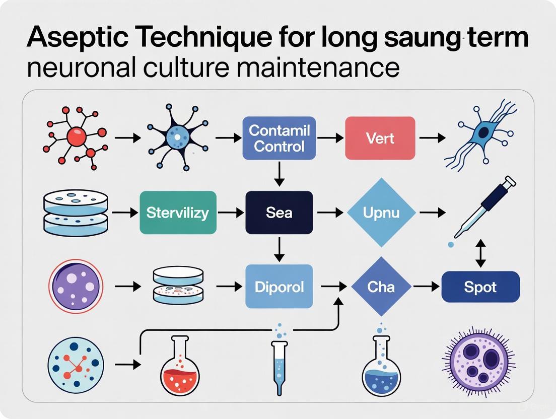
Workflow for Contamination Control
The following diagram illustrates the logical relationship and sequential application of sterile and aseptic techniques in a neuronal culture workflow, highlighting key decision points for contamination control.
Diagram 1: Workflow for contamination control in neuronal culture.
The distinction between sterile and aseptic technique is foundational for successful long-term neuronal culture research. Sterile processes are used to prepare the initial environment and materials, ensuring a clean starting point. Aseptic practices are the ongoing, vigilant methods that preserve this sterility throughout the culture's lifespan. Adherence to the detailed protocols and reagent guidelines outlined in this document will significantly reduce the risk of contamination, protect the integrity of valuable neuronal samples, and ensure the generation of reliable, reproducible data essential for meaningful research and drug development.
In the context of long-term neuronal culture maintenance, the consequences of microbial contamination extend far beyond the simple loss of an experiment. Contamination represents a critical failure in aseptic technique that directly compromises scientific integrity, leading to altered cellular physiology, irreproducible data, and significant financial losses [7]. For researchers investigating delicate processes such as neurite outgrowth, synaptic maturation, and network formation, even low-level contaminants can profoundly influence experimental outcomes by introducing uncontrolled variables that distort the very phenomena under investigation [8]. This application note delineates the multifaceted impacts of contamination, provides advanced protocols for its detection and prevention, and establishes a framework for quality control essential for maintaining the phenotypic fidelity of neuronal models in CNS drug discovery.
Quantitative Impacts of Contamination on Research Outcomes
The consequences of contamination manifest across three primary domains: direct cellular compromise, resource depletion, and scientific validity erosion. The tabulated data below synthesizes findings from controlled studies on contaminated cultures.
Table 1: Documented Consequences of Culture Contamination in Biomedical Research
| Impact Category | Specific Effect | Quantitative/Measurable Outcome | Experimental Consequence |
|---|---|---|---|
| Cellular Viability & Function | Reduced cell density and proliferation | Up to 100% culture loss in bacterial/yeast contamination [9] | Complete experiment termination |
| Altered neuronal metabolism | Shift from OXPHOS to glycolysis in hyperglycemic conditions [10] | Skewed metabolic studies | |
| Disrupted neurite outgrowth | Inhibition of neurovascular maturation and axon elongation [11] | Compromised neurodevelopment models | |
| Compromised network activity | Decreased synchronous bursting and correlation metrics [12] | Invalid neurophysiological data | |
| Resource Implications | Direct financial costs | Loss of specialized media, reagents, and primary cells | Increased research expenditures |
| Time investment | Weeks to months of lost research time | Delayed project timelines | |
| Personnel effort | Redundancy in experimental repeats | Decreased laboratory productivity | |
| Data Integrity | Uncontrolled variables | Non-physiological inflammatory responses [10] | Confounded mechanistic insights |
| Irreproducible results | Inconsistent inter-laboratory findings | Compromised scientific validity |
Advanced Detection Methodologies for Contaminant Identification
Early contaminant detection is paramount for mitigating downstream consequences. While conventional microscopy reveals advanced contamination, emerging technologies now enable identification at previously undetectable stages.
Protocol: Label-Free Contamination Monitoring Using Quantitative Phase Imaging
Principle: Quantitative Oblique Back-illumination Microscopy (qOBM) exploits refractive index properties to generate contrast from unlabeled samples, enabling continuous, non-destructive monitoring of culture status without compromising sterility [9].
Materials:
- Compact qOBM imaging system (fits standard incubators)
- Vertical wheel bioreactor or specialized culture vessel
- Closed-loop tubing with peristaltic pump
- Imaging flow cell with scattering layer
- Standard culture media for neuronal cells
Procedure:
- Integrate the qOBM system within the cell incubator, maintaining stable environmental conditions (37°C, 5% CO₂).
- Connect bioreactor to imaging flow cell via closed-loop tubing, ensuring complete sterility at connection points.
- Program peristaltic pump to transport cells from bioreactor to flow cell at predetermined intervals (e.g., every 6-12 hours).
- Acquire qOBM images at ~10 Hz frame rate during each monitoring cycle.
- Return imaged cells to bioreactor culture vessel for continued expansion.
- Analyze acquired images for early morphological indicators of contamination:
- Detect microscopic yeast cells (larger, elliptical structures) among neuronal cells
- Identify bacterial contaminants (smaller, rod-shaped or coccoid structures)
- Monitor changes in cell density and viability markers
Validation: qOBM reliably detects microbial contaminants 12-24 hours before conventional in-line sensors (e.g., oxygen electrodes) register anomalous culture conditions [9]. The technology differentiates between yeast and bacterial contamination based on distinct morphological signatures, enabling targeted response protocols.
Protocol: Assessment of Contamination-Induced Metabolic Shifts
Principle: Contamination can alter neuronal bioenergetics by competing for nutrient resources or inducing stress responses. This protocol assesses metabolic function through respirometry and glycolytic flux measurements.
Materials:
- Extracellular flux analyzer (e.g., Seahorse XF)
- Customized neuronal media with physiological glucose (5 mM)
- Mitochondrial stress test compounds (oligomycin, FCCP, rotenone/antimycin A)
- Glycolytic stress test compounds (glucose, oligomycin, 2-DG)
Procedure:
- Culture primary neurons from embryonic mice (C57BL/6NCrl) in parallel conditions:
- Experimental: Intentionally contaminated with low-level microbial burden
- Control: Maintained under strict aseptic conditions
- Plate neurons at equal density (20,000-40,000 cells/well) in XF microplates.
- Maintain cultures in physiological glucose (5 mM) to prevent artifactual glycolytic dependence [10].
- At predetermined timepoints (DIV 7, 14, 21), perform mitochondrial and glycolytic stress tests.
- Quantify key parameters:
- Basal and maximal respiration
- ATP-linked respiration
- Proton leak
- Glycolytic capacity and reserve
Anticipated Results: Contaminated cultures typically demonstrate suppressed oxidative phosphorylation, enhanced glycolytic flux, and reduced mitochondrial spare respiratory capacity compared to aseptic controls [10]. These metabolic alterations precede overt culture collapse but significantly compromise neuronal functionality.
Visualization of Contamination Impacts and Detection Workflows
Diagram 1: Contamination Impact Pathways on Neuronal Research
Diagram 2: Contamination Response Decision Workflow
The Scientist's Toolkit: Essential Reagents and Technologies
Table 2: Research Reagent Solutions for Aseptic Neuronal Culture
| Reagent/Technology | Function/Purpose | Application Notes |
|---|---|---|
| qOBM Imaging System | Label-free, in-line contamination monitoring | Enables continuous sterile culture assessment; detects contaminants 12+ hours earlier than conventional sensors [9] |
| Physiological Media (5 mM glucose) | Maintains physiological neuronal metabolism | Prevents artifactual glycolytic dependence seen in standard 25 mM glucose media [10] |
| IncuCyte NeuroBurst Orange | Genetically encoded calcium indicator for neuronal activity | Enables longitudinal monitoring of network function; transduction efficiency indicates culture health [12] |
| Barrier Technology (Isolators) | Physical separation of culture from environment | Critical for potent compound handling; reduces contamination risk in aseptic processing [7] |
| Engineered Silk Fibroin Scaffolds | 3D structural support for neurovascular cultures | Enhances axon elongation and provides physiologically relevant microenvironment [11] |
| Automated Live-Cell Imaging (IncuCyte S3) | Non-invasive neuronal activity quantification | Tracks neurite outgrowth and network maturation without fixation artifacts [8] |
The consequences of contamination in neuronal culture systems represent a critical challenge that transcends mere technical inconvenience. As demonstrated through the quantitative impacts and detection methodologies outlined in this application note, compromised aseptic technique directly generates unreliable scientific data, particularly problematic for the extended timelines required in neuronal network maturation studies. The integration of advanced monitoring technologies like qOBM with physiological culture conditions establishes a new standard for quality control in neuroscience research. By implementing the protocols and frameworks described herein, researchers can significantly mitigate the risks of compromised viability, altered growth parameters, and wasted resources, thereby enhancing both the efficiency of drug discovery pipelines and the validity of fundamental neurobiological insights.
For researchers investigating the intricate mechanisms of brain function and neurodegeneration, maintaining healthy neuronal cultures over extended periods is a fundamental yet challenging task. The unique cellular biology of neurons—characterized by their post-mitotic nature and high metabolic rate—renders them particularly susceptible to stress in vitro. This application note details the primary vulnerabilities of neuronal cultures and provides optimized protocols designed to maintain genomic integrity and cellular health within the critical context of aseptic long-term culture maintenance.
Core Vulnerabilities of Neuronal Cultures
The challenges in long-term neuronal culture maintenance stem from intrinsic physiological properties that are essential for neuronal function in vivo but become liabilities in a culture environment.
High Metabolic Activity and Genomic Instability
Neurons are highly metabolically active cells, consuming approximately 25% of the body's glucose to produce the energy required for electrical signaling. This comes at a cost: mature neurons generate about 4.7 billion molecules of adenosine triphosphate (ATP) per second, during which 1-3% of consumed oxygen is converted to reactive oxygen species (ROS). These ROS pose a significant threat to genomic DNA, creating a higher inherent risk of genome instability compared to other somatic cells [13] [14].
Table 1: Sources of Genomic Stress in Neurons
| Stress Category | Specific Stressors | Impact on Genomic Integrity |
|---|---|---|
| Endogenous Metabolic | Reactive oxygen species (ROS) from oxidative phosphorylation [13] [14] | DNA strand breaks, base modifications |
| Physiological Activity | Neuronal firing, immediate early gene (IEG) expression [13] [14] | Controlled double-strand breaks (DSBs) for gene regulation |
| Exogenous Chemical | Alcohol, cocaine, methamphetamine [13] [14] | Accumulation of DSBs and other DNA lesions |
| Age-Related | Accumulation of DNA damage, failure of repair systems [15] | Increased mutation load, aberrant splicing |
The Challenge of DNA Repair in Post-Mitotic Cells
Unlike most cell types, neurons have limited regenerative capacity and cannot be easily replaced once damaged. Postnatal neurogenesis in the brain is restricted to few regions. Consequently, neurons must rely on sophisticated molecular mechanisms to maintain genomic integrity throughout their long lifespan [13] [14]. Failure in DNA damage response (DDR) pathways is linked to severe neurological disorders. For example, mutations in ATM (involved in DSB repair) cause ataxia-telangiectasia, while XRCC1 mutations (involved in single-strand break repair) lead to cerebellar ataxia [13] [14].
Age-Associated Deterioration of RNA Biology
Aging neurons experience a broad dysregulation of RNA metabolism. Key RNA-binding proteins, particularly spliceosome components, are downregulated and mislocalized from the nucleus to the cytoplasm. The dementia-associated protein TDP-43 mislocalizes in aged neurons, leading to widespread alternative splicing errors [15]. Furthermore, aged neurons suffer from chronic cellular stress that impairs the proper sequestration of splicing proteins into stress granules, compromising the cellular stress response and overall neuronal resilience [15].
Essential Protocols for Long-Term Neuronal Culture
Protocol: Culturing of Adult Central Nervous System (CNS) Neurons
The inability to culture mature adult CNS neurons has historically limited the study of adult neuronal physiology. This protocol, adapted from van Niekerk et al. (2022), enables the culture of neurons from adult mice (up to postnatal day 90) from various brain regions, including the hippocampus, cortex, brainstem, and cerebellum [16].
Key Modifications for Adult Neurons:
- Gentle Dissociation: Use of a gentle mechanical dissociator (Octo Dissociator) with enzymes (papain and DNase) at 37°C for 30 minutes minimizes shear stress [16].
- Tissue Handling: Process individual brain regions as single 4-8 mm tissue blocks without further chopping to minimize trauma [16].
- Enhanced Survival: Addition of 20 ng/mL Brain-Derived Neurotrophic Factor (BDNF) after the Percoll gradient step is critical for mature cortical neuron survival [16].
- Neuronal Enrichment: Use a negative selection process with a cocktail of biotinylated antibodies against non-neuronal cells (astrocytes, oligodendrocytes, microglia, endothelial cells) and magnetic separation to enrich for neurons [16].
Protocol: Primary Culture of Mouse Fetal Hindbrain Neurons
This protocol provides a reliable method for dissociating and culturing embryonic mouse hindbrain neurons, a region critical for vital functions like breathing and heart rate control, but for which culture protocols are scarce [17].
Key Steps:
- Dissection: Isolate hindbrains from E17.5 mouse fetuses, carefully removing the cortex, cerebellum, and meninges.
- Dissociation: Use a combination of enzymatic loosening (Trypsin/EDTA) and gentle mechanical trituration with fire-polished Pasteur pipettes of decreasing diameter.
- Culture Medium: Use Neurobasal Plus Medium supplemented with B-27 Plus Supplement and GlutaMax.
- Glial Control: To prevent astrocyte overgrowth, add CultureOne supplement at the third day in vitro (DIV3). Cultures develop extensive branching and form functional synapses by DIV10 [17].
Protocol: Transient Transfection of Primary Neurons
Genetic manipulation in primary neurons is essential for functional studies. This protocol outlines two non-viral methods suitable for different stages of neuronal development [18].
Electroporation (for freshly isolated neurons):
- Application: Best for neurons in suspension before plating.
- Efficiency: Can reach up to 30%.
- Advantage: Less toxic than some other methods and versatile for different macromolecules.
Cationic Lipid Transfection (for adherent neurons):
- Application: For neurons that have been cultured for a few days and have developed neurites.
- Efficiency: Lower (1-2%), but results in higher transgene expression levels.
- Advantage: Avoids physical stress from electric pulses, leading to better survival of delicate, adherent neurons [18].
The Scientist's Toolkit: Essential Reagents
Table 2: Key Research Reagent Solutions for Neuronal Culture
| Reagent/Catalog Number | Function in Protocol |
|---|---|
| BDNF (450-02) [16] | Critical survival factor for mature cortical neurons; added post-dissociation. |
| B-27 Supplement (17504044) [18] | Serum-free supplement used in maintenance media to support neuronal health. |
| Papain & DNase [16] | Enzymatic cocktail for gentle tissue dissociation in adult neuron culture. |
| CultureOne (A3320201) [17] | Chemically defined supplement added at DIV3 to control astrocyte expansion. |
| Poly-L-Lysine (P2636) [18] | Substrate coating for plate preparation to promote neuronal attachment. |
| MACS Neurobasal Medium [16] | Optimized base medium for culturing adult CNS neurons. |
| Antibody Cocktail (Anti-astrocyte, -oligodendrocyte, etc.) [16] | For negative selection and enrichment of neurons during adult CNS culture. |
Visualization of Vulnerability Pathways and Workflows
Pathways of Neuronal Vulnerability and Integrity
Experimental Workflow for Adult CNS Neuron Culture
The successful long-term maintenance of neuronal cultures demands a rigorous aseptic technique coupled with a deep understanding of neuronal cell biology. The protocols and analyses presented here provide a framework for supporting the viability and genomic integrity of these sensitive cells. By addressing their unique metabolic, genomic, and age-related vulnerabilities, researchers can create more physiologically relevant models to advance our understanding of brain function and disease.
Maintaining aseptic conditions is the cornerstone of successful long-term neuronal culture. The integrity of research on neuronal development, function, and disease mechanisms hinges on the ability to sustain cultures free from biological contamination and non-neuronal cell overgrowth. For neuronal cultures, which often require weeks or months to fully mature and model age-related processes, a single contamination event can compromise months of dedicated work [19] [20]. This application note details the essential components of aseptic technique—sterile work area, personal hygiene, and sterile reagents—within the specific context of long-term neuronal culture maintenance. Adherence to these protocols ensures the reliability and reproducibility of data generated from these sophisticated in vitro models.
The Sterile Work Area
A dedicated and properly maintained sterile work area is the first line of defense against contamination in neuronal cell culture.
Primary Equipment: The Biosafety Cabinet
The laminar flow hood, or biosafety cabinet, creates a physical barrier between the user and the sterile cell culture.
- Setup and Placement: The cabinet should be positioned in an area with no through traffic, free from drafts from doors, windows, and other equipment to maintain uninterrupted laminar airflow [1]. The cabinet must be left running at all times, only being turned off for extended non-use periods [1].
- Routine Decontamination: Meticulous cleaning is non-negotiable. The work surface must be wiped thoroughly with 70% ethanol before work is initiated, during work (especially after any spillage), and after work is completed [1]. The use of ultraviolet light to sterilize the air and exposed surfaces between uses is also recommended [1].
- Aseptic Work Practices: The work surface should be uncluttered, containing only items required for the specific procedure. It is critical to work slowly and deliberately, minimizing the movement of hands and materials over open sterile containers to prevent the introduction of airborne contaminants [1].
Special Considerations for Long-Term Neuronal Cultures
The extended duration of neuronal cultures introduces unique challenges. A key advancement is the use of culture dish lids that form a gas-tight seal and incorporate a transparent hydrophobic membrane. This membrane is selectively permeable to O₂ and CO₂ but highly impermeable to water vapor, which drastically reduces media evaporation in non-humidified incubators and prevents airborne contamination. This approach has been shown to support the robust health and spontaneous electrical activity of dissociated cortical neuron cultures for over a year [19].
Table: Key Requirements for Maintaining a Sterile Work Area
| Component | Key Requirement | Purpose in Neuronal Culture |
|---|---|---|
| Biosafety Cabinet | Located in low-traffic, draft-free area; surface wiped with 70% ethanol before/after use [1] | Protects sterile cultures, media, and reagents from airborne contaminants during frequent feeding and manipulation. |
| Work Surface | Uncluttered; contains only items for the immediate procedure [1] | Minimizes turbulence and accidental contamination during complex, multi-step neuronal differentiation protocols. |
| Incubation | Use of membrane-sealed culture dishes for long-term studies [19] | Prevents media osmolarity shifts from evaporation, ensuring neuronal health over months of culture. |
Sterile Work Area Maintenance Workflow
Personal Hygiene and Protective Equipment
The researcher is a primary source of contamination. Rigorous personal hygiene and correct use of personal protective equipment (PPE) are essential to form a barrier between the operator and the sterile cell culture [1] [21].
Foundational Hygiene Practices
- Hand Washing: Thorough hand washing is the first critical step before entering the laboratory or handling culture materials. The process should last at least 20 seconds using warm water and antimicrobial soap, paying particular attention to areas between fingers and under nails where microorganisms accumulate [21].
- Protective Apparel: A clean, properly fastened laboratory coat or gown prevents particles from personal clothing from entering the workspace. Sterile surgical gloves create a crucial barrier between the hands and the cell culture environment [1] [21]. Gloves should be changed when contaminated.
- Hair and Behavior: Long hair must be securely tied back and contained within a head cap to prevent contamination and maintain a clear field of view [1] [21]. Researchers should avoid talking, singing, or whistling when performing sterile procedures to minimize the production of aerosols and droplets [1].
Maintaining Glove Sterility
During extended procedures, such as the dissection of primary neuronal tissues or the passaging of neural stem cells, glove sterility must be actively maintained.
- Systematic Sterilization: Gloves should be sterilized regularly with 70% isopropanol—every 15-20 minutes during prolonged work, after touching any non-sterile surface (e.g., microscope, laboratory notebook), and when moving between different areas of the workspace [21].
- Proper Technique: The application must ensure complete coverage of all glove surfaces, including the wrists and between fingers. The isopropanol must be allowed to air dry completely (approximately 30 seconds) before resuming work to ensure maximum antimicrobial activity and to prevent introducing the chemical into the cell culture system [21].
Table: Essential Personal Protective Equipment (PPE) and Hygiene Practices
| PPE/Hygiene Element | Protocol Specification | Rationale |
|---|---|---|
| Hand Washing | 20+ seconds with antimicrobial soap, focusing on nails and between fingers [21] | Removes transient microorganisms and loose skin cells, the most common contamination vectors from the researcher. |
| Laboratory Gloves | Sterile, disposable; sterilized with 70% isopropanol every 15-20 min during long procedures [21] | Creates a primary sterile barrier; regular decontamination maintains this barrier throughout complex protocols. |
| Laboratory Gown | Clean, dedicated, and properly fastened [1] [21] | Prevents contamination from personal clothing, such as lint and skin flakes, from entering the sterile field. |
| Hair Management | Securely tied back and fully contained [1] | Prevents hair and associated scalp microorganisms from falling into cultures or obstructing the aseptic field. |
Sterile Reagents and Media
The sterility of all reagents, media, and solutions that contact the cells is paramount. This is especially true for neuronal cultures, which often use complex, nutrient-rich media that can readily support the growth of contaminants.
Sourcing, Handling, and Quality Control
- Sterilization: All reagents and media prepared in the laboratory must be sterilized using the appropriate procedure, typically filtration through a 0.22 µm filter [1].
- Surface Decontamination: The outside of all bottles, flasks, and plates must be wiped with 70% ethanol before being introduced into the sterile work area [1].
- Aseptic Handling: To avoid contamination, pouring media and reagents directly from bottles or flasks should be avoided. Instead, sterile glass or disposable plastic pipettes with a pipettor should be used. Each pipette should be used only once to avoid cross-contamination [1]. Containers must be capped immediately after use, and multi-well plates should be sealed with tape or stored in resealable bags to prevent microbial entry [1].
- Quality Checks: Reagents and media should be inspected visually before use. Any signs of cloudiness, unusual color, or floating particles indicate potential contamination, and the item should be decontaminated and discarded immediately. Any foul smell is also a clear indicator of contamination [1].
Reagents for Neuronal Culture
The culture of specific neuronal cell types requires tailored media formulations to support survival and maturation. The table below outlines key reagents used in various neuronal culture protocols from the literature.
Table: Research Reagent Solutions for Neuronal Cell Culture
| Reagent / Material | Example Function in Neuronal Culture | Application Example |
|---|---|---|
| Neurobasal Medium | Base medium optimized for long-term survival of hippocampal and other CNS neurons [5] [20]. | Primary cortical and hippocampal neuron culture [5]. |
| B-27 Supplement | Serum-free supplement providing hormones, antioxidants, and other essential factors for neuron health [5] [20]. | Added to Neurobasal medium for primary neurons and hiPSC-derived neurons [5] [20]. |
| N2 Supplement | Defined supplement supporting the growth and differentiation of neural progenitor cells [22]. | Used in neural progenitor cell and motor neuron differentiation media [22]. |
| Matrigel | Basement membrane matrix providing a physiological substrate for cell attachment and differentiation. | Coating culture vessels for hiPSCs and neuronal cultures [22] [20]. |
| Growth Factors (BDNF, GDNF, NT-3) | Trophic factors that promote neuronal survival, maturation, and synaptic development. | Component of neuronal maturation medium for hiPSC-derived motor neurons [22]. |
| Cytosine Arabinoside (Ara-C) | Antimitotic agent used to inhibit the proliferation of non-neuronal cells like glia. | Added to primary neuronal cultures to enhance neuronal purity [20]. |
Sterile Reagent Handling Workflow
Integrated Protocol for Aseptic Technique in Neuronal Culture
The following consolidated protocol integrates the three core components for a common procedure: feeding a long-term neuronal culture.
Application: Routine media change for hiPSC-derived neurons or primary neuronal cultures. Objective: To replenish nutrients and remove waste products without introducing contamination.
Step-by-Step Method:
Preparation:
- Gather all necessary reagents in the biosafety cabinet: pre-warmed neuronal culture medium (e.g., Neurobasal/B-27), sterile pipettes, and waste container.
- Wipe the exterior of all reagent bottles with 70% ethanol before placing them in the cabinet.
- Wash hands thoroughly and don a laboratory coat and sterile gloves.
Aseptic Setup:
- Wipe the gloved hands with 70% ethanol.
- Organize the work area to ensure a clear, logical workflow.
Media Exchange:
- Carefully remove the neuronal culture vessel from the incubator.
- Inside the biosafety cabinet, slowly aspirate the spent media using a sterile pipette, being careful not to disturb the neuronal monolayer.
- Slowly add the fresh, pre-warmed neuronal culture medium to the side of the vessel.
- If using standard dishes, replace the lid immediately. For long-term studies, transfer cultures to membrane-sealed dishes [19].
Completion:
- Return the culture to the incubator.
- Dispose of all waste materials appropriately.
- Wipe down the biosafety cabinet surface with 70% ethanol.
By systematically applying the principles of a sterile work area, impeccable personal hygiene, and the use of sterile reagents, researchers can reliably maintain the health and integrity of long-term neuronal cultures, thereby ensuring the generation of robust and meaningful scientific data.
Laminar Flow Hoods: Principles and Selection for Neuronal Culture
The maintenance of long-term neuronal cultures is a cornerstone of neuroscience research, requiring an environment that is meticulously controlled to prevent microbial contamination and ensure the validity of experimental outcomes. The foundation of this aseptic environment is the laminar flow hood, which provides a continuous stream of HEPA-filtered air to create an ISO Class 5 workspace, containing fewer than 100 particles (≥0.5μm) per cubic foot [23]. Proper utilization of this technology can reduce contamination rates from typical laboratory levels of 15-20% to below 3%, a critical threshold for the success of sensitive and long-duration neuronal studies [23].
Scientific Principles and Contamination Control
Laminar flow hoods operate by directing HEPA-filtered air in parallel, unidirectional streamlines across the work surface. This laminar flow, characterized by a Reynolds number below 2,300, is essential for predictably moving airborne particles away from the critical work zone [23]. The High-Efficiency Particulate Air (HEPA) filter is the core component, demonstrating 99.97% efficiency at capturing particles ≥0.3μm through a combination of interception, impaction, and diffusion mechanisms [23] [24]. For neuronal culture, where even minor contamination can compromise weeks of work, this level of protection is indispensable. The efficacy of this system is demonstrated in the following contamination data:
Table 1: Contamination Control Efficacy in Different Laboratory Environments [23]
| Environment Type | Particles ≥0.5μm per ft³ | Microbial CFU/m³ | ISO Class | Typical Contamination Rate |
|---|---|---|---|---|
| Uncontrolled Laboratory | 500,000+ | 100+ | 9 | 15-20% |
| Standard Laboratory | 100,000-350,000 | 20-50 | 7-8 | 8-12% |
| Laminar Flow Hood | <100 | <1 | 5 | 0.5-3% |
| Clean Room | <10 | <0.1 | 3-4 | <0.5% |
Selecting the Appropriate Laminar Flow Hood
The two primary designs are vertical and horizontal laminar flow hoods, each with distinct advantages for specific applications.
Vertical Laminar Flow Hoods: In these units, clean air is directed from the top of the hood downward toward the work surface. This design is space-efficient, requires less depth, and minimizes the risk of airflow obstruction. Critically, it offers improved operator safety by directing potentially aerosolized materials away from the user, a significant consideration when working with viral vectors or other bioactive agents in neuronal transduction studies [24].
Horizontal Laminar Flow Hoods: These hoods direct air horizontally from the back of the unit, through the HEPA filter, and across the work surface toward the user. This provides a consistent, uniform cleansing effect with a uniform velocity, which is highly effective at sweeping contaminants away from open culture vessels [23] [24]. However, it provides less operator protection as the user is downstream of the work materials.
For standard, non-hazardous neuronal culture maintenance and manipulation, a horizontal flow hood is typically ideal due to its excellent product protection and clear work visibility [23]. When working with potentially hazardous materials, such as those involved in certain gene therapy approaches, a Class II Biological Safety Cabinet (a type of vertical flow cabinet) must be used to ensure both product and operator protection [23].
Table 2: Comparison of Laminar Flow Hood Types for Neuronal Culture
| Feature | Horizontal Flow Hood | Vertical Flow Hood | Biological Safety Cabinet |
|---|---|---|---|
| Airflow Pattern | Back to front [24] | Top to bottom [24] | Top to bottom, with exhaust [23] |
| Product Protection | Excellent [23] | Good | Excellent [23] |
| Operator Protection | Limited [23] | Good [24] | Excellent [23] |
| Ideal Application | Non-hazardous culture work, media prep [23] | General sterile work; tasks requiring operator safety [24] | Work with hazardous/infectious agents [23] |
| Work Visibility | Excellent [23] | Good | Variable |
Laboratory Layout: Traffic Control and Dedicated Culture Areas
The sterile field within a laminar flow hood is highly susceptible to disruption from external air currents. Therefore, the broader laboratory layout and traffic control are not merely logistical concerns but are integral to maintaining aseptic conditions.
Optimal Laminar Flow Hood Placement
The positioning of the hood within the laboratory is a critical first step. Key guidelines include [23]:
- Minimum Clearances: The hood should be positioned at least 30 cm from walls or obstructions and 1.5 meters from doors, high-traffic walkways, or opposing workstations.
- Draft Avoidance: The unit must be placed away from air conditioning vents, doors, windows, and any other equipment that generates air currents.
- Stable Environment: The hood should be on a stable, level surface in a room with controlled ambient conditions (18-25°C, 30-60% relative humidity) and a positive pressure relative to adjacent spaces to minimize infiltration of unfiltered air [23].
Establishing a Unidirectional Workflow and Dedicated Zones
Adopting a "clean to dirty" workflow within the laboratory minimizes the risk of cross-contamination. This involves designating separate areas for different activities.
Diagram 1: Laboratory Material and Workflow
The workflow should enforce a clear, logical progression. All materials should enter through a designated "clean" preparation area before being introduced into the laminar flow hood. Cultures are then moved to a dedicated incubation area, and finally to analysis stations, with clear procedures to prevent back-tracking of contaminated materials into clean zones.
Protocols for Laminar Flow Hood Operation and Maintenance
A laminar flow hood is only as effective as the protocol governing its use. The following application note details a standardized procedure for its operation in the context of neuronal culture maintenance.
Pre-Use Setup and Sanitization Protocol
- Activation: Turn on the laminar flow hood and allow it to operate for at least 15-30 minutes before use. This purge time is essential to establish stable, laminar airflow patterns and remove particulate contaminants from the work zone [23] [24].
- Personal Hygiene: Remove all jewelry and scrub hands and arms up to the elbows with an antibacterial agent. Wear sterile, lint-free gloves, a lab coat, and a mask that fully covers hair [24].
- Surface Sanitization:
- Remove all items from the work surface.
- Using a lint-free cleanroom wipe or microfiber cloth, thoroughly wipe down all interior surfaces with 70% isopropyl alcohol or ethanol [23] [24].
- Employ a systematic cleaning pattern: start with the back wall (top to bottom), then the side walls (top to bottom, side-to-side), and finally the base (back to front) [24].
- Allow a minimum 2-minute contact time for the disinfectant to be effective before allowing surfaces to air-dry [23].
- Workspace Organization: Assemble all necessary pre-sanitized supplies. Position smaller items closer to the HEPA filter and larger items further away to minimize disruption of the unidirectional airflow. Never allow objects to block the grille or the path between the HEPA filter and the work area [23] [24].
Aseptic Technique During Neuronal Culture Procedures
Diagram 2: Aseptic Workflow in Laminar Hood
- Material Handling: Work should be performed within 6-10 inches of the filter face, where the laminar flow is most effective [25]. Keep all open culture vessels (dishes, flasks) within this "clean" airstream at all times.
- Movement and Technique: Use slow, deliberate movements to avoid creating turbulence. When working, hold tools and vessels at an angle to avoid contaminating the sterile interior by reaching over open tops. Minimize the time that culture vessels are open.
- Liquid Transfers: When using pipettes, ensure the tip does not touch non-sterile surfaces. Wipe ampules and vials with 70% alcohol before opening. Avoid passing your hands or body over open sterile containers.
Routine Cleaning, Maintenance, and Certification
- Daily/Pre-Use Cleaning: Wipe all surfaces with 70% alcohol before and after each use [24].
- Weekly Cleaning: Perform a more thorough cleaning of the interior and exterior of the hood at least once a week, using a combination of 70% ethanol and a surface disinfectant [24].
- Filter Maintenance: Replace the pre-filter regularly (every 3-6 months, depending on use) to maintain airflow and protect the lifespan of the expensive HEPA filter [25]. The HEPA filter itself should last for several years but requires regular integrity testing.
- Professional Certification: The hood must be professionally certified upon installation, after any relocation or repair, and at least annually thereafter. Certification includes HEPA filter integrity testing (DOP/PAO challenge), airflow velocity measurement (target 0.45-0.55 m/s), and smoke pattern testing to verify laminar conditions [23].
The Scientist's Toolkit: Essential Reagents and Materials
The following table details key reagents and materials essential for maintaining aseptic conditions and supporting long-term neuronal cultures.
Table 3: Essential Research Reagent Solutions for Aseptic Neuronal Culture
| Item | Function/Application | Key Considerations |
|---|---|---|
| 70% Isopropyl Alcohol | Primary surface and skin disinfectant [23] [24]. | Effective contact time of ≥2 minutes is critical; higher concentrations evaporate too quickly [23]. |
| HEPA Filter | Primary filtration unit for laminar flow hood; removes 99.97% of particles ≥0.3μm [23] [24]. | Requires annual integrity certification; lifespan of 1-2 years under proper use and pre-filtration [23]. |
| Pre-Filter | Captures large particles (dust) to extend the service life of the HEPA filter [25]. | Typically 30-40% efficiency; should be replaced every 3-6 months [23] [25]. |
| Lint-Free Wipes | For applying disinfectants to hood surfaces without shedding particles [24]. | Microfiber or cleanroom wipes are preferred over cotton, which can leave fibers. |
| Sterile Pipettes & Tips | For aseptic transfer of media, reagents, and neuronal cell suspensions. | Must be pre-sterilized (e.g., gamma-irradiated) and used within the laminar flow hood. |
| Neuronal Culture Media | Nutrient-rich solution supporting survival and growth of neurons. | Often contains neurotrophic factors (e.g., BDNF, GDNF); must be filter-sterilized (0.22μm) before use. |
| Antimycotics/Antibiotics | Suppress the growth of latent bacterial or fungal contaminants in culture. | Use is debated; can mask low-level contamination. For critical work, antibiotic-free conditions are preferable. |
A Step-by-Step Aseptic Protocol for Neuronal Culture Maintenance
Within the context of long-term neuronal culture maintenance, the aseptic technique is not merely a best practice but an absolute necessity. Successful neuronal culture research hinges on the ability to maintain sterile conditions, thereby preserving the physiological relevance and genetic stability of delicate cultures over weeks or months [26]. The pre-work phase—sterilizing the work surface and organizing materials—establishes the foundational barrier between the external environment and the sterile cell culture. This protocol is specifically designed for researchers and scientists engaged in drug development, where the integrity of neuronal cultures is critical for generating reliable, high-quality data.
Key Principles and Definitions
- Aseptic Technique: A set of procedures designed to create a barrier between microorganisms in the environment and the sterile cell culture, thereby reducing the likelihood of contamination. It focuses on preventing the introduction of contaminants into a previously sterilized environment [26].
- Sterile Technique: Methods that ensure a space is completely free of any microorganisms that could cause contamination. For example, a cell culture hood is initially sterilized using sterile techniques, while aseptic techniques maintain its sterility during use [26].
- Critical Sites and Parts: In Aseptic Non-Touch Technique (ANTT), "key parts" are the critical parts of equipment that must remain sterile, and "key sites" are the open pathways and insertion points on a patient or, by analogy, the culture vessel [27].
Protocols for Sterilizing the Work Surface
Preparation of the Biosafety Cabinet (BSC)
The biosafety cabinet is the cornerstone of a sterile work area for neuronal cultures. Proper setup and preparation are critical.
- Location and Pre-Cleaning: Position the BSC in an area free from drafts, doors, windows, and high-traffic flow to minimize airborne disturbances [26]. Before use, clear the work surface of all clutter, as it should not be used as a storage area [26].
- Surface Decontamination: Thoroughly wipe the entire work surface, interior walls, and any stationary equipment with 70% ethanol before commencing work and after any spillage [26]. This is the primary method for disinfecting the work area.
- Ultraviolet (UV) Light Sterilization: Use ultraviolet light to sterilize the air and exposed work surfaces in the cell culture hood between uses. Note: The BSC should be left running at all times, only turned off for extended non-use periods [26].
Creating a Sterile Field with a Bunsen Burner
In laboratories without a BSC, a Bunsen burner can be used to create a sterile field on an open bench.
- Mechanism of Action: Working beside a Bunsen burner creates an upward flow of air through convection, which lowers the risk that dust or other contaminants will settle on the sterile surface or equipment [28].
- Setup: Set up the Bunsen burner to your dominant side on the bench (e.g., to your right if you are right-handed) and arrange your materials to the other side. Work slowly and deliberately within this sterile field [29].
Table 1: Comparison of Work Surface Sterilization Methods
| Feature | Biosafety Cabinet (BSC) | Bunsen Burner |
|---|---|---|
| Primary Sterile Barrier | HEPA-filtered laminar airflow | Convection updraft from flame |
| Best Suited For | BSL-1 and BSL-2 organisms; long-term cultures | BSL-1 organisms only [29] |
| Key Preparation Step | Wipe surfaces with 70% ethanol; UV sterilization | Clear clutter; flame the work area vicinity |
| Use in Cell Culture Hood | Recommended and essential | Not recommended or necessary [26] |
Protocols for Organizing Materials
Efficient organization of materials within the sterile field is paramount to maintaining asepsis and ensuring a smooth workflow.
Pre-Sterilization of Materials and Reagents
- Liquid Reagents and Media: All reagents, media, and solutions must be sterilized using the appropriate procedure (e.g., autoclaving or sterile filtration) prior to use [26]. Wipe the outside of all bottles, flasks, and plates with 70% ethanol before introducing them into the sterile work area [26].
- Sterile Instruments: Use sterile glass or disposable plastic pipettes for liquid handling. Sterilize metal instruments like forceps or spatulas by flaming until they are red-hot before use and between different samples [29] [28].
Spatial Organization within the Work Area
A logical arrangement of materials prevents unnecessary movements and cross-contamination.
- Zonal Organization: Arrange all supplies to maximize work efficiency. A common practice is to place agar plates or culture dishes to your left, cell cultures and media bottles in the center, and the Bunsen burner (if used) or waste receptacle to your right [29].
- Handling Practices: Loosen the caps of all tubes, flasks, and bottles before starting the procedure so they can be easily opened with one hand [29]. When removing a cap, never place it with the opening facing up; always place it face down on the sterile work surface [26]. Use each sterile pipette only once to avoid cross-contamination [26].
The following workflow diagram illustrates the logical sequence of pre-work preparation for neuronal culture maintenance.
The Scientist's Toolkit: Essential Materials
The following table details key reagents and materials essential for the pre-work preparation phase in neuronal culture maintenance.
Table 2: Research Reagent Solutions and Essential Materials for Pre-Work Preparation
| Item | Function/Brief Explanation |
|---|---|
| 70% Ethanol Solution | The primary disinfectant for decontaminating work surfaces, the exterior of reagent containers, and gloved hands [26]. |
| Personal Protective Equipment (PPE) | Lab coat, gloves, and safety glasses form a barrier to protect the culture from shed skin and the researcher from hazardous agents [26]. |
| Sterile Wipes (e.g., Kimwipes) | Used in conjunction with 70% ethanol for effective surface decontamination without leaving lint. |
| Biosafety Cabinet (BSC) | Provides a HEPA-filtered, sterile work environment, protecting both the culture and the researcher from aerosols [26]. |
| Pre-sterilized Pipettes and Tips | For sterile liquid handling; single-use to prevent cross-contamination between different reagents and cultures [26]. |
| Sterile Culture Media & Reagents | Nutrient-rich solutions specifically formulated to support the growth and maintenance of neuronal cells. |
Validation and Troubleshooting
Understanding common contamination sources informs the emphasis on rigorous pre-work preparation.
Table 3: Common Sources of Contamination in Cell Culture
| Contamination Source | Relative Risk/Impact | Mitigation Strategy from Pre-Work Protocol |
|---|---|---|
| Nonsterile Work Surfaces | High | Systematic disinfection with 70% ethanol before and during work [26]. |
| Airborne Particles | High | Use of BSC or Bunsen burner convective field; working slowly and deliberately [29] [26]. |
| Nonsterile Reagents/Media | Critical | Sterilization prior to use; wiping exteriors with ethanol; inspection for cloudiness or unusual color [26]. |
| Improper Handling | Moderate | Using sterile instruments; not touching critical parts; proper cap placement [29] [26]. |
Troubleshooting Common Pre-Work Issues
- Condensation on Agar Plates or Culture Dishes: If plates stored at 4°C show condensation, remove them several hours before use and spread them in small stacks to dry at room temperature. Wet surfaces can facilitate contamination spread during streaking or manipulation [29].
- Suspected Contaminated Reagents: If reagents appear cloudy, contain floating particles, or have an unusual color or smell, they should be decontaminated and discarded immediately [26].
Proper Personal Protective Equipment (PPE) and Personal Hygiene Practices
Maintaining aseptic conditions is paramount for the success and reproducibility of long-term neuronal culture research. Contamination can compromise cellular viability, alter phenotypic expression, and invalidate experimental outcomes, leading to costly delays and unreliable data. Proper Personal Protective Equipment (PPE) and stringent personal hygiene practices form the primary barrier between the researcher and the sterile cell culture environment, especially critical when working with sensitive neuronal cells that require extended maintenance periods. This protocol outlines evidence-based procedures for establishing effective contamination control, providing researchers with a standardized approach to safeguarding valuable neuronal cultures throughout their lifecycle.
Core Principles of Aseptic Technique
Aseptic technique refers to a set of procedural guidelines designed to prevent contamination by pathogens and other microorganisms. In cell culture laboratories, these techniques create a barrier between microorganisms in the environment and the sterile cell culture [1]. The core distinction between related terms is foundational:
- Clean Technique: Focuses on reducing the overall number of germs but does not completely eliminate them. An example is using boxed gloves that are free from dirt but not sterile [30].
- Aseptic Technique: A stricter standard aimed at eliminating pathogens altogether. This involves using sterile gloves, gowns, and drapes to create a controlled environment [30].
- Sterile Technique: Often used interchangeably with "aseptic" to describe the outcome, though "sterile" typically describes the condition of the environment and instruments, while "aseptic" describes the processes used to achieve it [1] [30].
The consequences of improper technique in neuronal culture are severe, potentially leading to altered growth patterns, compromised cellular viability, and complete loss of irreplaceable primary cultures or long-term experiments [1].
Personal Protective Equipment (PPE) Requirements
PPE serves as the primary physical barrier protecting both the researcher and the cell culture from cross-contamination. The following table summarizes the essential PPE components and their specific functions in a neuronal culture context.
Table 1: Essential Personal Protective Equipment for Neuronal Cell Culture
| PPE Component | Specifications & Material | Primary Function in Neuronal Culture |
|---|---|---|
| Gloves | Sterile, non-powdered nitrile or latex | Creates a crucial barrier between hands and the culture environment; prevents introduction of skin flora and contaminants [1] [21]. |
| Laboratory Gown | Clean, properly fastened, dedicated for lab use | Prevents particles from personal clothing and skin from entering the sterile workspace [1] [21]. |
| Eye Protection | Safety glasses or face shield | Protects eyes from potential splashes of media, reagents, or other hazardous materials [1]. |
| Respiratory Protection | Face masks or, when required for aerosols, N-95 respirators | Reduces the risk of contamination from talking, singing, or whistling during sterile procedures [1] [31]. |
| Head Cap | Disposable bouffant cap | Contains hair and dander, preventing direct contamination of cultures and maintaining a clear working field [21]. |
Special Considerations for Neuronal Cultures
Given the extended duration and sensitivity of neuronal cultures, additional precautions are necessary. Sterile gloves are mandatory rather than simply clean gloves, as used in some clinical settings [30]. Furthermore, during extended culture sessions, such as those involving lengthy patch-clamp recordings or complex transfections, regular sterilization of gloves with 70% (v/v) sterile isopropanol is critical to maintain aseptic conditions throughout the procedure [21].
Personal Hygiene Protocols
Meticulous personal hygiene is the first line of defense in contamination control. The following protocols must be rigorously followed.
Hand Hygiene Protocol
Hand hygiene is the single most important practice for reducing the transmission of infectious agents [31] [32]. The "Five Moments for Hand Hygiene" framework, adapted for the cell culture laboratory, dictates hand cleaning at these critical times [31]:
- Immediately before touching any sterile equipment or entering the culture hood.
- Before performing an aseptic task like handling cultures or placing an indwelling device (e.g., a recording electrode).
- Before moving from a soiled body site to a clean body site (e.g., after touching a non-sterile surface).
- After touching a potentially contaminated surface or equipment.
- After contact with any biological material or contaminated surfaces, including after glove removal [31] [32].
Table 2: Hand Hygiene Methods and Specifications
| Parameter | Alcohol-Based Hand Rub (ABHR) | Soap and Water Handwashing |
|---|---|---|
| Preferred Use Case | Unless hands are visibly soiled [32]. | When hands are visibly soiled, before eating, or after using the restroom [32]. |
| Procedure | 1. Apply product to palm.2. Rub over all surfaces of hands and fingers.3. Continue until hands are dry (~20 seconds) [31] [32]. | 1. Wet hands with water.2. Apply soap.3. Rub hands together vigorously for at least 15-20 seconds.4. Rinse well and dry with disposable towels [31] [32]. |
| Efficacy & Rationale | More effective at killing germs than soap; less irritating to skin with improved adherence [32]. | Physically removes debris and is essential when dealing with certain contaminants like C. difficile spores [32]. |
Additional Personal Hygiene Measures
- Hair Management: Long hair must be securely tied back and contained within a disposable head cap. This prevents direct contamination of cultures and maintains clear visibility during precise manipulations [1] [21].
- Jewelry: Remove rings, watches, and bracelets before the surgical hand scrub or donning gloves, as they can harbor microorganisms and puncture gloves [32].
- General Conduct: Avoid talking, singing, or whistling when performing sterile procedures. Work deliberately and slowly to minimize the creation of aerosols and turbulence [1].
Integrated Experimental Workflow for Aseptic Neuronal Culture
The diagram below illustrates the integrated workflow for maintaining asepsis during a typical neuronal culture maintenance session, combining PPE, hygiene, and bench practices.
The Scientist's Toolkit: Essential Research Reagent Solutions
The following reagents and materials are fundamental for executing the aseptic protocols described and maintaining the health of long-term neuronal cultures.
Table 3: Essential Reagents and Materials for Aseptic Neuronal Culture
| Item | Function/Application | Example from Literature |
|---|---|---|
| 70% Ethanol Solution | Primary disinfectant for work surfaces and the external surfaces of bottles, flasks, and equipment before introduction into the biosafety cabinet [1]. | Used for wiping work surfaces and equipment in standard cell culture protocols [1]. |
| 70% Isopropanol (v/v) | Sterilizing gloved hands during extended procedures to maintain aseptic conditions without leaving the hood [21]. | Critical for prolonged sessions like patch-clamp recording on neurons to prevent contamination [21]. |
| Sterile, Disposable Plastic Pipettes | Manipulating all liquids; used only once to avoid cross-contamination between reagents and cultures [1]. | Standard practice in neuronal culture protocols to ensure sterility [1] [33]. |
| Neurobasal Plus Medium | A defined, serum-free culture medium optimized for promoting neuronal survival and growth while inhibiting glial proliferation [33]. | Used as the base medium for culturing embryonic mouse fetal hindbrain neurons [33]. |
| B-27 Plus Supplement | A serum-free supplement designed to support the long-term survival and growth of primary CNS neurons [33]. | Component of the NB27 complete medium for hindbrain neuron cultures [33]. |
| CultureOne Supplement | A chemically defined, serum-free supplement used to control astrocyte expansion in mixed neural cultures [33]. | Added at the third day in vitro to hinder excessive astrocyte growth in mouse hindbrain cultures [33]. |
| Penicillin-Streptomycin | Antibiotic solution added to culture media to prevent bacterial contamination, particularly crucial during initial culture establishment [33]. | Included in the NB27 complete medium for primary neuronal cultures [33]. |
Adherence to the detailed protocols for PPE and personal hygiene outlined in this document is non-negotiable for the integrity of long-term neuronal culture research. These practices, when consistently and correctly applied, form a robust defense against contamination, ensuring the reliability of experimental data and the successful maintenance of sensitive neuronal networks in vitro. Integrating these aseptic techniques into every aspect of cell culture work is a fundamental responsibility of every researcher in the neuroscience and drug development fields.
In the context of long-term neuronal culture maintenance, the sterile handling of reagents is a critical determinant of experimental success. Contamination can compromise months of work, alter cellular physiology, and invalidate research findings. This application note details a targeted approach to an often-overlooked aspect of aseptic technique: the decontamination of reagent containers prior to use in a biosafety cabinet (BSC). Specifically, we provide a validated protocol for wiping container surfaces and emphasize the practice of avoiding pouring to minimize the risk of cross-contamination. This methodology is essential for researchers, scientists, and drug development professionals working with sensitive primary neurons or induced pluripotent stem cell (iPSC)-derived neural cultures, where even minor contaminant introductions can disrupt delicate cellular networks and long-term experiments.
The Critical Risk: Surface Contamination
The external surfaces of reagent containers, including bottles of culture medium, buffers, and enzyme solutions, are significant vectors for introducing contamination into sterile cell culture work areas. The risk is twofold:
- Particulate and Microbial Contamination: Dust and microbial spores can settle on containers stored in refrigerators, freezers, or on laboratory benchtops.
- Cross-Contamination with Active Agents: In laboratories handling multiple biologicals, there is a measurable risk of cross-contamination from residues of potent active pharmaceutical ingredients (APIs) or other cell products on container surfaces [34]. Research has demonstrated that residual culture medium, including its constituent proteins and nucleic acids, can persist on work surfaces and pose a cross-contamination risk during cell product processing [35]. Once dried, these residues can become particularly difficult to remove [35].
The practice of pouring, as opposed to pipetting, exacerbates these risks. The liquid stream can flow over the non-sterile outer surface of the container, carrying contaminants directly into the sterile media or onto the culture vessels.
Application Note & Protocol: External Decontamination of Reagent Containers
Principle
This protocol outlines a standardized procedure for the external decontamination of reagent containers entering a BSC. The primary objective is to eliminate surface contaminants, thereby preventing their introduction into the sterile work zone and protecting sensitive neuronal cultures.
Key Research Reagent Solutions
Table 1: Essential Materials for Container Decontamination
| Material | Function & Rationale |
|---|---|
| Sterile Wipes (Lint-Free) | To physically remove debris and apply disinfectant without shedding particles. |
| 70% (v/v) Ethanol | A broad-spectrum disinfectant that rapidly evaporates, minimizing residue. Effective against many bacteria and fungi. |
| Benzalkonium Chloride with Corrosion Inhibitor (BKC+I) | A disinfectant shown to be effective at removing residual proteins and DNA from surfaces, addressing cross-contamination risks [35]. |
| Distilled Water (DW) | Used in conjunction with wiping to remove residues; effective for both proteins and DNA without causing immobilization [35]. |
| Validated Cleaning Agent (e.g., TFD4 PF) | A phosphate-free, alkaline detergent for manual cleaning of labware, validated for removing difficult APIs [34]. |
Step-by-Step Procedure
- Preparation: Within the laboratory anteroom or a designated preparation area, gather all reagent containers required for the culture session.
- Initial Inspection: Visually inspect each container for any leaks, spills, or significant particulate matter on the exterior.
- Primary Decontamination: a. Moisten a sterile, lint-free wipe with a sufficient volume of 70% ethanol or a validated alternative disinfectant. b. Thoroughly wipe the entire external surface of the container, with particular attention to the cap or closure mechanism and the area around the neck. c. Employ a systematic wiping pattern (e.g., spiraling from the top down) to ensure complete coverage and avoid missing areas. d. Allow the disinfectant to air-dry completely. This contact time is critical for microbial inactivation.
- Transfer to BSC: Place the decontaminated containers into the BSC, arranging them logically to maintain an organized work area.
- Final Decontamination (within BSC): Upon introducing the container into the BSC, wipe its exterior again with a sterile wipe moistened with 70% ethanol. This step serves as a final safeguard against contaminants introduced during handling.
- Aseptic Dispensing: Avoid pouring. Always use a sterile serological pipette to aspirate and transfer liquids from the container to your culture vessel or other sterile tubes.
- Post-Use Handling: Before returning a partially used container to storage, wipe the exterior with 70% ethanol to remove any potential contamination acquired in the BSC.
Workflow Visualization
The following diagram illustrates the logical sequence and decision points for the decontamination protocol.
Experimental Validation & Data
Quantitative Evaluation of Cleaning Methods
The effectiveness of wiping and various disinfectants has been quantitatively assessed in studies evaluating cleaning methods to avoid cross-contamination during cell product processing. The following table summarizes key findings on the efficacy of different agents for removing residual biomolecules.
Table 2: Efficacy of Cleaning Methods for Removing Residual Proteins and DNA from Dried Culture Medium [35]
| Cleaning Method | Residual Protein | Residual DNA | Key Findings & Notes |
|---|---|---|---|
| Wiping with Distilled Water (DW) | Significantly Lower | Significantly Lower | Effective for both proteins and DNA; does not cause immobilization. |
| Wiping with Benzalkonium Chloride + Inhibitor (BKC+I) | Significantly Lower | Significantly Lower | Effective; resulted in an undetectable number of residual cells. |
| Wiping with Ethanol (ETH) | Not Effective | Not Effective | Caused protein immobilization, making residues harder to remove. |
| Peracetic Acid (PAA) | Not Effective | Effective | Suitable for nucleic acid decontamination but not for proteins. |
| UV Irradiation | Not Effective | Not Effective | Ineffective against both residual proteins and DNA. |
Establishing Residue Acceptable Limits (RALs)
For contamination control, it is essential to define acceptable limits. In cleaning validation for pharmaceuticals, a commonly referenced threshold is no more than 10 ppm of a substance in another product [34]. This principle can be adapted for critical neuroscience research, where the RAL would be a concentration of a contaminant that has no measurable effect on neuronal physiology, synapse formation, or gene expression. Analytical methods used for quality control, such as swab sampling followed by HPLC, must offer sufficient sensitivity to detect residues at or below these defined RALs [34].
Integration with Neuronal Culture Systems
The sterile handling practices described herein are foundational for maintaining the integrity of long-term neuronal cultures, which are central to modern neuroscience research. These cultures, whether derived from primary rodent tissue [5] [17] or from human induced pluripotent stem cells (iPSCs) [36], are particularly vulnerable. They are often maintained for weeks in complex, serum-free media optimized for neuronal health and synapse development [5] [17] [36]. The use of B-27 supplement and other growth factors in these media provides a rich environment not only for neurons but also for contaminating microbes. A single lapse in aseptic technique can lead to culture loss, invalidating data from sophisticated applications like patch-clamp electrophysiology, live-cell imaging of synaptic activity, or studies on neuroinflammation in iPSC-derived tri-culture systems [36]. Therefore, the rigorous decontamination of every component entering the culture environment is non-negotiable for generating reliable and reproducible results.
Maintaining the sterility of neuronal cultures is a persistent challenge that demands meticulous attention to detail. The protocol for wiping reagent containers and avoiding pouring is a simple yet powerfully effective strategy to mitigate the risk of contamination and cross-contamination. By integrating this validated practice with a comprehensive aseptic technique, researchers can significantly enhance the reliability and reproducibility of their long-term neuronal culture studies, thereby strengthening the foundation of their research in neurobiology and drug development.
Maintaining the health and integrity of primary neuronal cultures is paramount for generating reliable and reproducible data in neuroscience research. This application note details the critical laboratory techniques of slow deliberate movements and the principle of minimized exposure within the broader framework of aseptic technique for long-term neuronal culture maintenance. Evidence indicates that the variability and nature of movement in a laboratory environment can significantly impact neural activity and, by extension, the physiological state of cultured neurons [37]. Adopting these techniques is essential for minimizing external stressors and preserving the delicate homeostasis required for accurate modeling of neuronal function, development, and pathology.
Conceptual Foundation: Linking Movement to Neural Variability
Recent research underscores a profound relationship between movement patterns and neural activity. Studies in vivo have demonstrated that as subjects transition into states of disengagement, their movements become less stereotyped and more idiosyncratic. This change in movement structure is a strong predictor of both task performance and overall neural engagement state [37]. Although this research was conducted in behaving animals, the principle translates to the in vitro context: unpredictable or jarring environmental movements can induce analogous states of variability in neural cultures, potentially compromising the stability of neural encoding and increasing experimental noise.
Cultured neurons exist in a carefully balanced milieu, and external vibrations, rapid temperature shifts from open incubator doors, or sudden physical disturbances can disrupt this equilibrium. The core principle is that slow, deliberate movement minimizes unpredictable physical and acoustic vibrations, thereby supporting a more stable neural environment.
Quantitative Data for Experimental Planning
Precise execution of laboratory protocols is critical for success. The following tables summarize key quantitative data for the isolation and culture of primary neurons from different regions of the rat nervous system, enabling effective planning and standardization.
Table 1: Animal and Tissue Source Specifications for Primary Neuron Isolation
| Neural Tissue Source | Animal Age | Key Dissection Considerations |
|---|---|---|
| Cortex [5] | Embryonic Day 17-18 (E17-E18) | Limit dissection time to 2-3 minutes per embryo; total dissection time should not exceed 1 hour. |
| Hippocampus [5] | Postnatal Day 1-2 (P1-P2) | Induce hypothermia with an ice pad and use isoflurane anesthesia before dissection. |
| Spinal Cord [5] | Embryonic Day 15 (E15) | Requires skilled dissection technique to isolate the specific neural region effectively. |
| Dorsal Root Ganglia (DRG) [5] | 6-week-old young adult | Customized enzymatic and mechanical dissociation is required for this peripheral neural tissue. |
Table 2: Culture Medium Composition for Different Primary Neurons
| Component | Cortical, Hippocampal, & Spinal Cord Neurons [5] | Dorsal Root Ganglia (DRG) Neurons [5] |
|---|---|---|
| Base Medium | Neurobasal Plus Medium | F-12 Medium |
| Supplements | 1x P/S, 1x GlutaMAX, 1x B-27 Supplement | 1x P/S, 10% Fetal Bovine Serum (FBS), 20 ng/mL Nerve Growth Factor (NGF) |
Detailed Experimental Protocols
Protocol 1: Aseptic Dissection of Embryonic Rat Cortex
This protocol is fundamental for obtaining viable primary cortical neurons while adhering to the principles of minimized exposure [5].
Workflow Overview:
Procedure:
- Preparation: Pre-chill an adequate volume of Hanks' Balanced Salt Solution (HBSS). Place a 100-mm cell culture dish filled with cold HBSS on an ice tray. Sterilize all surgical instruments (fine forceps, scissors) [5].
- Embryo Extraction: Euthanize a pregnant rat (E17) following approved institutional protocols. Quickly open the abdominal cavity to separate the embryos. Transfer the embryos to a 60-mm culture dish containing cold HBSS on ice [5].
- Brain Isolation: Under a dissection microscope, position one embryo prone. Using two pairs of #5 fine forceps, gently press the neck with one hand and carefully remove the skin and skull with the other to expose the brain. Critical Step: Avoid applying direct pressure or puncturing the brain to preserve morphology [5].
- Meninges Removal: Place the brain in a dorsal view. Carefully grasp and remove the meninges surrounding the brain. Critical Step: Incomplete removal of the meninges will reduce neuron-specific purity by allowing contamination of non-neuronal cells [5].
- Cortex Isolation: Position the cerebral hemispheres with the inner surface facing up. The C-shaped hippocampus will be visible in the posterior third of the hemisphere. Use fine forceps to carefully isolate and remove the hippocampus, leaving the cortical tissue. Critical Step: Work quickly but methodically. The total dissection time for all embryos should be kept within 1 hour to maintain neuronal health [5].
- Collection: Transfer the purified cortical tissues to a 15-mL tube containing cold HBSS. Keep the tube on ice until the enzymatic dissociation step begins.
Protocol 2: General Aseptic Technique for Culture Maintenance
This protocol enforces the core principles of slow movements and minimized exposure during routine culture handling.
Workflow Overview:
Procedure:
- Pre-planning: Before beginning, ensure all required materials (media, pre-warmed reagents, pipettes, tips, waste container) are present within easy reach inside the biosafety cabinet. This prevents unnecessary reaching and air turbulence.
- Aseptic Field: Thoroughly disinfect all surfaces of the biosafety cabinet and the outside of all reagent bottles with 70% ethanol before placing them inside. Turn on the UV light for at least 15 minutes with the cabinet empty before use.
- Slow and Deliberate Motions: Once working, perform all actions slowly and deliberately. Avoid rapid arm movements, quick pipetting, and abruptly setting down bottles. Rationale: Sudden movements create significant air turbulence within the cabinet, breaking the sterile air barrier and increasing the risk of contamination.
- Minimized Exposure:
- Incubator Access: When retrieving or returning culture plates, open the incubator door for the shortest time possible. Know exactly where your plates are located to avoid prolonged searching.
- Plate Lid Handling: When feeding or observing cells, do not place the lid face down on the cabinet surface. Hold it at a slight angle over the plate or use a dedicated lid holder. Never leave a culture dish open and unattended.
- Media Bottles: Loosen reagent bottle caps only when immediately needed and tighten them immediately after use.
The Scientist's Toolkit: Essential Research Reagent Solutions
Table 3: Key Reagents for Primary Neuronal Culture
| Reagent | Function | Application Note |
|---|---|---|
| Neurobasal Plus Medium [5] | A optimized base medium designed to support the long-term survival and maturation of central nervous system neurons. | Used for cortical, hippocampal, and spinal cord cultures. Superior to DMEM/F12 for reducing astrocyte background. |
| B-27 Supplement [5] | A serum-free supplement containing hormones, antioxidants, and other essential factors for neuronal health. | Critical for the survival of postnatal hippocampal neurons and other primary CNS neurons in culture. |
| Nerve Growth Factor (NGF) [5] | A neurotrophic factor essential for the survival, development, and maintenance of sensory neurons. | A mandatory component of the culture medium for Dorsal Root Ganglia (DRG) neurons. |
| Poly-D-Lysine [5] | A synthetic polymer used to coat culture surfaces, providing a charged substrate for neuronal attachment. | Promotes strong adhesion of neurons to the culture vessel, which is a prerequisite for neurite outgrowth. |
| ROCK Inhibitor (Y-27632) [36] | A chemical compound that inhibits Rho-associated kinase, reducing apoptosis in dissociated single cells. | Used in hiPSC passaging and primary neuron plating immediately after dissociation to improve cell viability. |
| Growth Factor-Reduced (GFR) Matrigel [36] | A basement membrane matrix extract providing a complex biological substrate for cell attachment and differentiation. | Used for coating plates in hiPSC-derived neuronal cultures; requires cold handling to prevent polymerization. |
The consistent application of slow deliberate movements and strict minimized exposure protocols is not merely a matter of good laboratory practice but a critical determinant in the success of long-term neuronal culture experiments. By understanding the conceptual link between movement and neural variability and implementing the detailed, quantitative protocols provided, researchers can significantly enhance the viability, purity, and functional relevance of their neuronal models, thereby increasing the reliability and impact of their research outcomes.
Maintaining the viability and physiological relevance of long-term neuronal cultures, whether primary neurons or complex organoids, is a cornerstone of modern neuroscience research. The integrity of these precious cultures over weeks or months is paramount for studying neurodevelopment, disease mechanisms, and drug efficacy. Central to this maintenance is the routine procedure of medium changes and feeding, a process that, if performed incorrectly, can introduce contaminants or cause cellular stress, thereby compromising entire experiments. Aseptic technique is, therefore, not merely a best practice but a fundamental requirement. This application note details the critical protocols for using sterile pipettes and single-use tips during feeding procedures, providing a structured framework to safeguard neuronal cultures and ensure the reliability of research outcomes within the context of long-term culture maintenance.
Key Considerations for Neuronal Culture Maintenance
Long-term neuronal cultures have specific needs that dictate the feeding regimen. The choice of medium itself is tailored to the culture type; for example, primary cortical neurons are often maintained in Neurobasal Plus Medium supplemented with B-27 and GlutaMAX to support neuronal health and minimize glial overgrowth [5] [17]. In contrast, dorsal root ganglion (DRG) neuron cultures may require F-12 medium supplemented with fetal bovine serum (FBS) and nerve growth factor (NGF) [5]. The frequency of medium changes is equally critical. As cells metabolize nutrients and release waste products, the medium gradually acidifies and becomes depleted of essential factors. Regular partial or complete medium changes are necessary to maintain a stable pH and provide consistent nutrition, which is especially vital for sensitive cultures like brain organoids that can develop necrotic cores if nutrient diffusion is limited [38]. The overarching principle governing every interaction with these cultures is aseptic technique. The goal is to prevent the introduction of microbial contaminants (e.g., bacteria, fungi, mycoplasma) that can outcompete and kill neuronal cells, while also avoiding cross-contamination between cell lines [39] [40].
Table 1: Common Media Formulations for Neuronal Cultures
| Culture Type | Basal Medium | Key Supplements | Function of Supplements |
|---|---|---|---|
| Cortical/Hippocampal Neurons [5] | Neurobasal Plus | B-27, GlutaMAX | Provides optimized nutrition and stable glutamine for neuronal survival and function. |
| Hindbrain Neurons [17] | Neurobasal Plus | B-27 Plus, GlutaMAX, CultureOne | Supports diverse neuronal subtypes and controls astrocyte expansion. |
| DRG Neurons [5] | F-12 Medium | 10% FBS, NGF | Provides essential components for the survival and maturation of peripheral sensory neurons. |
| Neural Stem Cells (Proliferation) [41] | Neural Stem Cell Basal Medium | B-27, EGF, FGF-2 | Promotes the self-renewal and expansion of neural stem cell populations. |
Aseptic Pipetting Protocols for Medium Exchange
Preparing the Sterile Field
A properly prepared workspace is the first and most critical step in preventing contamination.
- Clear and Disinfect: Before beginning, clear the laboratory bench of all clutter. Thoroughly wipe down the entire work area with a disinfectant, such as 70% ethanol or 1% Virkon solution, and allow the surface to air-dry. Desiccation is a key mechanism for decontaminating surfaces [39] [40].
- Organize Materials: Arrange all necessary supplies within immediate reach, including sterile pipettes, single-use tips, fresh media in sterile bottles, waste containers, and the culture vessels themselves. This minimizes unnecessary movement once the sterile containers are open [39] [40].
- Create an Updraft: If working on an open bench and not in a biosafety cabinet, light a Bunsen burner. The flame creates a sterile field through an updraft, forcing warm air upward and away from the immediate work area, which carries away dust particles and microorganisms. All subsequent open-container work should be performed slowly and deliberately within this sterile field [39].
Using Serological Pipettes for Bulk Fluid Transfer
Serological pipettes are ideal for removing large volumes of spent medium or adding fresh medium, typically in the milliliter range [39].
- Selection: Choose an appropriately sized sterile, plugged serological pipette (e.g., 5 mL, 10 mL, 25 mL). Ensure it is a TD (To Deliver) pipette, calibrated to leave a tiny, specified volume in the tip, unless otherwise specified [39].
- Unpacking: For plastic pipettes in a sleeve, carefully peel the paper wrapper from the top (plugged end), avoiding contact between the sterile tip and the non-sterile exterior of the sleeve or your hands. For glass pipettes in a canister, flame the canister's open end before removing a single pipette [39].
- Aspiring Liquid: Affix a sterile pipette aid (bulb, pump, or gun) firmly to the top of the pipette. Hold the pipette aid in your dominant hand. With your non-dominant hand, pick up the bottle of sterile media or the culture flask. Remove the cap of the bottle by holding it between your ring finger and palm, as shown in the figure below. Gently pass the neck of the bottle through the Bunsen burner flame to create an updraft from the opening. Place the pipette tip into the liquid and use the pipette aid to draw up the required volume, aligning the meniscus with the calibration marks for an accurate reading [39] [40].
- Dispensing and Disposal: Transfer the liquid to its destination, then carefully eject the used serological pipette into a container of disinfectant waste without touching the tip to the outside of the container [40].
Using Micropipettors and Single-Use Tips for Precision
Micropipettors with sterile, single-use tips are essential for adding precise, small-volume supplements (e.g., growth factors, cytokines) or for handling miniaturized culture systems [39].
- Setting the Volume: Precisely set the required volume on the micropipettor, ensuring it is within the instrument's calibrated range.
- Tip Attachment: Firmly attach a sterile, single-use tip to the shaft of the micropipettor, avoiding any contact between the tip and non-sterile surfaces.
- Aspirating and Dispensing: Use the two-stop mechanism correctly. Press the plunger to the first stop, immerse the tip into the liquid, and then release the plunger slowly to draw up the liquid. To dispense, press the plunger to the first stop to release the liquid, pause briefly, and then press to the second stop to expel any residual liquid from the tip [39].
- Ejection: Eject the used tip directly into a disinfectant waste container by pressing the tip ejector button. Never reuse tips, as this is a primary source of cross-contamination.
The following workflow diagram summarizes the decision process and key steps for a complete medium exchange procedure.
The Scientist's Toolkit: Essential Research Reagent Solutions
Successful long-term neuronal culture relies on a suite of specialized reagents and materials. The table below details key items referenced in the protocols and their critical functions.
Table 2: Essential Materials and Reagents for Neuronal Culture Maintenance
| Item | Function/Application | Key Considerations |
|---|---|---|
| Serological Pipettes [39] | Aseptic transfer of bulk liquids (e.g., culture media). | Available in plastic (standard) or glass (for organic solvents). Always use pre-sterilized, plugged variants. |
| Micropipettors & Filter Tips [39] | Precise, aseptic transfer of small volumes (µL range) and supplements. | Single-use filter tips prevent aerosol contamination of the pipettor shaft and cross-contamination. |
| Neurobasal Plus Medium [5] [17] | A common basal medium for primary neurons and neural stem cells. | Optimized to support neuronal growth and minimize glial cell proliferation. |
| B-27 Supplement [5] [17] | Serum-free supplement for neuronal culture media. | Provides hormones, antioxidants, and other essential factors for neuronal survival and growth. |
| CultureOne Supplement [17] | Chemically defined, serum-free supplement. | Used to control the expansion of astrocytes in mixed neuronal cultures. |
| Nerve Growth Factor (NGF) [5] | A critical neurotrophic factor. | Essential for the survival and maturation of specific neuronal populations, such as DRG neurons. |
| Poly-L-ornithine & Laminin [41] | Substrate for coating culture vessels. | Provides an optimal matrix for the adhesion and growth of neurons and neural stem cells in 2D cultures. |
Troubleshooting and Best Practices
Even with careful technique, issues can arise. The table below outlines common problems and their solutions.
Table 3: Troubleshooting Guide for Medium Change Procedures
| Problem | Potential Cause | Corrective Action |
|---|---|---|
| Persistent Microbial Contamination | Non-sterile technique, contaminated reagents. | Always work within a sterile field (flame or cabinet), verify reagent sterility, and disinfect surfaces thoroughly [39] [40]. |
| Poor Cell Health After Feeding | pH shock from rapid medium change, improper medium formulation. | For sensitive cultures, consider a partial medium change. Pre-warm fresh medium to 37°C before addition. Double-check supplement concentrations [5]. |
| Necrotic Core in Organoids [38] | Limited nutrient diffusion into the organoid center. | Implement regular cutting of organoids using sterile methods to reduce size and improve nutrient access [38]. |
| Inaccurate Volume Delivery | Incorrect use of pipettor, using wrong pipette type. | Ensure pipettors are calibrated. Use the first and second stops correctly on micropipettors. Confirm pipette is TD (to deliver) unless blowing out is required [39]. |
The meticulous execution of medium changes using sterile pipettes and single-use tips is a foundational technique that directly influences the success and reproducibility of long-term neuronal culture research. By adhering to the aseptic protocols outlined here—from preparing a sterile workspace and correctly selecting pipettes to understanding the specific nutritional needs of neuronal cultures—researchers can significantly mitigate the risks of contamination and cellular stress. This disciplined approach ensures that valuable neuronal models remain viable and physiologically relevant, thereby providing a robust platform for meaningful experimentation in neurodevelopment, disease modeling, and drug discovery.
Substrate and Coating Preparation Under Sterile Conditions
Maintaining sterile conditions during substrate and coating preparation is a foundational requirement for successful long-term neuronal culture. The integrity of primary neuronal networks and the validity of data generated in studies of neurodevelopment, neurodegeneration, and drug efficacy are critically dependent on the initial setup of a contamination-free culture environment [42] [5]. This document details standardized protocols for the preparation of sterile substrates and coatings, framed within the context of a broader thesis on aseptic technique for long-term neuronal culture maintenance.
The challenge of preventing microbial contamination—bacterial, fungal, or viral—is magnified in long-term cultures, which can extend for several weeks [42]. Contaminants can compete for nutrients, alter pH, secrete toxic metabolites, and ultimately lead to the loss of precious neuronal samples. The procedures outlined herein are designed to integrate seamlessly with established neuronal culture techniques, providing a robust framework for generating reliable and reproducible experimental models [5].
Foundational Principles of Aseptic Technique
Aseptic technique encompasses all procedures used to prevent the introduction of microorganisms into the culture system. For substrate preparation, which often occurs prior to the introduction of cells, a lapse in sterility can compromise an entire experiment.
Establishing a Sterile Workspace
All procedures must be performed within a controlled environment. For non-pathogenic neuronal cultures (Biosafety Level 1, BSL-1), a laboratory bench area equipped with a Bunsen burner can create an effective sterile field via its updraft [43]. However, for procedures involving tissues or primary cells where sterility is paramount, a Class II Biological Safety Cabinet (BSC) is the gold standard. The BSC provides an ISO Class 5 environment through HEPA-filtered, laminar airflow, protecting both the sample and the user [43] [44].
Before starting, the work surface must be thoroughly cleaned with a disinfectant, such as 70% ethanol. All instruments, solutions, and media must be sterilized prior to use, typically by autoclaving or sterile filtration [43]. Organizing the work area to maximize efficiency and minimize unnecessary movements is crucial to reduce the exposure time of sterile materials to the open environment [43].
Sterile Workflow and Material Handling
Working within a sterile field requires meticulous planning. The workflow should be organized such that materials are arranged logically: agar plates or culture vessels to the left, cell cultures and reagents in the center, and the Bunsen burner or primary work zone to the right [43]. Caps of tubes and bottles should be loosened beforehand so they can be opened and closed easily with one hand, preventing the need to set down caps on a non-sterile surface [43].
Instruments such as forceps, spatulas, and pipettes must remain sterile. Metal instruments can be sterilized by flaming in a Bunsen burner until red-hot, ensuring that all surfaces, including the handle near the tip, are heated [43]. When manipulating sterile substrates or coatings, avoid direct contact with non-sterile surfaces, including the inner surface of culture plate lids and the outer rims of bottles and tubes.
Preparation of Common Substrates and Coatings for Neuronal Culture
The growth and differentiation of neurons are profoundly influenced by their physical and chemical microenvironment. The following section provides detailed protocols for preparing commonly used substrates and coatings in neuronal research.
Protocol: Preparation of Poly-D-Lysine (PDL) and Laminin-Coated Surfaces
This is the most widely used coating combination for promoting neuronal attachment, survival, and neurite outgrowth. PDL provides a positively charged substrate that facilitates cell adhesion, while laminin, an extracellular matrix protein, provides specific biochemical cues for neuronal development.
Materials:
- Sterile tissue culture-grade vessels (e.g., plates, coverslips)
- Poly-D-Lysine hydrobromide (sterile, typically 1 mg/mL stock)
- Laminin (sterile, typically 1 mg/mL stock)
- Sterile Dulbecco's Phosphate-Buffered Saline (DPBS), without calcium and magnesium
- Sterile ultrapure water
Procedure:
- Prepare Coating Solutions: Dilute the PDL stock solution in sterile PBS to a final working concentration of 0.1 mg/mL (100 µg/mL). Similarly, dilute laminin in cold, sterile PBS to a final working concentration of 0.02 mg/mL (20 µg/mL). Keep the laminin solution on ice to prevent degradation.
- Apply PDL Solution: Aseptically add enough PDL solution to completely cover the surface of the culture vessel. For a 35 mm dish, 1 mL is typically sufficient.
- Incubate: Incubate the culture vessels at room temperature or at 37°C for a minimum of 1 hour. For optimal results, incubation can be extended overnight at 4°C.
- Aspirate and Rinse: After incubation, carefully aspirate the PDL solution using a sterile pipette. Rinse the surface three times with sterile, ultrapure water to remove any unbound salt crystals that can affect neuronal health. Allow the vessels to air dry completely under the sterile hood.
- Apply Laminin Solution: Once the PDL-coated vessels are dry, aseptically add the diluted laminin solution to cover the surface.
- Second Incubation: Incubate the laminin-coated vessels at 37°C for a minimum of 2 hours.
- Final Preparation: Immediately before plating cells, aspirate the laminin solution. The vessels can be rinsed once with sterile PBS or used directly. Do not allow the coated surface to dry after laminin application.
Protocol: Preparation of Extracellular Matrix (ECM)-Coated Polydimethylsiloxane (PDMS) Substrates
For more advanced culture systems, such as those using microfluidic devices or engineered 3D environments, PDMS is a common polymer. However, its hydrophobic surface is unsuitable for cell attachment and must be modified and coated with ECM proteins [45].
Materials:
- Pre-fabricated and autoclaved PDMS substrates
- Oxygen plasma cleaner (located in or near a sterile environment)
- (3-Aminopropyl)triethoxysilane (APTES), sterile-filtered
- Glutaraldehyde solution, sterile-filtered
- Extracellular Matrix protein (e.g., Collagen, Matrigel, or specific ECM mix)
- Sterile PBS
Procedure:
- Surface Activation (Hydroxylation): Expose the PDMS substrates to oxygen plasma. This treatment replaces surface methyl groups (-CH3) with hydroxyl groups (-OH), creating a hydrophilic and reactive surface (V-OH) [45].
- Silane Functionalization (Amination): Immediately after plasma treatment, immerse the substrates in a sterile-filtered solution of APTES. This step covalently attaches amine groups (-NH2) to the surface, creating V-A samples [45].
- Crosslinker Application: Rinse the aminated substrates with sterile water and then incubate them in a sterile-filtered glutaraldehyde solution. The aldehyde groups of glutaraldehyde react with the surface amines, presenting free aldehyde terminals (V-A-G) [45].
- ECM Immobilization: Aspirate the glutaraldehyde and rinse the substrates thoroughly with sterile PBS. Incubate the activated PDMS with the desired ECM protein solution (e.g., 0.1-0.2 mg/mL in PBS) for several hours at room temperature or overnight at 4°C. The primary amines in the ECM proteins will form stable covalent bonds with the aldehyde groups on the surface [45].
- Final Rinse and Storage: Aspirate the ECM solution and rinse the coated substrates three times with sterile PBS to remove any unbound protein. The substrates can be used immediately for cell plating or stored with PBS in a sealed sterile container at 4°C for a short period.
Table 1: Key Parameters for Neuronal Culture Substrates and Coatings
| Coating Type | Typical Working Concentration | Incubation Time & Temperature | Key Function in Neuronal Culture |
|---|---|---|---|
| Poly-D-Lysine (PDL) | 50 - 100 µg/mL | 1 hr (RT) to O/N (4°C) | Provides a positively charged surface for electrostatic cell attachment. |
| Laminin | 10 - 20 µg/mL | 2 - 4 hours (37°C) | Enhances neurite outgrowth and neuronal survival via integrin binding. |
| Poly-L-Ornithine | 0.1 - 0.5 mg/mL | O/N (37°C) | Alternative to PDL for promoting neuronal adhesion. |
| ECM Proteins (e.g., on PDMS) | 0.1 - 0.2 mg/mL | 2 hrs (RT) to O/N (4°C) | Recapitulates biochemical cues of the native extracellular matrix. |
Quality Control and Sterility Assurance
Maintaining sterility is an ongoing process that extends beyond initial preparation.
Environmental Monitoring
For facilities engaged in prolonged or large-scale sterile work, formal environmental monitoring (EM) is essential. This includes:
- Nonviable Particle Monitoring: Continuous or routine monitoring of airborne particles ≥0.5 µm to verify that the work area meets ISO Class 5 standards [44].
- Viable Air and Surface Sampling: Using air samplers and contact plates to detect microbial contamination in the air and on critical surfaces like workbenches and instruments [44]. Results are reported as colony-forming units (CFUs) and must be within established action limits.
- Pressure Differential Monitoring: In cleanrooms, pressure cascades (e.g., positive pressure in ISO 7 buffer rooms relative to adjacent areas) must be continuously monitored to prevent inflow of contaminated air [44].
Functional Coating Verification
The efficacy of a coating protocol should be verified functionally by the successful attachment, spread, and neurite outgrowth of primary neurons. A negative control (e.g., an uncoated surface) should be included to demonstrate the coating's necessity. Furthermore, visual inspection for cloudiness or rapid pH change in the culture medium can provide an early indication of microbial contamination.
The Scientist's Toolkit: Essential Reagents and Materials
Table 2: Essential Research Reagents for Sterile Substrate Preparation
| Reagent/Material | Function | Sterilization Method |
|---|---|---|
| Poly-D-Lysine | Synthetic polymer that promotes neuronal attachment by increasing surface charge. | Sterile filtration (0.22 µm) |
| Laminin | Natural extracellular matrix protein that supports neurite outgrowth and cell differentiation. | Supplied as sterile solution; keep frozen. |
| Polydimethylsiloxane (PDMS) | Silicone-based organic polymer used for microfabricated devices and flexible substrates. | Autoclaving (121°C, 15-20 psi) |
| Dulbecco's PBS (DPBS) | Balanced salt solution used for rinsing and diluting reagents without affecting cell viability. | Autoclaving or sterile filtration |
| (3-Aminopropyl)triethoxysilane (APTES) | Silane coupling agent used to functionalize surfaces (e.g., glass, PDMS) with amine groups. | Sterile filtration (0.22 µm) |
| Glutaraldehyde | Homobifunctional crosslinker used to covalently link aminated surfaces to proteins. | Sterile filtration (0.22 µm) |
Workflow and Experimental Planning
The following diagrams summarize the logical workflow for sterile substrate preparation and its integration into a broader experimental plan for neuronal culture.
Diagram 1: Sterile Coating Preparation Workflow. This chart outlines the sequential steps for preparing culture substrates under aseptic conditions, from initial planning to cell plating.
Diagram 2: From Substrate to Thesis. This diagram illustrates the logical relationship where a properly prepared sterile substrate underpins the quality of cellular and functional outcomes, ultimately contributing to the validity of a research thesis.
Within the context of long-term neuronal culture maintenance, the integrity of every container and plate seal is a critical determinant of experimental success. Aseptic technique extends beyond the initial cell handling to the continuous protection of cultures from microbial contamination and the prevention of evaporation that can alter media osmolarity over weeks or months of cultivation. Proper capping and sealing practices are simple yet fundamental components of a robust aseptic protocol, directly impacting the health, reliability, and reproducibility of neuronal models essential for neuroscience research and drug development.
Core Principles and Quantitative Guidelines
The foundation of long-term maintenance lies in establishing and adhering to standardized practices. The table below summarizes the core principles and their specific applications for neuronal cultures.
Table 1: Core Principles for Capping and Sealing in Long-Term Neuronal Culture
| Principle | Specific Practice | Rationale & Quantitative Impact |
|---|---|---|
| Maintaining a Sterile Barrier | Always cap bottles and flasks immediately after use [1]. | Prevents airborne contaminants from entering media and reagents. |
| Securing Multi-Well Plates | Seal multi-well plates with paraffin or place in resealable plastic bags [1]. | Creates a primary barrier against microorganisms and airborne contaminants; prevents evaporation and gas exchange during extended culture. |
| Preventing Cross-Contamination | Avoid pouring media directly from bottles or flasks; use sterile pipettes instead [1]. | Pouring increases the risk of touching the bottle's rim, a common vector for contamination. |
| Ensuring Sterile Reagents | Wipe the outside of all containers with 70% ethanol before placing them in the cell culture hood [1]. | Decontaminates the external surface, preventing the introduction of contaminants from handling and storage into the sterile work area. |
| Managing Container Exposure | Never leave sterile containers (flasks, bottles, Petri dishes) uncovered. If a cap must be placed down, position it with the opening face down [1]. | Minimizes the time the sterile interior is exposed to the environment and protects the inner surface of the cap from contact with the non-sterile work surface. |
The Scientist's Toolkit: Essential Materials
Successful long-term neuronal culture relies on a specific set of reagents and materials. The following table details key items referenced in established protocols.
Table 2: Research Reagent Solutions for Neuronal Culture Maintenance
| Reagent/Material | Function in Protocol | Example Usage in Neuronal Culture |
|---|---|---|
| Poly-L-Ornithine / Poly-D-Lysine | Substrate coating for cell adhesion. | Used to coat culture surfaces (e.g., plates, coverslips) to promote neuronal attachment and growth [46] [47] [48]. |
| Laminin | Extracellular matrix protein coating. | Often used in combination with poly-ornithine/lysine to enhance neuronal adhesion, polarization, and neurite outgrowth [46] [16]. |
| B27 Supplement | Serum-free supplement. | Provides essential hormones, antioxidants, and other factors for the long-term survival and maturation of primary neurons and stem cell-derived neurons [46] [16] [49]. |
| N2 Supplement | Defined supplement for neural cells. | Supports the growth and maintenance of neural progenitor cells (NPCs) and neurons [46] [49]. |
| Brain-Derived Neurotrophic Factor (BDNF) | Trophic factor. | Critical for the survival of mature cortical neurons; added to culture media to support long-term health [16]. |
| Resealable Sterile Bags | Secondary containment for plates. | Used for storing sealed multi-well plates, providing an additional layer of protection against contamination [1]. |
Experimental Protocol: Sealing and Maintenance for Long-Term Neuronal Culture
This protocol provides a detailed methodology for the sealing and maintenance of neuronal cultures, particularly those destined for long-term studies exceeding several weeks.
Materials
- Culture Vessels: Multi-well plates (e.g., 24-well plate) [48]
- Sealing Materials: Laboratory film (e.g., Parafilm) or sterile, resealable plastic bags [1]
- Media: Appropriate neuronal culture medium (e.g., Neurobasal, BrainPhys) supplemented with B27 or N2 [49]
- Sterile Work Area: Laminar flow hood, disinfected with 70% ethanol [1]
- Personal Protective Equipment (PPE): Gloves, lab coat [1]
Procedure
- Aseptic Setup: Ensure the laminar flow hood is clean and uncluttered. Wipe the work surface and all materials, including the exterior of media bottles and the multi-well plate, with 70% ethanol [1].
- Media Exchange: Perform partial or complete media changes according to the specific neuronal culture protocol. Use sterile pipettes for all liquid handling; avoid pouring media directly from stock bottles to prevent contamination of the bottle rim [1].
- Primary Sealing:
- For multi-well plates, stretch a strip of laboratory film (e.g., Parafilm) around the entire perimeter of the plate lid to create an airtight seal. Alternatively, place the entire plate into a sterile, resealable plastic bag [1].
- For bottles and flasks, ensure caps are tightened immediately after use.
- Incubation and Storage: Place the securely sealed culture vessel into the humidified 37°C, 5% CO₂ incubator.
- Long-Term Maintenance:
- For cultures maintained over several weeks, the sealing should be checked and potentially replaced during each scheduled media change (e.g., every 3-4 days for hippocampal neurons [48], or weekly for stabilized cultures).
- Neuronal culture medium or differentiation medium can typically be stored for a maximum of 1 week at +4°C once prepared [46].
Workflow and Logical Relationships
The following diagram illustrates the decision-making workflow and the logical relationships between the key steps in the long-term maintenance of sealed neuronal cultures.
Troubleshooting Common Issues
Even with meticulous technique, issues can arise during long-term culture. The table below outlines common problems, their probable causes, and recommended solutions.
Table 3: Troubleshooting Guide for Sealing and Contamination Issues
| Problem | Potential Cause | Solution |
|---|---|---|
| Cloudy media, unusual color, or floating particles [1] | Microbial contamination (bacteria, fungi). | Discard the contaminated culture and media immediately after decontamination. Review aseptic technique, ensure all containers are properly capped when not in use, and verify the integrity of plate seals. |
| Excessive media evaporation (noted by decreased volume and increased osmolarity) | Inadequate or failed seal on the culture vessel. | Check the sealing method. Re-apply laboratory film, ensuring a complete seal around the entire plate. For long-term cultures, using a humidified incubator is essential to minimize evaporation. |
| Unexplained cell death or poor neuronal health | Could be due to chemical contamination from improper cleaning agents or changes in media composition from gas exchange. | Ensure only 70% ethanol is used for wiping down containers in the hood, as other disinfectants may leave a toxic residue. Confirm that plates are securely sealed to prevent pH shifts. |
Solving Common Contamination Issues and Optimizing Your Workflow
Aseptic Techniques Checklist for Routine Self-Audit
Maintaining sterile conditions is a cornerstone of successful long-term neuronal culture. The delicate nature of primary neurons and the extended duration required for maturation and experimentation in models like brain organoids make them exceptionally vulnerable to microbial contamination [50] [5]. Compromised aseptic technique can lead to culture loss, experimental variability, and unreliable data, ultimately wasting valuable resources and time. This application note provides a detailed checklist for the routine self-audit of aseptic technique, specifically framed within the context of long-term neuronal culture maintenance. By implementing this standardized protocol, researchers can ensure the integrity of their models, from primary neuronal cultures to complex, microglia-integrated brain organoids maintained for over nine weeks [50].
The Scientist's Toolkit: Essential Reagents and Materials
The table below outlines key reagents and materials essential for successful neuronal culture and aseptic technique, as identified from the cited protocols.
Table 1: Key Research Reagent Solutions for Neuronal Culture
| Item | Function/Application | Example from Literature |
|---|---|---|
| Neurobasal Plus Medium | A optimized basal medium for the long-term support of primary neurons, helping to minimize cellular stress. | Used in cultures of cortical, spinal cord, and hippocampal neurons [5]. |
| B-27 Supplement | A serum-free supplement crucial for neuronal survival and growth in vitro. | A key component of the culture medium for central nervous system neurons [5]. |
| Nerve Growth Factor (NGF) | A specific protein essential for the growth, maintenance, and survival of certain neuronal populations. | Required for the culture of Dorsal Root Ganglia (DRG) neurons [5]. |
| Colony-Stimulating Factor 1 (CSF-1) / IL-34 | Cytokines critical for microglia differentiation, survival, and maintenance within neural environments. | Used in various protocols for integrating and maintaining microglia in brain organoids [50]. |
| BioMed Clear Resin | A biocompatible resin for 3D printing lab equipment, allowing for the creation of sterilizable custom tools. | Used to fabricate organoid cutting jigs, supporting aseptic mechanical sectioning [38]. |
| Poly-D-Lysine / Laminin | Common substrate coatings for culture vessels to enhance neuronal attachment and outgrowth. | Used as a coating for cell culture plates in primary neuron protocols [5]. |
Core Aseptic Technique Checklist for Self-Audit
This checklist is adapted from the core principles of Aseptic Non-Touch Technique (ANTT) and tailored for the specific challenges of neuronal culture [51]. It should be used as a routine self-audit tool.
Table 2: Aseptic Techniques Self-Audit Checklist
| Category | Checkpoint | Compliant (Y/N) | Notes |
|---|---|---|---|
| Personal & Environmental Preparation | Work area is cleaned with a suitable disinfectant (e.g., 70% ethanol) before and after work. | ||
| Personal protective equipment (PPE) including a lab coat and gloves is worn. | |||
| Hands are effectively decontaminated before starting the procedure and after removing gloves [51]. | |||
| Equipment & Reagent Management | All culture vessels, media, and solutions are sterilized and their expiration dates are verified before use. | ||
| Water baths used for thawing reagents are regularly cleaned and contain a biocidal agent. | |||
| Pipettors and other frequently handled equipment are periodically decontaminated. | |||
| Critical Site & Key Part Protection | The necks of media bottles and culture flasks are briefly flamed before opening and after closing. | ||
| Sterile pipette tips are used and changed between handling different reagents and samples. | |||
| Caps of tubes and bottles are never placed face-down on the benchtop. | |||
| Technique During Procedures | The non-touch technique is used for all critical procedures, avoiding contact between sterile instruments and non-sterile surfaces [51]. | ||
| Manipulations are performed quickly and efficiently to minimize the exposure of cultures to the open environment. | |||
| When using tools like forceps or blades, they are sterilized (e.g., autoclaved or ethanol-dipped/flamed) before contact with cultures [38] [5]. | |||
| Post-Procedure | All biohazardous waste is disposed of promptly and correctly. | ||
| Incubators are regularly cleaned and monitored for contamination. |
Detailed Protocol: Generation of Microglia-Integrated Brain Organoids (μbMPS)
The following protocol, derived from current research, details the creation of a complex neural model that requires stringent aseptic technique for long-term culture [50].
Background
Microglia, the brain's resident immune cells, are essential for healthy neural development and function. However, because they originate from the yolk sac rather than the neuroectoderm, they are naturally absent from many human-induced pluripotent stem cell (hiPSC)-derived brain organoid models. This protocol describes a method for aggregating hiPSC-derived neural and microglia progenitors to form a microglia-integrated brain microphysiological system (μbMPS). This model allows for the study of microglial roles in synaptic pruning, neuroinflammatory responses, and neuronal maturation over extended periods exceeding nine weeks [50].
Materials
- Cell Sources: hiPSC-derived neural progenitors and hiPSC-derived microglia progenitors.
- Equipment: U-bottom 96-well plates (low attachment), biosafety cabinet, tissue culture incubator (37°C, 5% CO₂), centrifuge.
- Reagents: Appropriate neural basal medium (e.g., without exogenous microglia-specific growth factors).
Step-by-Step Methodology
- Preparation: Sterilize the biosafety cabinet and pre-warm all reagents. Ensure the U-bottom 96-well plates are ready.
- Progenitor Combination: Combine hiPSC-derived neural progenitors and microglia progenitors in a defined ratio within the U-bottom 96-well plates. The cited method achieves controlled and reproducible incorporation by aggregating them together from the outset [50].
- Aggregation and Culture: Centrifuge the plates to encourage aggregate formation at the bottom of the wells.
- Long-term Maintenance: Culture the aggregates in a specialized neural basal medium. A key advantage of this protocol is that it does not require the addition of costly exogenous microglia-specific growth factors (e.g., CSF-1, IL-34) for microglia survival, as the neural environment provides necessary support [50].
- Monitoring and Analysis: Over the culture period (≥9 weeks), the organoids can be assessed for:
- Microglia Integration & Function: Immunostaining for microglia markers (e.g., IBA1), and functional assays like phagocytosis.
- Neuronal Maturity: Electrophysiology and immunostaining for neuronal and synaptic markers (e.g., MAP2, Synapsin). The μbMPS model has been shown to exhibit enhanced neuronal activity and maturity [50].
- Response to Stimuli: Challenge with pro-inflammatory stimuli (e.g., LPS, IFN-γ) to model neuroinflammation.
The workflow for this protocol is summarized in the diagram below.
Supporting Technique for Long-Term Culture: Organoid Cutting
Maintaining organoids for extended periods (e.g., five months) is challenging due to hypoxia and nutrient deprivation in the core. Aseptic cutting is a key strategy to mitigate this.
- Jig Preparation: 3D print organoid cutting jigs using BioMed Clear resin. Sterilize the jigs and surgical blades prior to use.
- Transfer: Under a biosafety cabinet, collect organoids and deposit them into the channel of the cutting jig base.
- Alignment: Use a sterile fine-point tweezer to gently align organoids in the jig channel without touching adjacent organoids.
- Cutting: Position the sterile blade guide onto the jig. Push the blade down through the guide to cleanly slice the organoids.
- Recovery: Flush the cut organoid halves with medium and return them to the bioreactor for continued culture. This method improves nutrient diffusion and supports long-term viability [38].
The self-audit process for such complex procedures is outlined below.
Maintaining the integrity of long-term neuronal cultures is a cornerstone of reliable neuroscience, toxicology, and drug development research. Contamination can compromise months of meticulous work, leading to unreliable data and costly experimental delays. Aseptic technique forms the primary defense against this threat, creating a barrier between sterile cell cultures and environmental microorganisms. This application note provides a detailed framework for identifying and mitigating the principal sources of contamination—air, surfaces, and reagents—within the specific context of long-term neuronal culture maintenance. By integrating current market data, established protocols, and emerging research on environmental neurotoxins, we present a comprehensive strategy to safeguard your valuable neuronal models.
Airborne Contamination: Particulate Matter and Microbial Transfer
Airborne contamination presents a dual threat: biological (bacteria, fungi, spores) and chemical (neurotoxic particulate matter). Controlling the laboratory air environment is therefore critical for both culture sterility and physiological relevance.
Biological Contaminants and Airflow Control
The first line of defense against biological contaminants is a properly maintained laminar flow hood or biosafety cabinet (BSC), which provides a sterile work area by passing air through High-Efficiency Particulate Air (HEPA) filters [52]. These systems must be situated in locations free from drafts, doors, windows, and through traffic to prevent disruption of the unidirectional airflow [1]. Key practices include:
- Wiping all surfaces with 70% ethanol before and during work [1].
- Minimizing talking or singing while performing sterile procedures to reduce aerosolized droplets from personnel [1].
- Ultraviolet (UV) light sterilization of the BSC interior and exposed surfaces between uses, though this is a supplement to, not a replacement for, physical barriers and disinfection [1].
Particulate Matter (PM) as an Experimental Contaminant
For neuronal culture research, air pollution represents a potent, often-overlooked chemical contaminant. Particulate Matter (PM), a key component of air pollution, is a complex mixture of solids and liquids suspended in the air, with toxicity often inversely related to particle size [53] [54]. The table below summarizes the characteristics and documented neurotoxic effects of different PM size fractions.
Table 1: Neurotoxicity of Particulate Matter (PM) in Experimental Systems
| Particle Size Fraction | Size Range (Aerodynamic Diameter) | Primary Sources | Key Documented Neurotoxic Effects in Culture Models |
|---|---|---|---|
| Ultrafine (UFPM) | < 0.1 µm (100 nm) | Mobile source tailpipe emissions [53] | Significant loss of N27 dopaminergic neurons at low concentrations (>12.5 µg/mL); induces reactive nitrogen species (nitrite) and apoptosis in primary rat striatal cultures [54]. |
| Fine (PM2.5) | < 2.5 µm | Combustion, industrial activities, power plants [53] | Associated with oxidative stress, microglial activation, and elevated pro-inflammatory cytokines; linked to Alzheimer's and Parkinson's disease pathology [53] [55]. |
| Coarse (PM10) | 2.5 - 10 µm | Road dust, agricultural dust, mining [53] | Less directly implicated in neurotoxicity compared to finer fractions, but a carrier for biological contaminants. |
The mechanisms of PM-induced neurotoxicity are multifaceted, triggering oxidative stress and a neuroinflammatory response largely mediated by microglia, which subsequently produce reactive oxygen species (ROS) that damage nearby neurons [53] [55]. Furthermore, exposure can impair the blood-brain barrier (BBB), increasing its permeability and allowing greater entry of harmful substances into the brain parenchyma [53] [55]. This is particularly relevant when considering the effects of serum components in culture media.
The following diagram illustrates the primary pathways through which airborne contaminants threaten neuronal culture integrity.
Surface and Environmental Contamination
Non-sterile surfaces are a major reservoir for microorganisms that can be introduced to cultures via direct contact or airborne shedding.
Critical Control Points
The entire cell culture environment must be treated as a potential source of contamination. Key control points include:
- Work Surfaces: Must be uncluttered and thoroughly disinfected before and after use with 70% ethanol or other appropriate sterilants [1].
- Incubators, Refrigerators, and Freezers: Require routine cleaning and sterilization according to a strict schedule [1].
- Equipment and Containers: The outside of all bottles, flasks, and plates must be wiped with 70% ethanol before introduction into the sterile work area [1].
Personal Protective Equipment (PPE) and Aseptic Handling
Personnel are a primary source of shedding. Proper PPE—including lab coats, gloves, and masks—forms an immediate protective barrier [1] [52]. Sterile handling further requires:
- Slow, deliberate movements to minimize air turbulence.
- Avoiding direct contact between sterile items (e.g., pipette tips) and non-sterile surfaces (e.g., bottle threads).
- Never leaving culture vessels open to the environment; caps and covers should be placed facing down on a sterile surface if removed [1].
Reagent Contamination and Purity
The quality and sterility of reagents are fundamental to neuronal health and experimental reproducibility. Contamination can originate from the reagents themselves, the water used, or during handling.
Reagent Purity Grades
Selecting the appropriate reagent grade is application-dependent. The transition from research use only (RUO) to Good Manufacturing Practice (GMP)-grade reagents is often required for clinical therapy development and can necessitate lengthy revalidation [56]. The table below outlines common purity grades and their suitability.
Table 2: Reagent Purity Grades for Neuronal Culture Applications
| Purity Grade | Definition and Standards | Typical Use in Neuronal Research |
|---|---|---|
| GMP-Grade | Produced under Good Manufacturing Practice guidelines; highest level of quality control for therapeutic use. | Pre-clinical and clinical manufacturing of cell/gene therapies; final product formulation. |
| USP/ACS Grade | Meets standards of U.S. Pharmacopeia (USP) or American Chemical Society (ACS); high chemical purity. | Preparation of culture media, buffers, and solutions for sensitive in vitro applications. |
| Molecular Biology Grade | Tested for contaminants like DNases, RNases, and proteases; ensures nucleic acid integrity. | PCR, cloning, and molecular analyses performed on cultured neurons. |
| Research Use Only (RUO) | General-purpose reagents for basic research; not intended for diagnostic or therapeutic use. | Early-stage proof-of-concept studies; cost-effective for large-scale screening. |
Sterilization Techniques
The choice of sterilization method depends on the heat sensitivity of the reagent [52]:
- Autoclaving: Uses steam under pressure for heat-stable items like glassware and salt solutions.
- Filter Sterilization: Employ 0.22 µm membranes for heat-labile liquids like serum, growth factors, and enzyme solutions.
- Depyrogenation: Uses dry heat to remove bacterial endotoxins (pyrogens) from glassware, which can cause adverse reactions in biological systems [52].
Protocols for Mitigating Contamination in Neuronal Culture
Protocol 1: Aseptic Technique Checklist for Routine Culture Maintenance
This checklist, adapted from established guidelines [1], should be followed for all culture manipulations.
- Work Area: Laminar flow hood is clear, uncluttered, and wiped with 70% ethanol.
- Personal Hygiene: Hands washed; appropriate PPE (gloves, lab coat) worn; long hair tied back.
- Reagents & Media: All bottles wiped with 70% ethanol; solutions inspected for cloudiness or floating particles; no reagents used past expiration date.
- Sterile Handling: Pipettes are sterile and used only once; caps are not placed upright; work is performed swiftly and deliberately.
- Post-Procedure: All spills are mopped immediately with ethanol; work surface is cleaned; waste is properly disposed of.
Protocol 2: Primary Hippocampal Neuron Culture from Mice
This detailed protocol, based on a 2024 methodology [57], highlights critical aseptic steps.
Key Resources:
- Animals: P0-P2 mouse pups.
- Coated Coverslips: 18 mm glass coverslips coated with Poly-L-Lysine (100 µg/mL).
- Dissection Solution: Hibernate-E or HBSS-based medium.
- Papain Solution: For tissue dissociation.
- Plating & Maintenance Media: Neurobasal Plus medium supplemented with B-27, FBS, and antibiotics (Gentamicin, Amphotericin B).
Aseptic Procedure:
- Preparation (Day Before):
- Under a laminar flow hood, place sterile Poly-L-Lysine coated coverslips on a rack.
- Wash 4x with sterile PBS, leaving the final wash on the coverslips. Place the rack in a 37°C, 5% CO₂ incubator until use [57].
- Filter-sterilize (0.22 µm) all prepared media and solutions.
Dissection and Dissociation (Day of Experiment):
- All instruments must be sterilized by autoclaving or ethanol immersion.
- Perform dissections in a sterile Petri dish. Isolate hippocampi and transfer to a sterile tube containing pre-warmed Papain solution.
- Incubate in Papain at 37°C for 20-30 minutes.
- CRITICAL: All subsequent steps must be performed in the laminar flow hood.
- Carefully aspirate the Papain. Wash the tissue 3x with sterile Plating Medium.
- Gently triturate the tissue using sterile, fire-polished Pasteur pipettes of decreasing diameter until no visible clumps remain. This creates a single-cell suspension.
Plating and Maintenance:
- Count cells using a hemocytometer with a sterile cover slip.
- Plate neurons at the desired density (e.g., 60,000–70,000 cells per 18mm coverslip) in pre-equilibrated Plating Medium.
- Carefully transfer the culture plate to the incubator.
- After 24 hours, replace the Plating Medium with serum-free Maintenance Medium to inhibit glial overgrowth. Subsequently, replace half the medium every 5-7 days.
The workflow below summarizes the key stages of this protocol and their critical control points for contamination.
The Scientist's Toolkit: Essential Materials for Aseptic Neuronal Culture
Table 3: Key Research Reagent Solutions for Aseptic Neuronal Culture
| Item | Function | Aseptic Considerations |
|---|---|---|
| Laminar Flow Hood/BSC | Provides a sterile, HEPA-filtered work area for all culture manipulations. | Must be certified regularly; surfaces decontaminated with 70% ethanol before/after use. |
| Personal Protective Equipment (PPE) | Gloves, lab coat, mask. Creates a barrier to prevent contamination from personnel. | Changed when contaminated; gloves wiped with ethanol before handling sterile items. |
| 70% Ethanol | Broad-spectrum disinfectant for work surfaces, equipment, and gloved hands. | Prepared with sterile water; used generously for wiping surfaces. |
| Sterile Pipettes and Tips | For precise, aseptic transfer of liquids. | Use sterile, single-use plastic or autoclaved glass; never used more than once. |
| 0.22 µm Filters | For sterilization of heat-sensitive liquids (media, enzymes, serum). | Ensure membrane integrity; pre-sterilized disposable units are recommended. |
| Poly-L-Lysine | Coats culture surfaces to promote neuronal adhesion. | Filter-sterilized; applied aseptically to coverslips in a sterile hood. |
| B-27 & N-2 Supplements | Serum-free supplements providing essential factors for neuronal survival and growth. | Purchased as sterile solutions; aliquots avoid freeze-thaw cycles. |
| Antibiotic-Antimycotic | e.g., Gentamicin, Amphotericin B. Supplements media to prevent microbial growth. | Used at recommended concentrations; not a substitute for aseptic technique. |
Vigilance against contamination from air, surfaces, and reagents is not merely a procedural requirement but a fundamental aspect of scientific rigor in long-term neuronal culture. By understanding the specific threats posed by different particulate matter sizes, adhering to stringent aseptic protocols, and selecting reagents of appropriate purity, researchers can significantly enhance the reliability and reproducibility of their experiments. The protocols and guidelines provided here offer a actionable framework for maintaining the health and integrity of precious neuronal models, thereby supporting the advancement of neuroscience and drug discovery.
Microbial contamination poses a significant and persistent threat to the integrity of long-term neuronal cultures, potentially compromising experimental outcomes and resulting in substantial losses of valuable biological samples and research time. The challenges are particularly acute when working with sensitive primary neurons, which require extended culture periods to mature and establish functional networks. This application note provides a comprehensive framework for preventing, identifying, and addressing bacterial, fungal, and yeast contamination within the specific context of neuronal culture maintenance. By integrating structured data, detailed protocols, and visual workflows, we aim to equip researchers with practical strategies to safeguard their cultures throughout extended experimental timelines, thereby enhancing the reliability and reproducibility of neuroscience research.
Effective contamination control begins with recognizing the adversary. The table below catalogs the most common microbial contaminants in cell culture, their visual identifiers, and their typical sources, enabling researchers to implement targeted prevention strategies.
Table 1: Characteristics and Sources of Common Microbial Contaminants in Cell Culture
| Contaminant Type | Typical Morphology Under Microscope | Common Sources | Effect on Culture Medium |
|---|---|---|---|
| Bacteria | Small, rod-shaped or spherical particles exhibiting rapid, Brownian motion [58] | Non-sterile reagents, poor aseptic technique, contaminated incubators [58] | Turbidity, subtle yellow color change, fine granules [58] |
| Fungi | Thin, branching hyphae forming mycelial networks [58] | Laboratory air, surfaces, personnel [58] | Visible floating puffball-like structures [58] |
| Yeast | Ovoid or spherical particles, larger than bacteria, often budding [58] | Non-sterile reagents, poor aseptic technique [58] | Turbidity, distinct cloudiness [58] |
Foundational Aseptic Technique for Culture Maintenance
Rigorous aseptic technique forms the cornerstone of contamination prevention. The following protocols, adapted from established neuronal culture methods, are critical for maintaining sterility during routine maintenance activities.
Routine Medium Exchange Protocol
This procedure for half-medium changes, essential for nourishing long-term co-cultures, must be performed with meticulous attention to sterality [58].
- Step 1: Preparation. Inside a certified biosafety cabinet, pre-warm the required volume of fresh, complete Maintenance Medium (e.g., Neurobasal Medium supplemented with B-27, GlutaMAX, and specific growth factors) [58]. Ensure all surfaces are thoroughly disinfected with 70% ethanol, and all pipette tips, tubes, and reagents are sterile.
- Step 2: Removal of Spent Medium. Working quickly but carefully to minimize the time the culture plate is open, remove approximately 50% of the spent medium from each well. Use a sterile aspirating pipette or a vacuum aspiration system with sterile tips. Avoid touching the pipette tip to the monolayer of cells.
- Step 3: Addition of Fresh Medium. Gently add an equal volume of fresh, pre-warmed Maintenance Medium to each well. Do not forcefully expel the medium directly onto the cells, as this can dislodge them.
- Step 4: Incubation. Return the culture vessel to the humidified CO₂ incubator set at 37°C immediately after the procedure. Frequency: For long-term neuron-microglia co-cultures, perform this half-medium change twice per week (e.g., on days 9, 12, and 15 post-plating) [58].
Proactive Prevention Strategies
Preventing contamination is vastly more efficient than remediating it. A multi-layered approach addressing reagents, equipment, and technique is paramount.
Reagent and Material Quality Control
The use of certified, sterile reagents is non-negotiable. Key components of neuronal culture media, such as B-27 Supplement and GlutaMAX, should be aliquoted upon first use to minimize repeated freeze-thaw cycles and the risk of introduction of contaminants [58]. All lots of Fetal Bovine Serum (FBS) should be confirmed sterile before use in supporting cultures, such as those of Dorsal Root Ganglia (DRG) neurons [59]. Furthermore, the practice of adding antibiotics like Penicillin-Streptomycin (P/S) to dissection and washing media, as seen in protocols for cortical and hippocampal neuron isolation, provides a critical barrier against microbial introduction during complex, high-risk procedures [59].
Laboratory Workflow and Personal Practices
Personal responsibility and disciplined workflow are critical. Researchers must wear appropriate personal protective equipment, including lab coats and gloves, which should be disinfected with 70% ethanol before working in the biosafety cabinet. The workflow should proceed from clean to dirty tasks, and all manipulations within the cabinet should be performed quickly, efficiently, and with minimal disruption to the sterile field. Regular cleaning of shared equipment, especially water baths and incubator interiors, with sporicidal agents is essential to eliminate common environmental reservoirs of contamination.
Contamination Management and Culture Rescue
Despite best efforts, contamination can occur. A predetermined response plan is crucial.
Incident Response and Decontamination Protocol
- Step 1: Immediate Isolation. Upon suspected contamination, immediately seal the affected culture vessel with parafilm and remove it from the shared incubator. Place the vessel in a secondary container and relocate it to a designated quarantine area, such as a Class II biosafety cabinet.
- Step 2: Confirmation and Documentation. Under a microscope, examine the culture to confirm the type of contaminant based on morphology (refer to Table 1). Document your findings, including the date, contaminant type, and affected culture.
- Step 3: Safe Disposal. The only safe course of action for contaminated primary neuronal cultures is autoclaving. Do not attempt to treat the culture with antibiotics or antifungals for experimental use, as this can alter neuronal physiology and compromise data. Add bleach to the culture vessel to a final concentration of 10% and submerge all contents. Soak for at least 1 hour before transferring all materials into a biohazard bag for autoclave sterilization.
- Step 4: Incubator and Equipment Sanitization. If a contaminated culture was in a shared incubator, promptly remove and sanitize all shelving and interior surfaces with a sterilizing agent like 70% ethanol or a diluted bleach solution. Check other cultures that were in the same incubator for cross-contamination.
The Scientist's Toolkit: Essential Reagents for Aseptic Neuronal Culture
The following table outlines key reagents and their critical functions in maintaining healthy, contamination-free neuronal cultures.
Table 2: Key Research Reagent Solutions for Neuronal Culture and Contamination Control
| Reagent/Material | Primary Function in Culture | Role in Contamination Control |
|---|---|---|
| Penicillin-Streptomycin (P/S) [59] | Antibiotic to suppress bacterial growth. | Used in dissection and washing media during neuron isolation to prevent bacterial introduction [59]. |
| Antibiotic/Antimycotic [60] | Broad-spectrum combination against bacteria and fungi. | Added to rinse and culture media for sensitive preparations like enteric neurons [60]. |
| Neurobasal / F-12 Medium [59] [58] | Nutrient-rich base medium supporting neuronal health. | High-quality, sterile-filtered medium denies microbes nutrients. |
| B-27 & N-2 Supplements [58] | Serum-free supplements providing essential growth factors. | Eliminates risks associated with using FBS; aliquoting prevents contamination. |
| Poly-D-Lysine (PDL) / Laminin [61] [60] | Coating substrates for cell attachment. | Sterile filtration of PDL solutions and proper storage prevent introducing contaminants. |
| Sterile Filter Pipette Tips | Aspiration and medium transfer. | Single-use barrier preventing aerosol and liquid cross-contamination. |
| 70% Ethanol | Surface and glove disinfectant. | Standard for decontaminating hood surfaces, incubator interiors, and gloves. |
Workflow for Contamination Control
The following diagram synthesizes the key proactive and reactive procedures detailed in this note into a single, coherent workflow for managing contamination risk in long-term neuronal cultures.
Managing Cross-Contamination and Human Error in Busy Labs
Maintaining aseptic technique is paramount in neuroscience research, particularly for long-term neuronal cultures which are highly susceptible to contamination and environmental stressors. These sensitive cultures require specialized protocols that address both microbiological threats and human factor limitations to ensure experimental integrity over weeks or months of maintenance. This application note provides evidence-based strategies to manage cross-contamination and mitigate human error, specifically tailored for laboratories working with primary neuronal cells and long-term culture models.
Foundation Practices for Contamination Prevention
Core Aseptic Techniques
Implementing and consistently adhering to fundamental aseptic techniques forms the first line of defense against contamination in neuronal culture laboratories.
- Barrier Protection: Always wear appropriate personal protective equipment including gloves and lab coats dedicated only to cell culture work. Use certified biological safety cabinets that undergo regular servicing to maintain proper airflow and filtration integrity [62].
- Surface Decontamination: Spray all items entering the biosafety cabinet, including gloves, reagents, and instruments, with 70% ethanol. This concentration demonstrates optimal efficacy for killing bacteria and some viruses when mixed with water [62].
- Environmental Control: Clean cell culture incubators and water baths regularly to prevent microbial reservoirs. Change water bath treatments frequently and consider adding antimicrobial agents compatible with the equipment materials [62].
- Workflow Optimization: Minimize exposure of neuronal cultures to non-sterile environments by reducing transfer time between incubator and biosafety cabinet. Designate separate incubators for time-lapse imaging or other procedures requiring extended external exposure [62].
Instrument Management
Surgical instruments like scissors and forceps represent significant contamination vectors if not properly managed.
- Functional Dedication: Assign specific instruments to exclusive roles (tissue dissection, suture cutting, general prep) using color-coded handles or storage systems. This prevents scissors used for non-sterile prep from accidentally entering sterile surgical fields [63].
- Immediate Processing: Clean instruments immediately after use since dried biological material becomes exponentially harder to remove, creating microscopic contamination reservoirs. Implement protocols including rinsing with distilled water, enzymatic cleaning, and ultrasonic treatment for hinges and serrations [63].
- Proper Sterilization: Utilize validated autoclave parameters (typically 120°C/250°F for 15 minutes) as the gold standard for pathogen elimination. For heat-sensitive instruments, confirm chemical sterilants or low-temperature plasma methods meet IACUC guidelines [63].
- Correct Storage: Maintain sterility of cleaned instruments using sterilization pouches with indicators, covered trays, or cassette systems. Establish visual cues in shared labs to distinguish sterile versus processing instruments [63].
Quantitative Contamination Data
Understanding contamination transfer dynamics informs effective prevention strategies. The following table summarizes key quantitative findings from contamination studies relevant to laboratory settings.
Table 1: Quantitative Data on Contamination Transfer in Laboratory Environments
| Transfer Scenario | Transfer Fraction | Key Factors Influencing Transfer | Prevention Recommendations |
|---|---|---|---|
| Meat to cutting board [64] | High impact route | Surface texture, pressure applied, contact duration | Replace utensils between sample types |
| Bacterial transfer during slicing [64] | Varies by contaminated side | Which side of meat is contaminated | Implement dedicated cutting surfaces |
| Hand to surface transfer [64] | Bidirectional | Surface type, glove material, pressure | Regular glove changes, strategic workflow |
| Environmental contamination [65] | 40.5% of outbreaks occur at home | Airflow, surface cleanliness, HVAC systems | HEPA filtration, positive pressure rooms |
Protocols for Long-Term Neuronal Culture Maintenance
Primary Mesencephalic Dopaminergic Neuron Culture
This optimized protocol enables high viability and extended maintenance of primary mesencephalic dopaminergic neurons, crucial for Parkinson's disease research [66].
Materials and Reagents
Table 2: Essential Reagents for Primary Neuronal Culture
| Reagent | Specification | Function |
|---|---|---|
| Laminin | 1 mg/ml in DMEM/F12 | Substrate coating for neuronal attachment |
| Poly-L-ornithine | 0.01% in PBS | Pre-coating to enhance laminin adhesion |
| Dissociation Enzyme | 0.05% trypsin-EDTA or papain | Tissue dissociation |
| Deactivation Medium | 50% FBS in HBSS | Enzyme neutralization |
| Complete Medium | DMEM/F12 with supplements | Neuronal maintenance |
Coating Protocol
- Clean coverslips by boiling in 70% ethanol for 30 minutes followed by autoclaving
- Place coverslips in 24-well plates and add 500 µl Poly-L-ornithine solution per well
- Incubate 1 hour at room temperature under tissue culture hood
- Wash three times with 500 µl sterile water
- Add 500 µl laminin solution (1-2 µg/cm²) and incubate overnight at 37°C in humidified incubator
- Critical Note: Incomplete washing of poly-L-ornithine results in neuronal death within 24 hours [66]
Dissection and Dissociation
- Dissect ventral midbrain from E12.5 mouse (E14.5 rat) embryos within 1 hour to maintain viability
- Remove meninges completely using two forceps - critical step for optimal neuronal yield and survival
- Transfer tissue pieces to ice-cold HBSS in 15 ml conical tube
- Incubate tissue in pre-warmed 0.05% trypsin-EDTA at 37°C for 5-10 minutes
- Remove trypsin-EDTA and add deactivation medium
- Wash tissue twice with complete medium, avoiding complete medium removal to prevent discarding dissociated cells
- Triturate with fire-polished glass pipette (8-10 passes) until single cell suspension achieved
- Centrifuge at 400 × g for 5 minutes at room temperature and resuspend in complete medium [66]
Plating and Maintenance
- Adjust cell density to 1,500 cells per µl using trypan blue counting
- Plate cells onto prepared coverslips placed on sterilized microcentrifuge tube caps in Petri dishes
- Maintain cultures in neuronal growth medium with 50% medium changes every 24 hours for first 48 hours
- Subsequently change 50% of medium every 3-4 days
- Cultures remain viable for up to six weeks, enabling long-term neurodegeneration studies [66]
The following workflow diagram illustrates the key stages in establishing long-term neuronal cultures:
Adult Human Neuron Culture from Neurosurgical Specimens
This protocol enables isolation and culture of functional adult human neurons from neurosurgical brain specimens, providing a more physiologically relevant model than stem cell-derived neurons [67].
Specialized Materials
- Transport Medium: Hibernate-A medium supplemented with 2% B27 supplement and 10 µM ROCK inhibitor Y-27632 2HCl
- Digestion Solution: Transport medium supplemented with 2.5 U/ml papain and 100 U/ml DNase I
- Neuronal Growth Medium: DMEM/F12 with 2% B27, penicillin/streptomycin, GlutaMAX, 10 µM Y-27632 2HCl, 2 µg heparin, and neurotrophic factors (NGF, BDNF, NT-3, GDNF, IGF-1 at 40 ng/ml each) [67]
Isolation Procedure
- Mechanically dissociate grey matter regions into <1 mm² pieces
- Digest tissue with papain/DNase solution for 20 minutes at 37°C with gentle rotation
- Halt digestion with equivolume of transport medium
- Dissociate remaining tissue through gentle trituration
- Pass cell suspension through 100-µm cell strainer
- Centrifuge at 170 × g for 7 minutes and resuspend in neuronal growth medium
- Plate onto poly-D-lysine-coated surfaces
- Exchange 50% of culture media every 24 hours for first 48 hours, then every 3-4 days thereafter [67]
Human Error Reduction Strategies
Error Classification and Prevention
Human errors in laboratory settings can be broadly classified into latent and active errors, each requiring different prevention approaches.
Table 3: Human Error Classification and Prevention Strategies
| Error Type | Definition | Examples | Prevention Strategies |
|---|---|---|---|
| Slips [68] | Automatic behavior errors | Using wrong reagent due to similar packaging | Optimal workspace organization, minimal distractions |
| Lapses [68] | Memory failures | Forgetting medium change or incubation step | Cognitive aids, checklists, electronic reminders |
| Mistakes [68] | Knowledge or rule-based errors | Incorrect interpretation of protocol | Enhanced training, supervision, decision support |
| Violations [68] | Intentional protocol deviations | Skipping sterilization steps under time pressure | Safety culture development, realistic workload |
Cognitive Aids and System Safeguards
Implementing structured cognitive aids and system-based approaches significantly reduces human error in complex laboratory environments.
- Checklist Implementation: Develop and utilize standardized checklists for repetitive procedures like media preparation, feeding schedules, and subculturing. Ensure checklists are sequentially organized with verification steps to prevent omissions [68].
- Workplace Optimization: Establish optimal working conditions by limiting extended shifts, minimizing distractions during critical procedures, and implementing "sterile cockpit" principles during sensitive manipulations [68].
- Equipment Design: Select equipment with error-prevention features such as non-interchangeable connectors, automatic volume detection, and clear status indicators to reduce manipulation errors [68].
- Culture Ownership: Foster personal responsibility among team members by explaining the scientific impact of contamination events and celebrating consistent aseptic practice. Regular feedback and morning discussions reinforce the importance of each team member's contributions [65].
The following diagram illustrates the relationship between error types and corresponding prevention strategies:
Advanced Contamination Control Technologies
Automated Sample Dispensing Systems
Novel fluid dispensing technologies eliminate direct contact between biological samples and pump mechanisms, significantly reducing cross-contamination risks.
- Barrier Media Protection: Implement contamination-free sample dispensing using protective barrier media (buffer solutions, high-purity water, or air) that keeps samples completely isolated from pump internals while maintaining precision [69].
- Three-Step Process: Prime with barrier media, create additional air barrier if required, then aspirate and dispense samples without pump contact. This approach prevents residual material contamination and protein adhesion to internal surfaces [69].
- Application Flexibility: Select appropriate barrier media based on sample characteristics - air technique for visual confirmation needs, high-purity water for air-sensitive samples, or buffer solutions for specific environmental requirements [69].
Single-Use Systems and Real-Time Monitoring
Advanced manufacturing approaches from pharmaceutical applications offer valuable strategies for neuronal culture laboratories.
- Single-Use Technologies: Implement pre-sterilized disposable components for media preparation and sampling to eliminate cleaning validation challenges and reduce cross-contamination risks between production cycles [65].
- Real-Time Monitoring: Deploy continuous monitoring systems for critical environmental parameters including particulate levels, air quality, and microbial presence. These systems enable immediate corrective actions before contamination occurs [65].
- Automation Integration: Utilize robotic systems for repetitive tasks like media changes, feeding schedules, and subculturing to reduce human intervention and associated contamination risks [65].
Troubleshooting and Incident Management
Even with robust protocols, contamination concerns may arise. Implement clear response procedures to manage potential incidents.
- Immediate Isolation: Quarantine suspected contaminated cultures in a designated area away from main culture spaces. Clearly label potentially compromised samples to prevent accidental use [63].
- Backup Systems: Maintain duplicate culture sets and reagent aliquots to ensure research continuity when primary materials must be discarded due to contamination [63].
- Incident Documentation: Record contamination events including date, suspected cause, affected materials, and corrective actions taken. This documentation identifies recurring issues and protocol weaknesses [63].
- Systematic Review: Use contamination incidents as learning opportunities to strengthen protocols. Conduct quarterly reviews of instrument handling, cleaning procedures, and storage practices to identify and correct developing bad habits [63].
Effective management of cross-contamination and human error in neuronal culture laboratories requires a multifaceted approach combining solid aseptic technique, intelligent system design, and continuous team training. The protocols and strategies outlined in this application note provide a framework for maintaining the integrity of long-term neuronal cultures, particularly valuable for neurodegenerative disease modeling and drug development research. By treating contamination prevention as an integral component of experimental design rather than an ancillary concern, laboratories can significantly enhance research reproducibility while protecting valuable biological resources and research investments.
Maintaining an optimal and sterile culture environment is a cornerstone of successful long-term neuronal culture. Primary neurons and stem cell-derived neural models are exceptionally vulnerable to environmental fluctuations and microbial contamination, which can compromise data integrity and lead to experimental failure. This application note details established and emerging protocols for incubator monitoring and cleaning, framed within the critical context of aseptic technique for neuronal culture maintenance. Implementing these procedures is essential for preserving the health of delicate neuronal networks over weeks or months, enabling robust studies in neurodevelopment, disease modeling, and drug discovery.
Monitoring the Incubator Environment
Continuous monitoring of the incubator's internal conditions is vital for neuronal health. Evaporation of culture media, often an underappreciated factor, leads to increased osmotic strength and is a major contributor to the gradual decline in the health of primary neuron cultures, which conventionally survive less than two months [19].
Key Parameters for Neuronal Culture
Table 1: Critical Incubator Parameters for Long-Term Neuronal Culture
| Parameter | Optimal Range for Neurons | Impact on Culture | Monitoring Method |
|---|---|---|---|
| Temperature | 37.0°C ± 0.2°C | Critical for enzymatic activity and cell division; fluctuations induce stress. | Continuous digital probe with external display and alarms. |
| CO₂ Concentration | 5.0% ± 0.2% | Maintains physiological pH (typically ~7.4) of bicarbonate-buffered media. | Infrared (IR) sensor; daily verification with Fyrite kit is recommended. |
| Humidity | ≥95% relative humidity (RH) | Prevents excessive evaporation from culture dishes, maintaining media osmolarity [19]. | Resistive or capacitive humidity sensor; use of water pans. |
| Contamination | None | Bacterial, fungal, or mycoplasma contamination destroys cultures and invalidates data. | Regular microbiological monitoring (e.g., settle plates). |
Advanced culture systems can mitigate these risks. Using gas-tight seal culture dish lids that incorporate a transparent hydrophobic membrane selectively permeable to oxygen and carbon dioxide can greatly reduce evaporation and prevent contamination, allowing the use of a non-humidified incubator. This approach has demonstrated the maintenance of robust spontaneous electrical activity in dissociated cortical neuron cultures for over a year [19].
Protocols for Incubator Cleaning and Decontamination
A proactive and scheduled cleaning regimen is the most effective strategy to prevent contamination.
Routine Weekly Cleaning Procedure
Materials:
- Personal protective equipment (PPE): lab coat, gloves, and safety glasses
- Sterile distilled water
- 70% ethanol or isopropyl alcohol
- Clean, lint-free wipes
- Biohazard bag for waste
Method:
- Preparation: Retrieve all necessary materials. Briefly open the incubator door to add fresh sterile water to the humidity pan if required, then close it to allow conditions to stabilize.
- Access and Removal: Turn off the incubator. Carefully remove all internal components (shelves, shelf brackets, humidity pans, and fans). Place them on a clean, stable surface.
- Cleaning of Components: Thoroughly wipe all removed parts with a lint-free cloth soaked in 70% ethanol. Ensure all surfaces are clean and rinse with sterile water if specified by the manufacturer. Allow to air dry.
- Chamber Cleaning: Wipe the entire interior of the incubator—walls, ceiling, floor, and door seal—using a cloth soaked in 70% ethanol. Pay close attention to corners and seams where contaminants may accumulate.
- Reassembly and Restart: Once all parts and the chamber are dry, reassemble the incubator. Turn it on and allow it to reach the set temperature, CO₂ level, and humidity before returning cultures.
Major Decontamination Procedure (Quarterly or after Contamination)
This protocol is for deep cleaning or in the event of a confirmed contamination event (e.g., fungal growth).
Materials:
- All materials from the weekly protocol
- Copper-containing biocide for the humidity pan (e.g., CuSO₄ solution)
- Hydrogen peroxide (H₂O₂) vapor system or a bleach solution (1:10 dilution) for severe contamination
- Fume hood (if using volatile disinfectants)
Method:
- Clearance: Remove all cultures and store them in a backup incubator.
- Disassembly: Follow steps 1-2 of the weekly protocol.
- Decontamination: Submerge removable parts in a 70% ethanol solution for at least 30 minutes. For the incubator chamber, use a hydrogen peroxide vapor generator according to the manufacturer's instructions, or meticulously wipe all surfaces with a 1:10 bleach solution, followed by a rinse with sterile water and a wipe-down with 70% ethanol to remove residual bleach.
- Humidity Pan Treatment: Replace the water in the humidity pan with sterile water containing a copper-based biocide to inhibit microbial growth.
- Final Steps: Reassemble the incubator and run it empty for at least 24 hours. Perform microbiological monitoring (e.g., with settle plates) to confirm decontamination success before reintroducing cultures.
Experimental Workflow for Culture Maintenance
The following diagram integrates incubator management into the broader workflow of long-term neuronal culture, highlighting key decision points and aseptic techniques.
The Scientist's Toolkit: Essential Reagents and Materials
Table 2: Key Research Reagent Solutions for Neuronal Culture and Incubator Maintenance
| Item | Function/Application | Example/Benefit |
|---|---|---|
| Gas-Permeable Membrane Lids | Forms a seal on culture dishes, permeable to O₂/CO₂ but impermeable to water vapor. | Prevents media evaporation, maintains osmolarity, reduces contamination risk; enables year-long neuronal culture [19]. |
| Defined Laminin Isoforms | Extracellular matrix (ECM) coating for cell adhesion and differentiation. | Human-derived LN511 supports neuronal maturation; synergistic with optimized media to mitigate phototoxicity in imaging [70]. |
| Specialized Neuronal Media | Supports metabolic needs and health during culture or stress. | Brainphys Imaging medium contains light-protective compounds and antioxidants, supporting viability in phototoxic environments better than Neurobasal [70]. |
| Water-Jacketed CO₂ Incubator | Provides precise, stable temperature control. | Minimizes temperature fluctuations critical for sensitive neuronal cultures. |
| Copper-Based Biocides | Added to humidity pan water. | Inhibits fungal and bacterial growth in the incubator's humidifying reservoir. |
| 70% Ethanol | Broad-spectrum disinfectant for surfaces. | Effective for routine wiping down of incubator interiors and external surfaces. |
| Dual SMAD Inhibitors | Small molecules for efficient neural differentiation of iPSCs. | Noggin (BMP inhibitor) and SB431542 (TGF-β inhibitor) drive differentiation into neural progenitor cells [71]. |
Decontamination and Spill Management Protocols
Within the context of research on aseptic technique for long-term neuronal culture maintenance, effective decontamination and spill management are not merely supplementary laboratory skills; they are fundamental components of experimental integrity. The health and predictability of primary neuronal cultures, which are highly sensitive to microbial contamination and chemical exposure, are directly dependent on a rigorously controlled environment [1]. A single spill event can compromise months of research by introducing contaminants or creating hazardous conditions that jeopardize both the cellular models and researcher safety. This document provides detailed protocols to prepare researchers for the swift and effective management of spills, thereby safeguarding valuable experiments and maintaining a safe laboratory workspace.
Spill Prevention: The First Line of Defense
The most effective spill management strategy is to prevent spills from occurring. Proactive prevention minimizes risk, preserves the sterility of neuronal cultures, and ensures the continuity of long-term studies.
Engineering and Procedural Controls
- Secure Containment: Caps and covers for all chemical and media containers should be securely in place whenever the container is not in immediate use [72].
- Proper Container Selection: Utilize containers compatible with their contents. For instance, corrosive wastes or halogenated solvents often require plastic carboys or lined metal containers instead of standard metal, as the latter can corrode [72].
- Routine Inspection: Periodically inspect all chemical containers for signs of rust, deformation, or leakage [72].
- Aseptic Technique: Adherence to aseptic technique is a critical preventive measure. This includes working in a properly set up and disinfected laminar flow hood, using sterile pipettes only once, and never pouring media directly from bottles [1].
Preparedness: Spill Kits and Equipment
Every laboratory should maintain clearly identified, fully stocked spill kits in accessible locations. Regular monthly checks are essential to ensure all components are present and in good condition [73].
Table 1: Essential Components of a General Spill Kit
| Component | Function |
|---|---|
| Personal Protective Equipment (PPE) | Gloves, lab coats, goggles, and face shields to create a protective barrier. |
| Absorbent Materials | Pads, socks, and loose absorbent (e.g., vermiculite) to contain and soak up the spilled liquid. |
| Containment Tools | Absorbent socks to create a dike and prevent the spread of the spill. |
| Neutralizers | Specific agents for particular chemicals (e.g., acid or base neutralizers). |
| Disposal Materials | Heavy-duty bags, tags, and containers for the collection of contaminated waste. |
| Tools | Dustpan, brush, and forceps for collecting broken glass and debris. |
Emergency Response and Classification
A spill must be classified immediately upon discovery to determine the appropriate response level. The following flowchart provides a clear, actionable decision pathway.
Spill Assessment and Decision Pathway
Major vs. Minor Spill Classification
A spill's classification dictates the response protocol. The following table summarizes the defining criteria.
Table 2: Classification of Chemical Spills
| Parameter | Minor Spill | Major Spill |
|---|---|---|
| Volume | Generally ≤ 4 liters [72] | Generally > 4 liters [72] |
| Toxicity | Low to moderate hazard | Highly toxic, reactive, volatile, or corrosive (even volumes < 1 liter) [72] |
| Location | Contained, non-public area | Involves or contaminates a public area [72] |
| Personal Impact | No injury, no significant exposure potential | Causes injury, chemical exposure, or creates a fire hazard [72] |
| Response Capability | Within the training and equipment of lab personnel | Beyond the ability of laboratory personnel [72] |
Detailed Clean-up and Decontamination Procedures
Minor Chemical Spill Clean-up
For a minor spill, where the material and volume are within the laboratory's capacity to handle, follow this detailed methodology [72]:
- Initial Response: Do not panic, but react quickly. Notify nearby persons and evacuate the immediate area if necessary.
- Personal Protection: Don appropriate PPE before approaching the spill. This includes gloves, goggles, and a lab coat at a minimum. If volatile toxic vapors are possible, a respirator may be necessary.
- Spill Characterization: Identify the substance using the Safety Data Sheet (SDS), which will also provide specific clean-up procedures.
- Containment: If safe to do so, prevent the spill from spreading. Create a dike around the perimeter using absorbent socks or a compatible absorbent material.
- Ventilation & Ignition Control: If the vapors are non-toxic, open windows or doors to increase ventilation. If the material is flammable, turn off all potential ignition sources (e.g., hot plates, electrical equipment).
- Clean-up Execution:
- Slowly sprinkle an appropriate absorbent over the spill, working from the outside edges toward the center to avoid spreading.
- Use a brush and dustpan to collect the solid residue. For broken glass or sharps, use mechanical means like tongs or forceps—never your hands.
- Place all contaminated clean-up materials into a sealed, compatible container (e.g., a plastic bag within a rigid bin).
- Decontamination: Thoroughly clean the spill area with a compatible detergent and water.
- Waste Disposal and Reporting: All waste generated from the spill clean-up must be disposed of as hazardous chemical waste through the Environmental Health and Safety Office (EHSO). The spill must be reported to the EHSO, and any required incident documentation completed [72].
Biological Spill Management (e.g., Blood, Tissue)
Biological spills require specific protocols to mitigate exposure to bloodborne pathogens. Universal precautions must be observed, and cleaning should be limited to trained personnel [72].
- Personal Protective Equipment: Wear disposable gloves of sufficient strength and a lab coat. If splashing is anticipated, a face shield and protective gown are required. Contaminated PPE must be disposed of as biomedical waste [72].
- Disinfectants: Use an EPA-registered "hospital disinfectant" with a label claim for tuberculocidal activity. Follow the manufacturer's instructions for dilution and required contact time to ensure effective disinfection [72] [73].
- Clean-up Execution:
- Cover the spill with absorbent towels to soak up the liquid.
- Pour the appropriate disinfectant over the spill area, ensuring it covers the entire affected surface. Allow it to stand for the required contact time (typically 10-30 minutes).
- Carefully wipe up the disinfected spill, working from the edges to the center.
- All clean-up materials, including any broken glass retrieved with mechanical tools, must be placed in a biohazard bag for autoclaving or incineration.
Decontamination of Laboratory Equipment
Post-spill or as part of routine maintenance, proper decontamination of equipment is vital.
- Work Surfaces: Wipe down all surfaces, including the interior of biosafety cabinets, with 70% ethanol or an appropriate disinfectant before and after all procedures, and especially after any spillage [1].
- Equipment: Clean all reusable equipment (e.g., centrifuges, microscopes) that may have been contaminated. Incubators, refrigerators, and freezers should be routinely cleaned and sterilized according to the laboratory's schedule [1].
The Scientist's Toolkit: Research Reagent Solutions
The following table outlines key reagents and materials referenced in these protocols and their critical functions in spill management and decontamination.
Table 3: Key Reagents and Materials for Spill Management
| Reagent/Material | Function in Protocol |
|---|---|
| 70% Ethanol | Broad-spectrum disinfectant for routine decontamination of work surfaces and equipment in cell culture labs [1]. |
| EPA-registered Tuberculocidal Disinfectant | Hospital-grade chemical germicide for disinfecting biological spills and ensuring inactivation of bloodborne pathogens [72]. |
| Absorbent Socks and Pads | Physical containment and absorption of liquid spills; socks create a perimeter dike, pads absorb the bulk material [73]. |
| Safety Data Sheet (SDS) | Primary information source for chemical hazards, first-aid measures, and specific spill response procedures [72] [1]. |
| Personal Protective Equipment (PPE) | Creates a barrier between the researcher and the hazard; includes gloves, goggles, face shields, and lab coats [72] [1]. |
A robust decontamination and spill management program is a non-negotiable element of high-quality neuroscience research involving primary neuronal cultures. By integrating proactive prevention strategies, maintaining a state of preparedness with well-stocked spill kits, and ensuring all personnel are trained in the execution of these detailed response protocols, laboratories can significantly mitigate risks. This structured approach protects the integrity of sensitive neuronal cultures, ensures the safety of researchers, and upholds the compliance standards essential for a modern, productive research environment.
Maintaining healthy long-term neuronal cultures is fundamental to neuroscience research, providing critical insights into neural function, disease mechanisms, and therapeutic development. This guide outlines a systematic approach to identifying and resolving common issues in neuronal cell culture, ensuring the reliability and reproducibility of your experimental data.
Aseptic Technique and Contamination Control
Contamination is a primary cause of culture failure and can compromise long-term experiments. Early detection and prevention are crucial for maintaining culture integrity.
Table 1: Identifying and Addressing Common Contaminants in Neuronal Cultures
| Contaminant Type | Visual Indicators | Impact on Culture | Corrective Actions |
|---|---|---|---|
| Bacterial | Cloudy, yellowish medium; fine "black sand" under microscope [74] | Rapid pH change; cell death | Discard culture; review sterile technique; use antibiotic/antimycotic media [74] |
| Fungal | Filamentous, fuzzy mycelial structures in medium [74] | Nutrient depletion; metabolic waste accumulation | Discard culture; disinfect incubator [74] |
| Mycoplasma | No visible cloudiness; accelerated medium color change; unexplained cell death [74] [75] | Alters cellular function and metabolism; promotes cell detachment | Test with DNA fluorochrome stain, PCR, or ELISA; discard contaminated cultures [74] |
Culture Media and Environmental Stability
The culture environment must be tightly controlled to support sensitive neuronal cells. Even minor fluctuations can induce stress, alter gene expression, and lead to cell death.
Table 2: Troubleshooting Media and Incubation Problems
| Problem Source | Common Symptoms | Underlying Causes | Preventive Solutions |
|---|---|---|---|
| Media Evaporation & Osmolality | Slowed growth; altered cell morphology; increased cell death [75] | Low incubator humidity; infrequent media changes | Keep water reservoirs full; schedule regular media changes [75] |
| Incubator Temperature & Gas | Poor cell health; growth arrest; medium color change (pH shift) [74] [75] | Frequent door opening; faulty CO2 regulator; depleted water jacket | Use separate incubators for short/long-term cultures; monitor and calibrate regularly [75] |
| Media & Supplement Quality | Reduced viability; slow growth; failure to mature | Improper storage; light exposure; use beyond expiration date | Store media in dark; aliquot unstable supplements (e.g., L-glutamine); check phenol red [75] |
Cell Health and Morphology Issues
Unhealthy neurons often display morphological changes. Addressing the root causes requires a methodical approach to dissection, passaging, and substrate selection.
Figure 1: A diagnostic workflow for troubleshooting common neuronal cell health and morphology issues.
Detailed Solutions for Cell Health Problems
Poor Cell Adhesion and Detachment
- Cause: Degradation of adhesion substrates like poly-lysine (PLL) by cellular proteases, or over-trypsinization during passaging which damages cell surface proteins [74] [75].
- Solution: Replace PLL with the D-enantiomer, poly-D-lysine (PDL), which is more resistant to enzymatic degradation. For persistent issues, consider non-peptide polymers like dPGA [75]. During passaging, strictly control trypsin concentration and exposure time, and ensure all reagents are pre-warmed to at least room temperature to minimize cell stress [74] [75].
Low Yield and Viability Post-Thaw
- Cause: Incorrect cryopreservation techniques leading to ice crystal formation or osmotic shock [75].
- Solution: Freeze cells from early passages at approximately 80% confluency during exponential growth. Always use a cryoprotectant like DMSO and a controlled-rate freezing device (e.g., "Mr. Frosty"). When thawing, warm rapidly to 37°C, dilute gently with pre-warmed medium, and plate at a higher density to account for initial post-thaw death [75].
The Scientist's Toolkit: Essential Reagents for Primary Neuronal Culture
Table 3: Key Research Reagent Solutions for Neuronal Isolation and Culture
| Reagent/Category | Specific Examples | Primary Function in Protocol |
|---|---|---|
| Dissociation Enzymes | Trypsin-EDTA; Papain [76] [75] | Digests extracellular matrix and intercellular proteins to create single-cell suspensions from tissue. |
| Cell Separation Media | Percoll Gradient [76] | Density-based centrifugation medium for isolating specific cell types (e.g., microglia, astrocytes) from mixed brain cell populations. |
| Immunocapture Beads | CD11b (ITGAM) microbeads; ACSA-2 microbeads [76] | Antibody-conjugated magnetic beads for positive selection or depletion of specific brain cells (microglia, astrocytes) for high-purity isolation. |
| Basal Culture Media | Neurobasal Plus Medium; DMEM/F12 [5] [33] | The nutrient foundation of the culture medium, providing salts, vitamins, and energy sources. |
| Critical Media Supplements | B-27 Supplement; GlutaMAX; CultureOne [5] [33] | Provides essential growth factors, antioxidants, and hormones for neuronal survival and maturation; provides stable source of L-glutamine; defined supplement to control astrocyte expansion. |
| Adhesion Substrates | Poly-D-Lysine (PDL); Poly-L-Ornithine (PLO); Matrigel [36] [75] | Coats culture surfaces to provide a positively charged matrix that enhances neuronal attachment and neurite outgrowth. |
| Cell Type Markers (ICC) | Neurons: MAP-2, NeuN, βIII-tubulin (Tuj1) [76] [36]Astrocytes: GFAP, CD44 [76] [36]Microglia: IBA1, P2RY12, TMEM119 [76] [36] | Protein markers used in immunocytochemistry to confirm the identity and purity of isolated and cultured cell populations. |
Proactive Practices for Long-Term Culture Success
Beyond troubleshooting immediate problems, adhering to foundational practices is key to sustaining healthy cultures over weeks or months.
- Validate Cell Identity and Purity: Before starting long-term experiments, characterize each batch of isolated cells using immunocytochemistry for cell type-specific markers (see Table 3). This confirms you are working with the intended neuronal population and alerts you to potential contamination by non-neuronal cells, which can overgrow the culture [76] [36].
- Standardize and Document Protocols: Inherent batch-to-batch variability exists in primary isolations due to factors like animal age and dissection timing [76] [5]. Detailed record-keeping of dissection duration, enzymatic digestion times, and plating densities is essential for identifying sources of variability and ensuring experimental reproducibility [76].
- Understand Inherent Limitations: Primary neurons have a finite lifespan and cannot be passaged indefinitely like cell lines. Their high sensitivity means environmental control is paramount. Plan experiments within the viable window of the culture and always include proper controls to account for natural senescence over time [76].
Validating Culture Purity and Assessing Technique Efficacy
The morphological features of a neuron are fundamental to its function, reflecting its cellular health, maturity, and integrative capabilities within a network. In the context of long-term neuronal culture maintenance, rigorous aseptic technique is paramount not only for preventing contamination but also for ensuring the consistency and reliability of morphological data. Quantitative assessment of neuronal morphology serves as a critical, non-destructive checkpoint for researchers and drug development professionals to evaluate culture purity, neuronal differentiation, and the effects of experimental manipulations over time. These morphological analyses provide direct insights into the structural integrity and developmental state of neurons, complementing molecular and functional data to build a comprehensive picture of neuronal health in vitro [77].
The establishment of standardized morphological checkpoints is particularly vital given the considerable diversity of neuronal types, each with distinct dendritic and axonal arborization patterns that define their input and output capabilities [78]. This protocol details methodologies for the consistent quantification of key morphological parameters, enabling the tracking of neuronal development and the early detection of phenotypic changes in response to genetic, pharmacological, or toxicological interventions.
Quantitative Morphometric Parameters for Assessment
Systematic quantification of specific morphological features provides objective criteria for assessing neuronal health and maturation. The parameters outlined in Table 1 form the core metrics for evaluation across two-dimensional (2D) and three-dimensional (3D) culture systems. Regular monitoring of these parameters at established checkpoints throughout long-term cultures enables the creation of developmental baselines and facilitates the identification of aberrant phenotypes.
Table 1: Essential Morphometric Parameters for Neuronal Assessment
| Parameter | Description | Significance in Assessment | Common Measurement Techniques |
|---|---|---|---|
| Soma Area | Cross-sectional area of the neuronal cell body. | Indicator of neuronal health and metabolic activity; significant deviations may suggest stress or degeneration. | Measured from fluorescence images of transfected or immunostained neurons [77]. |
| Dendritic Length | Total length of primary and secondary dendrites. | Reflects neuronal maturity and integration capacity; longer, more branched dendrites typically indicate advanced maturation. | Tracing and measurement from high-resolution images using software plugins [77]. |
| Axonal Length | Length of the single, typically elongated axon. | Crucial for assessing network formation and connectivity potential. | Often requires specific axonal markers for unambiguous identification and measurement. |
| Branching Complexity | Number and pattern of dendritic branches (arborization). | Measure of computational capacity; increased complexity allows for more synaptic inputs. | Quantified by Sholl analysis or simply by counting branch points [77]. |
| Neurosphere Area & Perimeter | Size and boundary length of 3D neural aggregates. | Indicators of growth rate and structural uniformity in 3D culture models; used for quality control. | Measured from immunostained whole neurospheres [77]. |
The following workflow diagram illustrates the integrated process of maintaining long-term neuronal cultures and applying these morphological checkpoints, highlighting the critical role of aseptic technique.
Detailed Experimental Protocols
Protocol 1: Morphometric Analysis of 2D hPSC-Derived Neurons
This protocol allows for the quantification of soma area and dendrite length in neurons differentiated from human pluripotent stem cells (hPSCs) in 2D culture [77].
Before you begin:
- Neural Stem Cell (NSC) Generation: Generate NSCs from hPSCs using a validated protocol, such as with PSC Neural Induction Medium. Confirm NSC phenotype by expression of SOX1, SOX2, NESTIN, and PAX6 via immunostaining [77].
- Coating: Coat culture plates or coverslips with a suitable substrate (e.g., Geltrex at 1/100 dilution in DMEM/F12) for at least 1 hour at 37°C.
- Transfection (Optional but Recommended): Transfect neurons with a plasmid encoding a fluorescent protein (e.g., GFP) to enable clear visualization of fine neuronal processes. For early-stage neurons, electroporation provides high efficiency (~30%). For adherent neurons with neurites (a few days in vitro), use cationic lipid transfection (1-2% efficiency, but higher expression levels) to minimize physical stress [18].
Procedure:
- 2D Neuronal Differentiation: Plate validated NSCs at an appropriate density (e.g., 1 x 10^5 cells/cm²) on coated surfaces in neuronal differentiation medium.
- Culture Maintenance: Maintain cultures in a humidified incubator at 37°C, 5% CO₂. Feed cells with fresh pre-warmed neuronal differentiation medium every 2-3 days using strict aseptic technique to prevent contamination for the duration of the long-term culture.
- Fixation: At the desired time point, aspirate the medium and rinse once with warm PBS. Fix cells with 4% paraformaldehyde for 15 minutes at room temperature.
- Immunostaining: Permeabilize cells with 0.2% Triton X-100 in PBS for 10 minutes. Incubate with a blocking solution (e.g., 2% normal goat serum in PBS) for 1 hour. Incubate with primary antibodies against neuronal markers (e.g., MAP2 for dendrites, Tau for axons) overnight at 4°C, followed by appropriate fluorescent secondary antibodies.
- Image Acquisition: Acquire high-resolution, z-stack images using a fluorescence or confocal microscope. Ensure images are taken at a sufficient magnification to resolve fine dendritic processes.
- Morphometric Analysis:
- Soma Area: Manually trace the circumference of the neuronal cell body using the freehand selection tool in Fiji/ImageJ. The software will automatically calculate the area.
- Dendrite Length: Use the Simple Neurite Tracer plugin in Fiji/ImageJ to trace the length of the main dendrite and all secondary dendrites. Sum these lengths for the total dendritic length per neuron.
Protocol 2: Assessment of 3D Neurospheres
This protocol outlines the measurement of neurosphere area and perimeter, key indicators of growth and structural consistency in 3D cultures [77].
Procedure:
- 3D Neurosphere Formation: Aggregate NSCs in low-attachment U-bottom 96-well plates or via other hanging-drop methods to form uniform neurospheres.
- Long-Term Maintenance: Culture neurospheres in neural expansion medium, with regular, careful half-medium changes to support viability while minimizing mechanical disturbance.
- Fixation and Staining: Transfer neurospheres to a microcentrifuge tube and allow them to settle. Fix with 4% PFA, then process for whole-mount immunostaining using standard protocols.
- Image Acquisition: Place stained neurospheres on a glass slide and image them under a microscope capable of capturing the entire structure in a single frame (e.g., a stereomicroscope or a low-power objective on a compound microscope).
- Morphometric Analysis:
- Area and Perimeter: Open the image in Fiji/ImageJ. Convert it to 8-bit. Adjust the threshold to clearly define the neurosphere boundary. Use the "Analyze Particles" function to measure the area and perimeter of the neurosphere.
Protocol 3: Primary Culture of Mouse Fetal Hindbrain Neurons
This protocol provides a reliable method for obtaining primary cultures representative of the diverse neuronal populations of the hindbrain, a region critical for vital functions [33].
Before you begin:
- Prepare all dissection and culture media, sterile-filter through 0.22 µm filters, and warm to 37°C.
- Coat culture vessels with poly-L-lysine (working solution: 100 µg/mL in boric acid buffer) for at least 1 hour at 37°C, then rinse with sterile water [18].
Dissection and Dissociation:
- Dissection: Sacrifice a timed-pregnant mouse at E17.5. Decapitate embryos and isolate the whole brain in cold PBS or HBSS. Under a dissecting microscope, remove the cortex, cerebellum, and meninges to isolate the hindbrain.
- Dissociation: Pool hindbrains (up to 4 per tube) in 4 mL of calcium/magnesium-free HBSS. Mechanically dissociate with a plastic pipette. Add 350 µL of 0.5% Trypsin/0.2% EDTA and incubate for 15 minutes at 37°C.
- Trituration: Loosen the tissue further by trituration with a fire-polished glass Pasteur pipette. Add 4 mL of Solution 2 (HBSS with Ca²⁺/Mg²⁺, HEPES, and sodium pyruvate) to stop trypsinization.
- Plating: Centrifuge the cell suspension, resuspend the pellet in neuronal plating medium (e.g., MEM with 5% FBS, 0.6% D-glucose, 2 mM L-glutamine), and pass through a 70 µm cell strainer. Plate cells on coated vessels at the desired density.
- Maintenance: After 24 hours, replace the plating medium with neuronal maintenance medium (e.g., Neurobasal Plus medium supplemented with B-27 Plus and GlutaMAX). To control glial proliferation, add CultureOne supplement on the third day in vitro. For long-term culture, feed cells twice weekly with fresh maintenance medium [33].
The Scientist's Toolkit: Essential Research Reagent Solutions
Table 2: Key Reagents for Neuronal Culture and Morphological Analysis
| Reagent / Solution | Function / Application | Example Composition / Notes |
|---|---|---|
| Neurobasal/B-27 Medium | Serum-free medium optimized for long-term survival of primary neurons; supports high neuronal purity. | Neurobasal Plus Medium + 1x B-27 Plus Supplement + 0.5-2 mM GlutaMAX + Penicillin/Streptomycin [33] [18]. |
| Neural Induction Medium | Directs pluripotent stem cell differentiation toward a neural fate. | Commercial PSC Neural Induction Medium (e.g., Neurobasal + Neural Induction Supplement) [77]. |
| CultureOne Supplement | Chemically defined supplement used to control astrocyte expansion in primary cultures, enhancing neuronal purity. | Added to the culture medium at 1x concentration on day 3 in vitro [33]. |
| Poly-L-Lysine | Positively charged polymer used as a coating substrate to promote neuronal adhesion. | 100 µg/mL in boric acid buffer (pH 8.5); filter sterilized [18]. |
| Geltrex/Matrigel | Basement membrane extract used as a complex biological coating for both 2D and 3D cultures. | Diluted 1/100 in DMEM/F12; gels at 37°C to provide a scaffold for cells [77]. |
| Accutase | Enzyme blend for gentle cell detachment, ideal for passaging sensitive neural stem cells. | Preferable to trypsin for minimizing damage to cell surface proteins [77]. |
| ROCK Inhibitor (Y-27632) | Improves survival of single cells and dissociated neural cells after passaging or thawing. | Used at 5-10 µM during plating; typically only for the first 24 hours [77]. |
Aseptic Technique and Long-Term Culture Maintenance
Maintaining neuronal cultures for weeks or months requires meticulous aseptic technique to preserve morphological integrity and experimental validity. Key practices include performing all medium changes and manipulations in a biosafety cabinet, using sterile filtered reagents, and regularly checking cultures for signs of contamination. For 3D organoids, which are particularly susceptible to necrotic core formation during long-term culture, periodic cutting using sterile 3D-printed jigs can improve nutrient diffusion and viability, thereby preserving morphological health for analysis [38]. Furthermore, non-destructive morphological selection of cerebral organoids under a microscope within a biosafety cabinet allows researchers to ensure the collection of desired organoid types for subsequent long-term experiments, enhancing experimental accuracy [79]. Consistent application of these techniques ensures that observed morphological changes are due to experimental variables and not cultural artifacts or contamination.
Molecular and Immunocytochemical Validation of Neuronal Markers
Within the framework of aseptic technique for long-term neuronal culture maintenance, the reliable identification and validation of neurons is paramount. This application note details standardized protocols for the molecular and immunocytochemical (ICC) validation of key neuronal markers. These procedures are essential for researchers and drug development professionals to confirm neuronal identity, assess purity, and monitor maturation in primary cultures and stem cell-derived neuronal models, ensuring the integrity and reproducibility of experimental data [5] [80].
The critical foundation of this work is the precise use of terminology. Immunocytochemistry (ICC) refers to techniques used on individual cells (e.g., cultured neurons), preserving cellular but not extracellular matrix architecture. When fluorescent detection is employed, the more precise term immunocytofluorescence (ICF) is recommended to clarify both the sample type and detection method [81]. This distinguishes it from tissue-based methods (IHC) and avoids the ambiguous term "immunofluorescence," which only describes the detection system [81].
Core Concepts and Marker Selection
Neuronal markers are specific proteins, molecules, or genetic sequences uniquely expressed or highly prevalent in neurons, enabling their identification, visualization, and quantification within complex cultures [80]. The selection of appropriate markers depends on the experimental goals, such as confirming general neuronal identity, assessing maturity, or visualizing specific cellular compartments.
Table 1: Common Neuronal Markers for Validation Studies
| Marker | Localization | Primary Function | Indication |
|---|---|---|---|
| NeuN | Nucleus | RNA splicing factor [80] | Mature neuronal identity [80] |
| βIII-Tubulin | Cytoskeleton (neurites) | Microtubule component [82] | Neuronal differentiation, neurite outgrowth [82] |
| MAP2 | Dendrites & Cell Body | Microtubule-associated protein [80] | Dendritic architecture and maturity [80] |
| Nestin | Cytoskeleton | Intermediate filament protein [82] | Neural stem/progenitor cells (absence marks differentiation) [82] |
Analytic validation of these ICC assays is crucial. According to the College of American Pathologists (CAP) guidelines, laboratories must validate or verify the performance characteristics of all assays before issuing patient results, a standard that should be upheld in research for reliability [83]. Key validation parameters include sensitivity, specificity, precision, and reproducibility [84].
Experimental Protocols
Protocol 1: Aseptic Isolation and Culture of Primary Hippocampal Neurons
This protocol is optimized for the aseptic isolation and long-term culture of primary hippocampal neurons from postnatal day 1-2 (P1-P2) rats or mice [57] [5]. The entire procedure must be performed under a laminar flow hood using sterilized tools to prevent contamination [57].
Materials and Reagents:
- Animals: P1-P2 rat pups [5].
- Dissection Solution: Ice-cold Hanks' Balanced Salt Solution (HBSS) [5].
- Enzymatic Dissociation: Papain or 0.05% Trypsin-EDTA [57] [82].
- Culture Vessels: Poly-L-lysine-coated coverslips or plates [57] [85].
- Neuronal Culture Medium: Neurobasal Plus medium, supplemented with B-27, GlutaMAX, and antibiotics [57].
Procedure:
- Preparation: Sacrifice P1-P2 pups following institutional animal care guidelines. Place the brain in a dish of ice-cold HBSS [5].
- Dissection: Under a dissecting microscope, carefully remove the meninges to avoid damaging the hippocampus. Isolate the C-shaped hippocampal structure from each hemisphere [5].
- Tissue Dissociation:
- Incubate the pooled hippocampal tissue in enzymatic solution (e.g., papain) for 10-20 minutes at room temperature [82].
- Add a trypsin inhibitor to terminate digestion [82].
- Triturate the tissue gently using a fire-polished glass pipette until no clumps remain to achieve a single-cell suspension. Avoid generating air bubbles [82].
- Plating and Maintenance:
- Centrifuge the cell suspension at 200 x g for 5 minutes. Resuspend the pellet in pre-warmed neuronal culture medium [82].
- Plate cells onto poly-L-lysine-coated coverslips at a density of 60,000–70,000 cells per 18 mm coverslip [57].
- Maintain cultures in a humidified incubator at 37°C and 5% CO₂.
- Perform half-medium changes every 2-3 days, taking care not to disturb the adherent neuronal network [57].
Protocol 2: Immunocytochemical Validation of Neuronal Markers
This protocol outlines the steps for immunocytofluorescence (ICF) to validate the presence and localization of neuronal markers in cultured cells [85].
Materials and Reagents:
- Fixative: 4% Paraformaldehyde (PFA) in PBS [85].
- Permeabilization Buffer: PBS with 0.1-0.2% Triton X-100 [85].
- Blocking Buffer: 2-10% normal serum (from the host species of the secondary antibody) or BSA in PBS [85].
- Antibodies: Validated primary antibodies against target neuronal markers (e.g., Anti-βIII-Tubulin, Anti-MAP2) and species-specific secondary antibodies conjugated to fluorophores [85].
- Mounting Medium: Antifade medium with DAPI for nuclear counterstain.
Procedure:
- Fixation: Aspirate the culture medium from cells grown on coverslips. Gently add 4% PFA and incubate for 10-20 minutes at room temperature. Wash cells three times with PBS [85].
- Permeabilization (Optional for intracellular targets, required after PFA fixation): Incubate cells with 0.1% Triton X-100 in PBS for 2-5 minutes at room temperature. Wash three times with PBS [85].
- Blocking: Incubate cells in blocking buffer for 1-2 hours at room temperature to minimize non-specific antibody binding [85].
- Primary Antibody Incubation: Apply the primary antibody diluted in blocking buffer to the cells. Incubate overnight at 4°C in a humidified chamber. The optimal antibody dilution must be determined empirically [85].
- Secondary Antibody Incubation: Wash cells three times with PBS. Apply the fluorophore-conjugated secondary antibody (e.g., Alexa Fluor 488, 568) diluted in blocking buffer. Incubate for 1-2 hours at room temperature in the dark [85].
- Mounting and Imaging: Perform final PBS washes. Mount the coverslip onto a glass slide using an antifade mounting medium. Seal the edges and store in the dark. Acquire images using a fluorescence or confocal microscope [57].
The following workflow diagram summarizes the key stages of the immunocytofluorescence protocol.
The Scientist's Toolkit: Research Reagent Solutions
Table 2: Essential Materials and Reagents for Neuronal Culture and ICC Validation
| Item Category | Specific Examples | Function / Application |
|---|---|---|
| Culture Substrate | Poly-L-Lysine, Poly-D-Lysine, Laminin [57] [85] | Coats surfaces to promote neuronal adhesion and neurite outgrowth. |
| Culture Medium | Neurobasal Plus Medium [57] | A optimized basal medium designed to support the long-term survival of primary neurons. |
| Medium Supplements | B-27 Supplement, GlutaMAX [57] | Provides essential hormones, antioxidants, and stabilized glutamine to maintain neuronal health. |
| Fixatives | 4% Paraformaldehyde (PFA), Cold Methanol [85] | Preserves cellular morphology and immobilizes antigens for subsequent staining. |
| Permeabilization Agents | Triton X-100, Tween-20, Saponin [85] | Solubilizes cell membranes to allow antibody access to intracellular targets. |
| Blocking Agents | Normal Goat/Donkey Serum, Bovine Serum Albumin (BSA) [85] | Reduces non-specific background binding of antibodies. |
| Detection Tools | Primary Antibodies (e.g., anti-βIII-Tubulin), Fluorophore-conjugated Secondary Antibodies [82] [85] | Enable specific binding to neuronal markers and subsequent fluorescent detection. |
Data Analysis and Interpretation
Following ICF, accurate image acquisition and quantification are critical. For synaptic protein analysis, such as in studies of synaptic plasticity, high-resolution images should be acquired using confocal microscopy (e.g., a CLSM 800 Airyscan) [57]. Quantification of fluorescence intensity or puncta density can be performed using image analysis software like ImageJ or custom Python scripts designed for cluster analysis [57].
Expected outcomes for a successfully validated neuronal culture include:
- High Purity: A high percentage of cells positive for mature neuronal markers like NeuN and βIII-Tubulin.
- Morphology: Well-developed neurite networks stained intensely by MAP2 (dendrites) and βIII-Tubulin (all neurites).
- Specificity: Clear, crisp staining localized to the correct cellular compartment (nuclear for NeuN, cytosolic/dendritic for MAP2).
- Low Background: Minimal non-specific signal in the secondary antibody-only controls.
Troubleshooting and Best Practices
- High Background Staining: Ensure the blocking step is sufficient and that the secondary antibody is not cross-reacting. Using pre-adsorbed secondary antibodies and optimizing their dilution can mitigate this [85].
- Weak or No Signal: Confirm that the primary antibody is specific for the target species and has been validated for ICC/IF. Check that the fixation and permeabilization conditions are appropriate for the target antigen [81] [85].
- Poor Cell Health/Morphology: Strict adherence to aseptic technique is non-negotiable for long-term cultures. Minimize the time embryos/pups are kept on ice during dissection to under 1 hour to maintain neuronal viability [5]. Always use freshly prepared or properly aliquoted culture supplements.
- Antibody Validation: For reproducible results, use antibodies that have been specifically validated for use in your sample type (cells, not just tissue) and detection method (ICC/IF) [81].
Within the rigorous context of long-term neuronal culture maintenance research, aseptic technique is not merely a preliminary skill but a foundational component for generating reliable and reproducible data. The integrity of months-long maturation studies hinges on the consistent prevention of microbial contamination, which can alter neuronal health, synapse formation, and ultimately, electrophysiological properties. This application note details protocols for the functional validation of neuronal cultures, where electrophysiological maturity serves as a critical endpoint. We provide a structured framework for assessing this maturity, encompassing quantitative parameters, detailed methodologies for patch-clamp electrophysiology and calcium imaging, and a curated toolkit of essential reagents.
Quantitative Endpoints for Electrophysiological Maturity
A mature neuronal phenotype is characterized by specific, measurable electrophysiological properties. The following parameters, derived from key techniques, provide a quantitative assessment of functional maturity. These benchmarks are often established by comparing immature cells (e.g., after 20-40 days in vitro) to their late-stage counterparts (e.g., after 80-120 days) [86].
Table 1: Key Quantitative Endpoints for Electrophysiological Maturity
| Parameter | Description | Measurement Technique | Immature Phenotype | Mature Phenotype |
|---|---|---|---|---|
| Action Potential (AP) Properties | Patch-Clamp Electrophysiology | |||
| Maximum Diastolic Potential (MDP) | Resting membrane potential of spontaneously active cells. | Current clamp | Relatively depolarized | Hyperpolarized [86] |
| Action Potential Amplitude (APA) | Voltage difference between peak and resting potential. | Current clamp | Reduced amplitude | Increased amplitude [86] |
| Upstroke Velocity (dV/dt_max) | Maximum rate of AP depolarization. | Current clamp | Slower upstroke | Faster upstroke [86] |
| Calcium Handling | Calcium Imaging | |||
| Calcium Transient Amplitude | Peak intensity of intracellular calcium release. | Fluorescence (e.g., Fura-2) | Lower amplitude | Increased release [86] |
| Calcium Release & Reuptake Rates | Kinetics of calcium flux. | Fluorescence kinetics | Slower kinetics | Faster release and reuptake [86] |
| Synaptic Activity | Patch-Clamp / Calcium Imaging | |||
| Spontaneous Post-Synaptic Currents (sPSCs) | Neurotransmitter release events. | Voltage clamp | Infrequent, small events | Frequent, large-amplitude events |
| Synchronous Network Bursting | Coordinated firing across a neuronal network. | Calcium imaging / MEA | Limited or absent coordination | Regular, synchronized bursts |
Experimental Protocols
The following protocols are designed to be integrated into long-term culture workflows, with strict adherence to aseptic technique to ensure data integrity.
Protocol: Patch-Clamp Electrophysiology of Cultured Neurons
This protocol assesses the intrinsic electrical properties of individual neurons, such as action potential characteristics and passive membrane properties [86].
Materials & Reagents:
- Recording Setup: HEKA EPC-10 or similar patch-clamp amplifier, vibration-isolation table, Faraday cage [86].
- Microscopy: Inverted microscope with 40x objective.
- Pipette Puller: To fabricate recording pipettes (2-4 MΩ resistance) [86].
- Solutions:
- External (Tyrode's): 140 mM NaCl, 4 mM KCl, 2 mM CaCl₂, 1 mM MgCl₂, 10 mM HEPES, 10 mM Glucose (pH 7.4 with NaOH).
- Internal (Pipette): 130 mM K-gluconate, 10 mM KCl, 10 mM HEPES, 1 mM EGTA, 2 mM Mg-ATP, 0.3 mM Na-GTP (pH 7.2 with KOH).
Procedure:
- Preparation: Transfer the culture coverslip to a recording chamber on the microscope stage. Continuously perfuse with oxygenated Tyrode's solution at 35–37°C [86].
- Pipette Fabrication & Filling: Pull borosilicate glass capillaries to achieve a resistance of 2-4 MΩ. Back-fill with the internal pipette solution.
- Gigaohm Seal Formation: Approach a healthy, isolated neuron with the pipette. Apply gentle suction to form a high-resistance seal (>1 GΩ).
- Whole-Cell Configuration: Apply brief, strong suction or a voltage zap to rupture the membrane patch within the pipette, establishing whole-cell access.
- Data Acquisition:
- Current Clamp Mode: To record action potentials. Set the amplifier to zero current (I=0) for spontaneously active cells, or inject small current steps to elicit action potentials.
- Voltage Clamp Mode: To record ionic currents. Hold the cell at a specific potential (e.g., -70 mV) and apply voltage steps.
- Data Analysis: Use software such as PatchMaster and IgorPro to analyze parameters including MDP, AP amplitude, and upstroke velocity [86].
Protocol: Calcium Transient Imaging
This protocol evaluates intracellular calcium dynamics, which are tightly coupled to neuronal signaling and health [86].
Materials & Reagents:
- Imaging System: Inverted fluorescence microscope (e.g., Nikon) coupled to a photomultiplier tube or high-speed camera, such as an Ionoptix or similar system [86].
- Calcium Indicator: Fura-2 AM, a ratiometric dye (1 μM working concentration) [86].
- Solution: HEPES-buffered Tyrode's solution.
Procedure:
- Dye Loading: Incubate neurons in 1 μM Fura-2 AM dye for 20 minutes at 37°C in the dark [86].
- Washing & De-esterification: Rinse the cells thoroughly with pre-warmed PBS and then incubate in Tyrode's solution for 15-20 minutes to allow for complete de-esterification of the AM ester.
- Data Acquisition: Place the cells in a stimulus chamber on the microscope stage. Monitor spontaneous calcium transients by alternately exciting the dye at 340 nm and 380 nm, and measuring the emission at 510 nm. The ratio of fluorescence (F340/F380) is proportional to the intracellular calcium concentration [86].
- Data Analysis: Use acquisition software (e.g., Ionwizard) to analyze the kinetics (rise time and decay time constants) and amplitude of the calcium transients. A minimum of five traces should be averaged for each cell [86].
The workflow below illustrates the logical progression of a long-term neuronal culture study, from initial preparation to final functional validation.
The Scientist's Toolkit: Essential Research Reagents
Successful long-term culture and validation depend on a consistent supply of high-quality, well-characterized reagents.
Table 2: Key Research Reagent Solutions for Neuronal Culture and Validation
| Reagent / Material | Function / Application | Example Product Notes |
|---|---|---|
| Poly-D-Lysine / Poly-L-Lysine | Coats culture surfaces to promote neuronal adhesion [57] [87]. | Dilute stock to 50-100 μg/mL in sterile borate buffer or PBS for coating [57] [87]. |
| Neurobasal Plus Medium | Serum-free basal medium optimized for long-term neuronal survival and growth, minimizing glial overgrowth [57] [87]. | Superior to DMEM for primary neuronal culture. |
| B-27 Plus Supplement | Serum-free supplement containing hormones, antioxidants, and other factors crucial for neuronal health [57] [87]. | Used at 2% v/v in Neurobasal Plus to create complete medium [57] [87]. |
| Papain | Proteolytic enzyme for gentle dissociation of neural tissue into single-cell suspensions during initial isolation [57] [87]. | Used at 2 mg/mL for enzymatic digestion at 30°C [87]. |
| L-Glutamate / GlutaMAX | Provides a stable source of L-glutamine, essential for neurotransmitter synthesis and energy metabolism. | GlutaMAX is a stable dipeptide that reduces cytotoxic ammonia buildup [57]. |
| Fura-2, AM | Ratiometric, cell-permeant fluorescent dye for quantitative measurement of intracellular calcium transients [86]. | Ratiometric measurement (F340/F380) minimizes artifacts from cell thickness or dye concentration. |
| Ion Channel Modulators (e.g., TTX, CNQX, Bicuculline) | Pharmacological tools to probe specific ion channel and receptor function during electrophysiology. | Tetrodotoxin (TTX) blocks voltage-gated sodium channels; CNQX is an AMPA/kainate receptor antagonist [57]. |
Electrophysiological maturity is the definitive endpoint for validating that in vitro neuronal cultures have recapitulated critical aspects of the in vivo phenotype. The consistent application of aseptic technique across the entire experimental timeline—from subculture to final recording—is non-negotiable for obtaining reliable and trustworthy data. The structured protocols and quantitative frameworks provided here offer a pathway to robust functional validation, supporting advanced research in disease modeling and drug discovery.
Flow Cytometry for Quantifying Neural Progenitor and Neuronal Populations
The characterization of neural stem and progenitor cells (NSPCs) and neuronal populations is fundamental to advancing our understanding of neurodevelopment and neurological disease. Flow cytometry offers a powerful, quantitative alternative to traditional methods like immunohistochemistry, enabling rapid, multiparameter analysis at the single-cell level [88]. However, the brain's unique characteristics—including its cellular complexity, high lipid content, and significant autofluorescence—present distinct challenges for flow cytometric analysis [88]. This application note provides a detailed protocol for the isolation, staining, and quantification of neural progenitor and neuronal cells from brain tissue, framed within the essential context of aseptic technique required for long-term neuronal culture maintenance.
Critical Factors for Brain Tissue Analysis
Successful flow cytometric analysis of neural tissue requires careful consideration of several pitfalls unique to the central nervous system.
- High Autofluorescence: Brain tissue exhibits intrinsic autofluorescence, which varies significantly by region. The diencephalon, mesencephalon, and hindbrain demonstrate higher autofluorescence compared to the olfactory bulb and telencephalon [88]. This background signal can interfere with detection of specific fluorescence and must be accounted for with proper controls.
- Myelin Debris: The high lipid content of myelin generates substantial debris during tissue dissociation, which can obscure analysis. Centrifugation in 24-26% stock isotonic Percoll (SIP) effectively separates cells from myelin debris, with 26% SIP yielding pellets virtually free of myelin contamination [88].
- Protease Selection: The choice of dissociation enzyme significantly impacts cell viability and yield. Studies comparing collagenase and papain show differences in apoptosis rates for specific neural cell types, underscoring the need for protease optimization [88].
Markers for Identifying Neural Cell Populations
The selection of appropriate cell surface and intracellular markers is crucial for the precise identification and isolation of neural populations.
Table 1: Key Markers for Neural Cell Populations by Flow Cytometry
| Cell Population | Key Markers | Cellular Localization | Validation Notes |
|---|---|---|---|
| Radial Glia | CD24⁻ THY1⁻/lo [89] | Membrane | Enriched for multipotent cells capable of engrafting and differentiating into neurons, astrocytes, and oligodendrocytes [89]. |
| Glial Progenitor Cells (GPCs) | THY1ʰⁱ EGFRʰⁱ PDGFRA⁻ [89] | Membrane | Identifies a bipotent population lineage-restricted to astrocytes and oligodendrocytes [89]. |
| Committed Neuronal Lineages | CD24⁺ THY1⁻/lo [89] | Membrane | Marks excitatory and inhibitory neuronal lineages committed to a neuronal fate [89]. |
| Neurons (General) | CD200, NCAM, NeuN [88] | Membrane (CD200, NCAM), Nuclear (NeuN) | CD200 requires no permeabilization. NCAM requires membrane permeabilization. NeuN requires optimized permeabilization [88]. |
| GAD65+ Neurons | GAD65 [88] | Cytosol | Recognizes GABAergic neurons; requires cell membrane permeabilization [88]. |
| Oligodendrocyte Precursors | THY1ʰⁱ [89] | Membrane | Marks unipotent precursors committed to an oligodendroglial fate [89]. |
| Microglia | CD11b, CD45 [88] | Membrane | - |
Experimental Protocols
Aseptic Technique Framework
All procedures must be conducted using a standardized aseptic technique, such as the Aseptic Non-Touch Technique (ANTT) Clinical Practice Framework, to prevent microbial contamination [90]. This is critical for maintaining cell viability for subsequent culture and ensuring sample integrity. The core competencies include [90]:
- Hand Hygiene: Effective hand cleaning before and after procedures.
- Correct Glove Use: Appropriate use of sterile gloves.
- Key-Part and Key-Site Protection: Identifying and protecting critical parts of procedure equipment from touch contamination.
- Non-Touch Technique: The skill of not touching Key-Parts or Key-Sites.
- Key-Part Disinfection: Disinfection of critical parts of equipment.
- Aseptic Field Management: Using appropriate aseptic fields to protect Key-Parts.
Tissue Dissociation and Cell Isolation
Materials:
- Hanks' Balanced Salt Solution (HBSS)
- Stock Isotonic Percoll (SIP)
- Dissociation Enzymes (e.g., Collagenase or Papain)
- Fetal Bovine Serum (FBS)
Procedure:
- Dissociation: Harvest brain regions of interest under aseptic conditions. Mechanically dissociate tissue in cold HBSS using a sterile pipette or gentle trituration.
- Enzymatic Digestion: Incubate tissue with a selected protease (e.g., collagenase or papain). Optimize concentration and incubation time to maximize viability for your cell type of interest [88].
- Myelin Removal:
- Resuspend the single-cell suspension in 24% SIP solution [88].
- Centrifuge at sufficient g-force to pellet cells while leaving myelin debris in the supernatant.
- Carefully aspirate the supernatant, including the myelin layer.
- Wash the cell pellet with HBSS supplemented with FBS to inactivate the enzyme.
- Cell Counting: Resuspend the final pellet and count using a hemocytometer or automated cell counter. Assess viability via Trypan Blue exclusion; viability should exceed 90% for optimal results [6].
Staining and Flow Cytometry
Materials:
- Fluorescently-conjugated antibodies (see Table 1)
- Flow Cytometry Staining Buffer
- Fixation and Permeabilization Buffer (for intracellular markers)
- Viability Dye (e.g., 7-AAD)
Procedure:
- Viability Staining: Resuspend up to 1x10⁷ cells in buffer containing a viability dye like 7-AAD to exclude dead cells from analysis [88].
- Surface Staining:
- Aliquot cells into staining tubes.
- Add Fc receptor blocking agent to prevent non-specific antibody binding.
- Add fluorochrome-conjugated antibodies against surface markers (e.g., CD24, THY1).
- Incubate for 30 minutes in the dark at 4°C.
- Wash cells twice with staining buffer.
- Intracellular Staining (if required):
- For nuclear or cytosolic markers (e.g., NeuN, GAD65), fix and permeabilize cells using a commercial kit according to the manufacturer's instructions [88].
- Incubate with antibodies against intracellular targets.
- Wash cells twice with permeabilization buffer, then resuspend in staining buffer for analysis.
- Data Acquisition: Resuspend cells in buffer and acquire data on a flow cytometer. Collect a minimum of 10,000 events per sample for statistically robust analysis.
Data Analysis and Gating Strategy
A sequential gating strategy is essential to accurately identify and quantify rare progenitor populations within a heterogeneous brain cell mixture.
Diagram 1: Gating strategy for neural progenitor and neuronal populations.
Data Visualization and Interpretation
Flow cytometry data can be visualized in several formats, each with distinct advantages [91] [92]:
- Histograms: Plot signal intensity (x-axis) against cell count (y-axis), ideal for showing the expression level of a single marker. A positive signal shows a rightward shift compared to the negative control [91].
- Scatter Plots (Dot Plots): Display two parameters simultaneously (e.g., CD24 vs. THY1), allowing for the identification of single-positive and double-positive subpopulations [91]. These can be converted to density plots or contour plots to better visualize areas of high event density [92].
When analyzing gated populations, ensure calculations reflect the proportion of the total population. For example, if 30.1% of total cells are neutrophils and 14.5% of neutrophils express a marker, then 4.36% (30.1 x 0.145) of the total sample are positive for that marker [92].
The Scientist's Toolkit: Essential Reagents and Materials
Table 2: Key Research Reagent Solutions
| Item | Function/Purpose | Example Notes |
|---|---|---|
| Percoll | Density gradient medium for myelin debris removal. | Use at 24-26% concentration for effective separation of cells from myelin [88]. |
| Collagenase/Papain | Proteolytic enzymes for tissue dissociation. | Selection significantly affects cell viability; requires optimization [88]. |
| Anti-CD24 Antibody | Identifies committed neuronal lineages and helps define radial glia. | Used in combination with THY1 for prospective isolation of major NSPC types [89]. |
| Anti-THY1 Antibody | Critical marker for defining radial glia, oligodendrocyte precursors, and glial progenitors. | Expression level (negative/low/high) distinguishes different progenitor states and lineages [89]. |
| Anti-NeuN Antibody | Classical nuclear marker for mature neurons. | Requires optimized cell membrane permeabilization for flow cytometry [88]. |
| Anti-NCAM Antibody | Membrane protein marker for neurons. | Requires cell membrane permeabilization for intracellular epitope access in flow cytometry [88]. |
| Viability Dye (7-AAD) | Membrane-impermeant dye to exclude dead cells. | Distinguishes late apoptotic/necrotic cells; used with Annexin V for apoptosis assays [88]. |
| DMSO | Cryoprotective agent for cell freezing. | Prevents ice crystal formation; typically used at 5-10% in serum [6]. |
| ANTT Framework | Standardized aseptic practice for invasive procedures. | Protects culture viability and prevents contamination during cell preparation and culture [90]. |
Within the context of long-term neuronal culture maintenance research, the implementation of rigorous aseptic technique is not merely a preliminary skill but a foundational determinant of experimental success and reproducibility. Contamination by microorganisms can compromise the integrity of months-long studies, leading to data loss, wasted resources, and unreliable scientific conclusions. This application note examines the quantitative impact of different aseptic training methodologies on skill acquisition and details advanced protocols designed to preserve the sterility and health of sensitive primary neuronal cultures over extended periods. By framing these techniques within the specific demands of neuroscience research, this document provides a critical resource for researchers, scientists, and drug development professionals aiming to generate robust and reproducible in vitro data.
Quantitative Comparison of Aseptic Training Method Outcomes
The method used to teach aseptic techniques can significantly influence a researcher's proficiency. A quasi-experimental study compared the effectiveness of video-assisted teaching against traditional face-to-face demonstration in teaching surgical aseptic skills to nursing students, providing valuable, transferable data for laboratory research training [93].
Table 1: Comparison of Knowledge and Skill Scores Between Training Methods
| Assessment Metric | Intervention Group (Video-Assisted) | Control Group (Traditional Demonstration) | Statistical Significance |
|---|---|---|---|
| Post-Test Knowledge Score | High (No significant difference from control) | High | ( p > 0.05 ) (Not Significant) |
| Gown and Glove Wearing Skill Score | Higher | Lower | ( p < 0.05 ) (Significant) |
| Sterile Technique Skill Level | Higher | Lower | ( p < 0.05 ) (Significant) |
| Surgical Hand-Washing Skill Level | Higher | Lower | ( p < 0.05 ) (Significant) |
| Satisfaction with Teaching Method | Lower | Higher | Information not provided |
The data demonstrates that while both methods effectively convey theoretical knowledge, the video-assisted approach led to superior performance in critical psychomotor skills essential for maintaining a sterile field [93]. This suggests that interactive video learning can be a highly effective strategy for standardizing and enhancing practical aseptic technique among research staff.
Protocols for Aseptic Technique in Laboratory Settings
Fundamental Aseptic Procedures for the Bench
For routine work with non-pathogenic organisms (BSL-1), a well-executed aseptic technique at a laboratory bench involves creating and working within a sterile field. The following protocol is adapted from established microbiological methods [29] [28].
- Preparation of a Sterile Workspace: Clear the work area of all non-essential materials and disinfect the surface thoroughly with an appropriate agent, such as 70% ethanol. Arrange all necessary reagents, media, and equipment—properly labeled—for easy access to minimize unnecessary movements. Loosen the caps of all tubes and bottles for easy one-handed manipulation [29].
- Creation of a Convective Sterile Field: Work slowly and deliberately beside a lit Bunsen burner. The upward flow of hot air created by the flame prevents airborne contaminants from settling on sterile surfaces and equipment. This convective current is the cornerstone of open-bench aseptic work [28].
- Handling of Sterile Equipment: Sterilize metal inoculation loops by flaming until they become red-hot along the entire length, ensuring all microorganisms are destroyed. Allow the loop to cool briefly before contacting biological material. When opening vessels like test tubes or bottles, flame the lip of the container briefly after opening and before re-closing to destroy any potential contaminants [29] [28].
- Transfer of Cultures: Work quickly and purposefully to minimize the exposure of sterile media and cell cultures to the open environment. When using agar plates, open them as little as possible and with the lid facing away from the user to reduce contamination from airborne fungal spores [28].
Advanced Aseptic Protocol for Long-Term Neuronal Culture
Long-term neuronal cultures are exceptionally vulnerable to contamination and subtle environmental stresses. The following advanced protocol is crucial for studies spanning weeks or months, such as investigations into long-term plasticity or network development.
- Use of a Biosafety Cabinet: All cell culture manipulations must be performed within a certified Class II Biosafety Cabinet (BSC). The HEPA-filtered, laminar airflow provides a sterile work environment by preventing the entry of airborne contaminants. Note: A Bunsen burner must not be used inside a BSC as it disrupts the laminar airflow pattern essential for its function [29].
- Implementation of Membrane-Sealed Culture Dishes: To combat evaporation, which increases osmotic strength and is a major contributor to the gradual decline in health of long-term cultures, use culture dish lids that form a gas-tight seal. These specialized lids incorporate a transparent hydrophobic membrane (e.g., fluorinated ethylene-propylene) that is selectively permeable to oxygen and carbon dioxide but highly impermeable to water vapor. This technology greatly reduces evaporation and prevents contamination, allowing for the maintenance of healthy, sterile neuronal cultures for over a year, as evidenced by robust spontaneous electrical activity [19].
- Rigorous Personal Protective Equipment (PPE): Wear appropriate PPE, including a lab coat (preferably a Howie-style with a wrap-around lapel for better protection), disposable gloves, and safety glasses, at all times. This protects both the experiment and the researcher [28].
- Proper Sterilization and Disposal: All instruments and media must be sterilized prior to use, typically by autoclaving or sterile filtration. All materials that have come into contact with cultures must be decontaminated (e.g., by autoclaving) before disposal as biohazardous waste [29] [94].
The Scientist's Toolkit: Essential Reagents for Aseptic Neuronal Culture
Table 2: Key Research Reagent Solutions for Primary Neuronal Culture
| Reagent / Material | Function / Application | Example from Protocol |
|---|---|---|
| Neurobasal Plus Medium | A serum-free medium optimized for the long-term survival and growth of primary neurons, minimizing glial cell proliferation. | Used as the base for complete culture medium for cortical, hippocampal, and spinal cord neurons [33] [5]. |
| B-27 Supplement | A defined serum-free supplement providing hormones, vitamins, and other essential factors for neuronal health. | Added to Neurobasal medium to create a "complete" neuronal culture medium [33] [5]. |
| CultureOne Supplement | A chemically defined, serum-free supplement used to control the expansion of astrocytes and other glial cells in mixed cultures. | Incorporated into the culture medium on the third day in vitro to maintain neuronal enrichment [33]. |
| Hanks' Balanced Salt Solution (HBSS) | An isotonic salt solution used to maintain osmotic balance and pH during tissue dissection and cell preparation steps. | Used as a cold dissection buffer for handling embryonic brain tissues [33] [5]. |
| L-Glutamine / GlutaMAX | Provides a stable source of glutamine, an essential amino acid and precursor for neurotransmitters. Critical for neuronal metabolism. | Standard component of neuronal culture media; GlutaMAX is a more stable dipeptide form [33]. |
| Poly-D-Lysine / Laminin | Substrate coating agents for culture vessels. They promote strong neuronal attachment and axonal outgrowth. | Used to coat culture plates and glass coverslips prior to plating cells [5]. |
Workflow and Decision-Making Visualizations
The following diagrams outline the core experimental workflow and the logical decisions involved in selecting the appropriate aseptic method.
Diagram 1: Long-Term Neuronal Culture Workflow
Diagram 2: Aseptic Technique Selection Logic
The Role of Aseptic Technique in Ensuring Data Integrity for Drug Screening
In the field of neuropharmacology, the integrity of data generated from drug screening assays using long-term neuronal cultures is paramount. The reliability of this data is fundamentally dependent on the consistent application of aseptic technique, which serves as the first line of defense against microbial contamination that can compromise both cell viability and experimental results. Current Good Manufacturing Practice (cGMP) enforcement data from 2025 reveals that lapses in aseptic technique and data integrity failures remain among the most cited violations by regulatory authorities, underscoring their critical importance in scientific research and development [95] [96]. For researchers working with sensitive primary neuronal cultures, where experiments may span weeks or months, a single contamination event can invalidate months of work, resulting in significant scientific and financial losses. This application note examines the mechanistic relationship between aseptic practice and data integrity, provides validated protocols for long-term neuronal culture maintenance, and presents quantitative data on contamination risks to support robust, reproducible drug discovery workflows.
The Critical Link Between Aseptic Technique and Data Integrity
Regulatory Framework and Definitions
According to current regulatory standards, data integrity requires that all generated data adhere to the ALCOA+ principles, meaning it must be Attributable, Legible, Contemporaneous, Original, and Accurate, with the additional requirements of being Complete, Consistent, Enduring, and Available [97]. In the specific context of neuronal cell culture and drug screening, these principles translate to:
- Attributable: Knowing which researcher performed each specific culture manipulation or experimental procedure.
- Accurate: Ensuring data accurately reflects true cellular responses without alteration by contamination.
- Complete: Documenting all experimental observations, including any potential contamination events that might affect data interpretation.
The U.S. Food and Drug Administration (FDA) and other global regulators have increasingly emphasized that quality culture and technical fundamentals form the foundation of reliable scientific data [95]. The agency's enforcement actions in 2025 demonstrate a continued focus on these areas, with numerous warning letters citing failures in both aseptic processing controls and data integrity protocols [98].
Mechanisms of Contamination-Induced Data Compromise
Contamination affects drug screening data through multiple mechanistic pathways, as illustrated below. Microbial presence fundamentally alters the cellular microenvironment, inducing effects that can be misattributed to pharmacological activity.
Figure 1: Mechanistic pathways through which microbial contamination compromises drug screening data quality in neuronal cultures.
Quantitative Impact of Contamination on Screening Data
The consequences of contamination extend beyond culture loss to subtle alterations in cellular responses that generate misleading data. The following table summarizes documented effects of common contaminants on neuronal screening assays.
Table 1: Documented Effects of Microbial Contamination on Neuronal Drug Screening Assays
| Contaminant Type | Impact on Neuronal Viability | Effect on Screening Assays | Data Integrity Compromise |
|---|---|---|---|
| Bacterial | Rapid pH shift; nutrient depletion within 24-48h | Complete assay failure; non-specific cytotoxicity | False positive neurotoxicity results; complete data set loss |
| Mycoplasma | Subtle metabolic alterations; progressive deterioration over 2-4 weeks | Altered gene expression profiles; modified receptor responses | Misinterpretation of drug mechanisms; skewed dose-response curves |
| Fungal | Metabolic competition; physical space occupation | Variable assay interference; sporadic results | Inconsistent data across plates; unreliable statistical analysis |
| Viral | Cell-type specific vulnerability; immune activation | Unpredictable neuronal death; altered synaptic function | Confounded neuroprotective drug assessment; increased variability |
Analysis of FDA warning letters from 2023-2025 reveals that environmental monitoring deficiencies and inadequate contamination investigations account for approximately 32% of citations in pharmaceutical manufacturing settings, with similar principles applying to research laboratories [98]. Furthermore, recent case studies demonstrate that data integrity violations frequently occur as secondary consequences of contamination events, as personnel may attempt to document results from compromised assays without proper annotation of the confounding variables [99].
Essential Protocols for Long-Term Aseptic Neuronal Culture
Primary Neuron Isolation and Culture
Based on optimized methodologies for rat neural tissues, the following protocol ensures maximal viability and minimal contamination risk for long-term drug screening applications [5]:
Dissection and Isolation
- Perform all procedures in a Class II biological safety cabinet using aseptic technique
- Pre-chill dissection tools and solutions to 4°C to enhance cell viability
- Sacrifice timed-pregnant Sprague-Dawley rats (E17-E18 for cortical neurons) using approved ethical guidelines
- Rapidly dissect embryos and isolate brain tissues in cold Hanks' Balanced Salt Solution (HBSS)
- Remove meninges completely to minimize fibroblast contamination
- Dissociate cortical tissue using enzymatic digestion (0.25% trypsin-EDTA, 15 min at 37°C) followed by gentle mechanical trituration
- Terminate digestion with complete culture medium containing serum or trypsin inhibitor
Plating and Maintenance
- Plate cells on poly-D-lysine coated surfaces at optimal densities (50,000-70,000 cells/cm² for cortical neurons)
- Utilize serum-free Neurobasal Plus medium supplemented with B-27 and GlutaMAX to support neuronal health while inhibiting non-neuronal cell growth
- Implement scheduled medium exchanges (50% every 3-4 days) with strict aseptic technique
- Maintain cultures in humidified incubators at 37°C with 5% CO₂, with regular monitoring for contamination indicators (media turbidity, pH shifts, microscopic anomalies)
Comprehensive Aseptic Technique Workflow
The following diagram outlines the critical decision points and procedures for maintaining aseptic conditions throughout the neuronal culture and drug screening workflow.
Figure 2: Comprehensive aseptic technique workflow for long-term neuronal culture maintenance and drug screening applications.
Essential Research Reagent Solutions
The following table details critical reagents and materials required for implementing robust aseptic technique in neuronal culture and drug screening workflows.
Table 2: Essential Research Reagents for Aseptic Neuronal Culture and Drug Screening
| Reagent/Material | Function | Aseptic Technique Consideration |
|---|---|---|
| Neurobasal Plus Medium | Optimized nutritional support for long-term neuronal health | Purchase in small aliquots; avoid repeated warming/cooling cycles; pre-warm only volume needed |
| B-27 Supplement | Serum-free formulation to support neurons while inhibiting glial overgrowth | Use dedicated sterile pipettes for aliquoting; never enter stock bottle with used pipettes |
| Poly-D-Lysine | Coating substrate to promote neuronal adhesion and differentiation | Filter sterilize (0.22µm) before use on culture surfaces; verify sterility with negative control wells |
| Trypsin-EDTA (0.25%) | Enzymatic dissociation of neural tissues | Aliquot upon receipt; avoid contamination with non-sterile instruments during tissue processing |
| Antimycotic/Antibiotic | Emergency use for salvage of irreplaceable cultures | Not recommended for routine use as they can mask low-level contamination; document use in metadata |
| Sterility Testing Media | Regular monitoring of contamination in culture environment | Include negative controls (medium alone) alongside cultures; monitor weekly for turbidity |
Data Integrity Documentation Protocols
Contamination Event Documentation
When contamination occurs despite preventive measures, comprehensive documentation is essential to maintain overall data integrity:
Immediate Response Documentation
- Record date/time of first detection and the specific individual making the observation
- Document morphological characteristics of contamination (bacterial, fungal, yeast) with photographic evidence when possible
- Immediately annotate all associated culture records and experimental datasets to prevent inclusion of compromised data in analyses
- Implement and document quarantine procedures to prevent cross-contamination
Investigation and Corrective Actions
- Conduct root cause analysis following established investigation methodologies [99]
- Document all potential sources (reagents, techniques, equipment, environmental factors)
- Implement specific corrective and preventive actions (CAPAs) with assigned responsibilities and timelines
- Verify CAPA effectiveness through subsequent monitoring and document results
Culture and Experiment Metadata Tracking
Consistent with FDA expectations for robust quality systems [95], maintain comprehensive documentation for all neuronal cultures and screening experiments:
Essential Metadata Elements
- Donor animal information (strain, age, extraction date)
- Culture passage number and split history (for cell lines)
- Reagent lot numbers and preparation dates
- Environmental conditions (CO₂ levels, incubator temperature, humidity)
- Personnel performing specific procedures
- Regular sterility testing results and environmental monitoring data
In long-term neuronal cultures for drug screening applications, aseptic technique transcends traditional good laboratory practice to become an indispensable component of data integrity. The technical procedures that prevent microbial contamination simultaneously protect the biological relevance of experimental models and the veracity of resulting data. As regulatory scrutiny of data integrity intensifies across the pharmaceutical sector [98] [96], implementing and documenting robust aseptic practices becomes increasingly critical for research credibility. By adopting the protocols, monitoring systems, and documentation practices outlined in this application note, researchers can significantly enhance the reliability, reproducibility, and regulatory alignment of their neuropharmacological screening data, ultimately accelerating the development of novel therapeutics for neurological disorders.
Conclusion
Robust aseptic technique is not merely a preliminary skill but a continuous practice that underpins every stage of long-term neuronal culture, from initial plating to final data collection. By integrating a deep understanding of foundational principles, meticulous application of methodological protocols, proactive troubleshooting, and rigorous validation, researchers can significantly enhance the reliability and translational value of their neuroscience models. As the field advances towards more complex systems like 3D organoids and patient-derived iPSC neurons, the principles outlined here will become even more critical. Mastering these techniques is fundamental for generating high-quality, reproducible data that can drive meaningful discoveries in neurodevelopmental research and the development of novel therapeutics for neurological disorders.
References
- 1. Aseptic Laboratory Techniques and Safety in Cell Culture [https://www.thermofisher.com/us/en/home/references/gibco-cell-culture-basics/aseptic-technique.html]
- 2. Aseptic vs Sterile Techniques: Key Differences [https://www.ossila.com/pages/aseptic-vs-sterile-technique]
- 3. The Difference Between Aseptic and Sterile in a ... [https://www.prudentialuniforms.com/blog/whats-the-difference-between-sterile-and-aseptic/]
- 4. Why Aseptic and Sterile Techniques Matter in Microbiology [https://joyfulmicrobe.com/aseptic-technique/]
- 5. Optimized Protocols for the Isolation and Culture of Primary ... [https://www.integrmed.org/journal/view.php?number=64]
- 6. Cell culture and maintenance protocol [https://www.abcam.com/en-us/technical-resources/protocols/cell-culture-and-maintenance-protocol]
- 7. Aseptic Processing of Pharmaceutical and Biotech ... [https://www.lelezard.com/en/news-21995433.html]
- 8. Real-Time Analysis of Neuronal Cell Cultures for CNS ... [https://www.mdpi.com/2076-3425/14/8/770]
- 9. Label-free in-line characterization of immune cell culture ... [https://www.nature.com/articles/s41536-025-00442-x]
- 10. Low‐Glucose Culture Conditions Bias Neuronal Energetics ... [https://pmc.ncbi.nlm.nih.gov/articles/PMC12178110/]
- 11. Bioengineering an improved three-dimensional ... [https://www.sciencedirect.com/science/article/pii/S2452199X25004141]
- 12. Visualizing and Quantifying Long-Term Live-Cell Neuronal ... [https://www.news-medical.net/whitepaper/20200714/Visualizing-and-Quantifying-Long-Term-Live-Cell-Neuronal-Activity.aspx]
- 13. How do neurons live long and healthy? The mechanism of ... [https://pmc.ncbi.nlm.nih.gov/articles/PMC11961891/]
- 14. How do neurons live long and healthy? The mechanism of ... [https://www.frontiersin.org/journals/neuroscience/articles/10.3389/fnins.2025.1552790/full]
- 15. Neuronal aging causes mislocalization of splicing proteins ... [https://www.nature.com/articles/s41593-025-01952-z]
- 16. Methods for culturing adult CNS neurons reveal a CNS ... [https://pmc.ncbi.nlm.nih.gov/articles/PMC9308166/]
- 17. Primary Cell Cultures in Neurobiology: Optimized Protocol ... [https://www.mdpi.com/2073-4409/14/11/758]
- 18. Primary Neuronal Culture and Transient Transfection [https://en.bio-protocol.org/en/bpdetail?id=5169&type=0]
- 19. A new approach to neural cell culture for long-term studies [https://pubmed.ncbi.nlm.nih.gov/11564520/]
- 20. Protocols for the application of human embryonic stem cell ... [https://pmc.ncbi.nlm.nih.gov/articles/PMC11867521/]
- 21. The Role of Personal Hygiene in Aseptic Laboratory ... [https://www.cytion.com/us/Knowledge-Hub/Blog/Maintaining-Sterile-Conditions-The-Role-of-Personal-Hygiene-in-Aseptic-Laboratory-Technique/]
- 22. Assembling a Coculture System to Prepare Highly Pure ... [https://pmc.ncbi.nlm.nih.gov/articles/PMC11289586/]
- 23. Laminar Flow Hood Setup: Creating Sterile Work Environment [https://atlasspores.academy/storage-handling/sterility-techniques/laminar-flow-hood-setup-creating-sterile-work-environment/]
- 24. Laminar Flow Hood: Essential Cleaning Tips and Best ... [https://plantcelltechnology.com/blogs/blog/laminar-flow-hood-essential-cleaning-tips-and-best-practices?srsltid=AfmBOoqVv9TVYfKWkj6UZrKybq07lHyeVVCFiF2ZbxD_q1ezbHBuv5ge]
- 25. How to Build Your Own Laminar Flow Hood [https://www.centraltexasmycology.org/blog/2025/9/16/how-to-build-your-own-laminar-flow-hood]
- 26. Laboratory Aseptic and Safety in Cell Techniques Culture [https://www.thermofisher.com/za/en/home/references/gibco-cell-culture-basics/aseptic-technique.html]
- 27. Examining the knowledge level of nurses regarding aseptic ... [https://bmcnurs.biomedcentral.com/articles/10.1186/s12912-024-02154-x]
- 28. in Microbiology Aseptic Techniques [https://www.news-medical.net/life-sciences/Aseptic-Techniques-in-Microbiology.aspx]
- 29. Laboratory Aseptic : Plating Methods Techniques [https://www.jove.com/t/3064/aseptic-laboratory-techniques-plating-methods]
- 30. Aseptic Technique: What It Is & What To Know [https://my.clevelandclinic.org/health/treatments/aseptic-technique]
- 31. Chapter 4 Aseptic Technique - Nursing Skills - NCBI - NIH [https://www.ncbi.nlm.nih.gov/books/NBK593203/]
- 32. Clinical Safety: Hand Hygiene for Healthcare Workers [https://www.cdc.gov/clean-hands/hcp/clinical-safety/index.html]
- 33. Optimized Protocol for Culture of Mouse Fetal Hindbrain ... [https://pmc.ncbi.nlm.nih.gov/articles/PMC12153581/]
- 34. Cleaning validation in pharmaceutical quality control ... [https://pmc.ncbi.nlm.nih.gov/articles/PMC12229404/]
- 35. Evaluation of cleaning methods for change-over after the ... [https://www.sciencedirect.com/science/article/pii/S2352320424001305]
- 36. Protocol for generating a human iPSC-derived tri-culture ... [https://pmc.ncbi.nlm.nih.gov/articles/PMC12590421/]
- 37. Spontaneous movements and their relationship to neural ... [https://pubmed.ncbi.nlm.nih.gov/40602403/]
- 38. An Efficient Organoid Cutting Method for Long-Term ... [https://link.springer.com/article/10.1007/s13770-025-00731-y]
- 39. Aseptic Laboratory Techniques: Volume Transfers with ... [https://pmc.ncbi.nlm.nih.gov/articles/PMC3941987/]
- 40. Aseptic techniques [https://practicalbiology.org/standard-techniques/aseptic-techniques?highlight=WyJwcmFjdGljYWwiXQ==]
- 41. Neural Stem Cell Culture Protocols [https://www.sigmaaldrich.com/BR/en/technical-documents/protocol/cell-culture-and-cell-culture-analysis/stem-cell-culture/neural-stem-cell-culture-protocols?srsltid=AfmBOoo6j_30OdXll9CQtyaW99KXqSEjp0A4vkNMk8ac4zLGuzMmn8uI]
- 42. Validation of long-term primary neuronal cultures and ... [https://pubmed.ncbi.nlm.nih.gov/21858786/]
- 43. Aseptic Laboratory Techniques: Plating Methods - PMC [https://pmc.ncbi.nlm.nih.gov/articles/PMC4846335/]
- 44. Sterile Compounding Cleanrooms | USP 797/800 & ... [https://www.gconbio.com/sterile-compounding/]
- 45. Biomimetic culture substrates for modelling homeostatic ... [https://www.nature.com/articles/s41467-025-59459-x]
- 46. Protocol for the derivation, culturing, and differentiation of ... [https://pmc.ncbi.nlm.nih.gov/articles/PMC8121988/]
- 47. A Reproducible Protocol for Differentiation of Human ... [https://pmc.ncbi.nlm.nih.gov/articles/PMC10753927/]
- 48. A Simplified Method for Ultra-Low Density, Long-Term ... [https://pmc.ncbi.nlm.nih.gov/articles/PMC4828221/]
- 49. Neuronal Culture Microenvironments Determine ... [https://www.frontiersin.org/journals/molecular-neuroscience/articles/10.3389/fnmol.2017.00305/full]
- 50. Microglia-containing neural organoids as brain ... [https://pmc.ncbi.nlm.nih.gov/articles/PMC12528120/]
- 51. Aseptic Non Touch Technique UHL Guideline | PDF [https://www.scribd.com/document/661845608/Aseptic-Non-Touch-Technique-UHL-Guideline]
- 52. Aseptic Techniques During Reagent Preparation – A PRO ... [https://herbilabs.com/aseptic-techniques-during-reagent-preparation/]
- 53. Air Pollution: Mechanisms of Neuroinflammation & CNS Disease [https://pmc.ncbi.nlm.nih.gov/articles/PMC2743793/]
- 54. Particulate matter neurotoxicity in culture is size-dependent [https://www.sciencedirect.com/science/article/abs/pii/S0161813X11001914]
- 55. Air pollution and neurological diseases, current state ... [https://www.frontiersin.org/journals/neuroscience/articles/10.3389/fnins.2024.1351721/full]
- 56. How Pure is Pure? Understanding Reagent Purity Grades [https://www.biopharminternational.com/view/how-pure-is-pure-understanding-reagent-purity-grades]
- 57. Protocol for the culturing of primary hippocampal mouse ... [https://pmc.ncbi.nlm.nih.gov/articles/PMC11017351/]
- 58. Guide: Protocol and Microglia Co- Neuron Assay Culture [https://www.sigmaaldrich.com/CZ/en/technical-documents/protocol/cell-culture-and-cell-culture-analysis/stem-cell-culture/protocol-guide-neuron-microglia-co-culture-assay]
- 59. Optimized Protocols for the Isolation and Culture of Primary Neurons ... [https://integrmed.org/journal/view.php?number=64]
- 60. An In-vitro Preparation of Isolated Enteric Neurons and Glia from the... [https://www.jove.com/t/50688/an-vitro-preparation-isolated-enteric-neurons-glia-from-myenteric]
- 61. Hippocampal Neuron Dissociation Transfection and Culture in... [https://bio-protocol.org/en/bpdetail?id=235&type=0]
- 62. and Avoiding Cell Culture Preventing Contamination [https://www.technologynetworks.com/cell-science/how-to-guides/preventing-and-avoiding-cell-culture-contamination-299231]
- 63. - Preventing with Cross Scissors Contamination Lab [https://www.wpiinc.com/blog/post/preventing-cross-contamination-with-lab-scissors]
- 64. Modelling and magnitude estimation of cross‐ ... [https://pmc.ncbi.nlm.nih.gov/articles/PMC7691616/]
- 65. The Human and Technological Edge in Aseptic ... [https://www.americanpharmaceuticalreview.com/Featured-Articles/617758-Beyond-the-Cleanroom-The-Human-and-Technological-Edge-in-Aseptic-Manufacturing/]
- 66. Isolation, Culture and Long - Term of Primary... Maintenance [https://www.jove.com/t/52475/isolation-culture-long-term-maintenance-primary-mesencephalic]
- 67. Isolation and culture of functional adult human neurons from ... [https://pmc.ncbi.nlm.nih.gov/articles/PMC7660143/]
- 68. Human errors and their prevention in healthcare - PMC [https://pmc.ncbi.nlm.nih.gov/articles/PMC8562433/]
- 69. How to Prevent - Cross in Diagnostic Sample Dispensing Contamination [https://www.fluidmetering.com/blog/prevent-cross-contamination-in-diagnostic-sample-dispensing]
- 70. Optimizing the in vitro neuronal microenvironment to mitigate ... [https://stemcellres.biomedcentral.com/articles/10.1186/s13287-025-04591-0]
- 71. Neural Stem Cell Culture Protocols [https://www.sigmaaldrich.com/US/en/technical-documents/protocol/cell-culture-and-cell-culture-analysis/stem-cell-culture/neural-stem-cell-culture-protocols?srsltid=AfmBOorxU7PiRa_On8DaDELwA7TVV-94jwJg9IRYK95zhLdCn4qDm0UK]
- 72. Spill Control/Emergency Response - EHSO Manual 2025-2026 [https://labman.ouhsc.edu/hbSections.aspx?ID=685]
- 73. Spill Management Steps: A Comprehensive Guide in 2025 [https://spillxorb.com/blog/spill-management-steps/]
- 74. Common cell culture problems and causes [https://www.bocsci.com/resources/common-cell-culture-problems-and-causes.html?srsltid=AfmBOooPS3IqW3z7UwV3BPFhJ2NZO_z6HLIt7xDIXIZ-mGivtjpHWF3O]
- 75. The possible causes of cells dying in cell culture [https://dendrotek.ca/blogs/posts/the-possible-causes-of-cells-dying-in-cell-culture]
- 76. Isolation of Primary Brain Cells: Challenges and Solutions [https://pmc.ncbi.nlm.nih.gov/articles/PMC12320896/]
- 77. Protocol for morphometric analysis of neurons derived from ... [https://pmc.ncbi.nlm.nih.gov/articles/PMC9214874/]
- 78. Morphological diversity of single neurons in molecularly ... [https://www.nature.com/articles/s41586-021-03941-1]
- 79. Validation of non-destructive morphology-based selection ... [https://www.sciencedirect.com/science/article/pii/S2213671124002674]
- 80. What is Neuronal ? Uses, How It Works & Top... Markers [https://www.linkedin.com/pulse/what-neuronal-markers-uses-how-works-amp-top-companies-swxzc]
- 81. Immunohistochemistry vs Immunocytochemistry: Updated ... [https://www.stressmarq.com/blog/immunohistochemistry-vs-immunocytochemistry/?srsltid=AfmBOopuXvkOCYBOL5HVjRUH1SBXM4m74GI56Br1BKZYruyUp4RTXuCH]
- 82. Neural Stem Cell Culture Protocols [https://www.sigmaaldrich.com/BR/en/technical-documents/protocol/cell-culture-and-cell-culture-analysis/stem-cell-culture/neural-stem-cell-culture-protocols?srsltid=AfmBOoqzZkxbctqMK09r95LSNwPRQlzpjg_Tk61XNv7oEe1qShoLNjOk]
- 83. Principles of Analytic Validation of Immunohistochemical ... [https://www.cap.org/protocols-and-guidelines/cap-guidelines/current-cap-guidelines/principles-of-analytic-validation-of-immunohistochemical-assays]
- 84. sciencedirect.com/topics/biochemistry-genetics-and- molecular -biology... [https://www.sciencedirect.com/topics/biochemistry-genetics-and-molecular-biology/biomarker-validation]
- 85. Immunocytochemistry protocol [https://www.abcam.com/en-us/technical-resources/protocols/icc-protocol]
- 86. Structural and Functional Maturation of Cardiomyocytes ... [https://pmc.ncbi.nlm.nih.gov/articles/PMC3699903/]
- 87. Isolation, Culture, and Characterization of Cortical and ... [https://www.thermofisher.com/us/en/home/references/protocols/neurobiology/neurobiology-protocols/isolation-culture-and-characterization-of-cortical-and-hippocampal-neurons.html]
- 88. Critical factors for flow cytometry analysis of the brain - PMC [https://pmc.ncbi.nlm.nih.gov/articles/PMC12220843/]
- 89. Purification and characterization of human neural stem and ... [https://www.sciencedirect.com/science/article/pii/S0092867423001605]
- 90. Implementing the Aseptic Non Touch Technique (ANTT®) clinical... [https://pmc.ncbi.nlm.nih.gov/articles/PMC5753945/]
- 91. How to Interpret Flow Cytometry Data [https://www.fortislife.com/resources/antibody-resources/how-to-interpret-flow-cytometry-data]
- 92. Data analysis in flow cytometry [https://www.abcam.com/en-us/technical-resources/guides/flow-cytometry-guide/data-analysis-in-flow-cytometry]
- 93. An investigation of the effectiveness of two different methods ... [https://bmcsurg.biomedcentral.com/articles/10.1186/s12893-025-02870-4]
- 94. Cell Culture Techniques: Aseptic & 3D Methods | StudySmarter [https://www.studysmarter.co.uk/explanations/medicine/pharmacology-toxicology/cell-culture-techniques/]
- 95. FDA Enforcement In 2025: What The Data Reveals So Far [https://compliancearchitects.com/fda-enforcement-in-2025/]
- 96. FDA Enforcement in 2025: What the Data Reveals So Far [https://www.linkedin.com/pulse/fda-enforcement-2025-what-data-reveals-so-far-diluks-de-silva-uqw6e]
- 97. Data Integrity [https://ispe.org/topics/data-integrity]
- 98. 2025 FDA Warning Letter Trends: What Pharma Can Learn ... [https://www.leucine.io/qms-blogs/2025-fda-warning-letter-trends-pharma-lessons]
- 99. Data Integrity violations and ineffective Quality systems [https://www.gmp-compliance.org/gmp-news/data-integrity-violations-and-ineffective-quality-systems]