Electromagnetic vs Pneumatic CCI Devices: A Researcher's Guide to Selection, Optimization, and Application in Preclinical TBI Models
This article provides a comprehensive comparative analysis of electromagnetic and pneumatic controlled cortical impact (CCI) devices for researchers and drug development professionals.
Electromagnetic vs Pneumatic CCI Devices: A Researcher's Guide to Selection, Optimization, and Application in Preclinical TBI Models
Abstract
This article provides a comprehensive comparative analysis of electromagnetic and pneumatic controlled cortical impact (CCI) devices for researchers and drug development professionals. It covers the foundational mechanics and history of both systems, details methodological applications across species and injury severities, offers troubleshooting and parameter optimization strategies, and presents a critical validation of device reproducibility and performance. The goal is to equip scientists with the evidence needed to select the optimal CCI platform for their specific preclinical traumatic brain injury research, enhancing experimental rigor and translational potential.
Understanding CCI Device Fundamentals: From Pneumatic Origins to Electromagnetic Innovation
The Biomechanical Principle of Controlled Cortical Impact
Controlled Cortical Impact is a highly standardized mechanical model of traumatic brain injury developed nearly three decades ago to study the biomechanical properties of brain tissue exposed to direct mechanical deformation [1] [2]. Originally created to model TBIs from automotive crashes, the CCI model has evolved into a preclinical testing platform for investigating injury mechanisms and evaluating potential therapies [1]. The fundamental principle involves using a mechanical impactor to deliver precise, quantifiable energy to the brain tissue, resulting in reproducible injury patterns that mimic key aspects of human TBI [2].
The core biomechanical premise of CCI centers on the controlled transfer of energy to neural tissue through a rapidly accelerated rod that impacts either the exposed cortical dural surface (following craniectomy) or the intact skull [1] [3]. This approach provides researchers with unprecedented control over injury parameters including impact velocity, depth, duration, and location, enabling the creation of graded injury severities from mild to severe [1] [4]. The model produces characteristic morphological and cerebrovascular responses resembling human TBI, including cortical contusion, blood-brain barrier disruption, inflammation, axonal injury, and cognitive impairments [2].
Fundamental Biomechanical Principles
Core Mechanical Components
The biomechanical efficacy of CCI stems from precise control over several interdependent physical parameters that collectively determine the nature and extent of neural tissue damage. The impactor tip characteristics, including size, geometry, and material composition, define the contact area and stress distribution pattern upon impact [1] [5]. The impact velocity controls the rate of tissue deformation (strain rate), which directly influences the mechanical response of neural tissue and the resulting pathophysiology [6] [5]. The deformation depth determines the maximum tissue compression and spatial extent of the primary mechanical insult [1] [4]. The dwell time (duration of tissue compression) affects the temporal characteristics of the mechanical load application [1] [4]. Additionally, the impact angle influences the directionality of force application and the resulting strain patterns within the brain parenchyma [5].
Table 1: Core Biomechanical Parameters in Controlled Cortical Impact
| Parameter | Biomechanical Significance | Typical Range (Mouse Model) | Physiological Effect |
|---|---|---|---|
| Impact Velocity | Determines strain rate and energy transfer | 0.43-6.0 m/s [5] [7] | Higher velocities increase hemorrhage risk and lesion volume |
| Deformation Depth | Controls tissue compression magnitude | 1.0-3.0 mm [6] [7] | Deeper impacts increase cortical and hippocampal damage |
| Dwell Time | Duration of tissue compression | 50-150 ms [4] [8] | Longer dwell times may exacerbate vascular compromise |
| Tip Diameter | Defines contact area and stress distribution | 3-4 mm [5] [7] | Larger tips distribute force over broader area |
| Impact Angle | Influences strain directionality | Vertical to 20° from vertical [5] | Affects pattern of tissue deformation and functional deficits |
Tissue Biomechanics and Injury Mechanisms
The primary biomechanical event in CCI involves rapid tissue deformation that generates complex stress-strain patterns throughout the brain parenchyma [5]. When the impactor tip contacts the brain surface, it creates a direct compressive strain beneath the impact site that radiates outward, generating shear strains in deeper structures and contralateral regions [5]. The strain rate (rate of deformation) significantly influences the tissue response, with higher rates typically producing more severe tissue damage [5]. At the cellular level, these mechanical forces trigger primary injury mechanisms including immediate neuronal membrane disruption, vascular rupture, and axonal stretching [1] [8].
The mechanical insult initiates a cascade of secondary injury processes that evolve over hours to days post-impact. These include blood-brain barrier disruption, which permits influx of blood-derived factors into the brain parenchyma [1] [8]. The impact triggers excitotoxic processes through excessive glutamate release and receptor activation [8]. Mitochondrial dysfunction impairs cellular energy metabolism and increases oxidative stress [8]. Additionally, neuroinflammatory pathways become activated, characterized by microglial activation and cytokine production [1] [8]. These secondary processes collectively contribute to progressive tissue damage and functional impairments that characterize the TBI pathology [8].
Device Comparison: Electromagnetic vs. Pneumatic Systems
Technology and Operating Principles
Pneumatic CCI devices utilize a small-bore reciprocating double-acting pneumatic piston with an adjustable stroke length (typically ~50 mm) that is driven by compressed gas (usually nitrogen) [1] [4]. The cylinder is rigidly mounted to a crossbar with multiple mounting positions, allowing for vertical or angled impacts relative to the brain surface [1]. Impact velocity is controlled by regulating gas pressure and monitored by a sensor to ensure consistency [4]. These systems require a pressurized gas source, which can limit portability and increase setup complexity [1] [4].
Electromagnetic CCI devices employ a voice coil actuator similar to those used in audio speakers, where electrical current through a coil generates a magnetic field that propels the impactor [6] [4]. The moving coil design creates a back electromotive force that opposes the driving current, requiring higher voltages at increased speeds [6]. These systems are typically mounted directly on stereotaxic frames, eliminating the need for large crossbar assemblies [6]. Electronic control allows precise regulation of impact parameters through software interfaces, often integrated with data acquisition systems [6].
Performance Comparison and Experimental Data
Direct comparisons between electromagnetic and pneumatic CCI devices reveal distinct performance characteristics. A key study evaluating both systems found that electromagnetic devices demonstrated superior reproducibility with less velocity-dependent overshoot compared to pneumatic systems [6] [2]. The electromagnetic device produced consistent injuries across different impact depths (1.0-3.0 mm), with behavioral impairments observed at 2.0 mm and above in mouse models [6]. Both systems can generate a broad spectrum of injury severities through parameter adjustments, but the electromagnetic system offers more precise electronic control without frequent calibration requirements [6] [4].
Table 2: Direct Performance Comparison of Electromagnetic vs. Pneumatic CCI Devices
| Performance Metric | Electromagnetic CCI | Pneumatic CCI | Experimental Evidence |
|---|---|---|---|
| Impact Velocity Range | 0.43-6.0+ m/s [6] [5] | 4.0-6.0+ m/s [4] [8] | Both cover typical TBI ranges |
| Velocity Control | Electronic regulation via servo amplifier [6] | Gas pressure regulation with sensor feedback [1] | EM shows less overshoot [6] [2] |
| Reproducibility | High inter-operator reliability [6] | Good with proper calibration [1] | EM demonstrates superior consistency [6] [2] |
| Portability | Lightweight, frame-mounted [4] | Requires crossbar and gas source [1] | EM more portable [4] |
| Calibration Needs | Minimal after initial setup [6] | Frequent gas pressure adjustments [1] | EM requires less maintenance [6] |
| Commercial Examples | Leica Impact One, Hatteras PinPoint PCI3000 [1] [4] | AMSCIEN AMS 201, PSI TBI-0310 [1] [4] | Multiple suppliers for both |
Experimental Applications and Methodologies
Standardized CCI Protocol for Preclinical Research
A well-established CCI methodology has been optimized across numerous laboratories for consistent TBI induction. The procedure begins with animal anesthesia typically using isoflurane (4% induction, 2% maintenance) delivered via nose mask in a gas mixture of 70% N₂O and 30% O₂ [7]. Following adequate anesthesia, the subject is positioned in a stereotaxic frame on a heated pad to maintain normal body temperature (37°C) [7]. A midline scalp incision (approximately 10 mm) exposes the skull, and soft tissues are reflected to identify cranial landmarks [7]. A craniotomy is performed over the target region (e.g., 5 mm diameter on the central aspect of the temporoparietal bone between bregma and lambda) [7]. The impactor tip is positioned to contact the dural surface (or skull for closed-head models) and then reset to the predetermined impact depth [7]. The impact is delivered using preset parameters (velocity, depth, dwell time) appropriate for the desired injury severity [7]. Following injury induction, the surgical site is closed with sutures or wound clips, anesthesia is discontinued, and the animal is monitored during recovery with thermal support [7].
Injury Parameter Optimization for Different TBI Severities
The gradable nature of CCI allows researchers to model different clinical TBI severities through precise parameter adjustments. For mild TBI modeling, studies typically utilize lower impact velocities (0.43-3.0 m/s) and shallow depths (1.0-1.5 mm) with modified tip designs to minimize hemorrhagic lesions while still producing functional deficits [5] [3]. Moderate TBI parameters generally involve intermediate velocities (3.0-5.0 m/s) and depths (1.5-2.5 mm) that produce consistent cortical contusions and hippocampal damage with robust cognitive and motor impairments [6] [7]. Severe TBI models employ higher velocities (5.0-6.0+ m/s) and greater depths (2.5-3.0+ mm) that generate extensive tissue destruction, significant functional deficits, and potentially higher mortality rates [6] [8].
Table 3: Injury Parameter Optimization for Different TBI Severities in Mouse Models
| TBI Severity | Impact Velocity | Deformation Depth | Histopathological Features | Functional Outcomes |
|---|---|---|---|---|
| Mild TBI | 0.43-3.0 m/s [5] [3] | 1.0-1.5 mm [6] [5] | Minimal hemorrhage, axonal injury, BBB disruption [5] | Transient motor deficits, no cognitive impairment [5] |
| Moderate TBI | 3.0-5.0 m/s [6] [7] | 1.5-2.5 mm [6] [7] | Cortical contusion, hippocampal damage, inflammation [7] | Persistent motor and cognitive deficits [6] [7] |
| Severe TBI | 5.0-6.0+ m/s [6] [8] | 2.5-3.0+ mm [6] [8] | Extensive tissue loss, significant hemorrhage, ventricular expansion [8] | Profound, persistent motor and cognitive deficits [6] |
Research Applications and Outcome Assessment
The Scientist's Toolkit: Essential Research Reagents and Materials
Successful CCI research requires specific instrumentation, surgical materials, and assessment tools. CCI devices form the core of the experimental setup, with both electromagnetic and pneumatic systems available from commercial suppliers [1] [4]. Stereotaxic frames provide precise positioning for consistent injury placement [6] [7]. Anesthesia systems typically utilize isoflurane vaporizers for controlled anesthetic delivery [7]. Temperature maintenance equipment is critical, including heated pads and post-operative thermal support systems [7]. Surgical instruments for craniotomy include scalpel handles, bone drills, and delicate scissors [7]. Behavioral testing apparatus assesses functional outcomes, including rotarod for motor function, beamwalk for fine motor coordination, and Morris water maze for cognitive assessment [6] [7].
Table 4: Essential Research Materials for CCI Experiments
| Category | Specific Items | Research Function |
|---|---|---|
| Core Instrumentation | Electromagnetic or Pneumatic CCI Device [1] [4] | Precise injury induction with controlled parameters |
| Stereotaxic Frame with Manipulator Arms [6] [7] | Accurate positioning of impactor tip and animal | |
| Surgical Microscope [7] | Visualization of cranial landmarks and surgical site | |
| Surgical Supplies | Isoflurane Anesthesia System [7] | Controlled anesthetic delivery and maintenance |
| Temperature Maintenance System [7] | Prevention of hypothermia during and after surgery | |
| Bone Drill and Craniotomy Burrs [7] | Creation of precise cranial opening for injury | |
| Suture Materials and Wound Clips [7] | Surgical closure and post-operative care | |
| Assessment Tools | Rotarod Apparatus [6] [7] | Evaluation of motor coordination and balance |
| Beamwalk Task Equipment [7] | Assessment of fine motor coordination | |
| Morris Water Maze [6] [4] | Spatial learning and memory testing | |
| Tissue Processing for Histology [8] [7] | Analysis of lesion volume and cellular changes |
Outcome Measures and Data Interpretation
Comprehensive assessment of CCI effects incorporates histopathological, functional, and molecular endpoints. Histopathological outcomes include quantification of lesion volume through serial sectioning and staining techniques, hippocampal neuron counting using stereological methods, evaluation of blood-brain barrier integrity, and assessment of axonal injury with specialized staining [8] [7]. Functional outcomes encompass motor function assessment using rotarod (latency to fall) and beamwalk (foot fault counts), cognitive evaluation through Morris water maze (escape latency, platform crossings) and fear conditioning, and neurological scoring similar to clinical assessment scales [6] [8] [7]. Molecular outcomes include quantification of inflammatory markers (cytokines, microglial activation), oxidative stress indicators, apoptosis markers, and neurotransmitter alterations [8].
The interpretation of CCI data requires careful consideration of injury parameters and their relationship to observed outcomes. Studies consistently demonstrate that impact depth correlates with injury severity, with 1.0 mm impacts producing minimal cognitive deficits while 2.5-3.0 mm impacts cause significant impairments in water maze performance [6]. Impact velocity influences tissue strain rates, with higher velocities (>4 m/s) generating strain rates exceeding 1000 s⁻¹ associated with more severe vascular injury and hemorrhage [5]. Tip geometry affects injury pattern, with rounded tips producing more diffuse strain distributions while flat tips create more focal lesions [5]. Appropriate sham control groups (anesthesia and craniotomy without impact) are essential for controlling for surgical effects [1] [2].
The biomechanical principles underlying Controlled Cortical Impact have established it as a fundamental tool in traumatic brain injury research. Through precise control of physical parameters including impact velocity, deformation depth, dwell time, and tip characteristics, CCI enables researchers to create highly reproducible injuries that model key aspects of human TBI pathology [1] [2]. The comparison between electromagnetic and pneumatic actuation systems reveals distinct advantages for each approach, with electromagnetic devices offering superior portability and reproducibility while pneumatic systems provide robust performance with established historical usage [6] [4] [2].
The continued refinement of CCI methodology, including the development of modified impact protocols for mild TBI and closed-head adaptations, has expanded the utility of this model to address diverse research questions [5] [3]. Standardized assessment protocols incorporating motor, cognitive, and histological endpoints facilitate meaningful comparisons across laboratories and experimental conditions [6] [7]. As TBI research advances, the fundamental biomechanical principles of controlled cortical impact will continue to inform therapeutic development and enhance our understanding of injury mechanisms, ultimately contributing to improved outcomes for TBI patients.
Controlled Cortical Impact (CCI) represents one of the most significant advancements in experimental traumatic brain injury (TBI) research, providing researchers with a highly reproducible platform for studying brain trauma mechanisms and evaluating potential therapies. The development of the pneumatic CCI device in the late 1980s marked a turning point in neuroscience research, enabling unprecedented control over injury parameters that previous models lacked. This technology emerged from the need to create standardized, biomechanically precise injury models that could reliably replicate features of human TBI in laboratory settings. Initially developed to model TBIs from automotive crashes, the pneumatic CCI model rapidly transformed into a standardized technique that has driven TBI research for nearly three decades [1].
The historical development of pneumatic CCI is inextricably linked to the broader scientific narrative of electromagnetic versus pneumatic actuation systems in neuroscience research. This historical analysis traces the technological evolution, experimental applications, and comparative standing of pneumatic CCI within the landscape of preclinical TBI research tools, providing researchers and drug development professionals with critical insights for model selection and experimental design.
The Genesis of Pneumatic CCI Technology
Historical Background and Initial Development
The genesis of pneumatic CCI technology dates to the late 1980s, when Lighthall and colleagues pioneered the first CCI device for inducing TBI in ferrets [1] [9]. This innovation responded to significant methodological limitations in existing models, including the weight-drop technique, which offered poor control and reproducibility. The original pneumatic CCI system featured a constrained-stroke pneumatic cylinder mounted on an adjustable crosshead frame, capable of producing injuries with a high degree of mechanical reproducibility—a crucial advancement for comparative trauma studies [1].
Early CCI devices utilized pressurized gas to drive a piston that delivered precise mechanical impacts to the brain tissue. This technological approach represented a radical departure from previous methods, as it allowed researchers to independently control key injury parameters for the first time. The initial success with ferret models demonstrated the system's potential for creating graded, reproducible brain injuries that mimicked human TBI pathology, prompting rapid adoption and adaptation across the neuroscience community [9].
Technological Expansion and Adaptation
By 1991, the pneumatic CCI platform had been successfully scaled for rat models, significantly expanding its research applications [1]. Subsequent adaptations further refined the technology for mice, taking into account their thinner cortical structures [9]. This scalability demonstrated one of pneumatic CCI's most significant advantages: cross-species applicability. Later technological iterations extended pneumatic CCI to larger animal models, including swine and non-human primates, facilitating translational research across different brain sizes and anatomical structures [1].
The expansion of pneumatic CCI applications progressed through several distinct phases:
- Characterization phase: Initial studies focused on biomechanical and physiological changes
- Pathological investigation: Research expanded to histopathological and cellular characterization
- Therapeutic testing: The model became a platform for evaluating novel drug treatments
- Genetic exploration: With transgenic animals, CCI helped identify critical genes and gene products
Throughout these developmental stages, pneumatic CCI devices maintained their fundamental operating principle: using pressurized gas to mechanically transfer energy onto neural tissue with precise control over location and force [1].
Pneumatic CCI Experimental Methodology
Fundamental Operating Principles
The pneumatic CCI system operates on relatively straightforward mechanical principles that belies its precision. A typical device consists of a small-bore reciprocating double-acting pneumatic piston with an adjustable stroke length of approximately 50 mm, rigidly mounted to a crossbar with multiple mounting positions [1]. This configuration allows the impactor to be positioned vertically or at specific angles relative to the skull and underlying brain tissue, enabling targeted injury induction.
The core mechanical operation involves the pneumatic piston propelling a defined tip into exposed neural tissue or, in closed head injury adaptations, the intact skull. The key parameters controlled in standard pneumatic CCI protocols include:
- Impact velocity: Typically ranging from 1-6 m/s depending on desired injury severity
- Depth of penetration: Usually 0.5-3.0 mm for cortical deformation
- Dwell time: Typically 50-500 ms of tissue compression
- Tip size and geometry: Customizable for specific research requirements
This mechanical precision enables researchers to produce highly consistent cortical contusions that replicate specific aspects of human TBI pathology, from mild concussive injuries to severe tissue deformation [1].
Standardized Experimental Protocols
A typical pneumatic CCI experiment follows a standardized protocol that begins with anesthetized craniectomy to expose the dura mater, followed by precise injury induction using the pneumatic device. The methodology has been refined over decades to ensure reproducibility across laboratories, with key parameters documented according to NIH Common Data Elements for preclinical TBI [1].
Impact Conditions in Pneumatic CCI Studies [10]:
| Impact Parameter | Range/Variation | Biological Significance |
|---|---|---|
| Velocity | 0.6-6.0 m/s | Determines injury severity; higher velocities increase tissue deformation rate |
| Angle | 0°-45° from vertical | Affects stress distribution; influences directional mechanical strain |
| Depth | 0.1-3.0 mm | Controls extent of tissue compression and contusion volume |
| Dwell Time | 50-500 ms | Regulates duration of tissue compression; affects blood flow disruption |
| Tip Geometry | Flat, convex, beveled | Influences contact pressure distribution and tissue stress patterns |
The experimental workflow typically includes sham-operated control groups that undergo identical surgical procedures without impact induction, controlling for potential confounding effects of the craniectomy itself. Post-injury, animals are monitored for functional deficits using standardized behavioral tests, with histological and molecular analyses conducted at predetermined endpoints to quantify injury severity and progression [1] [10].
Comparative Analysis: Pneumatic vs. Electromagnetic CCI
Technical and Performance Characteristics
The emergence of electromagnetic CCI devices in the 2000s created a new technological landscape for preclinical TBI research. While both systems share the common goal of producing controlled mechanical brain injuries, their operational principles and performance characteristics differ significantly. The table below summarizes the key distinctions between these two widely used technologies:
Comparative Analysis of Pneumatic vs. Electromagnetic CCI Devices [1] [9]:
| Characteristic | Pneumatic CCI | Electromagnetic CCI |
|---|---|---|
| Actuation Method | Pressurized gas (N₂ or compressed air) | Electromagnetic field driving voice coil |
| Key Commercial Suppliers | Amscien Instruments, Precision Instruments & Instrumentation | Hatteras Instruments, Leica Biosystems |
| Impact Velocity Range | 1.0-6.0 m/s | 0.1-8.0 m/s |
| Portability | Lower (requires gas source) | Higher (compact, electrical operation) |
| Reported Overshoot | Velocity-dependent overshoot noted in some studies | Minimal overshoot; more consistent impact profiles |
| Impact Angle Flexibility | Multiple mounting positions; vertical or angled impacts | Typically vertical; some models offer angled impacts |
| Dwell Time Control | Good control | Excellent control |
| Scalability to Large Animals | Suitable with appropriate equipment modifications | Suitable with articulated support arm accessories |
A critical comparative study examining both systems identified that pneumatic devices demonstrated velocity-dependent overshoot not observed in electromagnetic models, along with greater overall overshoot [9]. This technical difference potentially gives electromagnetic systems an advantage in impact profile consistency, though both systems can produce highly reproducible injuries when properly calibrated and operated.
Experimental and Practical Considerations
Beyond technical specifications, researchers must consider multiple practical factors when selecting between pneumatic and electromagnetic CCI platforms. Pneumatic systems generally offer greater flexibility in impact angles due to their multiple mounting positions on crossbars, potentially enabling more complex injury biomechanics studies [1]. However, this advantage must be balanced against the potentially superior impact consistency reported for electromagnetic systems.
From a practical laboratory standpoint, electromagnetic CCI devices offer advantages in portability and operational simplicity, as they function without requiring pressurized gas sources [1]. This makes them potentially more suitable for facilities with space constraints or limited access to high-purity compressed gases. Additionally, electromagnetic systems typically produce less operational noise, which may reduce potential stress responses in laboratory animals.
Despite these differences, numerous studies have confirmed that both systems can produce the graded, reproducible injuries necessary for rigorous preclinical TBI research when properly operated and maintained [9]. The choice between platforms often depends on specific research requirements, available infrastructure, and historical laboratory experience rather than clear functional superiority of one technology over the other.
Research Applications and Experimental Data
Biomechanical Characterization of Brain Tissue
Pneumatic CCI has been instrumental in advancing our understanding of brain tissue biomechanics following traumatic injury. A sophisticated 2019 study utilized a custom-built pneumatic CCI device with tunable impact velocities and directions to systematically evaluate viscoelastic property changes in injured mouse brain tissue [10]. The research employed ramp-hold tests to measure both instantaneous shear modulus (G₀) and long-term shear modulus (G∞) across different brain regions following controlled impact.
The findings revealed that instantaneous shear modulus at the impact region showed significant variation across different impact angles (0°, 22.5°, 45°), while long-term shear modulus remained relatively consistent across different angles and velocities [10]. This suggests that the immediate mechanical response of brain tissue is more sensitive to impact direction than prolonged viscoelastic properties. Additionally, researchers observed an increased radius of vasculature in injured tissue compared to controls using CLARITY method analysis, indicating microstructural alterations following mechanical trauma [10].
Signaling Pathways in Traumatic Brain Injury
Pneumatic CCI studies have been instrumental in mapping the complex signaling pathways activated following traumatic brain injury. Research has demonstrated that the initial mechanical insult triggers immediate calcium influx in neurons and glial cells, initiating a cascade of secondary injury processes [11]. This ionic imbalance activates specific phosphorylation patterns that drive downstream pathways including neuroinflammatory responses characterized by microglial activation, astrocyte dysregulation, and peripheral leukocyte recruitment [12].
The cytokine signaling network (IL-1β, TNF-α, IL-6) emerges as a central pathway amplified through pneumatic CCI injury, propagating neuroinflammation through both innate and adaptive immune mechanisms [12]. Recent investigations have identified the IL-23/IL-17 axis as a significant amplifier of neuroinflammatory responses, while autoantibody-mediated neurodegeneration represents another mechanism contributing to progressive tissue damage. These intricate signaling relationships illustrate how pneumatic CCI models recapitulate the complex immunopathological cascades observed in human TBI.
The Scientist's Toolkit: Essential Research Reagents
Essential Research Reagents for Pneumatic CCI Experiments:
| Reagent/Category | Specific Examples | Research Application |
|---|---|---|
| Animal Models | C57BL/6 mice, Sprague-Dawley rats, transgenic strains | Species-specific injury modeling; genetic mechanism studies |
| Anesthetics | Isoflurane, urethane, ketamine/xylazine | Surgical anesthesia; physiological monitoring during impact |
| Immunohistochemical Markers | Iba-1 (microglia), GFAP (astrocytes), NeuN (neurons) | Cellular response quantification; glial activation mapping |
| Molecular Biology Reagents | ELISA kits, PCR primers, Western blot antibodies | Cytokine measurement; gene expression analysis; protein detection |
| Calcium Indicators | GCaMP6f transgenic mice, chemical calcium dyes | Real-time calcium dynamics monitoring in neurons and glia |
| Vascular Imaging Agents | Dextran conjugates, lectin perfusion | Blood-brain barrier integrity assessment; vasculature visualization |
| Behavioral Test Equipment | Morris Water Maze, Elevated Plus Maze, rotarod | Cognitive, anxiety-like, and motor function assessment |
This comprehensive toolkit enables researchers to fully characterize the multifaceted effects of pneumatic CCI across molecular, cellular, histological, and functional domains. The selection of appropriate reagents and methods depends heavily on specific research questions, with different combinations required for neuroinflammatory studies versus biomechanical investigations or therapeutic evaluations.
Advancements and Modern Applications
Evolution of Pneumatic CCI Technology
Since its initial development, pneumatic CCI technology has undergone significant refinement and expansion of applications. Modern pneumatic systems offer enhanced control systems for precise parameter adjustment and monitoring, addressing earlier limitations in impact consistency [1]. The development of specialized impactor tips with various sizes and geometries has expanded the range of injury patterns achievable with pneumatic systems, enabling researchers to model everything from focal contusions to more diffuse injury profiles.
A significant advancement in pneumatic CCI methodology has been its adaptation for closed head injury models, eliminating the need for craniectomy and better replicating the biomechanical conditions of many human TBIs, particularly mild and repetitive injuries [9]. This innovation has substantially expanded the translational relevance of pneumatic CCI, allowing investigation of concussion and mild TBI mechanisms without the confounding effects of skull disruption. The incorporation of real-time monitoring techniques during pneumatic CCI, including laser speckle contrast imaging for cerebral blood flow and two-photon microscopy for cellular responses, has further enhanced the research utility of this model [11].
Contemporary Research Applications
Modern pneumatic CCI applications span diverse research domains, reflecting the model's versatility. In neuroimmunology studies, pneumatic CCI has been instrumental in characterizing the dual role of neuroinflammatory processes in both secondary damage and recovery mechanisms [12]. Research using pneumatic CCI has revealed how TBI-induced immunosuppression presents as generalized T lymphocyte depletion and aberrant macrophage polarization, enhancing infection risk and impairing neurological recovery [12].
In the therapeutic development arena, pneumatic CCI serves as a crucial platform for evaluating novel treatment strategies, including immunomodulatory approaches such as cytokine blockade, complement inhibition, and targeted T lymphocyte modulation [12]. The model's reproducibility makes it particularly valuable for assessing drug efficacy across different injury severities and time windows. Additionally, pneumatic CCI has become a cornerstone in genetic mechanism studies, with researchers utilizing knockout models (e.g., GAL2/3R-KO mice) to elucidate the roles of specific genes and proteins in trauma responses and recovery processes [13].
The development of pneumatic CCI represents a landmark achievement in preclinical TBI research, providing generations of neuroscientists with a versatile, reproducible platform for investigating trauma mechanisms and therapeutic interventions. From its initial description in the late 1980s to its current status as a research mainstay, pneumatic CCI has consistently evolved to meet emerging research needs while maintaining its fundamental principle of controlled mechanical impact delivery.
While electromagnetic CCI systems offer certain technical advantages in impact consistency and operational convenience, pneumatic technology remains a widely used and highly productive platform that continues to generate valuable insights into TBI pathophysiology. The historical progression of pneumatic CCI reflects broader trends in neuroscience toward increasingly precise, standardized, and translatable experimental models capable of bridging the gap between basic mechanistic discoveries and clinical therapeutic applications. As TBI research enters an era of increasing complexity, with emphasis on multi-system interactions and personalized medicine approaches, the well-characterized and adaptable pneumatic CCI platform will undoubtedly continue to play a vital role in advancing our understanding and treatment of traumatic brain injury.
The Rise of Electromagnetic CCI Technology
In the field of preclinical traumatic brain injury (TBI) research, the controlled cortical impact (CCI) model stands as one of the most widely utilized and respected experimental platforms. Originally developed in the late 1980s using pneumatic technology, CCI has evolved significantly with the introduction of electromagnetic impact devices that offer enhanced precision and control. These devices use a mechanical piston to induce brain trauma in laboratory animals, allowing researchers to study the complex pathophysiology of TBI and test potential therapeutic interventions. As the field advances, the choice between electromagnetic and pneumatic CCI systems has become increasingly relevant for researchers, scientists, and drug development professionals seeking optimal experimental outcomes. This comparison guide examines the technological capabilities, performance metrics, and practical considerations of electromagnetic CCI technology relative to established pneumatic alternatives, providing evidence-based insights to inform equipment selection and experimental design.
Pneumatic CCI Systems
Pneumatic CCI devices were the first to be developed and remain commonly used in TBI research today. These systems employ a small-bore reciprocating double-acting pneumatic piston with a maximum adjustable stroke length of approximately 50 mm [4]. The cylinder is rigidly mounted to a crossbar, often with multiple mounting positions to allow the impactor to be positioned vertically or at an angle relative to the brain tissue [2]. The piston propels a tip of specified size and geometry into the exposed neural tissue or intact skull, with velocity monitored by a sensor to promote uniform injury across test animals [4]. Pneumatic systems require a compressed nitrogen gas source to operate and depend on careful calibration and adjustment of gas pressures to ensure reproducible impact velocities [6].
Electromagnetic CCI Systems
Electromagnetic CCI devices represent a more recent technological advancement that uses an electromagnetic actuator to drive the impactor tip. These systems feature a voice coil mechanism similar to those found in audio speakers, where an electric current through a coil generates a magnetic field that interacts with permanent magnets to produce linear motion [6]. This design eliminates the need for compressed gas sources and reduces the mechanical complexity of the system. The electromagnetic controller allows precise digital control over impact parameters through specialized software, with some systems capable of delivering impact velocities ranging from 1.0 to 6.0 m/s [6] [3]. The moving components are lighter than in pneumatic systems, potentially allowing for more responsive control and reduced mechanical overshoot.
Comparative Performance Analysis
Technical Specifications and Control Parameters
Both electromagnetic and pneumatic CCI devices allow researchers to control key injury parameters including impact depth, velocity, and dwell time (the duration the tip remains in the brain tissue after impact). However, the mechanisms for achieving this control differ significantly between technologies, leading to variations in performance characteristics and operational consistency.
Table 1: Technical Comparison of Electromagnetic vs. Pneumatic CCI Devices
| Parameter | Electromagnetic CCI | Pneumatic CCI |
|---|---|---|
| Power Source | Electrical current | Compressed nitrogen gas |
| Control Mechanism | Digital software with servo amplifier | Gas pressure regulation |
| Velocity Range | 1.0-6.0 m/s [6] | Variable, depending on pressure settings |
| Impact Depth Control | Stereotaxic adjustment [6] | Stereotaxic adjustment [4] |
| Dwell Time Control | Adjustable via software [6] | Adjustable via pneumatic controls |
| Portability | Higher (lighter weight, no gas tank) [2] [4] | Lower (requires gas source) |
| Mechanical Overshoot | Minimal reported [3] | Velocity-dependent overshoot observed [3] |
| Commercial Suppliers | Leica Biosystems, Hatteras Instruments [4] | Precision Systems, Pittsburgh Precision Instruments, AmScien Instruments [4] |
Experimental Performance and Reproducibility
Direct comparative studies between electromagnetic and pneumatic CCI devices are limited in the literature, but available evidence suggests meaningful differences in experimental performance and reproducibility. One published study that compared a prototype electromagnetic device with a commercially available pneumatic device found greater reproducibility with the electromagnetic system [3]. The researchers observed that the pneumatic device resulted in velocity-dependent overshoot that was not present in the electromagnetic model, along with greater overall overshoot [3]. This improved consistency in impact delivery translates to more predictable lesion volumes and behavioral outcomes, potentially reducing the number of animals needed to achieve statistical power in therapeutic studies.
Electromagnetic CCI devices have demonstrated capability to produce a broad range of injury severities by varying impact depth. Research shows that varying the depth of impact between 1.0 and 3.0 mm in mice can create injury severities ranging from mild to severe, with 2.0-mm impacts impairing hidden platform and probe trial water maze performance, while 1.5-mm impacts did not produce significant deficits [6]. This graded injury response allows researchers to tailor the model to specific research questions regarding TBI severity.
Table 2: Experimental Outcomes by Impact Depth in Electromagnetic CCI
| Impact Depth (mm) | Histological Outcomes | Behavioral Deficits | Injury Classification |
|---|---|---|---|
| 1.0-1.5 | Minimal tissue damage | No significant cognitive or motor deficits | Very mild |
| 2.0 | Cortical contusion, some hippocampal involvement | Impaired hidden platform and probe trial water maze performance | Moderate |
| 2.5-3.0 | Significant cortical and hippocampal damage | Deficits in rotorod and visible platform water maze tasks | Severe |
Experimental Applications and Methodologies
Standardized Protocol for Electromagnetic CCI
The experimental workflow for electromagnetic CCI follows a standardized sequence that ensures reproducible injuries across test subjects. The process begins with proper animal preparation and surgical exposure of the skull, followed by precise impactor positioning and parameter configuration, and concludes with impact delivery and post-operative care.
Species-Specific Considerations
The electromagnetic CCI model has been successfully adapted for use across multiple species, demonstrating its versatility for different research applications. Impact parameters must be appropriately scaled to account for differences in brain size, cortical thickness, and anatomical organization.
Mouse Models: For mice, electromagnetic CCI typically uses 3 mm impactor tips with depths ranging from 0.5-3.0 mm depending on desired injury severity [6] [4]. The high reproducibility of electromagnetic CCI is particularly valuable in mouse studies, where genetic uniformity aims to minimize biological variability.
Rat Models: In rats, commonly used impactor tips measure 5-6 mm in diameter with injury depths adjusted to produce mild, moderate, or severe TBI [4]. The consistent performance of electromagnetic devices helps account for the slightly greater brain size and structural complexity.
Large Animal Models: Electromagnetic CCI has been scaled for use in swine and non-human primates, requiring larger impactor tips (10-15 mm) and specialized stereotaxic frames that can accommodate the larger subjects [2] [4]. The electromagnetic design facilitates these applications through its adaptable mounting systems.
Research Reagent Solutions and Essential Materials
Successful implementation of electromagnetic CCI studies requires specific laboratory materials and surgical supplies. The following table details essential components for standard CCI experiments in rodent models.
Table 3: Essential Research Materials for Electromagnetic CCI Studies
| Item Category | Specific Examples | Research Function |
|---|---|---|
| Anesthetic Agents | Isoflurane, Ketamine/Xylazine | Surgical anesthesia and analgesia |
| Surgical Instruments | Scalpel, Forceps, Scissors, Drill | Craniectomy and surgical exposure |
| Sterilization Supplies | Betadine, Ethanol, Sterile drapes | Aseptic technique maintenance |
| Physiological Monitoring | Thermostatic heating pad, Pulse oximeter | Vital sign maintenance during surgery |
| Impactor Tips | Flat, Beveled, Round (3mm for mice, 5-6mm for rats) | Tissue deformation and injury induction |
| Histological Materials | Paraformaldehyde, Sucrose, Cryostat | Tissue preservation and sectioning |
| Behavioral Testing | Morris Water Maze, Rotorod, Foot Fault apparatus | Functional outcome assessment |
Discussion and Research Implications
Advantages of Electromagnetic CCI Technology
The evidence supporting electromagnetic CCI technology reveals several distinct advantages for preclinical TBI research. The digital control system provides precise command over impact parameters, potentially reducing inter-operator variability and improving experimental reproducibility [6] [3]. This enhanced consistency is particularly valuable in therapeutic studies where minimizing mechanical variability helps isolate drug effects. The compact design and portability of electromagnetic systems offer practical benefits for laboratory settings, eliminating the need for bulky gas cylinders and associated plumbing [2] [4]. Furthermore, the reduced mechanical overshoot observed in electromagnetic systems may produce more consistent tissue deformation profiles, leading to more predictable secondary injury cascades and functional outcomes [3].
Considerations for Research Applications
While electromagnetic CCI devices offer significant advantages, researchers must consider several factors when selecting appropriate TBI models. The initial investment cost for electromagnetic systems may be higher than basic pneumatic setups, though this may be offset by reduced operational costs and potential animal savings through improved consistency. Additionally, the commercial availability of both device types provides researchers with multiple options from established suppliers [4]. Different research questions may warrant specific technical approaches – for example, studies focusing exclusively on mild closed-head injury may prioritize different features than investigations of severe penetrating trauma.
Future Directions
As electromagnetic CCI technology continues to evolve, several emerging trends are likely to shape future applications. Integration with advanced monitoring systems such as laser Doppler flowmetry or intracranial pressure sensors could provide real-time physiological feedback during impact. Computational modeling of impact biomechanics based on precise electromagnetic parameters may enhance our understanding of tissue deformation patterns. Additionally, standardized reporting of electromagnetic CCI parameters using Common Data Elements (CDEs) will facilitate better comparison across studies and laboratories [4] [14].
Electromagnetic CCI technology represents a significant advancement in preclinical TBI research, offering improved precision, reproducibility, and operational convenience compared to traditional pneumatic systems. The digital control mechanisms, reduced mechanical overshoot, and broad range of inducible injury severities make electromagnetic CCI particularly well-suited for therapeutic screening studies and investigations requiring high experimental consistency. While both electromagnetic and pneumatic devices remain viable options with their own respective advantages, the technological features of electromagnetic systems align with the field's increasing emphasis on standardization and reproducibility. As TBI research continues to evolve, electromagnetic CCI stands as a powerful tool for unraveling the complex mechanisms of brain trauma and developing effective interventions for this devastating condition.
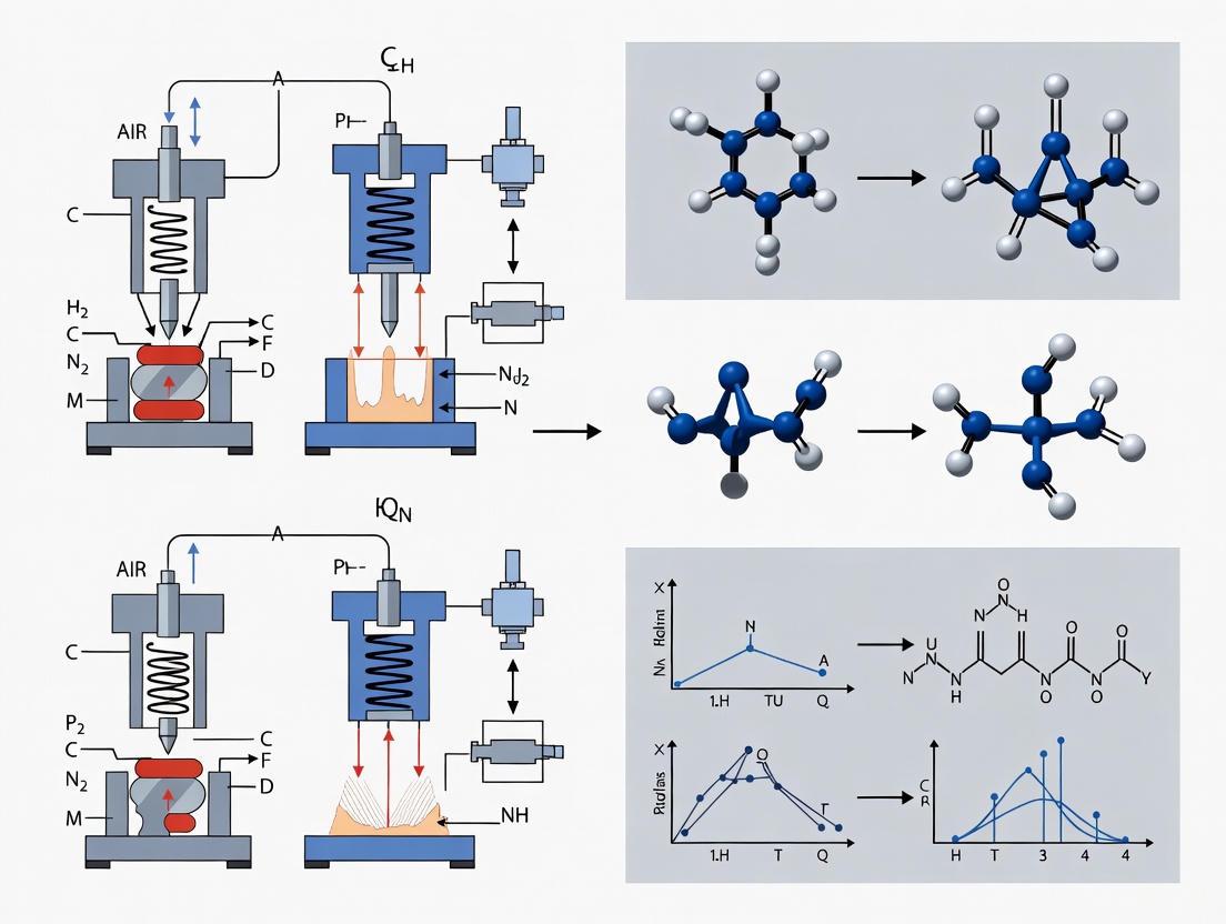
Controlled Cortical Impact (CCI) devices are indispensable tools in pre-clinical traumatic brain injury (TBI) research, enabling scientists to model brain trauma with high precision and reproducibility. These devices function by mechanically transferring energy to the brain tissue to simulate injuries that resemble human TBI. The core components of any CCI system—the impactor, the tip, and the control system—directly determine the characteristics and consistency of the resulting injury. Within neuroscience research, a fundamental division exists between pneumatic and electromagnetic actuation systems, each with distinct mechanical principles and performance implications. Understanding these core components is essential for researchers selecting equipment, designing experiments, and interpreting results within the broader thesis of comparing electromagnetic and pneumatic CCI technologies. This guide provides an objective, data-driven comparison of these subsystems to inform evidence-based device selection [2] [1].
Impactor Drive Systems: Pneumatic vs. Electromagnetic
The impactor is the core actuating component of a CCI device, responsible for delivering a controlled mechanical impact to the brain. The choice between pneumatic and electromagnetic drive systems represents a significant trade-off in terms of performance, cost, and operational convenience.
Pneumatic Impactor Systems
Pneumatic impactors utilize pressurized gas to drive a piston. A typical pneumatic CCI device features a small-bore reciprocating double-acting pneumatic piston with a maximum adjustable stroke length of approximately 50 mm. This piston is rigidly mounted to a crossbar, often with multiple mounting positions allowing the impactor to be oriented vertically or at an angle relative to the skull and underlying brain tissue. These systems have a long history in TBI research, being the original technology used in the earliest CCI models [2] [1].
Electromagnetic Impactor Systems
Electromagnetic impactors, a more recent technological development, use an electromagnetic field to accelerate the impactor tip. Like pneumatic devices, they are typically used with a commercial stereotaxic frame for precise positioning. Some electromagnetic models are also compatible with an articulated support arm, facilitating their use in large animal models such as swine. A key advertised advantage is their greater portability due to a smaller physical footprint and the elimination of a pressurized gas source [2] [1].
Direct Performance Comparison
A critical empirical study directly compared the reproducibility of pneumatic and electromagnetic CCI models and suggested superior reproducibility with the electromagnetic system [2] [1]. The following table summarizes the key characteristics of each drive system:
Table 1: Comparison of Pneumatic vs. Electromagnetic Impactor Drive Systems
| Feature | Pneumatic Impactor | Electromagnetic Impactor |
|---|---|---|
| Drive Principle | Pressurized gas (pneumatic piston) [2] | Electromagnetic actuator [2] |
| Typical Mounting | Crossbar with multiple angles [2] | Stereotaxic frame or articulated arm [2] |
| Portability | Lower (requires gas source) [2] | Higher (smaller, no gas source) [2] |
| Reproducibility | Standard | Suggested to be higher [2] |
| Historical Context | Original CCI technology; long-established use [1] | More recent development; gaining popularity [2] |
Impact Tip Design and Configuration
The impact tip is the component that makes direct contact with the dura or skull, and its physical characteristics are primary determinants of the injury's biomechanical profile. The size, shape, and material of the tip must be carefully selected based on the experimental model and desired injury severity.
Tip Characteristics and Selection
Tips are available in a variety of diameters and geometries. Common shapes include flat, convex, and beveled designs, each influencing the distribution of force and the pattern of tissue deformation. Manufacturers offer a range of removable tips to accommodate different species and research goals. For instance, one leading electromagnetic system offers seven different tip sizes, while another provides standard tips of 1, 1.5, 2, 3, and 5 mm diameters. This scalability is a key strength of the CCI model, allowing its application from mice to non-human primates [2] [1].
Table 2: Standard Commercially Available Impact Tip Sizes
| Tip Diameter (mm) | Typical Application Scope |
|---|---|
| 1.0 - 2.0 | Mouse models [2] |
| 3.0 - 5.0 | Rat models and larger species [2] |
| Custom sizes | Specialized applications or species [1] |
Control Systems and Injury Parameters
The control system is the interface through which researchers define and monitor the injury parameters. It is critical for ensuring the precision and repeatability of the experimental TBI.
Controlled Injury Parameters
CCI devices provide a high degree of control over several key mechanical factors that define the impact event [2] [1]:
- Impact Velocity: The speed at which the tip travels, typically controlled and measured by the system.
- Impact Depth: The distance the tip penetrates into the brain tissue past the dura or skull surface.
- Dwell Time: The duration for which the tip remains at the maximum depth of penetration before retraction.
- Impact Angle: The angle of approach, which can often be adjusted via the stereotaxic frame or mounting.
System Operation and Feedback
Modern CCI systems, particularly electromagnetic models, often integrate digital controls and sensors that provide real-time feedback and verification of the actual impact parameters achieved. Some pneumatic systems offer an accessory unit to measure rod speed to enhance accuracy and reporting. This capability to precisely set and record the biomechanical forces applied is a foundational strength of the CCI model, enabling the production of a broad, graded spectrum of TBI severities from mild to severe [2] [1].
Experimental Protocol for Device Performance Comparison
To objectively compare the performance of electromagnetic and pneumatic CCI devices, a standardized experimental protocol is essential. The following workflow outlines a rigorous methodology for generating comparable data on injury reproducibility and histopathological outcomes.
Methodology Detail
- Animal Subjects: Adult male and female Sprague-Dawley rats (250-300g) or C57BL/6 mice (20-25g), randomly assigned to experimental groups with appropriate sample sizes to ensure statistical power [1].
- Surgical Preparation: Animals are anesthetized and placed in a stereotaxic frame. A craniectomy is performed to expose the dura mater, keeping it intact [2] [1].
- Impact Delivery: The impactor tip is positioned perpendicular to the brain surface. The impact is delivered to the exposed dura. To ensure a valid comparison, both devices should be configured to deliver an impact with identical parameters (e.g., 3.0 mm depth, 5.0 m/s velocity, 0.5 s dwell time with a 3 mm flat tip) [2].
- Parameter Verification: The actual impact should be verified using a high-speed camera and integrated sensors (if available) to confirm the achieved velocity, depth, and dwell time match the set parameters [2].
- Outcome Measures: The primary measure for device performance is the consistency of the resulting lesion. This is quantified by measuring lesion volume (e.g., via MRI or histology with Cresyl Violet staining) and calculating the coefficient of variation (CV) across subjects within the same treatment group. A lower CV indicates higher reproducibility [2].
The Scientist's Toolkit: Essential Research Reagents and Materials
Successful and reproducible CCI experiments require a standardized set of laboratory materials and reagents beyond the impact device itself. The following table details these essential components.
Table 3: Essential Research Reagents and Materials for CCI Experiments
| Item Name | Function/Application | Experimental Consideration |
|---|---|---|
| Stereotaxic Frame | Precise positioning of the animal and impactor device [2]. | Must be compatible with the chosen CCI device; stability is critical. |
| Anesthesia System (Isoflurane) | Maintenance of surgical-plane anesthesia during impact [2]. | Type and concentration can affect physiological responses and must be reported per NIH CDEs. |
| Temperature Control (Homeothermic Blanket) | Maintenance of core body temperature at ~37°C [1]. | Prevents hypothermia, a key confounder in injury outcomes. |
| Standardized Impact Tips | Definitive contact point for delivering the cortical impact [2]. | Size and shape (flat, convex) must be selected and reported. |
| Bone Drill with Burrs | Performance of a precise craniectomy [2]. | Must be careful to avoid damaging the underlying dura. |
| Sham-Control Animals | Control for the effects of anesthesia, surgery, and craniectomy [2]. | Undergo all procedures except the actual impact. |
| Perfusion Pump & Fixative (Paraformaldehyde) | Tissue fixation for subsequent histology [1]. | Ensures high-quality preservation of brain morphology. |
| Primary Antibodies (e.g., GFAP, Iba1, APP) | Immunohistochemical detection of astrocytes, microglia, and axonal injury [1]. | Key for characterizing the neuropathological response to CCI. |
Selecting between a pneumatic and electromagnetic CCI system involves weighing specific research priorities. The following decision pathway synthesizes the comparative data to guide researchers.
In conclusion, both pneumatic and electromagnetic CCI devices offer a high degree of control over key injury parameters, facilitating robust pre-clinical TBI research. The core components—impactor drive system, tip design, and electronic controls—differentiate their performance. Evidence suggests that electromagnetic systems may offer superior reproducibility and greater portability, while pneumatic systems have a long-established track record. The optimal choice is contingent upon the specific research priorities, model species, and operational constraints of the laboratory. As the field moves forward, adherence to reporting standards like the NIH Common Data Elements (CDEs) for pre-clinical TBI will be vital for translating findings from these sophisticated tools into clinical breakthroughs [2] [1].
In the rigorous field of pre-clinical traumatic brain injury (TBI) research, the controlled cortical impact (CCI) model is a cornerstone for studying injury mechanisms and evaluating potential therapeutics. A central debate in this area revolves around the choice of injury device: traditional pneumatic systems versus newer electromagnetic actuators. The core of this comparison lies in how these devices control and reproduce the three fundamental injury parameters—velocity, depth, and dwell time. These parameters directly dictate the severity and reproducibility of the resulting brain injury, making their precise control paramount for generating valid, reliable data. This guide provides an objective comparison of pneumatic and electromagnetic CCI devices, focusing on their performance in managing these key variables, supported by experimental data and detailed methodologies.
Device Comparison: Pneumatic vs. Electromagnetic CCI
The following table summarizes the core characteristics, performance data, and advantages of pneumatic and electromagnetic CCI devices based on current literature.
Table 1: Performance Comparison of Pneumatic and Electromagnetic CCI Devices
| Feature | Pneumatic CCI Device | Electromagnetic CCI Device |
|---|---|---|
| Actuation Mechanism | Pressurized gas (e.g., nitrogen) drives a piston [2]. | Electromagnetic coil and stationary magnet propel the impactor [6]. |
| Control over Parameters | Direct control over impactor velocity, depth, and dwell time [3] [2]. | Direct control over impactor velocity, depth, and dwell time via electronic software [6] [2]. |
| Reported Velocity Range | Used across a broad range; specific range varies by commercial system. | Capable of producing a broad range of injury severities; prototype achieved high velocities requiring a 72V power supply [6]. |
| Key Performance Findings | Can exhibit velocity-dependent overshoot, leading to greater variability in impact parameters [3] [6]. | Demonstrated greater reproducibility and consistency in producing graded injuries with minimal overshoot [3] [6] [2]. |
| Primary Advantages | Well-characterized, widely used for decades, capable of producing graded TBI [3] [2]. | High reproducibility, compact size, portability, no need for pressurized gas source, and convenient electronic control [3] [6] [2]. |
| Commercial Suppliers | Amscien Instruments, Precision Instruments & Instrumentation, LLC [1]. | Hatteras Instruments, Leica Biosystems [1]. |
Experimental Protocols and Supporting Data
Protocol for Electromagnetic CCI in a Mouse Model
This detailed protocol, adapted from a study characterizing an electromagnetic device, outlines the steps for inducing a graded CCI injury in mice [6].
- Device Setup: The electromagnetic impactor is mounted onto the arm of a stereotaxic frame. The desired velocity (e.g., 1.5 m/s) and dwell time (e.g., 0.1 s) are set via the control software. A impactor tip of an appropriate size (e.g., 3 mm diameter) is attached [6] [15].
- Animal Preparation: The mouse is anesthetized (e.g., with isoflurane) and secured in the stereotaxic frame. A heating pad is used to maintain body temperature at 37°C. The scalp is shaved and disinfected, and a midline incision is made to expose the skull [15] [16].
- Craniectomy: A high-speed drill is used to perform a craniectomy over the desired hemisphere (e.g., left parietal cortex), leaving the dura mater intact. The site is frequently irrigated with sterile saline to prevent overheating [15].
- Impact Parameter Calibration: The impactor tip is carefully lowered until it just touches the dura mater; this position is defined as Z = 0. The tip is then retracted and the stereotaxic frame is used to lower it to the desired impact depth (e.g., 1.0 mm, 2.0 mm below the dura) [6] [15].
- Injury Induction: The impact is triggered via the control software. The piston extends, driving the tip into the cortex at the preset velocity and depth, holding for the set dwell time, and then retracting [6].
- Post-Procedural Care: The surgical site is irrigated with saline, the scalp is sutured, and the animal is moved to a warm recovery cage with appropriate post-operative analgesics [15].
Key Supporting Experimental Data
- Reproducibility: A direct comparison study found that a pneumatic device resulted in "velocity-dependent overshoot" and "greater overall overshoot," whereas an electromagnetic prototype demonstrated superior reproducibility [3] [6].
- Graded Histological and Behavioral Deficits: Using an electromagnetic device, varying the impact depth from 1.0 mm to 3.0 mm in mice produced a broad range of injury severities. Histological analysis showed that a 2.0 mm impact with the electromagnetic device produced a lesion similar to a 1.0 mm impact from a commercial pneumatic device. Behaviorally, impacts of 2.0 mm, 2.5 mm, and 3.0 mm impaired water maze performance, while a 1.5 mm impact did not, confirming the ability to produce finely graded injuries [6].
Experimental Workflow Visualization
The diagram below illustrates the key steps and parameters involved in a typical CCI experiment for modeling Traumatic Brain Injury.
The Scientist's Toolkit: Key Research Reagent Solutions
Successful execution of a CCI study requires more than just the impact device. The following table details essential materials and their functions in a standard protocol.
Table 2: Essential Reagents and Materials for CCI Experiments
| Item | Function/Application | Example in Protocol |
|---|---|---|
| Anesthetic | To induce and maintain a surgical plane of anesthesia during the procedure. | Isoflurane delivered via a vaporizer with oxygen [15] [16]. |
| Stereotaxic Frame | To securely immobilize the animal's head in a standardized position, ensuring precise impact location. | A commercial stereotaxic frame with ear bars and a bite bar [6] [15]. |
| Heating Pad | To maintain the animal's core body temperature at 37°C, preventing hypothermia which can confound outcomes. | A feedback-controlled warming pad placed under the animal during surgery [15] [16]. |
| High-Speed Drill | To perform a craniectomy by carefully removing a section of the skull, exposing the dura mater. | A rotary tool with a round burr bit (0.6-0.8 mm) [15]. |
| Sterile Saline | For irrigation during drilling to remove bone debris and prevent thermal damage to underlying tissue. | Applied generously during the craniectomy and after impact [15]. |
| Immunosuppressant | For studies involving cell transplantation; to prevent rejection of xenografted cells. | Cyclosporine A (CsA) administered subcutaneously [15]. |
| Post-operative Analgesic | To manage pain following the surgical procedure, in accordance with animal welfare guidelines. | Acetaminophen provided in the drinking water [15]. |
The choice between pneumatic and electromagnetic CCI devices hinges on the specific demands of the research question, particularly regarding the precision and reproducibility of the key injury parameters: velocity, depth, and dwell time. While pneumatic devices have a long and established history in the field, empirical evidence indicates that electromagnetic devices offer superior mechanical consistency with less parameter overshoot. This enhanced reproducibility can reduce inter-subject variability, potentially decreasing the number of animals needed to achieve statistical power and accelerating the pace of pre-clinical discovery. Researchers must weigh these performance characteristics against other factors such as cost, portability, and the specific injury phenotype they aim to model.
Implementing CCI Models: From Standardized Protocols to Advanced Applications
Translational traumatic brain injury (TBI) research requires careful consideration of species-specific anatomical and physiological differences when scaling experimental models from rodents to large animals. The controlled cortical impact (CCI) model, a widely used mechanical model of TBI, can be implemented using either electromagnetic (EM) or pneumatic actuation systems [2] [9]. While both devices share the common goal of delivering controlled mechanical energy to brain tissue, they differ significantly in their operational principles, portability, and control mechanisms. Electromagnetic CCI devices utilize a current-carrying coil within a magnetic field to generate precise impact forces, while pneumatic devices employ compressed gas to drive a piston [6] [2]. Understanding the technical and practical considerations for scaling these devices between mice and swine is essential for generating reproducible, clinically relevant injury data across the preclinical research spectrum. This guide objectively compares the performance of both systems while providing detailed methodologies for implementing species-appropriate CCI protocols.
Device Operational Principles and Comparative Performance
Fundamental Operating Mechanisms
Electromagnetic CCI Devices function through a voice coil actuator system where an electric current through a wire coil generates a Lorentz force when placed within a stationary magnetic field. This force propels the impactor tip with high precision and minimal overshoot [6] [17]. Key advantages include compact size, stereotaxic arm-mounting capability, and elimination of frequent gas pressure calibration requirements [6]. A significant engineering consideration is the back electromotive force (EMF), a voltage opposing the current that increases proportionally with impactor velocity (VB = kt × v), requiring higher driving voltages to achieve target velocities [6].
Pneumatic CCI Devices utilize compressed nitrogen or air to drive a piston within a cylinder, with impact velocity controlled by regulating gas pressure [2]. These systems typically require a solid metal crossbar for stabilization rather than stereotaxic arm mounting, and may exhibit velocity-dependent overshoot according to some comparative studies [17]. The need for a compressed gas source reduces portability compared to electromagnetic systems.
Direct Performance Comparison
Table 1: Electromagnetic vs. Pneumatic CCI Device Characteristics
| Feature | Electromagnetic CCI | Pneumatic CCI |
|---|---|---|
| Actuation Mechanism | Voice coil actuator in magnetic field [6] | Compressed gas piston [2] |
| Portability | High (arm-mounted, compact) [6] | Low (requires crossbar, gas tank) [2] |
| Impact Control | Electronic precision with software control [6] | Gas pressure regulation [2] |
| Overshoot | Minimal reported overshoot [17] | Velocity-dependent overshoot reported [17] |
| Commercial Suppliers | Leica Biosystems [2] | Pittsburgh Precision Instruments, AmScien Technologies [2] |
Species-Specific Scaling Methodologies
Murine CCI Model Implementation
The mouse CCI model requires meticulous surgical exposure and impact parameter optimization to generate reproducible injuries appropriate for the smaller neuroanatomy.
Surgical Protocol for Mice [6] [18]:
- Anesthesia and Positioning: Induce and maintain anesthesia (e.g., isoflurane), then secure the mouse in a stereotaxic frame with head immobilization.
- Surgical Exposure: Make a midline scalp incision, retract soft tissue, and identify cranial landmarks (bregma, lambda).
- Craniotomy: Perform a unilateral craniotomy (typically 3-4mm diameter) centered at specific coordinates relative to bregma (e.g., 0.5mm anterior, 2.0mm lateral for parietal lobe impact) [18].
- Impact Delivery: Position the impactor tip perpendicular to the exposed dura and deliver the impact with predetermined parameters.
- Closure: Suture the surgical site and provide postoperative analgesia (e.g., l-methadone) and supportive care [19].
Standard Mouse Impact Parameters [6] [18]:
- Tip Diameter: 3mm
- Impact Velocity: 3-6m/s
- Depth of Displacement: 0.5-2.0mm (severity-dependent)
- Dwell Time: 50-500ms
Porcine CCI Model Implementation
Swine models require substantial scaling of both impact parameters and surgical approach to account for larger brain mass, gyrencephalic neuroanatomy, and thicker cranial structures.
Surgical Protocol for Swine [20]:
- Anesthesia and Monitoring: Induce and maintain general anesthesia with appropriate physiological monitoring throughout the procedure.
- Surgical Exposure: Perform a substantial craniotomy (several centimeters) over the target region, typically the frontal or parietal cortex, accounting for the extensive frontal sinus development in swine.
- Dura Exposure: Carefully expose the intact dura mater without causing premature damage or bleeding.
- Device Stabilization: Secure the impactor using a stereotaxic frame or customized stabilization system to prevent movement during impact. A portable, wheeled stand with a 3D axis arm system provides optimal positioning flexibility [20].
- Impact Delivery: Deliver the impact with species-appropriate parameters as detailed below.
- Closure and Recovery: Close the surgical site in layers and provide appropriate postoperative analgesia and monitoring.
Porcine Impact Parameters [20]:
- Tip Diameter: Scaled up significantly from murine parameters (exact size varies by specific model)
- Impact Force: 12.8-67.6N (modulatable via voltage control)
- Kinetic Energy: 0.045-0.338J
- Depth Control: 0-7mm adjustable range with real-time depth verification
Quantitative Scaling Comparison
Table 2: Species-Specific CCI Parameter Comparison
| Parameter | Murine Model | Porcine Model | Scaling Factor |
|---|---|---|---|
| Typical Impact Force | Not typically reported | 12.8-67.6 N [20] | Not directly comparable |
| Kinetic Energy | Not typically reported | 0.045-0.338 J [20] | Not directly comparable |
| Impact Velocity | 3-6 m/s [6] | Modulated via voltage control [20] | ~Similar range |
| Cortical Displacement | 0.5-2.0 mm [6] | 0-7 mm [20] | ~3.5-14x increase |
| Tip Diameter | 3 mm [18] | Significantly larger (specifics vary) | Substantial increase |
| Surgical Complexity | Moderate | High (extensive craniotomy) | Significant increase |
Analytical and Behavioral Assessment Across Species
Histopathological and Molecular Outcomes
Murine Assessment Methods:
- Histological Analysis: Standard histological staining reveals consistent cortical contusions and hippocampal damage following CCI, with injury severity correlating with impact depth [6].
- Blood-Brain Barrier Permeability: Evaluated using Evans Blue extravasation assay, demonstrating significant barrier disruption post-CCI [18].
- Apoptosis Assessment: TUNEL staining quantifies apoptotic cells in pericontusional regions, typically performed 7 days post-injury [18].
- Gene Expression Analysis: RT-qPCR and Western blotting assess inflammatory mediators (e.g., NLRP3, IL-1β, TNF-α) and cell death markers [18] [21].
Porcine Assessment Methods:
- Ultrasound Imaging: Intraoperative ultrasound verifies injury presence and characteristics through the thicker cranial structures [20].
- Histological Validation: Hematoxylin and eosin (H&E) staining confirms contusion injury and assesses lesion volume in larger tissue sections [20].
- Advanced Imaging Compatibility: Larger brain size enables serial MRI or CT imaging to track injury progression longitudinally.
Functional and Behavioral Testing
Murine Behavioral Paradigms:
- Morris Water Maze: Hidden platform and probe trials detect spatial learning and memory deficits, particularly sensitive to 2.0-3.0mm impact depths [6].
- Rotorod Test: Assesses motor coordination and balance, with deficits observed following moderate-severe injuries (2.5-3.0mm impacts) [6].
- Open Field Test: Evaluates anxiety-like behaviors and locomotor activity, with impaired performance observed post-CCI [18].
- Y-Maze: Tests spatial working memory and hippocampus-dependent cognitive function [18].
Porcine Neurological Assessment:
- Porcine Neurological Motor (PNM) Score: Species-specific motor function evaluation sensitive to spinal cord and brain injury severity, with assessments typically performed at postoperative day 1 and beyond [20].
- Customized Behavioral Testing: Larger size and cognitive capacity enable complex learning and memory tasks adapted from primate assessments.
Welfare Considerations and Humane Endpoints
Robust welfare assessment protocols are essential across species, particularly as injury severity increases in scaled models.
Murine Welfare Monitoring [22] [19]:
- TBI-Specific Scoresheet: Implementation of structured assessment tools evaluating appearance, physical exam parameters, behavior, and body condition score.
- Nest Building Performance: Sensitive indicator of wellbeing with significant impairment observed at 1 day post-CCI but recovery by day 7 with appropriate analgesia [19].
- Analgesia Protocol: Postsurgical analgesia with opioids (e.g., l-methadone) for 3 days post-CCI significantly improves welfare outcomes [19].
- Humane Endpoints: Cumulative scores of 9-11 on standardized assessment scales indicate moribund state requiring immediate euthanasia [22].
Porcine Welfare Considerations [20]:
- Postoperative Analgesia: Multimodal pain management essential following extensive craniotomy procedures.
- Veterinary Monitoring: Intensive postoperative observation with customized supportive care appropriate for large animal physiology.
- Mobility Support: Specialized housing accommodations during recovery from neurological deficits.
The Scientist's Toolkit: Essential Research Reagents and Materials
Table 3: Essential Research Materials for Cross-Species CCI Studies
| Item | Application | Species Utility |
|---|---|---|
| Stereotaxic Frame | Precise head stabilization and impactor positioning | Both (scaled sizes) |
| Electromagnetic CCI Device | Precise, reproducible impact delivery with minimal overshoot [6] [17] | Both (parameter-adjusted) |
| Isoflurane Anesthesia System | Maintained surgical anesthesia throughout procedure | Both (species-specific dosing) |
| Surgical Drill System | Craniotomy performance | Both (different bit sizes) |
| l-Methadone/Analgesics | Postsurgical pain management [19] | Both (weight-appropriate dosing) |
| Evans Blue Dye | Blood-brain barrier permeability assessment [18] | Primarily murine |
| TUNEL Staining Kit | Apoptosis detection in pericontusional regions [18] | Primarily murine |
| Ultrasound Imaging System | Non-invasive injury verification [20] | Primarily porcine |
| Porcine Neurological Motor Score Sheet | Species-specific functional assessment [20] | Porcine-specific |
Effective scaling of CCI models from mice to swine requires meticulous attention to species-specific anatomical differences, impact parameter optimization, and appropriate analytical methodologies. Electromagnetic CCI devices offer distinct advantages in impact precision, reproducibility, and portability compared to pneumatic systems, particularly valuable when transitioning between species with vastly different neuroanatomical characteristics [6] [17]. The murine model provides a high-throughput, genetically modifiable platform for mechanistic studies, while the porcine model delivers superior translational relevance through neuroanatomical similarity to humans [20]. Successful implementation requires rigorous welfare monitoring and species-appropriate behavioral assessment. Through careful consideration of these scaling principles, researchers can generate complementary datasets across species that significantly enhance the predictive validity of preclinical TBI research.
Clinical Classification of Traumatic Brain Injury Severity
Traumatic Brain Injury (TBI) severity is clinically classified as mild, moderate, or severe based on a combination of assessment criteria. The most widely recognized classification system utilizes the Glasgow Coma Scale (GCS), duration of loss of consciousness (LOC), and post-traumatic amnesia (PTA) [23] [24].
Table 1: Clinical Criteria for Classifying TBI Severity
| Criteria | Mild | Moderate | Severe |
|---|---|---|---|
| Structural Imaging | Normal | Normal or abnormal | Normal or abnormal |
| Loss of Consciousness (LOC) | < 30 minutes | 30 minutes to 24 hours | > 24 hours |
| Alteration of Consciousness/Mental State | A moment to 24 hours | > 24 hours | > 24 hours |
| Post-Traumatic Amnesia (PTA) | 0–1 day | >1 and <7 days | >7 days |
| Glasgow Coma Scale (GCS) | 13–15 | 9–12 | 3–8 |
The Glasgow Coma Scale is the most common tool, where a score of 13-15 indicates mild injury, 9-12 moderate, and 3-8 severe [23] [25] [24]. These categories help predict the injury's impact and guide initial patient management. Furthermore, prolonged unconsciousness exceeding 24 hours (coma) and abnormal neuroimaging are hallmark indicators of severe TBI [25].
Preclinical Modeling Using Controlled Cortical Impact (CCI)
CCI Devices: Pneumatic vs. Electromagnetic
In preclinical research, the Controlled Cortical Impact (CCI) model is a widely used method to study TBI. This model uses a mechanical impactor to deform brain tissue in a controlled manner, creating injuries that mimic human TBI. The two primary types of CCI devices are pneumatic and electromagnetic [2].
Table 2: Comparison of Pneumatic vs. Electromagnetic CCI Devices
| Feature | Pneumatic CCI Device | Electromagnetic CCI Device |
|---|---|---|
| Actuation Method | Pressurized gas (pneumatic piston) | Electromagnetic coil & stationary magnet |
| Key Components | Cylinder, crossbar frame, pneumatic piston | Voice coil, piston, servo amplifier, power supply |
| Portability | Less portable due to need for gas source | More portable, smaller size, no gas source |
| Calibration | Requires frequent calibration of gas pressures | Reproducible velocities without frequent calibration |
| Integration | Mounted on a solid metal frame | Attaches to an arm of a stereotaxic frame |
| Control & Reproducibility | High degree of control over parameters | High reproducibility; capable of graded injuries |
Both devices allow precise control over injury parameters, including impact depth, velocity, dwell time, and tip geometry, enabling researchers to produce a broad range of injury severities [6] [2]. The electromagnetic device offers advantages in portability and operational simplicity, while the pneumatic device has a long-established history of use.
Correlating Impact Parameters to Injury Severity in Mice
The CCI model enables the precise correlation of mechanical impact parameters with histological and behavioral outcomes, effectively modeling different clinical severity levels.
Table 3: Electromagnetic CCI Injury Parameters and Outcomes in Mice (Based on data from PMC2435168)
| Impact Depth | Histological Outcomes | Behavioral Outcomes (Morris Water Maze) | Inferred Clinical Severity Analogue |
|---|---|---|---|
| 1.0 mm | Minimal / Not reported | No impairment | Subclinical / Very Mild |
| 1.5 mm | Not reported | No hidden platform or probe trial impairment | Mild |
| 2.0 mm | Similar to 1.0-mm pneumatic impact | Impaired hidden platform and probe trial performance | Moderate |
| 2.5 mm | Not reported | Impaired hidden platform, probe trial, rotorod, and visible platform performance | Severe |
| 3.0 mm | Not reported | Impaired hidden platform, probe trial, rotorod, and visible platform performance | Severe |
A study using an electromagnetic CCI device demonstrated that varying the impact depth from 1.0 mm to 3.0 mm produces a broad range of injury severities [6]. Impacts of 2.0 mm depth resulted in cognitive deficits (impaired water maze performance), analogous to moderate TBI. Deeper impacts (2.5 mm and 3.0 mm) caused more widespread motor and cognitive deficits, analogous to severe TBI [6]. This graded relationship allows researchers to select parameters that model specific human injury conditions.
Experimental Protocols for Preclinical CCI
Standardized Surgical and Impact Procedure
The following protocol details the standard procedure for conducting a CCI experiment in rodents, integrating key steps from established methodologies [6] [2].
Key Steps in the CCI Experimental Workflow:
- Anesthesia and Surgical Preparation: The animal (e.g., mouse or rat) is placed under general anesthesia. The head is then shaved and securely fixed in a stereotaxic frame to ensure complete immobilization during the procedure [6] [2].
- Craniotomy: A midline incision is made in the scalp, and the soft tissues are retracted to expose the skull. A craniotomy (surgical opening in the skull) is performed over the desired brain region, typically the parietal or frontal cortex, leaving the underlying dura mater intact [2].
- Impact Delivery: The impactor tip (of defined size and shape) is positioned perpendicular to the exposed dura. The impactor is then retracted to a predefined "cocked" position. The critical injury parameters—depth (e.g., 1.0-3.0 mm for mice [6]), velocity (e.g., 5-6 m/s), and dwell time (typically 100-500 ms)—are set via the device's control software. The impact is triggered, indenting the brain tissue [6] [2].
- Post-operative Care: Following the impact, the impactor is retracted. The bone flap may or may not be replaced, the scalp is sutured or stapled closed, and the animal is monitored closely during recovery from anesthesia and in the post-operative period [2].
The Scientist's Toolkit: Essential Research Reagents and Materials
Table 4: Key Reagents and Materials for CCI Research
| Item | Function/Application |
|---|---|
| Controlled Cortical Impact Device | Pneumatic or electromagnetic device to deliver precise mechanical impact to the brain. |
| Stereotaxic Frame | Apparatus to securely hold the animal's head in a fixed position during surgery and impact. |
| Anesthetic Agents | (e.g., Isoflurane, Ketamine/Xylazine). To induce and maintain surgical anesthesia. |
| Physiological Monitoring System | To monitor and maintain body temperature, respiration, and other vital signs during surgery. |
| Morris Water Maze | Behavioral apparatus to assess spatial learning and memory deficits post-TBI. |
| Rotorod | Behavioral apparatus to evaluate motor coordination and balance. |
| Histology Reagents | (e.g., formalin, antibodies). For tissue fixation, staining, and analysis of lesion volume and pathology. |
Recent Advances and Future Directions in TBI Characterization
The field of TBI research is evolving beyond traditional unimodal classification. A major international initiative by the NIH-NINDS has proposed a novel, multidimensional framework for acute TBI characterization known as the CBI-M framework [26]. This framework incorporates four pillars to provide a more comprehensive picture of the injury:
- Clinical Pillar: Includes the full GCS score and pupillary reactivity.
- Biomarker Pillar: Incorporates blood-based biomarkers like GFAP and UCH-L1.
- Imaging Pillar: Utilizes advanced neuroimaging to identify pathoanatomical changes.
- Modifier Pillar: Accounts for patient-specific features that influence presentation and outcome, such as age, comorbidities, and socioeconomic factors [26].
Concurrently, predictive modeling is advancing. The novel MOST (Mortality Score for TBI) model, developed from a modern dataset, demonstrates superior accuracy in predicting in-hospital mortality compared to established models like CRASH-Basic and IMPACT-Core [27]. These developments highlight a shift towards more precise, individualized injury assessment, which will ultimately refine preclinical modeling efforts and therapeutic development.
Open-Head vs. Closed-Head Injury Protocols
In both clinical practice and neuroscience research, traumatic brain injuries (TBI) are fundamentally categorized as either open-head or closed-head injuries. This distinction is critical for diagnosis, treatment, and the development of accurate experimental models. An open-head injury (also known as a penetrating injury) involves a breach of the skull, where an object pierces the cranium and enters brain tissue [28]. In contrast, a closed-head injury occurs when external force trauma causes brain damage without penetrating the skull [28] [29].
The Controlled Cortical Impact (CCI) model is a cornerstone of preclinical TBI research that can be adapted to model aspects of both injury types. CCI devices mechanically induce brain trauma using a piston-like impactor, and modern systems are primarily powered by either pneumatic or electromagnetic actuators [1] [2] [4]. These devices provide researchers with precise control over injury parameters such as impact velocity, depth, and dwell time. The choice between electromagnetic and pneumatic systems, and the specific protocol employed, directly influences the type and severity of brain injury produced, enabling targeted study of human TBI conditions in animal models.
Clinical and Pathophysiological Comparison of Open- and Closed-Head Injuries
The fundamental difference between open and closed head injuries lies in the integrity of the skull and the mechanisms of tissue damage.
Open-Head (Penetrating) Injuries: These injuries result from projectiles, sharp objects, or skull fragments physically penetrating the brain parenchyma [28] [29]. They typically cause focal damage along the penetration path, including direct laceration of blood vessels and neural tissue. Common consequences include skull fractures, severe bleeding, and high risk of infection [29]. The damage is often localized and simpler to identify on imaging, but can be devastating depending on the brain regions affected.
Closed-Head Injuries: These result from blunt force, acceleration-deceleration, or explosive blasts that cause the brain to move within the intact skull [28]. This leads to a more complex and often diffuse injury pattern, including concussions, diffuse axonal injury (DAI), contusions, and hematomas [29]. The damage arises from shearing forces, impact against the inner skull surface, and subsequent biochemical cascades, making its pathology more widespread and heterogeneous than in penetrating injuries.
Table 1: Clinical Comparison of Open-Head vs. Closed-Head Injuries
| Feature | Open-Head Injury | Closed-Head Injury |
|---|---|---|
| Skull Integrity | Breached or fractured [28] | Intact [28] |
| Primary Mechanism | Penetration by foreign object [28] | Blunt force, acceleration-deceleration, shaking [28] [29] |
| Typical Pathology | Focal damage, direct tissue disruption, skull fragments [29] | Concussion, diffuse axonal injury, contusions, hematomas [29] |
| Common Causes | Gunshot wounds, sharp objects, projectile accidents [28] | Falls, motor vehicle accidents, sports collisions, assaults [28] |
| Research Model Focus | Controlled cortical impact (CCI) with craniotomy [1] | CCI on intact skull (CHI), weight-drop, impact acceleration [1] [4] |
Experimental Modeling: Bridging Clinical TBI and Preclinical Research
The CCI model has been extensively developed to replicate human TBI in a controlled laboratory setting. Its versatility allows it to be configured to model both open and closed-head injury paradigms.
Modeling Open-Head Injuries with Traditional CCI
The traditional CCI protocol models open-head injury by first performing a craniotomy—surgically removing a portion of the skull to expose the dura mater [4]. The impactor tip is then directed onto the exposed dura to induce a focal contusion. This method directly mimics the physical breach of the skull seen in clinical open-head injuries and produces highly reproducible cortical lesions, hemorrhage, and neuronal loss in a specific brain region [1] [2].
Modeling Closed-Head Injuries with Modified CCI
To model closed-head injury (CHI), the CCI device is used to impact the intact skull [1] [4]. This approach eliminates the craniotomy, better preserving the biomechanical properties of the intact head and creating a more diffuse injury pattern that includes concussion-like pathology and diffuse axonal injury, more closely aligning with the common human experience of TBI [4].
Electromagnetic vs. Pneumatic CCI Devices: A Technical Comparison
The core technology driving the CCI model has evolved, offering researchers two primary options for impactor actuation.
- Pneumatic CCI Devices: These were the first developed, using a pressurized gas (e.g., nitrogen) cylinder to drive a piston [1] [4]. They are characterized by their robust construction and are typically mounted on a large crossbar for stability.
- Electromagnetic CCI Devices: A more recent development, these devices use an electromagnetic coil to propel the impactor [6] [4]. They are generally more portable due to their smaller size and lack of a gas cylinder, and are noted for high reproducibility [1].
Table 2: Technical Comparison of Pneumatic vs. Electromagnetic CCI Devices
| Parameter | Pneumatic CCI Device | Electromagnetic CCI Device |
|---|---|---|
| Actuation Mechanism | Pressurized gas piston [1] [4] | Electromagnetic coil [6] [4] |
| Portability | Lower (requires gas source, larger footprint) [4] | Higher (more compact, lighter) [4] |
| Key Advantages | Well-established history, robust construction [1] | Portability, high reproducibility, no gas source needed [1] [6] |
| Impact Velocity | Controlled via gas pressure, requires calibration [6] | Precisely controlled via input current [6] |
| Reported Reproducibility | High | Potentially higher in some comparative studies [1] |
| Commercial Examples | AmScien Instruments AMS 201; Pittsburgh Precision Instruments CCI [1] | Leica Impact One; Hatteras Pinpoint PCI3000 [1] |
Detailed Experimental Protocols for Injury Induction
The following protocols detail the steps for inducing open and closed-head injuries using an electromagnetic CCI device, which is gaining prominence for its reproducibility and ease of use [1] [30].
Protocol 1: Open-Head Injury via Craniotomy and CCI
This protocol models penetrating brain injury and severe contusion, suitable for studying focal TBI pathophysiology and therapeutic interventions.
- Anesthesia and Preparation: Induce and maintain anesthesia (e.g., isoflurane). Place the animal in a stereotaxic frame. Maintain body temperature at 37°C using a homeothermic heating pad throughout the procedure to counteract anesthesia-induced hypothermia, which is critical for survival and outcome consistency [30].
- Craniotomy: Perform a midline scalp incision and retract the skin. Identify the cranial landmarks Bregma and Lambda. Perform a craniotomy (e.g., 4-5 mm diameter) over the desired hemisphere, typically centered at -2.5 mm Bregma, without damaging the underlying dura [5].
- Device Setup: Mount the electromagnetic CCI impactor on the stereotaxic frame. Set injury parameters based on desired severity:
- Impact: Position the impactor tip over the center of the craniotomy site. Activate the device to deliver the impact.
- Closure and Recovery: After impact, suture the scalp incision. Place the animal in a warm, clean cage for monitoring until fully ambulatory.
Protocol 2: Closed-Head Injury via Intact Skull Impact
This protocol models concussion and diffuse brain injury, with recent modifications aiming to better replicate the biomechanics of human mild TBI (mTBI) [5].
- Anesthesia and Preparation: Follow the same anesthesia and heating pad setup as in Protocol 1.
- Scalp Incision and Skull Exposure: Make a midline scalp incision to expose the skull. Clean the skull surface.
- Device Setup with Modified Tip: Use an electromagnetic CCI device fitted with a softer, larger-diameter tip (e.g., 4-6 mm diameter silicone tip) to distribute force and reduce skull fracture risk [5]. Set significantly reduced injury parameters for mTBI:
- Impact on Intact Skull: Position the tip onto the exposed skull at the desired location (e.g., lateral to the midline). Deliver the impact.
- Closure and Recovery: Suture the scalp and monitor the animal as in Protocol 1.
Diagram 1: Experimental workflow for open-head versus closed-head injury protocols using CCI devices, highlighting key methodological differences.
The Scientist's Toolkit: Essential Research Reagents and Materials
Successful execution of CCI experiments and subsequent analysis requires a suite of specialized reagents and equipment.
Table 3: Essential Research Reagents and Materials for CCI Studies
| Item | Function/Application | Examples / Notes |
|---|---|---|
| Electromagnetic CCI Device | Precise induction of traumatic brain injury. | Leica Impact One; Hatteras Pinpoint PCI3000; allows control of depth, velocity, dwell time [1]. |
| Stereotaxic Instrument | Precise positioning and stabilization of the animal during surgery. | Essential for accurate impact location and device mounting [30]. |
| Anesthesia System | Induction and maintenance of surgical anesthesia. | Isoflurane vaporizer; critical for animal welfare and procedure consistency [30]. |
| Homeothermic Warming System | Maintenance of core body temperature during surgery. | Active warming pad; prevents hypothermia, significantly improves survival and outcome consistency [30]. |
| Surgical Tools | Performance of craniotomy and surgical procedures. | Scalpel, forceps, drill (for open-head injury), suture materials [4]. |
| 3D-Printed Surgical Aids | Reducing operation time and improving accuracy. | Custom headers that combine impactor and measurement functions can decrease surgery time by >20% [30]. |
| Modified Impactor Tips | Altering injury biomechanics. | Silicone tips for closed-head injury to reduce focal strain and hemorrhage [5]. |
| Analysis Reagents & Kits | Histological and molecular assessment of injury. | Antibodies for biomarkers (e.g., GFAP, Iba1, β-APP); ELISA kits for inflammatory cytokines. |
The strategic choice between open-head and closed-head injury protocols, enabled by the precision of modern CCI devices, is fundamental to advancing TBI research. Open-head CCI protocols provide a highly controlled model for investigating focal contusion, targeted neuroinflammation, and the efficacy of interventions for severe, localized brain damage. Conversely, closed-head CCI protocols, especially those incorporating modified biomechanical parameters, offer superior models for studying the diffuse pathology of concussion and mild TBI, which constitute the majority of clinical cases.
The evolution from purely pneumatic to increasingly adopted electromagnetic CCI devices enhances the reproducibility and accessibility of these models. By aligning the selection of injury protocol and device technology with specific research questions on the focal or diffuse nature of brain injury, scientists can more effectively bridge the gap between preclinical findings and successful clinical translation for the treatment of traumatic brain injury.
Traumatic brain injury (TBI) represents a significant global public health challenge, with mild TBI (mTBI) and concussions constituting approximately 75-85% of all cases [31]. Controlled cortical impact is one of the most widely utilized and reproducible mechanical models for studying TBI in preclinical research [32] [16]. CCI devices function by using an impactor to indent the exposed surface of the brain at a controlled velocity and depth, creating consistent focal injuries that mimic human cerebral contusion [32] [33]. These models have become indispensable tools for investigating the complex pathophysiological cascade that follows brain trauma, including primary mechanical damage and secondary injury mechanisms involving excitotoxicity, neuroinflammation, and neurodegeneration [31] [32].
The evolution of CCI technology has produced two primary systems: pneumatic impact devices that use compressed gas to propel the impactor, and electromagnetic devices that employ electromagnetic coils for precise control of impact parameters [6] [16]. Both systems aim to recreate specific aspects of human TBI in rodent models, allowing researchers to examine injury mechanisms and potential therapeutic interventions with high reproducibility [32] [33]. The choice between electromagnetic and pneumatic systems significantly influences experimental outcomes through differences in impact consistency, operational characteristics, and methodological flexibility, making a comprehensive comparison essential for researchers designing mTBI studies.
Technical Comparison: Electromagnetic vs Pneumatic CCI Devices
Table 1: Technical Specifications and Performance Comparison of CCI Devices
| Feature | Electromagnetic CCI Devices | Pneumatic CCI Devices |
|---|---|---|
| Power Source | Electromagnetic coil & stationary magnet [6] | Compressed gas (e.g., nitrogen) [6] |
| Velocity Control | Electronic control via software; consistent without frequent calibration [6] | Requires gas pressure adjustment and calibration [6] |
| Impact Velocity Range | Capable of high velocities (e.g., 6 m/s) [33] | Variable depending on pressure settings [6] |
| Device Portability | Compact design; mounts on stereotaxic arm [6] | Requires solid metal frame support [6] |
| Impact Depth Precision | High precision with stereotaxic control [6] [16] | Subject to potential variability [16] |
| Dwell Time Control | Adjustable via software [6] | Limited control capabilities |
| Maintenance Requirements | Minimal after initial setup [6] | Requires regular calibration [6] |
| Integration with Stereotaxic Systems | Direct mounting capability [6] [16] | Requires milling table support [6] |
Electromagnetic CCI devices utilize a voice coil actuator mechanism where a stationary magnet propels a wire coil, with the direction of motion controlled by reversing current direction through the coil [6]. This design generates precise mechanical impacts while minimizing the mass of moving components. The electromagnetic system incorporates sophisticated electronic controls, including a servo amplifier and specialized software, to deliver consistent impact parameters without the need for frequent recalibration [6]. The compact nature of electromagnetic devices allows direct mounting onto stereotaxic instrument arms, enhancing stability and precision during surgical procedures [6] [16].
Pneumatic CCI devices operate using compressed gas systems (typically nitrogen) to drive a piston that impacts the brain tissue [6]. These systems require a solid metal frame for support and a milling table to position the animal precisely relative to anatomical landmarks. Pneumatic impactors need regular calibration and adjustment of gas pressures to maintain reproducible impact velocities, adding to operational complexity [6]. While pneumatic systems have proven effective in generating reproducible TBI models, they generally offer less precise control over certain parameters like dwell time compared to their electromagnetic counterparts.
Table 2: Experimental Outcomes Across Injury Severity Levels in Mouse CCI Models
| Injury Severity | Impact Depth | Velocity | Histological Features | Behavioral Deficits | Device Type |
|---|---|---|---|---|---|
| Mild | 1.0 mm | Variable | Minimal cortical damage | No rotorod or visible platform water maze deficits [6] | Electromagnetic |
| Moderate | 1.5-2.0 mm | Variable | Cortical contusion, hippocampal damage | Impaired hidden platform performance; intact rotorod performance [6] | Both systems |
| Severe | 2.5-3.0 mm | Variable | Significant cortical and hippocampal damage | Impaired rotorod and visible platform performance [6] | Both systems |
Experimental Protocols for mTBI Research
Standardized CCI Surgical Procedure
The foundational protocol for CCI induction requires strict adherence to stereotaxic surgical techniques to ensure reproducibility across experiments. The procedure begins with the anesthetic induction of rodents using isoflurane (typically 3-4% in oxygen), followed by maintenance at 1-2% throughout surgery [16]. Proper positioning in the stereotaxic frame is critical, with maintenance of body temperature at approximately 37°C using an active warming system to prevent anesthesia-induced hypothermia, which significantly improves postoperative survival rates [16]. A midline scalp incision exposes the skull, followed by identification of bregma and lambda landmarks for precise coordinate calculation.
A craniotomy is performed using a sterile trephine or drill to create a unilateral bone flap over the desired impact region (typically 4-5mm in diameter), carefully leaving the dura mater intact [32] [6]. The impactor tip is then positioned perpendicular to the brain surface at the designated coordinates, commonly centered between bregma and lambda on the left hemisphere for unilateral injuries [6]. Impact parameters are set according to the desired injury severity: for mild TBI models, electromagnetic devices typically use depths of 0.6-1.0mm at velocities of 4-6m/s with minimal dwell time (typically 50-100ms) [6] [33]. Following impact, the bone flap may be replaced or discarded, and the scalp is sutured closed. Postoperative care includes monitoring during recovery from anesthesia and administration of analgesics as approved by the institutional animal care committee.
Advanced Protocol Integration for mTBI Studies
Recent methodological advancements have integrated CCI with complementary techniques for comprehensive mTBI assessment. The modified stereotaxic system incorporates a 3D-printed header that allows simultaneous CCI induction and electrode implantation without changing stereotaxic adapters, reducing surgical time by approximately 21.7% and enhancing procedural consistency [16]. This integrated approach enables researchers to combine injury models with immediate neural recording or stimulation capabilities for investigating real-time neurophysiological changes post-mTBI.
For multi-site studies, the Translational Outcomes Project in Neurotrauma (TOP-NT) Consortium has established harmonized protocols across research facilities to minimize technical variability [34]. These standardized procedures include identical coordinate systems for impact location, consistent anesthetic regimens, and unified postoperative care protocols. Such standardization enables valid cross-site comparison of data, which is particularly crucial for preclinical drug development studies where reproducibility is essential [34].
Diagram 1: CCI Experimental Workflow for mTBI Research. This diagram illustrates the standardized procedural sequence for conducting controlled cortical impact studies, from surgical preparation through outcome assessment.
Signaling Pathways and Molecular Mechanisms in mTBI
The pathophysiological cascade following mild traumatic brain injury involves numerous interconnected signaling pathways that contribute to both secondary damage and recovery processes. CCI models have been instrumental in elucidating these complex molecular mechanisms, particularly through the use of electromagnetic devices that produce highly consistent injury patterns [33] [35].
The Akt/mTOR signaling pathway represents a crucial regulator of cellular homeostasis following TBI, with documented deactivation in both in vivo CCI models and 3D in vitro contusion systems [33]. This pathway modulates essential cellular processes including protein synthesis, metabolism, and survival, with its disruption contributing to neuronal dysfunction post-mTBI. Simultaneously, necroptotic pathway activation occurs through phosphorylation of the key mediator pMLKL, leading to membrane permeabilization, glutamate release, and ultimately neuronal death [33]. The spatiotemporal propagation of these molecular events mirrors the structural degeneration observed in neural networks following impact.
Complement system activation emerges as another significant mechanism in mTBI pathology, particularly in repetitive injury models [35]. Following closed head impact, complement proteins including C3 activation products deposit on perilesional synapses, marking them for phagocytic removal by microglia [35]. This aberrant synaptic pruning contributes to cognitive impairment, while complement inhibition strategies have demonstrated neuroprotective effects in experimental models. Additionally, calcium dysregulation represents a fundamental secondary injury mechanism, with loss of cellular calcium homeostasis triggering excitotoxic cascades that expand the initial contusion volume over time [32].
Diagram 2: Key Signaling Pathways in mTBI Pathogenesis. This diagram illustrates the primary molecular mechanisms activated following controlled cortical impact, from initial secondary injury processes through functional outcomes.
Research Reagent Solutions for CCI Studies
Table 3: Essential Research Reagents and Materials for CCI Experiments
| Reagent/Material | Application in CCI Research | Specific Examples | Function |
|---|---|---|---|
| Anesthetic Agents | Surgical anesthesia and maintenance | Isoflurane [16] | Induction and maintenance of anesthesia during CCI surgery |
| Analgesics | Postoperative pain management | Buprenorphine, Carprofen | Alleviation of postoperative pain following recovery |
| Stereotaxic Equipment | Precise positioning for impact | Stereotaxic frame with adapters [16] | Accurate positioning of animal and impactor device |
| Surgical Instruments | Craniotomy and surgical access | Scalpel, forceps, trephine drill [16] | Scalp incision, tissue reflection, and craniotomy performance |
| Electromagnetic CCI Device | Precise impact delivery | Custom electromagnetic impactor [6] | Controlled induction of cortical impact with adjustable parameters |
| Temperature Maintenance | Prevention of hypothermia | Active warming pad system [16] | Maintenance of normothermia during surgical procedures |
| Histological Stains | Tissue analysis and damage assessment | Antibodies against Tuj1, MAP2, Synapsin-1 [33] | Visualization of neuronal structure, dendrites, and synapses |
| Molecular Biology Reagents | Pathway analysis | Antibodies for pMLKL, pAkt, GSK3β [33] | Detection of activated signaling pathways post-CCI |
| Behavioral Assessment Tools | Functional outcome measures | Barnes Maze, Rotorod, Von Frey [6] [35] | Evaluation of cognitive, motor, and sensory function |
The selection of appropriate reagents and materials is crucial for obtaining reliable and reproducible results in CCI studies. Anesthetic protocols typically utilize isoflurane due to its rapid onset and adjustable depth of anesthesia, though researchers must implement active warming systems to counter the hypothermic effects that can significantly impact postoperative recovery and survival rates [16]. For electromagnetic CCI devices, specialized equipment includes the voice coil actuator assembly (e.g., BEI Kimco LA12-17-000A) with corresponding electronic control systems featuring servo amplifiers and customized software for parameter adjustment [6].
Advanced histological and molecular reagents enable detailed investigation of TBI pathophysiology. Primary antibodies against neuronal markers such as β3-tubulin (Tuj1) visualize neuronal structure, while microtubule-associated protein 2 (MAP2) antibodies highlight dendritic integrity [33]. Synaptic markers including synapsin-1 and PSD95 allow quantification of synaptic density changes following mTBI. For signaling pathway analysis, antibodies targeting phosphorylated forms of MLKL (pMLKL), Akt (pAkt), and S6 ribosomal protein (pS6) provide insights into necroptotic and metabolic pathway activation [33].
Functional assessment requires comprehensive behavioral tools tailored to mTBI outcomes. The Barnes Maze evaluates spatial learning and memory retention sensitive to hippocampal dysfunction [35], while rotorod testing assesses motor coordination and balance. Sensory abnormalities can be quantified using von Frey filaments for mechanical sensitivity and Hargreaves apparatus for thermal pain thresholds [35]. For studies incorporating neurophysiological measures, electroencephalography (EEG) systems capture seizure activity and functional connectivity changes relevant to post-traumatic epilepsy [14].
Comparative Experimental Data and Validation
Reproducibility and Inter-Operator Reliability
Electromagnetic CCI devices demonstrate superior inter-operator reliability compared to pneumatic systems, with studies reporting very good consistency across different operators when using standardized protocols [6]. This reproducibility is particularly valuable in multi-researcher settings and multi-site consortium studies where technical variability can compromise data interpretation. The electromagnetic system's software-controlled parameters minimize human error in impact delivery, creating more homogeneous injury groups that require smaller sample sizes to achieve statistical power - research indicates that 7-8 mice per group can distinguish between injury depths differing by 1.0mm, while 12 mice per group can detect differences of 0.5mm [6].
The TOP-NT consortium has systematically evaluated cross-site reproducibility using harmonized CCI protocols, implementing statistical methods like NeuroCombat to remove site-specific technical variance while preserving biological signals [34]. These efforts demonstrate that despite inherent methodological challenges, electromagnetic CCI devices produce consistent injury patterns across different research facilities when standardized protocols are implemented. Such consistency is paramount for preclinical drug development where reproducibility across laboratories strengthens the translational value of therapeutic findings.
Integration with Advanced Model Systems
Recent innovations have established 3D in vitro brain injury models that replicate key aspects of CCI for high-throughput therapeutic screening [33]. These bioengineered systems, typically comprising cortical neurons grown on silk scaffolds embedded in collagen, exhibit similar molecular responses to CCI as observed in vivo, including neural network degradation, glutamate release, and spatiotemporal propagation of damage [33]. The development of these complementary models provides researchers with scalable platforms for initial therapeutic screening before validation in whole-animal CCI studies.
Electromagnetic CCI devices have further proven adaptable for combined injury models that better represent clinical complexity. For example, the integration of CCI with chronic stress paradigms models the frequent comorbidity of TBI and psychological trauma in civilian and military populations [32]. Similarly, repetitive mild closed-head injury models conducted using electromagnetic impactors have elucidated complement system involvement in cognitive impairment following multi-hit injuries [35]. These advanced applications demonstrate the flexibility of electromagnetic CCI systems in addressing increasingly sophisticated research questions in mTBI pathophysiology.
Electromagnetic controlled cortical impact devices represent a technologically advanced platform for mTBI and concussion research, offering distinct advantages in impact precision, parameter control, and operational consistency compared to traditional pneumatic systems. The software-controlled electromagnetic delivery system minimizes technical variability while enabling sophisticated experimental designs through exact control of injury parameters. These characteristics make electromagnetic CCI particularly valuable for studies requiring high reproducibility, such as therapeutic efficacy testing, genetic manipulation experiments, and multi-site consortium projects [6] [34].
The translational relevance of CCI models continues to expand through integration with complementary approaches including 3D in vitro systems [33], multi-omics technologies [35], and advanced neuroimaging techniques [34]. As mTBI research increasingly focuses on the molecular mechanisms linking initial injury to chronic neurodegeneration, electromagnetic CCI devices provide the methodological precision necessary to dissect these complex pathological cascades. While pneumatic systems remain functionally adequate for many applications, electromagnetic CCI technology offers superior capabilities for researchers pursuing mechanistic studies requiring exact control over injury parameters and high inter-experimental consistency.
The study of repetitive neural trauma, crucial for understanding conditions like sports-related concussions and chronic traumatic encephalopathy, demands experimental models that deliver precise, consistent impacts. The controlled cortical impact (CCI) model, a mainstay in traumatic brain injury (TBI) research for over three decades, has been extensively adapted for this purpose [4] [9]. CCI devices mechanically induce brain trauma by driving a piston with a specified tip into the brain at a controlled velocity, depth, and duration [1]. Originally developed as an invasive model following craniectomy, CCI has been successfully adapted for closed head injury (CHI) studies, making it highly relevant for modeling the biomechanics of most human concussions which occur without skull fracture [4] [9]. The core strength of CCI in repetitive trauma research lies in its exceptional control and reproducibility, allowing researchers to deliver sequential injuries with consistent biomechanical parameters, thereby enabling the study of cumulative effects on histopathology, functional deficits, and long-term outcomes [1] [9].
The central debate in selecting CCI equipment revolves around the actuation mechanism: pneumatic versus electromagnetic systems. Pneumatic devices, the original CCI technology, use pressurized gas to propel the impactor [4] [1]. In contrast, electromagnetic CCI devices employ a voice coil actuator to drive the impactor tip, a design that offers distinct advantages for repetitive trauma studies, particularly in terms of portability, reduced calibration needs, and potentially superior reproducibility [6] [4] [1]. This guide provides a direct, data-driven comparison of these technologies with a specific focus on their application in modeling repeated injuries.
Device Comparison: Electromagnetic vs. Pneumatic CCI
The choice between electromagnetic and pneumatic CCI systems influences experimental design, outcomes, and operational convenience. The table below summarizes a direct, data-driven comparison of their core characteristics.
Table 1: Direct Comparison of Electromagnetic and Pneumatic CCI Devices
| Feature | Electromagnetic CCI | Pneumatic CCI |
|---|---|---|
| Actuation Mechanism | Voice coil actuator within a magnetic field [6] | Pressurized gas (e.g., N₂) [4] [1] |
| Key Advantage for Repetitive Trauma | High reproducibility; minimal velocity overshoot [6] [9] | Long-standing, well-characterized model [4] [1] |
| Portability | High (lighter weight, no gas cylinder) [4] [1] | Low (requires a gas cylinder and large frame) [1] |
| Impact Velocity Control | Excellent; electronic control overcomes "back EMF" [6] | Good; requires sensor monitoring and gas pressure calibration [1] |
| Commercial Suppliers | Leica Biosystems (Impact One), Hatteras Instruments (Pinpoint PCI3000) [4] [1] | Precision Systems & Instrumentation (TBI-0310), Amscien Instruments (AMS 201), Pittsburgh Precision Instruments [4] [1] |
A critical technical distinction lies in impact dynamics. One study directly comparing a prototype electromagnetic device with a commercial pneumatic device found that the electromagnetic system demonstrated greater reproducibility and less velocity-dependent overshoot [9]. This precision is paramount in repetitive injury studies, where subtle variations between impacts can confound the interpretation of cumulative effects.
Experimental Protocols for Repeated Injury Induction
The CCI model can be effectively scaled to induce mild traumatic brain injury (mTBI) or concussion, making it suitable for studies of repeated trauma with low mortality [9]. The following workflow diagram outlines the core procedural steps for a repeated closed-head CCI experiment, a common approach for modeling concussions.
Diagram 1: Repeated CCI Experimental Workflow
Key Experimental Parameters for mTBI/Concussion
The specific injury parameters must be carefully tailored to the subject species and desired injury severity. The following parameters are derived from established protocols for inducing mild TBI in rodents.
Table 2: Key Injury Parameters for Rodent Repeated mTBI Models
| Parameter | Typical Range for Mild Injury | Protocol Considerations |
|---|---|---|
| Tip Diameter | 3 mm (mice), 5 mm (rats) [4] [9] | Smaller tips create more focal injury; flat or rounded tips are common [1]. |
| Impact Velocity | 3-5 m/s [9] | Electromagnetic devices show superior control in this range [6] [9]. |
| Impact Depth | 1.0-1.5 mm (for mild TBI in mice) [6] [9] | Depth is a primary determinant of injury severity and must be carefully calibrated [6]. |
| Dwell Time | 100-500 ms [1] | The duration the tip remains in the brain after impact. |
| Inter-Injury Interval | 24 hours to several days [9] | Shorter intervals typically produce more significant cumulative deficits [9]. |
Protocol for Closed-Head Repeated mTBI
A typical protocol for a repeated closed-head impact study, which avoids a craniectomy and better mimics most human concussions, involves the following steps [9]:
- Anesthesia and Preparation: The animal (e.g., a mouse or rat) is anesthetized using an approved regimen (e.g., isoflurane) and secured in a stereotaxic frame. The head is shaved and cleaned.
- Impact Delivery (Closed Head): The CCI impactor tip is positioned perpendicular to the skull surface at a defined location (e.g., over the lateral cortex). A single impact is delivered with the pre-determined parameters for mild injury (e.g., 3 m/s velocity, 1.0 mm depth, 150 ms dwell time). The use of a foam pad beneath the animal can help limit rotational acceleration and promote linear forces, modeling sports-related concussions [4] [9].
- Recovery and Monitoring: The animal is removed from the frame and monitored until it fully recovers from anesthesia. Post-operative care, including analgesia and access to soft food, is provided.
- Subsequent Injuries: After a defined inter-injury interval (e.g., 24 hours), the procedure is repeated. Studies have shown that repeated injuries, even at mild severity, lead to more pronounced and persistent deficits in cognitive and motor function compared to a single injury [9].
- Functional and Histological Analysis: After the final injury, animals undergo a battery of behavioral tests (e.g., Morris water maze for memory, rotarod for motor function) followed by histological examination to assess tissue damage, axonal injury, and inflammatory responses [6] [9].
The Scientist's Toolkit: Essential Research Reagents & Materials
Successful execution of repetitive CCI studies requires a suite of specialized materials and reagents. The table below details the essential components of the research toolkit.
Table 3: Essential Reagents and Materials for Repeated CCI Studies
| Item | Function/Application | Specific Examples / Notes |
|---|---|---|
| CCI Device | Core equipment for inducing precise traumatic impact. | Electromagnetic (e.g., Leica Impact One) or Pneumatic (e.g., PSI TBI-0310) systems [4] [1]. |
| Stereotaxic Frame | Provides stable, precise positioning of the animal and impactor for targeted injury [6]. | Standard rodent stereotaxic instrument with ear bars and a nose clamp. |
| Anesthetic System | Ensures animal is unconscious and pain-free during surgery and impact. | Isoflurane vaporizer system or injectable anesthetics (e.g., ketamine/xylazine). |
| Physiological Monitor | Monitors vital signs (e.g., respiration, temperature) to ensure animal stability and standardize conditions. | Rectal temperature probe with a homeothermic heating pad. |
| Behavioral Assays | To quantify functional deficits post-injury. | Morris Water Maze (spatial learning/memory) [6] [4], Rotarod (motor coordination/balance) [6], Foot Fault Test (sensorimotor function) [4]. |
| Histological Reagents | For fixation, staining, and analysis of brain tissue to assess damage. | Paraformaldehyde, sucrose, antibodies for immunohistochemistry (e.g., Iba-1 for microglia [9]), Cresyl Violet for Nissl substance [36]. |
| Molecular Biology Kits | To analyze biochemical and molecular changes post-TBI. | ELISA or Western blot kits for injury biomarkers (e.g., GFAP, S100B, UCH-L1) [36]. |
For modeling repetitive trauma, the high reproducibility, control, and portability of electromagnetic CCI devices make them a superior choice for many research applications. The ability to deliver consistent impacts with minimal parameter variation is critical for isolating the cumulative effects of repeated trauma from experimental artifact. Future directions for CCI research include continued technical refinements to minimize mechanical sources of variation and the widespread adoption of the NIH common data elements (CDEs) for preclinical TBI to enhance rigor and cross-study comparability [1] [2]. When selecting a device, researchers must weigh the need for the precise control offered by electromagnetic systems against the cost and established history of pneumatic systems, ensuring their choice is aligned with the specific goals of their repetitive trauma research program.
Utilizing CCI in Therapeutic and Genetic Knockout Studies
The Controlled Cortical Impact (CCI) model is a cornerstone of modern traumatic brain injury (TBI) research, providing a platform for investigating injury mechanisms and evaluating potential therapies. A key strength of this model is the high degree of control it offers over injury parameters—such as impact depth, velocity, and dwell time—enabling researchers to produce reproducible and scalable brain injuries across different species [1] [2]. Initially powered by pressurized gas, traditional pneumatic CCI devices have more recently been complemented by electromagnetic alternatives, which offer greater portability and do not require a compressed gas source [1] [4]. This guide provides an objective comparison of these two device types, detailing their performance in the context of therapeutic screening and genetic knockout studies, complete with experimental data and methodologies.
CCI Device Comparison: Pneumatic vs. Electromagnetic
The choice between pneumatic and electromagnetic CCI devices influences the precision, reproducibility, and practical setup of experiments. The table below summarizes their core characteristics.
Table 1: Key Characteristics of Pneumatic and Electromagnetic CCI Devices
| Feature | Pneumatic CCI | Electromagnetic CCI |
|---|---|---|
| Actuation Method | Pressurized gas (e.g., N₂) [4] | Electromagnetic coil [6] [4] |
| Portability | Lower due to larger size and gas source [1] | Higher due to smaller size and no gas source [1] [4] |
| Key Strengths | Longstanding, widely used model [1] | High reproducibility; precise stereotaxic control [1] [6] |
| Commercial Examples | TBI-0310 Impactor; AMS 201 [1] [4] | Impact One; Pinpoint PCI3000 [1] [4] |
Both devices are capable of inducing a broad range of injury severities and are mounted on a stereotaxic frame to ensure precise impact location [1] [2]. One study directly comparing the two types found that the electromagnetic device demonstrated greater reproducibility [1].
Experimental Protocols and Workflow
A standardized CCI procedure is critical for generating reliable and comparable data. The following workflow outlines the key steps, from animal preparation to post-injury analysis, and can be adapted for both device types.
Figure 1: Standardized CCI Experimental Workflow.
Detailed Methodology
The general workflow is consistent, but specific parameters must be tailored to the research question. Below is a detailed protocol for a severe injury in a mouse model, a common application in genetic knockout studies.
1. Animal Preparation: Mice (typically 8-16 weeks old) are anesthetized, commonly using 4% isoflurane, and maintained on 2-3% isoflurane during surgery. The animal is then secured in a stereotaxic frame [37].
2. Craniectomy: A midline incision is made to expose the skull. A craniotomy (e.g., 5 mm in diameter) is performed over the left parieto-temporal cortex using a trephine or drill, and the bone flap is removed without damaging the underlying dura mater [37].
3. Impact Induction: The impactor tip (e.g., 3 mm flat) is aligned perpendicular to the brain surface. The injury is induced with precise parameters. For instance, a severe injury in mice may use a velocity of 6 m/sec, a depth of 0.6 mm, and a dwell time of 100 msec [37] [6].
4. Post-operative Care: The scalp is sutured closed, and the animal is monitored closely during recovery from anesthesia, typically ambulating within 15 minutes [37]. Sham control animals undergo an identical procedure, including craniectomy, but do not receive the impact [2].
CCI in Action: Applications and Supporting Data
The CCI model is highly valued for its ability to produce consistent histopathological and functional deficits that resemble human TBI, making it a powerful tool for therapeutic and genetic research.
Therapeutic Discovery and Screening
CCI is extensively used for pre-clinical testing of novel therapies aimed at improving outcomes after TBI [1] [2]. Its high survivability also makes it suitable for studying long-term, progressive tissue loss and behavioral deficits, which are critical for evaluating the sustained effects of interventions [4].
Genetic Knockout Studies
The advent of transgenic mice has positioned CCI as an indispensable model for probing the genetic underpinnings of TBI pathophysiology and recovery [1] [38]. The following table summarizes key experimental data from genetic knockout studies utilizing the CCI model.
Table 2: Experimental Data from Genetic Knockout CCI Studies
| Gene Knockout (KO) | Key Findings Post-CCI | Behavioral & Cognitive Assessments | Reference |
|---|---|---|---|
| TNFR2/Fas | Worse motor and cognitive outcomes vs. WT. Suggests a protective role for TNFR2/Fas. | Wire grip score; Morris Water Maze (MWM). | [37] |
| RBM5 (CNS-specific) | Exacerbated chronic tissue loss in male mice; impaired visual function. Mild spatial learning impairment in females. | Beam balance; Rotorod; MWM (spatial learning and visible platform). | [38] |
| TNFR1/Fas | No significant difference in motor/cognitive outcome or lesion size vs. WT. | Motor function; MWM; Lesion size analysis. | [37] |
These studies demonstrate how CCI can be used to dissect the specific roles of genes and signaling pathways following brain trauma. For example, the study on TNF receptors revealed a complex signaling dynamic, which can be visualized in the pathway below.
Figure 2: Signaling Pathways in TNF-α/Fas Knockout Studies. Note: Data from TNFR2/Fas KO mice supports a protective role for this pathway, while the hypothesized detrimental role of TNFR1/Fas was not observed [37].
The Scientist's Toolkit: Essential Research Reagents
Successful execution of CCI studies requires a suite of specialized reagents and equipment for inducing injury, assessing outcomes, and analyzing molecular mechanisms.
Table 3: Essential Reagents and Materials for CCI Research
| Item Category | Specific Examples | Primary Function / Notes |
|---|---|---|
| CCI Device | Pneumatic (e.g., AMS 201); Electromagnetic (e.g., Impact One) [1] | To deliver a precise mechanical impact to the brain. |
| Anesthetic | Isoflurane [37] | To induce and maintain surgical anesthesia. |
| Stereotaxic Frame | Standard rodent frame with manipulator arm | To securely position the animal and precisely locate the impact site. |
| Impactor Tips | 3 mm (mouse), 5-6 mm (rat); flat, beveled, or rounded geometries [1] [4] | Tips are scaled by species and can be changed to alter injury characteristics. |
| Behavioral Assays | Morris Water Maze, Rotorod, Beam Balance [37] [6] [38] | To assess cognitive, motor, and memory deficits post-TBI. |
| Histology Reagents | Paraformaldehyde, Cresyl Violet, H&E stain [38] | For tissue fixation and analysis of lesion volume and cell death. |
| Antibodies | Anti-RIMS2, Anti-RIMS1, Anti-GFAP, Anti-Iba1 [38] | For Western blot or immunohistochemistry to probe protein expression and inflammation. |
Both pneumatic and electromagnetic CCI devices are capable of generating highly reproducible brain injuries suitable for advanced research applications. The electromagnetic device offers practical advantages in portability and has evidence supporting high reproducibility, while the pneumatic device represents a well-established and widely adopted standard. The decision between them should be guided by specific laboratory needs, such as existing infrastructure and the requirement for device portability. Ultimately, the CCI model's robust framework for investigating therapeutics and genetic mechanisms ensures its continued centrality in the quest to understand and treat traumatic brain injury.
Maximizing Experimental Reproducibility: Troubleshooting and Parameter Optimization
In the rigorous field of preclinical traumatic brain injury (TBI) research, the controlled cortical impact (CCI) model is a cornerstone for studying injury mechanisms and potential therapeutics. [1] The choice between electromagnetic and pneumatic devices to deliver this impact is a critical first step for any researcher. However, an often-overlooked factor with profound implications for experimental outcomes is the selection of the impact tip itself. The tip is the final interface between the device and the brain tissue, and its size, shape, and composition directly govern the biomechanical forces transferred, thereby determining the nature and severity of the resulting injury. This guide provides an objective comparison of impact tip alternatives, underpinned by experimental data, to empower researchers in making informed, reproducible decisions.
The CCI model was developed to create a standardized and reproducible platform for studying TBI. [1] It involves a mechanical impact to the exposed dura of an anesthetized subject, traditionally delivered by a device whose key parameters—velocity, depth, and dwell time—can be precisely controlled. [1]
Two primary types of CCI devices are commercially available:
- Pneumatic Devices: The original CCI systems, powered by pressurized gas. They are typically rigidly mounted on a crossbar and use a pneumatic piston to propel the impact tip. [1]
- Electromagnetic Devices: A more recent development, these devices use an electromagnetic coil to actuate the impactor. They are often more portable, can be mounted directly on a stereotaxic frame, and require less frequent calibration than their pneumatic counterparts. [1] [6] Some evidence suggests electromagnetic devices may offer greater reproducibility. [1]
Impact Tip Characteristics and Experimental Outcomes
The tip's physical properties are not merely operational details; they are independent variables that directly define the biomechanical input and thus the pathological and behavioral output of an experiment.
Tip Size and Shape
The dimensions and geometry of the tip are the primary determinants of impact surface area and pressure, influencing the volume and contour of the resulting lesion.
Table 1: Commercially Available Impact Tip Options
| Supplier | Device Type | Available Tip Sizes (Diameter) | Tip Shape Options |
|---|---|---|---|
| Hatteras Instruments | Electromagnetic | 7 removable tips available [1] | Customizable |
| Leica Biosystems | Electromagnetic | 1, 1.5, 2, 3, and 5 mm [1] | Customizable |
| Amscien Instruments | Pneumatic | Not Specified | Customizable |
| Precision Instruments & Instrumentation | Pneumatic | 3 mm and 5 mm (standard) [1] | Custom tips for sale [1] |
The data from commercial suppliers indicates a standard range of tip diameters from 1.0 mm to 5.0 mm, with custom options available. [1] The choice of size is highly dependent on the subject species (e.g., mouse vs. rat) and the desired injury severity. For instance, a study using an electromagnetic device found that varying the impact depth between 1.0 mm and 3.0 mm in mice produced a broad range of injury severities. A 2.0-mm impact depth from an electromagnetic device produced histological injuries similar to a 1.0-mm impact from a pneumatic device, highlighting the complex interaction between device type and tip parameters. [6]
Behavioral outcomes are also directly linked to these parameters. The same study reported that in mice, impact depths of 2.0, 2.5, and 3.0 mm impaired performance in the Morris water maze, whereas a 1.5-mm impact did not. [6] This demonstrates the graded relationship between impact depth (often used in conjunction with tip size) and cognitive deficit.
Tip Composition
The material of the impact tip must satisfy two key requirements: biocompatibility and mechanical durability. While the search results do not explicitly list specific materials for CCI tips, the broader context of neural implants emphasizes that any material interfacing with neural tissue must be safe for long-term interaction. [39] Furthermore, the tip must be made of a hard, durable material that does not deform upon impact, ensuring the consistent transfer of energy. Materials like medical-grade stainless steel are commonly inferred, given their standard use in surgical instruments.
Comparative Performance Data
Selecting the right tip involves understanding how its characteristics translate into experimental outcomes. The following table synthesizes data from key studies to illustrate these relationships.
Table 2: Impact Tip Parameters and Corresponding Experimental Outcomes
| Tip Size / Impact Depth | Impact Velocity | Device Type | Subject | Key Histological Outcomes | Key Behavioral Outcomes | Source |
|---|---|---|---|---|---|---|
| Varied (1.0 - 3.0 mm) | Not Specified | Electromagnetic | Mouse | Graded histologic injuries; 2.0 mm EM injury similar to 1.0 mm pneumatic injury. | 2.0, 2.5, 3.0 mm impaired water maze; 1.5 mm did not. Rotorod deficits at 2.5 & 3.0 mm. | [6] |
| Standardized for multi-site studies | Standardized | Both | Rat | Reproducible injuries across 4 sites when protocols are harmonized. | N/A (focused on imaging harmonization) | [34] |
| Scalable for species | Controlled | Both | Ferret, Rat, Mouse, Swine, Non-human primate | Produces graded histologic and axonal derangements, BBB disruption, hematoma, edema. | Neurobehavioral and cognitive impairments similar to clinical TBI. | [1] |
Detailed Experimental Protocols
To ensure reproducibility, it is critical to document the methodology thoroughly. Below is a harmonized protocol for a rat CCI study, as used in multi-site consortium research. [34]
Animal Model: Adult male and female Sprague Dawley rats. Anesthesia: Subjects are anesthetized and placed in a stereotaxic frame. Craniotomy: A craniectomy is performed on the skull, exposing the dura mater. Impact Parameters: The impact tip is positioned perpendicular to the exposed dura. Key parameters are set as per the experimental design:
- Tip Size: e.g., 3 mm or 5 mm diameter (selected based on desired injury focus).
- Impact Velocity: e.g., 5-6 m/s (controlled by the device).
- Impact Depth: e.g., 1.5-3.0 mm (determines injury severity).
- Dwell Time: e.g., 100-500 ms (the duration the tip remains in the extended position). Post-operative Care: The wound is closed, and animals are monitored closely during recovery with ad libitum access to food and water. Outcome Measures: Subjects are assessed at predetermined time points (e.g., 3 and 30 days post-injury) using histology, neuroimaging (e.g., DWI for fractional anisotropy), and behavioral tests (e.g., Morris water maze, rotorod). [34] [6]
The Scientist's Toolkit: Essential Research Reagents and Materials
A successful CCI experiment relies on a suite of specialized equipment and materials. The following table details key components.
Table 3: Essential Materials for CCI Research
| Item | Function / Description | Example Suppliers / Notes |
|---|---|---|
| CCI Device | Delivers the precise mechanical impact to brain tissue. | Hatteras Instruments (Pinpoint PCI3000), Leica Biosystems (Impact One), Amscien Instruments (Pneumatic Impact Device). [1] |
| Impact Tips | The interchangeable interface that contacts the dura; size, shape, and composition define the injury profile. | Removable tips in various diameters (1-5 mm) are available; custom shapes can be fabricated. [1] |
| Stereotaxic Frame | Provides rigid, precise three-dimensional positioning of the animal and impactor device. | Standard laboratory equipment, essential for reproducible targeting. |
| Surgical Tools | For performing the craniotomy and tissue handling. | High-quality tools for incision, retraction, drilling, and suturing. |
| Anesthesia System | Maintains the subject in a surgical plane of anesthesia during the procedure. | Isoflurane vaporizers or injectable anesthetic systems. |
| Physiological Monitoring | Moners vital signs (e.g., body temperature, respiration) to ensure animal homeostasis. | Homeothermic heating pads, respiratory monitors. |
Decision Framework for Tip Selection
Choosing the correct tip is a strategic decision. The following diagram outlines a logical pathway to guide researchers through the selection process based on their experimental goals.
The impact tip is a fundamental component that bridges experimental design and biological outcome in CCI models. The decision for a specific size, shape, and composition is not arbitrary but should be a deliberate choice justified by the research question, subject species, and desired injury phenotype. As the field moves toward larger, multi-site preclinical studies, the harmonization of protocols—including the precise specification of impact tip parameters—becomes paramount for generating reproducible, statistically powerful data. [34] By systematically considering the factors outlined in this guide, researchers can optimize their CCI methodology to produce reliable and clinically relevant models of traumatic brain injury.
Calibration Best Practices for Consistent Velocity and Depth
In preclinical traumatic brain injury (TBI) research, the controlled cortical impact (CCI) model is a cornerstone for studying injury mechanisms and evaluating potential therapeutics [1]. The reliability of this model hinges on the precise calibration of the impact device to deliver consistent mechanical inputs, primarily velocity and depth of impact [6]. Researchers primarily utilize two types of devices: the established pneumatic impactors and the newer electromagnetic actuators [2]. This guide provides an objective comparison of their performance in achieving consistent velocity and depth, supported by experimental data and detailed calibration methodologies, to inform researchers and drug development professionals.
Device Comparison: Pneumatic vs. Electromagnetic CCI
The choice between pneumatic and electromagnetic CCI devices involves trade-offs between power, control, and operational convenience. The table below summarizes their core characteristics.
Table 1: Fundamental Characteristics of Pneumatic and Electromagnetic CCI Devices
| Feature | Pneumatic CCI Device | Electromagnetic CCI Device |
|---|---|---|
| Power Source | Pressurized gas [1] | Electrical current and magnetic fields [6] |
| Core Actuator | Double-acting pneumatic piston [1] | Moving voice coil within a stationary magnet assembly [6] |
| Typical Mounting | Rigid crossbar frame [1] | Arm of a stereotaxic instrument [6] |
| Key Strengths | High power, suitability for large animals, durability [1] | High portability, precise electronic control, minimal calibration needs [6] [1] |
Quantitative Performance and Experimental Data
Direct comparative studies are limited, but available data allows for a performance analysis. One study suggested greater reproducibility with electromagnetic CCI compared to pneumatic CCI [1].
Table 2: Performance Comparison of CCI Devices
| Performance Metric | Pneumatic CCI Device | Electromagnetic CCI Device | Supporting Experimental Evidence |
|---|---|---|---|
| Velocity Control | Requires calibration of gas pressures [6]. | Direct control via command voltage; capable of a broad velocity range without frequent calibration [6]. | An EM device achieved consistent, graded injuries at high velocities (e.g., 0.6 m/s) [6] [10]. |
| Depth Consistency | Mechanically set using a stereotaxic frame. | Depth specified and highly reproducible using stereotaxic controls [6]. | An EM device could statistically distinguish between injury depths differing by 0.5 mm using 12 mice per group [6]. |
| Impact on Tissue Mechanics | Produces characteristic cortical contusion [1]. | Produces graded histologic damage; impact parameters directly affect tissue viscoelasticity [6] [10]. | Studies using a custom EM CCI device found the instantaneous shear modulus of injured tissue was significantly affected by impact angle [10]. |
| Operational Reproducibility | Produces consistent lesions and behavioral deficits [6]. | Excellent inter-operator reliability; produces highly reproducible gross histological lesions [6]. | One study explicitly noted "greater reproducibility with electromagnetic CCI" [1]. |
Calibration Protocols for Consistent Velocity and Depth
Electromagnetic CCI Device Calibration
The electromagnetic actuator allows for precise electronic control.
- Velocity Calibration: Velocity is regulated by the input command voltage to a servo amplifier. The relationship is defined by V = k × I, where V is velocity, k is a device-specific constant, and I is current. However, technicians must account for back EMF, an opposing voltage that increases with coil speed (VB = kt × v). To overcome this, a high-voltage power supply (e.g., 72V) is used. Calibration involves inputting a specific voltage and validating the resulting impactor tip velocity with high-speed videography [6].
- Depth Calibration: The impactor is zeroed on the dural surface using the stereotaxic frame. A short current pulse retracts and holds the impactor at a "cocked" position. The user then lowers the entire device to the desired impact depth using the stereotaxic vernier scales, ensuring high precision [6].
Pneumatic CCI Device Calibration
Pneumatic systems require careful regulation of mechanical and fluid power systems.
- Velocity Calibration: Impact velocity is a function of the regulated gas pressure driving the piston. Establishing a pressure-velocity curve requires external validation using a high-speed camera or a built-in linear variable differential transformer (LVDT). This calibration must be checked frequently as it can drift with changes in temperature, gas supply, or O-ring friction [6].
- Depth Calibration: Depth of impact is mechanically set by adjusting a mechanical stop or a micrometer on the impactor shaft. The accuracy depends on the precision of these mechanical components and requires verification with feeler gauges or under a microscope [1].
This workflow outlines the core calibration and validation process for both device types:
The Scientist's Toolkit: Essential Research Reagents and Materials
Successful and reproducible CCI experimentation requires a suite of specialized materials and reagents.
Table 3: Essential Research Reagents and Materials for CCI Studies
| Item | Function/Application | Specific Examples/Notes |
|---|---|---|
| Commercial CCI Device | To deliver the precise mechanical impact to the brain. | Electromagnetic: Pinpoint PCI3000 (Hatteras), Impact One (Leica). Pneumatic: AMS 201 (Amscien), TBI-0310 (Precision Instruments) [1]. |
| Stereotaxic Instrument | To securely position the animal and precisely locate the impact site. | A standard frame is essential for both device types; electromagnetic models often mount directly to an arm [6]. |
| Variable Impact Tips | To vary the size and geometry of the impact, enabling scalability across species and injury severity. | Available in diameters from 1mm to 5mm; geometry (flat, rounded) influences injury pattern [1]. |
| Anesthesia System | To maintain the animal in a surgical plane of anesthesia during the craniectomy and impact. | Isoflurane is commonly used due to its controllability and rapid onset/offset. |
| Surgical Suite | To perform the aseptic craniectomy procedure. | Includes tools for soft tissue dissection (scalpels, forceps) and a dental drill for the craniectomy. |
| Behavioral Assays | To quantify functional deficits post-injury, a key outcome for therapeutic studies. | Morris water maze (cognitive), rotorod (motor), conditioned fear (memory) [6]. |
| Histology Reagents | To evaluate the structural and cellular consequences of the impact. | Perfusion fixatives (paraformaldehyde), stains for neurons (Cresyl Violet) and axons (Silver stain). |
The pursuit of reliable and translational TBI research demands rigorous calibration of CCI devices. While pneumatic impactors offer robust power, electromagnetic devices provide superior control and reproducibility for velocity and depth, parameters critical for modeling specific injury severities. The calibration protocols and experimental data presented herein offer a framework for researchers to make evidence-based decisions, optimize their models, and ultimately accelerate the pace of discovery in neurotrauma and drug development.
Addressing Overshoot and Mechanical Variability
In traumatic brain injury (TBI) research, the controlled cortical impact (CCI) model is a cornerstone for studying injury mechanisms and evaluating potential therapeutics. The model's reliability hinges on the precision of the injury device to deliver consistent, reproducible impacts. The core mechanical challenge faced by researchers is managing overshoot (the piston exceeding its target depth) and mechanical variability (unintended fluctuations in impact parameters), as these factors directly influence experimental outcomes and data interpretation. This comparison guide objectively evaluates the performance of electromagnetic and pneumatic CCI devices in mitigating these challenges, providing researchers and drug development professionals with experimental data to inform their model selection.
Device Mechanics and Fundamental Differences
The primary distinction between electromagnetic and pneumatic CCI devices lies in their actuation mechanisms, which inherently affect their dynamic response and control.
- Electromagnetic CCI Devices: These devices utilize a voice coil actuator within a stationary magnetic housing. A current is applied to the coil, producing a Lorentz force that propels the impactor piston. The direction of motion can be instantly reversed by reversing the current, allowing for precise electronic control over the impact and retraction phases [6]. A potential complication is the back EMF (electromotive force), a voltage induced in the moving coil that opposes the driving current; this effect increases with piston velocity and must be compensated for by the control system [6].
- Pneumatic CCI Devices: These devices employ a double-acting pneumatic piston driven by pressurized gas. Impact velocity is typically controlled by regulating the gas pressure. The pneumatic system's reliance on compressed gas and mechanical valves can introduce different dynamic characteristics compared to electromagnetic systems [1].
The following table summarizes the fundamental characteristics of the two systems:
Table 1: Fundamental Characteristics of CCI Device Types
| Feature | Electromagnetic CCI | Pneumatic CCI |
|---|---|---|
| Actuation Principle | Voice coil actuator in a magnetic field [6] | Pressurized gas driving a piston [1] |
| Primary Control Mechanism | Electronic current control [6] | Gas pressure regulation [1] |
| Key Design Consideration | Managing back EMF [6] | System friction and pressure stability |
| Inherent Capability | Instant direction reversal via current [6] | Requires additional mechanism for reversal |
Quantitative Performance Comparison
Direct empirical comparisons of these devices are limited, but key studies have quantified performance differences relevant to overshoot and variability.
Overshoot and Impact Dynamics
A pivotal study by Brody et al. directly compared a prototype electromagnetic device with a commercially available pneumatic device. Using high-speed trajectory analysis, they found that the pneumatic device demonstrated velocity-dependent overshoot, a phenomenon not observed with the electromagnetic device. Furthermore, the overall magnitude of overshoot was greater for the pneumatic device [6] [3]. This suggests superior dynamic control in the electromagnetic design, potentially leading to more precise tissue deformation.
Reproducibility and Lesion Variability
The ultimate measure of a device's precision is the reproducibility of the injuries it produces. A study leveraging high-speed imaging and MRI to evaluate a commercially available electromagnetic impactor identified several sources of technical variability, even with highly experienced operators. These included vertical oscillations resulting in multiple impacts of varying depths, lateral movements of the piston during and after impact, and velocity changes at the prescribed impact depth [40]. While this study focused on an electromagnetic device, it highlights mechanical sources of variability that all CCI devices must minimize. Evidence suggests that electromagnetic CCI may offer an advantage in reproducibility, with one study indicating greater reproducibility with electromagnetic CCI compared to pneumatic CCI [1].
Table 2: Experimental Performance Data on Overshoot and Reproducibility
| Performance Metric | Electromagnetic CCI Findings | Pneumatic CCI Findings |
|---|---|---|
| Overshoot | No significant velocity-dependent overshoot was detected [6] [3]. | Exhibits velocity-dependent overshoot and greater overall overshoot [6] [3]. |
| Impact Dynamics | High-speed imaging reveals vertical oscillations, multiple impacts, lateral movement, and velocity changes at depth [40]. | The pneumatic piston's dynamic response can contribute to impact variability. |
| Lesion Reproducibility | Capable of producing statistically distinct injuries with 7-12 mice per group [6]; considered to have high reproducibility [1]. | Can produce graded injuries, but may have lower reproducibility than electromagnetic systems [1]. |
| Typical Velocity Range | Capable of a broad range, from ~0.43 m/s for mild TBI [5] to 5.0±0.2 m/s for closed-head injury [41]. | Commonly used across a wide range of velocities, though specific overshoot occurs [6]. |
Detailed Experimental Protocols for Validation
To assess and validate device performance, researchers employ specific protocols focusing on impact mechanics and histological outcomes.
Protocol for High-Speed Imaging of Impact Dynamics
This protocol is designed to capture and quantify the real-time mechanical behavior of the impactor, identifying sources of technical variability [40].
- Objective: To identify sources of technical variability, including multiple impacts, velocity fluctuations, and lateral movements.
- Equipment Setup: A high-speed camera is positioned to capture the impactor's movement profile. The impactor's position is simultaneously tracked using its built-in sensor (e.g., a capacitive sensor).
- Validation: The image tracking technique is validated by comparing the piston position calculated from video analysis with the position measured by the impactor's internal sensor before the animal is placed in the stereotaxic device.
- Data Collection: Impacts are delivered according to standard parameters (e.g., 2.25 m/s velocity, 1.0 mm depth, 100 ms dwell time). The high-speed video is recorded.
- Analysis: Video analysis software tracks the impactor's vertical and lateral position over time. This data is used to calculate the number of impacts, impact depth consistency, velocity profile (especially at the target depth), and the magnitude of lateral movement.
Protocol for Graded Injury Severity and Behavioral Correlation
This protocol evaluates the device's ability to produce a consistent, dose-dependent injury response, a key requirement for preclinical studies [6].
- Objective: To establish a device's capability to produce a broad range of injury severities with low inter-operator variability.
- Animal Subjects: Genetically modified mice (e.g., C57BL/6) are commonly used.
- Injury Parameters: The impact depth is systematically varied (e.g., 1.0, 1.5, 2.0, 2.5, and 3.0 mm) while velocity and dwell time are held constant. The impact is typically delivered to the exposed dura over the left hemisphere following a craniectomy.
- Histological Assessment: Histological analysis (e.g., Nissl staining) is performed at set time points post-injury to quantify lesion volume and characterize tissue damage.
- Behavioral Testing: A battery of behavioral tests is administered:
- Morris Water Maze: Evaluates spatial learning and memory (hidden platform) and visual-motor function (visible platform).
- Rotorod: Assesses motor coordination and balance.
- Conditioned Fear: Tests hippocampal-dependent learning and memory.
- Outcome Correlation: Behavioral deficits are correlated with impact depth and histological damage to establish graded, reproducible injury outcomes.
The Scientist's Toolkit: Key Research Reagent Solutions
The following table details essential materials and equipment used in CCI research for investigating device variability and injury mechanisms.
Table 3: Essential Research Reagents and Equipment for CCI Variability Studies
| Item Name | Function/Application | Specific Examples/Notes |
|---|---|---|
| Electromagnetic CCI Device | Precise, electronically controlled induction of focal brain injury. | Pinpoint PCI3000 (Hatteras Instruments); Impact One (Leica Biosystems) [1]. |
| Pneumatic CCI Device | Pneumatically-driven induction of focal brain injury. | Pneumatic Impact Device (Amscien Instruments); TBI-0310 Impactor (Precision Instruments) [1]. |
| High-Speed Camera | Quantification of impactor trajectory, overshoot, and oscillations. | Critical for identifying technical sources of variability [40]. |
| Stereotaxic Frame | Precise, stable positioning of the animal's head during impact. | Kopf small animal stereotaxic frame [40]. |
| MRI System | Non-invasive, longitudinal quantification of lesion volume and white matter integrity. | Used to assess injury variability and outcomes [40] [34]. |
| Morris Water Maze | Behavioral test for assessing spatial learning and memory deficits post-TBI. | Deficits are correlated with impact depth [6]. |
| Rotorod | Behavioral test for evaluating motor coordination and balance. | Deficits appear following moderate-severe impacts [6]. |
Discussion and Technical Considerations
The choice between electromagnetic and pneumatic CCI systems involves trade-offs between precision, cost, and portability. Evidence indicates that electromagnetic systems offer superior control over impact dynamics, notably reducing the overshoot observed in pneumatic devices [6] [3]. This inherent precision contributes to their reputation for high reproducibility [1]. Furthermore, electromagnetic devices are generally more compact and do not require a pressurized gas source, enhancing their portability [1].
However, it is critical to recognize that no device is entirely immune to mechanical variability. High-speed imaging studies reveal that even advanced electromagnetic impactors can exhibit vertical oscillations, multiple impacts, and lateral movements [40]. Therefore, rigorous validation using the experimental protocols outlined above is essential for any research program.
For researchers, selecting a CCI device must align with the specific research questions. Electromagnetic devices may be preferable for studies requiring high precision in mild TBI modeling or for multi-site consortia where harmonizing data is paramount [34] [5]. Pneumatic devices remain a viable and well-characterized option for many applications. Ultimately, acknowledging and systematically addressing the sources of mechanical variability through rigorous calibration and validation, regardless of the device type, is fundamental to generating robust and reproducible preclinical TBI data.
The Importance of Sham Controls and Surgical Best Practices
In the field of biomedical research, particularly in studies involving surgical interventions and advanced neurotechnologies, the implementation of sham controls and rigorous surgical best practices is fundamental to producing valid, reproducible, and ethically sound results. This is especially critical in preclinical research utilizing models such as controlled cortical impact (CCI) for traumatic brain injury (TBI), where the choice between electromagnetic and pneumatic systems can significantly influence experimental outcomes and translational potential. Sham-controlled trials serve as the gold standard for distinguishing the specific physiological effects of an intervention from non-specific effects, including the placebo response and the natural history of the disease. Concurrently, refined surgical protocols—encompassing precise stereotactic techniques, physiological maintenance, and standardized injury parameters—enhance animal welfare, improve data quality, and reduce inter-experimental variability. Framed within the context of comparing electromagnetic and pneumatic CCI devices, this guide examines the experimental evidence, detailed methodologies, and essential practices that underpin high-quality surgical research.
The Scientific and Ethical Foundation of Sham Controls
The Necessity of Sham Surgery in Clinical and Preclinical Trials
The use of sham surgery controls is predicated on the need to isolate the true therapeutic effect of a surgical procedure from the potent placebo effect inherent in any invasive intervention. In clinical medicine, the placebo response can account for up to 35% of the observed therapeutic response [42]. This effect is magnified in surgical trials due to the powerful psychological impact of undergoing an operation, the surgeon-patient relationship, and the accompanying perioperative care. Sham surgeries are designed to mimic all aspects of the genuine procedure—including anesthesia, skin preparation, incisions, and postoperative care—except for the intended therapeutic maneuver [43].
Historical and contemporary evidence underscores the value of this approach. A seminal example is a 1959 study that demonstrated no difference in improvement between patients undergoing internal mammary artery ligation and those receiving a sham operation for angina pectoris [42]. More recently, a 2002 study on arthroscopic surgery for knee arthritis concluded that the procedure was no more effective than a sham operation [42]. In the context of sacroiliac joint pain, a 2025 randomized controlled trial incorporating functional magnetic resonance imaging (fMRI) found that while both genuine and sham surgery groups experienced pain reduction, the genuine group showed significantly greater improvement, which was driven by a few "super-responders" and correlated with distinct changes in brain network connectivity [43]. These findings highlight that without sham controls, the efficacy of surgical interventions can be easily overestimated.
Ethical Considerations and Implementation
The ethical justification for sham surgery is a subject of ongoing debate, balancing the principles of scientific rigor (Beneficence) against the imperative to "do no harm" (Non-maleficence) [42]. The Belmont Report's principles of Respect for Persons, Beneficence, and Justice provide a framework for this analysis [44]. Key considerations include:
- Risk-Benefit Analysis: The risks of the sham procedure (e.g., anesthesia, infection, pain) must be justified by the potential social value of the knowledge gained. Minor risks, such as small skin incisions, are generally considered more acceptable than procedures involving burr holes in the skull [42].
- Informed Consent: The informed consent process for trials involving sham operations must be exceptionally thorough. Investigators must clearly explain the sham procedure, the fact that it holds no therapeutic benefit, and the rationale for its use. They must also address the "therapeutic misconception," wherein participants may believe that every aspect of the research is designed to directly benefit them [42].
- Blinding: Effective blinding of patients, caregivers, and outcome assessors is critical to maintain the integrity of the trial and prevent bias [43].
Electromagnetic vs. Pneumatic Controlled Cortical Impact Devices: A Comparative Analysis
In preclinical TBI research, the CCI model is a widely adopted method for creating reproducible brain injuries. The two primary systems for delivering the impact are electromagnetic and pneumatic devices. The choice between them has significant implications for experimental reproducibility, animal survival, and the translational value of the research.
Table 1: Comparative Analysis of Electromagnetic vs. Pneumatic CCI Devices
| Feature | Electromagnetic CCI Devices | Pneumatic CCI Devices |
|---|---|---|
| Operating Principle | Uses an electromagnet to propel the impactor tip [30]. | Uses compressed gas to propel the impactor tip [30]. |
| Control Over Parameters | High degree of control over depth, velocity, and dwell time [30]. | Control over depth, velocity, and dwell time [30]. |
| Reproducibility & Consistency | Recognized for superior reproducibility and consistency of impact [30]. | Reproducible, but may have more variability compared to electromagnetic systems [30]. |
| Impact Angle Flexibility | Can be coupled with stereotaxic frames for adjustable impactor angles [30]. | Typically less flexible in adjusting the impactor angle. |
| Surgical Integration | Can be modified with mounted headers for multi-procedure workflows (e.g., simultaneous electrode implantation) [30]. | Less commonly integrated into multi-procedure workflows. |
| Key Advantage | High consistency and parameter control, enhancing reproducibility across experiments. | Proven, reliable technology for inducing focal brain injury. |
Evidence indicates that electromagnetic devices are becoming the preferred choice in contemporary research due to their superior reproducibility and consistency [30]. The precise control they offer over injury parameters is invaluable for investigating neurobiological responses at controllable severity levels and for testing therapeutic interventions [30].
Best Practices in Surgical Protocols for Preclinical CCI Models
Beyond the choice of impactor device, the overall surgical protocol is a major determinant of experimental success. Key best practices, derived from recent research, directly address common sources of variability and morbidity.
Prevention of Perioperative Hypothermia
The use of inhaled anesthetics like isoflurane promotes hypothermia in rodents by inducing peripheral vasodilation [30]. Hypothermia can lead to cardiac arrhythmias, increased vulnerability to infection, and prolonged recovery time, all of which can confound experimental outcomes [30].
- Protocol: The implementation of an active warming pad system throughout the surgical procedure is critical. One study used a custom-made PCB heat pad with a PID controller and a thermal sensor placed under the animal's body to maintain a stable body temperature of 40°C [30].
- Outcome: This intervention resulted in a dramatic improvement in survival rates. In an early experiment without active warming, no rats survived the stereotaxic surgery. With the active warming system, a 75% survival rate was achieved during the surgical protocol [30].
Streamlined Stereotaxic Workflows
The duration of anesthesia and surgery is a risk factor for complications. Modifications to the stereotaxic setup can significantly reduce operation time, thereby minimizing these risks.
- Protocol: A modified electromagnetic CCI device was mounted with a 3D-printed header that incorporated a pneumatic duct for electrode insertion [30]. This design allowed researchers to perform Bregma-Lambda measurements, CCI induction, and electrode implantation without changing the stereotaxic header.
- Outcome: This integrated system decreased the total operation time by 21.7% compared to the conventional stereotaxic system, which requires multiple header changes [30]. This not only improves animal welfare but also enhances the consistency of the surgical insult by reducing the time under anesthesia.
The following diagram illustrates this streamlined surgical workflow for a CCI procedure with integrated electrode implantation.
Objective Outcome Assessment with Neuroimaging and Sensory Testing
Relying solely on subjective behavioral measures can be limiting. Incorporating objective biomarkers strengthens the validity of conclusions in both preclinical and clinical studies.
- Protocol: The use of functional magnetic resonance imaging (fMRI) and Quantitative Sensory Testing (QST) in a clinical trial of sacroiliac joint surgery allowed researchers to identify differential neural mechanisms behind reported pain relief [43].
- Outcome: The study found that while both genuine and sham surgery groups reported pain reduction, only the genuine surgery group showed a significant decrease in functional connectivity between the somatosensory cortex and the default mode network on fMRI [43]. This demonstrates a distinct neurophysiological correlate of the genuine intervention that subjective reports alone could not reveal.
The Scientist's Toolkit: Essential Research Reagents and Materials
Successful execution of complex surgical models requires a suite of specialized equipment and materials. The following table details key solutions for CCI and related neuromodulation research.
Table 2: Essential Research Reagent Solutions for CCI and Stereotaxic Surgery
| Item | Function/Application |
|---|---|
| Electromagnetic CCI Device | Delivers a precise, reproducible cortical impact with controlled depth, velocity, and dwell time; preferred for high consistency [30]. |
| Stereotaxic Frame with Integrated Warming Pad | Provides stable head fixation for precise targeting and maintains normothermia during surgery to improve survival and recovery [30]. |
| 3D-Printed Stereotaxic Headers | Customizable headers that integrate multiple tools (e.g., impactor, pneumatic duct for electrodes) to streamline complex surgical workflows and reduce operation time [30]. |
| Implantable Electrodes/Arrays | For recording neural activity or delivering electrical stimulation in neuromodulation studies (e.g., DBS, iBCIs) [44] [45]. |
| Active Warming System with PID Control | A feedback-controlled heating system that actively maintains rodent body temperature at a set point (e.g., 40°C) to counteract anesthesia-induced hypothermia [30]. |
| Intraoperative Neuromonitoring System | Ensures accurate electrode placement in the brain or spinal cord by monitoring neural signals, optimizing stimulation efficacy and minimizing off-target effects [45]. |
The integration of rigorous sham controls and refined surgical best practices is non-negotiable for advancing the reliability and translational impact of biomedical research. As demonstrated in studies comparing electromagnetic and pneumatic CCI devices, the choice of technology directly influences the reproducibility and precision of preclinical models. Electromagnetic systems offer superior control and consistency, while modifications like integrated surgical headers and active warming systems significantly enhance animal welfare and data quality. Furthermore, the move toward incorporating objective biomarkers like fMRI provides a deeper, more mechanistic understanding of interventions beyond subjective reports. Adhering to these principles across preclinical and clinical domains ensures that scientific progress is built upon a foundation of ethical rigor, methodological excellence, and robust, interpretable data.
In the pursuit of effective therapeutics for traumatic brain injury (TBI), the controlled cortical impact (CCI) model stands as a cornerstone of preclinical research. Within this domain, two primary devices are employed: the traditional pneumatic impactor and the more recent electromagnetic alternative. The choice between these devices influences not only the immediate experimental outcomes but also the broader reproducibility and translational potential of the research. Adherence to the National Institutes of Health (NIH) Common Data Elements (CDEs) provides a critical framework for standardizing data collection across studies, making it easier to share and combine datasets, accelerate discovery, and fulfill NIH Data Management and Sharing Policy requirements [46] [47] [48]. This guide objectively compares electromagnetic and pneumatic CCI devices, providing experimental data and methodologies, all within the essential context of standardized reporting.
Device Comparison: Electromagnetic vs. Pneumatic Impactor Systems
The core mechanical difference between these systems lies in their actuation. Pneumatic CCI devices use a piston driven by compressed gas [4] [2], while electromagnetic CCI devices use an electromagnetic actuator to drive the impactor tip [6] [2]. This fundamental distinction leads to several key performance differences.
Table 1: Key Characteristics of Pneumatic vs. Electromagnetic CCI Devices
| Feature | Pneumatic CCI Device | Electromagnetic CCI Device |
|---|---|---|
| Actuation Method | Pressurized gas (pneumatic piston) [4] | Electromagnetic actuator [6] |
| Portability | Less portable due to need for compressed gas cylinder [4] | More portable due to lighter weight and no gas source [4] [2] |
| Key Strengths | Well-characterized, widely used historically [4] [2] | High portability; excellent control over impact velocity [6] [2] |
| Reproducibility | Can exhibit velocity-dependent overshoot, leading to greater variability [3] | High reproducibility with minimal overshoot [3] |
| Impact Velocity | Requires frequent calibration of gas pressures [6] | Delivers reproducible velocities without frequent calibration [6] |
| Commercial Examples | Precision Systems & Instrumentation TBI-0310; Pittsburgh Precision Instruments; AmScien Instruments AMS 201 [4] | Leica Impact One; Hatteras Instruments Pinpoint PCI3000 [4] |
Performance Analysis: Supporting Experimental Data
Empirical evidence directly comparing the two device types is limited but reveals important distinctions. A study that developed a prototype electromagnetic device found it capable of producing a broad range of injury severities by varying impact depth, with histologic injuries from a 2.0-mm impact depth being similar to those from a 1.0-mm impact depth produced by a commercially available pneumatic device [6]. Behaviorally, the same study demonstrated the ability to statistically distinguish between injury depths differing by as little as 0.5 mm using 12 mice per group [6].
A critical performance metric is reproducibility. One review highlighted a formal comparison showing that a pneumatic device resulted in "velocity-dependent overshoot" and "greater overall overshoot" compared to an electromagnetic prototype, which demonstrated superior reproducibility [3]. Furthermore, the electromagnetic design offers superior control, as its software-driven operation allows for precise specification of impact location and depth using stereotaxic instrument controls, with user-initiated impact delivering a current at the desired velocity and an adjustable dwell time [6].
Beyond device type, other mechanical factors significantly influence outcomes. Research using a custom-built CCI device found that the instantaneous shear modulus of injured brain tissue was significantly more affected by the direction (angle) of impact than by the impact velocity [10]. This underscores the importance of reporting all impact parameters, as required by CDEs.
Table 2: Quantitative Experimental Outcomes from CCI Studies
| Experimental Measure | Device Type | Key Finding | Research Implication |
|---|---|---|---|
| Reproducibility & Overshoot [3] | Pneumatic vs. Electromagnetic | Pneumatic device showed velocity-dependent overshoot; Electromagnetic showed minimal overshoot. | Electromagnetic impactors may produce more consistent, less variable injuries. |
| Injury Severity Scaling [6] | Electromagnetic | Varying depth from 1.0 to 3.0 mm produced a broad range of injury severities. | Enables precise modeling of mild, moderate, and severe TBI in the same device. |
| Tissue Biomechanics [10] | Custom CCI | Instantaneous shear modulus at impact site was significantly different with varying impact angles. | Impact angle is a critical, and often unreported, variable affecting tissue damage. |
| Behavioral Deficit Detection [6] | Electromagnetic | Could distinguish between injury depths differing by 0.5 mm (n=12/group). | Enhances statistical power and reduces the number of animals needed per experiment. |
Experimental Protocols for Device Application
To ensure reproducibility, detailed reporting of experimental protocols is essential. Below are generalized methodologies for CCI, highlighting parameters that must be documented per NIH CDE standards.
Protocol 1: Standard Open-Head CCI Model
This protocol describes the traditional CCI model involving a craniectomy [4] [2].
- Anesthesia and Sterilization: Induce anesthesia in the test animal (e.g., mouse or rat) using an institutionally approved regimen. Secure the animal in a stereotaxic frame and maintain anesthesia throughout the procedure. Shave the scalp and disinfect the surgical site.
- Surgery and Craniectomy: Make a midline scalp incision and retract the skin. Gently clear the fascia to expose the skull. Perform a craniectomy over the desired hemisphere (e.g., over the parietal cortex) using a dental drill, taking care to leave the dura mater intact.
- Device Setup and Impact: Set the CCI device parameters, which must be meticulously reported:
- Tip diameter and geometry (e.g., 3 mm flat tip for mice; 5-6 mm for rats) [4].
- Impact velocity (e.g., 3-6 m/s) [4] [6].
- Impact depth (e.g., 1.0-2.5 mm for moderate injury in mice) [6].
- Dwell time (e.g., 100-500 ms) [4]. Position the impactor tip perpendicularly or at a defined angle to the exposed dura and initiate the impact.
- Closure and Recovery: After impact and retraction of the tip, close the surgical site. Place the animal in a warmed, clean cage for monitoring until fully recovered from anesthesia. Sham-control animals undergo an identical procedure, including craniectomy, but no impact [4].
Protocol 2: Closed-Head Injury (CHI) Model
The CCI device has been adapted to model closed-head injury, which is more clinically relevant for concussion and mild TBI [4] [3].
- Animal Preparation: Anesthetize and secure the animal in the stereotaxic frame as in Protocol 1. The skull remains intact.
- Device Setup with Modifications: Set the CCI device parameters. For CHI, a different tip material or geometry is sometimes used (e.g., tips covered with vulcanized rubber) to distribute force [4]. The impact is delivered directly onto the intact skull. Some protocols place a foam pad under the animal to limit rotational acceleration and promote linear forces, modeling sports-related concussions [4] [3].
- Impact and Recovery: Initiate the impact on the closed skull. As with the open-head model, all device parameters must be rigorously reported. The animal is then recovered and monitored.
The following workflow diagram illustrates the key decision points and procedural steps in these two primary CCI protocols.
Essential Research Reagent Solutions and Materials
Successful and reproducible CCI experiments require a standardized set of materials and reagents. The following table details key components of the experimental toolkit.
Table 3: Research Reagent Solutions and Essential Materials for CCI Studies
| Item Category | Specific Examples | Function & Application |
|---|---|---|
| CCI Device | Pneumatic (e.g., Pittsburgh Precision Instruments); Electromagnetic (e.g., Leica Impact One) [4] | Core instrument for delivering controlled mechanical impact to the brain. |
| Stereotaxic Frame | Standard rodent stereotaxic instrument with ear bars and nose clip. | Provides precise and stable positioning of the animal's head during surgery and impact. |
| Anesthetic Agents | Isoflurane, Ketamine/Xylazine mixtures. | Induces and maintains a surgical plane of anesthesia for the duration of the procedure. |
| Surgical Tools | Scalpel, forceps, retractors, bone drill (e.g., dental drill), sutures or wound clips. | For performing the scalp incision, craniectomy (if applicable), and surgical closure. |
| Variable Impactor Tips | 3 mm (mouse), 5-6 mm (rat) tips; flat, beveled, or rounded geometries [4]. | Allows scaling of injury for different species and modification of the injury biomechanics. |
| Behavioral Assays | Morris Water Maze (MWM), Rotorod, Foot Fault Test [4] [6]. | Assess functional outcomes such as cognitive deficits, memory, and motor coordination. |
| Histology Reagents | Paraformaldehyde, cryoprotectants, antibodies for immunohistochemistry (e.g., Iba-1 for microglia [3]). | For tissue fixation, preservation, and analysis of histopathological damage and cellular responses. |
The choice between electromagnetic and pneumatic CCI devices involves a careful consideration of performance, reproducibility, and practical logistics. Evidence suggests that electromagnetic impactors offer advantages in velocity control, portability, and potentially, reduced operational variability. However, both devices are capable of producing highly reproducible, graded injuries when operated within well-defined parameters. Ultimately, the most critical factor for the advancement of TBI research is not the selection of one device over the other, but the consistent and thorough reporting of all experimental details as outlined by the NIH Common Data Elements. Adherence to CDEs ensures that data from any laboratory, regardless of the equipment used, can be meaningfully compared, combined, and validated, thereby accelerating the path toward successful clinical translation.
Electromagnetic vs. Pneumatic CCI: A Direct Comparison of Performance and Output
Within preclinical traumatic brain injury (TBI) research, the controlled cortical impact (CCI) model is a cornerstone for studying injury mechanisms and evaluating potential therapeutics. A central choice for researchers employing this model is the selection of an impact device, primarily between pneumatic and electromagnetic actuation systems. This guide provides a direct, data-driven comparison of these two technologies, focusing on the critical performance aspects of reproducibility and the control of mechanical overshoot, which are fundamental for generating reliable and interpretable scientific data. The ability to produce consistent, graded injuries with high precision is not merely a technical specification but a prerequisite for rigorous experimental outcomes [1] [4].
The CCI model involves using a mechanical impactor to deform brain tissue following a craniectomy, or to the intact skull in closed-head injury models. While both pneumatic and electromagnetic devices serve this purpose, their underlying mechanisms of operation differ significantly, leading to variations in performance.
- Pneumatic CCI Devices: These were the first type developed and remain widely used. They operate using a small-bore, double-acting pneumatic piston driven by compressed gas (typically nitrogen) to propel the impactor tip [1] [4]. The cylinder is often rigidly mounted to a large crossbar frame.
- Electromagnetic CCI Devices: A more recent development, these devices use an electromagnetic actuator (voice coil) to drive the impactor tip. This design is generally more compact and is characterized by its portability, as it does not require a pressurized gas source [6] [1] [3].
The core distinction lies in the actuation mechanism: pneumatic devices rely on gas pressure, while electromagnetic devices use a controlled magnetic field to generate tip movement. This fundamental difference is a primary contributor to the performance metrics discussed in this guide.
Performance Comparison: Reproducibility and Overshoot
The most critical performance differentiators between these devices are their mechanical reproducibility and the phenomenon of impactor overshoot. The following table summarizes the key comparative data and findings from direct experimental comparisons.
Table 1: Head-to-Head Performance Comparison of Pneumatic vs. Electromagnetic CCI Devices
| Performance Metric | Pneumatic CCI Device | Electromagnetic CCI Device |
|---|---|---|
| Reproducibility | Lower overall reproducibility in direct comparison [3] | Greater reproducibility reported in empirical testing [3] |
| Overshoot | Prone to velocity-dependent overshoot; greater overall overshoot [3] | No significant velocity-dependent overshoot observed [3] |
| Key Advantage | Long-standing, well-characterized history of use [1] | Superior control over impact parameters, enhancing reliability [6] [3] |
| Primary Limitation | Requires frequent calibration of gas pressures [6] | Design must account for and counteract "back EMF" during operation [6] |
Experimental Protocols and Methodology
The comparative data on device performance are derived from rigorous experimental characterization. The following workflow outlines the standard procedure for a CCI experiment, highlighting the universal steps while emphasizing the points where device-specific performance becomes critical.
CCI Experimental Workflow
Detailed Methodology for Device Characterization
The performance data summarized in Table 1 are typically generated through controlled experiments designed to test the limits of each device. Key methodological steps include:
- Parameter Calibration: Devices are set to deliver impacts at a range of predefined velocities (e.g., 1.5 - 6.0 m/s) and depths (e.g., 0.5 - 3.0 mm) [6] [49]. Dwell time (the duration the tip remains in the deformed tissue) is also a controlled variable.
- High-Speed Monitoring: The actual velocity and trajectory of the impactor tip are measured using high-speed sensors or video. This is crucial for detecting deviations from the commanded parameters, such as overshoot [6] [3].
- Overshoot Measurement: Overshoot is quantified as the extent to which the impactor tip exceeds its programmed depth of penetration due to momentum. This is directly measured from the trajectory data [3].
- Reproducibility Assessment: The consistency of the impactor's velocity and depth is measured across multiple, repeated actuations. Lower variability between actuations indicates higher reproducibility [6] [3] [49].
- Biological Validation: The functional consequences of device performance are assessed by correlating mechanical parameters with histological outcomes (e.g., lesion volume) and behavioral deficits (e.g., Morris water maze performance) in animal subjects [6] [49].
Technical Deep Dive: Overshoot Mechanisms
The issue of overshoot is a critical differentiator with a clear technical basis. The diagram below illustrates the distinct physical principles and resulting performance outcomes for each device type.
Overshoot Mechanisms in CCI Devices
The Electromagnetic Advantage in Motion Control
The electromagnetic device's superior control stems from its direct management of the impactor's motion:
- Back EMF as a Braking Force: As the coil moves through the magnetic field, it generates a voltage (back EMF) that opposes the current driving it. This creates a natural braking force proportional to velocity, inherently damping the motion [6].
- Active Retraction: The direction of the impactor's movement can be instantly reversed by simply reversing the current direction through the coil. This allows for precise termination of the downstroke and active retraction, minimizing dwell time and uncontrolled tissue deformation [6].
The Scientist's Toolkit: Essential Research Reagents & Materials
Successful and reproducible CCI research requires more than just the impact device. The table below lists key materials and reagents essential for conducting these experiments.
Table 2: Key Research Reagent Solutions for CCI Studies
| Item | Function/Application | Specification Notes |
|---|---|---|
| CCI Device | Induction of precise traumatic brain injury. | Choose pneumatic (e.g., AmScien AMS 201) or electromagnetic (e.g., Leica Impact One, Hatteras PCI3000) [1] [4]. |
| Stereotaxic Frame | Precise positioning and stabilization of the animal and impactor. | Critical for ensuring accurate and repeatable impact location [6] [1]. |
| Impactor Tips | Direct contact with dura or skull. | Available in various sizes (e.g., 1-10mm diameter) and geometries (flat, beveled, rounded); selection depends on species and desired injury focus [1] [4]. |
| Anesthetic System | Surgical anesthesia and physiological maintenance. | Typically includes isoflurane vaporizer and medical oxygen supply. |
| Physiological Monitor | Monitoring vital signs (e.g., heart rate, SpO₂, body temperature). | Essential for maintaining animal homeostasis during and after surgery, a key factor in outcome variability. |
| Behavioral Assays | Functional assessment of cognitive and motor deficits. | Morris Water Maze for spatial memory; Rotorod for motor coordination; Foot Fault test for sensorimotor function [6] [4]. |
| Histology Reagents | Tissue fixation, processing, and staining for outcome analysis. | Includes perfusate (e.g., paraformaldehyde), cryoprotectant, antibodies for immunohistochemistry, and standard stains (e.g., H&E). |
The choice between pneumatic and electromagnetic CCI devices has a direct and measurable impact on the quality of preclinical TBI research. Empirical evidence indicates that electromagnetic CCI devices offer superior mechanical performance, characterized by minimal overshoot and higher reproducibility, due to their more responsive motion-control system [3]. While pneumatic devices have a long history of successful use, their propensity for velocity-dependent overshoot introduces a source of variability that researchers must account for. For studies where the highest level of precision and reliability in injury induction is paramount—such as in therapeutic testing or mechanistic studies employing transgenic models—the electromagnetic controlled cortical impactor presents a compelling advantage.
Within the rigorous field of traumatic brain injury (TBI) research, the controlled cortical impact (CCI) model stands as a preeminent method for generating reproducible brain injuries in preclinical studies. This model primarily utilizes two types of devices: pneumatic and electromagnetic impactors. The choice between these devices can influence the specific histopathological outcomes observed, particularly in terms of cortical contusion volume and the pattern of hippocampal damage. Understanding these differences is critical for researchers and drug development professionals in selecting the appropriate model for testing therapeutic interventions and translating findings to the clinical context of human TBI. This guide provides a direct comparison of the histopathological outcomes generated by these two dominant CCI methodologies, framing the analysis within the broader investigation of their respective capabilities and limitations.
Device Mechanics and Experimental Protocols
The fundamental difference between pneumatic and electromagnetic CCI devices lies in their actuation mechanisms, which in turn influences the control and reproducibility of the impact.
Pneumatic Controlled Cortical Impact
The pneumatic CCI device was the first to be developed and remains widely used [2]. It employs a pneumatically driven piston to propel an impactor tip into the exposed brain tissue [4]. Key parameters such as impact velocity, depth, and dwell time are controlled to produce a broad range of injury severities. The device is typically mounted on a large, solid metal frame to ensure stability during impact [6]. A standard protocol involves performing a craniectomy on an anesthetized subject, positioning the impactor tip perpendicular to the exposed dura, and then delivering the mechanical impact with precise control over the biomechanical parameters [4] [2].
Electromagnetic Controlled Cortical Impact
The electromagnetic CCI device is a more recent innovation that uses an electromagnetic coil to drive the impactor tip [6]. This design offers several practical advantages, including greater portability due to its smaller size and the lack of a required pressurized gas source [2]. Like the pneumatic device, it allows for precise control over impact velocity, depth, and dwell time. A significant design consideration is the "back EMF" (electromotive force), a voltage generated in the coil that opposes the driving current and must be overcome to achieve high velocities [6]. This device can be mounted directly onto the arm of a stereotaxic frame, facilitating easier positioning [6].
Table 1: Key Characteristics of Pneumatic vs. Electromagnetic CCI Devices
| Characteristic | Pneumatic CCI Device | Electromagnetic CCI Device |
|---|---|---|
| Actuation Method | Pressurized gas (pneumatic piston) [4] | Electromagnetic coil [6] |
| Portability | Lower (requires gas source, larger frame) [2] | Higher (lighter, no gas source) [2] |
| Typical Setup | Mounted on a solid crossbar frame [4] | Often arm-mounted on a stereotaxic frame [6] |
| Control Parameters | Impact depth, velocity, dwell time, tip geometry [4] | Impact depth, velocity, dwell time, tip geometry [6] |
| Key Design Note | Provides quantitative control over biomechanical parameters [4] | Must account for "back EMF" to achieve desired velocity [6] |
Comparative Histopathological Outcomes
Both CCI models produce a well-characterized suite of histopathological features that resemble certain aspects of human TBI, including cortical contusion, hippocampal damage, and inflammation. However, the specific nature and extent of this damage can vary.
Cortical Contusion
The CCI model is particularly noted for inducing a significant cortical contusion at the impact site. The contusion volume is highly dependent on the selected impact parameters, such as depth and velocity.
- Severity and Location: A standard severe CCI injury in mice (e.g., 3.0 m/s velocity, 2.0 mm depth) results in a pronounced cortical tissue loss underlying the craniotomy site, often involving the lateral cortex and sometimes subcortical structures [50] [2]. The model produces graded histologic derangements, disruption of the blood-brain barrier, and intraparenchymal hematoma [2].
- Device-Specific Findings: One comparative study indicated that a 2.0-mm impact depth injury produced by an electromagnetic device resulted in histological outcomes similar to a 1.0-mm impact depth injury from a commercially available pneumatic device, suggesting potential differences in the force transfer between the two systems [6].
Hippocampal Damage
Despite the primary impact being delivered to the cortex, the hippocampus, particularly in the ipsilateral hemisphere, consistently shows vulnerability to secondary injury in the CCI model.
- Pathology in Hippocampal Subregions: Acute histopathological responses, including neuronal degeneration and glial activation, are readily observed in the CA2 and CA3 regions of the hippocampus on the injured side [50]. The damage is positively correlated with the severity of the impact [50].
- Progressive Cell Loss: The CCI model leads to chronic and progressive hippocampal cell loss [4] [51]. This is a key feature that makes the model relevant for studying cognitive deficits.
- Role of Impact Location: While not a direct function of the device type, the precise location of the cranial impact influences hippocampal pathology. Research shows that cranial vertex impacts can cause more pronounced cell loss in the left hippocampus and amygdala compared to temporal lobe impacts, highlighting the importance of standardized impact coordinates for reproducible outcomes [52].
Table 2: Comparative Histopathological Outcomes in Rodent CCI Models
| Histopathological Feature | Pneumatic CCI Findings | Electromagnetic CCI Findings | Correlation with Injury Severity |
|---|---|---|---|
| Cortical Contusion Volume | Focal tissue loss, necrosis, and cavitation at impact site [2] | Focal tissue loss; comparable histology to pneumatic at different depths [6] | Positive correlation; increases with impact depth/velocity [50] |
| Hippocampal Cell Loss | Consistent, moderate to severe cell loss in ipsilateral hippocampus (e.g., CA1, CA3) [51] | Graded cell loss across hippocampal subfields [6] | Positive correlation; more severe impacts cause greater loss [50] |
| Axonal Injury | Diffuse axonal injury observed, particularly in white matter tracts [2] | Reproducible axonal injury patterns [6] | Positive correlation with impact strength [52] |
| Blood-Brain Barrier Disruption | Acute disruption evident [2] | Acute disruption evident [6] | Most pronounced in acute phase post-injury |
| Inflammation & Gliosis | Chronic microglial activation and astrocytosis (GFAP staining) [50] [51] | Chronic microglial activation and astrocytosis [6] | Sustained chronic inflammatory response [51] |
Figure 1: CCI-Induced Histopathological and Functional Sequelae. This diagram illustrates the primary and secondary damage pathways initiated by a controlled cortical impact, culminating in measurable cellular pathologies and functional deficits.
The Scientist's Toolkit: Essential Research Reagents and Materials
Successful implementation and analysis of the CCI model require a suite of well-established reagents and materials. The following table details key items used in the featured experiments for inducing injury and evaluating histopathological outcomes.
Table 3: Essential Research Reagents and Materials for CCI Studies
| Item Name | Function/Application | Example Use in CCI Protocols |
|---|---|---|
| Electromagnetic CCI Device | To deliver a precise, reproducible mechanical impact to the brain. | The Pin-Point CCI device (e.g., Model PCI3000) is used to induce injury at defined velocities and depths [50]. |
| Pneumatic CCI Device | To deliver a controlled cortical impact via a pneumatic piston. | A device from suppliers like Precision Systems & Instrumentation (TBI-0310) is used for injury induction [4]. |
| Fluoro-Jade B (FJB) | A fluorescent histochemical stain that specifically labels degenerating neurons. | Used on brain sections 24 hours post-CCI to identify and quantify acute neuronal degeneration, particularly in the hippocampus [50]. |
| Anti-GFAP Antibody | An immunohistochemical marker for astrocytes; used to label reactive astrocytosis (gliosis). | Employed to assess astrocyte activation and proliferation, a key indicator of neuroinflammation, in regions like the cortex and hippocampus [50]. |
| Hematoxylin and Eosin (H&E) | A common histological stain for general tissue morphology assessment. | Used to evaluate overall tissue architecture, identify contusion areas, and visualize nuclear (hematoxylin) and cytoplasmic (eosin) details [50]. |
| Stereotaxic Frame | To provide rigid, precise immobilization of the animal's head during surgery and impact. | Essential for accurate positioning of the craniotomy and ensuring the impactor tip is aligned correctly [53] [6]. |
Discussion and Future Directions
The body of evidence demonstrates that both pneumatic and electromagnetic CCI devices are capable of producing reliable and clinically relevant histopathological outcomes, including cortical contusion and hippocampal damage. The electromagnetic device offers practical benefits in terms of portability and ease of integration with standard stereotaxic setups [2] [6]. In contrast, the pneumatic device has a longer history of use and is considered a well-established standard in the field [4] [2].
A critical consideration for researchers is that the specific impact parameters (depth, velocity, tip size) often have a greater influence on the resulting pathology than the actuation method itself. For instance, increasing impact depth directly correlates with larger cortical contusion volumes [50]. Furthermore, the CCI model produces chronic and progressive histopathological changes, such as ongoing tissue loss and ventricular expansion, which persist for up to a year post-injury, making it suitable for studying long-term sequelae and therapeutic interventions [4] [51].
Future refinements to CCI research will likely focus on technical improvements to minimize mechanical sources of variation and the rigorous reporting of injury parameters as outlined in the NIH common data elements (CDEs) for preclinical TBI [2]. This will enhance the reproducibility and translational potential of findings generated using both pneumatic and electromagnetic CCI platforms.
Figure 2: Standardized Workflow for CCI Histopathology Study. This diagram outlines the key steps in a typical CCI experiment, from surgical preparation and impact delivery to final histopathological assessment.
Analysis of Behavioral and Cognitive Deficit Profiles
In the field of pre-clinical traumatic brain injury (TBI) research, the controlled cortical impact (CCI) model stands as one of the most widely utilized and characterized experimental platforms [9] [1]. Initially developed in the late 1980s, the CCI model employs a mechanical impactor to deliver controlled, reproducible brain injuries in laboratory animals [4]. Today, researchers primarily utilize two types of CCI devices: traditional pneumatic systems powered by pressurized gas and more contemporary electromagnetic actuators [1] [6]. The choice between these systems significantly influences the precision, reproducibility, and specific pathological and behavioral outcomes of TBI experiments, making a comparative analysis essential for research design and interpretation. This guide objectively compares electromagnetic and pneumatic CCI devices, with particular focus on their applications in modeling behavioral and cognitive deficits, providing researchers and drug development professionals with evidence-based insights for experimental planning.
The fundamental principle of CCI involves using a mechanical impactor to deform brain tissue at a controlled velocity, depth, and duration [1]. Pneumatic CCI devices utilize a pressurized gas piston to drive an impact tip into the exposed dura or intact skull [4]. These systems are typically mounted on a crossbar and can be positioned vertically or at an angle relative to the brain tissue [2]. Electromagnetic CCI devices employ an electromagnetic coil to propel the impactor, offering similar control over injury parameters without requiring a compressed gas source [6] [2]. Both systems allow precise adjustment of impact velocity, depth, dwell time, and tip characteristics, enabling researchers to produce graded injuries from mild to severe intensity [9].
Table 1: Technical Comparison of Pneumatic vs. Electromagnetic CCI Devices
| Feature | Pneumatic CCI | Electromagnetic CCI |
|---|---|---|
| Power Source | Pressurized gas (e.g., compressed nitrogen) [4] | Electromagnetic coil [6] |
| Portability | Lower (requires gas cylinder) [2] | Higher (more compact, no gas source) [2] |
| Impact Control | Control over depth, velocity, dwell time [1] | Control over depth, velocity, dwell time [1] |
| Commercial Suppliers | Amscien Instruments, Precision Instruments & Instrumentation [1] | Hatteras Instruments, Leica Biosystems [1] |
| Reproducibility | Good, but potential for velocity-dependent overshoot [9] [6] | Potentially higher reproducibility with less overshoot [9] [6] |
| Typical Applications | Mild to severe TBI, open/closed head injury, repeated concussion models [9] | Mild to severe TBI, open/closed head injury, transgenic mouse studies [6] |
A critical technical consideration when selecting CCI devices is reproducibility. One published study directly comparing a pneumatic device with a prototype electromagnetic device found greater reproducibility with the electromagnetic system [9] [6]. Specifically, the pneumatic device demonstrated velocity-dependent overshoot not observed in the electromagnetic model, along with greater overall overshoot [6]. However, with proper calibration and operation, both systems can produce graded, reproducible injuries that model important features of human TBI [9].
Experimental Protocols for Behavioral Deficit Analysis
Standardized CCI Injury Induction
The following protocol outlines the harmonized methodology for CCI injury induction used in multi-site consortium studies, such as the Translational Outcomes Project in Neurotrauma (TOP-NT) [34]:
- Animal Preparation: Adult Sprague Dawley rats (typically 250-400g depending on sex and site) are anesthetized and positioned in a stereotaxic frame. Body temperature is maintained at ~37°C using a homeothermic heating pad, which is critical for survival and outcome consistency [34].
- Craniotomy: A craniectomy is performed on the left hemisphere, exposing the dura mater. For consistent positioning across animals, the center of the impact is typically located at -2.5 mm bregma [5].
- Impact Parameters: The impactor tip (size scaled to species) is aligned at a 20-90° angle to the brain surface. Standard injury parameters for moderate TBI include:
- Post-operative Care: Following impact, the wound is closed, and animals receive supportive care during recovery. Sham controls undergo identical procedures except for the impact itself [34].
Modified Mild TBI Protocol
To better model the biomechanics of mild TBI, a modified CCI (mCCI) protocol has been developed [5]:
- Tip Modification: A rounded silicone tip (4mm diameter) is used instead of traditional rigid tips to minimize strain concentrations and reduce hemorrhaging [5].
- Reduced Impact Velocity: Velocity is reduced to 0.43 m/s to achieve strain rates (12-75 s⁻¹) more consistent with human mild TBI [5].
- Computational Modeling: Finite element analysis of the mouse brain guides parameter adjustments to ensure tissue loading conditions replicate human mild TBI biomechanics [5].
Analysis of Behavioral and Cognitive Deficit Profiles
Behavioral and cognitive outcomes following CCI vary significantly based on injury parameters and the device used. The table below summarizes key deficit profiles associated with different CCI injury severities.
Table 2: Behavioral and Cognitive Deficit Profiles Following CCI Injury
| Domain Assessed | Test Paradigm | Mild CCI Deficits | Moderate-Severe CCI Deficits | Device Considerations |
|---|---|---|---|---|
| Spatial Learning & Memory | Morris Water Maze (hidden platform) | No significant deficits reported [5] | Significant, persistent deficits [4] [6] | Electromagnetic device produced cognitive deficits at 2.0-3.0mm depth in mice [6] |
| Motor Coordination & Function | Rotorod | Transient deficits early after injury [5] | Significant, long-term deficits (foot fault test) [4] | Deficits on rotorod and visible platform only at higher impact depths (2.5-3.0mm) [6] |
| Contextual Memory | Fear Conditioning | No impairment observed in mild CCI [5] | No impairment detected in moderate CCI [6] | Not significantly affected in CCI models across injury severities [6] [5] |
| Anxiety-like Behavior | Open Field Test | Not typically assessed in mild CCI | Not typically reported | Not a primary outcome in CCI validation studies |
| Cognitive Flexibility | Reversal Learning | Not assessed in mild CCI | Not reported for CCI (deficits reported in lateral FPI) [4] | CCI may not model this domain as effectively as other TBI models |
Electromagnetic CCI devices have demonstrated particular utility in producing graded behavioral deficits correlating with impact depth. One systematic study found that electromagnetic CCI at 2.0-, 2.5-, and 3.0-mm impact depths impaired hidden platform and probe trial water maze performance in mice, whereas 1.5-mm impacts did not [6]. Rotorod and visible platform water maze deficits were specifically observed at the higher impact depths (2.5 and 3.0 mm) [6]. This precise correlation between injury parameters and behavioral outcomes highlights the electromagnetic device's capability to produce graded injury severities with predictable functional consequences.
Molecular Signaling Pathways in Behavioral Deficit Pathogenesis
The behavioral and cognitive deficits observed following CCI result from a complex cascade of molecular events triggered by mechanical tissue deformation. The diagram below illustrates key signaling pathways linking CCI to functional impairments.
Molecular Pathways in CCI-Induced Deficits
This diagram illustrates the sequential pathophysiology from initial mechanical injury to behavioral manifestations, highlighting potential therapeutic targets. The initial mechanical deformation directly damages cells and disrupts the blood-brain barrier (BBB) [10] [5]. This primary injury triggers secondary cascades including neuroinflammation with microglial activation [9] [5], oxidative stress, excitotoxicity due to excessive glutamate release, and apoptotic signaling [14]. These pathways converge to produce cellular consequences including neuronal loss, axonal injury, and synaptic dysfunction, which ultimately manifest as the behavioral and cognitive deficits characterized in Table 2 [4] [6].
The Scientist's Toolkit: Essential Research Reagents and Materials
Table 3: Essential Research Materials for CCI Behavioral Studies
| Item | Function/Application | Examples/Specifications |
|---|---|---|
| Electromagnetic CCI Device | Precise induction of traumatic brain injury | Leica Impact One, Hatteras Pinpoint PCI3000 [1] |
| Stereotaxic Frame | Precise positioning of animal for injury induction | Standard rodent stereotaxic with ear bars and tooth bar [16] |
| Anesthesia System | Surgical anesthesia and maintenance | Isoflurane vaporizer system with induction chamber [16] |
| Homeothermic Heating Pad | Maintenance of body temperature during surgery | Rectal temperature probe with feedback control [16] |
| Morris Water Maze | Assessment of spatial learning and memory | Large circular pool with hidden platform and tracking software [4] [6] |
| Rotorod | Evaluation of motor coordination and balance | Automated apparatus with accelerating speed protocol [6] |
| Fear Conditioning System | Assessment of contextual and cued memory | Chambers with grid floors for foot shock delivery [6] |
| Tissue Processing Reagents | Histological analysis of brain pathology | Paraformaldehyde for fixation, sucrose for cryoprotection [10] |
| Immunohistochemistry Reagents | Detection of specific protein markers | Primary antibodies (Iba-1 for microglia, GFAP for astrocytes) [9] [5] |
Both electromagnetic and pneumatic CCI devices provide valuable platforms for modeling traumatic brain injury and associated behavioral deficits, yet they offer distinct advantages for specific research applications. Electromagnetic systems demonstrate superior reproducibility and portability, with precise correlation between impact parameters and functional outcomes [9] [6]. Pneumatic devices remain widely used and capable of producing valid injury models across species [1]. The modified CCI approach with reduced impact velocity and compliant tips enables better modeling of mild TBI biomechanics [5]. When selecting a CCI device, researchers should consider the specific behavioral domains of interest, desired injury severity, and need for multi-site consistency. Both platforms continue to evolve with technical improvements aimed at minimizing experimental variability and enhancing clinical relevance.
For researchers in trauma neurology and preclinical drug development, selecting the appropriate controlled cortical impact (CCI) device is a critical decision. The choice between electromagnetic (EM) and pneumatic models significantly impacts experimental flexibility, operational workflow, and budget. This guide provides an objective comparison of these technologies, focusing on the practical parameters of cost, portability, and ease of use.
At a Glance: EM vs. Pneumatic CCI Devices
The following table summarizes the core practical differences between the two main types of CCI devices.
| Feature | Electromagnetic (EM) CCI | Pneumatic CCI |
|---|---|---|
| Initial Cost & Maintenance | Lower initial cost; no compressed gas required [2]. | Requires a source of compressed gas (e.g., N₂ or air) [4]. |
| Portability & Size | Highly portable due to lighter weight and smaller size; easier to move between labs [2]. | Less portable; typically larger, bulkier, and requires a solid metal frame or crossbar for support [6] [4]. |
| Ease of Use & Calibration | Designed for reproducible velocities without frequent calibration [6]. | Requires calibration and adjustment of gas pressures to ensure reproducible impact velocities [6]. |
| Operational Setup | Often attaches directly to an arm of a stereotaxic frame [6]. | Typically rigidly mounted to a crossbar, which may have multiple mounting positions [2] [4]. |
| Key Practical Advantage | Convenience and portability for a streamlined experimental setup [6] [54]. | Long-standing, well-characterized history of use in the field [2]. |
Detailed Experimental Protocols and Data
A foundational study directly compared a prototype EM device with a commercially available pneumatic device, providing critical empirical data for this comparison [6] [54].
Experimental Methodology for Device Comparison
Objective: To characterize the performance and output of a novel electromagnetic CCI device against an established pneumatic device.
Subjects: Adult mice, including both wild-type and various genetically modified strains [6].
Injury Parameters:
- Impact Location: Surgically exposed dura following a craniectomy.
- Impactor Tip: Flat, typically 3 mm in diameter for mice.
- EM Device Testing: Impact depth varied between 1.0 mm and 3.0 mm to produce a broad range of injury severities [6].
- Pneumatic Device Benchmarking: 1.0-mm impact depth as a reference point [6].
Outcome Measures:
- Histological Analysis: Assessment of brain tissue damage post-injury.
- Behavioral Testing:
- Morris Water Maze: Evaluated hidden platform, probe trial, and visible platform performance.
- Rotorod: Tested for motor deficits.
- Conditioned Fear: Assessed memory and learning [6].
- Inter-Operator Reliability: The consistency of results when the procedure is performed by different researchers [6].
Key Comparative Findings
- Injury Equivalency: A 2.0-mm impact from the EM device produced histological damage similar to a 1.0-mm impact from the pneumatic device, indicating the devices have different biomechanical outputs even at similar depth settings [6].
- Graded Injury Severity: The EM device successfully created a spectrum of injuries. Behaviorally, impacts of 2.0, 2.5, and 3.0 mm impaired water maze performance, while a 1.5-mm impact did not [6].
- Reproducibility: The EM device demonstrated "very good" inter-operator reliability, which is crucial for generating consistent data across a research team [6].
- Statistical Power: Using the EM device with refined techniques, researchers could distinguish between injury depths differing by 0.5 mm with 12 mice per group, and by 1.0 mm with only 7-8 mice per group, enhancing experimental efficiency [6].
The Scientist's Toolkit: Essential Research Reagents and Materials
Successful CCI experiments require more than just the impactor. Below is a table of essential materials and their functions in a standard CCI study protocol.
| Item | Function in CCI Research |
|---|---|
| Stereotaxic Frame | Provides precise, stable positioning of the animal's head and the impactor device for accurate, reproducible injury location [6] [2]. |
| Anesthesia System | Delivers inhaled anesthetics (e.g., isoflurane) to maintain surgical-plane anesthesia during the craniectomy and impact procedures. |
| CCI Impactor Tips | The interchangeable tips that directly contact the dura or skull; varying size and geometry allows for scaling and injury customization [2] [4]. |
| Surgical Suite | Includes tools for craniectomy: scalpel, drill, forceps, and sutures for exposing the dura and closing the wound post-impact [4]. |
| Temperature Control | A homeothermic blanket or rectal probe system to maintain the animal's core body temperature, a critical variable that influences injury outcomes. |
Operational Workflow: From Setup to Impact
The core operational differences between EM and pneumatic devices are visualized in the workflow below. The EM device offers a more integrated and potentially simpler setup, while the pneumatic device relies on external gas systems.
The choice between electromagnetic and pneumatic CCI devices hinges on specific laboratory priorities. Electromagnetic devices are superior in portability, ease of integration with stereotaxic setups, and offer a potentially lower total cost of ownership by eliminating gas dependencies [6] [2]. They provide highly precise and reproducible injuries suitable for studies requiring minimal operational variability [6]. Pneumatic devices have a long and established history in the field, but their operational complexity and lack of portability are significant drawbacks for modern, dynamic research environments [6] [4].
For research teams focused on efficiency, space constraints, and streamlined protocols—particularly in genetically modified mouse studies—the electromagnetic CCI device presents a compelling and reliable option for advancing traumatic brain injury research.
Controlled Cortical Impact (CCI) is a highly regarded preclinical model of traumatic brain injury (TBI) that utilizes a mechanical piston to deliver a precise and reproducible impact to the brain tissue of laboratory animals [4] [2]. Developed nearly three decades ago, the CCI model was originally created to study the biomechanical properties of brain tissue during trauma and has since evolved into a standardized platform for investigating TBI pathophysiology and evaluating potential therapeutic interventions [2]. The model is characterized by its high degree of control over injury parameters, including impact velocity, depth, and duration, allowing researchers to create graded levels of injury severity that correspond to predictable behavioral and physiological changes [4] [55]. This controllability and reproducibility have made CCI one of the most widely used experimental TBI models in contemporary neuroscience research.
The core premise of CCI involves using a device to mechanically transfer energy onto the exposed dura mater (in traditional invasive models) or the intact skull (in closed-head injury models) of an anesthetized animal [2]. This impact produces a cortical contusion and generates pathophysiological responses that mimic certain aspects of human TBI, including blood-brain barrier disruption, inflammation, neuronal cell death, and cognitive deficits [4] [2]. A key historical development in CCI technology has been the emergence of two distinct power systems for actuating the impactor: pneumatic and electromagnetic drives. The ongoing scientific dialogue regarding the relative merits of these systems forms the critical context for this commercial landscape review, as researchers must weigh factors such as reproducibility, cost, portability, and technical support when selecting systems for their laboratories.
Comparative Analysis of Pneumatic vs. Electromagnetic CCI Systems
Pneumatic CCI Systems
Pneumatic CCI devices represent the original and historically most commonly used technology for delivering controlled cortical impacts [2]. These systems utilize a pneumatically driven piston—typically powered by compressed nitrogen gas—to propel an impactor tip into the brain tissue [4]. The standard pneumatic CCI device features a small-bore reciprocating double-acting pneumatic cylinder with an adjustable stroke length, rigidly mounted to a crossbar that allows stereotaxic adjustment of the impact angle [4] [2]. Key commercial suppliers of pneumatic CCI devices include Precision Systems and Instrumentation (TBI-0310 Impactor), Pittsburgh Precision Instruments (Pneumatic Powered Controlled Cortical Impact Device), and AmScien Instruments (Pneumatic Cortical Impact Device, Model AMS 201) [4] [55].
The pneumatic CCI model offers several distinct advantages. The technology has a long-established history of producing reliable and valid TBI models with well-characterized histopathological and behavioral outcomes [2]. The systems provide precise control over impact parameters, including velocity, depth, and dwell time (the duration the tip remains in the extended position), allowing researchers to create highly consistent injuries across experimental animals [4]. Pneumatic systems are also noted for their robust construction and ability to accommodate various tip sizes and geometries to accommodate different species and research questions [4]. A potential limitation of traditional pneumatic systems is their relative lack of portability due to the requirement for a compressed gas source and their generally larger physical footprint compared to electromagnetic alternatives [4].
Electromagnetic CCI Systems
Electromagnetic CCI devices represent a more recent technological development that utilizes an electromagnetic actuator to drive the impactor tip, eliminating the need for compressed gas [4] [2]. These systems share many features with their pneumatic counterparts, including compatibility with stereotaxic frames for precise positioning and the availability of various tip sizes and geometries [4]. Commercially, electromagnetic CCI systems are available from suppliers such as Leica Biosystems (Impact One Stereotaxic Impactor) and Hatteras Instruments (Pinpoint PCI3000 Precision Cortical Impactor) [4].
The primary advantages of electromagnetic CCI systems center on their enhanced portability and convenience [4] [2]. Without the need for a compressed gas cylinder, these devices are generally lighter, more compact, and easier to relocate within a laboratory setting. Some evidence suggests that electromagnetic systems may offer superior mechanical reproducibility compared to pneumatic devices, potentially resulting in more consistent injuries across experimental animals [2]. From a usability perspective, electromagnetic systems may offer quicker setup times between procedures as they don't require gas line connections. However, these systems typically represent a significant capital investment and may have different maintenance requirements compared to pneumatic systems.
Direct System Comparison
Table 1: Comparative Analysis of Pneumatic vs. Electromagnetic CCI Systems
| Feature | Pneumatic CCI | Electromagnetic CCI |
|---|---|---|
| Power Source | Compressed gas (typically N₂) | Electricity |
| Portability | Lower (requires gas cylinder) | Higher (more compact, lighter) |
| Impact Parameters Controlled | Velocity, depth, dwell time | Velocity, depth, dwell time |
| Tip Options | Various sizes and geometries available | Various sizes and geometries available |
| Reproducibility | High | Potentially higher [2] |
| Commercial Suppliers | Precision Systems & Instrumentation, Pittsburgh Precision Instruments, AmScien Instruments | Leica Biosystems, Hatteras Instruments |
| Historical Usage | Extensive, well-characterized | Growing adoption |
| Typical Applications | Focal contusion models, graded TBI severity | Same range as pneumatic, including closed-head injury |
Table 2: Key Commercial Suppliers of CCI Devices
| Supplier | Device Type | Specific Model(s) | Notable Features |
|---|---|---|---|
| Precision Systems & Instrumentation | Pneumatic | TBI-0310 Impactor | Established system with parameter control |
| Pittsburgh Precision Instruments | Pneumatic | Pneumatic Powered Controlled Cortical Impact Device | Adjustable impact angle |
| AmScien Instruments | Pneumatic | AMS 201 | PC-based accessory unit for automatic rod speed measurement [55] |
| Leica Biosystems | Electromagnetic | Impact One Stereotaxic Impactor | Integration with stereotaxic systems |
| Hatteras Instruments | Electromagnetic | Pinpoint PCI3000 Precision Cortical Impactor | Portable design |
Experimental Protocols and Methodological Considerations
Standardized CCI Experimental Procedure
The implementation of CCI models requires strict adherence to standardized surgical and impact protocols to ensure reproducibility and scientific validity. Below is a detailed methodology representative of standard CCI procedures in rodent models, compiled from established experimental approaches [4] [2].
Animal Preparation: Subjects (typically rats or mice) are anesthetized using an appropriate anesthetic regimen (e.g., isoflurane or ketamine/xylazine) and securely placed in a stereotaxic frame. Body temperature is maintained at 37°C throughout the procedure using a homeothermic heating pad.
Surgical Exposure: The scalp is shaved and aseptically prepared. A midline incision is made to expose the skull, and the underlying fascia is carefully reflected. A craniectomy is performed over the intended impact site (typically over the parietal or temporal cortex) using a high-speed drill, taking care to leave the dura mater intact. The bone flap is removed to expose the intact dura.
Impact Device Setup: The CCI device (pneumatic or electromagnetic) is positioned perpendicular to the exposed brain surface. The impactor tip (size typically 3-5mm for rodents) is zeroed to contact the dural surface. Impact parameters are set according to the desired injury severity:
- Velocity: 3-6 m/s
- Depth: 0.5-2.5 mm
- Dwell time: 50-500 ms
Impact Delivery: The impact is triggered, driving the tip into the cerebral cortex at the predetermined parameters. The tip retracts automatically after the specified dwell time.
Closure and Recovery: The bone flap may be replaced or discarded according to experimental protocol. The scalp incision is sutured or stapled, and the animal is monitored closely during recovery from anesthesia before returning to its home cage.
Sham Control Procedure: Sham animals undergo identical procedures including anesthesia, craniectomy, and suturing, but do not receive the cortical impact.
Parameter Optimization for Injury Severity
The biomechanical parameters of CCI can be systematically adjusted to produce injuries of varying severity, allowing researchers to model different clinical manifestations of TBI. The following table summarizes typical parameter ranges for mild, moderate, and severe injury levels in rodent models, though exact parameters should be empirically determined for each specific experimental setup.
Table 3: CCI Parameter Ranges for Different Injury Severities in Rodents
| Injury Severity | Impact Velocity (m/s) | Deformation Depth (mm) | Dwell Time (ms) | Primary Histopathological Outcomes |
|---|---|---|---|---|
| Mild | 2.5-3.5 | 0.5-1.0 | 50-150 | Minimal cavitation, scattered neuronal cell loss |
| Moderate | 3.5-4.5 | 1.0-1.5 | 150-300 | Clearly defined contusion, significant tissue loss |
| Severe | 4.5-6.0 | 1.5-2.5+ | 300-500 | Extensive cavitation, subcortical involvement |
The Scientist's Toolkit: Essential Research Reagents and Materials
Successful implementation of CCI studies requires careful selection of specialized equipment, surgical materials, and assessment tools. The following table details essential components of a CCI research pipeline, with particular emphasis on materials specific to electromagnetic versus pneumatic systems.
Table 4: Essential Research Reagents and Materials for CCI Studies
| Item Category | Specific Examples | Function/Application | System-specific Considerations |
|---|---|---|---|
| CCI Device | Pneumatic: TBI-0310; Electromagnetic: Impact One | Delivers controlled impact to brain tissue | Pneumatic requires gas source; electromagnetic requires electrical power |
| Impactor Tips | Flat, beveled, or rounded tips (3-5mm for rats) | Direct contact with brain tissue; size/shape affects injury profile | Ensure compatibility with specific device model |
| Stereotaxic Frame | Digital or manual models with ear bars and nose clamp | Precise positioning and stabilization of animal | Universal requirement regardless of impact mechanism |
| Anesthesia System | Isoflurane vaporizer or injectable anesthetics | Maintenance of surgical anesthesia | Same for both systems |
| Temperature Regulation | Homeothermic heating pad with rectal probe | Maintenance of core body temperature during surgery | Critical for both systems to prevent hypothermia |
| Drill System | High-speed surgical drill with burrs | Performing craniectomy | Same for both systems |
| Assessment Tools | Morris water maze, rotarod, foot fault test | Evaluation of functional deficits post-TBI | Outcome measures independent of impact mechanism |
Technical Specifications and Performance Metrics
When comparing CCI systems, researchers should consider several key technical specifications that directly impact experimental outcomes and reproducibility. The following comparative analysis highlights critical performance metrics for both pneumatic and electromagnetic systems.
Table 5: Technical Specifications and Performance Metrics
| Performance Metric | Pneumatic CCI | Electromagnetic CCI | Experimental Significance |
|---|---|---|---|
| Impact Velocity Range | Typically 1-6+ m/s | Similar range | Determines injury severity; higher velocities increase injury severity |
| Velocity Monitoring | Sensor-based monitoring | Integrated monitoring | Critical for reproducibility across experiments |
| Depth Control Precision | High (typically ±0.1 mm) | High (typically ±0.1 mm) | Affects cortical deformation and subcortical involvement |
| Dwell Time Control | Adjustable (0-1000+ ms) | Adjustable (0-1000+ ms) | Influences tissue compression duration and contusion volume |
| Impact Angle Flexibility | Adjustable via crossbar | Adjustable via stereotaxic arm | Enables targeting of specific brain regions |
| Tip Compatibility | Multiple options available | Multiple options available | Different sizes/shapes create varying injury profiles |
| Cooling Requirements | None | None for basic operation | Simplifies experimental setup |
Research Applications and Model Considerations
Species Scaling and Applications
A significant advantage of the CCI model is its scalability across multiple species, from mice to non-human primates, by adjusting impact parameters and tip sizes relative to brain volume [4] [2]. This scalability enables researchers to address different research questions using appropriate model organisms while maintaining the core principles of controlled mechanical impact. For murine studies, typical tip sizes range from 2-3mm, while rat studies commonly use 3-5mm tips. Large animal models including swine and non-human primates may require customized tips of 10-15mm or larger, with corresponding adjustments to impact velocity and depth [4]. This scalability extends the translational relevance of findings across different neural scales and complexities.
Closed-Head Injury Modifications
While traditional CCI involves a craniectomy, both pneumatic and electromagnetic systems have been adapted for closed-head injury (CHI) models that impact the intact skull [4] [2]. These modifications typically involve the use of smaller tip diameters and deformable tips or padding materials to distribute force and reduce skull fracture risk [4]. One research group has described using vulcanized rubber from a lacrosse ball to cover the impactor tip when modeling sports-related concussions [4]. CHI models are particularly valuable for studying mild TBI and repetitive concussion, as they better mimic the biomechanics of many human head injuries while eliminating potential confounds associated with surgical invasion.
The commercial landscape for controlled cortical impact devices offers researchers two well-established technological approaches: traditional pneumatic systems and newer electromagnetic alternatives. Both systems provide the precise control over impact parameters that has made CCI one of the most widely used and reproducible models of experimental traumatic brain injury. The choice between pneumatic and electromagnetic systems involves careful consideration of multiple factors, including portability needs, reproducibility requirements, budget constraints, and technical support availability.
Future directions in CCI technology will likely focus on enhanced standardization through improved mechanical designs that minimize sources of experimental variation [2]. The adoption of Common Data Elements (CDEs) for reporting preclinical TBI studies represents an important step toward improving cross-study comparisons and meta-analyses [4] [2]. Technical innovations may include more sophisticated integrated monitoring systems, improved tip designs for specific injury paradigms, and enhanced compatibility with stereotaxic navigation systems. As the field continues to evolve, both pneumatic and electromagnetic CCI systems will remain indispensable tools for advancing our understanding of traumatic brain injury mechanisms and evaluating novel therapeutic interventions.
Conclusion
The choice between electromagnetic and pneumatic CCI devices is multifaceted, with electromagnetic systems often offering superior reproducibility and lower overshoot, while pneumatic devices have a long-standing track record. The optimal device depends on specific research goals, desired injury model, and practical laboratory constraints. Future directions involve continued technical refinements to minimize variability, the development of even less invasive application methods, and a stronger emphasis on standardized reporting through CDEs to improve the translational value of preclinical TBI research. Ultimately, both devices remain powerful tools for modeling the complex pathophysiology of traumatic brain injury.
References
- 1. The Controlled Cortical Impact Model: Applications ... [https://pmc.ncbi.nlm.nih.gov/articles/PMC4987613/]
- 2. The Controlled Cortical Impact Model: Applications ... [https://www.frontiersin.org/journals/neurology/articles/10.3389/fneur.2016.00134/full]
- 3. Mini Review of Controlled Cortical Impact: A Well-Suited ... [https://www.mdpi.com/2076-3425/7/7/88]
- 4. The Controlled Cortical Impact Model of Experimental Brain ... [https://pmc.ncbi.nlm.nih.gov/articles/PMC5271598/]
- 5. A Modified Controlled Cortical Impact Technique to Model ... [https://www.frontiersin.org/journals/neurology/articles/10.3389/fneur.2014.00100/full]
- 6. Electromagnetic Controlled Cortical Impact Device for Precise ... [https://pmc.ncbi.nlm.nih.gov/articles/PMC2435168/]
- 7. Comparison of Beamwalk, Rotarod, and Automated Gait ... [https://pmc.ncbi.nlm.nih.gov/articles/PMC8024371/]
- 8. Controlled Cortical Impact Model - Brain Neurotrauma - NCBI [https://www.ncbi.nlm.nih.gov/books/NBK299183/]
- 9. Mini Review of Controlled Cortical Impact: A Well-Suited ... [https://pmc.ncbi.nlm.nih.gov/articles/PMC5532601/]
- 10. Viscoelastic characterization of injured brain tissue after ... [https://www.sciencedirect.com/science/article/abs/pii/S0165027019303206]
- 11. Subcellular and macrostructural immediate responders to ... [https://www.nature.com/articles/s41598-025-13288-6]
- 12. The immunological landscape of traumatic brain injury [https://pmc.ncbi.nlm.nih.gov/articles/PMC12488441/]
- 13. Impact of galanin receptors 2 and 3 double-knockout on ... [https://www.sciencedirect.com/science/article/pii/S0196978125000762]
- 14. Advances and challenges in experimental models of ... [https://www.sciencedirect.com/science/article/pii/S0031699725074885]
- 15. Controlled Cortical Model of Mouse Brain Injury with... Impact [https://www.jove.com/t/59561/controlled-cortical-impact-model-mouse-brain-injury-with-therapeutic]
- 16. Modified stereotactic neurosurgery techniques for rodent ... [https://pmc.ncbi.nlm.nih.gov/articles/PMC12214793/]
- 17. A simple practice guide for dose conversion between ... [https://pmc.ncbi.nlm.nih.gov/articles/PMC4804402/]
- 18. Gper1 inhibition exacerbates traumatic brain injury-induced ... [https://pmc.ncbi.nlm.nih.gov/articles/PMC12224363/]
- 19. Animal welfare assessment after controlled cortical impact ... [https://www.sciencedirect.com/science/article/pii/S1525505024005961]
- 20. A Novel Electromagnetic Impactor for a Consistent Porcine ... [https://link.springer.com/article/10.1007/s10439-025-03836-6]
- 21. Modulation of inflammatory response by electromagnetic ... [https://www.fortunejournals.com/articles/modulation-of-inflammatory-response-by-electromagnetic-field-in-neuronal-and-microglial-cells.html]
- 22. End-Stage Illness Scoring System - Research A-Z [https://az.research.umich.edu/animalcare/guidelines/end-stage-illness-scoring-system/]
- 23. Table 1, Criteria used to classify TBI severity - NCBI - NIH [https://www.ncbi.nlm.nih.gov/books/NBK98986/table/introduction.t1/]
- 24. Table A-1, Classification of TBI Severity - NCBI - NIH [https://www.ncbi.nlm.nih.gov/books/NBK189784/table/appc.t1/]
- 25. Brain Injury Severity [https://biausa.org/brain-injury/about-brain-injury/diagnosis/injury-severity]
- 26. the NIH-NINDS TBI Classification and Nomenclature Initiative [https://www.sciencedirect.com/science/article/abs/pii/S1474442225001541]
- 27. The MOST (Mortality Score for TBI): A novel prediction ... [https://www.sciencedirect.com/science/article/pii/S0020138324006855]
- 28. Open Head Injuries vs. Closed [https://www.brain-injury-law-center.com/blog/closed-versus-open-head-injuries]
- 29. Open vs. Closed Head Injuries [https://www.thebarnesfirm.com/open-vs-closed-head-injuries/]
- 30. Modified stereotactic neurosurgery techniques for rodent ... [https://www.nature.com/articles/s41598-025-05328-y]
- 31. Animal Models of Traumatic Brain Injury and Their Relevance ... [https://pmc.ncbi.nlm.nih.gov/articles/PMC12003924/]
- 32. An overview of preclinical models of traumatic brain injury ... [https://www.frontiersin.org/journals/cellular-neuroscience/articles/10.3389/fncel.2024.1371213/full]
- 33. Modeling controlled cortical impact injury in three-dimensional ... [https://pmc.ncbi.nlm.nih.gov/articles/PMC7395313/]
- 34. Improved injury detection through harmonizing multi-site ... [https://www.frontiersin.org/journals/neurology/articles/10.3389/fneur.2025.1612598/full]
- 35. Targeted complement inhibition ameliorates the ... [https://www.nature.com/articles/s41392-025-02466-7]
- 36. A novel hydro-pneumatic fluid percussion device for inducing ... [https://pmc.ncbi.nlm.nih.gov/articles/PMC10829088/]
- 37. Genetic Analysis of the Role of Tumor Necrosis Factor ... [https://pmc.ncbi.nlm.nih.gov/articles/PMC2943499/]
- 38. Gene knockout of RNA binding motif 5 in the brain alters ... [https://www.sciencedirect.com/science/article/abs/pii/S0014488624000165]
- 39. Comparative analysis of energy transfer mechanisms for ... [https://pmc.ncbi.nlm.nih.gov/articles/PMC10825050/]
- 40. Variability and uncertainty in the rodent controlled cortical ... [https://www.sciencedirect.com/science/article/abs/pii/S0165027018303418]
- 41. Electromagnetic Controlled Closed-Head Model of Mild ... [https://www.jove.com/t/64556/electromagnetic-controlled-closed-head-model-mild-traumatic-brain]
- 42. Randomized Surgical Trials and "Sham" Surgery [https://pmc.ncbi.nlm.nih.gov/articles/PMC1888585/]
- 43. Comparing genuine and sham surgery for sacroiliac joint ... [https://pmc.ncbi.nlm.nih.gov/articles/PMC12503142/]
- 44. Best practices for clinical trials of deep brain stimulation for ... [https://pmc.ncbi.nlm.nih.gov/articles/PMC12041084/]
- 45. Advances in Neuromodulation and Digital Brain–Spinal ... [https://www.mdpi.com/1422-0067/26/13/6021]
- 46. Simplify your research using Common ( Data ) Elements CDE [https://blog.lib.uiowa.edu/needtoknow/2025/01/14/simplify-your-research-using-common-data-elements-cde/]
- 47. ( NIH ) Repository | MSK Library Blog Common Data Element CDE [https://library.mskcc.org/blog/2024/12/nih-common-data-element-cde-repository/]
- 48. Standardize Your Research Data with the NIH ... Common Data Element [https://news.nnlm.gov/ncds/standardize-your-research-data-with-the-nih-common-data-element-repository/]
- 49. Electromagnetic controlled cortical impact device for ... - PubMed [https://pubmed.ncbi.nlm.nih.gov/17439349/]
- 50. Acute histopathological responses and long-term behavioral ... [https://pmc.ncbi.nlm.nih.gov/articles/PMC6404507/]
- 51. Chronic Histopathological and Behavioral Outcomes of ... [https://pmc.ncbi.nlm.nih.gov/articles/PMC4677114/]
- 52. A comparative study on the acute-phase behavioral and ... [https://www.sciencedirect.com/science/article/abs/pii/S0014488625001232]
- 53. 创伤性脑损伤诱导受控皮质冲击的母模型 [https://www.jove.com/cn/t/60027/murine-model-controlled-cortical-impact-for-induction-traumatic-brain]
- 54. Electromagnetic controlled cortical impact device for ... [https://profiles.wustl.edu/en/publications/electromagnetic-controlled-cortical-impact-device-for-precise-gra]
- 55. Pneumatic(Cortical) Impact Device [http://amscien.com/Ams%20pneumatic.htm]