DNA Helicase Inhibitor Screening and Characterization: Methods, Challenges, and Clinical Translation
This article provides a comprehensive guide for researchers and drug development professionals on the current methodologies for screening and characterizing DNA helicase inhibitors.
DNA Helicase Inhibitor Screening and Characterization: Methods, Challenges, and Clinical Translation
Abstract
This article provides a comprehensive guide for researchers and drug development professionals on the current methodologies for screening and characterizing DNA helicase inhibitors. It covers the foundational biology of DNA helicases as therapeutic targets, explores a range of established and emerging screening assays, addresses common challenges and optimization strategies, and outlines the critical pathway for preclinical validation. With a focus on practical application, the content synthesizes recent advances in the field, including the development of inhibitors for targets like WRN in MSI-high cancers, and provides a framework for advancing helicase-targeted therapies from the bench to the clinic.
DNA Helicases as Therapeutic Targets: Biology, Mechanisms, and Disease Links
The Central Role of DNA Helicases in Genomic Stability and DNA Repair Pathways
DNA helicases are essential molecular motors that utilize the energy from ATP hydrolysis to unwind double-stranded DNA, a critical process in nearly all aspects of nucleic acid metabolism. These enzymes play indispensable roles in DNA replication, transcription, repair, and telomere maintenance by resolving secondary DNA structures and displacing proteins bound to DNA [1] [2]. The human genome encodes at least 31 DNA helicases, which are classified into six superfamilies (SF1-SF6) based on their sequence homology and structural characteristics [1]. Their fundamental importance is underscored by the fact that mutations in several DNA helicases cause severe human genetic disorders characterized by genomic instability, cancer predisposition, and premature aging [3] [1] [4].
Helicases implicated in the DNA damage response typically belong to the SF2 superfamily, which includes the RecQ family helicases (BLM, WRN, RECQL1/4/5), iron-sulfur (Fe-S) cluster family helicases (DNA2, XPD, DDX11, FANCJ), and other SF2 helicases (XPB, CSB, FANCM, HELQ) [1]. These enzymes often demonstrate structure-specific DNA unwinding activity, preferentially targeting alternative DNA structures such as G-quadruplexes, replication forks, and Holliday junctions that arise during DNA repair processes [4] [5]. Through their catalytic functions and protein interactions, DNA helicases have emerged as central coordinators of genomic stability, making them promising targets for therapeutic intervention in cancer and other diseases [1] [6] [4].
DNA Helicase Families and Their Functional Roles
Major Helicase Families in DNA Repair
Table 1: Key DNA Helicase Families in Genomic Stability Maintenance
| Helicase Family | Representative Members | Primary Functions | Associated Human Disorders |
|---|---|---|---|
| RecQ | BLM, WRN, RECQL1, RECQL4, RECQL5 | Homologous recombination regulation, replication fork restart, G-quadruplex resolution | Bloom syndrome, Werner syndrome, Rothmund-Thomson syndrome [3] [4] |
| Fe-S Cluster | XPD, FANCJ, DNA2, DDX11 | Nucleotide excision repair, interstrand crosslink repair, Okazaki fragment processing | Xeroderma pigmentosum, Fanconi anemia, Warsaw breakage syndrome [1] [4] |
| Pif1 | Pif1, RRM3 | Telomere maintenance, mitochondrial DNA replication, G-quadruplex unwinding | Not fully established, potential cancer links [5] |
| Other SF2 | XPB, CSB, FANCM, HELQ | Transcription-coupled repair, transcription factor assembly, replication fork remodeling | Xeroderma pigmentosum, Cockayne syndrome, Fanconi anemia [1] |
RecQ Helicases: Guardians of Genomic Integrity
The RecQ helicase family represents one of the most extensively studied helicase groups in genome maintenance. Humans possess five RecQ helicases (RECQL1, BLM, WRN, RECQL4, and RECQL5), three of which are linked to autosomal recessive disorders marked by cancer predisposition and premature aging [3]. These enzymes play diverse roles in multiple DNA metabolic processes, with prominent functions in homologous recombination (HR) regulation, replication fork stabilization, and telomere maintenance [3] [4].
BLM (Bloom syndrome protein) prevents aberrant recombination by dissolving double Holliday junctions, thereby suppressing sister chromatid exchanges [4]. WRN (Werner syndrome protein), unique among human RecQ helicases in possessing both helicase and exonuclease activities, is crucial for resolving replication fork stalling and participates in multiple DNA repair pathways, including non-homologous end joining (NHEJ) through its interaction with the XRCC4-DNA ligase IV complex [3]. RECQL4 mutations cause Rothmund-Thomson syndrome and related disorders, with evidence suggesting roles in DNA replication initiation and base excision repair [3] [4].
Iron-Sulfur Cluster Helicases in DNA Repair Pathways
Helicases containing iron-sulfur (Fe-S) clusters constitute another critical family in DNA repair. These enzymes utilize their Fe-S clusters for structural stability, redox sensing, and DNA binding [4]. XPD functions as part of the transcription factor IIH (TFIIH) complex in nucleotide excision repair (NER), where it verifies DNA damage and facilitates DNA unwinding around lesion sites [1]. FANCJ (also known as BRIP1/BACH1) interacts with the breast cancer suppressor BRCA1 and plays essential roles in interstrand crosslink repair and replication of G-quadruplex-containing regions [1] [4]. DNA2 helicase/nuclease processes Okazaki fragments during lagging-strand DNA synthesis and participates in DNA end resection during double-strand break repair [4].
DNA Helicases in DNA Repair Pathways
Helicase Functions in Major DNA Repair Pathways
DNA helicases participate in virtually all DNA repair pathways, with particularly prominent roles in homologous recombination (HR), non-homologous end joining (NHEJ), nucleotide excision repair (NER), and interstrand crosslink (ICL) repair [1] [4]. Their activities include DNA end resection, Holliday junction branch migration, replication fork remodeling, and lesion verification.
Table 2: Helicase Involvement in DNA Repair Pathways
| DNA Repair Pathway | Key Helicases Involved | Specific Functions |
|---|---|---|
| Homologous Recombination (HR) | BLM, WRN, RECQL1, RECQL5, DNA2 | DNA end resection, D-loop migration, Holliday junction dissolution, Rad51 filament disruption [3] [1] [4] |
| Non-Homologous End Joining (NHEJ) | WRN, RECQL1, DNA2 | End processing, satellite RNA-mediated regulation, potentially stabilizing broken DNA ends [3] |
| Nucleotide Excision Repair (NER) | XPB, XPD, CSB | DNA unwinding at damage sites, transcription-coupled repair initiation, RNA polymerase II displacement [1] |
| Interstrand Crosslink (ICL) Repair | FANCJ, RTEL1, WRN, BLM | Unhooking of crosslinked DNA, replication traverse of lesions, Holliday junction processing [1] [4] |
| Base Excision Repair (BER) | DNA2, RECQL4, CSB | Strand displacement synthesis, recruitment of XRCC1, interaction with PARP1/2 [1] |
Specialized Functions in Replication Stress Response
At stalled replication forks, specialized DNA helicases play crucial roles in fork remodeling, replication restart, and lesion bypass. For example, WRN and BLM can catalyze the regression of stalled forks to form chicken-foot structures that allow fork restart after damage bypass [3] [4]. The Pif1 family helicases facilitate replication through hard-to-replicate regions such as telomeres, ribosomal DNA, and G-quadruplex motifs [5]. Recently, Pif1 has also been implicated in repair-associated DNA synthesis during homologous recombination, where it stimulates D-loop migration in conjunction with DNA polymerase δ [5].
The following diagram illustrates the coordination of helicase functions across major DNA repair pathways:
Experimental Protocols for Helicase Studies
Biochemical Screening for Helicase Inhibitors
The discovery and characterization of small molecule helicase inhibitors requires well-established biochemical assays. The following protocol describes a semi-high-throughput screening approach adapted from established methods for WRN helicase inhibitor identification [6].
Semi-High-Throughput Helicase Activity Screen
Principle: This radiometric-based assay measures a helicase's ability to separate a radiolabeled DNA substrate in the presence of potential inhibitory compounds, enabling medium-throughput screening of compound libraries [6].
Reagents and Materials:
- Purified recombinant helicase protein (e.g., WRN, BLM) devoid of contaminating nuclease activity
- Radiolabeled or fluorescently labeled DNA substrate (appropriate for target helicase)
- Reaction buffer (optimized for specific helicase)
- ATP or other nucleoside triphosphate energy source
- Compound library dissolved in DMSO
- Non-denaturing polyacrylamide gel electrophoresis (PAGE) system
- Phosphorimager or fluorescence scanner for detection
Procedure:
- Reaction Setup: Prepare a master mix containing reaction salts, water, and DNA substrate (final concentration typically 0.5 nM in 20 μL reaction volume).
- Compound Addition: Dispense appropriate volumes into reaction tubes and add small molecule compounds or DMSO control (final DMSO concentration ≤5%).
- Enzyme Addition: Add helicase enzyme (pre-incubate enzyme with compounds for 5 minutes if desired) at a concentration yielding 50-75% substrate unwound under control conditions.
- Reaction Initiation: Start reactions by adding ATP (typically 1-5 mM final concentration).
- Incubation: Incubate reactions for specified time (e.g., 15 minutes) at optimal temperature for the helicase.
- Reaction Termination: Quench reactions by adding EDTA (to 25 mM final concentration) with marker dyes (bromophenol blue, xylene cyanol), glycerol, and excess unlabeled oligonucleotide to prevent reannealing.
- Product Separation: Load samples on non-denaturing PAGE gel (appropriate percentage acrylamide/bis-acrylamide to resolve substrate from product).
- Electrophoresis: Run gel at constant voltage (e.g., 200 V for 1.5-2 hours) under standard conditions.
- Visualization and Quantification: Expose gel to phosphorimager screen or scan with appropriate instrumentation. Quantitate using ImageQuantTL or similar software.
- Data Analysis: Calculate percentage unwound DNA as: (unwound product / [unwound product + remaining substrate]) × 100. Normalize to DMSO control to determine percentage inhibition [6].
Troubleshooting Notes:
- Include appropriate controls: DNA substrate alone, heat-denatured DNA substrate, and helicase with DMSO only.
- For initial screening, use small molecule concentration of 50 μM to balance detection sensitivity with specificity.
- Test compound mixtures (e.g., 5 compounds per reaction) to increase throughput, followed by deconvolution of active compounds.
- Assess potential DNA intercalation by compounds using fluorescent displacement assays (e.g., Thiazole Orange displacement) to eliminate false positives [6].
Specificity Assessment for Helicase Inhibitors
Principle: To establish compound specificity, potential inhibitors are tested against multiple DNA helicases and related enzymatic activities to exclude non-selective compounds that target DNA substrates or general ATPase functions.
Procedure:
- Multi-Helicase Screening: Test active compounds against a panel of structurally and functionally diverse DNA helicases (e.g., RecQ family members, Fe-S cluster helicases, SF1/SF2 representatives).
- DNA Binding Assessment: Evaluate compound-DNA interaction through electrophoretic mobility shift assays or fluorescent intercalator displacement.
- ATPase Activity Measurement: Determine effect on helicase-catalyzed ATP hydrolysis using coupled enzymatic assays or direct ADP detection methods.
- Related Nuclease Activity: For helicase-nuclease enzymes like WRN, test effect on exonuclease activity to establish target specificity within multi-domain proteins [6].
Cellular Assays for Helicase Inhibitor Validation
Principle: Cell-based assays establish biological activity of helicase inhibitors, evaluating effects on DNA damage response, replication stress, and synthetic lethal interactions.
Key Methodologies:
- Immunofluorescence Microscopy: Monitor recruitment of helicases and DNA repair proteins to damage-induced foci after inhibitor treatment.
- Clonogenic Survival Assays: Assess cytotoxicity and potential synthetic lethality in isogenic cell lines with varying helicase or DNA repair status.
- DNA Fiber Spreading: Analyze replication fork dynamics and stability in response to helicase inhibition.
- Chromosomal Aberration Scoring: Quantify sister chromatid exchanges, chromosomal breaks, and radial formations characteristic of helicase deficiency [6].
The following workflow outlines the complete process from biochemical screening to cellular validation:
Research Reagent Solutions for Helicase Studies
Table 3: Essential Research Reagents for DNA Helicase Investigations
| Reagent Category | Specific Examples | Applications | Technical Notes |
|---|---|---|---|
| Recombinant Helicase Proteins | WRN, BLM, RECQL1, RECQL4, RECQL5, FANCJ, DNA2, XPD, XPB | Biochemical assays, inhibitor screening, enzymatic characterization | Ensure purity and nuclease-free preparations; verify helicase activity with control substrates [6] |
| DNA Substrates | Forked duplexes, Holliday junctions, G-quadruplex structures, bubble substrates, partial duplexes | Helicase activity assays, substrate specificity determination, inhibitor characterization | Radiolabeled or fluorescently labeled; structure-specific substrates reveal functional specialization [6] |
| Detection Systems | Radiolabeled (³²P) nucleotides, fluorescent tags (FAM, Cy3, Cy5), antibody-based detection | Reaction monitoring, gel-based assays, high-throughput screening | Fluorescent detection reduces radioactivity handling; antibody detection enables specific recognition [6] |
| Enzymatic Assay Kits | ATPase/GTPase activity kits, ADP detection assays, coupled enzyme systems | Helicase motor function assessment, high-throughput inhibitor screening | Transcreener ADP² ATPase Assay (BellBrook Labs) enables HTS-compatible detection [7] |
| Cell-Based Reporter Systems | DR-GFP assay for HR, EJ reporter for NHEJ, GFP-based G4 stability reporters | Cellular pathway analysis, functional consequences of helicase inhibition | Validate biochemical findings in cellular context; assess pathway-specific effects [6] |
Therapeutic Targeting of DNA Helicases
Helicases as Anticancer Targets
The essential roles of DNA helicases in DNA repair and replication stress response make them attractive targets for cancer therapy, particularly through synthetic lethal approaches [1] [4]. rapidly proliferating cancer cells experience high levels of replicative stress and depend on efficient DNA repair mechanisms for survival. Inhibiting specific helicases can exploit this dependency while sparing normal cells [4].
Notably, POLQ (DNA polymerase θ) has emerged as a promising synthetic-lethal target in homologous recombination-deficient cancers, such as those with BRCA1/2 mutations [7]. POLQ contains an N-terminal SF2 helicase-like domain that unwinds DNA and removes RPA and RAD51 from single-stranded overhangs, and a C-terminal polymerase domain that fills DNA gaps during repair [7]. The helicase domain has recently been targeted with specific inhibitors such as AB25583 (IC₅₀ ~6 nM), which binds the ATPase cleft and prevents RAD51 filament displacement, disabling theta-mediated end joining repair entirely [7].
Similarly, the WRN helicase has been identified as a synthetic lethal target in microsatellite-unstable cancers, with small molecule inhibitors currently in development [6] [4]. Other helicases, including RECQL1 and DNA2, are overexpressed in various cancers and represent potential targets, particularly in combination with DNA-damaging agents [4].
Diagnostic Applications of Helicase Enzymology
Beyond therapeutic targeting, helicases have been harnessed for diagnostic applications through techniques like helicase-dependent amplification (HDA), an isothermal DNA amplification method that utilizes helicase enzymes to unwind double-stranded DNA at constant temperature [8]. This approach eliminates the need for thermal cycling and enables rapid, portable, and cost-effective detection of pathogens, genetic mutations, and biomarkers, making it particularly valuable for point-of-care diagnostics in resource-limited settings [8].
Recent advancements have led to thermophilic HDA (tHDA) using thermostable helicases (e.g., Tte-UvrD from Thermoanaerobacter tengcongenesis) and DNA polymerases (e.g., Bst from Bacillus stearothermophilus), allowing amplification at 60-65°C with improved efficiency and specificity [8]. Further engineering has produced bifunctional helimerase proteins linking helicase with polymerase domains, enabling amplification of fragments up to 2.3 kb [8].
DNA helicases stand as central players in maintaining genomic stability through their diverse roles in DNA repair pathways, replication stress response, and telomere maintenance. Their fundamental importance is evidenced by the severe human genetic disorders resulting from helicase deficiencies and their frequent dysregulation in cancer. The development of specific helicase inhibitors represents a promising therapeutic strategy, particularly through synthetic lethal approaches that target DNA repair deficiencies in cancer cells while sparing normal tissues.
Continued investigation of helicase functions, regulatory mechanisms, and interactions within DNA damage response networks will yield critical insights into genome maintenance mechanisms and identify new opportunities for therapeutic intervention. The experimental approaches outlined herein provide a framework for advancing these efforts, from biochemical characterization to cellular validation of helicase-targeting compounds. As research progresses, DNA helicases will undoubtedly remain at the forefront of both basic science and translational efforts in genomic stability and cancer therapeutics.
Helicases are ubiquitous molecular motor enzymes that utilize the energy from nucleoside triphosphate hydrolysis (typically ATP) to unwind double-stranded nucleic acids (dsNA) and remodel nucleic acid-protein complexes [9] [10]. They are fundamental to virtually all aspects of DNA and RNA metabolism, including replication, repair, recombination, transcription, translation, and ribosome biogenesis [9] [11]. The broad functional scope of helicases makes them genetically and chemically tractable for therapeutic intervention, particularly in oncology, antiviral, and antibiotic applications [10] [12].
Helicases are classified into six superfamilies (SF1-SF6) based on sequence homology within conserved core motifs [9] [10]. SF1 and SF2 comprise the largest groups and include non-ring forming enzymes that often function as monomers or dimers, while SF3 to SF6 are primarily toroidal, hexameric enzymes that encircle nucleic acids [9] [10]. This review focuses on SF2 and related families—particularly RecQ and Fe-S cluster helicases—as emerging druggable targets, providing a structured overview of their classification, disease relevance, and experimental frameworks for inhibitor screening.
Table 1: Major Helicase Superfamilies and Key Characteristics
| Superfamily | Structural Organization | Nucleic Acid Preference | Representative Families/Groups |
|---|---|---|---|
| SF1 & SF2 | Non-ring; typically monomers/dimers; two RecA-like domains [9] [10] | DNA and/or RNA [9] | RecQ-like, DEAD-box, DEAH/RHA, Rad3/XPD, Swi/Snf [9] [11] |
| SF3 to SF6 | Ring-forming; hexameric; one RecA-like domain per monomer [10] | Primarily DNA [10] | Viral SF3 (e.g., SV40 T-ag), SF4 (e.g., DnaB), SF6 (e.g., MCM) [10] |
SF2 Helicase Families: Classification and Biological Roles
Superfamily 2 (SF2) represents the largest and most diverse group of helicases, involved in all facets of RNA metabolism and many DNA processing pathways [11]. SF2 helicases share a conserved catalytic core with two RecA-like domains but are divided into distinct families based on sequence, structural, and mechanistic features [9]. A comprehensive phylogenetic analysis identified at least 9 families and several groups within SF2, each with characteristic functions [9].
Table 2: Key SF2 Helicase Families and Their Functions
| SF2 Family | Representative Members | Core Activities | Primary Biological Roles |
|---|---|---|---|
| DEAD-box | eIF4A, Ded1p, Mss116p | RNA duplex unwinding; no translocation; ATP binding drives local strand separation [11] | Ribosome biogenesis, translation initiation, RNA splicing, mitochondrial RNA processing [11] |
| DEAH/RHA | Prp2p, Prp16p, Prp22p, Prp43p | ssRNA translocation; dsRNA unwinding [11] | Pre-mRNA splicing, ribosome biogenesis [11] |
| RecQ-like | BLM, WRN, RECQL1, RECQL4, RECQL5 | ssDNA translocation (3'→5'); dsDNA unwinding; resolution of complex DNA structures [13] [14] [11] | DNA repair, replication fork restart, telomere maintenance, suppression of homologous recombination [13] [14] |
| Rad3/XPD | XPD, RAd3, FANCJ, DDX11, RTEL1 | ssDNA translocation (5'→3'); dsDNA unwinding [11] [12] | Nucleotide excision repair, genome maintenance [11] |
| Swi/Snf | INO80, ISWI, Rad54, CSB, ATRX | dsDNA translocation; chromatin remodeling; no unwinding activity [11] | Transcription regulation, DNA repair, chromatin remodeling [11] |
The functional diversity of SF2 helicases means that defects in these enzymes are linked to a wide spectrum of human diseases, including cancer predisposition, premature aging, immunodeficiency, and neurological disorders [9] [11]. This strong disease association, particularly in oncology, underscores their potential as therapeutic targets.
RecQ Helicases: Genome Guardians and Disease Links
The RecQ family represents a major class of SF2 DNA helicases with crucial roles in preserving genomic stability. Humans encode five RecQ helicases, with mutations in three—BLM, WRN, and RECQL4—causing severe heritable syndromes [13] [14].
Disease-Associated RecQ Helicases
- BLM (Bloom Syndrome): Bloom syndrome is an autosomal recessive disorder caused by mutations in the BLM gene. Clinical features include growth retardation, immunodeficiency, sun-sensitive facial telangiectasia, and a profoundly increased risk of developing various cancers at an early age [13]. Cells from affected individuals exhibit genomic instability hallmarks, particularly elevated sister chromatid exchanges (SCEs) [13].
- WRN (Werner Syndrome): Werner syndrome is characterized by the premature onset of features associated with aging, including bilateral cataracts, osteoporosis, type 2 diabetes, and atherosclerosis, alongside a predisposition to specific cancer types, especially sarcomas [13]. The WRN protein is unique among RecQ helicases in possessing both helicase and exonuclease activities [14].
- RECQL4 (Rothmund-Thomson Syndrome): Mutations in RECQL4 cause Rothmund-Thomson syndrome (RTS), which presents with poikiloderma (skin rash), juvenile cataracts, skeletal dysplasias, and a high risk of osteosarcoma [13]. RECQL4 has a critical role in the initial step of DNA double-strand break repair, known as DNA end resection [14].
The specialized functions of RecQ helicases in resolving replication stress and preventing inappropriate recombination are particularly critical in rapidly dividing cancer cells. Many RecQ helicases are overexpressed in cancers, making them attractive for targeted therapy that exploits synthetic lethal relationships [12].
Fe-S Cluster Helicases: Structural Roles and Redox Regulation
A significant subset of DNA repair helicases contains a conserved iron-sulphur (Fe-S) cluster domain, an inorganic cofactor that is increasingly recognized for its structural and potential regulatory roles [15] [16].
Key Fe-S Cluster Helicases
- XPD: This 5'→3' helicase is a component of the TFIIH complex, essential for nucleotide excision repair. Its Fe-S cluster is critical for structural integrity and catalytic function [12].
- FANCJ: Mutations in FANCJ are linked to Fanconi anemia and breast cancer. Its Fe-S cluster is indispensable for helicase activity [12].
- DNA2: A multifunctional nuclease-helicase involved in DNA replication, Okazaki fragment processing, and double-strand break repair. Recent studies demonstrate that its Fe-S cluster is required for all biochemical activities, including nuclease, helicase, and ATPase functions [16].
Functional and Mechanistic Insights
The Fe-S cluster in human DNA2 plays a critical structural role. Loss of the cluster induces a conformational change that distorts the DNA-binding tunnel, severely impairing DNA binding and, consequently, all DNA-dependent enzymatic activities [16]. Some Fe-S cluster helicases, including DNA2, also exhibit redox-sensitive DNA binding in vitro, suggesting a potential role as cellular redox sensors, though this regulation in DNA2 is surprisingly independent of the Fe-S cluster itself [16].
Experimental Protocols for Helicase Inhibitor Screening
The discovery of biologically active small molecules that modulate helicase function provides powerful tools for basic research and potential therapeutic leads. The following section outlines a standardized biochemical approach for identifying and characterizing helicase inhibitors, using the Werner syndrome helicase (WRN) as a model system [6].
Semi-High-Throughput Biochemical Screen for Helicase Inhibitors
Objective: To screen a library of small molecules for compounds that inhibit the DNA unwinding activity of a target helicase.
Materials:
- Purified Helicase Protein: Recombinantly expressed and purified, devoid of contaminating nuclease activity [6].
- DNA Substrate: A radiolabeled or fluorescently labeled partial duplex DNA (e.g., a forked duplex or 3'-tailed duplex) relevant to the helicase. For WRN, a 3'-tailed duplex is often used [6].
- Reaction Buffer: Optimized for the specific helicase (typically containing Mg²⁺, NaCl, and a pH buffer like HEPES) [6].
- ATP Solution: Serves as the energy source for the helicase reaction [6].
- Small Molecule Library: Compounds dissolved in DMSO. The U.S. National Cancer Institute (NCI) Diversity Set is a common starting point [6].
- Equipment: Gel electrophoresis apparatus, phosphorimager, or fluorescence scanner for quantification.
Procedure:
- Reaction Setup: In a 96-well plate or individual tubes, assemble 20 µL reactions containing reaction buffer, 0.5 nM DNA substrate, 50 µM small molecule (or DMSO vehicle control), and purified helicase at a concentration that unwinds 50-75% of the substrate in the control reaction [6].
- Initiation and Incubation: Pre-incubate the helicase with the small molecule for 5 minutes. Start the reaction by adding ATP and Mg²⁺. Incubate at 37°C for 15-30 minutes [6].
- Reaction Termination: Stop the reaction by adding a quench solution containing EDTA (to chelate Mg²⁺ and inhibit the enzyme), SDS, glycerol, marker dyes, and a large excess of unlabeled oligonucleotide to prevent reannealing of the unwound strands [6].
- Product Separation and Analysis: Resolve the reaction products on a non-denaturing polyacrylamide gel. The intact duplex substrate and the unwound single-stranded product are separated based on size and structure. Quantify the bands corresponding to the substrate and product using a phosphorimager or fluorescence scanner. Calculate the percent unwinding for each reaction [6].
- Hit Identification: Compounds that significantly reduce the percentage of unwound product compared to the DMSO control are considered primary hits.
Counter-Screening and Specificity Assays
Objective: To confirm that primary hits are specific inhibitors of the target helicase and do not act through non-specific mechanisms (e.g., DNA intercalation).
Key Experiments:
- DNA Binding Interference: Assess whether the compound binds the DNA substrate using a fluorescent DNA intercalator displacement assay (e.g., Thiazole Orange). Compounds that quench the fluorescent signal are likely DNA binders and may be non-specific [6].
- ATPase Activity Assay: Measure the effect of the compound on the helicase's ATP hydrolysis activity using a colorimetric or radiometric assay. This determines if the inhibitor targets the ATP-binding pocket [6].
- Cross-Helicase Screening: Test active compounds against other, structurally related and unrelated helicases (e.g., BLM, RECQL1) to establish selectivity for the target helicase [6].
- Nuclease Activity Assay: For helicase-nucleases like WRN, test the compound's effect on the nuclease activity to further define the inhibitor's specificity [6].
The Scientist's Toolkit: Research Reagent Solutions
Table 3: Essential Reagents for Helicase Inhibitor Screening
| Reagent/Category | Specific Examples | Function/Application |
|---|---|---|
| Target Helicases | WRN, BLM, RECQL1, FANCJ, DNA2 | Recombinant purified proteins for in vitro biochemical assays and screening [6]. |
| DNA Substrates | Forked duplex, 3'-tailed duplex, 5'-tailed duplex, G-quadruplex | Defined nucleic acid structures to probe substrate specificity and unwinding polarity [6]. |
| Small Molecule Libraries | NCI Diversity Set, DNA-Encoded Libraries | Diverse chemical collections for primary high-throughput screening [6] [17]. |
| Detection Reagents | γ-³²P-ATP, Fluorescent dyes (Cy3, Cy5), Thiazole Orange | For radiolabeling or fluorescent labeling of DNA substrates and detection in activity/displacement assays [6]. |
| Cellular Assay Systems | Isogenic cell lines (e.g., WRN-proficient vs. deficient), Cell viability assays (MTT, CellTiter-Glo) | For validating inhibitor activity and synthetic lethality in a cellular context [6] [12]. |
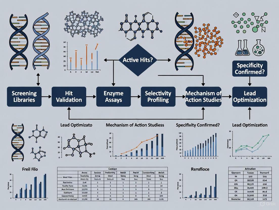
Strategic Workflow and Therapeutic Pathways
The following diagrams illustrate the integrated workflow for helicase inhibitor discovery and the strategic therapeutic concepts these inhibitors enable.
Diagram 1: Helicase Inhibitor Discovery Workflow
Diagram 2: Inhibitor Mechanisms and Therapeutic Strategies
Helicases of the SF2 superfamily, particularly RecQ and Fe-S cluster families, represent a promising yet underexplored class of therapeutic targets. Their central roles in genome maintenance pathways that are vital for cancer cell survival, combined with the genetic evidence from associated syndromes, provides a strong rationale for their targeted inhibition. The experimental frameworks outlined herein, encompassing biochemical screening, rigorous specificity testing, and cellular validation, provide a roadmap for the systematic discovery and characterization of novel helicase inhibitors. As structural and mechanistic understanding of these molecular machines deepens, structure-based drug design and the exploration of synthetic lethal relationships will likely yield increasingly potent and specific therapeutic candidates for oncology and beyond.
The discovery that Werner syndrome helicase (WRN) is a synthetic lethal target in microsatellite instability-high (MSI-H) cancers represents a paradigm shift in precision oncology, directly linking a rare genetic syndrome to a targeted cancer therapy strategy. This connection, first robustly demonstrated in 2019, reveals that cancer cells with defective DNA mismatch repair (dMMR) become fundamentally dependent on WRN helicase activity for survival, while normal cells remain unaffected [18] [19]. This application note details the mechanistic basis of this relationship and provides standardized protocols for exploiting this vulnerability through WRN inhibition, supporting ongoing drug discovery efforts for MSI-H colorectal, endometrial, and gastric cancers.
Werner syndrome is a rare autosomal recessive disorder caused by mutations in the WRN gene, characterized by premature aging and increased cancer susceptibility [20] [21]. The WRN protein, a member of the RecQ helicase family, possesses both 3' to 5' helicase and exonuclease activities and serves as a crucial genome caretaker involved in DNA replication, repair, recombination, and telomere maintenance [20] [21]. While Werner syndrome patients are cancer-prone, research has paradoxically revealed that inhibiting WRN specifically kills certain cancer cells while sparing normal cells—a phenomenon known as synthetic lethality [18].
In 2019, multiple independent research groups identified that WRN is a synthetic lethal vulnerability in MSI-H cancer cells [18] [22] [19]. MSI-H tumors, which frequently occur in colorectal (15%), gastric (15-22%), and endometrial (20-30%) cancers, arise from deficiencies in the DNA mismatch repair system [20]. This breakthrough established that while WRN is dispensable in microsatellite stable (MSS) cells, it becomes essential for maintaining genome integrity in MSI-H contexts, positioning WRN inhibitors as promising targeted therapies for MSI-H cancers [18].
Mechanistic Insights: WRN Dependency in MSI-H Cells
Molecular Basis of Synthetic Lethality
The synthetic lethal relationship between WRN inhibition and MSI-H status stems from the accumulation of TA-dinucleotide repeats throughout the genome of MMR-deficient cells [22]. During DNA replication, these expanded repetitive sequences form problematic secondary structures that create physical barriers to replication forks [22] [23]. WRN helicase is uniquely equipped to resolve these structures through its DNA unwinding activity [22]. When WRN is inhibited in MSI-H cells, unresolved DNA secondary structures persist, leading to replication fork collapse, double-strand breaks, and ultimately cell death [22] [20]. Importantly, MSS cells lack these problematic structures and therefore do not require WRN for survival, creating the therapeutic window [18].
Table 1: Key Evidence Establishing WRN-MSI Synthetic Lethality
| Evidence Type | Experimental Finding | Reference |
|---|---|---|
| Genetic Screens | Project DRIVE identified WRN as top dependency in MSI-H cell lines | [18] |
| Functional Validation | siRNA-mediated WRN depletion impaired viability in 15/18 MSI-H but 0/25 MSS cell lines | [18] [19] |
| Mechanism Studies | WRN helicase activity specifically required to resolve TA-repeat secondary structures | [22] |
| Rescue Experiments | ATP-binding deficient WRN mutants failed to rescue viability in WRN-depleted MSI-H cells | [18] |
Signaling Pathways in WRN Inhibition
The following diagram illustrates the key molecular pathways and cellular consequences following WRN inhibition in MSI-H cancer cells:
Diagram 1: Pathway of WRN inhibition in MSI-H cells. WRN helicase inhibition in MSI-H cancer cells leads to accumulation of unresolved DNA secondary structures, replication stress, double-strand breaks, and DNA damage response activation, ultimately resulting in cell death or growth arrest.
Current Clinical Landscape of WRN-Targeted Therapies
The translational potential of WRN inhibition is demonstrated by several candidates that have advanced to clinical trials. These compounds employ distinct mechanisms to target WRN, including both covalent and non-covalent inhibition strategies.
Table 2: WRN Inhibitors in Clinical Development
| Compound | Developer | Mechanism | Clinical Stage | Key Characteristics | |
|---|---|---|---|---|---|
| HRO761 | Novartis | Non-covalent allosteric inhibitor | Phase I (NCT05838768) | Binds D1-D2 interface; induces WRN degradation in MSI cells | [22] |
| RO7589831 | Roche | Covalent inhibitor | Phase I | Early clinical proof-of-concept; manageable safety profile | [24] |
| VVD-133214 | Vividion/Roche | Covalent inhibitor (targets C727) | Phase I (NCT06004245) | Covalently binds C727 residue in helicase domain | [23] |
Early clinical data from Phase I trials show promising signals of efficacy. For RO7589831, 5 of 37 evaluated patients across multiple MSI-H cancer types achieved partial responses, with 65.7% of patients experiencing durable disease stabilization [24]. The treatment was generally well-tolerated, with most adverse events being Grade 1-2 manageable nausea, vomiting, and diarrhea [24].
Experimental Protocols for WRN Inhibitor Characterization
Biochemical Helicase Inhibition Assay
Purpose: To quantitatively measure compound-mediated inhibition of WRN helicase activity in vitro.
Reagents:
- Purified recombinant WRN protein (full-length or helicase domain)
- Radiolabeled or fluorescently labeled DNA substrate (e.g., partial duplex with 3' or 5' overhang)
- ATP (1-5 mM in reaction buffer)
- Test compounds dissolved in DMSO
- Reaction buffer: 50 mM HEPES-KOH (pH 7.5), 50 mM KCl, 5 mM MgCl₂, 1 mM DTT, 0.1 mg/mL BSA
Procedure:
- Prepare reaction mixtures (20 μL final volume) containing reaction buffer, DNA substrate (0.5 nM), and WRN protein (concentration titrated to achieve 50-75% unwinding).
- Pre-incubate WRN with test compounds (typically 50 μM for initial screening) or DMSO control (≤5% final concentration) for 5 minutes at room temperature.
- Initiate reactions by adding ATP to 1-5 mM final concentration.
- Incubate at 37°C for 15-30 minutes.
- Terminate reactions by adding EDTA (20 mM final), glycerol (5%), and unlabeled competitor oligonucleotide.
- Resolve reaction products by non-denaturing PAGE (6-8% acrylamide).
- Visualize and quantify using phosphorimaging (radiolabeled) or fluorescence scanning.
- Calculate % inhibition relative to DMSO control: % Inhibition = [1 - (Unwound ProductCompound/Unwound ProductDMSO)] × 100 [6].
Cellular Viability and Proliferation Assays
Purpose: To determine selective anti-proliferative effects of WRN inhibitors in MSI-H vs. MSS cell lines.
Cell Models:
- MSI-H: HCT 116, RKO, SNU-C4 (colorectal); HEC-265, ISHIKAWA (endometrial)
- MSS: SK-CO-1, CaCo-2, SW480 (colorectal); MFE-280 (endometrial)
Short-term Viability Protocol (4-5 days):
- Seed cells in 96-well plates at optimized densities (500-3000 cells/well based on growth rate).
- After 24 hours, treat with compound serial dilutions (typically 0.1 nM - 10 μM range).
- Incubate for 4-5 days, then measure viability using ATP-based (CellTiter-Glo) or resazurin reduction assays.
- Calculate GI₅₀ values from dose-response curves [22].
Long-term Clonogenic Protocol (10-14 days):
- Seed cells at low density (200-1000 cells/well in 6-well plates).
- Treat with compounds 24 hours after seeding.
- Refresh compound-containing media every 3-4 days.
- After 10-14 days, fix with methanol and stain with crystal violet (0.1%).
- Image colonies and quantify using automated counting software [18] [22].
DNA Damage Response Assessment
Purpose: To evaluate mechanistic on-target effects of WRN inhibition through DNA damage marker analysis.
Protocol:
- Treat MSI-H and MSS control cells with WRN inhibitors for 24-72 hours.
- For immunofluorescence: Fix cells with 4% paraformaldehyde, permeabilize with 0.5% Triton X-100, block with 5% BSA, incubate with primary antibodies against γH2AX (DNA damage), followed by fluorescent secondary antibodies. Counterstain with DAPI and image using fluorescence microscopy. Quantify foci per nucleus [22].
- For immunoblotting: Harvest cells in RIPA buffer, separate proteins by SDS-PAGE, transfer to PVDF membranes, and probe with antibodies against γH2AX, p53, p21, and cleaved caspase-3. Use GAPDH or vinculin as loading controls [18] [22].
The Scientist's Toolkit: Essential Research Reagents
Table 3: Key Reagents for WRN-MSI Research
| Reagent/Cell Line | Type | Application | Key Characteristics | |
|---|---|---|---|---|
| HCT 116 | MSI-H colorectal cell line | Cellular validation | TP53 wild-type; MLH1-deficient; highly WRN-dependent | [18] |
| RKO | MSI-H colorectal cell line | Cellular validation | BRAF mutant; highly WRN-dependent | [18] |
| SK-CO-1 | MSS colorectal cell line | Negative control | Microsatellite stable; WRN-independent | [18] |
| Anti-WRN antibody | Immunoblot/IF | Target engagement | Confirms WRN depletion/degradation | [22] |
| Anti-γH2AX antibody | Immunoblot/IF | Mechanism studies | Detects DNA double-strand breaks | [22] |
| Recombinant WRN protein | Enzyme source | Biochemical assays | Full-length or helicase domain for in vitro screening | [6] |
The synthetic lethal interaction between WRN helicase and MSI represents a transformative approach for targeting mismatch repair-deficient cancers. Standardized protocols for assessing WRN inhibition across biochemical, cellular, and mechanistic studies will facilitate robust characterization of novel compounds and combination strategies. As clinical validation progresses, these application notes provide a framework for advancing the next generation of WRN-targeted therapies, potentially addressing the unmet needs of patients with MSI-H cancers who do not respond to current immunotherapies.
Synthetic lethality is a genetic phenomenon where the simultaneous disruption of two genes leads to cell death, while disruption of either gene alone remains viable [25]. This concept provides a powerful therapeutic rationale for selectively targeting cancer cells that harbor specific mutations, such as defects in DNA repair pathways, while sparing normal healthy cells [26] [27]. The foundational principle exploits the genetic vulnerabilities of cancer cells, creating a therapeutic window that maximizes efficacy while minimizing toxicity to normal tissues [28].
In clinical oncology, the most successful application of synthetic lethality to date involves PARP inhibitors in BRCA1/2-deficient cancers [27] [25]. Cancer cells with BRCA mutations lack functional homologous recombination repair, and when simultaneously exposed to PARP inhibitors that block base excision repair, the cumulative DNA damage becomes irreparable, leading to selective cancer cell death [26]. This review establishes the therapeutic rationale for expanding this approach to DNA helicase targets and their inhibitors, with particular emphasis on chemosensitization strategies that enhance the efficacy of conventional chemotherapeutic agents.
Molecular Mechanisms of DNA Repair and Helicase Function
DNA Damage Response Pathways
DNA helicases play crucial roles in maintaining genomic integrity through their involvement in multiple DNA repair pathways. The DNA damage response (DDR) network encompasses several specialized repair mechanisms, including base excision repair (BER), homologous recombination (HR), non-homologous end joining (NHEJ), nucleotide excision repair (NER), and mismatch repair (MMR) [26] [27]. These pathways are coordinated by kinase signaling cascades, primarily the ATR-CHK1-WEE1 pathway regulating replication stress checkpoints in M and G2 phases, and the ATM-CHK2-TP53 pathway managing stress checkpoints in S and G1 phases [27].
When one repair pathway is compromised in cancer cells, they become dependent on alternative pathways for survival. This dependency creates therapeutic opportunities for synthetic lethal targeting [26]. For example, microsatellite-unstable (MSI) cancers with mismatch repair deficiencies develop reliance on WRN helicase to resolve replication stress at expanded DNA (TA)n-dinucleotide repeats [29]. Inhibition of WRN in this context selectively targets MSI cancer cells while sparing microsatellite-stable (MSS) cells with functional MMR systems [29] [21].
DNA Helicases in Genome Maintenance
DNA helicases are motor proteins that catalyze the unwinding of double-stranded DNA into single strands using energy from ATP hydrolysis [8]. This function is essential for DNA replication, transcription, recombination, and repair. The human genome encodes several helicase families, with RecQ helicases being particularly important in maintaining genome stability [21].
Among RecQ helicases, Werner syndrome protein (WRN) possesses both 3'→5' helicase and 3'→5' exonuclease activities [21]. WRN rapidly accumulates at DNA damage sites and participates in multiple repair pathways, including base excision repair, non-homologous end joining, and homologous recombination [21]. The synthetic lethal relationship between WRN and MSI cancers has positioned WRN as a promising target for cancer therapy, particularly for tumors resistant to conventional treatments [29] [21].
Diagram 1: Synthetic lethality mechanism in DNA damage response. Cancer cells with HR deficiency become dependent on backup repair pathways. Inhibiting these pathways induces synthetic lethality.
Established Synthetic Lethal Targets and Helicase Dependencies
Clinical Synthetic Lethal Targets
Table 1: Established Synthetic Lethal Targets in Cancer Therapy
| Target | Synthetic Lethal Partner | Inhibitor Examples | Approved Cancer Indications |
|---|---|---|---|
| PARP | BRCA1/2, HR deficiency [27] | Olaparib, Niraparib, Rucaparib [27] | Ovarian, breast, pancreatic, prostate cancer [26] [27] |
| WRN | MSI/MMR deficiency [29] [21] | GSKWRN3, GSKWRN4, HRO761 [29] [21] | In clinical trials for MSI cancers (NCT05838768) [29] |
| ATR | ATM deficiency, ARID1A mutation [27] [28] | AZD6738, BAY 1895344 [28] | Various solid tumors (clinical trials) [28] |
| WEE1 | TP53 mutation [27] [28] | Adavosertib [30] [28] | Ovarian, pancreatic cancer (clinical trials) [30] [28] |
WRN Helicase as a Promising Synthetic Lethal Target
The recent discovery of synthetic lethality between WRN helicase and microsatellite instability has generated significant interest in pharmaceutical development [29] [21]. WRN is essential in MSI colorectal and endometrial cancer cell lines, where its inactivation selectively impairs genome integrity, induces double-strand breaks, alters cell cycles, promotes apoptosis, and decreases cell viability [21]. The mechanistic basis for this dependency stems from the accumulation of expanded DNA TA-dinucleotide repeats in MSI cells, which form cytotoxic DNA secondary structures requiring WRN for resolution [29].
Base editing screens using CRISPR-Cas9 technology have identified critical residues in WRN's ATP-binding helicase domain as essential for MSI cell survival, validating this domain as the primary drug target [29]. Covalent inhibitors targeting Cys727 in the WRN helicase domain have demonstrated remarkable selectivity due to this residue being unique to WRN among helicase family members [29]. Mass spectrometry-based chemoproteomic profiling revealed that of 23,602 distinct cysteine-containing peptides across the proteome, WRN Cys727 was the only site almost completely modified by the inhibitor GSK_WRN4, demonstrating exceptional specificity [29].
Experimental Protocols for Helicase Inhibitor Screening
Fragment-Based Screening for Covalent WRN Inhibitors
Purpose: To identify potent and selective covalent small molecule inhibitors targeting the WRN helicase domain through fragment-based screening approaches [29].
Materials and Reagents:
- Purified WRN helicase domain protein
- Methyl acrylate-based reactive fragment library
- LC-MS/MS system for tryptic digest analysis
- ATPase fluorescence-based assay kit
- Isogenic MSI and MSS cell line pairs (e.g., SW48 MSI vs. MSS)
- Bst-DNA polymerase from Bacillus stearothermophilus
- Thermostable reverse transcriptase for RNA targets
Procedure:
- Primary Screening: Screen fragment library against WRN helicase domain using intact-protein liquid chromatography-mass spectrometry (LC-MS).
- Hit Validation: Identify compounds with rapid covalent modification of WRN, measuring labeling efficiency at various concentrations and time points.
- Binding Site Mapping: Perform tryptic digest of WRN-inhibitor adducts followed by LC-MS/MS analysis to identify specific modified residues.
- Medicinal Chemistry Optimization: Initiate focused chemistry efforts to enhance biochemical potency and cellular selectivity, generating compound series with progressive improvements.
- Selectivity Assessment:
- Evaluate specificity against other RecQ helicases using fluorescence-based ATPase activity assays.
- Perform mass spectrometry-based quantitative chemoproteomic profiling for cysteine-ome coverage in relevant cell lines.
- Cellular Efficacy Testing: Test compounds in isogenic MSI vs. MSS cell line pairs, measuring DNA double-strand breaks, replication stress markers, and cell viability.
- Resistance Validation: Introduce knock-in mutations at identified binding sites (e.g., Cys727) to confirm target specificity and resistance mechanisms.
Expected Outcomes: Identification of lead compounds with pIC50 values >7.0, demonstrating >100-fold selectivity over other RecQ helicases and selective cytotoxicity in MSI versus MSS models [29].
CRISPR-Cas9 Base Editing for Essential Domain Mapping
Purpose: To map WRN protein residues critical for MSI cell survival using CRISPR-Cas9 base editing technology [29].
Materials and Reagents:
- Doxycycline-inducible adenine base editor (ABE) and cytosine base editor (CBE) constructs
- 3735-guide sgRNA library targeting WRN exons/promoters
- Control sgRNAs (non-targeting, intergenic, essential/non-essential gene targets)
- Next-generation sequencing platform
- MSI cancer cell lines (e.g., KM12 colorectal, RL95-2 endometrial)
- Antibiotics for selection (puromycin, blasticidin)
Procedure:
- Cell Line Engineering: Stably transduce MSI cell lines with doxycycline-inducible ABE and CBE editors.
- Library Transduction: Transduce base editor cell lines with the WRN-targeting sgRNA library.
- Base Editor Induction: Treat cells with doxycycline to induce base editor expression.
- Competitive Growth: Culture cells for 10-14 days to allow depletion of sgRNAs targeting essential WRN residues.
- Sequencing and Analysis:
- Harvest genomic DNA at multiple time points
- Amplify sgRNA sequences by PCR
- Perform next-generation sequencing to quantify sgRNA abundance
- Calculate depletion scores for each sgRNA compared to controls
- Data Integration: Combine ABE and CBE screen results to identify residues intolerant to variation.
- Structural Mapping: Map essential residues onto WRN helicase domain crystal structure to identify druggable pockets.
Expected Outcomes: Identification of essential WRN domains and specific residues, with significant hit enrichment in the ATP-binding helicase subdomain [29].
Diagram 2: Comprehensive workflow for helicase inhibitor screening and validation, integrating multiple experimental approaches.
Research Reagent Solutions for Helicase Studies
Table 2: Essential Research Reagents for Helicase Inhibitor Screening and Characterization
| Reagent/Category | Specific Examples | Application and Function |
|---|---|---|
| Screening Libraries | Methyl acrylate-based reactive fragments [29] | Covalent inhibitor discovery through structure-based screening |
| DNA Repair Assays | Fluorescence-based ATPase activity assay [29] | Quantify helicase ATP hydrolysis inhibition |
| Clonogenic survival assays [26] | Measure long-term cell viability after treatment | |
| γH2AX immunofluorescence [29] | Detect DNA double-strand breaks | |
| Cell Line Models | Isogenic MSI/MSS pairs (e.g., SW48) [29] | Controlled systems for synthetic lethality validation |
| Patient-derived organoids (PDOs) [29] | Physiologically relevant ex vivo models | |
| Patient-derived xenografts (PDXs) [29] | In vivo validation of efficacy and biomarkers | |
| Gene Editing Tools | CRISPR-Cas9 base editors (ABE, CBE) [29] | Functional domain mapping through targeted mutagenesis |
| sgRNA libraries [26] [29] | High-throughput gene function screening | |
| Biophysical Characterization | ThermoFluor assays [31] | Compound binding and stability assessment |
| Intact-protein LC-MS [29] | Covalent modification efficiency quantification | |
| Quantitative chemoproteomics [29] | Proteome-wide selectivity profiling |
Chemosensitization Strategies and Combination Therapies
Biomarker-Guided Chemosensitization
The integration of synthetic lethal approaches with conventional chemotherapy represents a promising strategy to overcome drug resistance and enhance therapeutic efficacy [28]. SLFN11 has emerged as a particularly important predictive biomarker for sensitivity to DNA-damaging agents, including topoisomerase I/II inhibitors, DNA synthesis inhibitors, and DNA cross-linking agents [30]. This putative DNA/RNA helicase is recruited to stressed replication forks and irreversibly triggers replication block and cell death in response to DNA damage [30].
Clinical evidence demonstrates that SLFN11 expression strongly predicts response to PARP inhibitors in small cell lung cancer (SCLC) [30]. In a Phase 2 trial of temozolomide plus veliparib versus temozolomide/placebo in relapsed SCLC, SLFN11-positive patients had significantly prolonged progression-free survival and overall survival in the combination arm [30]. Based on these findings, prospective validation of SLFN11 is now being incorporated into clinical trial designs, such as the Phase 2 randomized trial assessing maintenance atezolizumab with talazoparib versus atezolizumab alone in SLFN11-positive extensive-stage SCLC (SWOG1929, NCT04334941) [30].
Rational Combination Therapies
Table 3: Promising Synthetic Lethal Combination Strategies for Chemosensitization
| Combination Approach | Molecular Rationale | Experimental Evidence |
|---|---|---|
| PARP inhibitors + Chemotherapy | PARP inhibition impairs BER, increasing dependency on HR; chemotherapy induces DNA damage requiring functional repair [28] | Olaparib + doxorubicin enhances tumor growth inhibition in DLBCL models compared to doxorubicin alone [28] |
| ATR inhibitors + Chemotherapy in ATM deficiency | ATM-deficient cells rely on ATR-mediated checkpoint activation; ATR inhibition enhances chemotherapy efficacy [28] | ATR inhibitor AZD6738 + chemotherapy shows enhanced efficacy in ATM-defective chronic lymphocytic leukemia models [28] |
| WEE1 inhibitors + Gemcitabine in SLFN11-low cancers | SLFN11-deficient tumors resistant to DNA damage; WEE1 inhibition overcomes replication checkpoint dependency [30] | Adavosertib + gemcitabine shows efficacy in SLFN11-low ovarian and pancreatic cancer models [30] |
| WRN inhibitors + Immunotherapy in MSI cancers | WRN inhibition induces DNA damage in MSI tumors; enhances neoantigen load and immune recognition [29] [21] | WRN inhibitors suppress growth in immunotherapy-resistant PDX models [29] |
The combination of synthetic lethal approaches with standard chemotherapy represents a promising strategy to improve cancer treatment outcomes. By targeting backup DNA repair pathways that cancer cells depend on, these combinations can sensitize tumors to conventional chemotherapeutic agents, potentially overcoming resistance mechanisms and expanding therapeutic windows [28]. This approach is particularly valuable for aggressive cancers that develop resistance to initial therapies, such as MSI colorectal and endometrial cancers that progress after immune checkpoint inhibition [29].
The establishment of synthetic lethality as a therapeutic rationale provides a powerful framework for selective cancer targeting. The successful clinical translation of PARP inhibitors has validated this approach, while emerging targets like WRN helicase offer promising avenues for expanding synthetic lethal strategies to additional cancer types [29] [21]. The integration of advanced screening technologies, including fragment-based discovery, CRISPR-Cas9 base editing, and chemoproteomic profiling, has accelerated the identification and optimization of novel helicase inhibitors with exceptional potency and selectivity.
Future directions in this field will focus on several key areas: (1) prospective clinical validation of predictive biomarkers like SLFN11 and MSI status; (2) development of rational combination strategies that leverage synthetic lethality to overcome chemoresistance; (3) expansion of synthetic lethal approaches beyond DNA repair targets to other cancer vulnerabilities; and (4) advancement of computational methods for predicting synthetic lethal interactions [27] [30] [28]. As these approaches mature, synthetic lethality promises to transform cancer therapy by enabling truly precision medicine approaches that selectively target cancer cells based on their specific genetic vulnerabilities.
A Practical Guide to Helicase Inhibitor Screening Assays and Technologies
This application note provides a comprehensive guide for utilizing bioluminescence-based ATPase assays in high-throughput screening (HTS) formats. These assays are pivotal for characterizing ATP-dependent enzymes, particularly DNA and RNA helicases, and for discovering and characterizing potential inhibitors. We detail optimized protocols, key reagent solutions, and data analysis methods to enable robust evaluation of ATPase activity and compound effects in drug discovery research.
Adenosine triphosphatases (ATPases) represent a diverse class of enzymes that hydrolyze ATP to ADP and inorganic phosphate, a fundamental reaction that fuels essential cellular processes. DNA and RNA helicases are a crucial subset of ATP-dependent enzymes that unwind nucleic acid duplexes and are implicated in various diseases, making them promising therapeutic targets [1]. Consequently, robust assays for characterizing their ATPase activity and screening for inhibitors are indispensable tools in basic research and drug development.
Traditional methods for measuring ATPase activity, including colorimetric, fluorescent, and radiometric assays, often present limitations such as the use of hazardous substrates, extended detection times, and low sensitivity, complicating their adaptation for HTS [32]. Bioluminescence-based assays have emerged as a superior alternative, offering high sensitivity, rapid readouts, and excellent compatibility with automation. This note details the application of these assays for evaluating ATPase activity, with a specific focus on helicase inhibitor screening.
Principle of the Bioluminescence ATPase Assay
The bioluminescence ATPase assay is a coupling enzyme assay that indirectly measures ATPase activity by quantifying the consumption of its substrate, ATP.
- Core Principle: The assay couples the ATP-consuming reaction catalyzed by the ATPase to the ATP-dependent luciferase reaction from fireflies.
- Inverse Relationship: The luminescent signal is inversely proportional to ATPase activity. A high luminescence signal indicates low ATP consumption (low ATPase activity), whereas a low signal indicates high ATP consumption (high ATPase activity) [32] [33].
- Reaction Scheme:
- ATPase Reaction:
ATP + H₂O → ADP + Pi(catalyzed by the target ATPase) - Detection Reaction:
Luciferin + ATP + O₂ → Oxyluciferin + AMP + PPi + CO₂ + Light(catalyzed by luciferase)
- ATPase Reaction:
This homogeneous, "one-step" assay format is highly amenable to miniaturization, making it ideal for high-throughput screening in 384-well plates [34].
Assay Workflow and Logic
The following diagram illustrates the procedural workflow and logical relationship between assay components for a bioluminescence-based ATPase assay.
Experimental Protocols
Generic High-Throughput ATPase Assay Protocol
This protocol is adapted for a 384-well plate format and can be optimized for specific ATPases, such as helicases [34] [32].
Materials & Reagents
- Solid white 384-well plates
- Recombinant ATPase (e.g., helicase like MDA5, LGP2, DDX1, or VCP/p97)
- ATP solution (prepared in buffer)
- Assay buffer (containing Mg²⁺ and other required cofactors)
- Kinase-Glo Plus or similar luciferase-based detection reagent
- Test compounds or inhibitors (e.g., DBeQ for VCP)
- Multichannel pipettes
- Plate reader capable of luminescence detection
Procedure
- Plate Preparation: Dispense assay buffer into wells of a white 384-well plate.
- Compound Addition: Add test compounds or inhibitors to appropriate wells. Include controls: no-inhibitor (high ATPase activity) and no-enzyme (background/low ATPase activity).
- Enzyme Addition: Add the purified recombinant ATPase enzyme to initiate the reaction.
- ATPase Reaction Incubation: Incubate the plate at a defined temperature (e.g., 25-37°C) for a predetermined time (e.g., 30-60 minutes) to allow ATP hydrolysis.
- Detection: Add an equal volume of Kinase-Glo Plus reagent to each well. The formulation includes a thermostable luciferase and luciferin, which produces a stable luminescent signal (>5 hour half-life).
- Measurement: Incubate the plate for 10 minutes at room temperature to stabilize the signal, then measure luminescence using a plate reader.
- Data Analysis: Calculate ATPase activity based on the decrease in luminescence relative to the no-enzyme control.
Key Applications and Optimizations
- DNA/RNA Helicase Profiling: The assay has been successfully used to characterize the ATPase kinetics of human DExD/H-box RNA helicases (MDA5, LGP2, DDX1) using different RNA substrates, such as blunt-ended 24-mer ds-RNA or double-stranded RNA with a 3' overhang [34].
- Inhibitor Characterization: The protocol is effective for studying the effect of chemical compounds on ATPase function. For instance, it has been applied to characterize NPD8733, an inhibitor binding to the D1 domain of VCP/p97 [32].
- Toxicity Screening: The assay can be adapted to evaluate the effects of neurotoxic agents, such as heavy metal ions (Hg²⁺, Cu²⁺, Cd²⁺, Pb²⁺), on the activity of membrane-bound ATPases like Na,K-ATPase [33] [35].
Data Presentation and Analysis
Quantitative ATPase Activity Profiling
The table below summarizes quantitative data from published studies utilizing bioluminescence ATPase assays, demonstrating their application across different enzyme targets and inhibitor screenings.
Table 1: Summary of Bioluminescence ATPase Assay Applications and Results
| ATPase Target | Assay Context | Key Substrate/Cofactor | Reported IC₅₀ / Result | Reference |
|---|---|---|---|---|
| MDA5, LGP2, DDX1 | Helicase inhibitor screening | 24-mer ds-RNA or partial ds-RNA | Establishment of a robust HTS assay for inhibitor discovery | [34] |
| VCP/p97 | Domain-specific inhibitor study | ATP | NPD8733 compound binding to the D1 domain characterized | [32] |
| Synaptic Membrane ATPases | Neurotoxicity evaluation | ATP, Mg²⁺ | Inhibition potency: Hg²⁺ < Cu²⁺ < Cd²⁺ < Pb²⁺ | [33] [35] |
| XPB Helicase | NER pathway inhibition | DNA with lesions | Triptolide and spironolactone identified as inhibitors | [1] |
The Scientist's Toolkit: Essential Research Reagents
The following table catalogs key reagents and their critical functions for successfully implementing a bioluminescence ATPase assay.
Table 2: Key Research Reagent Solutions for Bioluminescence ATPase Assays
| Reagent / Material | Function / Role in the Assay | Example / Specification |
|---|---|---|
| Luciferase-Based Detection Reagent | Quantifies residual ATP by producing a luminescent signal proportional to ATP concentration. | Kinase-Glo Plus [32] |
| White Multiwell Plates | Provides an optimal surface for luminescence signal detection by reflecting light and minimizing cross-talk. | Solid white 384-well plates [32] |
| Recombinant ATPase Enzyme | The target enzyme of interest; purity and activity are critical for a robust assay signal. | GST-fused VCP or RNA helicases like MDA5 [34] [32] |
| ATP Solution | The substrate for the ATPase enzyme; concentration must be optimized for the kinetic range. | Prepared in assay buffer, often with Mg²⁺ as a cofactor [32] |
| Reference Inhibitors | Serve as positive controls for inhibition and for assay validation. | DBeQ for VCP/p97 [32] |
Concluding Remarks
Bioluminescence-based ATPase assays represent a powerful and versatile platform for biochemical investigation and drug discovery. Their high sensitivity, miniaturization capability, and operational simplicity make them particularly suited for the high-throughput screening and characterization of DNA/RNA helicase inhibitors. The protocols and data presented herein provide a framework for researchers to implement this technology, accelerating the development of novel therapeutics targeting ATP-dependent enzymes.
Unwinding assays are fundamental techniques in molecular biology for studying helicases, enzymes that catalyze the separation of nucleic acid duplexes into single strands. These assays are vital for understanding DNA and RNA metabolism, including replication, repair, and transcription. Furthermore, they provide a critical foundation for screening and characterizing potential helicase inhibitors, which have emerging therapeutic applications for treating diseases like cancer and viral infections [36]. This note details the core principles, quantitative parameters, and detailed protocols for key unwinding assay methodologies, with a focus on applications within inhibitor screening.
Core Principles and Quantitative Comparison of Unwinding Assays
The core principle of any unwinding assay is to differentiate the double-stranded substrate from the unwound single-stranded product. This is typically achieved by labeling the nucleic acid strands and exploiting the physical or spectroscopic differences between duplex and single-stranded states. The table below summarizes the key characteristics of the major assay types.
Table 1: Comparison of Key Unwinding Assay Methodologies
| Assay Type | Detection Principle | Throughput | Key Quantitative Measures | Advantages | Limitations |
|---|---|---|---|---|---|
| Gel-Based Radioactive Assay [36] | Separation of radiolabeled (e.g., ³²P) substrate and product by native gel electrophoresis; detection via phosphorimaging. | Low | Unwinding percentage, processivity, kinetics (with multiple time points). | Considered a "gold standard"; direct visualization of products; adaptable to various substrates. | Low temporal resolution; time-consuming; not suitable for high-throughput screening (HTS). |
| Plate-Based Fluorescence Assay (Quenched Probe) [36] | Fluorescent dye (e.g., FAM) on one strand is quenched by guanines on the complementary strand; unwinding causes fluorescence increase. | Medium to High | Unwinding kinetics in real-time, IC₅₀ for inhibitors. | Real-time kinetic data; adaptable to multi-well plates for inhibitor screening. | Requires specific substrate design; signal is indirect. |
| Dual-Labeled FRET Assay [37] | Fluorophore (e.g., Cy3) and quencher on two separate reporter strands; unwinding increases fluorescence. | Medium to High | Unwinding kinetics, coupling efficiency with ATPase activity. | Flexible for long, physiologically relevant RNA substrates; real-time data. | Fluorescent dyes can alter duplex stability. |
| Molecular Beacon Helicase Assay (MBHA) [37] | Fluorophore and quencher on a single hairpin-forming oligonucleotide; unwinding separates the pair. | Medium to High | Unwinding kinetics. | Prevents reannealing; no trap strand needed. | Not suitable for DEAD-box helicases that can unwind the dissociated beacon. |
Experimental Protocols
Protocol: Plate-Based Fluorescent Assay for Inhibitor Screening
This protocol is adapted for medium-to-high throughput screening of potential DNA helicase inhibitors using a quenched fluorescence system [36].
Research Reagent Solutions
Table 2: Essential Reagents for Plate-Based Fluorescent Unwinding Assay
| Reagent | Composition / Sequence | Function |
|---|---|---|
| Loading Strand Oligomer [36] | 5′-FAM-CATCATGCAGGACAGTCGGATCTTTTTTTTTTTTTTT-3′ | The fluorescently-labeled strand to be displaced. |
| Displaced Strand Oligomer [36] | 5′-GATCCGACTGTCCTGCATGATG-GGG-3′ | The quencher strand; three 3′ guanines quench the FAM dye. |
| Trapping Oligomer [36] | 5′-CATCATGCAGGACAGTCGGATC-3′ | Binds the displaced strand to prevent reannealing. |
| 5X Reaction Buffer [36] | 125 mM MOPS, pH 7.0, 250 mM NaCl, 10 mM β-mercaptoethanol, 500 μg/ml BSA, 0.5 mM EDTA | Provides optimal pH, ionic strength, and stabilizing conditions for the helicase. |
| HE Buffer [36] | 10 mM Hepes, pH 7.5, 1 mM EDTA | Storage and dilution buffer for oligonucleotides. |
| Purified Helicase | e.g., HCV NS3 in storage buffer (25 mM HEPES pH 7.5, 100 mM NaCl, 1mM EDTA, 5 mM β-mercaptoethanol, 20% Glycerol) | The enzyme target for which inhibitors are being screened. |
Step-by-Step Procedure
- Substrate Preparation: Anneal the Loading Strand and Displaced Strand oligonucleotides to form the duplex substrate. The final concentration of the annealed duplex for the assay is 200 nM [36].
- Reaction Setup: In a black, low-binding 96-well plate, assemble the reaction mixture:
- 1X Reaction Buffer (from the 5X stock)
- 2 mM ATP
- 4 mM MgCl₂
- ~20 nM DNA duplex substrate
- Test compound (inhibitor dissolved in DMSO) at desired concentration
- Purified helicase protein (concentration must be pre-optimized)
- Add Trapping Oligomer in excess (e.g., 5 μM) to prevent reannealing [36].
- Detection and Analysis: Immediately place the plate in a temperature-controlled plate reader. Kinetically measure fluorescence (Ex = 485 nm, Em = 528 nm) for 30-60 minutes. The increase in fluorescence over time corresponds to duplex unwinding. Compare the initial rates of fluorescence increase or the endpoint fluorescence in the presence and absence of inhibitor to identify hits [36].
Protocol: Gel-Based Radioactive Unwinding Assay (Validation)
This traditional method is used for validation and detailed mechanistic studies, often following a primary screen [36].
Research Reagent Solutions
- Loading Strand Oligomer: Unlabeled 37-mer (DNA or RNA) [36].
- Displaced Strand Oligomer: Unlabeled 22-mer complementary to the loading strand [36].
- [γ-³²P] ATP: Radioactive label for substrate preparation.
- T4 Polynucleotide Kinase (PNK): Enzyme to radiolabel the oligonucleotide.
- 10X TBE Buffer: For polyacrylamide gel electrophoresis.
- Non-Denaturing Polyacrylamide Gel: Typically 10-12%, cast between large glass plates.
Step-by-Step Procedure
- Substrate Labeling and Annealing:
- Label the 5' end of the displaced strand oligonucleotide using T4 PNK and [γ-³²P] ATP.
- Purify the labeled oligonucleotide using a nucleotide removal spin column.
- Anneal the radiolabeled displaced strand with the unlabeled loading strand to form the partial duplex substrate [36].
- Helicase Reaction:
- In a microtube, mix the helicase reaction components: reaction buffer, ATP-Mg²⁺, radiolabeled substrate (~1 ng), and the helicase enzyme.
- Include a no-enzyme control (background) and a heat-denatured control (100% unwinding).
- Incubate at 37°C for 30 minutes.
- Product Separation and Visualization:
- Stop the reaction by adding EDTA, SDS, and glycerol.
- Load the products onto a pre-run, native polyacrylamide gel.
- Run the gel in 1X TBE buffer at a constant voltage until sufficient separation is achieved.
- Dry the gel and expose it to a phosphorimager screen. Quantify the bands corresponding to the substrate (duplex) and product (single strand) to calculate the percentage of unwinding [36].
Workflow Visualization
The following diagram illustrates the logical workflow for using unwinding assays in a helicase inhibitor screening and characterization pipeline.
Substrate Design and Directionality
A critical aspect of unwinding assays is the design of the nucleic acid substrate, which must accommodate the specific requirements of the helicase under study, particularly its directionality (3'→5' or 5'→3') [38]. Helicases often require a single-stranded overhang for loading. Substrates with only one overhang can be used for directionality determination, but many helicases exhibit poor activity on them and require fork-like structures for efficient unwinding [39].
An advanced method to determine directionality for such helicases uses biotinylated oligonucleotides bound to streptavidin. The bulky streptavidin acts as a steric block, mimicking a fork structure and preventing strand reannealing, thereby enhancing helicase activity and enabling clear polarity determination [39]. For a helicase with 3'→5' polarity, activity will only be observed on a substrate with a 3' overhang and the streptavidin block on the 5' end, and vice-versa for a 5'→3' helicase [39].
Fragment-Based Screening and Pharmacophore Modeling (e.g., FragmentScout)
Fragment-based screening and pharmacophore modeling represent powerful methodologies in modern drug discovery, particularly for challenging targets such as DNA helicases. These approaches enable researchers to identify initial hit compounds and systematically develop them into potent inhibitors by focusing on essential molecular interactions. DNA helicases are crucial molecular motors involved in genome maintenance, and their dysfunction is implicated in various cancers and genetic disorders, making them promising therapeutic targets [1]. The integration of fragment-based screening with advanced pharmacophore modeling techniques provides a robust framework for identifying novel chemical starting points against these biologically complex targets. This application note details standardized protocols for implementing these methods, with specific emphasis on their application in DNA helicase inhibitor discovery, leveraging tools such as FragmentScout and ConPhar to streamline the identification and optimization of potential therapeutic compounds [40] [41].
Fragment-Based Screening with FragmentScout
The FragmentScout workflow represents a novel approach for systematically advancing from weakly binding fragments to potent inhibitors by leveraging structural data. This method is particularly valuable for helicase targets, which often feature dynamic binding sites and present challenges for traditional screening methods. The protocol utilizes publicly accessible structural data, such as that generated by XChem high-throughput crystallographic fragment screening, to generate comprehensive pharmacophore queries that aggregate feature information from multiple experimental fragment poses [40]. This approach was successfully validated through the discovery of 13 novel micromolar inhibitors of the SARS-CoV-2 NSP13 helicase, demonstrating its applicability to helicase targets and confirming hits through cellular antiviral and biophysical assays [40].
Detailed Experimental Protocol
Step 1: Data Preparation and Fragment Library Curation
- Obtain 3D structural data of the target helicase from fragment screening campaigns (e.g., XChem database)
- Curate a diverse fragment library with molecular weight <300 Da and appropriate chemical properties for helicase targets
- Format structural data for compatibility with FragmentScout and Inte:ligand LigandScout XT software
Step 2: Joint Pharmacophore Query Generation
- For each binding site, generate a joint pharmacophore query that aggregates all pharmacophore features from experimental fragment poses
- Define key interaction features including hydrogen bond donors/acceptors, hydrophobic regions, and aromatic interactions relevant to helicase binding pockets
- Set appropriate tolerance parameters to accommodate structural flexibility in helicase active sites
Step 3: Virtual Screening Implementation
- Use the joint pharmacophore query to search 3D conformational databases
- Apply filtering criteria to prioritize hits based on fit quality, chemical tractability, and novelty
- Output top-ranking compounds for experimental validation
Step 4: Experimental Validation
- Subject virtual screening hits to biochemical helicase activity assays (e.g., ATP hydrolysis, DNA unwinding)
- Confirm binding through biophysical methods such as ThermoFluor assay
- Evaluate cellular activity in relevant disease models
Table 1: Key Parameters for FragmentScout Implementation in Helicase Screening
| Parameter | Specification | Application to Helicase Targets |
|---|---|---|
| Fragment Library Size | MW <300 Da | Ensures appropriate sampling of helicase binding sites |
| Structural Data Source | XChem fragment screening | Provides experimental binding poses |
| Pharmacophore Features | HBD, HBA, hydrophobic, aromatic | Captures key helicase-inhibitor interactions |
| Screening Software | Inte:ligand LigandScout XT | Enables efficient 3D database searching |
| Validation Assays | Biochemical, ThermoFluor, cellular | Confirms helicase inhibition and binding |
Figure 1: FragmentScout Workflow for Helicase Inhibitor Discovery. This diagram illustrates the systematic process from data preparation to confirmed inhibitor identification.
Consensus Pharmacophore Modeling with ConPhar
Protocol for DNA Helicase Targets
Consensus pharmacophore modeling integrates molecular features from multiple ligands to create robust models that reduce bias from individual compounds and enhance predictive power. For DNA helicase targets with extensive structural data, this approach captures conserved interaction patterns essential for inhibition [42] [41]. The following protocol utilizes ConPhar, an open-source informatics tool specifically designed for identifying and clustering pharmacophoric features across multiple ligand-bound complexes.
Method 1: Complex Preparation and Feature Extraction
- Prepare and align protein-ligand complexes
- Collect all available helicase-ligand complex structures (e.g., PDB entries)
- Align complexes using PyMOL software based on protein backbone atoms [41]
- Extract each aligned ligand conformer and save as separate SDF files
- Generate pharmacophore JSON files using Pharmit
- Upload individual ligand files to Pharmit using the "Load Features" option
- Use the "Save Session" option to download corresponding pharmacophore JSON files
- Organize all JSON files in a single directory for processing [41]
Method 2: Consensus Model Generation with ConPhar
- Set up computational environment
- Launch Google Colab notebook with 2025.07 runtime version
- Install Conda and PyMOL using provided installation scripts
- Install ConPhar Python package and import required modules [41]
Process pharmacophore features
- Create a dedicated folder for pharmacophore JSON files
- Upload all JSON files to the designated folder
- Parse JSON files and extract pharmacophoric features into a consolidated DataFrame
- Implement exception handling to manage malformed files during processing [41]
Generate and apply consensus pharmacophore
- Execute ConPhar's consensus algorithm to identify clustered features across all ligands
- Save the resulting consensus model in multiple formats (JSON, PyMOL)
- Utilize the model for virtual screening of ultra-large molecular libraries targeting DNA helicases [41]
Table 2: ConPhar Implementation Parameters for Helicase-Targeted Pharmacophore Modeling
| Step | Tool/Software | Key Parameters | Output |
|---|---|---|---|
| Complex Alignment | PyMOL | Protein backbone atoms | Aligned helicase-ligand complexes |
| Feature Extraction | Pharmit | Default pharmacophore feature definitions | Individual pharmacophore JSON files |
| Data Consolidation | ConPhar | Automated exception handling | Unified feature DataFrame |
| Consensus Generation | ConPhar | Feature clustering algorithms | Consensus pharmacophore model |
| Virtual Screening | Compatible screening platforms | Fit tolerance parameters | Potential helicase inhibitors |
Application to SARS-CoV-2 Mpro Case Study
The ConPhar protocol was validated through a comprehensive case study on SARS-CoV-2 main protease (Mpro), utilizing one hundred non-covalent inhibitors co-crystallized with the target. The resulting consensus pharmacophore model successfully captured key interaction features in the catalytic region and enabled identification of novel potential ligands [42] [41]. This approach is directly applicable to DNA helicase targets with available structural data, particularly valuable for targets with extensive ligand datasets such as WRN helicase, where fragment-based screening has identified novel allosteric binding pockets [43] [1].
Figure 2: Consensus Pharmacophore Modeling Workflow. This visualization outlines the systematic process from structural data preparation to virtual screening application.
The Scientist's Toolkit: Research Reagent Solutions
Table 3: Essential Research Reagents and Tools for Fragment-Based Helicase Research
| Reagent/Tool | Function/Application | Example in Helicase Research |
|---|---|---|
| Fragment Libraries | Low MW compounds for initial screening | Diverse chemical space sampling for helicase binding sites [44] |
| X-ray Crystallography | High-resolution structure determination | Fragment binding mode analysis in helicase active sites [40] |
| Surface Plasmon Resonance (SPR) | Binding affinity and kinetics measurement | Detection of weak fragment-helicase interactions [43] |
| ThermoFluor Assay | Thermal stability measurement | Validation of helicase-fragment binding [40] |
| Pharmit | Pharmacophore feature extraction | Generation of initial pharmacophore models from helicase ligands [41] |
| ConPhar | Consensus pharmacophore generation | Integration of multiple helicase-ligand interaction patterns [41] |
| Inte:ligand LigandScout XT | 3D pharmacophore screening | Virtual screening for novel helicase inhibitors [40] |
| NMR Spectroscopy | Fragment binding confirmation | Mapping binding sites on helicase structures [44] |
Fragment-based screening and pharmacophore modeling represent complementary approaches that significantly advance the discovery of DNA helicase inhibitors. The FragmentScout workflow enables efficient translation of fragment screening data into viable leads, while consensus pharmacophore modeling with ConPhar provides a robust method for leveraging structural data from multiple ligand complexes. These methodologies are particularly valuable for challenging helicase targets such as WRN and hPIF1, where traditional screening approaches often struggle [45] [43] [1]. The standardized protocols presented in this application note offer researchers comprehensive frameworks for implementing these techniques, potentially accelerating the development of novel therapeutic agents targeting DNA helicases in cancer and other diseases.
Cellular phenotypic screens are indispensable in modern drug discovery, enabling the identification and characterization of compounds that induce specific biological outcomes. Within the context of DNA helicase inhibitor development, these assays provide a critical functional readout on compound efficacy and mechanism of action. By assessing parameters such as cell viability, DNA damage, and replication stress, researchers can rapidly triage potential inhibitors and gain insight into their cellular effects. This application note details established and emerging methodologies for evaluating these key phenotypic parameters, with a specific focus on applications within DNA helicase inhibitor screening. The protocols described herein facilitate the comprehensive characterization of compounds that target DNA helicases, enzymes essential for genomic maintenance and replication, and which represent promising targets for cancer therapy [6].
Assessing Cell Viability
Cell viability assays are a first-line approach in phenotypic screening, providing a quantitative measure of compound toxicity and its effect on cell proliferation.
Tetrazolium Reduction Assays (MTT Assay)
The MTT assay is a widely used colorimetric method for assessing viable cell number based on metabolic activity [46].
Principle: Viable cells with active metabolism reduce the yellow, water-soluble MTT (3-(4,5-dimethylthiazol-2-yl)-2,5-diphenyltetrazolium bromide) to purple, insoluble formazan crystals. The quantity of formazan, measured by absorbance, is proportional to the number of viable cells.
Table 1: Key Components of MTT Cell Viability Assay
| Component | Function | Typical Concentration/Format |
|---|---|---|
| MTT Substrate | Converted to formazan by metabolically active cells | 0.2 - 0.5 mg/mL in DPBS |
| Solubilization Solution | Dissolves formazan crystals for absorbance reading | 40% DMF, 2% acetic acid, 16% SDS (pH 4.7) |
| Multi-well Plates | Platform for cell culture and compound treatment | 96-well or 384-well format |
| Plate Reader | Measures absorbance of dissolved formazan | Absorbance at 570 nm |
Protocol:
- Cell Preparation: Plate cells in a 96-well plate at an optimal density (e.g., 1-5 x 10³ cells/well for adherent cells) and allow to adhere overnight.
- Compound Treatment: Add the helicase inhibitor compounds at various concentrations. Include a negative control (vehicle only) and a positive control for cell death.
- MTT Incubation: After the desired treatment period (e.g., 24-72 hours), add MTT solution to each well to a final concentration of 0.5 mg/mL. Incubate for 1-4 hours at 37°C.
- Solubilization: Carefully remove the medium and add the solubilization solution. Incubate until all formazan crystals are dissolved.
- Absorbance Measurement: Read the absorbance at 570 nm using a plate-reading spectrophotometer. A reference wavelength of 630 nm can be used to correct for background.
- Data Analysis: Calculate the percentage of cell viability relative to the vehicle-treated control wells.
Considerations:
- The MTT assay is an endpoint assay, as the reagents are cytotoxic [46].
- Optimization of cell density and MTT incubation time is required for each cell type.
- Reducing agents in the culture medium or test compounds can cause non-enzymatic reduction of MTT, leading to false positives. Include controls without cells to test for compound interference [46].
Detecting DNA Damage and Replication Stress
Beyond general viability, understanding a compound's specific impact on DNA integrity and replication dynamics is crucial for characterizing helicase inhibitors.
The DNAscent Assay for Replication Fork Dynamics
DNAscent is a high-resolution, single-molecule method that utilizes nanopore sequencing and artificial intelligence to map replication fork movement, stalling, and speed across the genome [47].
Principle: Cells are pulsed sequentially with two thymidine analogues, EdU and BrdU, which are incorporated into the nascent DNA strand by active replication forks. The resulting "footprint" of fork movement is then detected on single, nanopore-sequenced DNA molecules. Fork stalling manifests as a sharp drop-off in analogue incorporation, while the length of the analogue tracks relative to the pulse time provides a measure of fork speed [47].
Table 2: Key Components for DNAscent Assay
| Component | Function |
|---|---|
| Thymidine Analogues (EdU, BrdU) | Label nascent DNA strands during replication |
| Oxford Nanopore Sequencer | Long-read sequencing platform (e.g., MinION) |
| DNAscent Software | AI-based detection of analogue incorporation and fork calling |
| S-Phase Cell Enrichment | Fluorescence-activated cell sorting (FACS) |
Protocol:
- Pulse-Labelling: Pulse asynchronous or synchronized cells with EdU, then BrdU (e.g., 10-30 minutes each), followed by a thymidine chase.
- Cell Sorting: Enrich for S-phase cells using FACS.
- DNA Extraction: Isolate ultra-high-molecular-weight DNA.
- Nanopore Sequencing: Prepare sequencing libraries and run on an Oxford Nanopore platform (e.g., MinION). A typical run can yield over 150,000 reads longer than 20 kb from a single flow cell [47].
- Data Analysis: Use DNAscent software to call forks, calculate fork speeds (kb/min), and assign a "stall score" (0 to 1), which measures how abruptly BrdU incorporation ends, indicating a stalled or paused fork [47].
Application: This method can clearly distinguish between fork slowing (e.g., with hydroxyurea treatment) and fork stalling (e.g., with ATR inhibition) and has been used to show that different chemotherapies create distinct "replication stress signatures" [47].
Nanopore-Based Biodosimetry for DNA Damage
This novel technique rapidly assesses DNA damage, including that induced by replication stress, by measuring the fragmentation of DNA molecules [48].
Principle: Ionizing radiation and certain chemicals break DNA into smaller fragments. The method passes DNA through a nanopore with an electric current. The number and length of DNA fragments transiting the pore cause characteristic disruptions in the current, allowing for quantification of DNA damage.
Protocol:
- Sample Preparation: Extract DNA from cells treated with the helicase inhibitor.
- Nanopore Sensing: Load the DNA sample into the nanopore system.
- Current Monitoring: Monitor disruptions in the ionic current as DNA fragments pass through the nanopore.
- Data Analysis: Correlate the number and size distribution of DNA fragments with the level of genotoxic stress. This method can produce results within minutes and is effective for measuring doses in a crucial range of 2-10 Gray (Gy) [48].
Characterizing Helicase Inhibitors in a Cellular Context
Synthetic Lethality Screening
Synthetic lethality occurs when the simultaneous disruption of two genes is fatal, while disruption of either alone is not. This concept is powerful for targeting DNA repair pathways in cancers with specific vulnerabilities [49].
Principle: CRISPR interference (CRISPRi) is used to simultaneously repress the expression of a DNA helicase and another gene, then screen for synergistic effects on cell proliferation. This can identify backup pathways that cancer cells rely on when a specific helicase is inhibited.
Protocol (SPIDR Screen Overview):
- Library Design: Create a dual-guide CRISPRi library (e.g., the SPIDR library) targeting core DNA damage response (DDR) genes, including helicases [49].
- Cell Transduction: Stably express a catalytically inactive Cas9-KRAB in the cell line of interest. Transduce cells with the lentiviral dual-guide library.
- Phenotypic Selection: Harvest cells at an initial time point (T0) and after a period of competitive growth (e.g., T14 days).
- Sequencing & Analysis: Use next-generation sequencing to quantify sgRNA abundance over time. Identify sgRNA pairs that are synergistically depleted, indicating a synthetic lethal interaction [49].
Application: This approach has been used to comprehensively map genetic interactions in the DDR, revealing new synthetic lethal relationships that could be exploited therapeutically, such as between FANCM and SMARCAL1 helicases [49].
Targeting ecDNA Replication
Extrachromosomal DNA (ecDNA) is a key driver of oncogene amplification in cancer. Its replication is disorganized and vulnerable to replication stress, providing a potential therapeutic window [50].
Method: The combination of Repli-seq and DNAscent, applied to ecDNA isolated via FINE (Fluorescence-activated cell sorting-based Isolation of Native ecDNA), can be used to study how helicase inhibitors affect ecDNA replication.
Findings: ecDNA exhibits asynchronous replication, redistributed origins, reduced fork velocity, and increased stalling. Under replication stress induced by hydroxyurea, ecDNA replication is further compromised, leading to ecDNA loss [50]. This suggests that helicase inhibitors which induce replication stress could be particularly effective against ecDNA-containing cancers.
The Scientist's Toolkit
Table 3: Research Reagent Solutions for Phenotypic Screening
| Reagent/Assay | Function | Example Application |
|---|---|---|
| MTT Assay Kits (e.g., CellTiter 96) | Colorimetric measurement of cell viability | Initial cytotoxicity screening of helicase inhibitor compounds [46]. |
| CRISPRi Dual-guide Libraries (e.g., SPIDR) | Systematic repression of two genes to map genetic interactions | Identifying synthetic lethal partners for a target DNA helicase [49]. |
| DNAscent Software | Analysis of nanopore data to measure fork speed and stalling | Characterizing replication stress phenotypes induced by helicase inhibition [47] [50]. |
| FINE Method | Isolation of native, chromatinized ecDNA | Studying the specific vulnerability of ecDNA replication to helicase inhibition [50]. |
| Nanopore Biodosimeter | Rapid, single-molecule quantification of DNA fragmentation | Measuring direct DNA damage from compounds that cause replication fork collapse [48]. |
Workflow and Pathway Diagrams
Cellular Phenotypic Screening Workflow
DNA Replication Stress Response Pathway
DNA helicases are essential motor enzymes that utilize nucleoside triphosphate (NTP) hydrolysis to unwind double-stranded DNA and RNA, playing indispensable roles in fundamental cellular processes including DNA replication, transcription, repair, and recombination [51]. Their conserved catalytic cores and dynamic enzymatic cycles present both challenges and opportunities for therapeutic intervention. The dysregulation of helicase activity is implicated in various human diseases, with mutations in BLM and WRN helicases causing Bloom and Werner syndromes, respectively, both characterized by genomic instability and premature aging [51]. Furthermore, helicase overactivity in DNA repair pathways can diminish the efficacy of DNA-targeting chemotherapeutic agents, creating an urgent need for targeted helicase inhibitors to improve cancer treatment outcomes [51].
The therapeutic potential of helicase inhibition is exemplified by antiviral agents such as amenamevir (Amenalief), an approved helicase-primase inhibitor for treating herpes simplex virus (HSV) and varicella-zoster virus (VZV) infections [51]. Additionally, several helicase inhibitors have advanced to clinical trials, including MOMA-313 (polymerase theta helicase inhibitor) and HRO761 (Werner helicase inhibitor), demonstrating the growing pharmaceutical interest in this target class [51]. However, the development of selective helicase inhibitors has been hampered by the dynamic nature of these molecular machines and the conservation of their active sites, necessitating advanced structural techniques to guide rational drug design.
This application note explores the integrated use of cryo-electron microscopy (cryo-EM) and XChem fragment screening to overcome these challenges, providing detailed methodologies and structural insights that are transforming helicase inhibitor discovery. By combining high-resolution structural visualization with systematic fragment-based screening, researchers can now identify novel binding pockets and allosteric sites on helicase targets, accelerating the development of selective therapeutic compounds.
Cryo-EM Structural Elucidation of Helicase Mechanisms
Technical Advancements and Workflow
Cryo-EM has emerged as a powerful technique for determining high-resolution structures of helicase complexes in functionally relevant states. Recent technical improvements in direct electron detectors, image processing algorithms, and sample preparation methods have enabled researchers to capture these dynamic molecular machines at unprecedented resolution. The standard workflow begins with complex formation between the target helicase and its nucleic acid substrates, often followed by mild cross-linking to stabilize transient interactions. The sample is then vitrified in liquid ethane to preserve native structures and imaged under cryogenic conditions [52] [53].
Advanced single-particle analysis approaches have been developed to address the conformational heterogeneity common to helicase complexes. For the RECQL5-Pol II elongation complex, researchers implemented focused classification with signal subtraction to resolve flexible regions, particularly the helicase domain which was found to occupy a range of positions spanning a 60° arc around downstream DNA [52] [53]. This approach enabled the determination of multiple conformational states from a single sample, providing crucial insights into the structural dynamics underlying helicase function.
Key Structural Insights into Helicase Function
Recent cryo-EM studies have yielded groundbreaking insights into helicase mechanisms and inhibition. The 2025 structure of human RECQL5 bound to RNA polymerase II (Pol II) elongation complex at 3.2-Å resolution revealed how this helicase modulates transcription [52] [53]. The structure identified multiple RECQL5 domains that contact the Pol II complex, including the Internal Pol II-Interacting (IRI) module and the helicase D2 subdomain, which together stabilize RECQL5 binding and enable its function as a transcriptional roadblock [52]. Furthermore, the structures demonstrated that nucleotide-free RECQL5 twists downstream DNA in the elongation complex, and upon nucleotide binding, undergoes a conformational change that allosterically induces Pol II toward a post-translocation state, potentially helping restart stalled transcription [52] [53].
Similarly, cryo-EM analysis of the herpes simplex virus helicase-primase complex bound to inhibitors pritelivir (3.2 Å) and amenamevir (3.2 Å) elucidated the precise binding pocket enclosed by the UL52 α13 and α32 helices, the UL5 α17 helix, and the UL5 motif IV loop [54]. These structures revealed key polar interactions that anchor both compounds, particularly between UL5 K356 and the inhibitors' oxygen atoms, explaining resistance patterns observed in clinical variants and informing the design of next-generation inhibitors [54].
Table 1: Recent Cryo-EM Structures of Helicase Complexes
| Helicase Complex | Resolution | Key Insights | Publication |
|---|---|---|---|
| RECQL5-Pol II Elongation Complex | 3.2 Å | Molecular basis for transcription regulation; DNA twisting mechanism | Nature Structural & Molecular Biology (2025) [52] |
| HSV-1 Helicase-Primase with Pritelivir | 3.2 Å | Inhibitor binding pocket; resistance mutation mechanisms | Nature Microbiology (2025) [54] |
| HSV-1 Helicase-Primase with Amenamevir | 3.2 Å | Broad-spectrum inhibitor interactions; antiviral spectrum determinants | Nature Microbiology (2025) [54] |
Application to Inhibitor Design
The structural insights obtained through cryo-EM are directly applicable to rational inhibitor design. For RECQL5, the detailed interface between its KIX domain and Pol II's RPB1 subunit reveals specific residues (N595, K598, R610) that participate in hydrogen-bonding or ionic interactions, presenting potential target sites for small molecules aimed at modulating this protein-protein interaction [52]. For viral helicases, the mapping of inhibitor binding pockets enables structure-activity relationship studies that optimize compound affinity while circumventing resistance mechanisms.
The visualization of conformational changes during the helicase catalytic cycle further allows for targeting specific functional states. For example, the observation that RECQL5 transitions between nucleotide-free and bound states suggests opportunities for developing compounds that stabilize particular conformations, thereby modulating helicase activity with high specificity [52] [53].
Figure 1: Cryo-EM Workflow for Helicase Complex Analysis. The process begins with sample preparation and vitrification, proceeds through data collection and processing, and culminates in model building and functional analysis.
XChem Fragment Screening for Helicase Inhibitor Discovery
Principles and Implementation
Fragment-based drug discovery (FBDD) represents a powerful complementary approach to cryo-EM in helicase inhibitor development. The XChem platform at Diamond Light Source provides high-throughput crystallographic fragment screening, supporting the entire pipeline from crystal handling to data deposition [55]. This approach screens small, low molecular weight compounds (typically 150-250 Da) that bind weakly but efficiently to discrete binding sites on protein targets. The "minimalist" philosophy of FBDD recognizes that while fragments have lower affinity than drug-like molecules, they provide superior starting points for optimization due to their efficient binding per heavy atom [56].
XChem screening employs a diverse fragment library that samples chemical space efficiently, with each fragment serving as a potential scaffold for medicinal chemistry optimization. When combined with cryo-EM structural information, fragment screening can rapidly identify lead compounds that bind to functionally critical sites on helicase targets. The process typically involves soaking helicase crystals in fragment solutions, followed by high-throughput data collection and analysis to identify bound fragments [55].
Integration with Structural Biology
Fragment screening gains tremendous power when integrated with structural techniques like cryo-EM. The high-resolution structural information from cryo-EM guides the selection of target sites for fragment screening and provides context for interpreting fragment binding modes. For helicase targets, potential sites for fragment screening include:
- Active sites: Conserved motifs involved in NTP binding and hydrolysis
- Allosteric pockets: Regions that modulate helicase activity through conformational changes
- Protein-protein interfaces: Sites critical for helicase complex formation with partners
- Nucleic acid binding channels: Grooves that interact with DNA or RNA substrates
Once fragments are identified through XChem screening, cryo-EM can visualize their binding modes within the full helicase complex, revealing how they stabilize specific conformational states or disrupt functional interactions. This iterative process of screening and structural validation accelerates the optimization of fragments into potent, selective inhibitors.
Table 2: Fragment Screening Strategies for Helicase Targets
| Screening Strategy | Application to Helicases | Advantages | Considerations |
|---|---|---|---|
| X-ray Crystallography (XChem) | Identification of binding pockets in helicase domains | High-resolution structural information; direct visualization of binding modes | Requires protein crystallization; may not capture full dynamics |
| NMR-based Screening | Mapping allosteric sites and transient interactions | Solution-state analysis; detects weak binders | Lower throughput; requires significant protein amounts |
| Surface Plasmon Resonance | Validation of fragment binding and kinetics | Quantitative affinity measurements; low consumption | No structural information; potential false positives |
| Thermal Shift Assays | Rapid screening for stabilizing fragments | High throughput; low cost | Indirect binding measurement; confounded by buffer conditions |
Integrated Workflows: Combining Cryo-EM and Fragment Screening
Synergistic Applications in Helicase Research
The combination of cryo-EM and fragment screening creates a powerful synergistic workflow for helicase inhibitor discovery. Cryo-EM provides the architectural framework of full helicase complexes in functionally relevant states, while fragment screening identifies chemical starting points that target specific sites within these structures. This integrated approach is particularly valuable for addressing the dynamic nature of helicases, as it can capture different conformational states and identify compounds that stabilize specific configurations.
For example, in the study of RECQL5, cryo-EM revealed the twisting of downstream DNA in the nucleotide-free state and the subsequent conformational change upon nucleotide binding [52] [53]. This structural understanding creates opportunities for fragment screening to identify compounds that either prevent the DNA twisting motion or lock the helicase in a specific nucleotide-bound state, thereby modulating its transcriptional regulatory functions.
Similarly, for viral helicase targets like the HSV helicase-primase complex, cryo-EM structures with bound inhibitors reveal the molecular details of existing compound binding, which can then inform the design of focused fragment libraries that target the same pocket or adjacent allosteric sites to overcome resistance mutations [54].
Practical Implementation Protocol
Integrated Cryo-EM and Fragment Screening Workflow for Helicase Inhibitor Discovery:
Helicase Complex Preparation
- Express and purify recombinant helicase, ideally with truncations to improve stability
- Form complexes with relevant nucleic acid substrates and cofactors
- Validate functional activity through biochemical assays (ATPase, unwinding)
Initial Cryo-EM Analysis
- Prepare cryo-EM grids using vitrification devices
- Collect preliminary datasets to assess complex integrity and homogeneity
- Process data to obtain initial medium-resolution (4-5 Å) reconstruction
XChem Fragment Screening
- Crystalize the helicase complex or relevant domains
- Screen fragment library using high-throughput crystallography
- Identify hits based on electron density and binding site location
Structure-Guided Fragment Optimization
- Select fragment hits based on binding site, efficiency, and chemical tractability
- Synthesize analogs for initial structure-activity relationship studies
- Validate binding through orthogonal biophysical methods
High-Resolution Cryo-EM with Bound Fragments
- Prepare helicase complexes with optimized fragment compounds
- Collect high-resolution cryo-EM data (targeting <3.5 Å)
- Build atomic models to visualize precise binding interactions
Functional Validation
- Assess inhibitor potency in biochemical assays
- Evaluate cellular activity and selectivity
- Iterate design based on structural and functional data
Figure 2: Integrated Cryo-EM and Fragment Screening Workflow. The synergistic combination of these techniques accelerates the identification and optimization of helicase inhibitors.
Research Reagent Solutions for Helicase Studies
Table 3: Essential Research Reagents for Helicase Characterization and Inhibition Studies
| Reagent / System | Manufacturer / Source | Application | Key Features |
|---|---|---|---|
| Enzolution WRN Helicase ATPase Assay System | BellBrook Labs [57] | Measuring WRN helicase activity and inhibitor screening | Includes purified human WRN helicase (aa 500-946) and optimized DNA substrate |
| Transcreener ADP2 Assay Kits | BellBrook Labs [57] | Detection of ADP formation in helicase ATPase assays | Far-red fluorescence; compatible with FP, FI, and TR-FRET readouts |
| SPIDR (Systematic Profiling of Interactions in DNA Repair) Library | Custom [49] | CRISPRi screening for genetic interactions in DNA repair | Dual-guide RNA library targeting 548 DDR genes; 697,233 guide-level interactions |
| HSV Helicase-Primase Complex | Recombinant expression [54] | Structural and functional studies of viral helicase | Heterotrimeric complex (UL5, UL52, UL8); suitable for cryo-EM and inhibitor screening |
| RECQL5 Constructs (1-620) | Recombinant expression [52] [53] | Structural studies of human RECQL5-Pol II interactions | Truncated construct containing helicase, RQC, and IRI modules |
Detailed Experimental Protocols
Cryo-EM Sample Preparation and Data Collection for RECQL5-Pol II Complexes
Materials:
- Purified human Pol II elongation complex
- RECQL5 construct (amino acids 1-620, D157A catalytically inactive mutant)
- Nucleic acid scaffold (template/non-template DNA with hybridized RNA)
- Graphene oxide cryo-EM grids
- Vitrification device (e.g., Vitrobot)
Method:
- Complex Assembly: Incubate Pol II elongation complex with RECQL5 at 1:1.5 molar ratio in transcription buffer (20 mM HEPES pH 7.5, 40 mM KCl, 5 mM MgCl₂, 1 mM DTT) for 30 minutes at 25°C [52] [53].
Mild Cross-linking: Treat with 0.1% glutaraldehyde for 5 minutes on ice, then quench with 100 mM Tris-HCl pH 7.5.
Grid Preparation: Apply 3.5 μL of complex (0.5 mg/mL) to freshly plasma-cleaned graphene oxide grids. Blot for 3.5 seconds at 100% humidity and plunge-freeze in liquid ethane.
Data Collection: Collect movies on a 300 kV cryo-electron microscope with a K3 direct electron detector at 81,000× magnification (1.05 Å/pixel), with a defocus range of -0.8 to -1.8 μm and total electron exposure of 50 e⁻/Ų [52] [53].
Image Processing: Process data using cryo-EM software suites (e.g., RELION, cryoSPARC):
- Motion correction and CTF estimation
- Particle picking and extraction
- 2D classification to remove junk particles
- Initial 3D reconstruction without symmetry (C1)
- Focused classification with signal subtraction to address flexibility
- Bayesian polishing and final refinement
Model Building: Build atomic models into cryo-EM maps using Coot and refine with phenix.realspacerefine, validating with MolProbity [52].
Helicase ATPase Activity Assay for Inhibitor Screening
Materials:
- Enzolution WRN Helicase ATPase Assay System (BellBrook Labs) [57]
- Transcreener ADP2 Assay Kit (FP, FI, or TR-FRET format)
- Test compounds in DMSO
- 384-well low-volume assay plates
- Compatible plate reader
Method:
- Reaction Setup: Prepare enzyme reaction mix containing:
- 1× Enzyme Assay Buffer A (50 mM Tris pH 7.5, 1 mM MgCl₂, 0.01% Triton)
- 40 nM WRN helicase (amino acids 500-946)
- 200 nM WRN-H DNA substrate (37-bp annealed 3'-Flap duplex)
- 50 μM ATP
- Test compounds (typically 0.1-100 μM in 1% DMSO final) [57]
Reaction Incubation: Dispense 10 μL reaction mix per well in 384-well plates. Centrifuge briefly and incubate at 30°C for 60 minutes.
Detection: Add 10 μL of 2× Stop & Detect Buffer containing ADP detection reagents:
- For FP: ADP2 Antibody and Alexa Fluor 633 Tracer
- For TR-FRET: ADP2 Antibody-Terbium conjugate and ADP HiLyte647 Tracer
- Incubate 30-60 minutes at room temperature [57]
Readout: Measure signal according to detection format:
- FP: Excitation 620-640 nm, Emission 680-685 nm
- TR-FRET: Excitation 340 nm, Emission 490 nm & 665 nm with time-gated detection
Data Analysis: Calculate % inhibition relative to controls (100% activity = no compound; 0% activity = no enzyme). Generate dose-response curves and IC₅₀ values using appropriate software.
The integration of cryo-EM and XChem fragment screening represents a transformative approach in helicase research and drug discovery. Cryo-EM provides unprecedented insights into the structural dynamics and functional mechanisms of these essential molecular machines, while fragment screening identifies chemical starting points for targeting specific functional sites. Together, these techniques enable structure-based drug design against challenging helicase targets that have previously resisted conventional approaches.
Future developments will likely focus on increasing throughput and resolution for both techniques. For cryo-EM, advances in Volta phase plate technology, direct electron detectors, and processing algorithms promise to push resolution limits further while reducing data collection times. For fragment screening, the expansion of diverse chemical libraries and implementation of time-resolved crystallography will capture more transient binding events. The integration of these structural techniques with complementary methods such as single-molecule analysis, computational modeling, and cellular validation creates a comprehensive pipeline for helicase inhibitor development that will undoubtedly yield new therapeutic candidates in the coming years.
For researchers in this field, the key to success lies in leveraging the complementary strengths of these techniques—using cryo-EM to visualize the big picture of helicase complexes in action, and fragment screening to identify the chemical tools that can modulate their function with precision and selectivity.
Overcoming Pitfalls in Helicase Inhibition: Specificity, Artifacts, and Assay Design
Distinguishing True Inhibition from Nucleic Acid Substrate Binding Artifacts
In the pursuit of DNA helicase inhibitors for research and therapeutic applications, high-throughput screening (HTS) campaigns frequently identify compounds that appear to inhibit helicase activity. However, a significant portion of these initial "hits" are false positives resulting from non-specific interactions with the nucleic acid substrates rather than true enzymatic inhibition. These artifacts present a major challenge in helicase drug discovery, particularly for antiviral and anticancer development where helicases have emerged as promising targets due to their roles in viral replication and genome maintenance [1] [58] [59]. This application note provides detailed methodologies to distinguish true helicase inhibitors from compounds that function merely as nucleic acid binders, framed within the broader context of robust helicase inhibitor screening and characterization.
The Artifact Problem in Helicase Screening
Nucleic acid-binding artifacts represent a pervasive challenge in helicase inhibitor discovery. These compounds interfere with helicase activity not by interacting with the enzyme itself, but by stabilizing the duplex DNA or RNA structure, intercalating between base pairs, or otherwise modifying the substrate to make it resistant to unwinding. The prevalence of these artifacts necessitates rigorous counter-screening protocols to avoid costly follow-up on false positives [59] [60].
The table below outlines common artifact types and their mechanisms of interference:
Table 1: Common Nucleic Acid-Binding Artifacts in Helicase Screening
| Artifact Type | Mechanism of Interference | Example Compounds |
|---|---|---|
| Intercalators | Insert between DNA/RNA base pairs, stabilizing duplex structure | Ethidium bromide, actinomycin D |
| Groove Binders | Bind to minor/major grooves of duplex DNA, increasing melting temperature | Hoechst dyes, netropsin |
| Polycationic Molecules | Neutralize phosphate backbone charge, stabilizing nucleic acid structures | Spermine, spermidine |
| Aggregators | Form colloidal aggregates that non-specifically sequester enzymes | Various promiscuous inhibitors |
| Fluorescence Quenchers | Interfere with fluorescent readouts in HTS assays | Certain aromatic compounds |
Primary Helicase Assay Formats
Understanding the limitations of different helicase assay formats is essential for interpreting screening results and designing appropriate counter-screens. The most common biochemical approaches each present distinct vulnerabilities to artifacts.
Table 2: Helicase Assay Formats and Their Vulnerabilities to Artifacts
| Assay Format | Readout Principle | Primary Applications | Vulnerability to Artifacts |
|---|---|---|---|
| Fluorescent Dye Displacement | Fluorescence decrease as intercalating dye releases during unwinding | Kinetic studies, moderate-throughput screening | High - direct compound-dye interference |
| FRET-Based Unwinding | Fluorescence change as fluorophore-quencher separation increases | HTS, inhibitor profiling | Medium - inner filter effects, quenching |
| ADP Detection | Detects ADP produced from ATP hydrolysis | Primary HTS, universal screening | Low - but measures ATPase not unwinding |
| Gel-Based Unwinding | Separation of labeled duplex/unwound DNA or RNA by electrophoresis | Mechanistic validation, low-throughput | Low - but laborious and low throughput |
| Molecular Beacon/Hairpin | Fluorescence change upon hairpin opening | Mid-throughput kinetic assays | Medium - substrate design complexity |
Protocol 1: Primary High-Throughput Screening Using ADP Detection
Principle: This universal assay detects ADP generated from ATP hydrolysis by helicases, providing a homogeneous, "mix-and-read" format ideal for HTS. As it measures ATPase activity rather than direct unwinding, it is less susceptible to nucleic acid-binding artifacts, though it may identify compounds that target the ATPase site without affecting unwinding coupling [59] [61].
Materials:
- Transcreener ADP² Assay Kit (BellBrook Labs) or equivalent
- Recombinant helicase (e.g., WRN, BLM, POLQ, SARS-CoV-2 nsP13)
- ATP (1-10 mM stock in assay buffer)
- DNA or RNA substrate appropriate for target helicase
- Assay buffer (typically containing Mg²⁺, NaCl, HEPES, DTT)
- 384 or 1536-well microplates
- Fluorescence plate reader capable of FP or TR-FRET measurements
Procedure:
- Prepare assay buffer (e.g., 100 mM NaCl, 2.5 mM MgCl₂, 20 mM HEPES pH 7.4, 1 mM DTT).
- Dilute recombinant helicase in assay buffer to 2× final concentration (typically 0.1-10 nM depending on helicase).
- Prepare substrate/ATP mixture in assay buffer at 2× final concentration (typically 50-500 nM substrate, 2-10 mM ATP).
- Dispense 5 µL of helicase solution and 5 µL of substrate/ATP mixture into assay plates.
- Add 30 nL-100 nL compound solutions (typically 10 µM final concentration in 1% DMSO).
- Incubate at 30°C for 30-120 minutes (time optimized for each helicase).
- Develop reaction with 5 µL ADP detection mixture according to manufacturer's instructions.
- Read fluorescence polarization or TR-FRET signal.
- Calculate % inhibition relative to DMSO and enzyme-only controls.
Validation Parameters:
- Z' factor ≥ 0.7 indicates excellent assay robustness
- Signal-to-background ratio typically >3:1
- Coefficient of variation <10%
Orthogonal Assays for Hit Validation
Compounds identified in primary screening require validation through orthogonal assays that measure different aspects of helicase function to exclude artifacts.
Protocol 2: Gel-Based Strand Displacement Assay
Principle: This gold-standard method directly visualizes unwound products separated from substrate by native gel electrophoresis, providing unambiguous evidence of true helicase inhibition independent of ATPase activity [60].
Materials:
- Radiolabeled (³²P or ³³P) or fluorescently labeled DNA/RNA substrate
- Recombinant helicase
- ATP (typically 1-5 mM)
- Helicase reaction buffer
- Native gel electrophoresis equipment
- Phosphorimager or fluorescence gel scanner
Procedure:
- Prepare forked duplex DNA substrate by annealing complementary oligonucleotides (one 5'-end labeled with ³²P).
- Set up 20 µL reactions containing:
- 50 mM Tris-HCl (pH 7.5-8.0)
- 2-5 mM ATP
- 2-5 mM MgCl₂
- 50-100 mM NaCl
- 1 mM DTT
- 0.1 mg/mL BSA
- 1-10 nM DNA substrate
- 10-100 nM helicase
- Test compound (typically 0.1-100 µM)
- Incubate at 37°C for 15-60 minutes.
- Stop reaction with 5 µL stop solution (50 mM EDTA, 40% glycerol, 0.9% SDS, 0.05% bromophenol blue).
- Load samples on 8-12% non-denaturing polyacrylamide gel.
- Run gel in 0.5× TBE at 100 V for 60-90 minutes.
- Visualize using phosphorimager or fluorescence scanner.
- Quantify unwound product as percentage of total substrate.
Interpretation: True inhibitors show concentration-dependent decrease in unwound product without affecting substrate migration. Nucleic acid binders may cause shifted substrate bands or smearing.
Protocol 3: Fluorescence Anisotropy DNA Binding Assay
Principle: Directly measures compound binding to nucleic acid substrates through changes in molecular rotation of fluorescently labeled DNA/RNA, specifically identifying substrate-binding artifacts [60].
Materials:
- Fluorescein- or TAMRA-labeled DNA/RNA substrate
- Test compounds
- Assay buffer (compatible with helicase assay conditions)
- Black 384-well plates
- Fluorescence plate reader with polarization capability
Procedure:
- Prepare 20 µL reactions in assay buffer containing:
- 1-10 nM fluorescently labeled DNA/RNA substrate
- Serial dilutions of test compound (0.01-100 µM)
- Incubate at room temperature for 15-30 minutes.
- Measure fluorescence anisotropy (excitation 485 nm, emission 520 nm for fluorescein).
- Plot anisotropy vs. compound concentration.
- Fit data to determine apparent Kd for substrate binding.
Interpretation: Compounds with Kd < 10 µM for nucleic acid substrates likely function as artifacts in unwinding assays. True helicase inhibitors should show minimal substrate binding.
Advanced Counter-Screening Strategies
Protocol 4: Enzyme-Dependent DNA Binding Assessment
Principle: Some compounds require both enzyme and substrate for binding, representing a more subtle class of artifacts. This protocol detects such interactions.
Materials:
- Biotinylated DNA substrate
- Streptavidin-coated plates or beads
- Recombinant helicase
- Test compounds
- Detection antibodies (if needed)
Procedure:
- Immobilize biotinylated DNA substrate on streptavidin-coated plates.
- Block with BSA or casein-based blocking buffer.
- Add helicase with or without test compounds.
- Incubate 30-60 minutes at room temperature.
- Wash to remove unbound components.
- Detect bound helicase using specific antibodies or activity assays.
- Compare binding in presence vs. absence of test compounds.
Interpretation: Enhanced helicase retention in presence of compound suggests compound-mediated stabilization of helicase-DNA complex, potentially representing true inhibition. No change suggests compound does not affect enzyme-substrate interaction.
Protocol 5: Cross-Helicase Selectivity Profiling
Principle: True selective inhibitors should affect only specific helicases, while many nucleic acid binders show broad inhibition across multiple helicase families.
Materials:
- Multiple recombinant helicases from different families (e.g., WRN, BLM, FANCJ, SARS-CoV-2 nsP13)
- Appropriate substrates for each helicase
- ADP detection or unwinding assay reagents
Procedure:
- Perform primary helicase assays (Protocol 1 or 2) with each helicase.
- Test hit compounds at multiple concentrations (0.1-100 µM) against each helicase.
- Determine IC₅₀ values for each helicase-compound combination.
- Compare potency profiles across helicase family.
Interpretation: True selective inhibitors show >10-fold potency difference between target and off-target helicases. Broad-spectrum inhibition suggests nucleic acid binding artifacts.
Experimental Workflow Integration
The following diagram illustrates the integrated workflow for distinguishing true helicase inhibitors from artifacts:
The Scientist's Toolkit: Essential Research Reagents
Table 3: Key Research Reagent Solutions for Helicase Artifact Assessment
| Reagent/Assay System | Function | Key Features | Example Providers |
|---|---|---|---|
| Transcreener ADP² Assay | Universal ATPase activity detection | Homogeneous, HTS-compatible, Z' > 0.7 | BellBrook Labs |
| Heliscreener Unwinding Assay | Direct strand displacement measurement | Real-time, fluorescent, high sensitivity | BellBrook Labs |
| Forked Duplex DNA Substrates | Physiological helicase substrates | Custom designs, various labels | IDT, Sigma Aldrich |
| Recombinant Helicases | Target enzymes for screening | Full-length, catalytically active | Academic cores, BPS Bioscience |
| G-quadruplex Forming Oligos | Specialized substrate for specific helicases | Validates substrate specificity | IDT, Eurofins |
Case Studies and Applications
Case Study: WRN Helicase Inhibitor Screening
In a high-throughput screen of ~350,000 compounds against the Werner syndrome helicase (WRN), researchers employed a fluorometric unwinding assay followed by rigorous counter-screening [60]. Primary hits were evaluated for:
- Inhibition against multiple helicases (BLM, FANCJ)
- Direct DNA binding using fluorescence anisotropy
- Effects on DNA substrate integrity This systematic approach identified specific WRN inhibitors that impaired cancer cell proliferation in a lineage-dependent manner, validating the utility of rigorous artifact exclusion.
Case Study: SARS-CoV-2 nsP13 Helicase Screening
Recent efforts to target SARS-CoV-2 nsP13 helicase implemented a robust HTS campaign screening ~650,000 compounds [62] [63]. The workflow included:
- Primary screening in 1536-well format (Z' = 0.86)
- Orthogonal assay with alternative fluorophore
- Counterscreens against human helicases This identified 674 compounds with IC₅₀ <10 µM while excluding promiscuous nucleic acid binders.
Distinguishing true helicase inhibition from nucleic acid substrate binding artifacts requires a multifaceted approach combining primary ATPase assays, orthogonal unwinding measurements, direct binding assessments, and cross-helicase profiling. The protocols outlined herein provide a robust framework for validating helicase inhibitors, minimizing false positives in drug discovery campaigns. As helicases continue to emerge as important therapeutic targets in oncology, antiviral therapy, and beyond, these methodologies will prove essential for advancing high-quality chemical probes and drug candidates.
Achieving Selectivity Within Conserved Helicase Families
Targeting DNA helicases has emerged as a promising strategy in antiviral and anticancer drug discovery. However, a significant challenge exists in achieving selectivity for specific helicases within highly conserved families. Conserved ATP-binding and catalytic cores mean that inhibitors often cross-react, leading to potential off-target effects [1]. This application note details the structural and mechanistic insights that enable selective inhibition, supported by specific protocols for inhibitor screening and characterization. The content is framed within the broader objective of advancing DNA helicase inhibitor screening and characterization methods, providing researchers with practical tools for overcoming selectivity hurdles.
Structural Basis for Selective Inhibition
Conserved Families and Selectivity Challenges
DNA helicases are molecular motors that unwind double-stranded nucleic acids, playing essential roles in genome maintenance, replication, and transcription. They are classified into six superfamilies (SF1-SF6) based on sequence homology and mechanism [1]. The most prominent families involved in DNA damage response and repair include the RecQ family (BLM, WRN, RECQL1/4/5), iron-sulfur (Fe-S) cluster family (DNA2, XPD, DDX11, FANCJ), and other SF2 helicases (XPB, CSB, FANCM) [1]. The high structural conservation within these families, particularly in the catalytic cores that resemble RecA recombination protein folds, presents the fundamental challenge for achieving selective inhibition.
Druggable Pockets in Conserved Architectures
Recent structural studies have identified specific druggable pockets that enable selectivity. SARS-CoV-2 NSP13 helicase, a member of the 1B helicase superfamily, exemplifies this principle. Despite high sequence conservation, structural analyses reveal two potentially druggable pockets among the most conserved sites in the entire SARS-CoV-2 proteome [64]. These pockets include:
- Nucleotide-binding pocket: Situated in a cleft between the 1A and 2A RecA-like domains, with specific contacts provided by conserved helicase motifs I, II, and III in the 1A domain and IV, V, and VI in the 2A domain [64].
- Allosteric sites: Identified through crystallographic fragment screening, with 65 fragment hits across 52 datasets revealing alternative binding sites for structure-guided development [64].
Table 1: Key Conserved Helicase Families and Their Characteristics
| Helicase Family | Representative Members | Conserved Features | Associated Disorders |
|---|---|---|---|
| RecQ Family | BLM, WRN, RECQL1/4/5 | RecA-like catalytic core, zinc-binding domain | Bloom syndrome, Werner syndrome, Rothmund–Thomson syndrome |
| Fe-S Cluster Family | DNA2, XPD, DDX11, FANCJ | Iron-sulfur cluster domain, 5'-3' directionality | Fanconi anemia, xeroderma pigmentosum, trichothiodystrophy |
| SF2 Helicases | XPB, CSB, FANCM | Switch motifs, transducer domains | Xeroderma pigmentosum, Cockayne syndrome, combined syndromes |
Experimental Approaches for Selective Inhibitor Development
High-Throughput Screening (HTS) Assay Development
The establishment of robust HTS-compatible assays enables the identification of selective inhibitors through screening of large compound libraries. The following protocol details a helicase activity assay suitable for HTS campaigns:
Protocol 1: HTS-compatible SARS-CoV-2 NSP13 Helicase Activity Assay
Principle: This assay measures helicase activity through the unwinding of double-stranded DNA (dsDNA) substrates, with detection based on fluorescence polarization or intensity changes.
Materials:
- Purified SARS-CoV-2 NSP13 helicase (full-length, residues 1-601)
- dsDNA substrate: FAM/ATTO647-labeled strand annealed to complementary quencher strand
- Trap DNA: Unlabeled complementary strand to prevent re-annealing
- Assay buffer: 100 mM NaCl, 2.5 mM MgCl₂, 20 mM HEPES (pH 7.4), 2 mM ATP, 0.05% BSA
- Stop solution: 20 mM HEPES (pH 7.4), 0.2 M NaCl, 0.2 M EDTA
- 1,536-well microplates
- Fluorescence plate reader (e.g., PHERAstar)
Procedure:
- Reaction Setup: Dispense 2.5 μL of reaction mixture containing NSP13 (final concentration 0.075 nM) and trap DNA (500 nM) in assay buffer into each well of a 1,536-well plate.
- Compound Addition: Add 30 nL of test compound per well using acoustic dispensing. Include controls (DMSO for full activity, stop solution for background).
- Initiation: Add 2.5 μL of dsDNA substrate (final concentration 100 nM) to all wells.
- Incubation: Centrifuge plates at 1,200 rpm for 1 minute, then incubate at 30°C for 30 minutes.
- Termination and Detection: Add 1 μL of 5X stop solution, then measure fluorescence intensity using appropriate filters (Ex/Em: 485/520 nm for FAM, or corresponding settings for ATTO647) [62].
Validation: This assay demonstrated robustness with an average Z' factor of 0.86 ± 0.05, screening approximately 650,000 compounds and identifying 7,009 primary hits, with 1,763 confirmed upon retesting [62].
Structural Fragment Screening
Fragment-based drug discovery provides a powerful approach for identifying selective inhibitors that target unique sub-pockets within conserved helicase structures.
Protocol 2: Crystallographic Fragment Screening of Helicases
Principle: This method identifies small molecular fragments that bind to the target helicase by detecting electron density in crystal structures, enabling the discovery of novel binding sites.
Materials:
- Crystals of target helicase (e.g., SARS-CoV-2 NSP13)
- Fragment library (typically 500-1,000 compounds)
- X-ray diffraction facility
- Data processing software (e.g., HKL-2000, CCP4)
Procedure:
- Crystal Preparation: Grow crystals of the target helicase using optimized conditions. For SARS-CoV-2 NSP13, crystals were obtained in multiple forms: APO, phosphate-bound, and nucleotide-bound (AMP-PNP) states [64].
- Fragment Soaking: Soak crystals in solutions containing individual fragments at high concentrations (typically 50-100 mM) for varying durations.
- Data Collection: Collect X-ray diffraction data for each fragment-soaked crystal. For NSP13, datasets were collected at high resolution (1.9-3.0 Å) [64].
- Data Analysis: Process data to identify electron density corresponding to bound fragments. Map the binding sites and analyze protein-fragment interactions.
- Hit Validation: Confirm binding through complementary methods such as surface plasmon resonance or thermal shift assays.
Application: In a screen against SARS-CoV-2 NSP13, this approach identified 65 fragment hits across 52 datasets, providing starting points for structure-guided development of selective inhibitors [64].
Diagram 1: Fragment Screening Workflow for Selective Inhibitor Development
Research Reagent Solutions
Table 2: Essential Research Reagents for Helicase Inhibitor Screening
| Reagent/Category | Specific Examples | Function/Application | Key Characteristics |
|---|---|---|---|
| Recombinant Helicases | SARS-CoV-2 NSP13, XPB, XPD, BLM, WRN | Biochemical assays, structural studies, screening | Full-length constructs with activity tags (His-tag), high purity, verified enzymatic activity |
| Assay Substrates | Fluorescently labeled dsDNA/RNA, Quencher-labeled traps | Helicase activity measurement | Specific sequences, optimal length, high labeling efficiency, stability |
| Reference Inhibitors | Triptolide (XPB inhibitor), Spironolactone (XPB degrader) | Assay controls, mechanism studies | Known potency, well-characterized binding mode, selectivity profile |
| Screening Libraries | Fragment libraries, Diverse compound collections | Hit identification | Chemical diversity, favorable physicochemical properties, known helicase-targeting chemotypes |
| Crystallography Materials | Crystallization screens, Cryoprotectants, Fragment libraries | Structural studies | High-quality crystals, optimized conditions for fragment soaking |
Data Analysis and Validation Methods
Selectivity Profiling
Comprehensive selectivity profiling is essential to confirm that inhibitors targeting conserved helicase families do not cross-react with related human helicases, minimizing potential toxicity.
Protocol 3: Selectivity Profiling Against Human Helicase Panels
Principle: This protocol evaluates inhibitor selectivity by testing compound activity against a panel of human and viral helicases, identifying off-target effects early in development.
Materials:
- Panel of purified human helicases (XPB, XPD, BLM, WRN, etc.)
- Viral helicase (e.g., SARS-CoV-2 NSP13)
- Standardized helicase activity assay reagents
- IC₅₀ determination platform (dose-response capabilities)
Procedure:
- Assay Standardization: Establish and optimize activity assays for each helicase in the panel using validated substrates and conditions.
- Dose-Response Testing: Test compounds across a range of concentrations (typically 0.1 nM to 100 μM) against each helicase in the panel.
- Data Analysis: Calculate IC₅₀ values for each compound-helicase pair. Determine selectivity ratios by comparing IC₅₀ values between target and off-target helicases.
- Counter-Screening: Include unrelated enzymes (e.g., kinases, proteases) to assess general compound specificity.
Table 3: Quantitative Profiling of Helicase Inhibitor Selectivity
| Compound ID | Target Helicase IC₅₀ (μM) | Off-target 1 IC₅₀ (μM) | Off-target 2 IC₅₀ (μM) | Off-target 3 IC₅₀ (μM) | Selectivity Index |
|---|---|---|---|---|---|
| XPB-Inh-1 | 0.15 ± 0.02 | >50 (XPD) | 42.3 ± 3.1 (BLM) | >50 (WRN) | >333 |
| NSP13-Inh-A5 | 1.23 ± 0.11 | >100 (XPB) | >100 (XPD) | 85.6 ± 6.2 (DNA2) | >81 |
| Pan-Hel-Inh-22 | 0.08 ± 0.01 | 0.11 ± 0.02 (XPD) | 0.25 ± 0.03 (BLM) | 0.09 ± 0.01 (WRN) | 1.1 |
Mechanistic Characterization
Understanding the mechanism of inhibition provides critical insights for optimizing selectivity against conserved targets.
Protocol 4: Mechanism of Inhibition Studies
Principle: This protocol determines the inhibitory mechanism (competitive, non-competitive, allosteric) through kinetic analysis under varying substrate and inhibitor concentrations.
Materials:
- Purified helicase with verified activity
- dsDNA/RNA substrates at varying lengths
- ATP and analogs (AMP-PNP, ADP)
- Inhibitor compounds
Procedure:
- Substrate Variation: Measure helicase activity at fixed inhibitor concentrations while varying dsDNA substrate concentrations.
- ATP Variation: Measure helicase activity at fixed inhibitor concentrations while varying ATP concentrations.
- Pre-incubation Studies: Pre-incubate helicase with inhibitor before adding substrates to assess time-dependent inhibition.
- Data Fitting: Analyze data using appropriate enzyme kinetic models (Michaelis-Menten, Lineweaver-Burk) to determine inhibition modality.
Diagram 2: Comprehensive Inhibitor Screening and Characterization Workflow
Achieving selectivity within conserved helicase families remains challenging yet feasible through integrated structural, biochemical, and computational approaches. The protocols and methodologies detailed herein provide a roadmap for identifying and characterizing selective helicase inhibitors. Key strategies include leveraging fragment-based screening to identify novel binding pockets, implementing comprehensive selectivity profiling panels, and understanding inhibition mechanisms at the molecular level. As structural information expands and screening technologies advance, the rational design of selective helicase inhibitors will increasingly become a viable therapeutic strategy for cancer, genetic syndromes, and viral infections where specific helicases play pathogenic roles.
Counterscreening Strategies to Eliminate Non-Specific ATPase Inhibitors
Identifying specific inhibitors for ATP-dependent enzymes, particularly DNA helicases, is a cornerstone of modern drug discovery, especially in targeting DNA damage response pathways in oncology [1] [65]. However, High-Throughput Screening (HTS) campaigns are frequently plagued by false positives arising from nonspecific inhibitors [66]. These compounds often interfere with assay systems rather than the target enzyme itself, leading to wasted resources and misguided optimization efforts. A pervasive challenge is the prevalence of compounds that generically inhibit ATPase activity across multiple enzyme families, including helicases, kinases, and AAA+ proteins, without genuine target specificity [66] [67]. The generation of assay-ready plates, while increasing screening efficiency, can paradoxically enhance the occurrence of these nonspecific inhibitors [66]. This application note details strategies and protocols to eliminate such false positives, with a specific focus on DNA helicase targets like POLQ—an emerging synthetic-lethal target in homologous recombination-deficient cancers [65].
Core Counterscreening Strategies
A multi-faceted approach is essential to distinguish true target engagement from nonspecific ATPase inhibition. The following strategies form the foundation of a robust counterscreening workflow.
Orthogonal Assay Configurations
- Varied Order of Reagent Addition: Case studies across six kinase and protease targets reveal that a subtle change in the order of reagent addition to assay-ready plates can significantly reduce false-positive inhibition. This effect is unpredictable based on protein construct or inhibitor chemical scaffold, necessitating empirical testing [66].
- Direct vs. Coupled Detection: Traditional ATPase assays often rely on coupled enzyme systems or colorimetric detection of inorganic phosphate (e.g., malachite green) [68] [69] [70]. These can be susceptible to interference. Switching to a homogeneous, antibody-based ADP detection platform (e.g., Transcreener) provides a direct, non-coupled readout that is less prone to artifact [65] [67] [71]. This platform uses a competitive immunoassay where an anti-ADP antibody differentiates between ADP and ATP; accumulating ADP displaces a fluorescent tracer, generating a measurable signal change via Fluorescence Polarization (FP), Time-Resolved FRET (TR-FRET), or Fluorescence Intensity (FI) [67] [71].
Selectivity Profiling and Specificity Panels
- Profiling Against Related and Unrelated ATPases: A critical step in validating a putative helicase inhibitor is to test its activity against a panel of other ATPases. This includes:
- Utilizing ATPase Profiling Services: For researchers without an extensive enzyme collection, commercial profiling services are available. These services provide valuable data on inhibitor potency, selectivity, and mechanism of action across a pre-configured ATPase panel [67].
Mechanistic and Biophysical Interrogation
- Determining Mechanism of Action (MoA): Enzyme kinetic studies are indispensable. As demonstrated in a fragment-based screen against the bacterial ATPase Cagα, analyzing initial velocity data under varying ATP and inhibitor concentrations can distinguish competitive from non-competitive inhibition. A non-competitive mechanism may suggest an allosteric binding site, reducing the likelihood of pan-ATPase activity [70].
- Biophysical Confirmation of Binding: Techniques like Differential Scanning Fluorimetry (DSF) can identify fragments that bind and stabilize the target protein. For Cagα, binding of 16 fragments increased the protein's melting temperature by 1–4°C, providing confirmation of direct target engagement beyond functional inhibition [70].
Table 1: Summary of Core Counterscreening Strategies
| Strategy | Description | Key Benefit | Example/Tool |
|---|---|---|---|
| Orthogonal Assay | Using a biochemically distinct assay to re-test hits. | Identifies assay-specific artifacts. | Transcreener ADP² Assay (ADP detection) vs. Malachite Green (Phosphate detection) [68] [67]. |
| Selectivity Profiling | Screening hits against a panel of diverse ATPases. | Quantifies target specificity versus broad ATPase inhibition. | Enzolution Assay Systems for POLQ, DDX3, DHX9; VPS4B Assay [65] [71]. |
| Mechanism of Action | Enzyme kinetic analysis (Km/Vmax). | Suggests binding site; non-competitive can indicate allosteric inhibition [70]. | Michaelis-Menten kinetics with varying [ATP] and [Inhibitor]. |
| Biophysical Binding | Direct measurement of compound binding. | Confirms target engagement, separate from function. | Differential Scanning Fluorimetry (DSF) [70]. |
The following workflow diagrams the integration of these strategies into a coherent counterscreening pipeline, from primary screening to validated hit identification.
Diagram 1: Counterscreening workflow for ATPase inhibitors.
Recommended Experimental Protocols
Protocol 1: Direct ADP Detection Assay for Helicase ATPase Activity
This protocol is adapted from the Transcreener ADP² ATPase Assay Kit and is applicable to DNA helicases like POLQ [65] [67].
Principle
A homogeneous, antibody-based assay that directly quantifies ADP formation from ATP hydrolysis, suitable for HTS in 384- or 1536-well formats. The assay is compatible with FP, FI, and TR-FRET detection modes [67].
Materials
- Recombinant Target Helicase: e.g., POLQ helicase domain [65].
- Transcreener ADP² Assay Kit: Contains ADP tracer and antibody.
- ATP Solution: High-purity ATP in buffer (e.g., 200 mM Tris Base).
- MgCl₂ Solution: 100 mM in water.
- Assay Buffer: e.g., HEPES/NaCl/glycerol buffer.
- Low-Volume Microplates: 384- or 1536-well.
- Compatible Plate Reader: e.g., for FP, TR-FRET, or FI.
Procedure
- Reaction Setup: In a low-volume microplate, combine:
- Assay Buffer.
- ATP (final concentration typically 0.1–1000 µM, near Km for sensitivity).
- MgCl₂ (final concentration 1–10 mM).
- Test compound or DMSO control.
- Initiate the reaction by adding purified helicase.
- Incubation: Incubate at reaction temperature (e.g., 25–37°C) for 30–120 minutes to allow ATP hydrolysis. Ensure ATP turnover is <10–20% for linear kinetics [71].
- Detection: Add the detection mix containing the fluorescent ADP tracer and anti-ADP antibody. Incubate at room temperature for 15–60 minutes.
- Readout: Measure the signal using the chosen detection mode (FP, TR-FRET, or FI).
- Data Analysis: Calculate % inhibition and determine IC₅₀ values from dose-response curves. A true inhibitor will show concentration-dependent inhibition.
Protocol 2: Orthogonal Counterscreen Using Phosphate Release Assay
This protocol provides an orthogonal method using the classic malachite green phosphate detection system [68] [69] [70].
Principle
The malachite green molybdate reagent forms a complex with inorganic phosphate (Pi) released from ATP hydrolysis, resulting in a colorimetric change measurable at 650 nm [68].
Materials
- Malachite Green Phosphate Detection Kit.
- ATPase Reaction Components: (As in Protocol 3.1.2).
- 96-well or 384-well Plate.
- Plate Reader capable of Absorbance at 650 nm.
Procedure
- ATP Hydrolysis Reaction:
- Set up the enzymatic reaction in a 0.5 mL tube with purified helicase, ATP, MgCl₂, and compound/DMSO in assay buffer [68] [69].
- Remove a 5 µL aliquot at time zero and multiple time points (e.g., 15, 30, 45, 60 min).
- Dilute each aliquot 1:50 in assay buffer and immediately freeze in a dry ice/ethanol bath to stop the reaction [68].
- Phosphate Detection:
- Data Analysis:
- Generate a standard curve from the phosphate standards.
- Calculate the nmol of Pi released for each sample using the standard curve equation.
- Plot Pi release over time to determine the rate of ATP hydrolysis (nmol Pi/µmol protein/min). Compare rates in the presence and absence of test compounds [68].
Protocol 3: Selectivity Profiling Panel
This protocol outlines the use of selectivity panels to triage non-specific ATPase inhibitors.
Procedure
- Panel Design: Select a panel of 3–5 commercially available ATPases. This should include:
- Screening Execution:
- Test all primary hits from the target screen against this panel using the direct ADP detection assay (Protocol 3.1).
- Run all assays under identical conditions (buffer, ATP concentration, incubation time) for valid comparison.
- Data Analysis:
- Calculate % inhibition at a single concentration (e.g., 10 µM) for all ATPases.
- Compute a Selectivity Index (SI) for each compound: SI = IC₅₀ (Off-target ATPase) / IC₅₀ (Target Helicase).
- Prioritize compounds with high SI values (e.g., >10–100) for further development.
Table 2: Key Reagent Solutions for ATPase Counterscreening
| Research Reagent | Function / Utility | Example Application |
|---|---|---|
| Transcreener ADP² Assay Kit | Universal, direct detection of ADP for any ATPase; HTS-compatible [67]. | Primary screening and dose-response for POLQ helicase and selectivity panel [65]. |
| Enzolution POLQ Helicase Assay System | Ready-to-screen assay optimized for the specific ATPase activity of the POLQ helicase domain [65]. | Target-specific primary HTS and hit validation. |
| Enzolution DDX3/DHX9 Assay Systems | Assays for related SF2 helicases for selectivity profiling [65]. | Orthogonal counterscreening to eliminate non-specific helicase inhibitors. |
| Recombinant VPS4B / p97 Enzymes | AAA+ ATPases for broader selectivity profiling [67] [71]. | Counterscreening to identify compounds that broadly inhibit AAA+ ATPase family. |
| Malachite Green Phosphate Assay Kit | Orthogonal, colorimetric detection of inorganic phosphate release [68]. | Orthogonal confirmation of ATPase inhibition, separate from ADP detection. |
Data Interpretation and Hit Triage Strategy
Effective triage of screening data is critical for success. The following diagram and table outline a logical decision process for prioritizing hits.
Diagram 2: Hit triage logic for ATPase inhibitor screening.
Table 3: Triage Criteria for ATPase Inhibitor Hits
| Criterion | Acceptance Threshold | Rationale & Action |
|---|---|---|
| Potency in Primary Assay | IC₅₀ < 10 µM | Prioritize compounds with sub-micromolar to low micromolar potency for lead optimization. |
| Orthogonal Assay Confirmation | >50% Inhibition at 10 µM in Malachite Green assay. | Confirms activity is not an artifact of the primary detection method. Discard non-confirmed hits. |
| Selectivity Index (SI) | SI > 10 for at least 2 off-target ATPases. | Indicates specificity for the target over related and unrelated ATPases. Deprioritize pan-ATPase inhibitors. |
| Biophysical Binding (DSF) | ΔTm ≥ 1.5°C. | Confirms direct binding to the target protein, not just functional inhibition. |
| Mechanism of Action | Non-competitive inhibition preferred. | Suggests a potentially more selective allosteric mechanism versus competitive ATP-site binding [70]. |
The successful discovery of specific DNA helicase inhibitors requires a rigorous, multi-layered counterscreening strategy to overcome the pervasive challenge of non-specific ATPase inhibition. By integrating direct biochemical assays like the Transcreener platform, orthogonal detection methods, comprehensive selectivity profiling, and biophysical confirmation, researchers can effectively triage false positives and advance high-quality lead compounds. The protocols and workflows detailed herein provide a robust framework for screening campaigns targeting DNA helicases and other therapeutically relevant ATPases, ultimately enhancing the efficiency and success of drug discovery in areas such as DNA damage response and oncology.
Within the context of DNA helicase inhibitor screening and characterization, the robustness and sensitivity of biochemical assays are paramount. The reliability of high-throughput screening (HTS) campaigns and subsequent mechanistic studies of potential inhibitors directly depends on a thorough optimization of reaction conditions. Key parameters—including salt concentration, essential cofactors, and temperature—critically influence helicase activity, stability, and ultimately, the accurate identification of bioactive compounds. This document provides detailed application notes and protocols for systematically optimizing these parameters to establish robust helicase assays, enabling the discovery and characterization of novel antibacterial and antiviral agents targeting these essential motor proteins.
The Critical Parameters for Helicase Assay Optimization
Salt and Ionic Strength
Ionic strength is a fundamental parameter that directly influences nucleic acid binding, nucleoside triphosphate (NTP) binding, and the overall catalytic cycle of helicases.
- Mechanistic Impact: Elevated ionic strength typically weakens electrostatic interactions between the helicase and its nucleic acid substrate. This can reduce non-specific binding but may also inhibit essential translocation and unwinding activities if not properly calibrated [72].
- Optimization Guidance: A common starting buffer for SARS-CoV-2 nsp13 helicase assays consists of 100 mM NaCl and 20 mM HEPES (pH 7.4), which provides a physiologically relevant ionic environment [62]. For bacterial helicases like those from S. aureus and B. anthracis, similar ionic strength buffers are employed in FRET-based unwinding assays to identify inhibitors such as benzobisthiazole derivatives [73]. Optimization should involve titrating NaCl or KCl concentrations from 0 to 300 mM while monitoring unwinding activity.
Table 1: Effects of Increasing Reaction Parameters on Helicase Activities
| Parameter | RNA Binding | NTP Binding | NTP Hydrolysis | Oligomerization |
|---|---|---|---|---|
| Ionic Strength | ↓ | ↓ | - | - |
| Temperature | ↓ | ↓ | ↑ | - |
| pH | - | - | ↑ | - |
Note: "↓" indicates a decrease, "↑" an increase, and "-" no consistent direct effect. Adapted from [72].
Cofactors and Essential Components
Helicase reactions require several essential cofactors that work in concert to facilitate efficient nucleic acid unwinding.
- Magnesium Ions (Mg²⁺): As an essential cofactor for ATP hydrolysis, Mg²⁺ is crucial for coupling chemical energy to mechanical unwinding. Most helicase assays, including those for SARS-CoV-2 nsp13, use 2.5 mM MgCl₂ as a standard concentration [62]. The Mg²⁺ concentration should be optimized alongside ATP levels, as both affect the enzyme's kinetic parameters.
- Nucleoside Triphosphates (NTPs): ATP is the primary energy source. The SARS-CoV-2 nsp13 assay utilizes 2 mM ATP in the reaction buffer [62]. The Kₘ for ATP should be determined for each helicase to ensure saturating conditions during inhibitor screening.
- Stabilizing Agents: The addition of 1 mM DTT is standard practice to maintain reducing conditions and prevent oxidation of cysteine residues critical for helicase function, as used in SARS-CoV-2 nsp13 purification and storage buffers [62]. For HTS applications, 0.05% BSA can be included to prevent non-specific adsorption of enzymes and compounds to plastic surfaces [62].
Temperature
Temperature significantly affects enzyme kinetics, nucleic acid secondary structure stability, and the stringency of inhibitor binding.
- Kinetic Effects: Higher temperatures generally increase reaction rates, including NTP hydrolysis, but may also decrease substrate binding affinity (Table 1) [72].
- Assay Specific Considerations: SARS-CoV-2 nsp13 unwinding assays are typically conducted at 30°C for optimal activity and signal stability [62]. In contrast, bacterial helicase inhibitor screens for S. aureus and B. anthracis often run at 37°C to mimic host infection conditions [73]. The recent discovery that certain DEAD-box RNA helicases function over a wide temperature range, including those relevant to mammalian hosts, underscores the importance of selecting biologically relevant temperatures [74].
- Optimization Strategy: Perform activity time courses across a temperature gradient (e.g., 25°C-45°C) to identify the optimum that provides robust signal-to-background while maintaining enzyme stability throughout the assay duration.
Experimental Protocols for Key Assays
Protocol 1: HTS-Compatible Fluorescent Helicase Unwinding Assay
This protocol adapts the SARS-CoV-2 nsp13 helicase assay for HTS applications in a 1536-well plate format [62].
Workflow Overview:
Detailed Procedure:
Reagent Preparation:
- Prepare assay buffer: 100 mM NaCl, 2.5 mM MgCl₂, 20 mM HEPES (pH 7.4), 2 mM ATP, 0.05% BSA.
- Prepare helicase enzyme mixture: Dilute SARS-CoV-2 nsp13 to 0.075 nM in assay buffer containing 500 nM trap DNA (sequence: TCTAATGTAGTATAGTAATCCGCTC).
- Prepare dsDNA substrate: Anneal complementary strands (T20D25BHQ and FAM-T0D25) at 100 nM final concentration in assay buffer.
Assay Assembly:
- Dispense 2.5 µL of helicase/trap DNA mixture to all wells of a 1536-well plate.
- For high control wells (100% unwinding), dispense trap DNA only in assay buffer.
- Transfer 30 nL of compound library using an acoustic dispenser or pintool.
- Centrifuge plates briefly at 1,200 rpm for 1 minute to mix and collect contents.
- Pre-incubate plates for 10 minutes at room temperature.
Reaction Initiation and Measurement:
- Add 2.5 µL of dsDNA substrate (100 nM final) to all wells.
- For negative control wells (0% unwinding), add 1 µL of 5X stop solution (20 mM HEPES pH 7.4, 0.2 M NaCl, 0.2 M EDTA) before adding substrate.
- Incubate plates at 30°C for 30 minutes.
- Measure fluorescence intensity using a plate reader (e.g., PHERAstar) with Ex/Em 485/520 nm filters.
Data Analysis:
- Calculate % inhibition = 100 × [1 - (Sample - Low Control)/(High Control - Low Control)]
- High control = helicase + DMSO (maximum unwinding)
- Low control = no helicase or EDTA-stopped (minimum unwinding)
Protocol 2: Gel-Based Helicase Inhibition Assay
This radiometric gel-based assay provides direct visualization of unwinding products and is ideal for compound validation and mechanistic studies [6].
Detailed Procedure:
Reaction Setup:
- Prepare reaction mixture (20 µL final volume) containing:
- 50 mM Tris-HCl (pH 7.5)
- 50 mM NaCl
- 5 mM MgCl₂
- 2 mM ATP
- 1 mM DTT
- 0.5 nM ³²P-labeled DNA substrate
- Test compound (typically 50 µM for initial screening) or DMSO control
- Pre-incubate helicase with compound for 5 minutes on ice.
- Prepare reaction mixture (20 µL final volume) containing:
Reaction Initiation and Termination:
- Start reaction by adding DNA substrate and ATP.
- Incubate at 37°C for 15 minutes.
- Stop reaction by adding 5 µL of stop solution (50 mM EDTA, 0.5% SDS, 2 mg/mL Proteinase K, 30% glycerol, 0.25% bromophenol blue).
- Include heat-denatured DNA substrate control (boiled for 5 minutes).
Product Separation and Visualization:
- Load entire reaction on 8-12% non-denaturing polyacrylamide gel.
- Run gel in 0.5X TBE buffer at 200 V for 1.5-2 hours.
- Expose gel to phosphorimager screen overnight.
- Scan screen using Typhoon scanner or equivalent.
- Quantitate bands using ImageQuantTL software.
Table 2: Troubleshooting Guide for Helicase Assays
| Problem | Possible Cause | Solution |
|---|---|---|
| High background signal | Substrate degradation | Check substrate integrity; include no-enzyme controls |
| Low unwinding activity | Enzyme denaturation | Aliquot and freeze enzyme; check activity with positive control |
| High well-to-well variability | Inconsistent dispensing | Calibrate liquid handlers; include mixing step |
| Poor Z' factor | Insufficient signal window | Optimize enzyme concentration; extend incubation time |
| Compound interference | Fluorescence quenching | Use orthogonal assay; test compound alone |
Protocol 3: Dye-Displacement Helicase Assay
This continuous assay measures helicase activity through the displacement of fluorescent dyes from dsDNA, enabling real-time kinetic measurements [75].
Detailed Procedure:
Dye Selection and Preparation:
- Select appropriate DNA-binding dye: bis-benzimide (DAPI), Hoechst 33258, or thiazole orange.
- Prepare dye solution in assay buffer at optimal concentration (typically 1-5 µM).
Assay Assembly:
- Prepare reaction mixture containing:
- Standard helicase assay buffer
- dsDNA substrate (varies by helicase)
- Fluorescent dye at predetermined optimal concentration
- Pre-incubate helicase with inhibitor compounds for 5-10 minutes.
- Prepare reaction mixture containing:
Real-Time Measurement:
- Initiate reaction by adding ATP.
- Immediately monitor fluorescence decrease (excitation/emission wavelengths specific to dye).
- Collect data points every 10-30 seconds for 30-60 minutes.
- Calculate initial rates from the linear portion of the progress curves.
The Scientist's Toolkit: Research Reagent Solutions
Table 3: Essential Reagents for Helicase Inhibitor Screening Assays
| Reagent | Function | Example & Specification |
|---|---|---|
| Recombinant Helicase | Enzymatic unwinding activity | Nuclease-free preparation; >90% purity [76] |
| FRET-DNA Substrate | Unwinding reporter | Dual-labeled partial duplex; FAM/TAMRA or BHQ quencher [62] |
| Trap Oligonucleotide | Prevents reannealing | Unlabeled complementary strand [62] |
| ATP Regeneration System | Sustained energy supply | ATP + creatine phosphate/creatine kinase [76] |
| Fluorescent DNA Dyes | Continuous monitoring | Thiazole orange, DAPI, Hoechst 33258 [75] |
| Positive Control Inhibitor | Assay validation | Benzobisthiazoles (bacterial) [73] |
Data Analysis and Quality Control
Robust data analysis and quality control metrics are essential for successful helicase inhibitor screening campaigns.
- Quality Control Metrics: For HTS applications, the Z' factor should be calculated to assess assay quality: Z' = 1 - [3(σₚ + σₙ)/|μₚ - μₙ|], where σₚ and σₙ are the standard deviations of positive and negative controls, and μₚ and μₙ are their means. The SARS-CoV-2 nsp13 HTS campaign achieved an average Z' factor of 0.86 ± 0.05, indicating an excellent assay for screening [62].
- Inhibitor Characterization: Determine IC₅₀ values by testing compound serial dilutions. For benzobisthiazole inhibitors, IC₅₀ values against B. anthracis helicase ranged from >100 µM for weak inhibitors to 0.7 µM for the most potent analogs [73].
- Selectivity Assessment: Counter-screening against related helicases and human off-targets is crucial. Promising inhibitors should exhibit selectivity indices (SI = CC₅₀/IC₅₀) >500, as demonstrated by optimized benzobisthiazole compound 59 [73].
Systematic optimization of salt conditions, cofactors, and temperature is fundamental to developing robust helicase assays for inhibitor screening. The protocols outlined herein provide a framework for establishing sensitive and reproducible assays suitable for both high-throughput screening and detailed mechanistic studies of hit compounds. By carefully controlling these critical parameters, researchers can ensure the identification of truly bioactive helicase inhibitors with potential as novel anti-infective agents, contributing valuable tools and compounds to the drug development pipeline.
Addressing Challenges in Cellular Permeability and Toxicity
The discovery of DNA helicases as promising therapeutic targets for cancer and viral infections has accelerated the need for robust inhibitor screening platforms. A significant bottleneck in this pipeline lies in transitioning from identifying potent enzymatic inhibitors in biochemical assays to discovering compounds that are effective in cellular environments. The primary challenges are twofold: ensuring that lead compounds can effectively cross cell membranes to reach their intracellular targets (cellular permeability), and confirming that they do not cause undue harm to normal cells or exhibit off-target effects (toxicity). This application note details integrated experimental protocols designed to systematically address these challenges within the context of DNA helicase inhibitor development, providing a framework for de-risking the early-stage discovery process.
Quantitative Profiling of Key Parameters
Effective profiling of permeability and toxicity relies on quantifying a suite of interdependent parameters. The target values for a promising lead compound are summarized in Table 1.
Table 1: Key Quantitative Parameters for Profiling Helicase Inhibitors
| Parameter | Description | Target Value | Significance |
|---|---|---|---|
| Lipophilic Efficiency (LipE) | Measure of potency corrected for lipophilicity (pIC50 - LogP) [22]. | >5 | Optimizes balance of potency and permeability; reduces attrition risk. |
| Polar Surface Area (PSA) | Sum of surfaces of polar atoms in a molecule [22]. | <140 Ų | Predictive of good passive cellular permeability. |
| Cellular GI₅₀ | Concentration causing 50% inhibition of cell growth [22]. | MSI-H: ~50-1000 nM; MSS: No effect | Confirms selective anti-proliferative effect in target cell populations. |
| Thermal Shift (PS₅₀) | Concentration causing 50% protein stabilization in lysates [22]. | ~10-100 nM | Demonstrates direct intracellular target engagement. |
| Cytotoxicity (IC₅₀) | Concentration causing 50% cell death in non-target or normal cells. | >> Cellular GI₅₀ (e.g., >10x) | Indicates a wide therapeutic window and low general toxicity. |
Experimental Protocols
In Vitro Biochemical and Biophysical Profiling
This initial protocol focuses on characterizing the intrinsic properties of the inhibitor and its direct interaction with the helicase target.
Procedure:
- ATPase/Helicase Assay: Perform a biochemical assay to determine the IC₅₀ for enzymatic inhibition. For WRN, use an ATPase assay at high ATP concentrations (e.g., 20-fold Kₘ) to gauge potency under competitive conditions [22].
- Cellular Permeability Prediction: Calculate key physicochemical properties, including calculated LogP (cLogP) and 3D Polar Surface Area, using physics-based property prediction software. Compute Lipophilic Efficiency (LipE = pIC₅₀ - cLogP) to guide lead optimization towards molecules with an optimal balance of potency and permeability [22].
- Target Engagement via Thermal Shift Assay:
- Treat cell lysates (from both target-sensitive, e.g., MSI-H, and insensitive, e.g., MSS, cells) with a concentration gradient of the inhibitor.
- Subject the lysates to a gradient of thermal denaturation.
- Detect stabilized target protein using immuno-blotting or a thermal shift dye.
- Calculate the PS₅₀, the concentration at which 50% of the target protein is stabilized, as a measure of cellular target engagement [22] [77].
Cell-Based Viability and Selectivity Screening
This protocol assesses the functional outcome of helicase inhibition in a cellular context, determining selectivity and potency.
Procedure:
- Cell Line Panel Selection: Establish a panel of cell lines that includes:
- Short-Term Proliferation Assay: Plate cells in 384-well plates and treat with a 10-point serial dilution of the inhibitor for 4-5 days. Measure cell viability using a cell-titer glow luminescence assay. Calculate the GI₅₀ (50% growth inhibitory concentration) [22].
- Long-Term Clonogenic Assay: Seed cells at low density in 6-well plates and treat with a concentration range of the inhibitor for 10-14 days. Fix and stain the resulting colonies with crystal violet. Count colonies to determine the survival fraction. This assay is critical for detecting the effects of WRN inhibition, which often requires multiple cell cycles to manifest [22].
- Data Analysis: Plot dose-response curves for all cell lines. A promising inhibitor will show potent GI₅₀ in MSI-H cells (e.g., 50-1000 nM) and minimal effect in MSS cells, confirming a synthetic lethal interaction [22].
Mechanistic Confirmation and Toxicity Assessment
This protocol validates the on-target mechanism of action and evaluates indicators of toxicity.
Procedure:
- DNA Damage Response (DDR) Immunofluorescence:
- Plate MSI-H and MSS cells on glass coverslips and treat with the inhibitor for 24-72 hours.
- Fix, permeabilize, and stain cells with antibodies against phosphorylated histone H2AX (γH2AX), a marker of DNA double-strand breaks.
- Counterstain with DAPI to visualize nuclei.
- Image using a high-content imaging system and quantify γH2AX foci per nucleus. A positive result shows a significant increase in γH2AX foci specifically in MSI-H cells [22] [77].
- Cellular Toxicity Profiling:
- Treat immortalized non-malignant cell lines (e.g., MCF-10A) with the inhibitor and assess viability using the MTT or CellTiter-Glo assay after 72 hours to determine a general cytotoxicity IC₅₀.
- For functional toxicity, isolate peripheral blood mononuclear cells (PBMCs) from healthy donors. Treat PBMCs with the inhibitor and measure proliferation in response to a mitogen like PHA over 5 days.
- Apoptosis Assay: Treat MSI-H and MSS cells with the inhibitor for 48-96 hours. Detect apoptosis using an Annexin V/propidium iodide staining kit followed by flow cytometry analysis.
Pathway and Workflow Visualization
DNA Damage Response Pathway
The following diagram illustrates the key cellular pathway activated upon successful inhibition of a synthetically lethal helicase like WRN.
Integrated Screening Workflow
The integrated experimental workflow for addressing permeability and toxicity is outlined below.
The Scientist's Toolkit
Table 2: Essential Research Reagent Solutions
| Reagent/Assay | Function | Application Example |
|---|---|---|
| DNA-Conjugated Gold Nanoparticles | Colorimetric substrate for helicase activity; measures DNA unwinding via SPR shift [78]. | In vitro biochemical profiling of helicase inhibitors. |
| Cellular Thermal Shift Assay (CETSA) | Confirms direct target engagement by measuring compound-induced protein thermal stabilization in cells or lysates [22] [77]. | Measuring intracellular WRN engagement (PS₅₀). |
| High-Content Imaging (HCI) | Automated microscopy to quantify phenotypic changes (e.g., γH2AX foci) in cell populations [77]. | Mechanistic confirmation of DNA damage response. |
| ATPase/ADP-Glo Assay | Homogeneous, HTS-compatible assay measuring ATP consumption; indicator of helicase motor function [22] [77]. | Primary high-throughput biochemical screening. |
| Isogenic Cell Line Pairs | Genetically matched cell lines differing only in a key gene (e.g., MMR status or helicase mutation) [22]. | Controlling genetic background to confirm on-target, synthetic lethal effects. |
From Hit to Candidate: Validation, Mechanistic Profiling, and Lead Optimization
The discovery and characterization of biologically active small molecules, such as DNA helicase inhibitors, necessitates rigorous validation of direct target engagement [6]. Biophysical methods that quantify the binding affinity and thermodynamics of interactions between a protein and a ligand are fundamental to this process, providing unequivocal evidence beyond functional cellular assays [79]. Among the numerous techniques available, Isothermal Titration Calorimetry (ITC), Surface Plasmon Resonance (SPR), and Differential Scanning Fluorimetry (DSF) have emerged as cornerstone methodologies. These techniques are often employed in an integrated manner to overcome the inherent limitations of any single approach, delivering a comprehensive picture of the binding event—from the initial yes/no screening of potential ligands to the full kinetic and thermodynamic profiling of lead compounds [79]. This Application Note details the principles, protocols, and practical applications of ITC, SPR, and DSF, framed within the context of DNA helicase inhibitor research.
The following table summarizes the core attributes, advantages, and limitations of ITC, SPR, and DSF, guiding researchers in selecting the appropriate technique for their specific experimental phase.
Table 1: Comparison of Key Biophysical Binding Assays
| Method | Key Information | Throughput | Sample Consumption | Key Advantage | Key Limitation |
|---|---|---|---|---|---|
| ITC | Affinity (Kd), stoichiometry (n), and full thermodynamics (ΔH, ΔS, ΔG) [79] | Low | High (mg) | Label-free; provides direct measurement of enthalpy change | High protein consumption; limited throughput |
| SPR | Affinity (Kd) and kinetic data (association rate kon, dissociation rate koff) [80] [81] | Low to Moderate | Low (μg) | Label-free; real-time kinetic data; high sensitivity | Requires immobilization; complex system optimization [79] |
| DSF | Thermal shift (ΔTm); binding yes/no information [79] | High | Low (μg) | High-throughput; easy to use; low sample consumption | Prone to false positives/negatives; no quantitative affinity data [79] |
Isothermal Titration Calorimetry (ITC)
Principle and Application
ITC directly measures the heat released or absorbed during a molecular binding event [79]. By performing a series of controlled injections of one binding partner into the other, ITC can determine the binding affinity (Kd), stoichiometry (n), and the complete thermodynamic profile (enthalpy ΔH and entropy ΔS) of the interaction in a single experiment [79]. This makes it a gold-standard technique for characterizing the driving forces behind binding, such as distinguishing between enthalpically- or entropically-driven interactions. In helicase inhibitor studies, ITC is invaluable for confirming direct binding and understanding the molecular forces at play.
Detailed Protocol
Materials:
- Purified protein (e.g., DNA helicase) and ligand (inhibitor)
- ITC instrument (e.g., MicroCal PEAQ-ITC, Malvern Panalytical)
- Dialysis buffer or desalting column for buffer matching
- Degassing station
Method:
- Sample Preparation:
- Dialyze both the protein and the ligand into an identical, well-degassed buffer. Precise buffer matching is critical to avoid heats of dilution from buffer mismatches.
- Centrifuge samples to remove any particulate matter.
- Determine accurate concentrations. For a typical experiment with a protein in the cell, a concentration that yields a c-value (c = n[M<>t]Ka) between 10 and 100 is ideal.
Instrument Loading:
- Load the protein solution into the sample cell (typically ~200 μL).
- Load the ligand solution into the injection syringe.
Experiment Setup:
- Set the experimental temperature (commonly 25°C or 37°C).
- Design the titration program. A typical setup includes:
- A single initial injection (e.g., 0.4 μL) that is often discarded in data analysis.
- Followed by a series of 15-20 injections (e.g., 2-3 μL each) with constant stirring.
- Sufficient spacing between injections (e.g., 120-180 s) to allow the signal to return to baseline.
Data Collection and Analysis:
- Run the experiment, which measures the heat flow (μcal/s) for each injection.
- Integrate the raw heat peaks to obtain the total heat per injection.
- Subtract the heat of dilution, obtained from a control experiment (titrating ligand into buffer alone).
- Fit the corrected isotherm (plot of kcal/mol of injectant vs. molar ratio) to an appropriate binding model (e.g., a "One Set of Sites" model) using the instrument's software to derive Kd, n, and ΔH.
Surface Plasmon Resonance (SPR)
Principle and Application
SPR detects changes in the refractive index at a sensor surface, allowing for the label-free, real-time monitoring of biomolecular interactions [81]. One interactant (the ligand, e.g., the DNA helicase) is immobilized on a dextran-coated sensor chip, while the other (the analyte, e.g., the inhibitor) is flowed over the surface [80]. The resulting sensorgrams plot response (Resonance Units, RU) against time, enabling the calculation of association (kon) and dissociation (koff) rate constants, from which the equilibrium dissociation constant (Kd) is derived (Kd = koff/kon) [80]. SPR is particularly powerful for characterizing binding kinetics and can reveal complex mechanisms, as demonstrated in the kinetic analysis of BRCA1-BRCT domain interaction with phosphopeptides [80].
Detailed Protocol
Materials:
- SPR instrument (e.g., Biacore series, Carterra LSA)
- Sensor chip (e.g., CM5 for amine coupling, SA for streptavidin-biotin capture)
- Running buffer (e.g., HEPES-buffered saline with surfactant)
- Amine Coupling Kit (EDC, NHS, ethanolamine-HCl) or biotinylated ligand
- Regeneration solution (e.g., 10 mM glycine, pH 1.5-3.0)
Method:
- Immobilization (Ligand Attachment):
- Amine Coupling: Activate the carboxymethylated dextran surface with a mixture of EDC and NHS. Inject the ligand (helicase) in a low-salt buffer at a pH 0.5-1.0 units below its pI to ensure a positive charge. Deactivate remaining esters with ethanolamine [82].
- Capture Coupling: For higher activity and oriented immobilization, use a capture approach. For an antibody, immobilize an anti-species antibody first, then capture the ligand antibody. For other proteins, tags like His can be used with an appropriate capture surface [82]. An advanced strategy involves pre-forming the antibody-antigen complex before capture to protect the binding site [82].
- Immobilize the ligand to a low density (50-200 RU) to minimize mass transport effects and rebinding.
Binding Experiment (Kinetic Titration):
- Use a flow rate of 30-50 μL/min to reduce mass transport limitations.
- Inject a series of analyte (inhibitor) concentrations over the ligand and a reference surface for 2-5 minutes (association phase).
- Switch to running buffer and monitor the dissociation for a similar period.
- Regenerate the surface with a short injection (15-60 s) of regeneration solution to remove bound analyte without damaging the ligand.
Data Analysis:
- Subtract the signal from the reference flow cell and a buffer blank (double referencing) [80].
- Fit the resulting sensorgrams globally to a kinetic model. Start with a simple 1:1 Langmuir model. If the fit is poor, more complex models (e.g., a two-state or conformational change model) may be required, as was necessary for the BRCA1-BRCT interaction [80].
- Report kon, koff, and the calculated Kd.
Diagram 1: SPR kinetic analysis workflow.
Differential Scanning Fluorimetry (DSF)
Principle and Application
DSF, also known as the thermal shift assay, monitors the thermal denaturation of a protein. A fluorescent dye (e.g., SYPRO Orange) binds to hydrophobic patches of the protein that become exposed as the protein unfolds upon heating. Ligand binding often stabilizes the protein's native fold, leading to an increase in its melting temperature (ΔTm) [79]. DSF is primarily used as a high-throughput primary screening tool to identify potential binders from large compound libraries due to its low sample consumption and simplicity [79]. In a helicase inhibitor campaign, it can rapidly triage thousands of compounds to identify promising hits for further validation with ITC or SPR.
Detailed Protocol
Materials:
- Real-time PCR instrument
- Purified protein
- Compound library
- Fluorescent dye (e.g., SYPRO Orange, 1000X stock in DMSO)
- Multi-well PCR plates and seals
Method:
- Assay Setup:
- Prepare a master mix containing the protein (e.g., 1-5 μM) and dye (e.g., 5X final concentration) in an appropriate buffer.
- Dispense the master mix into a 96- or 384-well PCR plate.
- Add compounds to test wells, and include a DMSO-only control. The final DMSO concentration should be consistent across all wells (typically ≤1%).
- Seal the plate and centrifuge briefly to eliminate bubbles.
Run the Melting Curve:
- Place the plate in the real-time PCR instrument.
- Set the temperature ramp. A typical protocol ramps from 25°C to 95°C with a slow ramp rate (e.g., 1°C/min) while continuously monitoring the fluorescence of the dye.
Data Analysis:
- Plot the fluorescence intensity against temperature for each well.
- Determine the melting temperature (Tm) for each curve, which is the inflection point or the temperature at the fluorescence maximum.
- Calculate the ΔTm for each compound by subtracting the Tm of the DMSO control.
- A significant positive ΔTm shift (e.g., >1-2°C) suggests potential binding and stabilization of the protein.
Integrated Workflow and Reagent Solutions
A powerful strategy for helicase inhibitor discovery combines these techniques sequentially. DSF acts as the high-throughput filter to identify "hits." These hits are then validated and characterized for kinetics using SPR, and the most promising leads are subjected to a full thermodynamic profiling with ITC [79]. This multi-tiered approach efficiently allocates resources and builds a robust dataset for candidate selection.
Table 2: Essential Research Reagent Solutions for Binding Assays
| Reagent / Solution | Function / Application | Key Considerations |
|---|---|---|
| SYPRO Orange Dye | Binds hydrophobic regions of unfolded protein in DSF [79] | Stock solution is in DMSO; light-sensitive; compatible with standard real-time PCR filters. |
| CM5 Sensor Chip | Gold sensor surface with a carboxymethylated dextran matrix for ligand immobilization in SPR [82] | Versatile; used for amine coupling or capturing; requires maintenance to prevent clogging. |
| Anti-Mouse IgG Capture Kit | For oriented, site-specific immobilization of mouse monoclonal antibodies in SPR [82] | Preserves antigen-binding activity; increases assay sensitivity and consistency. |
| Amine Coupling Kit (EDC/NHS) | Cross-linking reagents for covalent immobilization of proteins via lysine residues in SPR [82] | Standard, robust chemistry; can lead to random orientation and loss of activity. |
| High-Purity Buffers | Provide a stable chemical environment for all binding assays (ITC, SPR, DSF) | Must be matched exactly in ITC; filtered and degassed for SPR and ITC. |
Diagram 2: Integrated biophysical validation workflow.
Within the broader research on DNA helicase inhibitor screening and characterization, confirming that a small molecule engages its intended protein target in a physiological cellular environment is a critical challenge. While biochemical assays, such as the semi-high-throughput helicase activity screens used to discover WRN helicase inhibitors, confirm direct enzymatic inhibition in vitro, they cannot verify cellular uptake or target binding within the complex milieu of a cell [6]. This Application Note details two powerful, complementary strategies for assessing cellular target engagement: the Cellular Thermal Shift Assay (CETSA) and Proteolysis Targeting Chimeras (PROTACs). CETSA provides a label-free method to directly measure drug-target binding in cells, while PROTACs leverage this engagement to induce targeted protein degradation, offering a functional readout and a novel therapeutic modality. Framed within research on DNA helicase inhibitors, these techniques enable the confirmation that a candidate inhibitor not only disrupts enzyme activity in a test tube but also binds and modulates its target in a living cell, thereby facilitating the characterization of potent and specific molecular tools for dissecting helicase function [6] [83].
The Cellular Thermal Shift Assay (CETSA)
Principle and Applications
Introduced in 2013, CETSA is a label-free biophysical technique that detects drug-target engagement by measuring ligand-induced thermal stabilization of proteins [83]. The core principle is that a ligand, upon binding to its target protein, often enhances the protein's thermal stability by reducing its conformational flexibility, thereby making it less susceptible to heat-induced denaturation and aggregation [83]. This thermal shift can be quantitatively measured, serving as a direct proxy for binding events. As a key application in the characterization of DNA helicase inhibitors, CETSA can be used to confirm that a putative inhibitor identified in a biochemical screen (e.g., the WRN helicase inhibitor NSC 19630) engages directly with the helicase protein in a cellular context [6] [83]. This is crucial for establishing a mechanistic link between observed cellular phenotypes and the intended molecular target. Furthermore, CETSA is invaluable for assessing off-target effects, analyzing drug resistance mechanisms, and studying membrane proteins and kinases in physiologically relevant conditions without requiring genetic modification of the target protein [83].
Table 1: Overview of CETSA Method Variants
| Method Variant | Detection Method | Throughput | Primary Application | Key Advantages |
|---|---|---|---|---|
| WB-CETSA | Western Blot | Medium | Validation of known target proteins [83]. | Simple, uses standard lab equipment, label-free. |
| ITDR-CETSA | Western Blot or MS | Medium-High | Quantitative assessment of drug-binding affinity (EC50) [83]. | Measures dose-dependent stabilization; ranks compound potency. |
| MS-CETSA/TPP | Mass Spectrometry | High | Proteome-wide identification of drug targets and off-target effects [83]. | Unbiased, comprehensive; analyzes thousands of proteins. |
| 2D-TPP | Mass Spectrometry | High | Multidimensional analysis of binding dynamics across temperature and concentration [83]. | Provides high-resolution view of ligand-target engagement. |
Detailed CETSA Protocol
This protocol outlines the standard Western blot-based CETSA (WB-CETSA) for validating target engagement of a DNA helicase inhibitor in cultured cells.
Research Reagent Solutions & Essential Materials
- Cell Line: Appropriate cell model (e.g., HeLa, HEK293, or a cancer cell line relevant to the helicase under study).
- Test Compound: Small molecule inhibitor (e.g., a candidate DNA helicase inhibitor dissolved in DMSO).
- Vehicle Control: High-purity DMSO.
- Phosphate-Buffered Saline (PBS): Ice-cold.
- Lysis Buffer: Compatible with subsequent protein quantification and Western blotting, supplemented with protease and phosphatase inhibitors.
- Protein Assay Kit: e.g., BCA or Bradford assay.
- Antibodies: Specific and validated antibodies against the target DNA helicase (e.g., anti-WRN) and a loading control protein (e.g., GAPDH, Actin).
Procedure
Cell Treatment and Heating:
- Culture cells to ~80% confluency. Treat with the candidate inhibitor or vehicle control (DMSO) at the desired concentration and duration (e.g., 1-24 hours).
- Harvest cells by trypsinization and wash twice with ice-cold PBS.
- Resuspend the cell pellet in a small volume of PBS. Divide the cell suspension into equal aliquots (e.g., 10-15) across PCR tubes.
- Subject the aliquots to a temperature gradient (e.g., from 40°C to 65°C in 2-5°C increments) using a thermal cycler for a fixed time (typically 3-10 minutes).
- Immediately after heating, place all samples on ice.
Cell Lysis and Soluble Protein Extraction:
- Lyse the heated cells by subjecting them to multiple freeze-thaw cycles (e.g., rapid freezing in liquid nitrogen followed by thawing at room temperature or 37°C) [83].
- Centrifuge the lysates at high speed (e.g., 20,000 x g for 20 minutes at 4°C) to separate the soluble (non-denatured) protein from the aggregated (denatured) protein.
Protein Quantification and Analysis:
- Carefully collect the supernatant containing the soluble protein.
- Quantify the soluble protein concentration using a protein assay kit.
- Prepare samples for Western blotting by adding Laemmli buffer. Analyze equal amounts of total soluble protein by SDS-PAGE, followed by immunoblotting with antibodies against the target helicase and a loading control.
Data Analysis:
- Quantify the band intensity from the Western blots using densitometry software.
- For each temperature point, calculate the fraction of soluble protein remaining relative to the lowest temperature point.
- Plot the fraction remaining against temperature to generate a thermal melting curve. A rightward shift in the melting temperature (Tm) in the drug-treated sample compared to the vehicle control indicates stabilization of the target protein due to ligand binding [83].
Figure 1: CETSA Experimental Workflow. The process involves treating cells, applying a temperature gradient, lysing cells, and analyzing soluble protein to detect ligand-induced thermal stabilization (∆Tm).
Proteolytic Targeting Chimeras (PROTACs)
Principle and Applications
PROTACs represent a groundbreaking therapeutic technology for the selective degradation of proteins of interest (POIs) [84]. These heterobifunctional molecules are composed of three elements: a ligand that binds the target POI, a ligand that recruits an E3 ubiquitin ligase, and a linker connecting them [84] [85]. The mechanism of action is elegant: the PROTAC molecule simultaneously engages both the POI and the E3 ubiquitin ligase, forming a ternary complex. This proximity induces ubiquitination of the POI, tagging it for destruction by the cell's proteasome system [84]. In the context of DNA helicase research, PROTACs offer a powerful alternative to small-molecule inhibitors. While traditional inhibitors merely block enzymatic activity, a helicase-targeting PROTAC can achieve complete protein degradation, offering a more profound and sustained loss-of-function phenotype. This is particularly useful for probing the biological functions of helicases like WRN in DNA replication, repair, and genome stability [6]. Furthermore, PROTACs have the potential to target proteins previously considered "undruggable," including non-enzymatic scaffolding functions of helicases, and can circumvent the off-target effects associated with some catalytic inhibitors [85].
Monitoring PROTAC Efficacy with Environment-Sensitive Reporters
A key challenge in PROTAC development is the rapid and non-invasive monitoring of protein degradation efficiency. Traditional methods like Western blotting are low-throughput and do not allow for live-cell or in vivo monitoring [84]. A recent innovation is the Environment-Sensitive Reporter (ESR) strategy. An ESR is a heterobifunctional molecule consisting of a POI-targeting ligand, an environment-sensitive fluorophore, and a linker [84]. In aqueous cellular environments, the fluorophore (e.g., Nile Red) rotates freely and emits weak fluorescence. However, when the ESR binds to its POI within a hydrophobic binding pocket, the fluorophore's motion is restricted, leading to a significant fluorescence increase [84]. Therefore, the fluorescence signal directly correlates with the levels of the intact POI, allowing for real-time, non-invasive quantification of PROTAC-mediated degradation in living cells and in vivo.
Table 2: Key Research Reagent Solutions for Target Engagement Studies
| Reagent / Solution | Function / Role | Example Applications |
|---|---|---|
| Purified Recombinant Protein | In vitro biochemical assays (helicase activity, binding affinity). | Initial screening of DNA helicase inhibitors [6]. |
| Cellular Thermal Shift Assay (CETSA) | Label-free confirmation of target engagement in a physiological cellular context. | Validating binding of a small-molecule helicase inhibitor to its target in cells [83]. |
| PROTAC Molecule | Induces targeted degradation of a protein of interest via the ubiquitin-proteasome system. | Probing non-catalytic functions of DNA helicases; therapeutic development [84] [85]. |
| Environment-Sensitive Reporter (ESR) | Non-invasive, fluorescence-based quantification of protein levels in live cells and in vivo. | High-throughput screening of PROTACs; monitoring degradation kinetics [84]. |
| Proteasome Inhibitor (e.g., MG132) | Blocks the 26S proteasome, inhibiting protein degradation. | Confirming that a PROTAC's action is mediated by the ubiquitin-proteasome system [84]. |
Figure 2: PROTAC Mechanism and ESR Monitoring. The PROTAC molecule brings the target protein and E3 ligase together, leading to ubiquitination and degradation. The ESR binds the target, and its fluorescence signal correlates with remaining target protein levels.
Microsatellite Instability-High (MSI-H) tumors arise from a deficient DNA mismatch repair (dMMR) system, which fails to correct errors that occur during the replication of repetitive DNA sequences known as microsatellites [86]. This results in a hypermutated phenotype and genomic instability. The MSI-H status is a critical biomarker in oncology, not only as a favorable prognostic indicator in certain cancers like stage II colorectal cancer (CRC) but also as a strong predictor of response to immunotherapy [86] [87]. Research into DNA helicase inhibitors represents a promising avenue for targeted therapy, particularly in the context of synthetic lethal approaches for cancers with specific DNA repair deficiencies [1] [88]. Functional validation of these novel therapeutic candidates in biologically relevant disease models is a crucial step in the preclinical drug development pipeline.
Patient-derived xenograft (PDX) models, established by implanting fresh human tumor tissue into immunodeficient mice, have emerged as a gold standard for in vivo cancer research. These models are prized for their ability to preserve the genomic characteristics, pathological structure, and tumor heterogeneity of the original patient tumor [89] [90]. For MSI-H cancers, PDX models provide a physiologically relevant system for studying tumor biology and therapy response. However, researchers must be aware that the inherent genomic instability of MSI-H tumors can present unique challenges during model development and maintenance, particularly affecting authentication methods that rely on stable short tandem repeat (STR) profiles [91].
MSI-H Model Systems and Their Applications
Types and Characteristics of MSI-H Models
MSI-H cancer research utilizes both cell line-based and patient-derived xenograft (PDX) models, each offering distinct advantages for different stages of drug discovery. Cell lines provide a cost-effective, scalable system for high-throughput initial screening, while PDX models deliver superior clinical predictability by maintaining the original tumor's complexity.
- Cancer Cell Lines: Traditional human cancer cell lines with confirmed MSI-H status (e.g., certain CRC lines) are used for high-throughput in vitro screening. They offer a cost-effective and scalable system for initial mechanistic studies and toxicity profiling.
- Patient-Derived Xenograft (PDX) Models: Created by implanting patient tumor fragments into immunodeficient mice, PDX models preserve the genomic landscape, histopathology, and heterogeneity of the original MSI-H tumor [89]. They are particularly valuable for assessing in vivo efficacy and tumor evolution.
The following table summarizes the core characteristics of these models, highlighting their suitability for helicase inhibitor screening:
Table 1: Comparison of MSI-H Cancer Model Systems for Drug Screening
| Feature | Cancer Cell Lines | Patient-Derived Xenografts (PDX) |
|---|---|---|
| Tumor Heterogeneity | Low (clonal) | High (preserves patient tumor heterogeneity) [89] |
| Microenvironment | Human stroma lost | Murine stroma replaces human component |
| Throughput | High | Low to medium |
| Cost & Timeline | Lower cost, rapid results | Higher cost, establishment takes 1-6 months [89] |
| Clinical Predictivity | Moderate | High for drug response [90] |
| Key Application in Inhibitor Screening | Primary high-throughput screens, mechanistic studies | Validation of in vivo efficacy, biomarker discovery |
Applications in DNA Helicase Inhibitor Discovery
MSI-H models are instrumental in validating novel therapeutic targets like DNA helicases. A prominent example is DNA polymerase θ (POLQ), a dual-domain enzyme with helicase activity that is upregulated in many MSI-H and homologous recombination (HR)-deficient cancers [88]. POLQ mediates the error-prone theta-mediated end joining (TMEJ) pathway, a backup DNA double-strand break repair mechanism.
In HR-deficient cells (such as those with BRCA1/2 mutations, which often co-occur with MSI-H), inhibition of POLQ creates a synthetic lethal interaction, leading to catastrophic genomic instability and cell death [88]. MSI-H PDX models and cell lines provide a biologically relevant context to:
- Validate the synthetic lethality of POLQ helicase inhibition.
- Evaluate the efficacy of POLQ inhibitors as single agents and in combination with PARP inhibitors or DNA-damaging chemotherapies.
- Investigate mechanisms of resistance that may emerge upon treatment.
Diagram 1: Synthetic Lethality of POLQ Inhibition. Targeting the POLQ helicase in HR-deficient or MSI-H cancer cells blocks a critical DNA repair pathway, leading to cell death.
Detailed Experimental Protocols
Protocol 1: Authentication of MSI-H PDX Models
Principle: The genomic instability of MSI-H tumors can cause shifts in Short Tandem Repeat (STR) profiles, complicating authentication. This protocol uses Single Nucleotide Polymorphism (SNP) analysis as a more reliable method for quality control [91].
Workflow:
Diagram 2: PDX Authentication Workflow. A combined STR/SNP approach ensures reliable authentication of unstable MSI-H models.
Procedure:
- DNA Isolation: Extract high-quality genomic DNA from the original patient tumor (if available) and from each passage of the PDX model (e.g., P0, P1, P2) using a commercial kit. Assess DNA quality and quantity via spectrophotometry.
- Primary Screening with STR Analysis:
- Amplify a standard panel of STR loci (e.g., the 8-loci ANSI/ATCC guideline) via PCR.
- Analyze PCR products by capillary electrophoresis.
- Calculate the match rate between the patient tumor and the PDX model.
- Interpretation and Confirmatory Testing:
- If the STR match rate is ≥ 90%, the model is considered authenticated ("Matched").
- If the STR match rate is < 89.8%, proceed to SNP analysis [91].
- SNP Analysis for Final Authentication:
- Perform SNP genotyping (e.g., using WES, microarray, or targeted sequencing) on the same DNA samples.
- A match rate of ≥ 90% in SNP analysis confirms the model's identity, even if STR analysis failed. A model in this category is classified as "Undeterminable" by STR but is validated by SNP, a pattern often associated with high MSI [91].
Protocol 2: In Vivo Efficacy Testing of a Helicase Inhibitor in an MSI-H PDX Model
Principle: This protocol evaluates the antitumor activity of a DNA helicase inhibitor (e.g., a POLQ inhibitor) in a validated MSI-H PDX model, measuring its impact on tumor growth.
Procedure:
- Model Expansion: Expand a low-passage (P2-P4) MSI-H PDX model subcutaneously in immunodeficient mice (e.g., NSG mice) until tumors reach a palpable size [89].
- Study Initiation:
- Once tumors reach a volume of 150-200 mm³, randomize mice into cohorts (n=5-10 per group).
- Treatment Groups: Vehicle control, POLQ inhibitor monotherapy, standard care (e.g., PARP inhibitor), and combination therapy (POLQi + standard care).
- Administer compounds via the intended route (e.g., oral gavage) at predetermined schedules and doses based on prior pharmacokinetic studies.
- Tumor Monitoring:
- Measure tumor dimensions 2-3 times per week using digital calipers.
- Calculate tumor volume using the formula: Volume = (Length × Width²) / 2.
- Monitor mouse body weight and overall health as indicators of toxicity.
- Endpoint Analysis:
- At the end of the study (e.g., when control tumors reach a predefined volume), harvest tumors and other relevant tissues.
- Tumor Tissue Processing: Preserve fragments in formalin for histology (IHC), snap-freeze in liquid nitrogen for molecular analysis (DNA/RNA/protein), and potentially re-implant for model preservation.
Data Analysis:
- Plot mean tumor volume ± SEM for each group over time.
- Calculate metrics such as Tumor Growth Inhibition (TGI %) and assess statistical significance using a two-way ANOVA.
- Best overall response can be categorized using the criteria in the table below.
Table 2: Efficacy Endpoint Definitions for In Vivo PDX Studies
| Endpoint | Calculation | Interpretation |
|---|---|---|
| Tumor Growth Inhibition (TGI) | [1 - (ΔT/ΔC)] × 100%ΔT/ΔC: Relative change in treated vs. control | Measures cytostatic activity; >100% indicates regression. |
| Complete Regression (CR) | Disappearance of the measurable tumor at any point. | Ablation of tumor. |
| Partial Regression (PR) | >50% decrease in volume from baseline but not CR. | Strong anti-tumor effect. |
| Progressive Disease (PD) | >20% increase in volume from baseline. | Lack of efficacy. |
| Stable Disease (SD) | Neither PR nor PD criteria met. | Potential cytostatic effect. |
Protocol 3: Deep Learning-Based MSI Status Prediction from H&E Images
Principle: Deep learning (DL) algorithms can predict MSI-H status directly from hematoxylin and eosin (H&E)-stained whole slide images (WSIs) of CRC tumors, offering a rapid, cost-effective pre-screening tool [87].
Procedure:
- Slide Digitization: Obtain H&E-stained sections from PDX or patient tumors. Scan slides using a high-resolution slide scanner to generate WSIs.
- Data Preprocessing:
- Tiling: Split each WSI into smaller, manageable image tiles (e.g., 256x256 or 512x512 pixels at 20x magnification).
- Quality Control: Automatically or manually exclude tiles with poor quality, excessive artifacts, or lacking tumor tissue.
- Model Application:
- Input the preprocessed tiles into a validated DL model (e.g., a convolutional neural network or transformer-based architecture trained to predict MSI status).
- The model generates an MSI-H probability score for each tile.
- Slide-Level Aggregation:
- Aggregate tile-level predictions to generate a single, slide-level MSI-H prediction and probability score.
- Validation:
- Compare the DL-predicted MSI status with the gold-standard results from PCR or IHC performed on the same tumor tissue [87].
Table 3: Performance Metrics of DL Models for MSI-H Detection in CRC (Meta-Analysis Data) [87]
| Validation Type | Sensitivity (Pooled) | Specificity (Pooled) | AUC (Pooled) | Key Consideration |
|---|---|---|---|---|
| Internal Validation | 0.88 (95% CI: 0.82–0.93) | 0.86 (95% CI: 0.77–0.92) | 0.94 (95% CI: 0.91–0.95) | Lower risk of overfitting. |
| External Validation | 0.93 (95% CI: 0.88–0.95) | 0.71 (95% CI: 0.57–0.82) | 0.92 (95% CI: 0.90–0.94) | Lower specificity indicates need for standardization. |
The Scientist's Toolkit: Research Reagent Solutions
Table 4: Essential Reagents and Tools for MSI-H Model Research and Helicase Inhibitor Screening
| Research Tool | Function/Application | Example & Notes |
|---|---|---|
| POLQ Helicase Assay System | High-throughput screening (HTS) to measure ATPase activity of the POLQ helicase domain for inhibitor discovery. | Enzolution POLQ Helicase Assay (BellBrook Labs); homogeneous, HTS-compatible format [88]. |
| Immunodeficient Mouse Strains | Hosts for PDX model establishment, allowing engraftment of human tumor tissue. | NSG (NOD-scid IL2Rγnull) mice offer high engraftment rates for various cancers [89]. |
| STR Authentication Service | Cell line and PDX model identity verification. | Commercial services from ATCC or others following ANSI/ASN-0002 standards. Use SNP analysis for MSI-H models [91]. |
| MSI Testing Kit (PCR-based) | Gold-standard detection of MSI status using capillary electrophoresis. | Kits assessing 5 mononucleotide markers (BAT-25, BAT-26, NR-21, NR-24, MONO-27) [86]. |
| Deep Learning MSI Prediction | Computational pre-screening of MSI status from standard H&E pathology slides. | MSIntuit (Owkin) – CE-marked for clinical use; custom models for research [87]. |
| cGAS-STING Pathway Assay | Measure immunogenic effects of DNA damage from helicase inhibition (e.g., micronuclei formation). | Transcreener cGAMP cGAS Assay; useful for combination therapy with immunotherapy [88]. |
The discovery of Werner syndrome RecQ helicase (WRN) as a synthetic lethal target in microsatellite instability (MSI) cancers represents a breakthrough in precision oncology [92]. MSI, a hypermutable phenotype resulting from defective DNA mismatch repair (dMMR), occurs in approximately 3% of all cancers, including 10-30% of colorectal, endometrial, and gastric carcinomas [92] [22]. Cancer cells with MSI characteristics demonstrate unique dependence on WRN for survival, creating a therapeutic window that can be exploited through targeted inhibition [93]. This application note provides a comprehensive framework for benchmarking novel WRN inhibitors against clinical-stage compounds, with detailed protocols for assessing compound efficacy, selectivity, and mechanism of action.
The dependency of MSI cells on WRN stems from the accumulation of expanded TA-dinucleotide repeats that form secondary DNA structures during replication, requiring WRN helicase activity for resolution [29]. Genetic depletion of WRN induces DNA damage, anti-proliferative effects, mitotic defects, and apoptosis specifically in MSI cancer models, while sparing microsatellite stable (MSS) cells [22] [93]. This synthetic lethal interaction has positioned WRN as a promising therapeutic target, particularly for MSI cancers resistant to immune checkpoint inhibitors [29].
Clinical-Stage WRN Inhibitors: Benchmarking Parameters
Key Clinical-Stage WRN Inhibitors
Table 1: Clinical-Stage WRN Inhibitors for Benchmarking Studies
| Compound | Mechanism of Action | Biochemical IC₅₀ | Cellular GI₅₀ (MSI) | Selectivity | Clinical Status | Key Characteristics |
|---|---|---|---|---|---|---|
| HRO761 | Allosteric, non-covalent inhibitor | 100 nM (ATPase assay) | 40-1000 nM (SW48: 40 nM) | High selectivity over other RecQ helicases | Phase I (NCT05838768) | Binds D1-D2 interface, induces conformational change, causes WRN degradation in MSI cells |
| Novel Covalent Inhibitors (e.g., GSK_WRN series) | Covalent binding to Cys727 | pIC₅₀ 7.6-8.6 | Not specified | Exceptional; WRN Cys727 unique among RecQ helicases | Preclinical | Fragment-based screening approach, remarkable specificity in cysteine-ome profiling |
Essential Benchmarking Assays
Table 2: Essential Assays for Comprehensive WRN Inhibitor Profiling
| Assay Category | Specific Assays | Key Measured Parameters | Benchmarking Applications |
|---|---|---|---|
| Biochemical Profiling | ADP-Glo assay, DNA unwinding assay, Surface Plasmon Resonance (SPR) | ATPase activity IC₅₀, unwinding inhibition, binding kinetics (KD, kon, koff) | Target engagement potency, mechanism of inhibition |
| Selectivity Assessment | RecQ family panel screening (RecQ1, RecQ4, RecQ5, BLM), quantitative chemoproteomics | Selectivity ratio over other RecQ helicases, cysteine modification profile | Target specificity, off-target potential |
| Cellular Activity | Cell viability assays (CTG, clonogenic), DNA damage response (DDR) markers, protein degradation assays | GI₅₀, colony formation inhibition, γH2AX formation, WRN protein levels | Functional potency, mechanism validation |
| In Vivo Evaluation | CDX models (SW48, HCT116), PDX models, resistance models | Tumor growth inhibition, DNA damage biomarkers, resistance emergence | Efficacy, pharmacodynamics, resistance mechanisms |
Experimental Protocols for WRN Inhibitor Characterization
Biochemical Assay Protocols
ADP-Glo Assay for Helicase Activity
Principle: Measures WRN helicase activity by detecting ADP generated during ATP hydrolysis using a luminescent signal [94].
Reagents:
- WRN protein (full-length or helicase domain, e.g., WRN(500-946))
- ADP-Glo Assay Kit
- DNA substrate (forked DNA structure preferred)
- ATP (1-10 mM stock solution)
- Assay buffer (20 mM TRIS-HCl, pH 7.6, 10 mM KCl, 5 mM MgCl₂, 2 mM DTT)
Procedure:
- Prepare serial dilutions of test compounds in DMSO (final DMSO concentration ≤1%).
- Set up reaction mixtures in white 96-well plates containing:
- 25 nM WRN protein
- 0.1-1 μM DNA substrate
- Test compounds at varying concentrations
- Assay buffer to adjust volume
- Initiate reactions by adding ATP to a final concentration of 1 mM.
- Incubate at 37°C for 60 minutes.
- Terminate reactions and detect ADP formation using ADP-Glo reagent according to manufacturer's instructions.
- Measure luminescence using a plate reader.
- Calculate IC₅₀ values using nonlinear regression analysis of inhibition curves.
Validation: Include HRO761 as a reference compound (expected IC₅₀ ~100 nM at 20× KM ATP) [22].
DNA Unwinding Fluorescence Assay
Principle: Monitors helicase-catalyzed DNA unwinding in real-time using a forked DNA substrate labeled with a fluorophore (Cy3) and quencher (BHQ2) [95] [94].
Reagents:
- Forked DNA substrate (32-mer with 10 nucleotide 3'-overhang)
- Cy3-labeled at blunt end, BHQ2 on opposite strand
- Recombinant WRN protein
- ATP (2 mM stock)
- Reaction buffer (20 mM TRIS-HCl, pH 7.6, 10 mM KCl, 5 mM MgCl₂, 2 mM DTT, 5% glycerol, 0.1 μg/μL BSA)
Procedure:
- Prepare DNA substrate (5 nM final) in reaction buffer.
- Pre-incubate test compounds with WRN protein (10-50 nM) for 10 minutes at room temperature.
- Initiate unwinding by adding ATP (2 mM final) and immediately transfer to quartz cuvette.
- Monitor fluorescence emission at 564 nm (excitation 515 nm) with time resolution of 200 ms/point.
- Continue measurements for 10-30 minutes.
- Determine unwinding rate constants (kobs = kU + kD) and processivities (P = kU/(kU + kD)) by fitting time-dependent fluorescence increase.
- Calculate percentage inhibition relative to DMSO control.
Cellular Assay Protocols
Cell Viability and Clonogenic Assays
Principle: Assess anti-proliferative effects of WRN inhibitors in MSI vs. MSS cell lines using metabolic activity (CellTiter-Glo) and long-term colony formation as endpoints [94].
Cell Lines:
- MSI models: SW48 (colorectal), KM12 (colorectal), RL95-2 (endometrial)
- MSS models: Appropriate tissue-matched controls
- Engineered isogenic pairs: HCT116-C727A/S, RKO-C727S for resistance studies
Procedure for CellTiter-Glo Viability Assay:
- Seed cells in 96-well plates at optimal density (500-2000 cells/well depending on growth rate).
- After 24 hours, treat with serial dilutions of test compounds (typically 0.1 nM - 10 μM).
- Incubate for 4-7 days (varies by cell line doubling time).
- equilibrate plates to room temperature for 30 minutes.
- Add CellTiter-Glo reagent equal to well volume.
- Shake orbital for 2 minutes, incubate for 10 minutes to stabilize signal.
- Measure luminescence.
- Calculate GI₅₀ values using four-parameter logistic curve fitting.
Procedure for Clonogenic Assay:
- Seed cells at low density (200-1000 cells/well) in 6-well plates.
- After cell attachment, treat with test compounds at relevant concentrations (include GI₅₀, 2×GI₅₀, 5×GI₅₀).
- Incubate for 10-14 days with medium refreshment every 3-4 days.
- Wash with PBS, fix with methanol, and stain with 0.5% crystal violet.
- Count colonies (>50 cells) manually or using automated colony counters.
- Calculate surviving fraction relative to vehicle control.
Benchmarking: HRO761 shows GI₅₀ of 40 nM in SW48 cells in 4-day proliferation assays, with increased effects in longer-term clonogenic assays [22].
DNA Damage Response and Protein Degradation Analysis
Principle: Evaluate mechanistic consequences of WRN inhibition through DNA damage markers and WRN protein stability [22] [29].
Procedure for Immunofluorescence Staining (γH2AX):
- Seed cells on glass coverslips in 12-well plates.
- Treat with test compounds for 24-72 hours.
- Fix with 4% paraformaldehyde for 15 minutes, permeabilize with 0.5% Triton X-100.
- Block with 5% BSA for 1 hour.
- Incubate with anti-γH2AX primary antibody (1:1000) overnight at 4°C.
- Incubate with fluorescent secondary antibody (1:2000) for 1 hour at room temperature.
- Counterstain with DAPI, mount on slides.
- Quantify foci per cell using fluorescence microscopy (≥50 cells per condition).
Procedure for Western Blot Analysis:
- Treat cells with test compounds for 24-96 hours.
- Lyse cells in RIPA buffer with protease and phosphatase inhibitors.
- Separate proteins by SDS-PAGE, transfer to PVDF membranes.
- Probe with antibodies against WRN, p53, p21, cleaved caspase-3, and loading control (GAPDH or vinculin).
- Detect using enhanced chemiluminescence.
- Quantify band intensities normalized to loading controls.
Expected Results: WRN inhibition induces γH2AX foci, p53 activation, and WRN degradation specifically in MSI cells [22].
Signaling Pathways and Experimental Workflows
WRN Inhibition Mechanism in MSI Cells
WRN Inhibitor Screening Workflow
The Scientist's Toolkit: Essential Research Reagents
Table 3: Key Research Reagent Solutions for WRN Inhibitor Development
| Reagent Category | Specific Products | Application | Key Features |
|---|---|---|---|
| Recombinant WRN Proteins | WRN(FL), WRN(500-946), WRN(517-1238) | Biochemical assays | Insect cell expression, high purity, validated helicase activity |
| RecQ Family Proteins | RecQ1, RecQ4, RecQ5, BLM | Selectivity screening | E. coli expression, compatible with ADP-Glo assays |
| Specialist Assay Systems | ADP-Glo assay, fluorescence unwinding assay | Enzymatic activity assessment | Luminescence or fluorescence-based, high throughput compatible |
| Cellular Models | MSI cell lines (SW48, KM12, RL95-2), MSS controls, C727 mutant lines | Cellular activity profiling | Authenticated, MSI status validated, engineered resistance models |
| Protein Analysis Tools | HiBiT WRN cell line, Western blot, JESS Simple Western | Target engagement & degradation | Quantitative WRN protein measurement |
| In Vivo Models | CDX models (SW48, HCT116), PDX models, resistance models | Efficacy assessment | Biomarker development, resistance mechanism studies |
Benchmarking against clinical-stage WRN inhibitors requires a multi-faceted approach encompassing biochemical, cellular, and in vivo assessments. The protocols outlined herein enable comprehensive characterization of novel WRN inhibitors, with HRO761 and covalent Cys727-targeting compounds serving as key benchmarks. Successful candidates should demonstrate potent inhibition of WRN helicase activity (IC₅₀ < 100 nM), selective anti-proliferative effects in MSI models (GI₅₀ 50-1000 nM), induction of DNA damage and WRN degradation in MSI cells, and efficacy in relevant in vivo models. The ongoing clinical evaluation of HRO761 (NCT05838768) will provide critical validation of WRN as a therapeutic target and establish clinical benchmarks for next-generation inhibitors [22].
Within the context of DNA helicase inhibitor discovery, the transition from initial screening hits to viable lead compounds represents a critical juncture in early drug discovery. This phase, often termed the "hit-to-lead" (H2L) process, requires a multi-faceted evaluation of chemical compounds to de-risk programs before committing to costly preclinical development [96]. DNA helicases, essential molecular motor enzymes involved in replication, repair, and transcription of DNA, have emerged as promising therapeutic targets for cancers, infectious diseases, and other conditions [1] [97]. The growing recognition of helicases as druggable targets necessitates robust and standardized frameworks for characterizing the chemical probes identified against them. This application note provides a detailed comparative analysis of screening hits, focusing on the essential triad of potency, selectivity, and drug-likeness, and offers standardized protocols for their evaluation within DNA helicase research programs.
Experimental Workflows for Hit Characterization
A systematic, multi-stage approach is essential to triage and prioritize helicase inhibitor hits from high-throughput screening (HTS) campaigns. The following workflow integrates both biochemical and cell-based assessments.
The Hit-to-Lead Characterization Cascade
The journey from a confirmed hit to a promising lead involves a cascade of experiments designed to gather increasingly detailed information on a compound's properties and potential liabilities. The logical flow of this characterization process is outlined below.
Key Assay Types in Helicase Inhibitor Profiling
A successful hit-to-lead campaign for DNA helicase inhibitors relies on a panel of complementary assays, each designed to answer specific questions about the compound's properties [96] [98].
- Biochemical Assays: These are cell-free systems that measure the direct interaction between the compound and the purified helicase target. They are the cornerstone for establishing direct target engagement and understanding the mechanism of inhibition.
- Enzyme Activity Assays: Measure the inhibition of the helicase-catalyzed DNA unwinding reaction. Data from these assays yield the primary measure of compound potency (IC50 value) [99] [98].
- Mechanistic Assays: Determine the mode of action, such as whether the compound is competitive with the ATP substrate or the DNA substrate [99]. Techniques like fluorescence polarization (FP) or time-resolved FRET (TR-FRET) are often used in homogeneous, "mix-and-read" formats for efficiency [96].
- Cell-Based Assays: These assays add a layer of physiological relevance by evaluating compound effects in a living cellular environment.
- Permeabilized Cell Replication Assays: Assess the inhibitor's ability to block DNA replication in cells with permeabilized membranes, confirming target engagement in a more complex system [99].
- Whole-Cell Antibacterial or Antiproliferative Assays: Determine the Minimal Inhibitory Concentration (MIC) for antibacterial compounds or the half-maximal inhibitory concentration (IC50) for anti-proliferative effects in cancer cell lines [99] [98].
- Cytotoxicity Counter-Screens: Evaluate compound toxicity against mammalian cells (e.g., HeLa cells) to calculate a therapeutic index (CC50/MIC), a critical metric for selectivity [99].
- Profiling & Counter-Screening Assays: These are essential for confirming selectivity and ruling out undesirable off-target activity.
- Selectivity Panels: Test the compound against a panel of related and unrelated enzymes (e.g., other helicases, polymerases, kinases) to identify off-target interactions [96] [99].
- False-Positive Assays: Eliminate compounds that inhibit helicase activity through non-specific mechanisms, such as DNA intercalation or aggregation-based inhibition, using ethidium bromide displacement or detergent-based assays [99].
Quantitative Comparison of Screening Hits
The following tables synthesize quantitative data from a published screening campaign for inhibitors of bacterial replicative helicases, providing a concrete example of how key parameters are compared during hit prioritization [99].
Table 1: Comparative Potency and Selectivity of Representative Helicase Inhibitor Hits
Data adapted from a screen against B. anthracis and S. aureus replicative helicases [99]. IC50: half-maximal inhibitory concentration; MIC: minimal inhibitory concentration; CC50: half-maximal cytotoxic concentration.
| Compound | Series | IC50 (B. anthracis) (μM) | IC50 (S. aureus) (μM) | MIC (B. anthracis) (μM) | MIC (S. aureus) (μM) | IC50 (HeLa cells) (μM) | Selectivity (CC50/MIC) |
|---|---|---|---|---|---|---|---|
| 1 | A | 6 | 65 | 6 | >100 | 6 | 1.0 |
| 2 | A | 12 | 180 | 10 | >100 | >100 | >10.0 |
| 3 | A | 10 | 42 | 20 | 24 | 10 | 0.5 |
| 4 | A | 4 | 15 | >100 | >100 | >100 | >1.0 |
| 12 | D | 24 | 47 | 50 | 75 | 68 | 1.4 |
Table 2: Analysis of Drug-Likeness and Scaffold Properties
GUIDE: +++ Excellent, ++ Moderate, + Poor. TPSA: Topological Polar Surface Area; HBD: Hydrogen Bond Donors; HBA: Hydrogen Bond Acceptors.
| Compound | Series | MW (g/mol) | cLogP | TPSA (Ų) | HBD | HBA | Solubility | Metabolic Stability | Lead-like Potential |
|---|---|---|---|---|---|---|---|---|---|
| 1 | A | ~350 | 2.1 | 85 | 2 | 5 | ++ | ++ | + |
| 2 | A | ~400 | 3.0 | 95 | 1 | 6 | + | +++ | +++ |
| 4 | A | ~300 | 1.5 | 110 | 3 | 4 | +++ | + | + |
| 12 | D | ~450 | 4.2 | 70 | 1 | 3 | + | + | ++ |
Detailed Experimental Protocols
Protocol 1: Biochemical FRET-Based Helicase Unwinding Assay for IC50 Determination
This protocol is adapted from HTS campaigns for SARS-CoV-2 nsP13 and bacterial helicases, utilizing a Förster Resonance Energy Transfer (FRET) readout in a high-throughput microtiter plate format [99] [62].
Reagent Preparation:
- Helicase Enzyme: Purify recombinant helicase (e.g., full-length nsP13 or bacterial DnaB) to homogeneity using affinity and size-exclusion chromatography. Store in buffer (e.g., 100 mM NaCl, 10 mM HEPES pH 7.4, 1 mM DTT) at -80°C [62].
- dsDNA Substrate: Anneal complementary oligonucleotides, one labeled with a fluorophore (e.g., FAM) and the other with a quencher (e.g., BHQ). A typical sequence is 5´-[FAM]-TCTAATGTAGTATAGTAATCCGCTC-3´ with its complement 5´-...[BHQ]-3´ [62].
- Assay Buffer: 100 mM NaCl, 2.5 mM MgCl₂, 20 mM HEPES (pH 7.4), 2 mM ATP. Add 0.05% BSA to prevent non-specific adsorption.
- Compound Plates: Prepare serial dilutions of test compounds in DMSO. Use a liquid handler to transfer 30 nL into 1536-well plates.
Assay Execution:
- Dispense 2.5 µL of assay buffer containing the helicase enzyme and a trap DNA strand (unlabeled complementary strand, 500 nM final) into all wells of the 1536-well compound plate. For high control wells (100% inhibition), omit the enzyme.
- Centrifuge the plate briefly (1,200 rpm for 1 min) to mix.
- Pre-incubate the plate at 30°C for 10 minutes to allow compounds to interact with the enzyme.
- Initiate the unwinding reaction by dispensing 2.5 µL of the dsDNA FRET substrate (100 nM final).
- Incubate the plate at 30°C for 30-60 minutes.
- Terminate the reaction by adding 1 µL of 5X stop solution (20 mM HEPES pH 7.4, 0.2 M NaCl, 0.2 M EDTA).
Data Acquisition and Analysis:
- Measure the fluorescence intensity (Ex/Em: 485/520 nm) using a plate reader (e.g., PHERAstar).
- Calculate % inhibition for each well:
[1 - (Fluorescence_Test - Fluorescence_HighControl) / (Fluorescence_LowControl - Fluorescence_HighControl)] * 100. - Plot % inhibition vs. compound concentration and fit the data to a sigmoidal dose-response curve to determine the IC50 value.
Protocol 2: Cellular Selectivity and Cytotoxicity Assessment
This protocol outlines a counter-screen to determine a compound's cytotoxicity and calculate its therapeutic index, a crucial metric for selectivity [99] [98].
Cell Culture:
- Maintain mammalian cell lines (e.g., HeLa cervical cancer cells) in appropriate media (e.g., DMEM + 10% FBS) at 37°C in a 5% CO₂ incubator.
Cell Viability Assay:
- Seed cells in 96-well tissue culture plates at a density of 5,000 - 10,000 cells per well in 100 µL of media. Incubate for 24 hours.
- Prepare serial dilutions of the test compound in culture media. Treat cells with the compound dilution series, including a DMSO vehicle control (e.g., 0.1% final). Each condition should have at least three replicates.
- Incubate the cells for 48-72 hours.
- Add a metabolic activity indicator (e.g., WST-1). Incubate for 1-4 hours.
- Measure the absorbance at 440 nm using a microplate reader.
Data Analysis:
- Calculate % cell viability:
(Absorbance_Test / Absorbance_VehicleControl) * 100. - Plot % viability vs. compound concentration and fit the data to a sigmoidal dose-response curve to determine the CC50 value.
- Calculate the Therapeutic Index (Selectivity) for antibacterial compounds as
CC50 (HeLa cells) / MIC (Bacteria)[99].
- Calculate % cell viability:
The Scientist's Toolkit: Research Reagent Solutions
Table 3: Essential Reagents and Tools for Helicase Inhibitor Characterization
| Category | Item | Function & Application | Example / Vendor |
|---|---|---|---|
| Enzyme Production | Expression Vector (pET-based) | Recombinant protein expression in E. coli | In-house systems [62] |
| Ni²⁺-Affinity Chromatography | Purification of His-tagged helicase | Qiagen, Bio-Rad NGC [62] [100] | |
| Assay Reagents | FRET/Oligonucleotide Probes | DNA substrate for unwinding assays | IDT [62] |
| Homogeneous Assay Kits | Mix-and-read assays for HTS/H2L | Transcreener Assays (BellBrook Labs) [96] | |
| Cell-Based Profiling | Cell Lines (Bacterial/Mammalian) | Cellular potency & cytotoxicity assessment | ATCC [99] [98] |
| Metabolic Viability Kits (WST-1) | Quantification of cell proliferation/viability | Various vendors [98] | |
| Characterization | β-Lactamase Counter-Screen | Detects promiscuous colloidal aggregators | In-house assay [99] |
| Ethidium Bromide Displacement | Identifies DNA intercalators (false positives) | Standard laboratory reagent [99] |
The path from identifying a screening hit to establishing a qualified lead for a DNA helicase target is a meticulous process of triage and validation. By implementing a structured workflow that integrates quantitative assessments of biochemical potency, cellular selectivity, and drug-like properties, researchers can effectively prioritize the most promising chemical matter for further optimization. The standardized protocols and comparative frameworks detailed in this application note provide a foundational roadmap for advancing DNA helicase inhibitors, with the ultimate goal of translating these molecular tools into novel therapeutic strategies for cancer and other diseases.
Conclusion
The field of DNA helicase inhibitor development is rapidly advancing, propelled by a deeper understanding of helicase biology and innovative screening methodologies. The successful translation of these inhibitors, particularly in precision oncology contexts like WRN inhibition for MSI-high tumors, demonstrates the clinical viability of this target class. Future directions will focus on overcoming selectivity challenges, exploiting novel binding sites revealed by structural biology, expanding the repertoire of druggable helicases, and developing rational combination therapies. As screening technologies continue to evolve, the pipeline of helicase-targeted therapeutics is poised for significant growth, offering new avenues for cancer treatment and beyond.
References
- 1. DNA repair helicases: from mechanistic understanding to ... [https://pmc.ncbi.nlm.nih.gov/articles/PMC12501782/]
- 2. Protein Degradation Pathways Regulate the Functions of Helicases in... [https://www.mdpi.com/2218-273X/5/2/590]
- 3. Roles of RECQ helicases in recombination based DNA ... repair [https://pmc.ncbi.nlm.nih.gov/articles/PMC2713741/]
- 4. involved in DNA and... | Nature Reviews Cancer helicases DNA repair [https://www.nature.com/articles/nrc3560?error=cookies_not_supported&code=02ee4990-37e9-4874-8d79-33e12f66ae8a]
- 5. To peep into Pif1 helicase : Multifaceted all the way from genome... [https://link.springer.com/article/10.1007/s12275-014-3524-3]
- 6. Biochemical and Cell Biological Assays to Identify ... [https://pmc.ncbi.nlm.nih.gov/articles/PMC5035559/]
- 7. POLQ Inhibitor Discovery: Targeting DNA Polymerase θ for HTS [https://bellbrooklabs.com/polq-inhibitor-discovery-targeting-dna-polymerase-%CE%B8-for-hts/?srsltid=AfmBOooX8VaB0MpkCdOf30WyhcMZ_9XkR_YVd8PQmKNBTxy3gaJuZ0-a]
- 8. Helicase-dependent isothermal amplification turns ... [https://www.sciencedirect.com/science/article/abs/pii/S0026265X25021198]
- 9. SF1 and SF2 helicases: family matters - PMC [https://pmc.ncbi.nlm.nih.gov/articles/PMC2916977/]
- 10. Discovering New Medicines Targeting Helicases [https://pmc.ncbi.nlm.nih.gov/articles/PMC4427233/]
- 11. Superfamily 2 helicases - PMC [https://pmc.ncbi.nlm.nih.gov/articles/PMC3775597/]
- 12. New Insights Into DNA Helicases as Druggable Targets for ... [https://www.frontiersin.org/journals/molecular-biosciences/articles/10.3389/fmolb.2018.00059/full]
- 13. The RecQ DNA helicases in DNA Repair - PMC [https://pmc.ncbi.nlm.nih.gov/articles/PMC4038414/]
- 14. RecQ helicase [https://en.wikipedia.org/wiki/RecQ_helicase]
- 15. DNA helicase and helicase-nuclease enzymes with ... - PubMed [https://pubmed.ncbi.nlm.nih.gov/22287629/]
- 16. The iron–sulphur cluster in human DNA2 is required for all ... [https://www.nature.com/articles/s42003-020-1048-4]
- 17. Project 5: Pandemic Virus Helicase Inhibitors [https://midwestavidd.umn.edu/research/project-5-pandemic-virus-helicase-inhibitors]
- 18. helicase is a selective vulnerability of microsatellite... Werner syndrome [https://elifesciences.org/articles/43333]
- 19. helicase is a selective vulnerability of microsatellite... Werner syndrome [https://pubmed.ncbi.nlm.nih.gov/30910006/]
- 20. Targeting the Werner protein in microsatellite instability... syndrome [https://link.springer.com/article/10.1007/s10238-025-01781-1]
- 21. Targeting WRN helicase: Discovery and development of its ... [https://www.sciencedirect.com/science/article/abs/pii/S0045206825006510]
- 22. Discovery of WRN inhibitor HRO761 with synthetic lethality ... [https://www.nature.com/articles/s41586-024-07350-y]
- 23. Targeted Degradation of Werner Syndrome Helicase ... [https://www.sciencedirect.com/science/article/abs/pii/S0223523425008864]
- 24. AACR: First-in-class covalent Werner helicase inhibitor ... [https://www.mdanderson.org/newsroom/aacr-first-in-class-covalent-werner-helicase-inhibitor-shows-clinical-proof-of-concept-in-phase-I-trial.h00-159775656.html]
- 25. What Is Synthetic Lethality? | Blog [https://www.aacr.org/blog/2024/06/04/what-is-synthetic-lethality/]
- 26. Synthetic lethality in cancer: a protocol for scoping review ... [https://pmc.ncbi.nlm.nih.gov/articles/PMC11978169/]
- 27. Perspectives on cancer therapy—synthetic lethal precision ... [https://www.nature.com/articles/s41420-025-02418-8]
- 28. Unleashing the Power of Synthetic Lethality [https://pmc.ncbi.nlm.nih.gov/articles/PMC10610204/]
- 29. Novel WRN Helicase Inhibitors Selectively Target ... [https://pmc.ncbi.nlm.nih.gov/articles/PMC7616858/]
- 30. The role of Schlafen 11 (SLFN11) as a predictive ... - Nature [https://www.nature.com/articles/s41416-020-01202-y]
- 31. A New Fragment‐Based Pharmacophore Virtual Screening ... [https://pmc.ncbi.nlm.nih.gov/articles/PMC12412281/]
- 32. Measurement of ATPase of Valosin-containing Protein/p97 Activity [https://bio-protocol.org/en/bpdetail?id=3516&type=0]
- 33. One-step bioluminescence for the... | BIOREpository ATPase assay [https://biore.bio.bg.ac.rs/handle/123456789/1085]
- 34. profiling of three human DExD/H-box RNA helicases ATPase activity [https://pubmed.ncbi.nlm.nih.gov/40194700/]
- 35. (PDF) One-step Bioluminescence ATP ase Assay for the Evaluation... [https://www.academia.edu/8883181/One_step_Bioluminescence_ATP_ase_Assay_for_the_Evaluation_of_Neurotoxic_Effects_of_Metal_Ions]
- 36. Identifying RNA Helicase Inhibitors Using Duplex Unwinding Assays - PMC [https://www.ncbi.nlm.nih.gov/pmc/articles/PMC8862664/]
- 37. Real-time fluorescence assays to monitor duplex ... [https://pmc.ncbi.nlm.nih.gov/articles/PMC5524973/]
- 38. Quantitative and kinetic single-molecule analysis of DNA ... [https://pmc.ncbi.nlm.nih.gov/articles/PMC8967476/]
- 39. DNA unwinding assay using streptavidin-bound oligonucleotides - PMC [https://pmc.ncbi.nlm.nih.gov/articles/PMC1684258/]
- 40. A New Fragment-Based Pharmacophore Virtual Screening ... [https://ucrisportal.univie.ac.at/en/publications/a-new-fragment-based-pharmacophore-virtual-screening-workflow-ide]
- 41. Pharmacophore Modeling for Targets with Extensive ... [https://www.jove.com/t/68933/pharmacophore-modeling-for-targets-with-extensive-ligand-libraries]
- 42. A Case Study on SARS-CoV-2 Mpro [https://pubmed.ncbi.nlm.nih.gov/41082460/?utm_source=FeedFetcher&utm_medium=rss&utm_campaign=None&utm_content=14mzXgUgXxOhz72eR8oGB-fhcdG9zbPCC1041iU1Zt22GVtQAD&fc=None&ff=20251024010210&v=2.18.0.post22+67771e2]
- 43. Fragment-Based Drug Discovery | April 15-16, 2025 [https://www.drugdiscoverychemistry.com/25/fragment-based-drug-discovery]
- 44. Fragment-based drug discovery: A graphical review [https://www.sciencedirect.com/science/article/pii/S2590257125000215]
- 45. Identification of hPIF1 helicase inhibitors by virtual ... [https://www.sciencedirect.com/science/article/abs/pii/S0960894X25002239]
- 46. Cell Viability Assays - Assay Guidance Manual - NCBI Bookshelf [https://www.ncbi.nlm.nih.gov/books/NBK144065/]
- 47. A high-resolution, nanopore-based artificial intelligence ... [https://www.nature.com/articles/s41467-025-63168-w]
- 48. New Technique for Measuring DNA Damage Could ... [https://www.nist.gov/news-events/news/2025/08/new-technique-measuring-dna-damage-could-improve-cancer-therapy-and]
- 49. Comprehensive interrogation of synthetic lethality in the ... [https://www.nature.com/articles/s41586-025-08815-4]
- 50. ecDNA replication is disorganized and vulnerable to ... [https://pmc.ncbi.nlm.nih.gov/articles/PMC12311790/]
- 51. Structural Chemistry of Helicase Inhibition - PMC [https://pmc.ncbi.nlm.nih.gov/articles/PMC11873931/]
- 52. Structural insights into transcriptional regulation by the ... [https://www.nature.com/articles/s41594-025-01611-8]
- 53. Structural insights into transcriptional regulation by the ... [https://pmc.ncbi.nlm.nih.gov/articles/PMC12262184/]
- 54. Structural and mechanistic insights into herpesvirus ... [https://www.nature.com/articles/s41564-025-02168-4]
- 55. XChem crystallographic fragment screening [https://www.protocols.io/view/xchem-crystallographic-fragment-screening-n2bvjnzwxgk5/v1]
- 56. Fragment-Based Drug Discovery Approaches [https://www.x-chemrx.com/projects/fragment-based-drug-discovery-approaches/]
- 57. Enzolution™ WRN Helicase ATPase Assay System ... [https://www.protocols.io/view/enzolution-wrn-helicase-atpase-assay-system-techni-hdtmb26k7.html]
- 58. Striking the toughest targets in antiviral research [https://www.sciencedirect.com/science/article/abs/pii/S016635422500110X]
- 59. What Is the Best Assay for Helicase Enzymes? Formats, ... [https://bellbrooklabs.com/best-assay-helicase-enzymes/?srsltid=AfmBOoornV96hxb63kbIRuEktK_1dw_oHgni9FeyE0TB6z1lJjM39mpS]
- 60. A high-throughput screen to identify novel small molecule ... [https://journals.plos.org/plosone/article?id=10.1371/journal.pone.0210525]
- 61. POLQ Inhibitor Discovery: Targeting DNA Polymerase θ for HTS [https://bellbrooklabs.com/polq-inhibitor-discovery-targeting-dna-polymerase-%CE%B8-for-hts/?srsltid=AfmBOortVbVd109-kwFBhHLQUwhflQctnvura_ZrFVVIzo7ZNy-t2hE9]
- 62. High Throughput Screening for SARS-CoV-2 Helicase ... [https://pmc.ncbi.nlm.nih.gov/articles/PMC12377702/]
- 63. High throughput screening for SARS-CoV-2 helicase ... [https://www.sciencedirect.com/science/article/pii/S247255522400042X]
- 64. Structure, mechanism and crystallographic fragment ... [https://www.nature.com/articles/s41467-021-25166-6]
- 65. POLQ Inhibitor Discovery: Targeting DNA Polymerase θ for HTS [https://bellbrooklabs.com/polq-inhibitor-discovery-targeting-dna-polymerase-%CE%B8-for-hts/?srsltid=AfmBOoodC6A9zkDWbob9_c8tj7x7G0xURPutEzn32E5qBSeLx0yeEtXk]
- 66. Case Studies of Minimizing Nonspecific Inhibitors in HTS ... [https://www.sciencedirect.com/science/article/pii/S2472555222075979]
- 67. Accelerating Drug Discovery With ATPase Activity Assays [https://bellbrooklabs.com/atpase-activity-assay-for-drug-discovery/?srsltid=AfmBOoqkaCaWJUf1TmOontmcV0_xTsPVYcMRIrokoYG1QfhUrZIWkAsj]
- 68. Measuring In Vitro ATPase Activity for Enzymatic ... [https://pmc.ncbi.nlm.nih.gov/articles/PMC5091952/]
- 69. Video: Measuring In Vitro ATPase Activity for Enzymatic ... [https://www.jove.com/v/54305/measuring-in-vitro-atpase-activity-for-enzymatic-characterization]
- 70. Fragment-based screening identifies inhibitors of ATPase ... [https://www.nature.com/articles/s41598-019-42876-6]
- 71. VPS4B Inhibitor Screening Assay: Targeting the ATPase ... [https://bellbrooklabs.com/vps4b-inhibitor-screening-assay-targeting-escrt-pathway/?srsltid=AfmBOorDdr8WaCXqmROYyUhM2f4G3JjfRndMoDLU_NvwUx_fBZMf0uup]
- 72. Measuring the impact of cofactors on RNA helicase activities [https://pmc.ncbi.nlm.nih.gov/articles/PMC9306305/]
- 73. Optimization of a novel potent and selective bacterial DNA helicase inhibitor scaffold from a high throughput screening hit - PMC [https://www.ncbi.nlm.nih.gov/pmc/articles/PMC3691018/]
- 74. Global effects of the DEAD-box RNA helicase DeaD (CsdA ... [https://pmc.ncbi.nlm.nih.gov/articles/PMC4048959/]
- 75. A helicase assay based on the displacement of fluorescent, nucleic acid-binding ligands. - PMC [https://www.ncbi.nlm.nih.gov/pmc/articles/PMC145774/]
- 76. Insight into the biochemical mechanism of DNA helicases ... [https://www.sciencedirect.com/science/article/abs/pii/S1046202321002723]
- 77. High-throughput evaluation of novel WRN inhibitors [https://www.sciencedirect.com/science/article/pii/S2472555225000590]
- 78. -conjugated gold nanoparticles based colorimetric assay to assess... DNA [https://www.nature.com/articles/srep44358?error=cookies_not_supported&code=be15b089-d09c-4996-8aa1-e022d275372b]
- 79. Differential scanning fluorimetry followed by microscale ... [https://pmc.ncbi.nlm.nih.gov/articles/PMC11885780/]
- 80. Kinetic Analysis of Interaction of BRCA1 Tandem Breast ... [https://pmc.ncbi.nlm.nih.gov/articles/PMC2574583/]
- 81. Accelerating antibody discovery and optimization with... [https://pmc.ncbi.nlm.nih.gov/articles/PMC12063268/]
- 82. Surface Plasmon Resonance kinetic analysis of the ... [https://pmc.ncbi.nlm.nih.gov/articles/PMC5988565/]
- 83. Applications of the Cellular Thermal Shift Assay to Drug ... [https://pmc.ncbi.nlm.nih.gov/articles/PMC12072176/]
- 84. Non-invasive in vivo monitoring of PROTAC-mediated ... [https://www.nature.com/articles/s41467-025-57191-0]
- 85. A comprehensive primer and review of PROTACs and their ... [https://www.sciencedirect.com/science/article/pii/S016926072500104X]
- 86. Current Gold-Standards for MSI Testing and the Clinical ... [https://www.promega.com/resources/pubhub/2025/current-gold-standards-for-msi-testing/]
- 87. Systematic review and meta-analysis of deep learning for ... [https://www.nature.com/articles/s41746-025-01848-z]
- 88. POLQ Inhibitor Discovery: Targeting DNA Polymerase θ for HTS [https://bellbrooklabs.com/polq-inhibitor-discovery-targeting-dna-polymerase-%CE%B8-for-hts/?srsltid=AfmBOopcTwLDBkhdjXbhngjRf3sd-_Xvuv1dZlqss99_n8HEqKRwgeq2]
- 89. Patient‐derived xenograft model in cancer - PubMed Central [https://pmc.ncbi.nlm.nih.gov/articles/PMC11742426/]
- 90. Cutting-edge insights from colorectal cancer patient ... [https://www.sciencedirect.com/science/article/pii/S2352304225001230]
- 91. Comprehensive DNA profiling strategy for patient-derived ... [https://www.nature.com/articles/s41598-025-21299-6]
- 92. Targeting the Werner syndrome protein in microsatellite ... [https://pmc.ncbi.nlm.nih.gov/articles/PMC12328481/]
- 93. Werner Syndrome Helicase Is Required for the Survival of ... [https://www.sciencedirect.com/science/article/pii/S2589004219300409]
- 94. Comprehensive WRN Target Capabilities: Proteins, Assays ... [https://en.ice-biosci.com/index/show?id=184&catname=ApplicationNote]
- 95. Inhibition of E. coli RecQ Helicase Activity by Structurally ... [https://www.mdpi.com/1422-0067/23/24/15654]
- 96. Hit to Lead Assays: Accelerating Early Drug Discovery [https://bellbrooklabs.com/hit-to-lead-assays/?srsltid=AfmBOoqoopxSg_qCGsTjsm2U7VbEA5TgnStVEER8_mKYfJkWK7RUq5ew]
- 97. New Insights Into DNA Helicases as Druggable Targets for ... [https://pmc.ncbi.nlm.nih.gov/articles/PMC6028597/]
- 98. Biochemical and cell biological assays to identify and ... [https://www.sciencedirect.com/science/article/abs/pii/S1046202316300792]
- 99. Discovery, Characterization and Comparison of Inhibitors of ... [https://pmc.ncbi.nlm.nih.gov/articles/PMC2776654/]
- 100. Preparation and characterization of polyclonal antibodies ... [https://jlpm.amegroups.org/article/view/10310/html]