Dissecting Addiction: A Comprehensive Guide to Optogenetic and Chemogenetic Circuit Analysis
This article provides a comprehensive overview of how optogenetic and chemogenetic technologies are revolutionizing our understanding of the neural circuits underlying addiction.
Dissecting Addiction: A Comprehensive Guide to Optogenetic and Chemogenetic Circuit Analysis
Abstract
This article provides a comprehensive overview of how optogenetic and chemogenetic technologies are revolutionizing our understanding of the neural circuits underlying addiction. Aimed at researchers, scientists, and drug development professionals, it covers the foundational principles of these tools, their specific methodological applications in mapping and manipulating reward pathways, key considerations for troubleshooting and experimental optimization, and a comparative analysis of their relative strengths and limitations. The content synthesizes the latest advances, including novel closed-loop chemogenetic approaches, to illustrate how these techniques are uncovering the cellular and circuit-based mechanisms of addictive behaviors and informing the development of targeted therapeutic strategies.
The Neuromodulation Toolkit: Principles of Optogenetics and Chemogenetics
Optogenetics is a bioengineering technology that integrates optics, genetic engineering, and electrophysiology to regulate the activity of specific cells within neural circuits with high temporal and spatial precision [1]. In the context of addiction research, this technique provides an unprecedented ability to dissect the neural circuitry underlying reward, reinforcement, and craving by enabling researchers to manipulate genetically defined neuronal populations during specific behavioral events [2] [3]. Unlike traditional pharmacological interventions that act on slower timescales and lack cell-type specificity, optogenetics allows for millisecond-precision control over neural activity in defined pathways, making it particularly valuable for establishing causal relationships between neural circuit function and addiction-related behaviors [4].
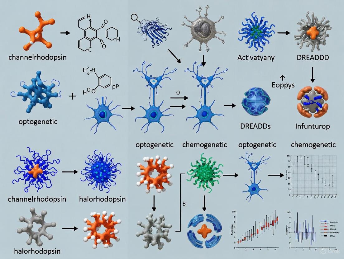
The foundation of optogenetics rests on the utilization of microbial rhodopsins—light-sensitive proteins that can be expressed in specific neuronal populations to render them responsive to light stimulation [1]. The most prominent tools in the optogenetics toolkit are channelrhodopsins (ChRs), which depolarize and excite neurons, and halorhodopsins (NpHR), which hyperpolarize and inhibit neuronal activity [5] [6]. This application note details the core principles, experimental protocols, and practical implementation of these key optogenetic tools within the framework of addiction circuit analysis.
Core Principles and Molecular Tools
Channelrhodopsins: Precision Neuronal Activation
Channelrhodopsin-2 (ChR2), a light-gated cation channel originally isolated from the green alga Chlamydomonas reinhardtii, serves as the primary tool for neuronal excitation in optogenetics [2] [4] [7]. Its molecular mechanism involves a covalently-bound retinal chromophore that undergoes photoisomerization upon illumination with blue light (approximately 470 nm), triggering a conformational change that opens the channel pore [6] [7]. This allows passive influx of cations (primarily Na⁺, with some Ca²⁺ and K⁺ permeability), leading to membrane depolarization and action potential generation with millisecond precision [4] [6].
The utility of ChR2 in addiction research has been demonstrated in foundational studies showing that phasic optogenetic stimulation of ventral tegmental area (VTA) dopamine neurons is sufficient to drive conditioned place preference and reinforcement behavior, mimicking key aspects of natural reward processing [3]. Continued protein engineering has yielded optimized ChR variants with improved properties for specific experimental needs, including accelerated kinetics (ChETA), red-shifted excitation spectra (C1V1, Chrimson), and step-function phenotypes that enable prolonged neuronal excitation following brief light pulses [4] [6].
Halorhodopsins and Archaerhodopsins: Targeted Neuronal Silencing
For neuronal inhibition, optogenetics employs two primary classes of microbial rhodopsins: light-driven ion pumps and light-gated ion channels. Halorhodopsin (NpHR), a yellow light-activated chloride pump from Natronomonas pharaonis, represents the first optogenetic tool used for neuronal silencing [5]. Upon illumination with yellow light (∼590 nm), NpHR actively pumps chloride ions into the cell, resulting in hyperpolarization and suppression of neuronal firing [5] [4].
Archaerhodopsin (Arch), a green light-activated outward proton pump from Halorubrum sodomense, provides an alternative silencing mechanism by extruding protons from the cytoplasm [5]. Archaerhodopsin-3 (ArchT), an improved variant, shows enhanced light sensitivity and membrane targeting, enabling more effective neural silencing at both cell bodies and axon terminals [5] [6].
Table 1: Key Optogenetic Actuators for Addiction Research
| Opsin | Type | Activation Wavelength | Ionic Mechanism | Effect on Neurons | Key Applications in Addiction Research |
|---|---|---|---|---|---|
| Channelrhodopsin-2 (ChR2) | Cation channel | ~470 nm blue light [4] [6] | Passive cation influx (Na⁺, Ca²⁺) [4] | Depolarization/Excitation [4] | Establishing causal links between specific neuronal activity and reward behavior [3] |
| ChETA | Engineered ChR2 variant | ~470 nm blue light [8] | Faster cation influx kinetics [8] | High-frequency neuronal excitation [8] | Investigating roles of specific firing patterns in addiction pathways |
| Halorhodopsin (NpHR/eNpHR) | Chloride pump | ~590 nm yellow light [5] [4] | Active Cl⁻ influx [5] | Hyperpolarization/Silencing [5] | Loss-of-function studies to determine necessity of specific circuits in drug-seeking [5] |
| Archaerhodopsin (Arch/ArchT) | Proton pump | ~560 nm green light [5] [4] | Active H⁺ extrusion [5] | Hyperpolarization/Silencing [5] | Inhibiting specific neural pathways during relapse behavior with high efficiency [5] |
| Jaws | Red-shifted halorhodopsin | Red light [8] | Active Cl⁻ influx [8] | Deep tissue silencing [8] | Targeting nuclei in deep brain structures involved in addiction |
Molecular and Circuit Mechanisms
The following diagram illustrates the fundamental mechanisms of these core optogenetic tools at the cellular level:
Figure 1: Molecular and circuit mechanisms of core optogenetic tools. Channelrhodopsins mediate neuronal activation via cation influx, while halorhodopsins and archaerhodopsins mediate silencing through chloride influx or proton extrusion, respectively, enabling bidirectional control of reward circuits.
Experimental Protocols for Addiction Circuit Analysis
Viral Vector Delivery and Cell-Type Specific Targeting
Protocol: Stereotaxic Delivery of Cre-Inducible Opsin Constructs
Objective: To achieve cell-type-specific opsin expression in defined neural circuits relevant to addiction, such as dopamine neurons in the Ventral Tegmental Area (VTA) projecting to the Nucleus Accumbens (NAc) [3] [4].
Materials:
- Cre-recombinase dependent adeno-associated virus (AAV) encoding opsin (e.g., AAV-EF1α-DIO-ChR2-EYFP or AAV-EF1α-DIO-eNpHR-EYFP) [4]
- Transgenic mouse or rat line expressing Cre recombinase under cell-type-specific promoter (e.g., TH-Cre for dopamine neurons, DAT-Cre for dopamine transporter-expressing neurons) [4]
- Stereotaxic apparatus
- Microsyringe pump and glass micropipettes or Hamilton syringe
- Anesthesia equipment (isoflurane vaporizer)
- Analgesics (meloxicam, bupivacaine)
Procedure:
- Anesthetize the animal using isoflurane (4% induction, 1.5-2% maintenance) and secure in stereotaxic apparatus with body temperature maintained at 37°C.
- Administer preoperative analgesics and prepare sterile surgical field.
- Calculate target coordinates for VTA (e.g., -3.3 mm AP, ±0.5 mm ML, -4.3 mm DV from bregma for mouse) using appropriate brain atlas.
- Slowly inject 300-500 nL of viral suspension (titer ≥ 10¹² viral genomes/mL) at a rate of 50 nL/min using a microsyringe pump.
- Allow 5-10 minutes for diffusion before slowly retracting the syringe.
- For projection-specific studies, inject retrograde tracer or use intersectional viral approaches to target specific pathways.
- Allow 3-6 weeks for sufficient opsin expression before commencing experiments [4].
In Vivo Optogenetic Stimulation During Behavioral Paradigms
Protocol: Real-Time Place Preference for Assessing Reward Valence
Objective: To determine whether activation or inhibition of a specific neural population is reinforcing or aversive, a key assay in addiction research [3] [4].
Materials:
- Laser system (473 nm blue laser for ChR2; 561 nm or 593 nm yellow laser for NpHR)
- Optical fiber (200 μm core diameter, 0.22 NA) with fiber-optic patch cord
- Rotary joint (for freely moving experiments)
- Behavioral tracking software with real-time triggering capability
- Two- or three-chamber place conditioning apparatus
Procedure:
- Implant optical fiber cannula above target brain region (e.g., VTA or NAc) during viral injection surgery or in a separate procedure.
- Habituate animals to patch cord connection for 2-3 days prior to testing.
- Conduct preconditioning trial: Place animal in apparatus with free access to all chambers for 15 minutes to assess baseline chamber preference.
- For conditioning trials: Over 2-3 days, confine animal to non-preferred chamber when laser stimulation is delivered (0.5-2 second pulses, 10-30 Hz, 5-15 mW at fiber tip for ChR2; continuous 5-15 mW for NpHR).
- On alternate days, confine animal to preferred chamber without laser stimulation.
- Conduct post-conditioning test: Place animal in apparatus with free access to all chambers without laser stimulation for 15 minutes.
- Measure time spent in each chamber; significant increase in time spent in stimulation-paired chamber indicates reinforcing effect, while decrease indicates aversive effect [3] [4].
Table 2: Standardized Optogenetic Stimulation Parameters for Behavioral Assays
| Behavioral Paradigm | Opsin Tool | Light Parameters | Stimulation Pattern | Typical Light Power at Fiber Tip | Key Measured Variables |
|---|---|---|---|---|---|
| Real-Time Place Preference | ChR2 [4] | 473 nm, 1-5 ms pulses [4] | 10-30 Hz [4] | 5-15 mW [4] | Time in paired chamber, movement velocity |
| Operant Self-Stimulation | ChR2 [4] | 473 nm, 0.5-1 s pulse duration | Continuous or 5-20 Hz | 5-15 mW | Active lever presses, reward learning curve |
| Cue-Induced Reinstatement | NpHR/Arch [5] | 561-593 nm, continuous | Continuous during cue presentation | 10-20 mW | Drug-seeking responses, latency to seek |
| In Vivo Electrophysiology | ChR2 [4] | 473 nm, 5 ms pulses | 1-50 Hz (varies by experiment) | 1-10 mW | Spike probability, latency, fidelity |
| Synaptic Circuit Mapping | ChR2 [3] | 473 nm, 1-5 ms pulses | 0.1-1 Hz (minimal stimulation) | 1-5 mW | EPSC amplitude, latency, failure rate |
Integrated Experimental Workflow
The following diagram outlines a comprehensive experimental workflow for implementing optogenetics in addiction circuit research:
Figure 2: Comprehensive experimental workflow for optogenetics in addiction research, spanning experimental design, surgical implementation, behavioral analysis, and circuit validation phases.
The Scientist's Toolkit: Essential Research Reagents
Table 3: Essential Research Reagents for Optogenetic Experiments
| Reagent Category | Specific Examples | Function & Application | Key Considerations |
|---|---|---|---|
| Viral Vectors | AAV1, AAV2, AAV5, AAV8, AAV9 [4] | Delivery of opsin genes to target cells; serotypes vary in tropism and spread | Titer (>10¹² vg/mL), promoter specificity (CaMKIIα for excitatory neurons, synapsin for pan-neuronal) |
| Opsin Constructs | ChR2(H134R), ChETA, C1V1, NpHR3.0, ArchT [4] [8] | Light-sensitive effectors for neuronal excitation or inhibition | Kinetics, light sensitivity, expression efficiency, spectral properties |
| Cell-Type Specific Drivers | Cre-recombinase lines (TH-Cre, DAT-Cre, D1-Cre, D2-Cre) [4] | Genetic targeting of specific neuronal populations | Specificity, developmental expression patterns, leakiness |
| Retinal Cofactor | All-trans-retinal (ATR) [9] | Essential chromophore for microbial opsins | Solubility (ethanol stock), concentration (1-10 mM in food), light sensitivity |
| Light Delivery | Solid-state lasers (473 nm, 561 nm), LEDs, optical fibers [4] | Precise light delivery to target brain regions | Power output, stability, thermal management, fiber numerical aperture |
| Control Constructs | GFP, YFP, mCherry (fluorescent reporters) [4] | Expression verification, control for viral injection and surgical procedures | Matching promoter and viral serotype to experimental conditions |
Critical Considerations and Technical Challenges
Experimental Design and Controls
Proper experimental design requires careful consideration of multiple control conditions to ensure specific interpretation of results. Critical controls include: (1) sham stimulation in opsin-expressing animals, (2) light stimulation in non-opsin-expressing animals (e.g., expressing fluorescent protein only), and (3) verification that observed effects are specific to the targeted cell population or pathway [4]. For addiction studies specifically, it is essential to demonstrate that optogenetic manipulations produce changes in addiction-relevant behaviors but not general locomotor or sensory function, unless those are the variables of interest.
Limitations and Potential Artifacts
While powerful, optogenetic approaches have important limitations. Halorhodopsin activation can alter intracellular chloride concentrations, potentially affecting GABAergic signaling and causing rebound excitation after light offset [5] [9]. Archaerhodopsin activation modifies proton gradients, potentially affecting pH-sensitive processes [5]. Both inhibitory tools require high light power for effective silencing, which can generate significant heat in brain tissue that may itself alter neuronal function [5]. Additionally, ectopic expression of microbial opsins may potentially interfere with normal cellular function, though current evidence suggests minimal disruption at moderate expression levels [1] [4].
The integration of channelrhodopsin and halorhodopsin technologies has fundamentally transformed addiction research by enabling precise causal interrogation of specific neural circuits with millisecond temporal precision. These tools have helped identify the roles of specific neuronal populations in the VTA, NAc, PFC, and other nodes of the reward circuit in drug seeking, relapse, and addiction-related plasticity [3] [4]. Current developments in red-shifted opsins, bidirectional control systems, and integration with in vivo imaging techniques promise to further enhance our ability to dissect the complex circuit mechanisms underlying addiction [8]. As these tools continue to evolve, they will undoubtedly yield deeper insights into addiction pathophysiology and potentially identify novel therapeutic targets for this devastating disorder.
Designer Receptors Exclusively Activated by Designer Drugs (DREADDs) represent a powerful chemogenetic approach for the precise and reversible modulation of cellular signaling in defined cell populations [10]. This technology has revolutionized neuroscience research, particularly in the dissection of neural circuits underlying complex behaviors such as addiction. DREADDs are engineered GPCRs that are unresponsive to their native endogenous ligands but can be selectively activated by biologically inert designer compounds [11]. Unlike optogenetics, which provides millisecond temporal precision but requires invasive light delivery, DREADDs offer temporal flexibility through systemic drug administration, making them particularly suitable for studying behavioral processes that develop over time, such as drug-seeking and relapse [10] [2].
The foundational development of DREADD technology involved introducing point mutations (Y3.33C and A5.46G) into muscarinic acetylcholine receptors, rendering them insensitive to acetylcholine while creating specificity for synthetic ligands like clozapine-N-oxide (CNO) [11]. This elegant solution allows researchers to manipulate specific neural pathways without interfering with endogenous cholinergic signaling, providing a robust tool for circuit mapping and behavioral analysis in intact organisms.
DREADD Systems and Their Mechanisms
The DREADD family consists of several receptor variants that couple to different intracellular signaling pathways, enabling either activation or inhibition of targeted neurons [10]. The table below summarizes the primary DREADD receptors and their characteristics:
Table 1: Key DREADD Receptors and Their Signaling Properties
| DREADD Type | G-protein Coupling | Primary Signaling Effect | Result on Neuronal Activity | Common Applications |
|---|---|---|---|---|
| hM3Dq | Gq | Activates phospholipase C, increases intracellular Ca²⁺ | Neuronal excitation [11] | Behavioral activation, circuit stimulation |
| hM4Di | Gi/o | Inhibits adenylate cyclase, reduces cAMP | Neuronal inhibition [10] | Suppression of specific behaviors, pathway silencing |
| rM3Ds/hM3Ds | Gs | Activates adenylate cyclase, increases cAMP | Context-dependent excitation [11] | Selective modulation of cAMP-sensitive neurons |
| KORD | κ-opioid | Activates GIRK channels | Neuronal inhibition [12] | Bidirectional control when combined with other DREADDs |
The cellular signaling pathways activated by these different DREADD classes are fundamental to their experimental utility. The following diagram illustrates the primary intracellular mechanisms:
DREADD Applications in Addiction Circuit Analysis
DREADD technology has provided transformative insights into the neural circuitry underlying addiction behaviors by enabling cell-type specific and circuit-specific manipulations that were previously unattainable [10]. In addiction research, DREADDs have been successfully applied to investigate three fundamental processes: psychomotor sensitization, drug self-administration, and relapse behavior [10].
Psychomotor Sensitization
Psychomotor sensitization refers to the progressive enhancement of locomotor responses to repeated, intermittent drug exposure and is considered a behavioral correlate of the incentive-motivational aspects of addiction [10]. Using Gi/o-coupled DREADDs (hM4Di) to inhibit specific neuronal populations, researchers have demonstrated that the expression of psychomotor sensitization involves adaptations in the ventral tegmental area (VTA) and its projection targets. These studies reveal that circuit-specific inhibition can reverse or prevent sensitization, highlighting the potential for targeted interventions.
Drug Self-Administration
DREADDs have been particularly valuable in elucidating the neural mechanisms underlying drug-taking behaviors and motivation. Interestingly, studies have shown that Gi/o signaling in indirect pathway medium spiny neurons (MSNs) in the nucleus accumbens core has no effect on responding for sucrose under a progressive ratio schedule, suggesting a complex, behavior-specific organization of reward circuits [10]. This specificity underscores the advantage of DREADDs in dissecting functionally distinct elements within anatomically overlapping pathways.
Relapse Modeling
A recurring challenge in treating addiction is the high propensity for relapse, which can be modeled in animals using extinction training or withdrawal paired with reinstatement tests [10]. DREADD studies have identified distinct functional contributions of subregions of the ventral pallidum (VP) in cue- and drug prime-induced reinstatement. Increasing Gi/o signaling in the rostral VP attenuates cue-induced reinstatement, while inhibition of the caudal VP reduces cocaine prime-induced reinstatement [10]. These findings demonstrate how DREADDs can reveal functional heterogeneity within brain regions previously considered unitary in their contribution to addiction behaviors.
Table 2: Key DREADD Findings in Addiction Research
| Addiction Phase | Brain Circuit | DREADD Intervention | Behavioral Effect | Research Significance |
|---|---|---|---|---|
| Psychomotor Sensitization | VTA and projections | hM4Di inhibition | Reduced sensitization | Identified critical nodes for behavioral plasticity |
| Drug Self-Administration | Nucleus Accumbens (indirect pathway MSNs) | hM4Di inhibition | No effect on sucrose reinforcement | Revealed dissociation between natural and drug reward processing |
| Relapse (cue-induced) | Rostral Ventral Pallidum | hM4Di inhibition | Attenuated reinstatement | Demonstrated functional sub-specialization within brain regions |
| Relapse (drug prime-induced) | Caudal Ventral Pallidum | hM4Di inhibition | Reduced reinstatement | Identified novel targets for preventing relapse |
| Compulsive Drug-Seeking | Prefrontal-Accumbens Pathway | hM3Dq activation | Enhanced compulsive seeking | Mapped circuits underlying loss of behavioral control |
Experimental Protocol: DREADD-Based Circuit Manipulation in Addiction Models
The following protocol details a representative approach for using DREADDs to investigate circuit-specific contributions to addiction-related behaviors, specifically focusing on the manipulation of hippocampal-amygdala circuits in fear memory segregation [12].
Experimental Workflow
The complete experimental workflow for a DREADD-based circuit manipulation study involves multiple stages from viral vector preparation to behavioral analysis, as visualized below:
Step-by-Step Methodology
Viral Vector Preparation
- AAV1-hSyn-Cre (2×10¹³ GC/mL): An anterograde transsynaptic vector that expresses Cre recombinase under the human synapsin promoter, enabling labeling of postsynaptic neurons [12].
- AAV1-hDlx-DIO-KORD-mCyRFP (4×10¹² GC/mL): A Cre-dependent inhibitory DREADD (κ-opioid receptor-based DREADD) specifically designed for expression in GABAergic neurons using the Dlx promoter [12].
- Aliquot viral preparations in sterile Eppendorf tubes and store at -80°C until the day of injection.
Stereotaxic Surgery for Viral Delivery
- Anesthesia and Positioning: Place the animal in an induction chamber with isoflurane (4%) and oxygen (2 L/min). After loss of consciousness, transfer to a stereotaxic frame and maintain anesthesia at 2% isoflurane [12].
- Skull Exposure and Calibration: Make a rostro-caudal incision along the scalp midline, expose the skull, and clean the bone. Calibrate the stereotaxic coordinate system by identifying bregma and lambda points [12].
- Viral Injections:
- Ventral Hippocampus Injection: Using stereotaxic coordinates (AP: -5.0 mm, ML: ±5.0 mm, DV: -5.0 mm from bregma), inject AAV1-hSyn-Cre (0.3-0.5 μL) at an infusion rate of 0.1-0.3 μL/min [12].
- Basolateral Amygdala Injection: Using appropriate coordinates for the target species, inject AAV1-hDlx-DIO-KORD-mCyRFP (0.3-0.5 μL) at the same infusion rate [12].
- Post-operative Care: Allow 3-4 weeks for adequate viral expression and receptor trafficking before initiating behavioral experiments.
Behavioral Assessment and Chemogenetic Manipulation
- Fear Conditioning: Train animals in a standard fear conditioning paradigm to establish contextual fear memories.
- DREADD Activation: Administer the KORD ligand Salvinorin B (3.0 mg/kg, i.p.) 30 minutes before behavioral testing to inhibit the targeted GABAergic neurons in the BLA that receive input from the ventral hippocampus [12].
- Memory Testing: Assess fear memory expression in the conditioned context and compare to appropriate control groups.
- Quantitative Analysis: Measure freezing behavior as an index of fear memory expression and perform statistical comparisons between experimental conditions.
Research Reagent Solutions
Table 3: Essential Reagents for DREADD Experiments
| Reagent / Tool | Function / Purpose | Example Specifications | Considerations |
|---|---|---|---|
| DREADD Viral Vectors | Delivery of engineered receptors to target cells | AAV serotypes (AAV1, AAV5, AAV8), Cell-type specific promoters (hSyn, CaMKIIa, Dlx) | Optimize titer, serotype, and promoter for target cells |
| Cre-dependent DREADDs | Enables cell-type specific expression in Cre-driver lines | AAV-DIO-hM3Dq, AAV-DIO-hM4Di, AAV-DIO-KORD | Requires appropriate Cre-recombinase expression |
| DREADD Agonists | Activation of DREADD receptors | CNO (0.1-10 mg/kg), DCZ (0.001-0.1 mg/kg), Salvinorin B (for KORD) | Dose-dependent effects; consider pharmacokinetics |
| Control Viral Vectors | Control for viral injection and expression | AAV expressing fluorescent proteins only (e.g., GFP, mCherry) | Critical for controlling for non-specific viral effects |
| Stereotaxic Apparatus | Precise targeting of brain regions | Digital stereotaxic instrument with micromanipulator | Surgical precision essential for circuit-specific targeting |
Recent Advances and Future Directions
Recent developments in DREADD technology have focused on improving its specificity, safety profile, and translational potential. A significant advancement is the creation of a fully sequence-humanized Gs-coupled DREADD (hM3Ds), designed to reduce potential immunogenicity concerns for future clinical applications [11]. This humanized DREADD maintains comparable ligand response profiles to its non-humanized counterpart while potentially offering improved biocompatibility.
The DREADD toolbox continues to expand with the development of novel receptor-effector couplings and ligand-gated systems. The KORD (κ-opioid receptor-based DREADD) system represents one such innovation, enabling orthogonal chemogenetic control when combined with other DREADDs [12]. This allows researchers to manipulate multiple neural circuits independently within the same animal, providing unprecedented analytical power for deciphering complex circuit interactions.
Future directions include the refinement of pathway-specific DREADD expression using retrograde and anterograde tracers, the development of β-arrestin-biased DREADDs for selective signaling pathway engagement, and the creation of clinically translatable DREADD systems for potential therapeutic applications in addiction and other neurological disorders [11]. As these tools evolve, they will undoubtedly continue to illuminate the complex neural circuitry underlying addiction and enable more precise interventions for this devastating disorder.
Optogenetics and chemogenetics have revolutionized the analysis of neural circuits underlying addiction by enabling precise, cell-type-specific manipulation of neuronal activity [2] [13]. These approaches rely on key molecular components—opsins, synthetic ligands, and viral delivery systems—to probe the complex neural networks mediating reward, reinforcement, and craving behaviors [2]. This Application Note provides a structured overview of these core components, summarizes quantitative data in comparative tables, and details standardized protocols for implementing these technologies in addiction circuit research. The integration of these tools offers unprecedented spatial and temporal control for dissecting the neural mechanisms of addiction, facilitating the identification of novel therapeutic targets [8].
Key Molecular Components
Opsins: Nature's Photoreceptors
Opsins are universal photoreceptive proteins in animals that function as G-protein-coupled receptors (GPCRs) [14] [15]. These molecules consist of a protein moiety with seven transmembrane domains and a non-protein chromophore, typically retinal, which covalently binds to a conserved lysine residue in the seventh helix [14] [15]. Upon light absorption, the retinal isomerizes, triggering conformational changes in the opsin that activate intracellular signaling cascades [15]. The opsin family is phylogenetically diverse, with seven major subfamilies identified in animals, including vertebrate visual and non-visual opsins, encephalopsin/tmt-opsin, Gq-coupled opsin/melanopsin, Go-coupled opsin, neuropsin, peropsin, and retinal photoisomerase subfamilies [14].
Table 1: Classification and Properties of Major Opsin Families
| Opsin Subfamily | G-Protein Coupling | Chromophore State | Primary Functions | Representative Members |
|---|---|---|---|---|
| Vertebrate visual & non-visual | Transducin (Gt) | 11-cis-retinal | Vision, non-visual photoreception | Rhodopsin, cone opsins, pinopsin, VA-opsin |
| Encephalopsin/Tmt-opsin | Unknown | Unknown | Non-visual photoreception | Encephalopsin, tmt-opsin |
| Gq-coupled opsin/Melanopsin | Gq | 11-cis-retinal | Circadian rhythm, pupillary reflex | Melanopsin, invertebrate visual opsins |
| Go-coupled opsin | Go | 11-cis-retinal | Photoreception in invertebrates | Mollusk opsin |
| Neuropsin | Unknown | Unknown | Non-visual photoreception | Neuropsin |
| Peropsin | Unknown | all-trans-retinal | Retinal pigment epithelium function | Peropsin |
| Retinal photoisomerase | Unknown | all-trans-retinal | Chromophore regeneration | RGR-opsin, retinochrome |
Molecular properties of opsins vary significantly between types. Vertebrate rhodopsins are "mono-stable," forming a metastable active state upon photoreception that cannot revert to the dark state without chromophore replacement via the visual cycle [16]. In contrast, many non-visual opsins and invertebrate rhodopsins are "bistable," maintaining a stable active state that can photorevert to the dark state, enabling them to function without external chromophore regeneration systems [16]. A newly identified opsin, Opn5L1, exhibits "photocyclic" properties similar to microbial channelrhodopsins, where illumination drives a cyclic reaction that controls its activity [16].
Table 2: Molecular Properties of Opsin Types
| Property | Vertebrate Rhodopsin (Mono-stable) | Invertebrate Rhodopsin (Bistable) | Opn5L1 (Photocyclic) |
|---|---|---|---|
| Active State Stability | Metastable | Thermally stable | Dark-active state spontaneously regenerates |
| Chromophore Regeneration | Requires retinal pigment epithelium | Photoreversion | Intrinsic photocycle |
| Retinal Isomer in Dark | 11-cis-retinal | 11-cis-retinal | all-trans-retinal (active state) |
| Photoreversibility | No | Yes | Yes |
| Primary Signaling Cascade | Gi/transducin → PDE → cGMP reduction | Gq → PLCβ → IP3/DAG | Unknown |
Optogenetic Tools Derived from Opsins
Optogenetics utilizes light-sensitive proteins, primarily microbial opsins (Type I rhodopsins), to control neuronal activity with high temporal precision [13]. These tools are categorized into excitatory and inhibitory opsins, with Channelrhodopsin-2 (ChR2) and halorhodopsin (NpHR) serving as foundational prototypes [8].
Table 3: Engineered Optogenetic Tools for Neural Control
| Opsin Tool | Type | Activation Wavelength | Ionic Mechanism | Physiological Effect | Key Features |
|---|---|---|---|---|---|
| Channelrhodopsin-2 (ChR2) | Cation channel | ~470 nm (Blue) | Na+ influx | Depolarization | Fast activation, millisecond precision |
| Halorhodopsin (NpHR) | Chloride pump | ~590 nm (Yellow) | Cl- influx | Hyperpolarization | Neural inhibition, light-driven pump |
| ChETA | Engineered channel | ~470 nm (Blue) | Na+ influx | Depolarization | Faster kinetics, improved spike fidelity |
| Jaws | Halorhodopsin | Red-shifted | Cl- influx | Hyperpolarization | Enhanced tissue penetration |
| GtACR | Anion channel | ~470 nm (Blue) | Cl- influx | Hyperpolarization | Potent inhibition, channel mechanism |
Advanced optogenetic tools continue to emerge through protein engineering approaches. "Red-shifted" opsins activated by longer wavelengths enable deeper tissue penetration, while dual-color opsins allow bidirectional control of the same neurons with different light wavelengths [8]. Luminopsins (LMOs) represent innovative fusion proteins combining light-emitting luciferase with light-sensing opsins, enabling both optogenetic and chemogenetic control through the same molecule [13].
Synthetic Ligands for Chemogenetics
Chemogenetics employs engineered receptors that respond exclusively to synthetic ligands, with Designer Receptors Exclusively Activated by Designer Drugs (DREADDs) being the most prominent platform [13]. These modified G-protein-coupled receptors (GPCRs) are unresponsive to endogenous ligands but can be activated by inert synthetic compounds like clozapine-N-oxide (CNO) to modulate neuronal activity through G-protein signaling pathways [13]. Unlike optogenetics, chemogenetics offers less temporal precision but enables less invasive manipulation of neural activity without implanted hardware, making it suitable for chronic studies across distributed neural circuits [13].
Viral Delivery Systems for Gene Therapy in Neuroscience
Effective delivery of optogenetic and chemogenetic components to specific neural populations relies primarily on viral vector systems. The choice of vector depends on multiple factors including payload capacity, tropism, expression kinetics, and immunogenicity.
Table 4: Comparison of Viral Delivery Systems for Neural Circuit Research
| Vector Type | Payload Capacity | Integration | Expression Onset | Expression Duration | Primary Applications | Advantages | Limitations |
|---|---|---|---|---|---|---|---|
| Adeno-Associated Virus (AAV) | ~4.7 kb | Non-integrating (episomal) | 2-3 weeks | Long-term (months to years) | Optogenetics, chemogenetics, gene expression | Low immunogenicity, high safety profile | Limited packaging capacity |
| Lentivirus (LV) | ~8 kb | Integrating (random) | 3-7 days | Long-term (stable integration) | Stable gene expression, RNA interference | Large payload, infects non-dividing cells | Potential insertional mutagenesis |
| Adenovirus (Ad) | ~8-36 kb | Non-integrating (episomal) | 1-2 days | Transient (weeks) | High-level transient expression, vaccine development | High transduction efficiency, very large capacity | Significant immune response |
Viral vectors are typically engineered with cell-type-specific promoters (e.g., CaMKIIα for excitatory neurons, TH for dopaminergic neurons) or using Cre-lox systems for precise targeting of defined neuronal populations [13]. For example, in addiction research focusing on dopaminergic circuits, AAV vectors carrying DREADDs or opsins under a Cre-dependent promoter can be injected into the ventral tegmental area (VTA) of tyrosine hydroxylase (TH)-Cre transgenic mice, restricting transgene expression to dopamine-producing neurons [13].
Experimental Protocols
Protocol: Stereotaxic Viral Delivery for Optogenetics in Rodent Models
Purpose: To express light-sensitive opsins in specific neural populations for circuit manipulation in addiction studies.
Materials:
- AAV vectors encoding opsins (e.g., AAV5-CaMKIIα-hChR2(H134R)-eYFP)
- Anesthetic (e.g., ketamine/xylazine or isoflurane)
- Stereotaxic apparatus
- Microinjection pump and glass micropipettes
- Fiber optic cannulas and dental cement
- Analgesics (e.g., meloxicam)
- Standard surgical instruments
Procedure:
- Surgical Preparation: Anesthetize the rodent and secure its head in the stereotaxic apparatus. Maintain body temperature throughout the procedure.
- Craniotomy: Shave the scalp, make a midline incision, and clean the skull. Identify bregma and lambda landmarks. Perform a small craniotomy at the target coordinates.
- Viral Injection: Load the AAV vector into a glass micropipette connected to a microinjection pump. Lower the pipette to the target coordinates (e.g., VTA: AP -3.3 mm, ML ±0.5 mm, DV -4.3 mm from bregma). Infuse the virus (e.g., 500 nL at 100 nL/min). Wait 10 minutes before slowly retracting the pipette.
- Optic Cannula Implantation: Implant an optic fiber cannula above the injection site and secure it with dental cement.
- Post-operative Care: Administer analgesics and monitor the animal until recovery. Allow 3-4 weeks for opsin expression before conducting experiments.
Validation:
- Confirm opsin expression histologically using fluorescent protein tags.
- Verify functional expression through electrophysiological responses to light stimulation.
Protocol: Neural Circuit Mapping with Optogenetics
Purpose: To characterize functional connectivity between opsin-expressing neurons and their projection targets.
Materials:
- Rodents expressing ChR2 in specific neuronal populations
- Light source (laser or LED) with precise temporal control
- Electrophysiology setup for patch-clamp or extracellular recording
- Artificial cerebrospinal fluid (aCSF)
Procedure:
- Preparation: Prepare acute brain slices containing both the opsin-expressing region and the projection target.
- Recording: Patch-clamp neurons in the projection target while delivering light pulses (1-5 ms, 470 nm) to ChR2-expressing axon terminals.
- Analysis: Measure postsynaptic currents to identify functional connections. Use receptor antagonists to characterize synaptic properties (AMPA vs. NMDA, GABAergic).
Applications in Addiction Research: This protocol can map connectivity between VTA dopamine neurons and nucleus accumbens (NAc) projections, revealing circuit adaptations following drug exposure.
The Scientist's Toolkit: Essential Research Reagents
Table 5: Key Research Reagent Solutions for Optogenetic-Chemogenetic Experiments
| Reagent Category | Specific Examples | Function | Key Considerations |
|---|---|---|---|
| Viral Vectors | AAV1, AAV5, AAV9 serotypes; LV-CaMKIIα, LV-hSyn promoters | Delivery of genetic constructs to target cells | Serotype affects tropism; promoter determines cell specificity |
| Opsins | ChR2(H134R), eNpHR3.0, Chronos, GtACR1 | Precise neuronal excitation or inhibition | Action spectrum, kinetics, conductance properties |
| Chemogenetic Receptors | hM3Dq, hM4Di DREADDs | Chemically control neuronal activity via Gq or Gi signaling | Ligand pharmacokinetics, receptor density |
| Synthetic Ligands | Clozapine-N-oxide (CNO), Deschloroclozapine (DCZ) | Activate chemogenetic receptors | Metabolic conversion, blood-brain barrier penetration |
| Cre-Driver Lines | TH-IRES-Cre, CaMKIIα-Cre, DAT-Cre mice | Cell-type-specific targeting | Specificity and efficiency of Cre expression |
| Light Delivery | Optic fibers, LEDs, lasers | Activate optogenetic tools | Light power, wavelength, temporal pattern control |
Signaling Pathways and Experimental Workflows
Optogenetic Signaling Pathway
Experimental Workflow for Addiction Circuit Analysis
The integration of opsins, synthetic ligands, and viral delivery systems provides a powerful toolkit for dissecting the neural circuits underlying addiction. Optogenetics offers millisecond-precision control of specific neuronal populations, while chemogenetics enables less invasive manipulation of distributed circuits. Viral vectors, particularly AAVs, facilitate targeted delivery of these tools to defined cell types. By implementing the standardized protocols and utilizing the key reagents outlined in this Application Note, researchers can systematically investigate addiction-related circuits, from molecular mechanisms to behavioral outcomes, accelerating the development of novel therapeutic strategies for addiction disorders.
Systems neuroscience has undergone a paradigm shift, moving from studying brain function through gross lesions to achieving unprecedented precision in manipulating specific neural circuits. Early techniques involving brain lesions or electrical stimulation, while foundational, were limited by their irreversibility, invasiveness, and lack of specificity, as they often affected multiple cell types and circuits within a targeted region [17]. The advent of genetic tools has overcome these limitations, enabling researchers to test how specific neural circuits mediate brain function and behavior with a precision that mirrors the molecular approaches of cell biology [17]. This evolution has been particularly transformative for addiction research, where understanding the precise neural circuits governing reward, reinforcement, and drug-seeking behaviors is paramount.
Foundational Strategies for Circuit Manipulation
Two overarching strategies have been developed to achieve circuit-specific manipulation, each with distinct advantages and applications in addiction research.
Genetic Identity Approach
This method leverages the unique genetic identity of a neuron—such as cell-type-specific transcription factors, neurotransmitter systems, or calcium-binding proteins—to drive the expression of molecules that can manipulate its function [17]. For example, promoters for vesicular glutamate transporters (e.g., Vglut2) can target glutamatergic neurons, while promoters for vesicular GABA transporter can target GABAergic neurons [17]. In the context of addiction, this allows researchers to selectively target distinct neuronal populations within key reward areas, such as dopamine neurons in the Ventral Tegmental Area (VTA) or medium spiny neurons in the Nucleus Accumbens (NAc).
Spatial Control (Connectivity-Based) Approach
This strategy utilizes the anatomical connectivity of a circuit for specificity. It typically involves introducing one genetic element at the origin of a circuit (e.g., the VTA) and another at its termination point (e.g., the NAc). Only neurons that are co-infected with both viruses—and are thus part of the specific VTA-NAc circuit—express the functional transgene that allows for circuit manipulation [17]. This approach is powerful for dissecting the role of specific projections in addictive behaviors.
Core Toolkits for Neural Circuit Manipulation
The implementation of the above strategies relies on a sophisticated toolkit of genetically encoded actuators.
Optogenetics
Optogenetics involves the expression of light-sensitive ion channels, known as opsins, in specific neuronal populations. These opsins allow for millisecond-scale control of neuronal activity in response to light of specific wavelengths [8].
- Principle: Viral vectors deliver genes encoding for opsins under cell-specific promoters. Once expressed, illumination with light depolarizes (activates) or hyperpolarizes (silences) the target neurons [8].
- Key Opsins:
- Channelrhodopsin-2 (ChR2): A light-activated cation channel. Blue light (~460 nm) causes cation influx, leading to neuronal activation [8].
- Halorhodopsin (NpHR): A light-activated chloride pump. Yellow light (~580 nm) causes chloride influx, leading to neuronal inhibition [8].
- Engineered Variants: Continued development has produced opsins with improved properties, such as ChETA (faster kinetics), red-shifted opsins (better tissue penetration for deep brain structures), and Jaws (red-light inhibited) [8].
Chemogenetics
Chemogenetics uses engineered receptors that are activated by biologically inert synthetic ligands, allowing for remote, non-invasive control of neuronal activity over longer timescales (hours).
- Principle: The most common chemogenetic approach involves Designer Receptors Exclusively Activated by Designer Drugs (DREADDs). These are modified G-protein-coupled receptors that, upon binding an inert ligand like Clozapine-N-oxide (CNO), activate specific intracellular signaling pathways to either excite or inhibit neurons [17] [8].
- Key Advance - Synthetic Physiology: A recent groundbreaking development is the creation of drug-gated ion channels. For example, researchers have engineered a cocaine-activated ion channel ("coca-5HT3") by mutating the ligand-binding domain of a chimeric α7 nicotinic receptor. When expressed in the lateral habenula, this channel activates in the presence of cocaine, blunting cocaine-induced dopamine release and suppressing drug-seeking behavior in rats without affecting natural reward motivation [18]. This represents a "closed-loop" intervention that is directly yoked to drug exposure dynamics.
The following table summarizes the quantitative properties of key optogenetic and chemogenetic actuators.
Table 1: Key Actuators for Circuit Manipulation in Addiction Research
| Tool | Type | Activating Trigger | Key Properties | Typical Use in Addiction Circuits |
|---|---|---|---|---|
| Channelrhodopsin-2 (ChR2) | Opsin (Excitatory) | Blue Light (~460 nm) | Cation channel; fast kinetics (ms timescale) | Evoking burst firing in VTA dopamine neurons to probe reward signaling |
| Halorhodopsin (NpHR) | Opsin (Inhibitory) | Yellow Light (~580 nm) | Chloride pump; silences neurons | Inhibiting projections from prefrontal cortex to NAc to study impulse control |
| Jaws | Opsin (Inhibitory) | Red Light (~630 nm) | Chloride pump; enhanced tissue penetration | Inhibiting neurons in deep brain structures like the lateral habenula |
| DREADDs (hM3Dq, hM4Di) | Chemogenetic (GPCR) | CNO (or similar) | Gq or Gi signaling; timescale of hours | Long-term modulation of specific circuit activity during withdrawal or relapse tests |
| Coca-5HT3 | Chemogenetic (Ion Channel) | Cocaine (EC~50~ = 1.5 µM) | Cocaine-gated cation channel; closed-loop | Synthetic physiology to directly counteract cocaine's effects in real-time [18] |
Detailed Experimental Protocol: Chemogenetic Suppression of Cocaine Seeking
This protocol details the methodology based on the recent synthetic physiology approach to blunt cocaine-seeking behavior [18].
Research Reagent Solutions
Table 2: Essential Materials and Reagents
| Item | Function | Example/Details |
|---|---|---|
| Coca-5HT3 AAV Plasmid | Gene delivery vector for cocaine-gated channel | pCAG::coca-5HT3-IRES-GFP for neuronal expression and visualization [18] |
| AAV Vector (e.g., AAV9) | Viral packaging for in vivo transduction | Serotype chosen for high neuronal tropism and efficacy |
| Stereotaxic Instrument | Precise intracranial virus injection | Target coordinates for lateral habenula (or other region of interest) |
| Cocaine HCl | Pharmacological agent for self-administration and channel activation | For intravenous self-administration paradigms; purity >98% |
| Food Pellets | Control for natural reward | To test specificity of the intervention for drug vs. natural reward |
| In Vivo Electrophysiology Setup | Measurement of neuronal activity | To confirm cocaine-induced activation of transfected neurons |
| Microdialysis/HPLC | Measurement of dopamine levels | To quantify cocaine-induced dopamine transients in the NAc |
Step-by-Step Methodology
Virus Preparation and Stereotaxic Injection:
- Package the coca-5HT3 construct into an adeno-associated virus (AAV) vector under a pan-neuronal promoter (e.g., CAG) or a cell-type-specific promoter.
- Anesthetize adult rats and secure them in a stereotaxic frame.
- Using aseptic technique, perform a craniotomy and bilaterally inject ~1 µL of the high-titer AAV suspension into the lateral habenula (coordinates from rat brain atlas). Control animals receive a control virus (e.g., encoding GFP only).
- Allow 3-4 weeks for robust viral expression.
Validation of Channel Function (Ex Vivo):
- Prepare acute brain slices containing the lateral habenula from transfected animals.
- Perform whole-cell patch-clamp recordings on GFP-positive neurons.
- Bath apply cocaine (1-10 µM) while in current-clamp mode to confirm dose-dependent depolarization and increased firing rate, validating functional coca-5HT3 expression.
Cocaine Self-Administration Training:
- Implant rats with intravenous catheters.
- Train rats in operant chambers to self-administer cocaine (e.g., 0.5 mg/kg/infusion) on a fixed-ratio schedule, where a nose-poke results in an infusion paired with a cue light. Conduct daily sessions until stable responding is achieved.
Testing the Intervention:
- After stable self-administration is established, test the effects of coca-5HT3 expression on drug-seeking.
- Compare the number of cocaine infusions earned and active nose-pokes between coca-5HT3 and control groups.
- Critical Specificity Control: Conduct sessions where a natural reward (e.g., food pellets) is available under a similar schedule to ensure that reduced cocaine seeking is not due to a general motor impairment or loss of motivation.
Neurochemical Verification:
- In a separate cohort, use in vivo microdialysis in the NAc to measure extracellular dopamine levels.
- Collect samples before and during a cocaine self-administration session. The coca-5HT3 group should show a blunted rise in dopamine compared to controls, directly demonstrating the circuit-level mechanism [18].
Schematic Workflows
The following diagrams illustrate the core logical and experimental relationships in circuit manipulation.
Diagram 1: Circuit Manipulation Experimental Framework
Diagram 2: Synthetic Physiology for Cocaine Addiction
Mapping the Addicted Brain: Circuit-Specific Applications in Preclinical Models
Substance use disorders are characterized by maladaptive learning and memory processes, where drug-related stimuli gain the capacity to elicit intense craving and relapse. The transition from voluntary drug use to compulsive seeking reflects a complex learning process mediated by specific neural circuits. The mesocorticolimbic pathway, particularly the dopamine projections from the ventral tegmental area (VTA) to the nucleus accumbens (NAc) and prefrontal cortex (PFC), forms the central hub of the brain's reward system and is profoundly altered by drugs of abuse [19] [3]. Advanced neuromodulation techniques, particularly optogenetics and chemogenetics, now enable researchers to move beyond correlation to establish causality between specific circuit nodes and addiction-related behaviors with unprecedented spatial and temporal precision [8] [3].
Optogenetics combines genetic and optical methods to control defined events within specific cells of living tissue. It utilizes microbial opsin genes, such as channelrhodopsin-2 (ChR2) for neuronal excitation and halorhodopsin (NpHR) for inhibition, which are delivered to target cells via viral vectors [8]. When illuminated with specific wavelengths of light, these proteins allow precise temporal control of neuronal activity. Chemogenetics, particularly Designer Receptors Exclusively Activated by Designer Drugs (DREADDs), offers an alternative approach using engineered G-protein-coupled receptors that are activated by inert compounds like clozapine-N-oxide (CNO) [19] [20]. While offering lower temporal resolution than optogenetics, chemogenetics provides longer-lasting modulation and does not require implanted optical hardware. Together, these techniques have revolutionized our ability to dissect the neural circuitry underlying addiction by enabling cell-type and projection-specific manipulation of discrete pathways.
Key Neural Nodes and Circuitry in Addiction
The neural circuitry of addiction extends beyond the classic mesolimbic dopamine system to include cortical, thalamic, and other limbic structures that form interconnected networks mediating reward, motivation, and executive control. The VTA serves as a critical origin point, containing not only dopamine neurons but also GABAergic, glutamatergic, and co-releasing populations that project to diverse targets including the NAc, PFC, amygdala, and lateral habenula [3]. These projections regulate reinforcement learning, aversion, and motivated behavior through complex microcircuitry.
The nucleus accumbens acts as a central integration hub, receiving inputs from the VTA, PFC, basolateral amygdala (BLA), hippocampus, and thalamus. Optogenetic circuit mapping has revealed remarkable complexity in the NAc, challenging earlier simplistic models of direct and indirect pathways [3]. For instance, contrary to dorsal striatal organization, approximately half of ventral pallidum neurons receive synaptic input from D1-receptor expressing medium spiny neurons (MSNs), and VP neurons projecting to the thalamus receive input from both D1- and D2-MSNs [3].
Cortical influences on addiction circuitry are mediated through projections from the prelimbic (PL) and infralimbic (IL) divisions of the prefrontal cortex to the NAc, VTA, and paraventricular thalamus (PVT). Recent research has identified the PL→PVT pathway as a key regulator of heroin seeking, with abstinence from heroin self-administration inducing strengthening of these synapses that can be normalized through optogenetic long-term depression [20]. Similarly, inputs from the BLA to the NAc undergo specific synaptic adaptations following cocaine exposure, including the generation and subsequent unsilencing of NMDA receptor-containing synapses [3].
Table 1: Key Neural Nodes in Addiction Circuitry
| Brain Region | Primary Function in Addiction | Key Projections | Cell Types |
|---|---|---|---|
| Ventral Tegmental Area (VTA) | Reward processing, reinforcement learning | NAc, PFC, amygdala, LHb | Dopamine, GABA, glutamate, co-releasing neurons |
| Nucleus Accumbens (NAc) | Reward integration, motivation, action selection | VP, VTA, lateral hypothalamus | D1-MSNs, D2-MSNs, interneurons |
| Prelimbic Cortex (PL) | Goal-directed behavior, drug seeking | NAc, PVT, VTA | Glutamatergic pyramidal neurons, GABAergic interneurons |
| Paraventricular Thalamus (PVT) | Relapse vulnerability, cue reactivity | NAc, amygdala, bed nucleus of stria terminalis | Glutamatergic neurons |
| Basolateral Amygdala (BLA) | Emotional memory, cue associations | NAc, PFC, central amygdala | Glutamatergic pyramidal neurons, GABAergic interneurons |
Experimental Protocols and Applications
Circuit-Specific Inactivation of VTA→NAc Pathway
Background and Rationale: The mesolimbic pathway from the VTA to NAc is crucial for encoding the reinforcing properties of cocaine and establishing associated memories [19]. To establish causality between this pathway and behavioral outcomes following mediated devaluation, researchers employed a dual-viral chemogenetic strategy to selectively inhibit VTA cells projecting specifically to the NAc during the critical devaluation phase [19].
Materials and Reagents:
- Retrograde virus: pENN.AAV.hSyn.HI.eGFP-Cre.WPRE.SV40 (Addgene #105540)
- Cre-dependent inhibitory DREADD: pAAV-hSyn-DIO-hM4D(Gi)-mCherry (Addgene #44362)
- Control vector: mCherry control (Addgene #114471)
- DREADD agonist: Clozapine-N-oxide (CNO) dissolved in 10% (2-Hydroxypropyl)-β-cyclodextrin/0.2M PBS
- Anesthesia: 4% isoflurane in oxygen
- Analytical: Tyrosine hydroxylase antibody, AlexaFluor 488 conjugate
Surgical Procedure:
- Anesthetize rats with 4% isoflurane and secure in stereotaxic apparatus.
- Bilaterally inject retrograde Cre-recombinase virus (0.25 µl per infusion) into NAc using coordinates: AP +2.2mm, ML ±1.6mm, DV -7.0mm; AP +1.8mm, ML ±1.2mm, DV -7.5mm; AP +1.8mm, ML ±0.75mm, DV -7.5mm relative to bregma.
- Bilaterally inject Cre-dependent hM4Di(Gi) DREADD or mCherry control into VTA using coordinates: AP -5.4mm, ML ±0.7mm, DV -7.5/-8.5mm; AP -6.2mm, ML ±0.7mm, DV -7.5/-8.5mm.
- Allow 4 weeks for viral expression and transport before behavioral experiments.
Validation and Confirmation:
- Confirm viral targeting through immunohistochemistry post-mortem.
- Verify functional inhibition through ex vivo patch-clamp electrophysiology: apply 10 µM CNO while recording action potentials in current-clamp mode with depolarizing steps from +10 to +100 pA.
- Assess colocalization of hM4Di-mCherry with tyrosine hydroxylase-positive neurons in VTA.
Behavioral Application:
- Administer CNO (0.3 mg/kg, i.p.) 30-45 minutes before mediated devaluation sessions.
- During mediated devaluation: present cocaine-associated conditioned stimulus (CS) in distinct context, immediately followed by LiCl (0.6 M, 5 ml/kg, i.p.) injection.
- Test cocaine-seeking behavior in subsequent extinction sessions [19].
Optogenetic Depotentiation of PL→PVT Pathway for Heroin Seeking
Background and Rationale: The PL→PVT pathway undergoes strengthening during abstinence from heroin self-administration, and reversing this plasticity represents a potential therapeutic strategy. This protocol uses an optogenetic long-term depression (LTD) approach to depotentiate this specific pathway and measure effects on heroin seeking [20].
Materials and Reagents:
- Virus: AAV encoding light-sensitive opsin (e.g., Channelrhodopsin-2)
- Optical fiber implants (200 µm core diameter)
- Laser system (473 nm blue light for ChR2)
- Heroin hydrochloride
- Surgical supplies: stereotaxic apparatus, isoflurane anesthesia, bone screws, dental acrylic
Surgical Procedure:
- Anesthetize rats and secure in stereotaxic apparatus.
- Inject AAV-ChR2 into PL cortex using region-appropriate coordinates.
- Implant optical fiber cannula above PVT using coordinates: AP -1.5mm to -3.5mm, ML ±0.3mm to ±1.5mm, DV -3.5mm to -6.5mm relative to bregma.
- Secure implant with bone screws and dental acrylic.
- Allow 4-6 weeks for opsin expression.
Optogenetic LTD Protocol:
- Connect implanted optical fiber to laser system via patch cord.
- Apply low-frequency stimulation (1-5 Hz) for 10-15 minutes to induce LTD.
- Use light power of 5-15 mW at fiber tip to activate ChR2.
- Apply stimulation immediately before cued heroin seeking tests.
Behavioral Testing:
- Train rats to self-administer heroin for 12 days.
- Enforce 14-day abstinence period.
- Apply optogenetic LTD protocol before cued seeking test.
- Measure active lever presses during seeking test without heroin available.
Validation:
- Confirm opsin expression and fiber placement histologically post-mortem.
- Verify LTD induction ex vivo by measuring AMPA/NMDA ratio in brain slices from stimulated vs. control animals [20].
Mediated Devaluation of Cocaine-Seeking Behavior
Background and Rationale: This innovative approach adapts mediated devaluation, previously used for natural rewards, to disrupt maladaptive cocaine reward memories. The procedure pairs retrieval of cocaine-associated memories with gastric malaise to reduce subsequent drug-seeking behavior [19].
Materials and Reagents:
- Cocaine hydrochloride (0.5 mg/kg/infusion)
- Lithium chloride (LiCl, 0.6 M solution)
- Sterile saline
- Operant chambers with cue lights (1.2 fc brightness) and tone generators (80 dB, 1 kHz)
- Jugular catheters and surgical supplies
Procedure: Phase 1: Self-Administration Training
- Train rats to self-administer cocaine in operant chambers (120 min sessions, maximum 70 infusions).
- Program active lever responses to deliver IV cocaine infusion (0.05 ml, 0.5 mg/kg) paired with tone-light CS (2 s on/off).
- Implement 20 s timeout after each infusion.
- Continue training until stable self-administration established (typically 10-14 days).
Phase 2: Mediated Devaluation
- Place rats in distinct context different from training chamber.
- Present cocaine-associated CS non-contingently (same parameters as during training).
- Immediately after CS presentation, administer LiCl (0.6 M, 5 ml/kg, i.p.).
- Control groups receive CS-saline pairing or LiCl alone.
- Repeat for multiple sessions.
Phase 3: Testing
- Assess cocaine-seeking behavior during extinction training (active lever presses no longer deliver cocaine or CS).
- Test cue-induced reinstatement: present CS contingently without cocaine.
- Test cocaine-primed reinstatement: administer cocaine (5 mg/kg, i.p.) before session.
Key Parameters:
- LiCl concentration: 0.6 M
- Volume: 5 ml/kg
- CS-drug interval: Immediate (<30 s)
- Context: Distinct from training environment
Table 2: Quantitative Outcomes from Key Addiction Circuit Manipulations
| Experimental Manipulation | Behavioral Effect | Neural Plasticity Changes | Key Measurements |
|---|---|---|---|
| VTA→NAc chemogenetic inhibition during mediated devaluation | Prevents reduction in cocaine-seeking | Blocks memory reconsoilidation | 70% reduction in extinction responding with intact VTA→NAc; no reduction with inhibition [19] |
| PL→PVT optogenetic LTD | Reduces heroin seeking | Normalizes increased AMPA/NMDA ratio | ~60% reduction in cued heroin seeking; AMPA/NMDA ratio decreased to saline control levels [20] |
| Mediated devaluation (CS + LiCl) | Disrupts cocaine-seeking | Alters cocaine reward memory | Substantial reduction in extinction responding vs. CS-saline or LiCl alone controls [19] |
| Phasic VTA dopamine neuron stimulation | Conditioned place preference, reinforcement | Induces CP-AMPAR expression | Phasic but not tonic stimulation produces CPP; mimics cocaine-induced plasticity [3] |
The Scientist's Toolkit: Research Reagent Solutions
Table 3: Essential Research Reagents for Circuit Manipulation in Addiction Models
| Reagent/Tool | Type | Primary Function | Example Applications | Key Considerations |
|---|---|---|---|---|
| AAV-hSyn-DIO-hM4D(Gi)-mCherry | Chemogenetic | Inhibitory DREADD for neuronal silencing | Circuit-specific inhibition of VTA→NAc projections [19] | Cre-dependent; requires retrograde Cre delivery to projection source |
| Channelrhodopsin-2 (ChR2) | Optogenetic | Cation channel for neuronal excitation | Circuit mapping, reward conditioning, LTD induction [20] [3] | Blue light activation (~470 nm); fast kinetics |
| Halorhodopsin (NpHR) | Optogenetic | Chloride pump for neuronal inhibition | Suppressing drug-seeking behaviors | Yellow light activation (~580 nm); produces hyperpolarization |
| pENN.AAV.hSyn.HI.eGFP-Cre.WPRE.SV40 | Retrograde tracer | Cre recombinase delivery to projection neurons | Retrograde labeling from specific target regions [19] | Enables projection-specific manipulation when combined with Cre-dependent effectors |
| Clozapine-N-oxide (CNO) | DREADD agonist | Activates DREADD receptors | Chemogenetic manipulation in behaving animals | 0.3 mg/kg i.p. typical dose; 30-45 minute pretreatment [19] |
| LiCl (Lithium chloride) | Pharmacological | Unconditioned stimulus for aversion | Mediated devaluation of drug rewards [19] | 0.6 M, 5 ml/kg i.p. immediately after CS presentation |
| ChETA | Optogenetic | Engineered ChR2 variant with faster kinetics | Precise temporal control for mimicking phasic firing [8] | Suitable for high-frequency stimulation |
| Jaws | Optogenetic | Red-shifted inhibitory opsin | Inhibition in deep brain structures [8] | Enhanced tissue penetration with red light |
Signaling Pathways and Experimental Workflows
Optogenetics and Chemogenetics Mechanisms
Experimental Workflow Comparison
The application of optogenetics and chemogenetics has fundamentally advanced our understanding of addiction circuitry by moving beyond correlation to establish causal relationships between specific neural pathways and addiction behaviors. The precise manipulation of key nodes within the mesocorticolimbic system and beyond has revealed the circuit basis of drug seeking, relapse, and associated synaptic plasticity. The experimental protocols detailed herein provide robust frameworks for investigating and manipulating these circuits, with demonstrated efficacy across multiple classes of drugs of abuse including cocaine, heroin, and other substances.
Future directions in this field will likely focus on increasing the specificity and temporal precision of circuit manipulations, perhaps through the development of novel opsins with improved kinetics and spectral properties [8]. The integration of these approaches with in vivo imaging techniques such as miniscope Ca2+ imaging and fiber photometry will enable simultaneous manipulation and observation of neural activity during addiction-related behaviors [3]. Additionally, targeting specific synaptic adaptations, such as the optogenetic depotentiation of strengthened PL→PVT synapses, represents a promising therapeutic strategy that moves beyond simple excitation or inhibition to reverse maladaptive plasticity underlying addiction [20]. As these technologies continue to evolve, they will undoubtedly provide deeper insights into the neural circuitry of addiction and identify novel targets for therapeutic intervention.
Substance use disorders (SUDs) are chronic brain diseases characterized by clinically significant impairments in health, social function, and voluntary control over substance use [21]. Advances in neuroscience have established that addiction involves specific alterations in brain circuits regulating reward, self-control, and affect [22]. The transition from controlled substance use to chronic misuse involves progressive neuroadaptations in three key brain regions: the basal ganglia (reward and habit formation), extended amygdala (stress and negative affect), and prefrontal cortex (executive control and regulation) [21].
Drugs of abuse exert their initial reinforcing effects by triggering supraphysiologic surges of dopamine in the nucleus accumbens (NAc), activating the direct striatal pathway via D1 receptors and inhibiting the indirect striato-cortical pathway via D2 receptors [22]. Repeated drug administration triggers neuroplastic changes in glutamatergic inputs to the striatum and midbrain dopamine neurons, enhancing the brain's reactivity to drug cues, reducing sensitivity to non-drug rewards, weakening self-regulation, and increasing sensitivity to stressful stimuli [22]. This application note provides detailed protocols for cell-type-specific manipulation of dopamine, GABA, and glutamate neurons to dissect their distinct contributions to addiction circuitry.
Table 1: Cell-Type-Specific Molecular Alterations in Substance Use Disorders
| Cell Type | Key Altered Genes/Proteins | Direction of Change | Functional Consequences |
|---|---|---|---|
| D1-MSNs | ΔFosB | Upregulated [23] | Enhanced reward sensitivity, compulsive drug seeking [23] |
| FTH1 (Ferritin Heavy Chain 1) | Upregulated [24] | Iron homeostasis disruption, oxidative stress response [24] | |
| SLC35F3 (Thiamine Transporter) | Downregulated [24] | Reduced thiamine transport, metabolic impairment [24] | |
| D2-MSNs | ΔFosB | Upregulated [23] | Reduced inhibitory control, habitual drug use [23] |
| FTH1 | Upregulated [24] | Iron homeostasis disruption, oxidative stress response [24] | |
| SLC35F3 | Downregulated [24] | Reduced thiamine transport, metabolic impairment [24] | |
| Dopamine Neurons | CREB | Variable | Altered reward valuation, enhanced drug craving [23] |
| BDNF | Variable | Modified synaptic plasticity, persistent addiction memories [23] | |
| Astrocytes | CTNNA3 (Catenin Alpha 3) | Upregulated [24] | Altered cell adhesion, synaptic reorganization [24] |
| LSAMP (Limbic System Associated) | Downregulated [24] | Impaired axon guidance, neural circuit maladaptation [24] |
Table 2: Epigenetic Modifications in Addiction-Relevant Neural Circuits
| Epigenetic Mechanism | Molecular Targets | Cell Types Affected | Functional Outcomes |
|---|---|---|---|
| DNA Methylation | PP1c promoter | NAc MSNs [23] | Enhanced behavioral sensitization [23] |
| DNMT3A/3B | NAc neurons [23] | Persistent transcriptional changes, relapse vulnerability [23] | |
| Histone Modifications | FosB, BDNF promoters | D1/D2-MSNs [23] | Long-term plastic adaptations, drug-seeking behavior [23] |
| Glutamate receptor genes | Cortical neurons [23] | Altered excitatory transmission, cue reactivity [23] |
Experimental Protocols for Cell-Type Specific Manipulation
Protocol 1: Cre-dependent DREADD Manipulation of Dopamine Neurons
Purpose: To selectively modulate midbrain dopamine neuron activity in addiction circuits using chemogenetics.
Materials:
- AAV5-hSyn-DIO-hM3D(Gq)-mCherry (Addgene #44361)
- AAV5-hSyn-DIO-hM4D(Gi)-mCherry (Addgene #44362)
- Clozapine-N-oxide (CNO) (Hello Bio HB1801)
- DAT-Cre or TH-Cre transgenic mice (Jackson Laboratory)
- Stereotaxic apparatus
- Microinjection pump
- Behavioral testing equipment
Procedure:
- Stereotaxic Surgery:
- Anesthetize adult DAT-Cre or TH-Cre mice (8-12 weeks) using isoflurane.
- Secure in stereotaxic frame with ear bars and confirm head leveling.
- Target ventral tegmental area (VTA) coordinates: AP -3.2 mm, ML ±0.5 mm, DV -4.3 mm from bregma.
- Inject 500 nL of AAV5-hSyn-DIO-hM3D(Gq)-mCherry or AAV5-hSyn-DIO-hM4D(Gi)-mCherry (titer: 1×10¹³ GC/mL) at 100 nL/min.
- Leave needle in place for 5 minutes post-injection before slow withdrawal.
- Allow 3-4 weeks for viral expression before behavioral testing.
Chemogenetic Manipulation:
- Prepare CNO solution at 1 mg/kg in sterile saline with 1% DMSO.
- Administer CNO or vehicle intraperitoneally 30 minutes before behavioral sessions.
- For self-administration experiments, inject CNO 30 minutes prior to sessions.
- For relapse tests, administer CNO during extinction or reinstatement sessions.
Validation:
- Confirm expression and functionality via immunohistochemistry for mCherry and c-Fos.
- Verify neuronal activation (hM3Dq) or inhibition (hM4Di) using electrophysiology.
Protocol 2: Optogenetic Control of GABAergic MSNs in Nucleus Accumbens
Purpose: To precisely control the activity of specific MSN subtypes in NAc during addiction behaviors.
Materials:
- AAV5-EF1α-DIO-ChR2-eYFP (Addgene #20298)
- AAV5-EF1α-DIO-eNpHR3.0-eYFP (Addgene #26966)
- D1-Cre or D2-Cre transgenic mice (Jackson Laboratory)
- Optic fibers (200 μm core diameter) and ceramic ferrules
- Blue (473 nm) and yellow (589 nm) lasers
- Optical patch cables
Procedure:
- Viral Delivery and Fiber Implantation:
- Anesthetize and secure D1-Cre or D2-Cre mice in stereotaxic apparatus.
- Target NAc core coordinates: AP +1.5 mm, ML ±1.5 mm, DV -4.2 mm from bregma.
- Inject 600 nL of AAV5-EF1α-DIO-ChR2-eYFP or AAV5-EF1α-DIO-eNpHR3.0-eYFP at 100 nL/min.
- Implant optic fiber 0.2 mm above injection site.
- Secure fiber with dental cement anchored to skull screws.
- Allow 4 weeks for opsin expression.
Optogenetic Stimulation/Inhibition:
- For ChR2 stimulation: Use 5-20 Hz, 5-15 ms pulse width, 5-20 mW output.
- For eNpHR inhibition: Use continuous light, 5-15 mW output.
- Time light delivery to specific behavioral events (e.g., cue presentation, lever press).
Behavioral Paradigms:
- Conditioned Place Preference: Pair light stimulation with specific context.
- Self-Administration: Deliver light contingent on drug-seeking behavior.
- Reinstatement: Apply stimulation during extinction and test for drug-seeking.
Validation:
- Verify opsin expression and fiber placement with histology.
- Confirm functional effects with c-Fos immunohistochemistry post-stimulation.
Protocol 3: Chemogenetic Modulation of Cortical Glutamate Inputs to Striatum
Purpose: To dissect the role of specific cortical glutamatergic inputs to striatum in compulsive drug seeking.
Materials:
- AAVretro-hSyn-Cre (Addgene #105553)
- AAV5-hSyn-DIO-hM3D(Gq)-mCherry
- AAV5-hSyn-DIO-hM4D(Gi)-mCherry
- CNO (1 mg/kg in saline with 1% DMSO)
Procedure:
- Retrograde Targeting:
- Anesthetize wild-type C57BL/6J mice.
- Inject AAVretro-hSyn-Cre into NAc (AP +1.5 mm, ML ±1.5 mm, DV -4.2 mm).
- Inject AAV5-hSyn-DIO-hM3D(Gq)-mCherry or AAV5-hSyn-DIO-hM4D(Gi)-mCherry into prefrontal cortex (PFC; AP +2.0 mm, ML ±0.5 mm, DV -2.0 mm).
- Allow 4 weeks for retrograde transport and expression.
Circuit-Specific Modulation:
- Administer CNO (1 mg/kg, i.p.) 30 minutes before behavioral testing.
- Test effects on drug-seeking, extinction learning, and reinstatement.
- Use different CNO administration timelines to target different addiction phases.
Circuit Verification:
- Use pathway-specific c-Fos mapping to confirm functional connectivity.
- Employ anterograde tracing to validate projection patterns.
Signaling Pathways and Experimental Workflows
Diagram 1: Molecular and Circuit Mechanisms of Addiction. This workflow illustrates the neurobiological progression from initial drug exposure to compulsive use, highlighting key cell-type-specific adaptations in striatal circuits.
Diagram 2: Optogenetic Workflow for Cell-Type Specific Manipulation. This protocol outlines the sequential steps for precise optogenetic control of specific neuronal populations in addiction circuits.
The Scientist's Toolkit: Research Reagent Solutions
Table 3: Essential Reagents for Cell-Type Specific Manipulation in Addiction Research
| Reagent/Tool | Supplier/Catalog # | Function | Application Notes |
|---|---|---|---|
| AAV5-hSyn-DIO-hM3D(Gq)-mCherry | Addgene #44361 | Chemogenetic neuronal activation | Use in Cre-driver lines for selective excitation; optimal titer: 1×10¹³ GC/mL |
| AAV5-hSyn-DIO-hM4D(Gi)-mCherry | Addgene #44362 | Chemogenetic neuronal inhibition | Use in Cre-driver lines for selective silencing; effective with 1 mg/kg CNO |
| AAV5-EF1α-DIO-ChR2-eYFP | Addgene #20298 | Optogenetic neuronal activation | Blue light (473 nm) sensitive; 5-20 ms pulses at 5-20 Hz |
| AAV5-EF1α-DIO-eNpHR3.0-eYFP | Addgene #26966 | Optogenetic neuronal inhibition | Yellow light (589 nm) sensitive; continuous illumination at 5-15 mW |
| AAVretro-hSyn-Cre | Addgene #105553 | Retrograde access to input neurons | Enables projection-specific manipulation; 4-week expression period |
| Clozapine-N-oxide (CNO) | Hello Bio HB1801 | DREADD actuator | Administer at 1 mg/kg i.p. 30 min pre-test; dissolve in saline + 1% DMSO |
| DAT-Cre mice | Jackson Laboratory #006660 | Selective targeting of dopamine neurons | Ideal for VTA/SNc dopamine neuron manipulation |
| D1-Cre mice | Jackson Laboratory #030778 | Selective targeting of D1-MSNs | Labels direct pathway neurons in striatum |
| D2-Cre mice | Jackson Laboratory #030779 | Selective targeting of D2-MSNs | Labels indirect pathway neurons in striatum |
| Ceramic Ferrules | Thor Labs #CFLC230 | Optic fiber implantation | 2.5mm diameter; compatible with 200μm core fibers |
Cell-type-specific manipulation technologies have revolutionized our ability to dissect the neural circuits underlying addiction. The protocols outlined here enable precise interrogation of dopamine, GABA, and glutamate neurons in reward and addiction circuits. Future directions will leverage increasingly sophisticated approaches, including cell-type-specific multi-omics analyses [24] that combine single-cell transcriptomics and epigenetics to identify novel molecular targets. The integration of these cutting-edge molecular profiling techniques with precise circuit manipulation will accelerate the development of targeted interventions for substance use disorders.
Understanding the precise neural circuitry underlying addiction requires sophisticated tools for dissecting its component afferent (inputs) and efferent (outputs) pathways. The advent of optogenetic and chemogenetic technologies has enabled researchers to move beyond correlational studies to establish causal relationships within these circuits. These approaches allow for cell-type-specific and projection-specific interrogation with unprecedented temporal and spatial resolution, facilitating a deeper understanding of the synaptic and circuit-level adaptations that drive addictive behaviors [25] [2]. This document provides detailed application notes and experimental protocols for the systematic dissection of afferent and efferent pathways in the context of addiction research, with a focus on key reward-related circuits such as those involving the Ventral Tegmental Area (VTA), Nucleus Accumbens (NAc), and the newly characterized claustrum (CLA) [26] [25].
Fundamental Concepts: Afferent and Efferent Pathways
In neural circuit analysis, the terms "afferent" and "efferent" define the directional flow of information relative to a brain structure of interest. Afferent pathways carry information toward the central nervous system or a specific neural structure, while efferent pathways carry information away from a brain or spinal cord center toward peripheral targets or other central structures [27] [28]. In the context of a specific brain region like the NAc, afferents constitute its inputs (e.g., from VTA, PFC, BLA), and efferents constitute its outputs (e.g., to VP, VTA) [25]. These pathways form the basic architectural units of the complex neural circuits that subserve reward processing and addiction.
Optogenetic and Chemogenetic Toolkits for Pathway Analysis
Core Principles and Technologies
Optogenetics uses light-sensitive ion channels (opsins), such as Channelrhodopsin-2 (ChR2) for neuronal activation or Inhibitory Chloride-Conducting Channelrhodopsins (iC1C2, SwiChR) for neuronal inhibition, to control neuronal activity with millisecond precision [2] [29]. These proteins are expressed in specific cell populations using viral vectors and controlled by transdermal or implanted fiber optic light delivery.
Chemogenetics employs engineered receptors that are activated by otherwise inert designer drugs. The most common platforms are Designer Receptors Exclusively Activated by Designer Drugs (DREADDs), such as the excitatory hM3Dq and the inhibitory hM4Di, which allow for sustained modulation of neuronal activity over minutes to hours [29]. A recent advancement is the development of drug-specific chemogenetic receptors, such as cocaine-gated ion channels (e.g., coca-5HT3, coca-GlyR), which provide a closed-loop intervention by only activating in the presence of the target drug [18].
Research Reagent Solutions
Table 1: Essential Research Reagents for Pathway-Specific Interrogation
| Reagent / Tool | Type | Key Function in Pathway Analysis | Example Application |
|---|---|---|---|
| Channelrhodopsin-2 (ChR2) | Optogenetic Actuator | Blue-light-gated cation channel for neuronal excitation. | Mapping functional efferent projections by stimulating axon terminals [2]. |
| SwiChR | Optogenetic Actuator | Step-function inhibitory channelrhodopsin; enables sustained inhibition with sparse light pulses. | Prolonged inhibition of nociceptors or afferent inputs to a region [29]. |
| hM4D(Gi) DREADD | Chemogenetic Inhibitor | Designer GPCR activated by CNO; suppresses neuronal firing via Gi signaling. | Long-term, reversible silencing of specific afferent populations [29]. |
| coca-5HT3 | Drug-gated Chemogenetic Tool | Engineered cation channel specifically activated by cocaine (~1.5 µM EC~50~). | Closed-loop suppression of cocaine-seeking by targeting specific neural populations [18]. |
| AAV-hSyn-ChR2-eYFP | Viral Vector | Adeno-associated virus with human synapsin promoter for neuron-specific opsin expression. | Anterograde labeling and control of efferent pathways from a defined injection site [29]. |
| Retrograde AAV Vectors | Viral Vector | Serotypes (e.g., AAVretro, AAVrg) that infect axon terminals and travel backward along the axon. | Targeting neurons based on their projection target (e.g., labeling VTA→NAc afferents) [26]. |
| Monosynaptic Rabies Virus | Viral Tracer | EnvA-pseudotyped, G-deleted rabies virus for retrograde tracing of direct presynaptic inputs. | Unbiased mapping of whole-brain afferent connectome to a defined starter cell population [26]. |
Quantitative Data on Key Addiction Circuitry
The following tables consolidate quantitative findings from recent studies on afferent inputs to the claustrum and the biophysical properties of a novel cocaine-gated tool, providing a reference for experimental design and data interpretation.
Table 2: Whole-Brain Presynaptic Inputs to the Mouse Claustrum (CLA). Data derived from quantitative retrograde rabies viral tracing reveals the distribution of inputs to the CLA, highlighting its extensive cortical connectivity [26].
| Brain Region | Fraction of Total Inputs (%) | Key Cell Types and Notes |
|---|---|---|
| Isocortex | 61% | Prefrontal module has the most cell types projecting to CLA; L5 IT neurons predominate. |
| Olfactory Areas (OLF) | 12% | - |
| Cortical Subplate (CTXsp) | 9% | - |
| Hippocampal Formation (HPF) | 7% | - |
| Thalamus (TH) | 7% | - |
| Striatum (STR) | 1% | - |
| Pallidum (PAL) | 1% | - |
| Other (HY, MB, P, MY, CB) | < 1% | Combined total. |
Table 3: Biophysical Properties of the Cocaine-Gated Channel coca-5HT3. This engineered channel allows for intervention precisely timed to cocaine presence, offering a novel closed-loop strategy [18].
| Parameter | Value | Experimental Context / Significance |
|---|---|---|
| Cocaine EC~50~ | 1.5 ± 0.3 µM | Below brain cocaine concentrations from self-administration (~2.5-20 µM). |
| Acetylcholine EC~50~ | 216 ± 35 µM | ~100-fold above peak physiological levels, ensuring minimal endogenous activation. |
| High-Affinity K~iH~ | 1.6 ± 0.9 nM | Higher affinity for cocaine than its endogenous target, DAT (0.23-2.0 µM). |
| Cocaine Metabolites | No activation | Specific for cocaine over ecgonine and benzoyl ecgonine. |
| Other Drugs | No activation | Not activated by amphetamine, methamphetamine, morphine, heroin, or oxycodone. |
Experimental Protocols
Protocol: Monosynaptic Retrograde Tracing of Afferent Inputs
Application: Identifying the direct presynaptic partners of a defined neuronal population, e.g., CLA principal neurons or NAc D1-MSNs [26].
Materials:
- Cre-dependent AAV helper virus (e.g., AAV-EF1a-FLEX-TVA-GFP)
- EnvA-pseudotyped, G-deleted Rabies-mCherry virus
- Cre-transgenic mouse line specific to target cell type (e.g., Gnb4-IRES2-Cre for CLA)
- Stereotaxic surgery apparatus
Procedure:
- Helper Virus Injection: Stereotaxically inject a Cre-dependent AAV helper virus encoding TVA receptor and rabies glycoprotein (G) into the target brain region (e.g., CLA: AP +0.5 mm, ML ±2.5 mm, DV -3.0 mm from Bregma).
- Incubation: Allow 3 weeks for helper virus expression and transport.
- Rabies Virus Injection: Inject the EnvA-pseudotyped, ΔG-Rabies-mCherry virus into the same coordinates.
- Incubation & Analysis: After 1 week, perfuse the animal and process the brain for imaging. The starter cells (infected with both viruses) will express GFP and mCherry, while presynaptic input neurons (infected only with rabies) will express only mCherry. Quantify mCherry+ cells across the entire brain to map the afferent connectome.
Protocol: Closed-Loop Chemogenetic Suppression of Cocaine Reinforcement
Application: Testing the causal role of a specific neural pathway in cocaine-seeking behavior using a cocaine-gated inhibitory receptor [18].
Materials:
- AAV vector encoding coca-GlyR (e.g., pCAG::coca-GlyR-IRES-GFP)
- Rats or mice trained in cocaine self-administration
- Stereotaxic surgery apparatus
Procedure:
- Viral Expression: Stereotaxically inject AAV-coca-GlyR into a target brain region implicated in cocaine aversion, such as the lateral habenula (LHb).
- Recovery and Expression: Allow 2-3 weeks for viral expression and recovery from surgery.
- Behavioral Testing: Subject animals to cocaine self-administration sessions.
- Assessment: The expression of coca-GlyR in LHb neurons will render them hyperpolarized specifically in the presence of cocaine, countering the drug's normal inhibitory effect on this region. Compare cocaine intake and motivation (e.g., on a progressive ratio schedule) between coca-GlyR and control groups. Measure extracellular dopamine in the NAc using microdialysis to confirm the blunting of the cocaine-induced dopamine rise.
Protocol: Projection-Specific Optogenetic Stimulation of Efferent Pathways
Application: Determining the functional consequences of activating a specific efferent pathway, e.g., from the Prefrontal Cortex (PFC) to the NAc, on drug-seeking behavior [25].
Materials:
- Retrograde AAV vector encoding ChR2 (e.g., AAVretro-EF1a-DIO-ChR2-eYFP)
- Cre-transgenic mouse line (e.g., D1-Cre for NAc-projecting PFC neurons)
- Optical fiber implant and laser system
Procedure:
- Retrograde Labeling: Inject AAVretro-DIO-ChR2-eYFP into the NAc core/shell. This virus will be retrogradely transported to and expressed in PFC neurons that project to the NAc.
- Fiber Implantation: Implant an optical fiber ferrule above the PFC.
- Behavioral Testing: After 3-4 weeks for opsin expression, test the animals in a behavioral paradigm (e.g., cue-induced reinstatement of cocaine-seeking).
- Stimulation: Deliver blue light pulses (e.g., 473 nm, 20 Hz, 10-ms pulses) via the implanted fiber to selectively activate the PFC→NAc efferent pathway during the test session. Compare behavior to trials without light stimulation.
Pathway Visualization and Experimental Workflows
The following diagrams, generated using DOT language, illustrate core signaling pathways and experimental designs for projection-specific interrogation.
Diagram 1: Afferent inputs to the Nucleus Accumbens (NAc).
Diagram 2: Closed-loop chemogenetic intervention workflow.
Diagram 3: Projection-specific optogenetic control workflow.
The systematic dissection of afferent and efferent pathways is fundamental to deconstructing the neural circuitry of addiction. The protocols and tools outlined here—from monosynaptic tracing for mapping inputs to projection-specific optogenetics and closed-loop chemogenetics for functional output analysis—provide a comprehensive framework for researchers. The integration of these high-precision techniques enables a more nuanced and causal understanding of how specific neural projections contribute to drug reinforcement, seeking, and relapse, thereby illuminating potential targets for future therapeutic interventions.
Application Notes
Drug-evoked synaptic plasticity refers to the persistent changes in neural communication that outlast the presence of the drug itself. These adaptations, particularly within the mesocorticolimbic dopamine system, are fundamental to the pathophysiology of addiction. The core circuit involves dopamine (DA) neurons in the ventral tegmental area (VTA) projecting to key regions such as the nucleus accumbens (NAc) and the prefrontal cortex (PFC) [30]. All major classes of addictive drugs, despite their diverse initial molecular targets, converge on a common mechanism: increasing dopamine concentrations in these reward-related areas [30].
A critical molecular adaptation observed is the alteration of excitatory synaptic transmission onto VTA DA neurons. A single exposure to an addictive drug can induce a transient increase in the AMPA receptor to NMDA receptor ratio (AMPAR/NMDAR ratio) at these synapses, a change that can last for approximately one week [30]. This specific form of synaptic potentiation is a shared feature across multiple drug classes, including cocaine, morphine, nicotine, and ethanol, but is not induced by non-addictive psychoactive substances [30]. Following prolonged exposure, such as extended self-administration, this synaptic change can become more persistent, lasting for months [30].
Modern circuit dissection tools, namely optogenetics and chemogenetics, have been pivotal in establishing causal links between these synaptic changes and specific behavioral facets of addiction. These techniques allow for the precise excitation or inhibition of specific neuronal populations within defined circuits, enabling researchers to probe their functional roles with unprecedented specificity [8]. For instance, mimicking burst-firing patterns in VTA DA neurons using channelrhodopsin-2 (ChR2) is sufficient to elicit synaptic plasticity similar to that induced by drugs of abuse [30].
Quantitative Synopsis of Drug-Evoked Synaptic Adaptations
Table 1: Summary of Key Synaptic Adaptations in the VTA Following Drug Exposure
| Drug Class | Primary Molecular Target | Key Synaptic Adaptation in VTA DA Neurons | Persistence | Key Induction Requirements |
|---|---|---|---|---|
| Psychostimulants (e.g., Cocaine) | Dopamine Transporter (DAT) [30] | ↑ AMPAR/NMDAR Ratio [30] | ~1 week (single exposure); up to 3 months (self-administration) [30] | NMDAR activation, D1/D5 receptor signaling [30] |
| Opioids (e.g., Morphine) | μ-opioid receptors (on GABA neurons) [30] | ↑ AMPAR/NMDAR Ratio [30] | ~1 week (single exposure) [30] | NMDAR activation, D1/D5 receptor signaling [30] |
| Nicotine | α4β2 nicotinic receptors [30] | ↑ AMPAR/NMDAR Ratio [30] | ~1 week (single exposure) [30] | NMDAR activation, D1/D5 receptor signaling [30] |
| Ethanol | Multiple (e.g., GABA-A, NMDA receptors) | ↑ AMPAR/NMDAR Ratio [30] | ~1 week (single exposure) [30] | NMDAR activation, D1/D5 receptor signaling [30] |
Table 2: Electrophysiological and Behavioral Correlates of Plasticity
| Measurement | Technique | Finding | Behavioral Correlation |
|---|---|---|---|
| Synaptic Strength | AMPAR/NMDAR Ratio measurement in ex vivo slice electrophysiology [30] | Increased ratio post-drug exposure [30] | Increased drug-seeking behavior [30] |
| Neuronal Activation | In vivo optogenetic stimulation of VTA DA neurons [30] | Sufficient to induce synaptic plasticity [30] | Supports conditioned place preference [30] |
| Receptor Trafficking | 2-photon glutamate uncaging & electrophysiology [30] | Changes in AMPAR subunit composition and reduced NMDAR transmission at some synapses [30] | Altered susceptibility to future LTP/LTD [30] |
Experimental Protocols
Protocol: Ex Vivo Electrophysiology for Measuring AMPAR/NMDAR Ratio in VTA Neurons
Application: This protocol is used to quantify drug-evoked changes in synaptic strength in dopamine neurons of the Ventral Tegmental Area (VTA) 24 hours after in vivo drug exposure [30].
Materials:
- Acute midbrain slices from rodents treated with drug or saline 24 hours prior.
- Standard artificial cerebrospinal fluid (aCSF) for slicing and recording.
- Recording aCSF containing CNQX (10 µM) and D-AP5 (50 µM) for isolation of NMDA-EPSCs, and picrotoxin (100 µM) to block GABA-A receptors.
- Patch-clamp electrophysiology rig with appropriate amplifiers and software.
Procedure:
- Animal Pre-treatment & Slice Preparation: Administer a single intraperitoneal injection of the drug of abuse (e.g., cocaine, 15-20 mg/kg) or saline to the rodent. After 24 hours, prepare acute horizontal midbrain slices (200-300 µm thick) in ice-cold, sucrose-based cutting solution.
- Neuron Identification: Transfer a slice to the recording chamber and identify VTA DA neurons under visual guidance using infrared-differential interference contrast (IR-DIC) microscopy. DA neurons can be identified by their location and electrophysiological properties (e.g., prominent Ih current) [30].
- Whole-Cell Patch-Clamp Recording: Establish a whole-cell voltage-clamp recording on a identified DA neuron.
- Synaptic Stimulation & AMPAR-EPSC Measurement: Place a bipolar stimulating electrode in the vicinity of the recorded neuron to activate afferent fibers. Record evoked excitatory postsynaptic currents (EPSCs) at a holding potential of -70 mV. The peak amplitude of this current represents the AMPAR-mediated component (AMPAR-EPSC).
- NMDAR-EPSC Measurement: Change the holding potential to +40 mV. In the presence of CNQX, the evoked EPSC at this potential is mediated primarily by NMDARs. Measure the amplitude of the NMDAR-EPSC at a fixed latency after the stimulus (e.g., 60 ms) to minimize the contribution of voltage-dependent sodium channels.
- Data Analysis: For each neuron, calculate the AMPAR/NMDAR ratio by dividing the peak AMPAR-EPSC amplitude (at -70 mV) by the NMDAR-EPSC amplitude (at +40 mV). Compare the average ratio between drug-treated and saline-treated control groups using an appropriate statistical test, such as an unpaired t-test [31].
Protocol: Optogenetic Induction of Synaptic Plasticity in VTA DA Neurons
Application: To test the sufficiency of specific neural activity patterns in driving synaptic plasticity, mimicking drug-induced changes [30] [8].
Materials:
- Recombinant adeno-associated virus (rAAV) vector encoding Channelrhodopsin-2 (ChR2, e.g., AAV5-CAG-ChR2-eYFP) [8].
- Stereotaxic surgery apparatus.
- Implantable optic fiber cannula.
- Blue laser (473 nm) or LED light source with a pulse generator.
Procedure:
- Stereotaxic Viral Injection: Anesthetize the rodent and secure it in a stereotaxic frame. Inject the ChR2-expressing AAV vector bilaterally into the VTA using calibrated coordinates [8].
- Optic Cannula Implantation: Immediately following the viral injection, implant an optic fiber cannula directly above the VTA to allow for future light delivery.
- Recovery and Expression: Allow the animal to recover for 3-6 weeks to ensure robust expression of ChR2 in VTA DA neurons.
- Optogenetic Stimulation: Following recovery, tether the animal to the laser system. Deliver a protocol of light pulses designed to mimic phasic burst firing in DA neurons (e.g., 5 pulses at 20 Hz, repeated every 10 seconds for 10-15 minutes) [30].
- Verification: 24 hours after the optogenetic stimulation, prepare acute midbrain slices and perform ex vivo electrophysiology as described in Protocol 2.1 to determine if the optogenetic stimulation alone was sufficient to increase the AMPAR/NMDAR ratio in VTA DA neurons.
Signaling Pathways and Workflows
Diagram 1: Core pathway of drug-evoked synaptic plasticity.
Diagram 2: Optogenetic workflow for plasticity induction.
Research Reagent Solutions
Table 3: Essential Reagents and Tools for Investigating Reward Circuit Plasticity
| Reagent / Tool | Function / Application | Example & Notes |
|---|---|---|
| Channelrhodopsin-2 (ChR2) | Excitatory optogenetic opsin; depolarizes neurons in response to blue light [8]. | Used to mimic phasic burst firing in VTA DA neurons to induce plasticity [30] [8]. |
| Halorhodopsin (NpHR) | Inhibitory optogenetic opsin; hyperpolarizes neurons in response to yellow light [8]. | Used to suppress activity in specific neural pathways to test necessity [8]. |
| Designer Receptors Exclusively Activated by Designer Drugs (DREADDs) | Chemogenetic tool to modulate neuronal activity via administration of an inert ligand (e.g., CNO) [8]. | Allows for remote, non-invasive control of neuronal populations over longer time scales. |
| Recombinant Adeno-Associated Virus (rAAV) | Viral vector for delivering genetic constructs (e.g., opsins) to specific brain regions [8]. | Serotypes (e.g., AAV5) and cell-type-specific promoters (e.g., TH for DA neurons) enable precise targeting. |
| CNQX / NBQX | AMPA receptor antagonist [30]. | Essential for pharmacologically isolating NMDAR-mediated synaptic currents in electrophysiology. |
| D-AP5 | NMDA receptor antagonist [30]. | Used to block NMDARs to confirm the identity of synaptic currents or to prevent plasticity induction. |
Traditional open-loop neuromodulation techniques, such as pharmacology and conventional chemogenetics, face a significant limitation in addiction research: their effects are not directly coupled to the dynamic, fluctuating brain concentrations of an addictive drug [18]. The rewarding properties of substances like cocaine are highly dependent on their precise temporal dynamics of brain exposure, which are influenced by dose, route of administration, and reinforcement schedule [18]. To address this fundamental challenge, researchers have developed a "synthetic physiology" approach inspired by biological control systems. This innovative strategy involves installing cocaine-dependent opposing signalling processes directly into the body-brain signalling loop to artificially disrupt the positive-feedback cycle of addiction without affecting basal neurological functions [18]. This article details the application notes and protocols for implementing these novel chemogenetic receptors, framing them within the broader context of optogenetic and chemogenetic approaches for addiction circuit analysis.
Cocaine-Activated Chemogenetic Receptors: Application Notes
Mechanism of Action and Receptor Design
The novel chemogenetic platform centers on engineered ion channels that are selectively gated by cocaine. These receptors were developed using a protein-engineering approach with the ligand-binding domain (LBD) of α7 nicotinic acetylcholine receptors (nAChRs) due to structural similarities between cocaine and nAChR agonists [18]. Through systematic mutagenesis, researchers created two primary classes of cocaine-gated channels:
Coca-5HT3: An excitatory cation channel formed by splicing a mutated α7 nAChR LBD (Leu141Gly, Gly175Lys, Tyr210Phe, Tyr217Phe) to the ion pore domain of the serotonin 5HT3 receptor. This channel demonstrates a biphasic cocaine binding curve with high-affinity binding sites (KiH = 1.6 ± 0.9 nM) and activates at cocaine concentrations (EC50 = 1.5 ± 0.3 µM) within the range observed during self-administration behaviors [18].
Coca-GlyR: An inhibitory chloride channel created by fusing a similar mutated α7 nAChR LBD to the ion pore domain of the glycine receptor. This channel also exhibits biphasic cocaine binding (KiH = 0.014 ± 0.01 nM; KiL = 850 ± 500 nM) and enables neuronal silencing in response to cocaine exposure [18].
Table 1: Pharmacological Properties of Engineered Cocaine-Gated Ion Channels
| Parameter | Coca-5HT3 | Coca-GlyR |
|---|---|---|
| Ion Selectivity | Cations | Chloride |
| Cocaine EC50 | 1.5 ± 0.3 µM | Not specified |
| High-Affinity Ki (KiH) | 1.6 ± 0.9 nM | 0.014 ± 0.01 nM |
| Low-Affinity Ki (KiL) | 10 ± 7 µM | 850 ± 500 nM |
| ACh EC50 | 216 ± 35 µM | Not specified |
| Choline EC50 | 1,212 ± 61 µM | Not specified |
Specificity and Selectivity Profiles
A critical advantage of this chemogenetic approach is its exceptional specificity for cocaine over other molecules:
- Endogenous Molecules: The receptors show minimal activation by physiological concentrations of endogenous nAChR agonists (ACh and choline), with potency approximately 100-fold above peak physiological levels [18].
- Cocaine Metabolites: No activation was observed with the main cocaine metabolites (ecgonine and benzoyl ecgonine), likely due to unfavorable interactions with their free carboxylate groups [18].
- Other Addictive Drugs: A panel of drugs with amine pharmacophores (amphetamine, methamphetamine, morphine, heroin, oxycodone) failed to activate the receptors [18].
- Nicotine: While nicotine is an agonist of unmutated α7–5HT3 channels, it showed >300-fold reduced potency for coca-5HT3 [18].
This specificity profile enables researchers to target cocaine effects specifically without interfering with responses to natural rewards or other pharmacological agents, making it a highly selective tool for dissecting cocaine-specific mechanisms in addiction circuits.
Experimental Protocols
Protocol 1: In Vitro Characterization of Cocaine-Gated Channels
Purpose: To validate the functional properties and pharmacological characteristics of engineered cocaine-gated ion channels in cell culture systems.
Materials:
- HEK293 cell line
- Expression vectors: pCAG::coca-5HT3-IRES-GFP or pCAG::coca-GlyR-IRES-GFP
- Whole-cell patch-clamp rig
- Cocaine hydrochloride (1 mM to 1 M stock solutions)
- Control compounds: ACh, choline, cocaine metabolites, other addictive drugs
Procedure:
- Cell Transfection: Culture HEK293 cells following standard protocols. Transfect with cocaine-gated channel plasmids using preferred transfection method (e.g., lipofection, electroporation).
- Electrophysiological Recording: 24-48 hours post-transfection, transfer cells to recording chamber. Perform whole-cell voltage-clamp recordings at -60 mV holding potential.
- Cocaine Dose-Response: Apply increasing concentrations of cocaine (0.1 µM to 100 µM) for 10-30 seconds each, with 2-minute washout periods between applications.
- Specificity Assessment: Apply endogenous agonists (ACh, choline), cocaine metabolites, and other drugs at physiologically relevant concentrations.
- Data Analysis: Plot normalized current amplitude against cocaine concentration to determine EC50 values using nonlinear regression.
Notes: The cocaine-activated currents in coca-5HT3-expressing HEK293 cells show robust, dose-dependent responses with steady-state window currents corresponding to EC50 values from membrane potential assays [18].
Protocol 2: Neuronal Modulation in Cultured Hippocampal Neurons
Purpose: To demonstrate the ability of cocaine-gated channels to modulate neuronal excitability.
Materials:
- Primary hippocampal neuronal cultures (E18 rat)
- Neurobasal medium with B27 supplement
- Expression vectors as in Protocol 1
- Current-clamp electrophysiology rig
- Cocaine solutions (1-10 µM)
Procedure:
- Neuronal Transfection: Transfect primary hippocampal neurons at days in vitro (DIV) 7-10 using calcium phosphate or viral transduction.
- Current-Clamp Recording: At DIV 14-21, perform current-clamp recordings from GFP-positive neurons.
- Cocaine Application: Bath apply cocaine (1-3 µM) while monitoring membrane potential and firing properties.
- Rheobase Measurement: Inject increasing current steps to determine the minimum current required to elicit action potentials before and during cocaine application.
- Frequency Analysis: Measure spontaneous and evoked firing rates pre- and post-cocaine application.
Notes: Current-clamp recordings of coca-5HT3 in cultured hippocampal neurons demonstrate dose-dependent depolarization and increased firing at 1-3 µM cocaine, with significant reduction in action potential rheobase [18].
Protocol 3: In Vivo Suppression of Cocaine Self-Administration
Purpose: To assess the efficacy of cocaine-gated channels in modifying addiction-relevant behaviors in rodent models.
Materials:
- Adult male Long Evans rats (250-400 g)
- AAV vectors encoding cocaine-gated channels (e.g., AAV2-hSyn-DIO-coca-5HT3)
- Stereotaxic apparatus
- Cocaine self-administration chambers
- Intravenous catheters
- Microinjection system
Procedure:
- Stereotaxic Surgery: Anesthetize rats with 2.0% isoflurane and position in stereotaxic frame. Inject AAV vectors (1 µL/hemisphere) targeting the lateral habenula (coordinates: AP: -3.6 mm, ML: ±0.8 mm, DV: -4.8 mm from bregma).
- Recovery and Expression: Allow 3-4 weeks for viral expression.
- Catheter Implantation: Implant intravenous catheters in the jugular vein for cocaine self-administration.
- Behavioral Training: Train rats to self-administer cocaine (0.75 mg/kg/infusion) on a fixed-ratio 1 schedule during daily 2-hour sessions.
- Testing: Compare self-administration behavior before and after viral expression. Include controls for natural reward motivation (e.g., food self-administration).
- Dopamine Measurement: Use fast-scan cyclic voltammetry in the nucleus accumbens to measure cocaine-induced dopamine changes.
Notes: Expression of excitatory cocaine-gated channels in the lateral habenula, a region normally inhibited by cocaine, suppresses cocaine self-administration without affecting food motivation, and reduces cocaine-induced extracellular dopamine rise in the nucleus accumbens [18].
Signaling Pathways and Experimental Workflows
Cocaine Chemogenetic Intervention Pathway
The Scientist's Toolkit: Research Reagent Solutions
Table 2: Essential Research Reagents for Chemogenetic Addiction Research
| Reagent / Tool | Function / Application | Example Use Case |
|---|---|---|
| Coca-5HT3 Plasmid | Excitatory cocaine-gated cation channel | Neuronal activation specifically in presence of cocaine |
| Coca-GlyR Plasmid | Inhibitory cocaine-gated chloride channel | Neuronal silencing specifically in presence of cocaine |
| AAV Vectors (e.g., AAV2) | In vivo gene delivery to specific brain regions | Targeted expression in lateral habenula or other addiction-relevant regions |
| TH:Cre Transgenic Rats | Cell-type specific targeting | Selective manipulation of dopamine neurons |
| DREADDs (hM3Dq, hM4Di) | Chemogenetic control for comparison | Multiplexed behavioral control when combined with cocaine-gated receptors [32] |
| Fast-Scan Cyclic Voltammetry | Real-time dopamine measurement in NAc | Quantifying cocaine-induced dopamine changes following chemogenetic manipulation [33] |
| Clozapine-N-Oxide (CNO) | DREADD actuator for control experiments | Comparing cocaine-specific vs. general chemogenetic effects |
Discussion and Future Directions
The development of cocaine-gated ion channels represents a significant advancement in addiction neuroscience, providing a closed-loop intervention strategy that operates specifically during drug exposure. This synthetic physiology approach effectively creates an artificial opposing signaling process that counters drug reinforcement by clamping dopamine release in the presence of cocaine [18]. The exceptional specificity of these receptors for cocaine over endogenous neurotransmitters, metabolites, and other drugs of abuse enables precise dissection of cocaine-specific mechanisms within complex reward circuits.
Future applications of this technology may include the development of similar chemogenetic receptors for other addictive drugs, potentially creating a toolkit for drug-specific circuit interventions. The approach also holds promise for gene therapy strategies for cocaine addiction, though significant translational challenges remain. Furthermore, combining these drug-gated channels with cell-type specific targeting approaches could enable even more precise circuit-level interventions for different aspects of addictive behavior, from initial reinforcement to craving and relapse.
This chemogenetic platform demonstrates how engineered receptors can be designed to interface with specific pharmacological signals in the brain, providing researchers with powerful new tools for probing the neural circuit mechanisms of addiction using a synthetic physiology approach [18]. As these technologies evolve, they will likely contribute significantly to both basic understanding of addiction neurobiology and the development of novel therapeutic strategies.
Understanding the neural circuitry that underpins drug seeking and relapse is a central goal of modern addiction research. The advent of optogenetic and chemogenetic technologies has provided researchers with unprecedented spatial and temporal precision to manipulate specific neural populations and pathways, thereby enabling the direct testing of causal hypotheses about circuit function in addictive behaviors [8] [3]. These techniques allow for the selective activation or inhibition of genetically defined neurons in awake, behaving animals, facilitating a detailed dissection of the circuit elements that drive relapse [2] [34]. This Application Note details key experimental protocols and findings that link the manipulation of specific neural circuits to behavioral correlates of drug seeking and relapse, providing a methodological framework for researchers in the field.
Key Experimental Findings and Quantitative Data
Recent studies have successfully identified several discrete neural circuits that critically regulate drug-seeking behaviors. The quantitative outcomes from manipulating these pathways are summarized in the table below.
Table 1: Behavioral Outcomes of Circuit Manipulation in Rodent Models of Relapse
| Targeted Neural Pathway | Modulation Technique | Drug Context | Key Behavioral Outcome | Reported Effect Size/Statistics |
|---|---|---|---|---|
| Prelimbic Cortex → Paraventricular Thalamus (PL→PVT) [20] | Chemogenetic Inhibition (Gi-DREADD) | Heroin Seeking | ↓ Cued heroin seeking | "Significantly reduced" |
| PL→PVT [20] | Optogenetic Depotentiation (LTD) | Heroin Seeking | ↓ Cued heroin seeking | "Significantly reduced" |
| Dorsal Raphe Nucleus (DRN) 5-HT Neurons [35] | Chemogenetic Activation (Gq-DREADD) | Anxiety-like behavior (Morphine CPP context) | ↑ Anxiety-like behavior (EPM) | Confirmed anxiogenic role; no significant effect on morphine CPP reinstatement |
| Lateral Habenula [18] | "Synthetic Physiology" (Cocaine-gated ion channel) | Cocaine Self-Administration | ↓ Cocaine self-administration | Suppressed without affecting food motivation |
Detailed Experimental Protocols
Protocol: Pathway-Specific Chemogenetic Inhibition of the PL→PVT Pathway in Heroin Seeking
Application: This protocol is used to assess the causal role of excitatory input from the prelimbic cortex (PL) to the paraventricular thalamus (PVT) in cued heroin seeking after a period of abstinence [20].
Materials & Reagents:
- Subjects: Male and female Long-Evans or Sprague-Dawley rats.
- Viral Vector: AAV encoding a Cre-dependent inhibitory DREADD (e.g., AAV8-hSyn-DIO-hM4D(Gi)-mCherry) [20] [35].
- Designer Drug: Clozapine N-Oxide (CNO), typically administered at 1-5 mg/kg (i.p. or s.c.) [35].
- Control Vector: AAV encoding a fluorescent reporter only (e.g., AAV8-hSyn-DIO-mCherry).
Procedure:
- Stereotaxic Surgery: Anesthetize rats and inject the Cre-dependent AAV vector into the prelimbic cortex (PL) of wild-type rats. The use of a wild-type model requires a dual-virus strategy for projection-targeting, which was implied in the source study [20].
- Viral Expression: Allow 3-4 weeks for sufficient viral expression and transport of the DREADD receptor to axon terminals in the PVT.
- Heroin Self-Administration Training: Train rats to self-administer intravenous heroin (e.g., 0.05-0.1 mg/kg/infusion) on a fixed-ratio schedule for 12 days. Each infusion is paired with a light cue.
- Abstinence: Subject rats to a 14-day forced abstinence period in their home cages.
- Chemogenetic Testing: On the test day, administer CNO approximately 30-45 minutes before placing the rat in the operant chamber.
- Cued Seeking Test: Conduct an extinction test session where lever presses reactivate the drug-paired light cue but do not result in heroin infusion. The number of active lever presses is the primary measure of drug seeking.
- Histological Verification: Perfuse animals and conduct immunohistochemistry to verify injection site accuracy and DREADD expression in PL cell bodies and PVT terminals.
Protocol: In Vivo Optogenetic Depotentiation of the PL→PVT Pathway
Application: This protocol is used to reverse abstinence-induced synaptic strengthening at PL→PVT synapses and test its necessity for heroin seeking [20].
Materials & Reagents:
- Subjects: Rats as above.
- Viral Vector: AAV encoding a Cre-dependent channelrhodopsin (e.g., ChR2).
- Optical Fiber: Implanted bilaterally above the PVT.
Procedure:
- Viral Delivery and Implantation: Follow steps 1-2 from the chemogenetic protocol, but use a ChR2-encoding virus and implant an optical fiber cannula above the PVT.
- Heroin Self-Administration and Abstinence: Follow steps 3-4 as above.
- Depotentiation Protocol: On the test day, apply a low-frequency stimulation (LFS) protocol via the optical fiber (e.g., 10-15 minutes of 1 Hz light pulses) to induce long-term depression (LTD) at the PL→PVT synapse.
- Seeking Test: Immediately following the LTD protocol, subject rats to the cued heroin seeking test as described in step 6 of the previous protocol.
Protocol: Chemogenetic Interrogation of Serotonergic DRN Neurons
Application: To investigate the role of dorsal raphe nucleus (DRN) serotonin (5-HT) neurons in anxiety-like behavior and stress-induced relapse [35].
Materials & Reagents:
- Subjects: Transgenic Tph2-iCre rats (male and female) and wild-type littermates.
- Viral Vectors: AAV8-hSyn-DIO-hM3D(Gq)-mCherry (for activation) or AAV8-hSyn-DIO-hM4D(Gi)-mCherry (for inhibition) [35].
- Designer Drug: CNO.
Procedure:
- Stereotaxic Surgery: Inject the Cre-dependent DREADD vector directly into the DRN of Tph2-iCre and WT rats.
- Cre Activation: Administer tamoxifen (40 mg/kg, i.p.) for 5 consecutive days to induce Cre recombinase activity in Tph2-iCre rats.
- Behavioral Testing: After 1-2 weeks, begin behavioral testing. Administer CNO prior to tests such as:
- Elevated Plus Maze (EPM): To assess anxiety-like behavior.
- Forced Swim Test (FST): To assess depression-like behavior.
- Stress-Induced Reinstatement: After establishing morphine conditioned place preference (CPP) and subsequent extinction, a swim stressor is applied to provoke reinstatement. CNO is administered to test the effect of DRN manipulation on this relapse-like behavior.
The Scientist's Toolkit: Research Reagent Solutions
Table 2: Essential Research Reagents for Circuit Manipulation in Addiction Models
| Reagent / Tool | Function / Application | Example Use Case |
|---|---|---|
| Channelrhodopsin-2 (ChR2) [8] | Excitatory opsin; blue light-gated cation channel for neuronal activation. | Precise, millisecond-timescale activation of specific neural projections during behavioral tasks [3]. |
| Halorhodopsin (NpHR) / Jaws [8] | Inhibitory opsins; yellow/red light-gated chloride pumps for neuronal silencing. | Inhibition of specific cell types or pathways to test their necessity for drug seeking [8]. |
| Excitatory DREADD (hM3Dq) [35] | Chemogenetic receptor activated by CNO to increase neuronal firing. | Sustained (30+ minutes) activation of a neural population to study its role in behaviors like anxiety or relapse [35]. |
| Inhibitory DREADD (hM4Di) [20] [35] | Chemogenetic receptor activated by CNO to decrease neuronal firing. | Temporally extended inhibition of a pathway to test its role in drug-seeking behavior without implanted hardware [20]. |
| Cre-dependent AAV Vectors [35] [34] | Enables opsin or DREADD expression in genetically defined cell types in Cre-driver lines. | Targeting specific neuronal subtypes (e.g., serotonin neurons in Tph2-iCre rats) for manipulation [35]. |
| Cocaine-gated Ion Channels (e.g., coca-5HT3) [18] | Engineered synthetic receptors that are specifically activated by cocaine. | "Closed-loop" intervention where neural activity is modulated only in the presence of cocaine, blunting drug-seeking without affecting natural rewards [18]. |
Signaling Pathways and Experimental Workflows
Diagram 1: Generalized workflow for circuit manipulation in relapse studies, integrating optogenetic and chemogenetic approaches.
Diagram 2: Mechanism of synthetic physiology for blunting cocaine seeking. Engineered cocaine-gated ion channels (e.g., coca-5HT3) in the lateral habenula (LHb) convert cocaine's presence into an inhibitory signal for dopamine activity, thereby clamping the drug-induced dopamine rise and reducing reinforcement [18].
Optimizing Experimental Design: Technical Considerations and Pitfalls
In the field of addiction circuit analysis, the ability to precisely manipulate specific neural populations has revolutionized our understanding of the neurobiological mechanisms underlying drug-seeking behaviors. Optogenetics and chemogenetics represent two powerful approaches that enable researchers to establish causal relationships between neural circuit activity and addictive behaviors. These techniques have been particularly instrumental in dissecting the roles of various brain regions—including the ventral tegmental area (VTA), nucleus accumbens (NAc), and lateral habenula—in reward processing, reinforcement, and relapse [36] [8]. As these methods continue to evolve, selecting the appropriate technique for specific experimental questions in addiction research requires careful consideration of multiple factors, including temporal resolution, invasiveness, and target population.
This application note provides a structured framework for researchers to guide their selection between optogenetic and chemogenetic approaches for addiction neuroscience studies. We present comparative data, detailed protocols, and decision-making resources to facilitate the optimal implementation of these transformative technologies.
Technical Comparison: Optogenetics vs. Chemogenetics
Table 1: Core Characteristics of Optogenetics and Chemogenetics
| Feature | Optogenetics | Chemogenetics |
|---|---|---|
| Temporal Resolution | Milliseconds [37] | Minutes to Hours [37] [38] |
| Spatial Resolution | High (restricted to illuminated area) [37] | Broad (affects all receptor-expressing cells) [37] |
| Mechanism of Action | Light-sensitive ion channels (opsins) [36] [8] | Engineered GPCRs (e.g., DREADDs) [38] |
| Stimulation Control | Precise, reversible, easily controlled light [37] | Less precise, depends on ligand pharmacokinetics [37] |
| Invasiveness | Requires intracranial implant for light delivery [37] | Less invasive; systemic ligand injection [37] |
| Typical Experimental Duration | Short-term, precise behavioral tasks | Long-term modulation studies |
Table 2: Common Tools and Reagents
| Category | Optogenetics | Chemogenetics |
|---|---|---|
| Common Excitatory Tools | Channelrhodopsin-2 (ChR2), ChETA [36] [8] | Gq-DREADD [38] |
| Common Inhibitory Tools | Halorhodopsin (NpHR), Jaws, GtACR [36] [8] | Gi-DREADD [38] |
| Key Actuators | Blue (e.g., 460 nm) or Yellow (e.g., 580 nm) light [36] | Designer ligands (e.g., CNO, DCZ) [37] |
| Primary Targeting Method | Viral vector delivery (e.g., AAV) with cell-specific promoters [36] [37] | Viral vector delivery (e.g., AAV) with cell-specific promoters [37] [38] |
Application in Addiction Circuit Analysis
Addiction research demands the precise dissection of complex neural circuits that mediate reward, motivation, and compulsive drug-seeking. Both techniques have yielded seminal insights.
- Circuit-Specific Manipulations: Optogenetics has been used to map the causal role of specific projections, such as those from the arcuate nucleus to the paraventricular hypothalamus in driving food consumption, a behavior with parallels to substance addiction [39] [40]. This level of projection-specificity is a key strength.
- Closed-Loop Interventions: Recent advances show the development of "synthetic physiology" approaches. A landmark 2025 study engineered a cocaine-gated ion channel ("coca-5HT3"). When expressed in the lateral habenula—a region normally inhibited by cocaine—this channel activated neurons in the presence of cocaine, suppressing self-administration without affecting natural reward motivation [18]. This represents a novel closed-loop chemogenetic strategy that directly interfaces with the pharmacokinetics of the drug of abuse.
- Modeling Behavioral States: Chemogenetics is particularly suited for modeling the prolonged neuroadaptations seen in addiction. Its ability to modulate activity over hours allows researchers to study the effects of chronic circuit manipulation on behavioral escalation, incubation of craving, and relapse [37] [38].
Detailed Experimental Protocols
Protocol: Optogenetic Inhibition of a Projection Pathway in a Self-Administration Model
This protocol details the inhibition of a specific GABAergic projection from the nucleus accumbens (NAc) to the ventral pallidum (VP) during cocaine-seeking behavior.
Research Reagent Solutions
| Item | Function |
|---|---|
| AAV5-hSyn-eNpHR3.0-eYFP | Viral vector for delivering the inhibitory opsin halorhodopsin to neurons under the hSyn promoter. |
| Stereotaxic Instrument | For precise viral injection and fiber optic cannula implantation. |
- Viral Delivery & Implantation:
- Anesthetize the rodent and secure it in a stereotaxic frame.
- Inject AAV5-hSyn-eNpHR3.0-eYFP (or a control virus) into the NAc of the rodent (e.g., AP: +1.5 mm, ML: ±0.8 mm, DV: -4.5 mm from bregma).
- Implant a fiber optic cannula above the VP (e.g., AP: -0.3 mm, ML: ±2.0 mm, DV: -5.0 mm) to illuminate the terminal field of the NAc→VP projection.
- Allow 4-6 weeks for adequate opsin expression and recovery.
- Behavioral Training & Testing:
- Train animals to self-administer cocaine on a fixed-ratio schedule.
- During a subsequent test session (e.g., extinction or cue-induced reinstatement), deliver continuous yellow light (e.g., 589 nm) to the VP via a laser connected to the implanted cannula.
- Quantify the number of active lever presses and compare between light-on and light-off epochs or between experimental and control groups. A significant reduction in seeking behavior during illumination indicates the contribution of this inhibitory pathway.
Protocol: Chemogenetic Activation of a Neuronal Population in a Relapse Model
This protocol uses excitatory DREADDs to activate VTA dopamine neurons and probe their role in priming-induced reinstatement of drug-seeking.
Research Reagent Solutions
| Item | Function |
|---|---|
| AAV8-CAMKIIa-hM3Dq-mCherry | Viral vector for delivering the excitatory Gq-DREADD to CaMKIIa-expressing neurons. |
| Clozapine-N-Oxide (CNO) | Inert designer ligand that activates DREADDs. Typically dissolved in saline or DMSO. |
- Viral Delivery:
- Inject AAV8-CAMKIIa-hM3Dq-mCherry into the VTA of rodents. A control group should receive a virus expressing a fluorescent reporter only.
- Allow 3-4 weeks for receptor expression.
- Behavioral Training & Testing:
- Train animals to self-administer cocaine until stable responding is achieved.
- Extinguish the drug-seeking behavior by discontinuing drug delivery.
- On the test day, administer CNO (e.g., 1-5 mg/kg, i.p.) or vehicle approximately 30-60 minutes before a priming injection of a low dose of cocaine or exposure to a drug-associated cue.
- Measure the reinstatement of drug-seeking behavior (non-reinforced lever presses). Enhanced reinstatement in the hM3Dq group compared to controls suggests that activation of VTA dopamine neurons lowers the threshold for relapse.
Selection Guide & Decision Framework
The choice between optogenetics and chemogenetics is not a matter of superiority but of strategic alignment with the experimental hypothesis and practical constraints. The following diagram and key questions provide a structured decision framework.
Key Questions to Guide Tool Selection:
- What is the required temporal precision? If your hypothesis concerns the real-time, causal role of a circuit in a specific behavior (e.g., the precise moment of cue-induced drug-seeking), optogenetics is the necessary tool [37]. If you are studying a sustained process (e.g., the effect of chronic circuit modulation on the incubation of craving), chemogenetics is more appropriate [38].
- How broad is the target neuronal population? For manipulating a specific, anatomically confined projection, optogenetics offers superior spatial precision. For modulating a broadly distributed cell population throughout the brain, chemogenetics is more practical, as the ligand can reach all expressing cells via systemic administration [37].
- What are the experimental constraints regarding invasiveness? The requirement for intracranial implants in optogenetics can limit its use in certain experiments or species. The less invasive nature of chemogenetics makes it suitable for long-term studies in freely behaving animals and for potential translational applications [37] [38].
- What is the technical expertise and resource availability? Optogenetics requires expertise in surgery, light physics, and hardware integration. Chemogenetics can be more readily adopted by labs with standard neurobiological techniques, focusing primarily on viral delivery and systemic drug administration.
Optogenetics and chemogenetics are complementary pillars of modern circuit neuroscience in addiction research. Optogenetics provides unparalleled temporal and spatial resolution for dissecting the precise moment and pathway of a behavior, while chemogenetics offers a less invasive and more translationally feasible approach for modulating circuit tone over behaviorally relevant timescales. The development of novel chemogenetic receptors, such as the cocaine-gated channels, further blurs the lines, offering closed-loop, drug-contingent interventions [18]. The informed selection between these tools, guided by the specific experimental question and the framework presented herein, will continue to be essential for unraveling the complex circuitry of addiction and identifying novel therapeutic targets.
Viral vectors are indispensable tools in modern neuroscience research, particularly for dissecting the neural circuits underlying addiction. These engineered viruses enable the delivery of optogenetic and chemogenetic actuators to specific neuronal populations, allowing researchers to precisely control and monitor neural activity [8] [36]. The fundamental challenge lies in selecting appropriate vectors and rigorously validating their performance to ensure experimental outcomes accurately reflect circuit function rather than vector artifacts. This application note provides a structured framework for viral vector selection and validation specifically tailored for addiction circuit research, integrating current best practices to ensure both specificity and efficacy in experimental outcomes. The validation lifecycle for viral vectors establishes a strong foundation for successful translation from laboratory to clinic and is comprised of three critical stages: process definition, process validation, and continued process verification [41].
Viral Vector Selection Criteria
Selecting the optimal viral vector requires careful consideration of multiple interdependent parameters. The choice significantly influences transduction efficiency, transgene expression levels, and ultimately, experimental validity.
Table 1: Comparative Analysis of Viral Vectors for Neuroscience Applications
| Vector | Payload Capacity | Tropism | Expression Onset | Expression Duration | Primary Applications in Addiction Research |
|---|---|---|---|---|---|
| AAV | ~4.7 kb [42] | Broad; serotype-dependent [43] [42] | 1-3 weeks [43] | Long-term (months to years) [43] | Optogenetic/chemogenetic expression in specific neural populations [8] [42] |
| Lentivirus | ~8 kb [43] | Broad (infects dividing and non-dividing cells) [43] | 3-7 days [43] | Long-term (stable integration) [43] | Expression of larger transgenes; constitutive actuator expression |
| Adenovirus (Ad) | ~8-36 kb [44] | Broad [44] | 1-3 days [43] | Short-term (weeks) [43] | Rapid, high-level expression; vaccine development [44] |
| VSV | ~6-11 kb [44] | Neurotropic [44] | Rapid (days) [44] | Short to medium term [44] | Anterograde tracing; vaccine vectors [44] |
Table 2: AAV Serotype Selection for Addiction-Related Brain Regions
| AAV Serotype | Neuronal Tropism | Recommended Brain Regions | Advantages for Circuit Mapping |
|---|---|---|---|
| AAV1 & AAV2 | Broad neuronal transduction [42] | Cortex, Striatum, Hippocampus [42] | Well-characterized; reliable expression |
| AAV5 | Preferential neuronal transduction [43] | NAc, PFC, Amygdala [45] | Efficient spread from injection site |
| AAV8 | Efficient neuronal transduction [43] | VTA, Hippocampus, Thalamus [45] | High transduction efficiency in deep structures |
| AAV9 | Crosses blood-brain barrier [43] | Widespread CNS delivery [43] | Suitable for systemic administration |
| AAV-retro | Efficient retrograde transport [42] | Projection-defined neurons [42] | Labels neurons projecting to injection site |
Validation Protocols for Specificity and Efficacy
Process Definition and Characterization
The initial validation stage establishes a foundation of process understanding through comprehensive characterization [41].
Protocol 1: Viral Vector Titer and Potency Validation
- Objective: Quantify functional vector particles and confirm biological activity.
- Materials: qPCR kit, cell line permissive for viral transduction (e.g., HEK293), culture media, transgene-specific antibody or functional assay reagents.
- Procedure:
- Physical Titer (qPCR): Extract vector genome (vg) DNA, quantify using transgene-specific primers, and calculate vector genome concentration (vg/mL) [43].
- Functional Titer (Transduction Assay): Serially dilute vector on permissive cells. After 48-72 hours, quantify transduced cells via immunocytochemistry or flow cytometry. Calculate functional titer as transducing units (TU)/mL [43].
- Potency Validation: Transduce cells with standardized MOI, measure transgene expression (e.g., opsin levels via Western blot) or function (e.g., light-evoked currents for optogenetic tools) [8] [36].
- Acceptance Criteria: Functional titer ≥ 1×10^8 TU/mL; >70% transduction efficiency at MOI=10^5; potency EC50 within expected range.
Protocol 2: Cell Line Suitability and Characterization
- Objective: Ensure cell lines support viral production and comply with regulatory requirements.
- Materials: Master Cell Bank (MCB), cell culture reagents, viral safety testing kits.
- Procedure:
- Cell Line History: Document complete lineage and manipulation history [43].
- BSL Compliance: Verify appropriate biosafety level for production [44].
- Viral Safety Testing: Test for adventitious agents and endogenous viruses [43].
- Performance Qualification: Confirm robust viral production and genetic stability over passages [43].
- Key Considerations: HEK293 cells and derivatives are commonly used for AAV, adenovirus, and lentivirus production; ensure cGMP compliance for clinical applications [43].
Process Performance Qualification
This stage confirms the manufacturing process consistently produces vectors meeting predetermined quality attributes [41].
Protocol 3: Specificity Validation in Neural Circuits
- Objective: Confirm targeted expression in defined neuronal populations within addiction circuits.
- Materials: Stereotaxic apparatus, appropriate animal model, viral vector, cell-type specific promoter, anesthesia, perfusion equipment, immunohistochemistry supplies.
- Procedure:
- Stereotaxic Injection: Inject viral vector into target brain region (e.g., NAc, VTA, PFC) using cell-type specific promoter (e.g., CaMKIIα for excitatory neurons, D1-Cre for D1-medium spiny neurons) [45] [42].
- Expression Analysis: After 3-4 weeks, perfuse and section brain tissue.
- Immunohistochemistry: Co-stain for transgene-encoded protein (e.g., opsin) and cell-type markers (e.g., NeuN, GFAP, specific neuronal markers).
- Quantification: Image and calculate co-localization percentage; >80% specificity is typically acceptable for circuit manipulation studies [8] [42].
- Troubleshooting: Optimize promoter selection, viral titer, and injection coordinates to minimize off-target expression.
Protocol 4: Functional Efficacy Validation for Optogenetics
- Objective: Verify optogenetic tools produce robust and specific neural modulation.
- Materials: Viral vector encoding opsin (e.g., ChR2, NpHR), light delivery system, electrophysiology setup or fiber photometry, behavioral apparatus.
- Procedure:
- In Vitro Validation: Transduce cultured neurons, patch-clamp recording while delivering specific light wavelengths (e.g., 460nm blue light for ChR2 activation) [8] [36].
- In Vivo Validation: Express opsin in target neural population, implant optic fiber, record light-evoked activity changes (electrophysiology) or calcium dynamics (fiber photometry) [42].
- Behavioral Correlation: Test light manipulation effects on addiction-related behaviors (e.g., drug seeking, conditioned place preference) [45].
- Acceptance Criteria: Light-evoked spike fidelity >90% for ChR2; behavioral effects statistically significant versus control conditions.
Workflow Visualization
Viral Vector Validation Workflow for Addiction Research
Signaling Pathways in Addiction Circuits
Addiction Circuit Pathology and Viral Vector Interventions
Research Reagent Solutions
Table 3: Essential Research Reagents for Viral Vector Experiments
| Reagent Category | Specific Examples | Function and Application | Validation Parameters |
|---|---|---|---|
| Viral Vectors | AAV2/5, AAV2/8, AAV2/9, AAV-retro, LV-CaMKIIα-ChR2 [43] [42] | Deliver genetic material to specific neural populations | Titer (vg/mL), purity, endotoxin levels, sterility [43] |
| Cell-Type Specific Promoters | CaMKIIα (excitatory neurons), GAD67 (GABAergic neurons), D1-Cre, D2-Cre (MSN subtypes) [45] [42] | Target transgene expression to defined cell types | Specificity (IHC colocalization), expression level (qPCR) |
| Opsins and Chemogenetic Receptors | ChR2 (excitatory), NpHR/Jaws (inhibitory), DREADDs [8] [36] [42] | Precisely control neuronal activity with light or designer drugs | Action spectrum, kinetics, conductance, ligand sensitivity |
| Neuronal Tracers | AAV-based anterograde tracers, RV-ΔG monosynaptic retrograde tracers [42] | Map neural circuit connectivity | Tracing specificity, synaptic resolution, minimal toxicity |
| Validation Antibodies | Anti-GFP (opsin tag), NeuN (neurons), GFAP (astrocytes), cell-type specific markers [42] | Confirm expression specificity and pattern | Specificity, sensitivity, species compatibility |
| Cell Lines | HEK293, HEK293T, Sf9 (AAV production) [43] | Produce and titer viral vectors | Doubling time, transfection efficiency, viral yield |
Rigorous viral vector selection and validation are fundamental to generating reliable data in addiction circuit research. The integrated approach presented here—combining systematic vector selection with comprehensive validation protocols—ensures both specificity and efficacy in experimental outcomes. By implementing these standardized methodologies, researchers can minimize technical variability and strengthen conclusions about neural circuit function in addiction. As viral vector technologies continue to evolve, maintaining rigorous validation standards will remain essential for advancing our understanding of addiction neurobiology and developing novel therapeutic strategies. The validation lifecycle approach, encompassing process definition, process performance qualification, and continued process verification, provides a robust framework for ensuring vector quality and experimental reproducibility [41].
Optogenetic and chemogenetic technologies have revolutionized the analysis of neural circuits underlying addiction, enabling researchers to probe causality with exceptional spatiotemporal precision. However, the interpretative power of findings from these approaches depends on rigorous management of technical caveats. Artifacts arising from phototoxicity, expression toxicity, and compromised physiological relevance can confound experimental outcomes and lead to erroneous conclusions. This Application Note provides a structured framework to identify, quantify, and mitigate these critical technical challenges within the specific context of addiction circuit analysis. The protocols and data presented herein are designed to empower researchers to design robust experiments and validate their findings against these potential pitfalls.
Phototoxicity: Mechanisms, Measurement, and Mitigation
Phototoxicity refers to light-induced damage to cells or tissues, a significant concern in optogenetics due to the high light intensities often required for opsin activation.
Mechanisms of Phototoxicity
The primary mechanisms identified include:
- Reactive Oxygen Species (ROS) Generation: Light exposure, particularly at shorter wavelengths, can lead to the production of ROS, which cause oxidative stress, damaging lipids, proteins, and DNA [46].
- Thermal Damage: Light absorption by tissue or culture medium can cause localized heating, leading to protein denaturation and cell death [46] [47].
- Direct Cellular Stress: High-intensity illumination can directly compromise cellular integrity, independent of opsin presence [47].
Quantitative Assessment of Phototoxicity
The table below summarizes key parameters and metrics for assessing phototoxicity in experimental settings.
Table 1: Quantitative Metrics for Phototoxicity Assessment
| Parameter | Measurement Method | Benchmark Indicators of Toxicity | Recommended Thresholds |
|---|---|---|---|
| Cell Viability Live/Dead staining (e.g., Calcein-AM/Propidium Iodide), MTT assay | Significant increase in cell death in illuminated vs. control regions | >20% reduction in viability relative to control [46] | |
| ROS Production | DCFDA assay, CellROX dyes | Significant increase in fluorescence post-illumination | N/A |
| Apoptosis Markers | TUNEL staining, Caspase-3/7 activity assays | Upregulation of apoptotic pathways | N/A |
| Tissue Inflammation | GFAP, NF-κB, CD45 immunohistochemistry [47] | Activation of glial cells, immune cell infiltration | N/A |
| Mitochondrial Function | TMRE, JC-1 staining for membrane potential | Loss of mitochondrial membrane potential | N/A |
Experimental Protocol for Phototoxicity Evaluation
Protocol 1: In Vitro Phototoxicity Assessment in Neuronal Cultures
Objective: To determine the maximum safe light dosage for optogenetic stimulation in primary neuronal cultures.
Materials:
- Primary neuronal cultures (e.g., cortical, ventral tegmental area)
- Optogenetic setup: LED or laser system (470 nm for ChR2)
- Live-cell imaging system with environmental control
- Cell viability dyes (e.g., Calcein-AM, Ethidium homodimer-1)
- ROS detection kit (e.g., DCFDA)
Procedure:
- Culture Preparation: Plate neurons expressing your optogenetic construct (e.g., AAV-hSyn-ChR2-eYFP) and control neurons (expressing fluorescent protein only).
- Illumination Paradigm:
- Divide cultures into groups exposed to varying light intensities (1-100 mW/mm²) and durations (1 ms to continuous).
- Use a pulsed stimulation pattern (e.g., 5-50 Hz) relevant to your addiction behavior paradigm.
- Include a non-illuminated control group.
- Viability Staining:
- Post-illumination, incubate cultures with Calcein-AM (2 µM) and Ethidium homodimer-1 (4 µM) for 30 minutes at 37°C.
- Wash with PBS and immediately image using a fluorescence microscope.
- Quantification:
- Count live (Calcein-AM positive, green) and dead (Ethidium homodimer-1 positive, red) cells in multiple fields of view.
- Calculate the percentage viability for each illumination condition.
- ROS Detection:
- Load illuminated and control cultures with 10 µM DCFDA for 30 minutes at 37°C.
- Wash, then measure fluorescence intensity (Ex/Em: 485/535 nm).
- Data Analysis:
- Plot viability and ROS levels against total light energy delivered (intensity × duration).
- Statistically compare each treatment group to the non-illuminated control (e.g., one-way ANOVA).
- Establish the highest light dosage that does not significantly reduce viability or increase ROS.
Expression Toxicity: Opsin and Viral Vector-Related Challenges
Expression toxicity encompasses adverse effects on cell health and function resulting from the introduction and sustained production of foreign proteins, such as opsins, or from the viral vectors used for delivery.
- Opsin Overexpression: High levels of opsin protein can cause endoplasmic reticulum (ER) stress, disrupt membrane properties by increasing capacitance, and lead to the formation of axonal blebs or puncta [47].
- Cellular Immune Response: The expressed opsin itself can be immunogenic. Studies have shown that ChR2 expression can trigger an immune response leading to motor neuron death and muscle atrophy in the spinal cord, even with peripheral nerve expression [47].
- Viral Vector Immunogenicity: The AAV capsid can provoke inflammatory responses, and the use of certain fluorescent reporters (e.g., GFP) can contribute to cytotoxicity [47].
Quantitative Profiling of Expression Toxicity
The following table outlines key findings and metrics from studies investigating expression toxicity.
Table 2: Expression Toxicity: Findings and Mitigation Strategies
| Toxicity Source | Experimental Findings | Observed Phenotypes | Successful Mitigation Strategies |
|---|---|---|---|
| Opsin Overexpression (ChR2) | Axonal blebbing, ER retention, membrane trafficking defects [47] | Loss of membrane integrity, slight loss-of-function | Use of weaker promoters, titrating viral titer [47] |
| Opsin Immunogenicity (ChR2) | T-cell mediated immune response, neuronal death, muscle atrophy [47] | Loss of opsin expression over time (weeks) | Pharmacological immunosuppression (e.g., Tacrolimus) [47] |
| AAV Capsid | Inflammatory responses to viral capsid proteins [47] | Local inflammation, potential neuronal damage | Use of different AAV serotypes, capsid engineering [47] |
| Fluorescent Reporter (eGFP) | Cytotoxicity and immunogenicity reported in vivo and in vitro [47] | Reduced cell health, immune activation | Use of less immunogenic tags (e.g., YFP) [47] |
Experimental Protocol for Assessing Expression Toxicity
Protocol 2: In Vivo Evaluation of Opsin Expression Toxicity
Objective: To monitor the long-term safety and stability of opsin expression in a target brain region relevant to addiction (e.g., Nucleus Accumbens, VTA).
Materials:
- Adult male and female mice or rats
- AAV vectors: Experimental (e.g., AAV-hSyn-ChR2-eYFP) and Control (e.g., AAV-hSyn-eYFP)
- Stereotaxic surgery equipment
- Immunosuppressant (e.g., Tacrolimus slow-release pellets, optional)
- Antibodies for IHC: Anti-GFAP (astrocytes), Anti-Iba1 (microglia), Anti-NeuN (neurons), Anti-CD3 (T-cells)
Procedure:
- Viral Injection: Perform stereotaxic injections of the experimental and control AAVs into the target brain region of separate animal groups. Include a sham-surgery group.
- Immunosuppression Sub-group: For the experimental AAV group, implant a subset of animals with a slow-release Tacrolimus pellet (or vehicle) at the time of surgery [47].
- Long-Term Monitoring:
- Conduct behavioral assays relevant to addiction (e.g., locomotor activity, sucrose preference) at 2, 4, 8, and 12 weeks post-injection to detect functional deficits.
- Tissue Collection and Histology:
- Perfuse animals at multiple timepoints (e.g., 4 and 12 weeks post-injection).
- Perform immunohistochemistry on brain sections for:
- Neuronal Integrity: NeuN
- Gliosis: GFAP (astrocytosis) and Iba1 (microglial activation)
- Immune Infiltration: CD3 (T-cells)
- Quantify fluorescence intensity or cell counts in the injected region.
- Expression Stability:
- Image opsin/fluorophore expression at each timepoint and quantify fluorescence intensity to assess potential loss-of-expression.
- Data Analysis:
- Compare gliosis and immune cell infiltration between Experimental, Control, and Sham groups.
- Assess whether immunosuppression preserves neuronal health and opsin expression in the experimental group.
- Correlate histological markers with behavioral outcomes.
The Scientist's Toolkit: Research Reagent Solutions
Table 3: Essential Reagents for Managing Technical Caveats in Optogenetics
| Reagent / Tool | Function / Purpose | Example Use Case |
|---|---|---|
| Red-Shifted Opsins (ReaChR, Chrimson) [46] | Activate with longer wavelength light, which scatters less and penetrates tissue more deeply, reducing required intensity and phototoxicity. | Targeting deep brain structures like the VTA or NAc for self-stimulation assays. |
| Step-Function Opsins (SwiChR) [29] | Requires only brief illumination for sustained activation/inhibition, drastically reducing total light exposure. | Sustained inhibition of pain pathways with sparse light pulses; applicable to withdrawal studies. |
| Cell-Type Specific Promoters [4] | Restricts opsin expression to genetically defined neuronal populations (e.g., CamKIIα for glutamatergic neurons, DAT for dopaminergic neurons). | Dissecting the specific role of VTA dopamine neurons vs. GABA neurons in reward circuits. |
| Cre-Dependent AAV Vectors [4] | Enables opsin expression only in cells expressing Cre recombinase, allowing for intersectional targeting in transgenic animals. | Targeting a specific neuronal projection from one region to another (e.g., BLA to NAc). |
| Pharmacological Immunosuppressants [47] | Suppresses adaptive immune responses against opsins or AAV capsids, preserving long-term expression. | For long-term studies (>4 weeks) to prevent immune-mediated loss-of-expression. |
| Titrated AAV Preparations [47] | Using the lowest effective viral titer minimizes cellular stress from overexpression while achieving sufficient functional opsin levels. | Finding the balance between robust neural control and minimal cellular perturbation. |
Experimental Workflow for Caveat Management
The following diagram illustrates a logical workflow for integrating the assessment of technical caveats into an optogenetic study of addiction circuits.
Ensuring Physiological Relevance in Addiction Circuit Interrogation
Beyond technical artifacts, a paramount concern is whether opto-/chemogenetic manipulations accurately recapitulate native neural activity patterns observed in addiction.
Key Considerations for Physiological Fidelity
- Temporal Patterning: Neurons fire in complex patterns (tonic vs. phasic firing, burst firing). Simple, high-frequency optogenetic stimulation may not mimic the phasic bursts of VTA dopamine neurons critical for reward prediction error [48] [4].
- Spatial Specificity: While optogenetics offers superior specificity, off-target effects can occur due to promoter leakage, axonal stimulation, or light scattering, potentially activating parallel circuits [46].
- Homeostatic Compensation: Chronic manipulation of neural activity, as is often required in addiction models, can trigger homeostatic plasticity mechanisms that alter synaptic strength and intrinsic excitability, potentially masking or distorting the true function of the circuit [49].
Strategies to Enhance Physiological Relevance
- Use of Patterned Stimulation: Design illumination protocols that mimic endogenous firing patterns observed in vivo during specific behaviors (e.g., cue-induced relapse). Closed-loop systems that trigger stimulation based on real-time neural activity or behavior are particularly powerful [46].
- Activity-Dependent Expression: Employ activity-dependent promoters (e.g., c-fos based) to target neurons that were naturally activated during a specific behavioral experience, such as a drug-associated memory.
- Combined Electrophysiology and Optogenetics: Validate that optogenetic activation or inhibition produces physiologically plausible changes in postsynaptic targets using ex vivo brain slice electrophysiology [4].
- Convergent Evidence: Never rely solely on a single technique. Corroborate opto-/chemogenetic findings with complementary approaches, such as pharmacological manipulations or recordings of native neural activity during behavior.
The precise dissection of neural circuits underlying addiction has been revolutionized by the advent of optogenetic and chemogenetic technologies. These tools allow for cell-type-specific manipulation of neural activity, providing causal insights into circuit function. However, a complete understanding of addiction neurobiology requires more than the ability to manipulate circuits; it demands the capacity to observe the consequent dynamic changes in neuronal activity and network-wide communication. The integration of electrophysiology and in vivo imaging with optogenetics and chemogenetics provides a powerful, multi-faceted approach to achieve this. These complementary techniques enable researchers to not only perturb specific neural pathways but also to record the immediate electrophysiological consequences and visualize population-level activity in behaving animals, thereby bridging the gap between cellular manipulation, circuit-level dynamics, and addictive behaviors [3] [50]. This protocol details methods for combining these approaches in the context of addiction circuit analysis, with a focus on key brain regions such as the nucleus accumbens (NAc), ventral tegmental area (VTA), and prefrontal cortex (PFC).
Application Notes: The Value of Integration
Combining manipulation with recording and imaging technologies provides a more comprehensive dataset than any single method could yield independently.
- Correlating Manipulation with Real-Time Activity: Optogenetics allows for millisecond-precise activation or inhibition of specific neuronal populations or projections. When combined with simultaneous electrophysiological recording or calcium imaging, one can directly observe the downstream effects of this manipulation, such as changes in firing rates in a target region, the induction of synaptic plasticity, or the synchronization of network activity [3] [42]. For example, stimulating VTA dopamine neuron terminals in the NAc while recording from NAc medium spiny neurons (MSNs) can reveal how addictive drugs alter synaptic strength and AMPA receptor composition [3].
- Validating and Contextualizing Chemogenetic Actions: While chemogenetics (e.g., DREADDs) offers temporal flexibility over hours, its effects are less immediate than optogenetics. In vivo imaging or electrophysiology provides critical validation that the administered chemogenetic ligand is producing the intended neural effect (e.g., increased or decreased firing) during a behavioral assay, ensuring that behavioral outcomes can be confidently linked to the circuit manipulation [8] [36].
- Mapping Circuit Connectivity and Plasticity: The integrated use of optogenetics with ex vivo electrophysiology (patch-clamp recording) is a gold standard for mapping functional connectivity and studying experience-dependent plasticity. This approach, often called "optogenetic circuit mapping," involves expressing channelrhodopsin in a presynaptic population and recording light-evoked postsynaptic currents in a target region. This has been instrumental in defining how inputs from the basolateral amygdala, prefrontal cortex, and other regions to the NAc are selectively strengthened during the incubation of cocaine craving [3].
- Bridging Cellular Activity with Network-Wide fMRI Signals: Simultaneous optogenetic stimulation and functional Magnetic Resonance Imaging (fMRI) is an emerging approach. Local circuit manipulations can be correlated with brain-wide changes in blood-oxygen-level-dependent (BOLD) signals, helping to interpret the network-level underpinnings of human fMRI studies on cue craving [50].
Experimental Protocols
Protocol: Simultaneous Optogenetics andIn VivoCalcium Imaging in the Nucleus Accumbens during a Cue-Induced Reinstatement Task
This protocol describes how to combine optogenetic perturbation of specific inputs to the NAc with simultaneous recording of calcium activity in NAc cells during a drug-seeking behavior.
I. Experimental Workflow
The following diagram outlines the major stages of this integrated experiment, from viral vector preparation to final data correlation:
II. Detailed Methodology
Animal Preparation and Surgery:
- Subjects: Adult male and female rodents.
- Viral Vectors:
- Presynaptic Manipulation: AAV5-CaMKIIa-ChR2-eYFP is injected into the prelimbic PFC to target excitatory projection neurons.
- Postsynaptic Imaging: AAV1-SYN-GCaMP6s is injected into the NAc core to drive expression of the calcium indicator in a broad population of neurons.
- Stereotaxic Surgery: Under anesthesia, perform bilateral injections into the PFL and NAc using appropriate coordinates. Immediately following injections, implant a custom-built "optrode" or a dual-cannula that seats both a GRIN lens for miniscope imaging and an optical fiber for light delivery adjacent to the NAc.
Recovery and Habituation:
- Allow 4-6 weeks for robust viral expression and full surgical recovery.
- Habituate animals to handling and the tethering system for the miniscope.
Behavioral Training (Standard Operant Chambers):
- Self-Administration: Train animals to self-administer cocaine or saline on a fixed-ratio 1 schedule. Each infusion is paired with a light/tone cue.
- Extinction: Once stable self-administration is achieved, extinguish the drug-seeking behavior by withholding the drug and cue presentation.
- Cue-Induced Reinstatement: Test for drug-seeking by re-presenting the conditioned cue without drug availability.
Integrated Test Session:
- Simultaneous Manipulation and Imaging: During the reinstatement test, deliver optogenetic stimulation (e.g., 473 nm, 10 ms pulses, 20 Hz) to PFC terminals in the NAc in a temporally precise manner (e.g., upon lever press initiation).
- Data Acquisition: Simultaneously record:
- Neural Activity: Use a head-mounted miniscope to capture GCaMP6s fluorescence changes in the NAc at a high frame rate.
- Behavior: Record lever presses, locomotor activity, and timestamps of optogenetic stimulation.
Data Analysis:
- Image Processing: Use computational pipelines (e.g., MIN1PIPE, CalmAn) to extract calcium traces from identified neurons.
- Event Alignment: Align calcium transients and behavioral events (lever presses, cue onsets) with optogenetic stimulation epochs.
- Statistical Comparison: Compare the rate and magnitude of cue-evoked calcium activity and drug-seeking behavior between trials with and without optogenetic stimulation.
Protocol: Combining Chemogenetics and Fiber Photometry to Monitor VTA Dopamine Dynamics
This protocol uses chemogenetics to inhibit VTA GABAergic neurons and fiber photometry to measure the consequent changes in dopamine release in the NAc.
I. Experimental Workflow
The workflow for this chemogenetics and photometry integration is as follows:
II. Detailed Methodology
Animal Preparation and Surgery:
- Viral Vectors:
- Chemogenetic Inhibition: Inject AAV5-hSyn-hM4D(Gi)-mCherry into the VTA of VGAT-cre mice to selectively express the inhibitory DREADD in GABAergic neurons.
- Dopamine Sensing: Inject AAV5-hSyn-dLight1.1 into the NAc to express a genetically encoded dopamine sensor.
- Implant: Unilaterally implant a fiber optic cannula above the NAc for photometry.
- Viral Vectors:
Fiber Photometry Recording:
- Use a fiber photometry system to excite dLight (e.g., 470 nm) and collect emitted fluorescence. A reference wavelength (e.g., 405 nm) is used to control for motion artifacts and autofluorescence.
- Record the dLight signal in a behavioral arena during a natural reward task (e.g., sucrose seeking) or in response to an acute drug challenge.
Experimental Paradigm:
- Perform a baseline photometry recording session.
- On a subsequent day, administer clozapine N-oxide (CNO, 3-5 mg/kg, i.p.) 45-60 minutes prior to a second photometry recording session under identical conditions.
Data Analysis:
- Calculate ΔF/F for the dLight signal.
- Compare the magnitude and duration of dopamine transients evoked by rewards or drugs between the saline and CNO conditions. Inhibition of VTA GABA neurons should produce a larger dopamine signal in the NAc upon CNO administration.
The selection of appropriate tools is critical for experimental success. The following tables summarize key performance characteristics of common optogenetic actuators, calcium indicators, and chemogenetic receptors used in these integrated approaches.
Table 1: Properties of Common Optogenetic Actuators for Circuit Manipulation
| Opsin Name | Type | Activation Wavelength | Ion Conductance | Key Features & Applications |
|---|---|---|---|---|
| Channelrhodopsin-2 (ChR2) [8] [3] | Excitatory | ~460 nm (Blue) | Cations (Na⁺, K⁺, Ca²⁺) | Fast kinetics, reliable neuronal depolarization. Standard for precise millisecond-timescale activation. |
| ChETA [8] [36] | Excitatory | ~460 nm (Blue) | Cations | Engineered variant of ChR2 with faster kinetics and reduced spike failure at high frequencies. |
| Halorhodopsin (NpHR) [8] | Inhibitory | ~580 nm (Yellow) | Chloride (Cl⁻) pump | Light-driven chloride pump for neuronal silencing. |
| Archaerhodopsin-3 (Arch) [36] | Inhibitory | ~560 nm (Green/Yellow) | Proton (H⁺) pump | Robust neural silencing with prolonged effects. Effective for sustained inhibition. |
| Jaws [8] | Inhibitory | ~630 nm (Red) | Chloride (Cl⁻) pump | Red-shifted inhibition for deeper tissue penetration and less light scattering. |
Table 2: Common Genetically Encoded Indicators for In Vivo Imaging
| Indicator Name | Target | Excitation/Emission (approx.) | Key Features & Applications |
|---|---|---|---|
| GCaMP6f / GCaMP6s [3] | Ca²⁺ | ~480 nm / ~510 nm | GCaMP6f (fast) and GCaMP6s (sensitive) are widely used for detecting action potentials in populations of neurons via miniscopes. |
| jRGECO1a [3] | Ca²⁺ | ~560 nm / ~580 nm | Red-shifted calcium indicator. Allows multiplexing with blue-light actuators or blue-shifted sensors. |
| dLight1 [3] | Dopamine | ~470 nm / ~510 nm | Specifically binds to extracellular dopamine, allowing direct measurement of neurotransmitter release dynamics via fiber photometry. |
Table 3: Common Chemogenetic Tools for Neuromodulation
| Receptor Name | Type | Ligand | Signaling Pathway | Effect on Neuronal Activity |
|---|---|---|---|---|
| hM3D(Gq) | Excitatory DREADD | CNO/Clozapine | Gq → ↑ IP₃ → ↑ Ca²⁺ | Neuronal depolarization and increased firing. |
| hM4D(Gi) | Inhibitory DREADD | CNO/Clozapine | Gi → ↓ cAMP → ↑ K⁺ | Neuronal hyperpolarization and decreased firing. |
| KORD | Inhibitory DREADD | Salvinorin B | Gi | Orthogonal to CNO-activated DREADDs, allowing for dual-circuit control. |
The Scientist's Toolkit: Research Reagent Solutions
Table 4: Essential Materials for Integrated Circuit Analysis
| Item | Function & Description | Example Use Case |
|---|---|---|
| AAV Vectors (Serotype 1, 2, 5, 8, 9) | Gene delivery vehicles with high neuronal tropism and varying transduction profiles. Serotypes differ in spread, cell-type specificity, and production yield. | AAV5 is often used for projection-specific optogenetics due to its efficient anterograde transport. |
| Channelrhodopsin-2 (ChR2) | Light-gated cation channel for precise excitatory optogenetics [8] [3]. | Stimulating prefrontal cortex terminals in the nucleus accumbens to test their role in cue-induced relapse. |
| GCaMP6 | Genetically Encoded Calcium Indicator (GECI) for inferring action potentials from changes in intracellular calcium [3]. | Imaging population activity in the NAc during drug-seeking behavior using a miniscope. |
| dLight | Genetically Encoded Dopamine Sensor for direct, real-time detection of dopamine release [3]. | Measuring dopamine transients in the NAc during reward consumption or cue presentation via fiber photometry. |
| hM4D(Gi) DREADD | Designer Receptor Exclusively Activated by a Designer Drug; inhibits neurons upon CNO binding [8] [36]. | Chronically inhibiting a specific neuronal population for hours to assess its role in long-term behavioral adaptations. |
| Clozapine N-oxide (CNO) | Pharmacologically inert ligand that activates DREADDs [36]. | Administered systemically (i.p.) to activate hM3Dq or hM4Di DREADDs in vivo. |
| Optic Fiber / Cannula | Implanted guide for delivering light to the brain for optogenetics and/or collecting fluorescence for photometry. | A 400 μm core, 0.48 NA fiber is standard for many optogenetics and photometry applications. |
| Miniscope | A head-mounted miniaturized fluorescence microscope for imaging calcium activity in freely behaving animals [3]. | Recording from hundreds of neurons in the hippocampus or striatum simultaneously during a spatial or reward-based task. |
| Fiber Photometry System | A setup for recording population-level fluorescence from a genetically encoded sensor in a specific brain region. | Measuring bulk dopamine (dLight) or calcium (GCaMP) dynamics in the NAc in response to a drug-paired cue. |
Integrated Data Analysis and Visualization
The power of these integrated approaches is fully realized only when the multi-modal data streams are combined and analyzed cohesively.
- Temporal Alignment: The absolute prerequisite for analysis is the precise temporal alignment of all data streams (e.g., stimulation pulses, calcium events, behavioral timestamps, and photometry signals) with a common master clock.
- Correlating Manipulation with Neural Readouts: The primary analysis involves comparing the recorded neural activity (e.g., calcium transients or dopamine signals) in the presence versus absence of the circuit manipulation (optogenetic or chemogenetic).
- Example: Does optogenetic stimulation of PFC→NAc terminals during a cue presentation significantly increase the amplitude of cue-evoked calcium responses in a subset of NAc neurons compared to trials without stimulation?
- Linking Neural Changes to Behavior: The final step is to determine how the experimentally induced neural change correlates with the behavioral output.
- Example: Are trials with the largest optogenetically-enhanced calcium responses in the NAc also the trials with the highest probability of a drug-seeking lever press? This type of analysis can establish a causal chain from circuit manipulation to neural dynamics to behavior.
A central challenge in modern neuroscience is to transition from observing neural activity to understanding the specific neural codes that drive perception and behavior. The neural code is defined not merely by neural features that carry sensory information, but specifically by those features that carry information used by the animal to drive appropriate behavior—the intersection between sensory coding and information readout [51]. In the context of addiction research, this framework is paramount: we must identify not just how neural circuits respond to drugs of abuse, but which specific activity patterns are read out to compel drug-seeking behavior.
Addiction fundamentally represents a hijacking of natural reward processing circuits. Research has firmly established that the mesolimbic dopaminergic system, particularly connections between the ventral tegmental area (VTA), nucleus accumbens (NAc), and prefrontal cortex (PFC), plays a key role in the acute rewarding effects of drugs [2] [3]. However, the field has moved beyond a simple dopamine-centric view, recognizing the critical contributions of VTA glutamate, GABA, and neurotransmitter co-releasing populations [3]. The application of optogenetics and chemogenetics has been instrumental in delineating this complex circuitry, allowing researchers to move from correlation to causation by precisely manipulating defined cell types and projections in vivo [8] [3].
A growing body of evidence indicates that endogenous neural activity—the spontaneous, patterned activity present in the brain even in the absence of explicit stimuli—is not mere noise, but rather a structured process that significantly influences stimulus processing and behavioral outcomes [52]. This endogenous activity reflects the stimulus processing properties of local neural circuitry and broad-scale brain network architecture [52]. In addiction, understanding and eventually mimicking these endogenous patterns represents the frontier for developing circuit-specific interventions that can restore normal processing to addicted circuits.
Table: Key Characteristics of Endogenous Neural Activity
| Characteristic | Description | Relevance to Addiction |
|---|---|---|
| Pre-stimulus Modulation | Pre-stimulus activity patterns modulate the strength of neural tuning to subsequent stimuli [52]. | May determine vulnerability to drug-associated cues. |
| Stimulus-Specificity | Different endogenous patterns selectively facilitate processing in distinct category-selective circuits [52]. | Could explain selective strengthening of drug-related circuits. |
| Behavioral Correlation | The same pre-stimulus features that modulate neural tuning correlate with perceptual behavior [52]. | Direct link between endogenous states and drug-seeking behavior. |
| Rich Temporal Structure | Exhibits complex oscillatory patterns and broadband dynamics beyond simple arousal signals [52]. | Provides multiple temporal dimensions for therapeutic targeting. |
The Scientist's Toolkit: Research Reagent Solutions
The investigation of endogenous activity and neural coding requires a sophisticated toolkit of reagents and technologies. The table below summarizes essential materials for designing experiments in this domain, with particular emphasis on applications in addiction research.
Table: Essential Research Reagents for Neural Coding and Modulation Studies
| Reagent / Technology | Function/Description | Application in Addiction Research |
|---|---|---|
| Channelrhodopsin-2 (ChR2) | Light-activated cation channel for neuronal excitation [8]. | Foundational opsin for establishing causal links between specific neural pathways and reward behaviors [3]. |
| Halorhodopsin (NpHR) | Light-activated chloride pump for neuronal inhibition [8]. | Inhibition of specific reward pathways to test necessity in drug-seeking behavior [3]. |
| Engineered ChR2 Variant (ChETA) | Mutated ChR2 with larger photocurrents and faster kinetics [8]. | Enables precise temporal control needed to mimic naturalistic firing patterns in reward circuits. |
| Red-Shifted Opsins | Opsins activated by longer-wavelength light with better tissue penetration [8]. | Allows modulation of deeper brain structures (e.g., VTA, NAc, habenula) relevant to addiction [8]. |
| Dual-Color Opsins | Opsins that can be activated and inhibited by different light wavelengths [8]. | Bidirectional control of the same neuronal population to probe addiction-related plasticity. |
| Jaws | Red-light sensitive halorhodopsin for enhanced inhibition in deep tissue [8]. | Effective inhibition of deep brain structures involved in addiction networks. |
| Guillardia theta Anion Channelrhodopsin (GtACR) | Blue-light sensitive anion channel for neuronal inhibition [8]. | Alternative inhibitory tool with different membrane targeting and kinetics for silencing reward circuits. |
| Designer Receptors Exclusively Activated by Designer Drugs (DREADDs) | Chemically engineered GPCRs activated by inert ligands like CNO [8]. | Allows non-invasive, prolonged modulation of addiction circuits without implanted hardware. |
| Genetically Encoded Calcium Indicators (GECIs) | Fluorescent protein-based sensors for imaging neural activity [3]. | Monitoring population activity in addiction circuits during behavior (e.g., with miniscopes). |
| Viral Vectors (AAV, Lentivirus) | Vehicles for delivering genetic constructs to specific brain regions [8]. | Enables cell-type-specific expression of opsins, DREADDs, and sensors in defined reward circuits. |
Quantitative Data on Endogenous Activity and Neural Coding
Empirical studies have generated substantial quantitative data characterizing how endogenous activity influences neural coding and behavior. The following tables summarize key findings that inform our understanding of these processes.
Table: Quantitative Measures of Endogenous Activity Effects on Neural Coding [52]
| Measure | Experimental Condition | Result | Implication |
|---|---|---|---|
| Classification Sensitivity (d') | Without pre-stimulus conditioning | d' = 1.06 | Baseline neural discriminability between visual categories. |
| Classification Sensitivity (d') | With pre-stimulus conditioning | d' = 1.19 | Pre-stimulus activity significantly improves category decoding (t(245) = 12.39; p < 1×10⁻¹⁰). |
| Reaction Time Difference | Preferred condition (bottom vs. top MI quartile) | 18.7 ms faster | Trials with favorable pre-stimulus state showed significantly faster perception (p = 0.014). |
| Reaction Time Correlation | Preferred condition (MI vs. RT) | Spearman's rho = 0.051 | Significant correlation between pre-stimulus modulation and behavior (t(245) = 2.78, p = 0.0058). |
| Reaction Time Correlation | Non-preferred condition (MI vs. RT) | Spearman's rho = 0.0031 | No significant correlation for non-preferred stimuli (t(245) = 0.376, p = 0.71). |
Table: Whole-Brain Connectome Statistics for Circuit Analysis [53]
| Connectome Metric | Drosophila melanogaster | Significance for Addiction Research |
|---|---|---|
| Total Neurons | 139,255 | Enables complete circuit mapping in a model system. |
| Total Synapses | 54.5 million | Provides resolution for microcircuit analysis relevant to conserved reward pathways. |
| Synapse Density | 7.4 synapses/µm³ | Highlights computational complexity and potential for adaptive plasticity. |
| Neuropil Volume | 0.0175 mm³ | Reference for scaling to mammalian systems. |
| Reconstruction Accuracy (F1 score) | 99.2% by volume | Validates reliability of wiring diagrams for mechanistic studies. |
Application Notes & Experimental Protocols
Protocol: Measuring Endogenous Activity Modulation of Neural Tuning
Objective: To quantify how pre-stimulus endogenous activity modulates stimulus-specific neural tuning and behavioral outcomes in a rodent model.
Background: This protocol adapts the human intracranial EEG findings from Shapcott et al. [52] for preclinical addiction research, allowing investigation of how endogenous states bias processing of drug-related cues.
Materials:
- Stereotaxic apparatus for precise implant placement
- Extracellular recording equipment or miniscope for GECI imaging
- Programmable stimulus delivery system
- Behavioral chamber with reward delivery
- Analysis software (Python, MATLAB)
Procedure:
- Animal Preparation: Implant recording electrodes or a miniscope in target regions (e.g., VTA, NAc, PFC) using stereotaxic surgery.
- Stimulus Design: Create distinct visual categories including drug-paired cues, natural reward cues, and neutral control stimuli.
- Behavioral Training: Train animals on a discrimination task where they must respond differently to various cue categories to obtain reward.
- Data Collection:
- Record neural activity for 500ms pre-stimulus and 1000ms post-stimulus onset.
- Extract features from pre-stimulus activity: single-trial potential (stP), single-trial broadband high-frequency activity (stBHA), and oscillatory phase.
- Record behavioral response times and accuracy.
- Two-Stage Discriminant Analysis:
- Train a baseline classifier to discriminate stimulus categories using only post-stimulus activity.
- Implement a modulation algorithm that adjusts classification boundaries trial-by-trial based on pre-stimulus features.
- Calculate a Modulation Index (MI) quantifying the adjustment magnitude for each trial.
- Statistical Analysis:
- Compare classification accuracy with and without pre-stimulus conditioning using paired t-tests.
- Correlate trial-by-trial MI with behavioral reaction times using Spearman's correlation.
- Test for stimulus-specificity by comparing MI-behavior correlations for preferred vs. non-preferred stimuli.
Troubleshooting:
- If classification accuracy is low, ensure sufficient trials per category (>50) and verify electrode placement.
- If no pre-stimulus modulation is detected, explore additional pre-stimulus features or longer pre-stimulus windows.
- Control for serial dependencies by analyzing trial sequences and excluding consecutive repeated stimuli.
Protocol: Optogenetic Mimicry of Endogenous Activity Patterns
Objective: To artificially recreate endogenous activity patterns in specific neural circuits and measure their impact on stimulus processing and drug-seeking behavior.
Background: This protocol leverages the finding that endogenous activity modulates neural tuning in a stimulus-specific manner [52], testing whether artificial induction of these patterns can bias circuit function and behavior in addiction models.
Materials:
- Optogenetic constructs (e.g., ChR2, ChETA) in appropriate viral vectors
- Optical fibers and laser system for light delivery
- Real-time signal processing hardware
- Behavioral apparatus with drug administration capability
- Electrophysiology or imaging equipment
Procedure:
- Viral Injection: Stereotaxically inject opsin-expressing virus (e.g., AAV5-CaMKIIα-ChR2-EYFP) into target brain region (e.g., PFC→NAc pathway).
- Fiber Implantation: Implant optical fiber above target region for light delivery.
- Endogenous Activity Characterization:
- Record spontaneous activity patterns in the target circuit during various behavioral states (rest, arousal, anticipation).
- Identify characteristic patterns (oscillatory rhythms, burst patterns, population synchrony) that correlate with enhanced processing.
- Pattern Generation:
- Program light stimulation protocols that mimic identified endogenous patterns.
- For phasic patterns, use brief pulses (1-5ms) at frequencies observed naturally (e.g., 4-8Hz theta, 30-80Hz gamma).
- Adjust light intensity to evoke physiological firing rates (confirmed with simultaneous recording).
- Circuit Interrogation:
- In anesthetized or behaving animals, deliver patterned stimulation prior to sensory cue presentation.
- Measure neural responses to drug-paired vs. neutral cues with and without pre-stimulation.
- Quantify changes in tuning specificity, signal-to-noise ratio, and population coding accuracy.
- Behavioral Testing:
- In animals trained to self-administer drugs, deliver pattern-specific stimulation before presentation of drug-associated cues.
- Measure subsequent drug-seeking behavior, comparing effects of "facilitatory" vs. "disruptive" patterns.
- Test specificity by examining effects on natural reward seeking.
Troubleshooting:
- If artificial patterns produce non-physiological responses, reduce light intensity or simplify pattern complexity.
- If behavioral effects are inconsistent, verify pattern timing relative to cue presentation and optimize based on natural temporal relationships.
- Control for non-specific effects of stimulation with control patterns (scrambled timing) and in control animals (no opsin expression).
Protocol: Connectome-Informed Circuit Targeting for Addiction
Objective: To leverage whole-brain wiring diagrams to identify and manipulate specific pathways involved in addiction-related behaviors.
Background: Recent advances in connectome mapping provide complete neuronal wiring diagrams that can identify all inputs and outputs of addiction-related brain regions [53]. This protocol uses this information to design targeted interventions.
Materials:
- Whole-brain connectome data (e.g., FlyWire for Drosophila, Mouse Light Project for mouse)
- Tissue clearing and imaging equipment (for validation)
- Cell-type-specific Cre driver lines
- Projection-specific viral strategies (e.g., retrograde CAV-Cre + Cre-dependent DREADDs)
Procedure:
- Circuit Identification:
- Query connectome databases to identify all monosynaptic inputs to key addiction nodes (e.g., VTA, NAc).
- Filter for statistically overrepresented connections in addicted vs. control states.
- Generate a projectome map highlighting addiction-relevant pathways.
- Validation:
- Use retrograde tracing (e.g., CTB, retroAAV) to confirm identified pathways in target species.
- Quantify connection strengths using synaptic counts or tracer intensity.
- Pathway-Specific Targeting:
- For input-specific manipulation, inject retrograde CAV-Cre into primary node (e.g., NAc).
- Inject Cre-dependent effector virus (e.g., DREADD-hM4Di) into source region (e.g., PFC).
- For output-specific manipulation, inject anterograde virus with region-specific promoter into source region.
- Functional Characterization:
- Measure circuit-specific physiological responses to drugs of abuse using fiber photometry.
- Quantify drug-induced synaptic plasticity in identified pathways using ex vivo electrophysiology.
- Behavioral Intervention:
- Activate or inhibit targeted pathways during key addiction behaviors (acquisition, maintenance, extinction, reinstatement).
- Compare efficacy of pathway-specific vs. region-wide manipulations.
Troubleshooting:
- If connectome data doesn't match empirical tracing, consider species differences or state-dependent plasticity.
- If pathway manipulation has unexpected effects, check for off-target expression and confirm monosynaptic specificity.
- For weak functional effects, consider combinatorial targeting of multiple parallel pathways.
Signaling Pathways and Experimental Workflows
Pre-Stimulus Modulation of Neural Tuning and Behavior
Optogenetic Mimicry of Endogenous Patterns
Connectome-Informed Circuit Targeting for Addiction
Benchmarking Neuromodulation Tools: A Strategic Comparison for Addiction Research
In the field of addiction circuit analysis, the precise temporal control of neural activity is paramount for dissecting the causal relationships between specific neuronal firing patterns and complex behaviors such as craving, reward, and relapse. Optogenetics and chemogenetics represent two powerful techniques that enable researchers to modulate neural circuits with exceptional cellular specificity, yet they operate on fundamentally different temporal scales. Optogenetics provides millisecond-scale precision, allowing neuronal activity to be synchronized with light pulses to mimic natural neural coding patterns. In contrast, chemogenetics operates on a minute-to-hour scale, enabling sustained modulation of neural circuits that is particularly useful for studying long-term behavioral adaptations and therapeutic interventions. The selection between these methodologies depends critically on the specific research question, whether it requires mimicking phasic neurotransmitter release events or modeling prolonged neuromodulatory states characteristic of addiction pathways.
The fundamental difference in temporal resolution stems from their distinct activation mechanisms. Optogenetics employs light-sensitive ion channels and pumps (opsins) that open or close within milliseconds of light exposure, while chemogenetics utilizes engineered G-protein-coupled receptors (DREADDs) that modify neuronal activity through second-messenger systems activated by systemically administered designer drugs. This article provides a detailed comparison of these techniques, with specific application notes and protocols tailored for addiction neuroscience research, focusing on their temporal characteristics, implementation requirements, and appropriate use cases for delineating addiction circuitry.
Technical Comparison: Mechanisms and Temporal Profiles
Fundamental Operating Principles
Optogenetics relies on the expression of microbial opsins—light-sensitive ion channels or pumps—in specific neuronal populations. When exposed to light of the correct wavelength, these proteins rapidly alter ion flow across the membrane, either depolarizing (e.g., Channelrhodopsin-2/ChR2) or hyperpolarizing (e.g., Halorhodopsin/NpHR) the target cells [8] [54]. This direct gating of ion channels enables single-spike precision with millisecond temporal control, allowing researchers to mimic natural firing patterns with high fidelity [55] [56]. The development of ultrafast opsins such as Chronos (turn-off time ~3.5 ms) has further enhanced this temporal precision, enabling more naturalistic neural coding [57].
Chemogenetics, particularly Designer Receptors Exclusively Activated by Designer Drugs (DREADDs), employs engineered G-protein-coupled receptors that are unresponsive to endogenous neurotransmitters but activated by inert designer compounds like clozapine-N-oxide (CNO) [55] [58]. Upon ligand binding, these receptors initiate intracellular signaling cascades (Gαq for excitation, Gαi for inhibition) that modulate neuronal firing over extended periods. The temporal profile is characterized by a slow onset (minutes after administration) and prolonged duration (several hours), reflecting the pharmacokinetics of the activating ligand and the downstream signaling mechanisms [59] [37].
Table 1: Key Characteristics of Optogenetics and Chemogenetics
| Characteristic | Optogenetics | Chemogenetics |
|---|---|---|
| Temporal Resolution | Milliseconds | Minutes to Hours |
| Activation Mechanism | Direct ion channel gating | G-protein coupled receptor signaling |
| Onset Time | Immediate (<10 ms) | 10-30 minutes |
| Duration of Effect | Limited to light illumination | Several hours |
| Spatial Precision | Limited by light diffusion and spread | Limited by receptor expression pattern |
| Invasiveness | Requires intracranial implant | Requires viral delivery only |
| Cell-Type Specificity | High (depends on promoter and delivery) | High (depends on promoter and delivery) |
| Therapeutic Potential | Medium (due to invasiveness) | High (non-invasive activation) |
Temporal Dynamics and Kinetics
The kinetic profiles of these techniques directly impact their application in addiction research. Optogenetic tools enable precise spike-timing control, with channelrhodopsins like ChR2 supporting single-spike precision at frequencies of 5-30 Hz, while engineered variants like ChETA can achieve precision up to 200 Hz [55]. The temporal kinetics vary significantly between different opsins—Chronos exhibits ultrafast kinetics (turn-off time ~3.5 ms) compared to ChR2 (turn-off time ~12 ms), while ChRmine shows slower kinetics (turn-off time ~50 ms) but much higher light sensitivity [57]. These properties directly influence the ability to generate specific neural firing patterns, with different temporal light pulse shapes (square, ramp, Gaussian) producing distinct photocurrent kinetics and spiking outputs [57].
Chemogenetic modulation follows markedly different temporal dynamics. Following systemic administration of the designer ligand (e.g., CNO), neuronal modulation begins after 10-30 minutes, peaks at approximately 1-2 hours, and can persist for 4-8 hours depending on the dose and pharmacokinetics [59] [58]. This prolonged timescale is ideal for studying behavioral paradigms that require sustained circuit modulation, such as the expression of addiction-related behaviors across extended sessions or the investigation of long-term synaptic adaptations.
Table 2: Temporal Kinetics of Common Optogenetic and Chemogenetic Tools
| Tool | Activation Stimulus | Onset Time | Duration of Effect | Key Applications in Addiction Research |
|---|---|---|---|---|
| ChR2 | 470 nm blue light | <1 ms | While light is present | Real-time place preference, reward conditioning |
| Chronos | 470 nm blue light | <1 ms | While light is present (faster offset) | High-frequency pattern stimulation |
| NpHR | 580 nm yellow light | <10 ms | While light is present | Immediate suppression of craving behaviors |
| hM3Dq | CNO or DCZ | 10-30 min | 4-8 hours | Long-term manipulation of reward pathways |
| hM4Di | CNO or DCZ | 10-30 min | 4-8 hours | Sustained inhibition of addiction circuits |
| KORD | Salvinorin B | 10-30 min | 4-8 hours | Bidirectional control in different circuits |
Experimental Design and Implementation
Targeting Strategies for Addiction Circuits
Both techniques employ similar initial targeting strategies based on genetic identity and neural connectivity. For circuit-specific manipulation in addiction research, researchers typically use Cre-recombinase driver lines targeting specific neuronal populations (e.g., dopamine neurons using TH-Cre, medium spiny neurons using D1-Cre or D2-Cre) in combination with Cre-dependent viral vectors [55] [58]. For projection-specific targeting, a double-viral strategy can be employed using anterograde or retrograde tracing techniques [60]. This approach is particularly valuable in addiction research for dissecting specific pathways such as the mesolimbic dopamine system (VTA to NAc projections) or corticostriatal circuits (PFC to NAc projections).
Advanced targeting systems continue to emerge, offering even greater specificity. FLARE and Cal-Light systems confer targeted expression upon coincident occurrence of increased calcium and blue light, restricting expression to neurons active during an experimenter-defined time window [55]. Similarly, vCAPTURE offers activity-dependent, pathway-specific opsin expression [55]. These tools are particularly powerful for targeting neurons activated during specific phases of addiction behaviors, such as drug seeking or withdrawal.
Optogenetics Protocol for Addiction Circuit Analysis
Protocol: Real-Time Place Preference for Assessing Reward Circuitry
This protocol details the use of optogenetics to assess the rewarding properties of specific neural circuits in rodent models, a fundamental paradigm in addiction research.
Materials and Reagents:
- AAV5-EF1a-DIO-ChR2-eYFP (or control virus AAV5-EF1a-DIO-eYFP)
- TH-Cre mice (for targeting dopamine neurons)
- Stereotaxic apparatus
- 473 nm blue laser with fiber optic cannula
- Bilateral ferrule fibers for light delivery
- Real-time place preference apparatus
Procedure:
Viral Injection and Fiber Implantation:
- Anesthetize TH-Cre mouse and secure in stereotaxic apparatus.
- Inject 0.5-1.0 µL of AAV5-EF1a-DIO-ChR2-eYFP (or control virus) into the VTA (coordinates from bregma: AP -3.2 mm, ML ±0.5 mm, DV -4.3 mm) at a rate of 0.1 µL/min.
- Leave needle in place for 10 minutes post-injection to prevent backflow.
- Implant bilateral fiber optic ferrules aimed 0.2 mm above the injection site.
- Secure implants with dental cement and allow 3-4 weeks for opsin expression.
Behavioral Testing:
- Habituate mouse to the testing room and patch cord for 10 minutes daily for 3 days.
- Conduct preconditioning test: Place mouse in neutral chamber and allow free exploration of the two-chamber apparatus for 15 minutes; record time spent in each chamber.
- Counterbalance the conditioning chamber for each animal.
- For conditioning: On Day 1, confine mouse to one chamber and deliver 473 nm light stimulation (5 ms pulses, 20 Hz, 5-10 mW) for 30 minutes. On Day 2, confine to the other chamber with no stimulation.
- Repeat conditioning for 3 cycles (6 days total).
- Conduct post-conditioning test: Identical to preconditioning test with no light delivery.
Data Analysis:
- Calculate preference score as (time in stimulated chamber - time in non-stimulated chamber) during post-test.
- Compare to preconditioning baseline to assess light-induced place preference.
- Verify opsin expression and fiber placement post-mortem with histology.
Temporal Considerations: This protocol leverages the millisecond precision of optogenetics to deliver phasic stimulation (20 Hz) that mimics natural dopamine neuron firing patterns associated with reward processing. The immediate onset and offset of stimulation enables precise temporal pairing with chamber exposure.
Chemogenetics Protocol for Long-Term Circuit Modulation
Protocol: Sustained Circuit Modulation for Relapse Behavior
This protocol utilizes chemogenetics to achieve sustained modulation of neural circuits during extended behavioral testing, such as extinction and reinstatement of drug-seeking behavior.
Materials and Reagents:
- AAV8-hSyn-DIO-hM3Dq-mCherry (or AAV8-hSyn-DIO-hM4Di-mCherry for inhibition)
- CamKIIa-Cre mice (for targeting excitatory neurons in PFC)
- Clozapine-N-oxide (CNO) or deschloroclozapine (DCZ)
- Saline for control injections
- Self-administration apparatus
Procedure:
Viral Injection:
- Anesthetize CamKIIa-Cre mouse and secure in stereotaxic apparatus.
- Inject 0.8-1.2 µL of AAV8-hSyn-DIO-hM3Dq-mCherry into the prefrontal cortex (coordinates from bregma: AP +1.8 mm, ML ±0.4 mm, DV -2.8 mm) at a rate of 0.1 µL/min.
- Leave needle in place for 10 minutes post-injection.
- Allow 3-4 weeks for DREADD expression before behavioral testing.
Drug Self-Administration and Extinction Training:
- Train mice to self-administer drug (e.g., cocaine) or saline in operant chambers for 2 hours daily until stable responding is established (typically 10-14 days).
- Conduct extinction training: Remove drug and make responses result in no drug delivery.
- Once extinction criteria are met (<20% of baseline responding), proceed to reinstatement testing.
Chemogenetic Manipulation During Reinstatement:
- Prepare CNO solution (1 mg/kg in saline) fresh daily.
- Administer CNO or vehicle intraperitoneally 30 minutes prior to reinstatement test session.
- For cue-induced reinstatement: Present drug-associated cues contingent on responding, but without drug delivery.
- Record number of active and inactive lever presses during the 2-hour test session.
Data Analysis:
- Compare active lever presses between CNO and vehicle conditions using within-subjects design.
- Verify DREADD expression with immunohistochemistry for mCherry.
- Confirm neuronal activation using c-Fos immunohistochemistry if required.
Temporal Considerations: The slow temporal dynamics of chemogenetics are ideal for this protocol, as the sustained modulation (4-8 hours) covers the entire reinstatement test session and any subsequent behavioral or molecular analyses. The 30-minute pre-treatment time accounts for the slow onset of CNO action.
Visualization of Experimental Workflows
Research Reagent Solutions
Table 3: Essential Research Reagents for Optogenetics and Chemogenetics
| Reagent Category | Specific Examples | Key Function | Considerations for Addiction Research |
|---|---|---|---|
| Excitatory Opsins | ChR2, ChETA, Chronos, ChRmine | Neuronal depolarization with light | ChRmine offers deep tissue penetration for subcortical reward circuits |
| Inhibitory Opsins | NpHR, Jaws, Arch, GtACR | Neuronal hyperpolarization with light | Jaws (red-shifted) enables non-invasive inhibition through skull |
| Excitatory DREADDs | hM3Dq, rM3Ds | Neuronal excitation via Gq signaling | hM3Dq enhances plasticity in addiction circuits |
| Inhibitory DREADDs | hM4Di, KORD | Neuronal inhibition via Gi signaling | KORD enables bidirectional control when combined with hM3Dq |
| Activating Ligands | CNO, DCZ, Salvinorin B | DREADD activation | DCZ offers improved blood-brain barrier penetration and specificity |
| Viral Vectors | AAV5, AAV8, AAV9 | Delivery of genetic constructs | Serotype affects tropism and spread in different addiction-related brain regions |
| Promoters | CaMKIIa, Synapsin, hSyn | Cell-type specific expression | CaMKIIa targets excitatory neurons in cortical addiction circuits |
| Cre-Driver Lines | TH-Cre, D1-Cre, D2-Cre | Genetic access to specific cell types | Critical for targeting specific elements of reward circuitry |
Application Notes for Addiction Circuit Research
Selecting the Appropriate Technique
The choice between optogenetics and chemogenetics in addiction research should be guided by the temporal requirements of the experimental question and the circuit dynamics under investigation.
Optogenetics is preferred when:
- Studying phasic firing patterns in reward prediction error or cue-induced craving
- Investigating millisecond-scale circuit dynamics in addiction pathways
- Establishing precise causal relationships between neural activity and discrete behavioral elements
- Mapping functional connectivity between addiction-related brain regions
- Mimicking burst firing of dopamine neurons during reward processing
Chemogenetics is more suitable for:
- Modeling long-term adaptations in addiction circuits
- Studying sustained neuromodulatory states during withdrawal or abstinence
- Investigating circuit manipulations during extended behavioral sessions
- Therapeutic explorations where sustained modulation is desirable
- Experiments where minimal tethering is important for complex behaviors
Integration with Other Methodologies
Both techniques can be powerfully combined with complementary approaches to provide comprehensive insights into addiction circuitry:
- In vivo electrophysiology during optogenetic stimulation can identify downstream neural correlates
- Fiber photometry combined with chemogenetics enables monitoring of neural activity during sustained modulation
- Microdialysis can quantify neurotransmitter release during DREADD-mediated modulation
- Behavioral economic approaches can assess motivation and decision-making in addiction models
The emerging integration of these tools with translational imaging modalities such as fMRI (chemogenetics) and MEG (optogenetics) offers promising avenues for bridging mechanistic insights across species, ultimately enhancing our understanding of human addiction pathophysiology and treatment development.
Optogenetics and chemogenetics provide complementary approaches for dissecting the neural circuits underlying addiction with distinct temporal capabilities. Optogenetics offers millisecond precision ideal for probing the phasic neural codes that mediate discrete aspects of addictive behaviors, while chemogenetics enables sustained modulation appropriate for investigating long-term adaptations and therapeutic interventions. The continued development of improved opsins, DREADDs, and targeting strategies will further enhance the temporal and cellular precision of these approaches. By selecting the appropriate technique based on temporal requirements and experimental goals, researchers can address fundamental questions about addiction circuitry with unprecedented specificity, ultimately advancing our understanding of this complex disorder and informing novel treatment strategies.
In the analysis of neural circuits underlying addiction, the selection of a neuromodulation technique is a critical strategic decision. Optogenetics (relying on fiber implants for light delivery) and chemogenetics (utilizing systemic ligand administration) represent two dominant approaches, each with a distinct profile of advantages and limitations pertaining to spatial resolution and invasiveness [61] [58]. These characteristics directly determine their applicability for probing specific scientific questions within the complex neural networks that govern reward and addictive behaviors.
Optogenetics enables unprecedented temporal precision and cell-type-specific control by combining laser light delivery via implanted optical fibers with the expression of light-sensitive opsins in targeted neurons [4] [8]. Conversely, chemogenetics, particularly using Designer Receptors Exclusively Activated by Designer Drugs (DREADDs), offers the capability for sustained neuromodulation without the need for chronic intracranial hardware, via systemic injection of inert designer ligands such as Clozapine-N-Oxide (CNO) [62] [58]. This application note provides a structured comparison of these technologies, detailing protocols and resources to guide researchers in deploying these tools effectively in addiction circuit analysis.
Comparative Technical Specifications
The core technical differences between fiber-optic-based optogenetics and systemic ligand-based chemogenetics are quantified in the table below across several performance and practicality metrics.
Table 1: Technical Comparison of Fiber Optic and Systemic Ligand Administration Methods
| Feature | Fiber Optic Implants (Optogenetics) | Systemic Ligand (Chemogenetics) |
|---|---|---|
| Spatial Resolution | Very High (Single cell type/circuit) [4] | High (Cell-type-specific, but region-wide) [58] |
| Temporal Resolution | Millisecond precision [4] [58] | Minutes to hours [62] [58] |
| Invasiveness | High (Requires chronic implant) [58] | Low (No implant needed post-injection) [58] |
| Stimulation Depth | Unlimited (Fiber delivers light directly to deep structures) [63] | Unlimited (Ligand circulates systemically) [58] |
| Typical Stimulation Duration | Milliseconds to minutes (Controlled by light pulses) | 30 minutes to 10 hours (Dictated by ligand pharmacokinetics) [58] |
| Hardware Requirement | Laser/LED, fiber optic implant, patch cord [4] [58] | Minimal (Standard injection equipment) |
| Key Advantage | Precise control over neural activity patterns in time and space [4] | Enables long-term modulation studies without tethered implants [58] |
Experimental Protocols for Addiction Circuit Analysis
Protocol: Pathway-Specific Manipulation with Fiber Optic Implants
This protocol is designed to investigate the causal role of specific neural projections in addiction-related behaviors, such as cue-induced drug-seeking.
3.1.1 Workflow Diagram
3.1.2 Materials and Reagents
- Cre-dependent AAV vector encoding ChR2 (e.g., AAV5-EF1a-DIO-ChR2-eYFP): For cell-type-specific opsin expression [4] [58].
- Sterotaxic injector and microliter syringes: For precise viral delivery.
- Single or multi-ferrule optic fiber implant (200 µm or 400 µm core diameter): For chronic light delivery [63] [4].
- 473 nm blue laser diode or LED source: For activating ChR2.
- Behavioral apparatus with real-time stimulus control: For integrating light delivery with behavioral tasks.
3.1.3 Procedure Steps
- Stereotaxic Surgery: Anesthetize the animal and secure it in a stereotaxic frame. Inject 500-1000 nL of the Cre-dependent ChR2 AAV into the brain region containing the cell bodies of the projection of interest (e.g., the Basolateral Amygdala (BLA) in a CaMKIIa-Cre mouse for glutamatergic neurons). Immediately lower and secure the optic fiber implant above the terminal region (e.g., the Nucleus Accumbens (NAc)) [4] [58].
- Recovery and Expression: Allow a minimum of 3-4 weeks for post-surgical recovery and robust opsin expression in the axonal terminals at the implantation site [58].
- Behavioral Training: Train animals in the chosen behavioral paradigm (e.g., cocaine self-administration).
- In Vivo Stimulation: During behavioral testing, deliver patterned light stimulation (e.g., 5-20 Hz, 5-15 ms pulse width, 5-15 mW at the fiber tip) through a patch cord connected to the implanted ferrule to activate the specific pathway during defined behavioral epochs [4] [58].
- Data Analysis and Verification: Correlate stimulation with behavioral outputs. Finally, perfuse the animal and verify opsin expression and fiber tip placement histologically.
Protocol: Sustained Circuit Modulation via Systemic Ligand Administration
This protocol is suited for investigating the effects of sustained neuronal activation or inhibition on longer-term addiction processes, such as the incubation of craving or withdrawal.
3.2.1 Workflow Diagram
3.2.2 Materials and Reagents
- Cre-dependent AAV vector encoding DREADDs (e.g., AAV8-hSyn-DIO-hM3Dq-mCherry for activation or AAV8-hSyn-DIO-hM4Di-mCherry for inhibition): For targeted receptor expression [62] [58].
- Clozapine-N-Oxide (CNO): The inert designer ligand for DREADD activation.
- Saline vehicle: For preparing CNO solution and control injections.
- Standard intraperitoneal (i.p.) injection supplies.
3.2.3 Procedure Steps
- Stereotaxic Surgery: Anesthetize the animal and inject the DREADD-encoding AAV into the brain region of interest (e.g., the Ventral Tegmental Area (VTA) in a DAT-Cre mouse for dopamine neurons). No chronic implant is required [58].
- Recovery and Expression: Allow 3-4 weeks for recovery and DREADD receptor expression.
- Ligand Administration: On the test day, prepare a fresh solution of CNO in saline. Administer systemically (e.g., via i.p. injection) at a standard dose of 1-5 mg/kg. A vehicle control injection should be administered in a counter-balanced manner on a separate day [62] [58].
- Behavioral Testing: Initiate behavioral testing approximately 30-60 minutes post-injection to coincide with peak CNO bioavailability. Behavioral modulation can be assessed for several hours [58].
- Post-hoc Analysis: Perfuse animals and verify DREADD expression. Analyze long-term behavioral data and, if required, tissue for molecular markers (e.g., c-Fos).
Advanced Applications and Novel Tools
Synthetic Physiology for Addiction Intervention
Emerging "synthetic physiology" approaches are creating closed-loop systems that intervene specifically during drug exposure. A landmark study engineered a cocaine-gated ion channel (coca-5HT3), a chemogenetic tool designed to be activated only by the presence of cocaine [18].
4.1.1 Signaling Pathway Diagram
Key Features of Coca-5HT3 [18]:
- High Specificity: Activated by cocaine (EC~50~ = 1.5 µM) but not its metabolites (ecgonine, benzoyl ecgonine) or other drugs of abuse (amphetamine, morphine).
- Physiological Precision: Engineered to have low potency for endogenous neurotransmitters (ACh EC~50~ = 216 µM), minimizing off-target effects.
- Mechanism: When expressed in the Lateral Habenula (LHb)—a region normally inhibited by cocaine—coca-5HT3 activates LHb neurons upon cocaine presence. This excitatory signal counteracts cocaine's rewarding effects by suppressing dopamine release in the Nucleus Accumbens, effectively blunting drug-seeking without affecting natural reward motivation.
Next-Generation Fiber Optic Technology
To address the spatial limitation of conventional single-fiber implants, the novel PRIME (Panoramically Reconfigurable IlluMinativE) fiber uses ultrafast-laser 3D microfabrication to inscribe thousands of microscopic grating light emitters into a single hair-thin fiber [63]. This allows multi-site, reconfigurable optical stimulation across different subregions of a brain structure through a single implant, enabling unprecedented panoramic control of neural circuits with minimal tissue damage [63].
The Scientist's Toolkit: Research Reagent Solutions
Table 2: Essential Research Reagents for Optogenetic and Chemogenetic Studies
| Reagent / Tool | Primary Function | Example Use-Case |
|---|---|---|
| Channelrhodopsin-2 (ChR2) | Excitatory opsin; depolarizes neurons with blue light (≈460 nm) [4] [8] | Millisecond-precision activation of glutamatergic projections from BLA to NAc during a reward-seeking task [4]. |
| Halorhodopsin (NpHR) / Jaws | Inhibitory opsins; hyperpolarizes neurons with yellow/red light (≈580 nm/>590 nm) [4] [58] | Silencing of VTA GABAergic neurons to disinhibit dopamine neurons and probe their role in reinforcement [4]. |
| DREADDs (hM3Dq / hM4Di) | Chemogenetic GPCRs activated by CNO; hM3Dq (Gq) excites, hM4Di (Gi) inhibits neurons [62] [58] | Sustained (hour-long) inhibition of prefrontal cortex projections to study their role in compulsive drug-seeking during withdrawal. |
| Coca-5HT3 | Cocaine-gated excitatory ion channel; a "closed-loop" chemogenetic tool [18] | Synthetic physiological intervention to reduce cocaine self-administration by activating LHb neurons specifically when cocaine is present. |
| Cre-dependent AAV vectors | Enable cell-type-specific transgene expression in Cre-driver rodent lines [4] [58] | Targeted expression of opsins or DREADDs in dopamine neurons by injecting a Cre-inducible AAV into the VTA of DAT-Cre mice. |
| PRIME Fiber | Single fiber implant capable of multi-site, reconfigurable light delivery [63] | Simultaneous or sequential stimulation of distinct neuronal populations in different sub-regions of the superior colliculus to map behavioral outputs. |
The strategic choice between fiber implants and systemic ligand administration is not a matter of selecting a superior technology, but rather the appropriate tool for the scientific question at hand. Fiber-optic-based optogenetics is unparalleled for experiments requiring millisecond precision and anatomical specificity to dissect the moment-to-moment causal contributions of defined circuits to behavior. Systemic ligand administration for chemogenetics offers a less invasive and more practical approach for investigating the roles of neural populations in long-term behavioral and physiological processes, such as addiction development and relapse. Emerging technologies like PRIME fibers and closed-loop chemogenetic receptors [63] [18] are pushing the boundaries of both approaches, offering ever-greater precision and minimal invasiveness. By understanding the technical profiles and adeptly applying the protocols outlined here, researchers can effectively dissect the neural circuitry of addiction to identify novel therapeutic targets.
Longitudinal research designs, characterized by multiple measurements of the same variables over an extended period, provide a powerful framework for investigating the speed, sequence, direction, and duration of changes in neural circuits relevant to addiction [64]. Unlike cross-sectional approaches that offer only a snapshot in time, longitudinal designs enable researchers to establish clear temporal relationships between neuromodulation interventions (e.g., optogenetic or chemogenetic manipulations) and subsequent neural and behavioral outcomes, thereby providing critical insight into causal mechanisms and pathways in addiction circuitry [64]. This temporal resolution is particularly valuable in addiction research, where the progression from initial drug exposure to compulsive drug-seeking involves complex neuroadaptations that unfold over time.
In the context of optogenetic and chemogenetic approaches to addiction circuit analysis, longitudinal designs allow researchers to track how specific manipulations of defined neural populations influence circuit function and behavioral outputs across different stages of the addiction cycle. These designs are essential for understanding how transient manipulations during early drug exposure might alter long-term trajectory of circuit adaptation, or how chronic neuromodulatory interventions might reverse or compensate for addiction-related maladaptations. However, implementing these approaches in longitudinal frameworks presents unique methodological challenges, including the need for stable expression of optogenetic/chemogenetic actuators over extended periods, maintenance of neural access ports (e.g., fiber optics) for chronic manipulations, and control for potential compensatory mechanisms that may emerge over time.
Key Methodological Considerations for Longitudinal Neural Circuit Studies
Structural Foundations for Longitudinal Success
Implementing longitudinal designs for chronic neural circuit manipulations requires careful attention to study infrastructure and protocols to ensure data validity and reliability over time. Robust experimental infrastructure must be established to support consistent tracking of participants, monitoring of response rates, and systematic data management [64]. This includes implementing unique subject identifiers that allow matching of responses across data collection periods while maintaining subject confidentiality. For studies involving chronic neural manipulations, this infrastructure must also track technical parameters such as opsin expression stability, fiber optic patency, and actuator functionality over time.
Consistency in methodological execution across extended time periods is equally critical. Longitudinal studies require standardization of procedures in data collection necessitating careful and continuous training and supervision of research staff [64]. In the context of optogenetics and chemogenetics, this includes standardized protocols for viral injections, optical fiber implantation, behavioral testing, and neural recording that are maintained consistently throughout the study duration. Development of a strong study identity through consistent naming and branding can help both participants and staff maintain connection with the study over extended periods [64].
Table 1: Advantages and Limitations of Longitudinal Designs in Addiction Circuit Research
| Advantage | Description | Considerations for Chronic Manipulation Studies |
|---|---|---|
| Tracking Change Over Time | Allows observation of circuit adaptation and behavioral progression throughout addiction stages [65] | Requires stable expression and function of optogenetic/chemogenetic actuators across timepoints |
| Establishing Temporal Precedence | Enables determination of cause-effect relationships between manipulation and outcome [64] | Must account for potential compensatory mechanisms that develop in response to chronic manipulation |
| High Validation | Pre-established objectives and protocols enhance study validity [65] | Protocols must accommodate technical advances without compromising longitudinal consistency |
| Reduced Recall Bias | Minimizes reliance on subject memory of past events or states [65] | Particularly valuable for tracking subtle behavioral changes in addiction models |
| Individual Differences Analysis | Permits identification of interindividual variability in intraindividual change [65] | Enables identification of circuit-specific resilience/vulnerability factors in addiction |
Mitigating Attrition and Maintaining Sample Integrity
Attrition management represents a critical challenge in longitudinal research, as participant dropout threatens the validity and representativeness of findings [64]. In addiction neuroscience studies employing chronic neural manipulations, attrition can result from technical failures (e.g., loss of viral expression, fiber optic damage), health issues, or diminished motivation to participate over extended periods. Strategic approaches to minimize attrition include developing strong rapport between research staff and participants, implementing flexible scheduling for experimental sessions, and maintaining regular contact between assessment timepoints [64].
The social exchange theory framework suggests that research participation incurs costs that participants seek to keep below expected rewards [64]. Consequently, researchers should implement strategies to increase perceived benefits and minimize participation burdens. In the context of chronic neural manipulation studies, this might involve optimizing surgical procedures to minimize discomfort, streamlining behavioral testing protocols to reduce time commitments, and providing regular feedback to maintain engagement. During periods where monetary incentives are not feasible due to funding constraints, response rates may decrease, as demonstrated by a 9% reduction in participation when monetary incentives were discontinued in a 25-year longitudinal study [64].
Quantitative Data Management in Longitudinal Neural Circuit Studies
Data Collection and Distribution Analysis
Systematic data summarization is essential for making sense of quantitative information gathered across multiple timepoints in longitudinal studies. The distribution of quantitative variables—describing what values are present in the data and how often those values appear—provides crucial insights into neural and behavioral changes over time [66]. In addiction circuit research, this might include distributions of neuronal firing rates, behavioral response latencies, or preference scores across different stages of addiction.
For continuous quantitative data (e.g., neural activity measurements, reaction times), researchers must carefully construct frequency tables by grouping variables into appropriate intervals or "bins" that are exhaustive (covering all values) and mutually exclusive (with observations belonging to only one category) [66]. To avoid ambiguity in continuous data classification, bin boundaries should be defined to one more decimal place than the recorded data, ensuring no values lie exactly on the border between bins [66]. This precision is particularly important when tracking gradual changes in neural response properties over extended periods of chronic manipulation.
Table 2: Quantitative Data Presentation Methods for Longitudinal Neural Data
| Method | Description | Application in Chronic Manipulation Studies |
|---|---|---|
| Frequency Tables | Groups quantitative data into intervals with corresponding counts [66] | Useful for summarizing neural activity distributions at different addiction stages |
| Histograms | Series of rectangles showing frequency distribution; width represents interval, height represents frequency [66] [67] | Ideal for visualizing population-level changes in circuit response to chronic manipulation |
| Frequency Polygons | Line graphs connecting midpoints of histogram intervals [67] | Effective for comparing multiple distributions (e.g., different treatment groups) across time |
| Line Diagrams | Frequency polygons with time as the class interval [67] | Optimal for displaying trends in neural or behavioral measures across longitudinal timepoints |
| Scatter Diagrams | Plots relationship between two quantitative variables [67] | Suitable for examining correlations between neural activity and behavioral measures across sessions |
Visualization Strategies for Longitudinal Neural Data
Appropriate graphical representation of quantitative data facilitates understanding of complex longitudinal patterns in neural circuit function. Histograms provide effective visualization for moderate to large datasets, with the width of bars representing value intervals and height representing observation frequency [66]. For continuous neural data, careful attention must be paid to bin size and boundary definitions, as these choices can substantially influence the appearance and interpretation of distributions [66].
For tracking temporal trends in neural or behavioral measures, line diagrams represent a particularly valuable visualization approach, as they effectively display changes in quantitative variables over time [67]. In addiction circuit research, this might include plotting the progression of neural responsivity to drug-associated cues across different stages of addiction, or changes in behavioral preference following chronic neuromodulatory interventions. When creating such visualizations, optimal bin selection through trial and error may be necessary to find intervals that best display the overall distribution while maintaining scientific accuracy [66].
Experimental Protocols for Chronic Neural Circuit Manipulations
Protocol 1: Longitudinal Assessment of Chemogenetic Interventions in Addiction Models
Objective: To evaluate long-term effects of chemogenetic manipulations on addiction-relevant circuits and behaviors across multiple timepoints.
Materials:
- Chemogenetic actuators (e.g., DREADDs, cocaine-activated channels [18])
- Appropriate viral vectors for targeted delivery (e.g., AAV, lentivirus)
- Stereotaxic surgical equipment
- Small animal behavioral equipment (operant chambers, place conditioning apparatus)
- Ligands for chemogenetic activation (e.g., CNO, cocaine [18])
Procedure:
- Stereotaxic Viral Delivery: Inject appropriate viral vector carrying chemogenetic construct into target brain region (e.g., lateral habenula, nucleus accumbens, ventral tegmental area) using stereotaxic coordinates specific to species and strain.
- Expression Period: Allow 3-6 weeks for sufficient expression of chemogenetic receptors, verifying expression via immunohistochemistry or fluorescent reporting where possible.
- Baseline Assessment: Conduct pre-manipulation behavioral testing (e.g., baseline self-administration, preference testing) to establish individual baselines.
- Longitudinal Manipulation & Testing: Implement regular manipulation sessions paired with behavioral assessments at predetermined intervals (e.g., weekly, biweekly) across study duration.
- Control Sessions: Include control sessions without ligand administration to assess manipulation-specific effects.
- Terminal Verification: Conduct terminal procedures (histology, electrophysiology) to verify targeting and manipulation efficacy.
Longitudinal Considerations: This protocol requires maintenance of chemogenetic receptor expression and consistent ligand efficacy over extended periods. For cocaine-activated channels such as coca-5HT3, researchers should verify sustained sensitivity to cocaine across timepoints, noting this receptor's EC50 of 1.5 ± 0.3 µM and selectivity for cocaine over metabolites and other drugs [18].
Protocol 2: Chronic Optogenetic Interrogation of Addiction Circuits
Objective: To implement repeated optogenetic manipulations across different stages of addiction to establish causal contributions of specific neural pathways.
Materials:
- Optogenetic actuators (e.g., ChR2, NpHR, Jaws [8])
- Optic fibers and implants
- Light delivery system (lasers, LEDs)
- Real-time place preference or operant self-stimulation apparatus
- Neural recording equipment (where applicable)
Procedure:
- Combined Viral/Fiber Implant Surgery: Co-implant optogenetic viral vector and optic fiber into target brain region, ensuring precise alignment for effective light delivery.
- Opsin Expression Period: Allow 3-4 weeks for sufficient opsin expression before beginning manipulations.
- Habituation and Baseline: Habituate subjects to tethering and light delivery procedures while collecting baseline measures.
- Repeated Manipulation Sessions: Implement optogenetic manipulations at strategic timepoints (e.g., during acquisition, maintenance, extinction, or reinstatement of drug-seeking).
- Cross-Within-Subject Designs: Where possible, employ designs that allow both within-subject comparisons (across time) and between-condition comparisons.
- Post-hoc Verification: Verify fiber placement and opsin expression histologically upon study completion.
Longitudinal Considerations: This protocol requires maintenance of fiber optic integrity and stable opsin expression over extended periods. For deep brain structures, red-shifted opsins such as Jaws may be preferable due to their greater tissue penetration [8]. Regular verification of light transmission through implanted fibers is recommended.
Research Reagent Solutions for Chronic Manipulation Studies
Table 3: Essential Research Reagents for Longitudinal Optogenetic and Chemogenetic Studies
| Reagent Category | Specific Examples | Function in Longitudinal Studies |
|---|---|---|
| Excitatory Opsins | Channelrhodopsin-2 (ChR2), ChETA [8] | Enables repeated neuronal activation across multiple timepoints; ChETA offers faster kinetics for precise temporal control |
| Inhibitory Opsins | Halorhodopsin (NpHR), Jaws [8] | Permits chronic neuronal inhibition; Jaws provides red-light sensitivity for deeper tissue penetration |
| Chemogenetic Receptors | DREADDs, engineered cocaine-activated channels (coca-5HT3, coca-GlyR) [18] | Allows pharmacological control of neural activity; engineered cocaine-activated channels offer drug-contingent modulation [18] |
| Viral Delivery Systems | AAV vectors, lentivirus | Provides stable long-term expression of actuators necessary for longitudinal designs |
| Neural Access Hardware | Chronic implantable optic fibers, cannulae | Enables repeated access to neural circuits for manipulation or recording across sessions |
| Control Reagents | Fluorescent reporters (GFP, RFP), empty vectors | Critical for controlling for viral expression and procedural effects in long-term studies |
Visualizing Experimental Workflows and Signaling Pathways
Diagram 1: Longitudinal Experimental Workflow for Chronic Neural Manipulation Studies
Diagram 2: Signaling Pathway for Cocaine-Activated Chemogenetic Intervention
Data Analysis Strategies for Longitudinal Neural Manipulation Studies
Advanced statistical approaches are required to appropriately analyze longitudinal data from chronic manipulation studies. The key objectives of longitudinal data analysis include identifying intraindividual change (within-subject changes over time), interindividual differences in intraindividual change (how change trajectories differ between subjects), analyzing interrelationships in change (how multiple processes co-evolve), and analyzing causes of intraindividual change [65]. In addiction circuit research, this might involve examining how chronic optogenetic manipulation of prefrontal inputs to nucleus accumbens alters the trajectory of behavioral sensitization across weeks of drug exposure.
Handling missing data represents a particular challenge in longitudinal neural studies. Attrition over time is the main source of missing data, which can reduce statistical power and introduce bias if dropout is nonrandom [65]. Modern approaches to missing data include maximum likelihood estimation and multiple imputation, which are preferable to traditional methods like listwise deletion. Proactive strategies to minimize attrition include maintaining current contact information, regular participant engagement, and appropriate incentive structures [64].
Measurement invariance must be established when comparing constructs across multiple timepoints. This involves evaluating whether the same neural or behavioral construct is being measured consistently across time through assessment of configural, metric, and scalar invariance [65]. In chronic manipulation studies, this is particularly important when using behavioral measures that might be subject to practice effects or changing response strategies over extended periods.
A core challenge in modern neuroscience is moving beyond correlation to definitively establish causal links between specific neural circuit activity and behavioral outcomes. Optogenetics and chemogenetics have emerged as the premier tools for addressing this challenge, each providing unique and powerful means to experimentally manipulate neural circuits and observe resulting behavioral effects. Optogenetics offers unparalleled millisecond-temporal precision control of neuronal activity using light-sensitive proteins, allowing researchers to mimic natural neural firing patterns with high temporal fidelity [8] [13]. In contrast, chemogenetics (particularly DREADDs - Designer Receptors Exclusively Activated by Designer Drugs) enables longer-duration modulation of neuronal activity through systemically administered inert compounds, making it ideal for investigating sustained circuit manipulations relevant to chronic conditions like addiction [8]. These techniques have revolutionized addiction research by enabling precise interrogation of the mesocorticolimbic reward circuitry—including the ventral tegmental area (VTA), nucleus accumbens (NAc), prefrontal cortex (PFC), and associated pathways—that undergo profound neuroadaptations in addiction [3] [25] [68]. This Application Note details the specific methodological approaches for using these techniques to establish causality in addiction circuit analysis.
Technical Foundations: Mechanisms of Causality Establishment
Core Principles of Optogenetic Control
Optogenetics establishes causality through light-sensitive ion channels and pumps (opsins) genetically targeted to specific neuronal populations [8]. The fundamental mechanism involves precise depolarization or hyperpolarization of neuronal membranes in response to specific light wavelengths, enabling researchers to directly control action potential generation in defined cell types with millisecond precision [8] [13].
Table 1: Key Optogenetic Actuators for Establishing Causality
| Opsin Type | Mechanism of Action | Light Wavelength | Neuronal Effect | Temporal Precision | Primary Use Cases |
|---|---|---|---|---|---|
| Channelrhodopsin-2 (ChR2) | Cation channel | ~460 nm (Blue) | Depolarization/Excitation | Millisecond | Testing sufficiency of neuronal activation in reward behaviors |
| Halorhodopsin (NpHR) | Chloride pump | ~580 nm (Yellow) | Hyperpolarization/Inhibition | Millisecond | Testing necessity of neuronal activity in addiction pathways |
| Jaws | Chloride pump | ~590 nm (Red) | Enhanced inhibition | Millisecond | Inhibition in deep brain structures with better tissue penetration |
| GtACR | Anion channel | ~470 nm (Blue) | Enhanced inhibition | Millisecond | Potent inhibition of neuronal activity |
Core Principles of Chemogenetic Control
Chemogenetics, primarily using DREADDs (Designer Receptors Exclusively Activated by Designer Drugs), establishes causality through engineered G-protein coupled receptors that respond exclusively to inert compounds like clozapine-N-oxide (CNO) [8]. Unlike optogenetics, DREADDs modulate neuronal activity through second messenger systems rather than direct ion flow, resulting in longer-lasting neuromodulatory effects ideal for studying sustained circuit adaptations in addiction [8].
Table 2: Key Chemogenetic Actuators for Establishing Causality
| DREADD Type | Mechanism of Action | Ligand | Neuronal Effect | Temporal Profile | Primary Use Cases |
|---|---|---|---|---|---|
| hM3Dq (Gq-DREADD) | Gq-coupled signaling | CNO, DCZ | Neuronal excitation | Minutes to hours | Sustained circuit activation in addiction models |
| hM4Di (Gi-DREADD) | Gi-coupled signaling | CNO, DCZ | Neuronal inhibition | Minutes to hours | Prolonged circuit inhibition to test necessity |
| rM3Ds (Gs-DREADD) | Gs-coupled signaling | CNO | Enhanced neuronal excitability | Minutes to hours | Modulating synaptic plasticity mechanisms |
Experimental Protocols for Establishing Causality
Optogenetic Sufficiency Testing Protocol
Objective: Determine whether specific neuronal population activation is sufficient to drive addiction-relevant behaviors.
Workflow:
- Viral Vector Design: Package ChR2 or other excitatory opsin under cell-type specific promoter (e.g., CaMKIIα for glutamatergic neurons, TH for dopaminergic neurons) in AAV or lentiviral vector [8] [13].
- Stereotaxic Surgery: Inject viral vector into target brain region (e.g., VTA, NAc, PFC) using coordinates from brain atlas. For addiction studies targeting VTA dopamine neurons: AP -3.3 mm, ML ±0.5 mm, DV -4.3 mm from bregma (mouse) [3].
- Optic Fiber Implantation: Implant optic fiber (200μm core) above injection site or terminal regions (e.g., VTA fibers for local stimulation, NAc fibers for terminal stimulation).
- Post-operative Recovery: Allow ≥2 weeks for opsin expression and recovery.
- Behavioral Testing:
- Real-time Place Preference: Pair one chamber with laser stimulation (20 Hz, 10 ms pulses, 5-15 mW) to test rewarding/aversive effects.
- Operant Self-stimulation: Train animals to nose-poke for laser delivery to measure reinforcing efficacy.
- Cue-induced Seeking: Pair laser stimulation with drug cues to test enhancement of drug-seeking behavior.
Controls: eYFP-only controls (no opsin), sham laser stimulation, off-target wavelength stimulation.
Optogenetic Necessity Testing Protocol
Objective: Determine whether specific neuronal population activity is necessary for natural addiction behaviors.
Workflow:
- Viral Vector Design: Package NpHR, Jaws, or GtACR in inhibitory opsin under cell-type specific promoter.
- Stereotaxic Surgery & Fiber Implantation: Follow same procedure as sufficiency testing.
- Behavioral Testing:
- Extinction Training: Deliver continuous or pulsed inhibition during extinction sessions.
- Drug-seeking Tests: Inhibit target circuits during expression of drug-seeking behavior.
- Real-time Inhibition: Inhibit neurons during specific behavioral phases to establish temporal necessity.
Key Parameters: For NpHR: Continuous 5-15 mW 589 nm light; For Jaws: 5-15 mW 590-630 nm light.
Chemogenetic Sufficiency Testing Protocol
Objective: Determine whether prolonged neuronal activation drives addiction-relevant behavioral states.
Workflow:
- Viral Vector Design: Package hM3Dq in AAV vector under cell-type specific promoter.
- Stereotaxic Surgery: Inject viral vector into target brain region (no fiber implantation needed).
- Expression Period: Allow ≥3 weeks for robust DREADD expression.
- Ligand Administration: Administer CNO (0.1-3 mg/kg, i.p. or s.c.) or deschloroclozapine (DCZ, 0.001-0.1 mg/kg) 30-45 minutes before behavioral testing.
- Behavioral Testing:
- Conditioned Place Preference: Test if DREADD activation alone establishes preference.
- Operant Self-administration: Measure changes in drug-taking behavior.
- Social Interaction Tests: Assess DREADD effects on addiction-relevant social behaviors.
Chemogenetic Necessity Testing Protocol
Objective: Determine whether sustained neuronal inhibition prevents addiction behaviors.
Workflow:
- Viral Vector Design: Package hM4Di in AAV vector under cell-type specific promoter.
- Stereotaxic Surgery: Same as chemogenetic sufficiency testing.
- Expression Period: Allow ≥3 weeks for DREADD expression.
- Ligand Administration: Administer CNO (0.1-3 mg/kg) 30-45 minutes before behavioral challenges.
- Behavioral Testing:
- Reinstatement Tests: Inhibit circuits during drug, cue, or stress-primed reinstatement.
- Withdrawal Measurements: Assess DREADD effects on withdrawal symptoms.
- Compulsivity Tests: Measure persistence of drug-seeking despite adverse consequences.
Experimental Design & Workflow Visualization
Figure 1: Causal Experimental Design Workflow. Parallel pathways for optogenetics (green) and chemogenetics (blue) approaches to establish circuit-behavior causality in addiction models.
Research Reagent Solutions
Table 3: Essential Research Reagents for Causal Circuit Analysis
| Reagent Category | Specific Examples | Function in Causality Experiments | Key Considerations |
|---|---|---|---|
| Viral Vectors | AAV5-CaMKIIα-ChR2-eYFP, AAV8-hSyn-DIO-hM4Di-mCherry | Cell-type specific targeting of opsins/DREADDs | Serotype determines tropism and spread; promoters determine specificity |
| Opsins | ChR2(H134R), eNpHR3.0, Chronos, GtACR2 | Light-sensitive effectors for neuronal control | Kinetics, light sensitivity, and conductance properties determine experimental suitability |
| DREADDs | hM3Dq, hM4Di, rM3Ds, KORD | Chemogenetic control via engineered GPCRs | Ligand selectivity, signaling pathway, and expression levels critical for efficacy |
| Light Delivery | 473nm blue laser, 589nm yellow laser, rotary joints | Precise light delivery for optogenetic control | Fiber diameter, numerical aperture, and light power determine stimulation volume |
| Designer Ligands | CNO, DCZ (deschloroclozapine), SalB | Selective activation of DREADDs without endogenous activity | Pharmacokinetics, metabolism, and potential off-target effects must be characterized |
| Cre-driver Lines | DAT-Cre, CaMKIIα-Cre, VGAT-Cre | Genetic access to specific neuronal populations | Pattern and efficiency of Cre recombination determines targeting specificity |
Data Interpretation & Analytical Approaches
Establishing Sufficiency Evidence
Behavioral Criteria: Activation of specific neural populations is considered sufficient to drive behavior if:
- Stimulation Alone elicits behavioral responses (e.g., place preference, self-stimulation) in absence of natural reward [3].
- Stimulation Potentiates drug-seeking behaviors when paired with subthreshold drug cues [3].
- Stimulation Parameters (frequency, pattern) determine behavioral efficacy, mimicking natural firing patterns [69].
Controls for Interpretation: Include eYFP-only controls, off-target stimulation, and behavioral specificity tests (e.g., ensure stimulation doesn't affect general locomotion unless studying locomotor effects of drugs).
Establishing Necessity Evidence
Behavioral Criteria: Inhibition of specific neural populations is necessary for behavior if:
- Inhibition Blocks expression of addiction behaviors (seeking, reinstatement, consumption) [3].
- Inhibition Prevents acquisition or consolidation of drug-related behaviors when applied during learning phases.
- Temporal Specificity of inhibition effects reveals critical time windows for circuit involvement.
Controls for Interpretation: Include vehicle injection controls for chemogenetics, sham light controls for optogenetics, and test effects on natural motivated behaviors to establish behavioral specificity.
Integration with Complementary Approaches
For comprehensive causal analysis, integrate optogenetics and chemogenetics with:
- In vivo Calcium Imaging: Use GCaMP indicators to observe natural activity patterns before causal manipulation [3].
- * Fiber Photometry*: Measure neurotransmitter release (dopamine, glutamate) during behavioral tasks with dLight or iGluSnFR sensors [3].
- * Electrophysiology*: Conduct patch-clamp recordings to verify opsin/DREADD efficacy and examine synaptic changes ex vivo [3].
- * Behavioral Paradigms*: Employ established addiction models (self-administration, conditioned place preference, behavioral economics) to ensure translational relevance [68] [69].
The strategic application of these causal techniques, following the detailed protocols outlined above, enables researchers to move beyond correlation to definitively establish how specific neural circuit activity controls addiction-related behaviors, accelerating the development of targeted interventions for substance use disorders.
The study of addiction neurocircuitry has been revolutionized by the development of techniques that allow for precise, cell-type-specific manipulation of neuronal activity. Among these, optogenetics and chemogenetics have emerged as cornerstone technologies, enabling researchers to deconstruct the complex neural networks underlying addiction behaviors with unprecedented spatial and temporal precision [8]. More recently, a novel "synthetic physiology" approach has been introduced, creating artificial, drug-gated signaling processes to directly oppose the pathophysiological cycles of addiction [70]. This application note provides a comparative analysis of these key tools and details the experimental protocols for their use in addiction circuit analysis, offering a practical guide for researchers and drug development professionals.
Table 1: Comparative Analysis of Neuromodulation Tools for Addiction Research
| Parameter | Optogenetics | Chemogenetics (DREADDs) | Synthetic Physiology (e.g., Cocaine-gated channels) |
|---|---|---|---|
| Core Principle | Light-sensitive ion channels (opsins) control neuronal depolarization/hyperpolarization [8] | Engineered GPCRs activated by inert ligands (e.g., CNO, DCZ) to modulate neuronal activity [8] | Engineered ion channels gated directly by the addictive drug (e.g., cocaine) [70] |
| Spatial Resolution | Very High (can target specific neural projections) [8] | High (cell-type-specific) [8] | High (cell-type-specific) [70] |
| Temporal Resolution | Millisecond precision [2] | Minutes to Hours [8] | Seconds to Minutes (mirrors drug pharmacokinetics) [70] |
| Key Reagents | Opsins (e.g., ChR2, NpHR), viral vectors, optical fiber implants [8] | DREADD receptors, viral vectors, designer ligands (CNO) [8] | Engineered ion channels (e.g., coca-5HT3, coca-GlyR), viral vectors [70] |
| Mode of Operation | Open-loop (experimenter-controlled) | Open-loop (experimenter-controlled) | Closed-loop (drug-concentration-dependent) [70] |
| Invasiveness | High (requires intracranial surgery and optic fiber implantation) | Moderate (requires intracranial surgery for viral delivery) | Moderate (requires intracranial surgery for viral delivery) |
| Key Advantage | Unmatched temporal precision and bidirectional control [8] | Non-invasive manipulation after initial surgery; scalable to large neural populations [8] | Highly selective intervention that only occurs in the presence of the target drug, sparing normal physiology [70] |
| Primary Limitation | Limited tissue penetration of light; invasive hardware [8] | Slow temporal kinetics; potential off-target effects of ligands [8] | Highly specific to a single molecule; complex protein engineering required for new drugs [70] |
Research Reagent Solutions
Table 2: Essential Research Reagents and Materials
| Reagent/Material | Function/Description | Example Application |
|---|---|---|
| Channelrhodopsin-2 (ChR2) | Excitatory opsin; blue light-gated cation channel for neuronal activation [8] | Activating projections from the prelimbic cortex to the nucleus accumbens to study cocaine sensitization [71] |
| Halorhodopsin (NpHR) | Inhibitory opsin; yellow light-gated chloride pump for neuronal silencing [8] | Inhibiting lateral habenula neurons to study aversion circuits in addiction [8] |
| Coca-5HT3 | Engineered cocaine-gated excitatory cation channel [70] | Expressed in lateral habenula to induce firing upon cocaine exposure, suppressing drug self-administration [70] |
| Coca-GlyR | Engineered cocaine-gated inhibitory chloride channel [70] | Provides a means for cocaine-dependent silencing of specific neuronal populations. |
| AAV Vectors | Adeno-associated virus; safe and efficient gene delivery vehicle for opsins and chemogenetic receptors in vivo [8] | Stereotaxic delivery of genetic constructs to specific brain regions like VTA, NAc, or LHb [8] [70] |
| Wireless Optogenetic System | Enables untethered light delivery in freely behaving animals, reducing movement artifacts [71] | Studying the effect of PL-NAc core circuit stimulation on cocaine-induced locomotor sensitization without restrictive tethers [71] |
Experimental Protocols
Protocol: Wireless Optogenetic Modulation of a Cortico-Striatal Circuit in Cocaine Sensitization
Application: Investigating the causal role of the prelimbic cortex (PL) to nucleus accumbens (NAc) core circuit in cocaine-induced behavioral sensitization [71].
Workflow:
Detailed Methodology:
- Stereotaxic Surgery:
- Anesthetize adult male Sprague-Dawley rats and secure them in a stereotaxic frame.
- Inject an adeno-associated virus (AAV) encoding the excitatory opsin Channelrhodopsin-2 (ChR2) under the CaMKIIa promoter (e.g., AAV-CaMKIIa-ChR2-eYFP) into the prelimbic cortex (PL). Coordinates: +3.0 mm AP, ±0.7 mm ML, -3.5 mm DV from bregma [71].
- Implant an optic cannula unilaterally or bilaterally above the nucleus accumbens (NAc) core (coordinates: +1.5 mm AP, ±1.8 mm ML, -5.8 mm DV) to allow for light delivery.
- Allow 2-4 weeks for sufficient viral expression and recovery.
Behavioral Sensitization Paradigm:
- Development Phase: Administer cocaine (e.g., 15 mg/kg, i.p.) or saline once daily for 5-7 consecutive days. Measure locomotor activity in open-field chambers after each injection.
- Withdrawal Phase: Leave animals undisturbed in their home cages for 7-14 days.
- Expression Phase/Challenge: On the test day, administer a challenge dose of cocaine (e.g., 7.5 mg/kg, i.p.) to all animals to elicit the sensitized locomotor response.
Optogenetic Intervention:
- During the expression phase, activate the wireless optogenetic system to deliver blue light (473 nm) in pulses (e.g., 20 Hz frequency, 5-15 ms pulse width) for the duration of the behavioral test.
- Control groups should receive the same viral injection and surgery but with light delivery off or with a control virus (e.g., eYFP only).
Tissue Processing and Analysis:
- Perfuse and collect brain tissues shortly after the behavioral test.
- Perform immunohistochemistry for c-Fos protein in the NAc core to quantify neuronal activation.
- Use Golgi-Cox staining or dye-filled microelectrodes to analyze dendritic spine density and morphology (thin, stubby, mushroom) on medium spiny neurons in the NAc core [71].
Protocol: Closed-Loop Chemogenetic Suppression of Cocaine Seeking
Application: Utilizing a synthetic physiology approach to selectively blunt cocaine reinforcement by installing a cocaine-dependent opposing signal in the lateral habenula (LHb) [70].
Signaling Pathway and Mechanism:
Detailed Methodology:
- Viral Vector Delivery:
- Perform stereotaxic surgery on rats to inject an AAV vector encoding the engineered cocaine-gated ion channel (e.g., AAV-hSyn-coca-5HT3-IRES-GFP) into the lateral habenula (LHb). Allow several weeks for robust expression.
Cocaine Self-Administration:
- Train rats to self-administer cocaine intravenously (e.g., 0.5 mg/kg/infusion) on a fixed-ratio schedule in operant chambers. A lever press results in a cocaine infusion paired with a cue light.
- Compare self-administration behavior with a natural reward, such as food pellets, to assess selectivity.
Assessment of Chemogenetic Effect:
- After stable self-administration is achieved, test the effect of the expressed channel on drug-seeking.
- The intervention is closed-loop and automatic; the coca-5HT3 channel is only activated when cocaine is present in the brain [70]. No external ligand or light is applied by the experimenter.
- Key metrics include the number of cocaine infusions earned, the breakpoint in a progressive ratio schedule (measuring motivation), and reinstatement of drug-seeking after extinction.
Validation:
- Use in vivo microdialysis or fast-scan cyclic voltammetry to confirm that LHb activation via coca-5HT3 reduces the cocaine-induced rise in extracellular dopamine in the NAc [70].
- Verify expression and functionality of the channel post-hoc using electrophysiology on brain slices.
The choice between optogenetics, chemogenetics, and the emerging synthetic physiology approaches depends critically on the specific research question. Optogenetics remains the gold standard for probing the causal role of neural circuits with millisecond precision, as demonstrated in studies dissecting the PL-NAc core circuit in behavioral sensitization [71]. Chemogenetics offers a more scalable and less invasive means to manipulate neuronal populations over longer timescales. The novel synthetic physiology paradigm, exemplified by cocaine-gated ion channels, represents a significant leap forward by enabling closed-loop, drug-concentration-dependent interventions that selectively disrupt the positive-feedback cycle of addiction without altering basal neural function, offering a highly targeted potential strategy for future therapies [70].
Conclusion
Optogenetics and chemogenetics have fundamentally transformed addiction research by enabling unprecedented causal dissection of specific neural circuits. While optogenetics offers unmatched temporal precision for probing rapid, phasic neural events, chemogenetics provides a less invasive tool for sustained neuromodulation and longitudinal studies. The convergence of these tools with advanced mapping and imaging techniques is painting an increasingly detailed picture of the addicted brain, revealing input-specific synaptic plasticity and distinct circuit motifs that drive compulsive drug-seeking. Future directions will focus on developing next-generation tools with greater specificity and minimal invasiveness, including novel closed-loop systems like drug-gated ion channels, and translating these precise circuit-based insights into targeted neurotherapy strategies for addiction. The continued refinement of these technologies promises not only to deconstruct the pathophysiology of addiction but also to pioneer a new era of precision medicine for neuropsychiatric disorders.
References
- 1. Applications and challenges of rhodopsin-based optogenetics ... [https://pmc.ncbi.nlm.nih.gov/articles/PMC9537737/]
- 2. Optogenetics: potentials for addiction research - PMC [https://pmc.ncbi.nlm.nih.gov/articles/PMC5767107/]
- 3. Application of optogenetics and in vivo imaging approaches ... [https://pmc.ncbi.nlm.nih.gov/articles/PMC9190069/]
- 4. Optogenetic Strategies to Dissect the Neural Circuits that ... [https://pmc.ncbi.nlm.nih.gov/articles/PMC3543095/]
- 5. Halorhodopsin - an overview [https://www.sciencedirect.com/topics/medicine-and-dentistry/halorhodopsin]
- 6. Channelrhodopsin - an overview [https://www.sciencedirect.com/topics/medicine-and-dentistry/channelrhodopsin]
- 7. Channelrhodopsins: From Phototaxis to Optogenetics [https://link.springer.com/article/10.1134/S0006297923100115]
- 8. Optogenetics and chemogenetics: key tools for modulating ... [https://www.frontiersin.org/journals/neural-circuits/articles/10.3389/fncir.2025.1516839/full]
- 9. Hyperpolarization by activation of halorhodopsin results in ... [https://pmc.ncbi.nlm.nih.gov/articles/PMC6029800/]
- 10. Using DREADDs to investigate addiction behaviors [https://www.sciencedirect.com/science/article/abs/pii/S2352154614000199]
- 11. A humanized Gs-coupled DREADD for circuit and behavior ... [https://pmc.ncbi.nlm.nih.gov/articles/PMC12015759/]
- 12. Protocol for selective DREADD-based chemogenetic ... [https://pmc.ncbi.nlm.nih.gov/articles/PMC12506550/]
- 13. Optogenetic investigation of neural mechanisms for alcohol ... [https://www.sciencedirect.com/science/article/abs/pii/S0741832918300454]
- 14. The opsins - PMC - PubMed Central - NIH [https://pmc.ncbi.nlm.nih.gov/articles/PMC1088937/]
- 15. Opsin [https://en.wikipedia.org/wiki/Opsin]
- 16. Discovery and design of photocyclic animal opsins: potential ... [https://eandv.biomedcentral.com/articles/10.1186/s40662-025-00463-z]
- 17. Selective Manipulation of Neural Circuits - PMC [https://pmc.ncbi.nlm.nih.gov/articles/PMC4824027/]
- 18. Cocaine chemogenetics blunts drug-seeking by synthetic ... [https://www.nature.com/articles/s41586-025-09427-8]
- 19. The mesolimbic reward pathway is necessary for ... [https://www.nature.com/articles/s41386-025-02119-x]
- 20. Chemogenetic Inhibition and Optogenetic Depotentiation ... [https://pubmed.ncbi.nlm.nih.gov/41219131/]
- 21. THE NEUROBIOLOGY OF SUBSTANCE USE, MISUSE, AND ... [https://www.ncbi.nlm.nih.gov/books/NBK424849/]
- 22. Review The Brain on Drugs: From Reward to Addiction [https://www.sciencedirect.com/science/article/pii/S0092867415009629]
- 23. Cell-type specific epigenetic and transcriptional mechanisms ... [https://pmc.ncbi.nlm.nih.gov/articles/PMC11985801/]
- 24. Cell type-specific multi-omics analysis of cocaine use ... [https://www.nature.com/articles/s41467-025-57339-y]
- 25. Reward Circuitry in Addiction - PMC [https://pmc.ncbi.nlm.nih.gov/articles/PMC5509624/]
- 26. Regional and cell-type-specific afferent and efferent ... [https://pmc.ncbi.nlm.nih.gov/articles/PMC10415534/]
- 27. Efferent Pathway - an overview | ScienceDirect Topics [https://www.sciencedirect.com/topics/neuroscience/efferent-pathway]
- 28. Neural Circuits - Neuroscience - NCBI Bookshelf - NIH [https://www.ncbi.nlm.nih.gov/books/NBK11154/]
- 29. Optogenetic and chemogenetic strategies for sustained ... [https://www.nature.com/articles/srep30570]
- 30. Drug-evoked synaptic plasticity in addiction - PubMed Central [https://pmc.ncbi.nlm.nih.gov/articles/PMC4046255/]
- 31. 14 Comparing quantitative data between individuals [https://bookdown.org/pkaldunn/SRM-Textbook/BetweenQuantData.html]
- 32. A New DREADD Facilitates the Multiplexed Chemogenetic ... [https://www.sciencedirect.com/science/article/pii/S0896627315002895]
- 33. Chemogenetic Manipulation of Dopamine Neurons ... [https://pmc.ncbi.nlm.nih.gov/articles/PMC7643298/]
- 34. Developments from Bulk Optogenetics to Single-Cell ... [https://pmc.ncbi.nlm.nih.gov/articles/PMC7799172/]
- 35. Behavioral impact of chemogenetic manipulations of 5-HT ... [https://link.springer.com/article/10.1007/s00213-025-06947-z]
- 36. Optogenetics and chemogenetics: key tools for modulating ... [https://pmc.ncbi.nlm.nih.gov/articles/PMC11893610/]
- 37. Chemogenetics vs. Optogenetics: Which Method Should I ... [https://blog.addgene.org/chemogenetics-vs-optogenetics-which-method-should-i-choose]
- 38. and Optogenetics | Neuroscience Class... | Fiveable chemogenetics [https://fiveable.me/neuroscience/unit-13/optogenetics-chemogenetics/study-guide/PhAGfFhMW2Jdc5dB]
- 39. Optogenetic and chemogenetic insights into the food addiction ... [https://pmc.ncbi.nlm.nih.gov/articles/PMC3937547/]
- 40. Frontiers | Optogenetic and chemogenetic insights into the food... [https://www.frontiersin.org/journals/behavioral-neuroscience/articles/10.3389/fnbeh.2014.00057/full]
- 41. Best practices for viral vector process validation [https://www.patheon.com/us/en/insights-resources/blog/viral-vector-process-validation.html]
- 42. Neural Circuit Mapping and Neurotherapy-Based Strategies [https://link.springer.com/article/10.1007/s10571-025-01595-5]
- 43. Guide To Viral Vector Production & Development [https://bataviabiosciences.com/guide-to-viral-vector-production-from-bench-to-bedside-faster/]
- 44. Strategic and Technical Considerations in Manufacturing ... [https://pmc.ncbi.nlm.nih.gov/articles/PMC11769106/]
- 45. Molecular and Genetic Mechanisms of Synaptic Changes ... [https://www.neurologyletters.com/article_226998.html]
- 46. 9.4 Challenges and future directions in optogenetics [https://fiveable.me/biophotonics/unit-9/challenges-future-directions-optogenetics/study-guide/yNgKct7vTSmhavV9]
- 47. Optogenetic Peripheral Nerve Immunogenicity [https://www.nature.com/articles/s41598-018-32075-0]
- 48. Making Sense of Optogenetics - PMC [https://pmc.ncbi.nlm.nih.gov/articles/PMC4756725/]
- 49. Review Reward Circuitry in Addiction [https://www.sciencedirect.com/science/article/pii/S1878747923014472]
- 50. Electrophysiology and functional MRI of cue craving [https://www.sciencedirect.com/science/article/pii/S2667174325001764]
- 51. Cracking the neural code for sensory perception by ... [https://pmc.ncbi.nlm.nih.gov/articles/PMC5308795/]
- 52. Endogenous activity modulates stimulus and circuit ... [https://www.nature.com/articles/s41467-020-17729-w]
- 53. Neuronal wiring diagram of an adult brain [https://www.nature.com/articles/s41586-024-07558-y]
- 54. Optogenetics: the new molecular approach to control functions ... [https://pmc.ncbi.nlm.nih.gov/articles/PMC6220144/]
- 55. Wiring the depressed brain: optogenetic and chemogenetic ... [https://pmc.ncbi.nlm.nih.gov/articles/PMC6461994/]
- 56. Temporally precise single-cell resolution optogenetics - PMC [https://pmc.ncbi.nlm.nih.gov/articles/PMC5726564/]
- 57. Optogenetic Generation of Neural Firing Patterns with ... [https://www.mdpi.com/2304-6732/10/5/571]
- 58. Optogenetics and Chemogenetics - PMC [https://pmc.ncbi.nlm.nih.gov/articles/PMC6984397/]
- 59. Chemogenetic modulation in stroke recovery: A promising ... [https://www.sciencedirect.com/science/article/pii/S1935861X2500107X]
- 60. Review Selective Manipulation of Neural Circuits [https://www.sciencedirect.com/science/article/pii/S1878747923015878]
- 61. Emerging techniques for precision neuromodulation [https://www.the-innovation.org/article/doi/10.59717/j.xinn-life.2025.100150]
- 62. Contemporary approaches to neural circuit manipulation and ... [https://pmc.ncbi.nlm.nih.gov/articles/PMC4528822/]
- 63. Prime time for fiber optics to take a deep dive into brain ... [https://source.washu.edu/2025/11/prime-time-for-fiber-optics-to-take-a-deep-dive-into-brain-circuits/]
- 64. Nurturing Longitudinal Samples 2.0 - PMC [https://pmc.ncbi.nlm.nih.gov/articles/PMC11528941/]
- 65. Longitudinal Study Design: Definition & Examples [https://www.simplypsychology.org/longitudinal-study.html]
- 66. 11 Summarising quantitative data | Scientific Research and ... [https://bookdown.org/pkaldunn/SRM-Textbook/SummariseQuantData.html]
- 67. Presentation of Quantitative Data [https://ihatepsm.com/blog/presentation-quantitative-data]
- 68. Neurobiology of addiction: a neurocircuitry analysis - PMC [https://pmc.ncbi.nlm.nih.gov/articles/PMC6135092/]
- 69. Dopamine Circuit Mechanisms of Addiction-Like Behaviors [https://www.frontiersin.org/journals/neural-circuits/articles/10.3389/fncir.2021.752420/full]
- 70. Cocaine chemogenetics blunts drug-seeking by synthetic ... [https://pmc.ncbi.nlm.nih.gov/articles/PMC12527922/]
- 71. Wireless optogenetic stimulation on the prelimbic to ... [https://www.sciencedirect.com/science/article/pii/S0969996124003358]