Complete FLUOR DE LYS HDAC Assay Protocol: From Basic Principles to Advanced Applications in Drug Discovery
This comprehensive guide details the FLUOR DE LYS HDAC fluorometric assay system, a critical tool for researchers and drug development professionals.
Complete FLUOR DE LYS HDAC Assay Protocol: From Basic Principles to Advanced Applications in Drug Discovery
Abstract
This comprehensive guide details the FLUOR DE LYS HDAC fluorometric assay system, a critical tool for researchers and drug development professionals. It covers foundational principles of histone deacetylase biology and the unique two-step mechanism of this non-radioactive, high-throughput compatible assay. The article provides detailed methodological protocols for diverse applications including cellular activity assessment, inhibitor screening using provided controls like Trichostatin A, and specialized procedures for specific HDAC isoforms. It further addresses troubleshooting, optimization strategies for complex samples like immunoprecipitates, and validation techniques to ensure data accuracy and reproducibility, serving as an essential resource for advancing epigenetic research and therapeutic development.
Understanding HDAC Biology and the FLUOR DE LYS Assay Principle
Histone deacetylases (HDACs) are crucial epigenetic regulators that modulate gene expression by removing acetyl groups from lysine residues on histone and non-histone proteins. This article provides a comprehensive overview of HDAC classification, biological functions, and regulatory mechanisms, with specific emphasis on practical applications in research and drug discovery. We detail standardized protocols for quantifying HDAC activity using fluorometric assays, particularly the FLUOR DE LYS system, and present quantitative data on isoform-selective inhibitors. Within the broader context of FLUOR DE LYS HDAC deacetylase assay protocol research, this work serves as an essential resource for scientists investigating epigenetic mechanisms and developing targeted therapeutics for cancer, neurological disorders, and other diseases.
HDAC Classification and Biological Functions
Histone deacetylases (HDACs) represent a family of epigenetic enzymes that catalyze the removal of acetyl groups from ε-acetylated lysine residues on histones and various non-histone proteins [1]. This deacetylation reaction restores the positive charge on lysine residues, increasing histone affinity for the negatively charged DNA backbone and resulting in chromatin condensation. The compressed chromatin structure limits accessibility for transcription factors, ultimately leading to gene expression suppression [1].
HDACs are classified into four main groups based on their structure, enzymatic mechanism, and cellular localization. To date, 18 HDAC enzymes have been identified in humans [2]. The table below summarizes the classification and characteristics of zinc-dependent HDACs:
Table 1: Classification and Characteristics of Zinc-Dependent HDACs
| Class | Subclass | Members | Catalytic Mechanism | Cellular Localization | Tissue Expression |
|---|---|---|---|---|---|
| Class I | Ia | HDAC1, HDAC2 | Zinc-dependent | Nuclear | Ubiquitous, high in brain [1] |
| Class I | Ib | HDAC3 | Zinc-dependent | Nuclear/Cytoplasmic | Ubiquitous |
| Class I | Ic | HDAC8 | Zinc-dependent | Nuclear | Ubiquitous |
| Class II | IIa | HDAC4, HDAC5, HDAC7, HDAC9 | Zinc-dependent | Nuclear/Cytoplasmic | Heart, skeletal muscle, brain [1] |
| Class II | IIb | HDAC6, HDAC10 | Zinc-dependent (two catalytic domains) | Primarily Cytoplasmic | HDAC6: Ubiquitous; HDAC10: Enriched in spleen, liver, kidney [2] [3] |
| Class IV | - | HDAC11 | Zinc-dependent | Nuclear | Limited, primarily in brain, heart, muscle |
Class III HDACs, known as sirtuins (SIRT1-7), differ fundamentally from other classes as they are NAD+-dependent rather than zinc-dependent [1] [2]. HDACs regulate numerous critical cellular processes beyond histone modification, including autophagy, cell cycle control, apoptosis, and DNA damage repair [2]. Dysregulation of HDAC activity has been implicated in various disease states, including cancer, neurodegenerative disorders, epilepsy, and traumatic brain injury [1].
HDAC Activity Assay Principles and Protocols
Fundamental Assay Principle
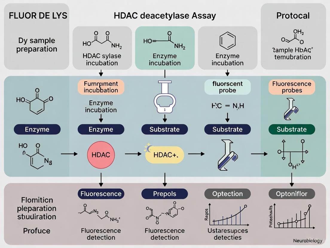
The FLUOR DE LYS HDAC assay and similar fluorometric methods operate on a two-step enzymatic reaction principle [4]. In the first step, active HDAC enzymes catalytically remove an acetyl group from a synthetic peptide substrate (Boc-Lys(Ac)-AMC). In the second step, the deacetylated product is incubated with a developer containing trypsin, which cleaves the modified lysine to release the highly fluorescent compound 7-Amino-4-methylcoumarin (AMC) [4]. The fluorescence intensity, measured with excitation at 340-360 nm and emission at 440-460 nm, is directly proportional to HDAC activity in the sample [4].
Diagram 1: HDAC Fluorometric Assay Principle
Nuclear Protein Extraction Protocol
Basic Protocol 1: Isolation of Nuclear Protein from Brain and Other Tissues [1]
Materials Required:
- Nuclear Extraction Kit (e.g., Abcam, cat. no. ab113474)
- Tissue of interest (e.g., brain regions: hippocampus, cortex, amygdala)
- 1.5-ml microcentrifuge tubes, 15-ml conical tubes
- Pipettes and tips
- Ice or cold room (4°C)
- Homogenizer (e.g., Branson Sonifier)
- Vortex mixer
- Refrigerated benchtop centrifuge (capable of 18,400 × g)
Procedure:
- Rapidly dissect desired tissues and micro-dissect specific regions if necessary. Weigh the tissue and store immediately at -80°C until needed.
- Prepare working reagents:
- 1× Pre-Extraction Buffer: Dilute 10× Pre-Extraction Buffer with distilled water
- Add 1000× Dithiothreitol (DTT) and 1000× Protease Inhibitor Cocktail (PIC) to Extraction Buffer immediately before use
- Homogenize tissue in Pre-Extraction Buffer using a mechanical homogenizer
- Centrifuge homogenate at 12,000 × g for 20 minutes at 4°C
- Discard supernatant and resuspend pellet in Extraction Buffer containing DTT and PIC
- Vortex the suspension for 15 seconds, then incubate on ice for 30 minutes with intermittent mixing
- Centrifuge at 18,400 × g for 20 minutes at 4°C
- Collect supernatant (nuclear protein extract) for HDAC activity assay
- Determine protein concentration using Pierce bicinchoninic acid (BCA) assay
Fluorometric HDAC Activity Assay Protocol
Basic Protocol 2: HDAC Activity Fluorometric Assay in Brain and Other Tissues [1] [4]
Materials Required:
- FLUOR DE LYS HDAC Fluorometric Cellular Activity Assay Kit (Enzo Life Sciences, BML-AK503-0001) [5]
- Black 96-well half-area plates
- Boc-Lys(Ac)-AMC substrate
- Trypsin from bovine pancreas
- HDAC inhibitor controls (e.g., Trichostatin-A, vorinostat/SAHA)
- Microplate reader capable of fluorescence measurements (e.g., BMG LABTECH)
Procedure:
- Prepare reaction buffer (e.g., FB188 buffer: 15 mM Tris-HCl pH 8.0, 50 mM KH₂PO₄/K₂HPO₄, 250 mM NaCl, 250 μM EDTA, and 0.001% Pluronic F-68) [4]
- Set up reactions in black 96-well plates:
- Experimental wells: Nuclear protein extract (10-50 μg) + substrate
- Negative controls: Sample + substrate + known HDAC inhibitor
- Blank: Buffer + substrate only
- Initiate reaction by adding Boc-Lys(Ac)-AMC substrate to a final concentration of 10-20 μM
- Incubate plates at 30°C for 60 minutes to allow deacetylation reaction
- Stop reaction and develop fluorescence by adding trypsin to a final concentration of 1.7 mg/mL
- Incubate for additional 30 minutes at room temperature
- Measure fluorescence using microplate reader (excitation: 340-360 nm, emission: 440-460 nm)
- Calculate HDAC activity based on AMC standard curve, normalized to protein concentration
Note: For cell-based assays, the FLUOR DE LYS substrate is cell-permeable and can be added directly to cultured cells, enabling measurement of HDAC activity in an undisturbed cellular environment [5].
Quantitative Analysis of HDAC Inhibitors
HDAC inhibitors are categorized based on their specificity toward different HDAC classes. Pan-inhibitors target multiple HDAC classes, while isoform-selective inhibitors specifically target individual HDAC isoforms, potentially reducing side effects [2] [3]. The table below summarizes inhibitory concentration (IC₅₀) values for representative HDAC inhibitors:
Table 2: Selectivity Profiles and Potency of HDAC Inhibitors
| Inhibitor | HDAC1 | HDAC6 | HDAC8 | HDAC10 | Primary Target | Clinical/Research Status |
|---|---|---|---|---|---|---|
| Vorinostat (SAHA) | 374 nM [4] | - | - | - | Class I [1] | FDA-approved for CTCL [2] |
| Compound 2a | No significant impact [2] | Selective over HDAC6 [2] | - | 0.41 ± 0.02 nM [2] | HDAC10 | Preclinical research [2] |
| Compound 2c | No significant impact [2] | Selective over HDAC6 [2] | - | 4.5 ± 0.3 nM [2] | HDAC10 | Preclinical research [2] |
| Compound 2f | No significant impact [2] | 2.5 ± 0.3 nM [2] | - | 110 ± 10 nM [2] | HDAC6 | Preclinical research [2] |
| PZ48 | - | - | - | Active at 5-15 μM [3] | HDAC10 | Preclinical research [3] |
The determination of IC₅₀ values follows standardized protocols where HDAC enzymes are incubated with serial dilutions of inhibitors followed by substrate addition [4]. For example, to evaluate SAHA against HDAC1, the enzyme is pre-incubated with a 1:3 serial dilution of SAHA (initial concentration 35 μM) for 15 minutes at room temperature before adding substrate at 20 μM final concentration [4].
The Scientist's Toolkit: Essential Research Reagents
Table 3: Essential Research Reagents for HDAC Activity Studies
| Reagent/Category | Specific Examples | Function/Application |
|---|---|---|
| HDAC Activity Assay Kits | FLUOR DE LYS HDAC Fluorometric Cellular Activity Assay Kit (Enzo) [5] | Cell-based HDAC activity measurement |
| COLOR DE LYS HDAC Colorimetric Activity Assay Kit (Enzo) [6] | Colorimetric HDAC activity detection | |
| HDAC Substrates | Boc-Lys(Ac)-AMC [1] [4] | Fluorogenic substrate for HDAC activity assays |
| HDAC Inhibitors (Research Tools) | Trichostatin-A [1] | Pan-HDAC inhibitor for control experiments |
| Vorinostat (SAHA) [1] [4] | FDA-approved pan-HDAC inhibitor | |
| PZ48 [3] | HDAC10-selective inhibitor | |
| Nuclear Extraction Reagents | Nuclear Extraction Kits [1] | Isolation of nuclear proteins for HDAC activity assays |
| Protease Inhibitor Cocktails [1] | Prevention of protein degradation during extraction | |
| Detection Instruments | Fluorescence Microplate Readers [4] | Quantification of fluorometric HDAC assay signals |
Research Applications and Therapeutic Implications
HDAC activity assays serve crucial roles in both basic research and drug discovery. In basic research, these assays help elucidate HDAC functions in physiological and pathological processes, including the study of HDAC dysregulation in neurological disorders such as Alzheimer's disease, Parkinson's disease, epilepsy, and traumatic brain injury [1]. In drug discovery, HDAC activity assays are indispensable for screening putative HDAC inhibitors, evaluating inhibitor potency and selectivity, and assessing the effects of therapeutic interventions on epigenetically modulated phenotypes [1] [2].
The therapeutic relevance of HDAC inhibitors is particularly prominent in oncology, where several HDAC inhibitors have received FDA approval. Vorinostat and belinostat are approved for T-cell lymphomas, romidepsin for cutaneous and peripheral T-cell lymphoma, and panobinostat for multiple myeloma [2] [3]. More recently, selective HDAC inhibitors have shown promise in preclinical studies, with HDAC10 inhibitors such as PZ48 demonstrating efficacy against acute lymphoblastic leukemia cells while sparing normal blood cells [3]. This selective toxicity toward malignant cells highlights the potential of isoform-selective HDAC inhibitors to achieve therapeutic efficacy with reduced side effects.
The workflow below illustrates a typical HDAC drug discovery and validation pipeline incorporating FLUOR DE LYS assay technology:
Diagram 2: HDAC Inhibitor Screening Workflow
HDACs represent crucial epigenetic regulators with diverse biological functions and profound therapeutic implications. The FLUOR DE LYS HDAC deacetylase assay protocol provides a robust, sensitive, and reproducible method for quantifying HDAC activity in various biological samples, from purified enzyme preparations to intact cells. As research continues to elucidate the distinct roles of individual HDAC isoforms, the development of isoform-selective inhibitors holds promise for targeted epigenetic therapies with improved efficacy and reduced side effects. The protocols and quantitative frameworks presented herein provide researchers with essential tools to advance our understanding of HDAC biology and accelerate the development of novel epigenetic therapeutics.
The FLUOR DE LYS (FDL) assay platform represents a significant advancement in the measurement of histone deacetylase (HDAC) activity, providing researchers with a robust, non-radioactive alternative to traditional methods that relied on radiolabeled substrates or cumbersome HPLC-based separation techniques [7] [8]. This technology has revolutionized HDAC and sirtuin research by enabling high-throughput screening (HTS) for candidate inhibitors and activators, characterization of enzyme kinetics, and cellular deacetylase activity assessments within an undisturbed cellular environment [9] [10]. The core innovation lies in its unique two-step fluorometric mechanism that combines an acetylated substrate with a developer to generate a highly sensitive, fluorescent signal proportional to deacetylase activity. This technical note details the fundamental principles, procedural methodologies, and practical applications of the FDL assay system, providing researchers with a comprehensive framework for implementing this technology in both biochemical and cellular contexts for drug discovery and epigenetic research.
The Biochemical Mechanism of the Two-Step Assay
Conceptual Framework and Reaction Chemistry
The FLUOR DE LYS assay operates on a elegantly designed two-stage mechanism that cleanly separates the enzymatic deacetylation reaction from the signal detection event. This separation is crucial for minimizing interference and providing a highly specific measurement of HDAC activity. The first stage involves the enzymatic deacetylation of a specially designed substrate, while the second stage encompasses the chemical development of the fluorescent signal [9] [8].
The FDL substrate comprises an acetylated lysine side chain embedded within a peptide backbone that is optimized for recognition by specific HDAC classes [9]. The deacetylation reaction, catalyzed by active HDAC enzymes, removes the acetyl group from the lysine residue, thereby "sensitizing" the substrate [9]. This sensitized intermediate then serves as the precursor for the subsequent development reaction. The developer reagent contains a developing agent that specifically recognizes the deacetylated lysine product and reacts with it to produce a highly fluorescent fluorophore [9]. This two-step approach provides significant advantages over single-step assays, including reduced compound interference, enhanced signal-to-noise ratios, and greater flexibility in assay optimization.
Visualizing the Two-Step Mechanism
The schematic below illustrates the sequential biochemical workflow of the FLUOR DE LYS assay mechanism:
Experimental Protocols and Methodologies
Standard Biochemical HDAC Activity Assay
The following protocol describes the procedure for measuring HDAC activity from purified enzymes or nuclear extracts using the FLUOR DE LYS system, adapted from established methodologies [11] [12].
Materials Required:
- FLUOR DE LYS substrate (specific for HDAC class being tested)
- FLUOR DE LYS developer
- Recombinant HDAC enzyme or HeLa nuclear extract (positive control)
- HDAC assay buffer
- Test compounds/inhibitors (e.g., Trichostatin A, Scriptaid)
- White or clear half-volume 96-well microplates
- Fluorescence plate reader capable of excitation ~350-380 nm and emission ~440-460 nm
Procedure:
- Preparation of Reaction Mixture: In a 96-well plate, combine the following components per well:
- 50 μL assay buffer
- 10 μL FLUOR DE LYS substrate (optimal concentration determined empirically)
- 10 μL HDAC enzyme source (recombinant enzyme or nuclear extract)
- 10 μL test compound or inhibitor (diluted in appropriate solvent)
Enzymatic Reaction (Step 1):
- Incubate the reaction mixture at 25°C or 37°C for 30-120 minutes, depending on enzyme activity.
- The incubation time should be optimized to ensure the reaction is within the linear range.
Signal Development (Step 2):
- Stop the enzymatic reaction and develop the fluorescence by adding 50 μL of FLUOR DE LYS developer.
- Include a developer-only blank to account for background fluorescence.
- Incubate the plate at 25°C for 15-30 minutes. The developer reaction typically goes to completion in less than 1 minute at 25°C [8].
Fluorescence Measurement:
- Read fluorescence using a plate reader with excitation at ~360 nm and emission at ~460 nm.
- For the FLUOR DE LYS-Green variant, use excitation/emission at 485/530 nm [10].
Data Analysis:
- Subtract background fluorescence (developer-only control).
- Calculate relative fluorescence units (RFU) and plot against standard concentrations if quantified.
- For inhibitor studies, express activity as percentage of control (no inhibitor).
Cellular HDAC Activity Assay
The cellular HDAC activity assay utilizes a cell-permeable variant of the FLUOR DE LYS substrate to measure deacetylase activity within intact cells [10].
Procedure:
- Cell Preparation: Seed cells in a 96-well plate and culture until desired confluence.
- Substrate Loading:
- Add cell-permeable FLUOR DE LYS substrate directly to culture media.
- Incubate cells for 30-120 minutes to allow substrate penetration and deacetylation by intracellular HDACs.
- Cell Lysis and Development:
- Remove media and lyse cells using provided lysis buffer.
- Add developer solution to the lysate and incubate for 15-30 minutes at room temperature.
- Fluorescence Measurement:
- Measure fluorescence as described in the biochemical assay protocol.
- Normalize readings to protein concentration or cell number.
Research Applications and Quantitative Data
HDAC Inhibitor Screening and Profiling
The FLUOR DE LYS platform is extensively utilized for screening and characterizing HDAC inhibitors, providing critical quantitative data on inhibitor potency and isoform selectivity. The table below summarizes representative data from a study investigating [6]-shogaol derivatives as HDAC inhibitors [13]:
Table 1: HDAC Inhibitory Activity of Selected [6]-Shogaol Derivatives
| Compound | % HDAC Inhibition (at 100 μM) | IC₅₀ (μM) | Selectivity Profile |
|---|---|---|---|
| TSA | 92% | N/D | Pan-inhibitor |
| [6]-Shogaol (4) | 86% | N/D | Broad-spectrum |
| 4c | 80% | 61 ± 0.92 | Selective for HDAC3 |
| 4d | 80% | 60 ± 0.84 | Selective for HDAC1, HDAC3 |
| 5j | 85% | 51 ± 0.82 | Selective for HDAC1 |
| 5k | 83% | 65 ± 1.12 | Selective for HDAC2 |
These quantitative results demonstrate the utility of the FDL assay in structure-activity relationship (SAR) studies, revealing that subtle structural modifications significantly impact both inhibitor potency and isoform selectivity—critical considerations for developing targeted epigenetic therapies.
Isoform-Selective Assay Applications
Specialized FDL drug discovery kits are available for specific HDAC isoforms, each optimized with isoform-specific substrates and recombinant enzymes. The table below outlines key isoforms and their research applications:
Table 2: FLUOR DE LYS Drug Discovery Kits for Selective HDAC Isoforms
| HDAC Isoform | Class | Cellular Functions | Research Applications |
|---|---|---|---|
| HDAC1 | I | Transcriptional regulation, cell cycle progression | Cancer research, inhibitor selectivity profiling [10] [13] |
| HDAC6 | IIb | Cytoplasmic tubulin deacetylation, stress response | Cancer metastasis, neurodegenerative diseases [9] |
| HDAC8 | I | Tubulin deacetylation, cancer proliferation | Cervical cancer research, substrate identification [12] |
| SIRT1 | III | Metabolic regulation, genotoxic stress response | Aging, metabolism, DNA damage response [7] [14] |
| SIRT3 | III | Mitochondrial function, energy metabolism | Metabolic disorders, cancer biology [14] |
The availability of isoform-specific assays has been instrumental in deciphering the distinct biological functions of individual HDACs and developing targeted inhibitors with potentially reduced side-effect profiles compared to pan-HDAC inhibitors [13].
The Scientist's Toolkit: Essential Research Reagents
Successful implementation of the FLUOR DE LYS technology requires specific reagents and components, each serving a distinct function in the assay workflow:
Table 3: Essential Research Reagents for FLUOR DE LYS Assays
| Reagent/Component | Function | Application Notes |
|---|---|---|
| FLUOR DE LYS Substrate | Acetylated peptide substrate containing sensitized lysine side chain | Optimal substrate varies by HDAC class; selected from panel of acetylated sites in p53 and histones [9] |
| FLUOR DE LYS Developer | Chemical developer that reacts with deacetylated substrate | Produces fluorophore upon reaction; different formulations available (e.g., Developer II for sirtuins) [14] |
| Recombinant HDAC Enzymes | Catalyze the deacetylation reaction in biochemical assays | Supplied in drug discovery kits; specific isoforms available (HDAC1, HDAC6, HDAC8, SIRT1, etc.) [9] [10] |
| HeLa Nuclear Extract | Rich source of multiple HDAC classes (1, 2, 3) | Used as positive control or enzyme source in general HDAC assays [8] [11] |
| HDAC Assay Buffer | Provides optimal pH and ionic conditions for deacetylation | Typically Tris-based buffer with magnesium chloride and potassium chloride [14] |
| Reference Inhibitors | Control compounds for assay validation (e.g., Trichostatin A, Scriptaid) | Establish baseline inhibition and assay performance [9] [10] |
| NAD⁺ | Essential cofactor for sirtuin (Class III HDAC) assays | Required for sirtuin activity; not needed for zinc-dependent HDACs [10] [14] |
Technical Considerations and Optimization Strategies
Experimental Design and Workflow
Implementing the FLUOR DE LYS assay requires careful experimental planning and optimization. The workflow below outlines the key decision points and procedures:
Troubleshooting and Optimization
Several factors critically impact the performance and reliability of FLUOR DE LYS assays:
Enzyme Concentration and Incubation Time: Must be optimized to maintain reaction linearity and ensure signal detection falls within the dynamic range of the fluorescence reader.
Compound Interference: Fluorescent or quenching compounds in test samples may interfere with signal detection. The FLUOR DE LYS-Green variant (excitation/emission: 485/530 nm) helps minimize this issue by avoiding the UV range where many compounds absorb [10].
Cofactor Requirements: Sirtuin assays (Class III HDACs) require the addition of NAD⁺ to the reaction mixture, whereas classical HDACs (Classes I, II, IV) do not [10] [14].
Cellular Assay Considerations: For cellular applications, substrate permeability, cellular esterase activity, and efflux mechanisms may influence signal intensity and require optimization of loading conditions and incubation times.
The FLUOR DE LYS two-step fluorometric mechanism represents a cornerstone technology in modern epigenetic research and drug discovery. Its elegant biochemical design, combining enzymatic specificity with sensitive fluorescence detection, has enabled researchers to overcome the limitations of traditional HDAC assay methods. The platform's flexibility—spanning biochemical, cellular, and isoform-specific applications—makes it an invaluable tool for characterizing HDAC function, screening therapeutic compounds, and advancing our understanding of epigenetic regulation in health and disease. As research continues to illuminate the complex roles of individual HDAC isoforms in cellular physiology and pathology, the FLUOR DE LYS technology remains well-positioned to support the next generation of discoveries in epigenetic therapeutics.
Within epigenetic research and drug discovery, the accurate quantification of histone deacetylase (HDAC) enzyme activity is fundamental. Traditional methods, particularly those relying on radioactive substrates or high-performance liquid chromatography (HPLC), present significant operational and safety challenges [15] [16]. This application note details the FLUOR DE LYS (FdL) HDAC fluorometric activity assay, a protocol that eliminates these drawbacks while providing robust, high-throughput compatible data. Developed within the context of a broader thesis on optimized HDAC assay research, this protocol offers researchers a safer, more efficient, and highly adaptable methodological framework.
The core principle of the FdL assay involves a two-step, mix-and-read procedure conducted entirely in a single microplate. This streamlined workflow is a significant advancement over traditional techniques, enabling efficient screening of HDAC inhibitors, which are of considerable interest as potential anticancer therapeutics and probes for studying epigenetic mechanisms [15] [4].
Key Advantages Over Traditional Methods
The FLUOR DE LYS system provides distinct benefits by replacing outdated and cumbersome technologies. A direct comparison of these advantages is summarized in the table below.
Table 1: Comparison of HDAC Activity Assay Methodologies
| Feature | Traditional Radioactive Assays | HPLC-Based Methods | FLUOR DE LYS Fluorometric Assay |
|---|---|---|---|
| Detection Principle | Use of radiolabeled (e.g., 3H) acetylated histones [15] | Separation and quantification of deacetylated product via HPLC | Fluorogenic substrate sensitized by deacetylation, followed by developer addition to generate a fluorophore [15] |
| Safety Concerns | Handling and disposal of radioactive materials [15] | Minimal safety concerns | No radioactivity; minimal safety risks [15] |
| Workflow Complexity | Multi-step, requires extractions and scintillation counting [15] | Multi-step, requires skilled operation and long run times | Simple, "mix-and-read" protocol in one microplate; no extractions [15] |
| Throughput | Low to medium | Low | High-throughput screening (HTS) friendly [15] |
| Quantitative Data | Provides kinetic data (e.g., IC50) | Provides kinetic data (e.g., IC50) | Enables determination of kinetic parameters (e.g., KM, IC50) [4] |
This streamlined approach is particularly valuable for research focused on class I HDACs—such as HDAC1, HDAC2, and HDAC3—which are critical targets in cancer development and gaining significant interest as clinically viable targets [17]. The protocol's compatibility with various biological sources, including cell lysates, immunoprecipitates, and purified enzymes, makes it a versatile tool for biochemical and pharmacological applications [15].
Experimental Protocol
This section provides a detailed step-by-step guide for determining the inhibitory potency (IC50) of a compound against HDAC1 using the FLUOR DE LYS system.
The Scientist's Toolkit: Essential Research Reagents
Table 2: Key Materials and Reagents for the FLUOR DE LYS Assay
| Item | Function / Description | Example Source / Specification |
|---|---|---|
| FLUOR DE LYS Substrate | Acetylated peptide substrate that is deacetylated by active HDAC enzymes. | Boc-Lys(Ac)-AMC [4] |
| HDAC Enzyme | Recombinant enzyme or native protein from cell extracts used as the assay target. | Recombinant HDAC1 (e.g., BPS Bioscience) [4] |
| FLUOR DE LYS Developer | Second-step reagent that cleaves the deacetylated substrate, releasing the fluorescent signal. | Contains Trypsin (e.g., 1.7 mg/mL) to stop the reaction and develop fluorescence [4] |
| Reference Inhibitor | Well-characterized inhibitor for assay validation and control experiments (e.g., determination of IC50). | SAHA (Suberoylanilide Hydroxamic Acid; Vorinostat) [4] |
| Assay Buffer | Provides optimal pH and ionic conditions for HDAC1 enzymatic activity. | FB188 buffer (15 mM Tris-HCl pH 8.0, 250 mM NaCl, etc.) [4] |
| Microplate Reader | Instrument for detecting and quantifying the fluorescent signal. | Fluorescence-capable reader (e.g., BMG LABTECH) with 340-360 nmEx/440-460 nmEm filters [4] |
| Microplates | Vessel for conducting the assay. | Black 96-well or 384-well plates to minimize crosstalk and background [4] |
Detailed Step-by-Step Procedure
Part A: Inhibitor Dilution and Pre-incubation
- Prepare a serial dilution of the test inhibitor (e.g., SAHA) in assay buffer. A 1:3 serial dilution starting from 35 µM is a typical range [4].
- Dispense the diluted inhibitor solutions into a black 96-well plate.
- Add recombinant HDAC1 (e.g., 4.5 nM final concentration) to the wells containing the inhibitor. Include control wells with buffer only (no enzyme, for background) and enzyme only (no inhibitor, for maximum activity).
- Incubate the plate for 15 minutes at room temperature to allow the inhibitor to bind the enzyme.
Part B: Enzymatic Reaction and Signal Development
- Initiate the reaction by adding the FLUOR DE LYS substrate (e.g., Boc-Lys(Ac)-AMC) at a final concentration near its KM value (e.g., 20 µM) for sensitive inhibition studies [4].
- Incubate the plate for 1 hour at 30°C to allow the deacetylation reaction to proceed.
- Stop the reaction and develop the fluorescent signal by adding the FLUOR DE LYS Developer, which contains trypsin and a high concentration of SAHA (e.g., 5 µM) to instantly and completely halt HDAC activity [4].
- Measure the fluorescence immediately using a microplate reader with excitation at 340-360 nm and emission at 440-460 nm.
Workflow and Mechanism Visualization
The following diagram illustrates the two-step mechanism and workflow of the FLUOR DE LYS assay:
Expected Results and Data Analysis
Kinetic and Inhibitor Characterization
A well-optimized FdL assay yields high-quality quantitative data. Prior to inhibition studies, it is crucial to determine the Michaelis constant (KM) for the substrate under specific assay conditions, which informs the appropriate substrate concentration to use in subsequent experiments. Using a substrate concentration near the KM value is recommended for sensitive inhibition studies [4].
Table 3: Exemplary Kinetic and Inhibitor Data for HDAC1 with Boc-Lys(Ac)-AMC
| Parameter | Description | Exemplary Value |
|---|---|---|
| KM Value | Michaelis constant for substrate Boc-Lys(Ac)-AMC | 58.89 µM [4] |
| IC50 for SAHA | Half-maximal inhibitory concentration of the reference inhibitor | 374 nM [4] |
| Recommended [Substrate] | Optimal substrate concentration for inhibition assays | ~20 µM (near KM) [4] |
For IC50 determination, fluorescence data from the inhibitor dilution series is collected. The relative fluorescence units (RFU) are normalized, typically defining the signal from the enzyme-only control as 0% inhibition and the background signal (no enzyme) as 100% inhibition. A dose-response curve is generated by plotting the percentage of inhibition versus the logarithm of the inhibitor concentration. The IC50 value is derived by fitting the data to a four-parameter logistic model (e.g., using software like GraphPad Prism).
Advantages in a Physiological Context
While fluorogenic substrates like Boc-Lys(Ac)-AMC offer unparalleled convenience for biochemical screening, it is important to acknowledge that short peptide substrates may not fully recapitulate the activity of HDACs, particularly sirtuins, toward complex physiological substrates like nucleosomes [18]. For investigations requiring high physiological relevance, chemically defined nucleosome core particles (NCPs) can be employed as substrates in Western blot-based deacetylation assays [18]. The FdL assay's primary strength lies in its utility for efficient, high-throughput inhibitor screening and mechanistic enzymology, providing a critical first step in the drug discovery pipeline.
The FLUOR DE LYS HDAC fluorometric assay protocol represents a significant methodological advancement for researchers in epigenetics and drug discovery. By eliminating radioactive materials and complex HPLC separations, it offers a safe, streamlined, and robust platform for characterizing HDAC activity and inhibitor potency. This protocol aligns with the ongoing development of class I selective HDAC inhibitors, such as novel benzamide-based compounds, which show improved antitumour profiles and represent the next generation of targeted epigenetic therapies [17]. The methodology's adaptability for use with purified enzymes, cell lysates, and even in cell-based assays ensures its continued relevance in the pursuit of novel therapeutic agents and a deeper understanding of epigenetic regulation.
FLUOR & Sirtuin DE - Enzo LYS HDAC Assays
The FLUOR DE LYS (Fluorogenic Histone deAcetylase Lysyl Substrate/Developer) system represents a transformative methodology in epigenetic research, providing a non-radioactive, high-throughput compatible platform for assessing histone deacetylase (HDAC) and sirtuin enzyme activity [7]. This technology has liberated researchers from the cumbersome protocols traditionally associated with radiolabeled or other modified histone-based methods, enabling more efficient screening of potential HDAC inhibitors for drug development [7]. The assay's core innovation lies in its patented substrate/developer chemistry, which, when combined with high-activity, high-purity enzymes, delivers reliable data quality essential for both basic research and pharmaceutical applications [7].
Within the broader context of FLUOR DE LYS HDAC deacetylase assay protocol research, this application note details the specific system components—substrates, developers, assay buffers, and controls—that form the foundation of this widely cited technology [7]. The flexibility of available screening formats (chemiluminescent, fluorescent, and colorimetric) allows researchers to select the optimal configuration for their specific experimental needs, particularly in drug discovery workflows where interference from test compounds can pose significant challenges [7].
System Components
The FLUOR DE LYS HDAC assay system comprises several integral components that work in concert to facilitate accurate measurement of deacetylase activity. Each component serves a specific function in the two-step assay process, which can be adapted for various sample types including cell or nuclear extracts, immunoprecipitates, and purified enzymes [19].
Core Substrate/Developer Chemistry
FLUOR DE LYS Substrate: The core substrate features an acetylated lysine side chain that serves as the enzymatic target for HDAC activity [19]. Upon deacetylation during the incubation step, the substrate becomes sensitized for subsequent development. The standard substrate is suitable for general HDAC assessment, while specialized formulations like FLUOR DE LYS-Green offer higher sensitivity with excitation/emission at 485/530nm, effectively minimizing quenching and fluorescent interference from compounds absorbing in the near UV and blue range [19].
FLUOR DE LYS Developer: The developer reagent reacts specifically with the deacetylated substrate to generate a fluorophore [19] [8]. This reaction typically proceeds to completion in less than one minute at 25°C, enabling rapid results [8]. The developer is added after the enzymatic deacetylation step is complete, ensuring that the fluorescence signal directly correlates with HDAC activity in the sample.
Assay Buffers and Solutions
The complete assay system includes optimized buffers that maintain enzymatic activity and ensure reagent stability. While specific buffer formulations are proprietary, they are designed to support HDAC activity across multiple classes while maintaining compatibility with the fluorescent detection system. The buffers facilitate analysis of diverse sample types, including crude extracts and purified enzyme preparations [15].
Controls and Calibrators
HeLa Nuclear Extract: Provided as a positive control, HeLa nuclear extract represents a rich source of HDACs 1 and 2, serving as a reliable reference for assay validation and normalization [15]. This control enables researchers to verify proper assay performance across experimental runs.
Enzyme Controls: The system utilizes high-purity, high-activity control enzymes manufactured in-house to ensure reproducible results and reliable quantification of inhibitory effects [7]. These controls are essential for establishing standard curves and calculating specific activity in experimental samples.
Quantitative Assay Specifications
Table 1: FLUOR DE LYS HDAC Assay Kit Configurations and Specifications
| Parameter | FLUOR DE LYS HDAC Assay | FLUOR DE LYS-Green HDAC Assay |
|---|---|---|
| Product Code | BML-AK500-0001 [15] | BML-AK530-0001 [19] |
| Well Format | 96 wells [15] | 96 wells [19] |
| Assay Type | Fluorometric [15] | Fluorometric [19] |
| Excitation/Emission | Standard fluorophore | 485/530 nm [19] |
| Key Features | Useful for lysates, immunoprecipitates, inhibitor screening; includes HeLa nuclear extract [15] | Higher sensitivity; avoids quenching and fluorescent interference [19] |
| Sample Compatibility | Cell/nuclear extracts, immunoprecipitates, purified enzymes [15] | Cell/nuclear extracts, immunoprecipitates, purified enzymes [19] |
| HDAC Class Compatibility | Class I & IIb HDACs and sirtuins (with addition of NAD+) [15] | Broad HDAC and sirtuin coverage [7] |
Table 2: HDAC Enzyme Compatibility and Experimental Applications
| HDAC Class | Compatible HDACs | Sample Types | Research Applications |
|---|---|---|---|
| Class I | HDAC1, HDAC2, HDAC3, HDAC8 [15] | Purified enzymes, nuclear extracts [15] | Cancer research, enzyme characterization [12] |
| Class II | HDAC4, 5, 6, 7, 9, 10 [15] | Cytoplasmic extracts, immunoprecipitates [12] | Subcellular localization studies [12] |
| Class III | SIRT1 (with NAD+ addition) [15] | Whole cell extracts, purified sirtuins [7] | Aging research, metabolic studies [7] |
| Class IV | HDAC11 [12] | Various cellular fractions [12] | Specialized HDAC function studies [12] |
Research Reagent Solutions
Table 3: Essential Research Reagents for FLUOR DE LYS HDAC Assays
| Reagent/Component | Function/Description | Example Usage |
|---|---|---|
| FLUOR DE LYS Substrate | Acetylated lysine-containing peptide that is deacetylated by active HDACs [19] | Serves as the enzymatic target in the first step of the assay [8] |
| FLUOR DE LYS Developer | Chemical developer that generates fluorophore from deacetylated substrate [19] | Added in the second step to produce measurable fluorescence signal [8] |
| HeLa Nuclear Extract | Positive control rich in HDACs 1 and 2 [15] | Verification of assay performance; normalization between experiments [15] |
| HDAC8-Specific Inhibitor (PCI-34051) | Selective HDAC8 inhibitor for target validation [12] | Confirmation of HDAC8-specific activity in mechanistic studies [12] |
| HDAC6-Specific Inhibitor (Tubastatin) | Selective HDAC6 inhibitor for isoform discrimination [12] | Determination of HDAC6 contribution to total deacetylase activity [12] |
| NAD+ | Cofactor required for sirtuin (Class III HDAC) activity [15] | Essential for assessing sirtuin activity in addition to classical HDACs [15] |
Experimental Protocol
Sample Preparation Guidelines
The FLUOR DE LYS assay system accommodates diverse sample types, each requiring specific preparation techniques to preserve HDAC activity while maintaining compatibility with the fluorescent detection system.
Cell Lysate Preparation: Cultured cells (e.g., HeLa, HEK293T) should be harvested at 80-90% confluency [12]. After trypsinization, wash cells with 1X PBS and lyse using appropriate lysis buffer (e.g., 50 mM Tris pH 8.0, 150 mM NaCl, 10% Glycerol, 0.5% Triton X-100, 1X Protease Inhibitor Cocktail) [12]. Clear lysates by centrifugation and determine protein concentration for normalization.
Subcellular Fractionation: For compartment-specific HDAC analysis, separate nuclear and cytoplasmic fractions using differential centrifugation techniques [12]. Validate fraction purity using markers for specific cellular compartments (e.g., GAPDH for cytoplasm, histone proteins for nucleus).
Immunoprecipitated Samples: For immunoprecipitation-based assays, incubate approximately 500 μg of total protein with 2 μg of specific HDAC antibody (e.g., anti-HDAC8) overnight at 4°C [12]. Capture immune complexes using Protein A Agarose beads, wash with appropriate buffer (e.g., 50 mM Tris pH 8.0, 150 mM NaCl, 1 mM EDTA, 0.1% NP-40), and resuspend in assay buffer for activity measurements [12].
HDAC Activity Assay Procedure
The standard FLUOR DE LYS assay follows a two-step procedure that can be completed in a single 96-well plate, facilitating high-throughput applications [15].
Step 1: Enzymatic Deacetylation
- In a 96-well plate, combine HDAC-containing sample (cell/nuclear extract, immunoprecipitate, or purified enzyme) with FLUOR DE LYS substrate [19] [8].
- Include appropriate controls: positive control (HeLa nuclear extract), negative control (sample with HDAC inhibitor), and blank (substrate without enzyme) [15].
- Incubate the reaction mixture at an appropriate temperature (typically 37°C) for 1-2 hours to allow deacetylation of the substrate [8]. The incubation time may be optimized based on enzyme activity.
Step 2: Fluorophore Development
- Stop the deacetylation reaction by adding FLUOR DE LYS developer [19] [8].
- Mix thoroughly and incubate at room temperature for 15-30 minutes to allow complete fluorophore development [8].
- Measure fluorescence using a microplate reader with appropriate filters (excitation/emission: 485/530 nm for FLUOR DE LYS-Green substrate) [19].
Data Analysis and Interpretation
Calculate HDAC activity based on fluorescence intensity relative to controls. The developer reaction typically goes to completion in less than one minute at 25°C, ensuring stable fluorescence readings [8].
Activity Calculation:
- Subtract blank fluorescence values from all samples.
- Normalize sample readings to positive controls (set as 100% activity).
- For inhibitor screening, calculate percentage inhibition using the formula: % Inhibition = [1 - (Sample - Blank)/(Positive Control - Blank)] × 100.
- Generate dose-response curves for IC50 determination using appropriate statistical software.
Application Example: HDAC8 Functional Characterization
A research application employing the FLUOR DE LYS system demonstrated the role of HDAC8 in cervical cancer cells [12]. This study utilized the HDAC8 FLUOR DE LYS fluorometric assay kit (BML-AK518-0001) to investigate HDAC8-mediated deacetylation of alpha-tubulin in HeLa cells [12].
Experimental Design
Cell Culture and Treatment: HeLa cells and HEK293T controls were cultured in DMEM with 10% FBS and 1% penicillin/streptomycin [12]. Cells were treated with HDAC8-specific inhibitor PCI-34051 (10-20 μM) and/or HDAC6-specific inhibitor tubastatin (5 μM) for 24 hours to assess isoform-specific contributions to deacetylase activity [12].
Activity Measurements: HDAC8 enzyme activity was measured in control and inhibitor-treated samples, including immunoprecipitated HDAC8 from cytoplasmic and nuclear fractions of HeLa and HEK293T cells [12]. The FLUOR DE LYS assay was performed according to the manufacturer's protocol, enabling precise quantification of HDAC8-specific activity [12].
Key Findings
The FLUOR DE LYS-based analysis revealed that HDAC8 and its phosphorylated form (pHDAC8) localized predominantly in the cytoplasm in both cancerous (HeLa) and non-cancerous (HEK293T) cells, with additional nucleolar localization observed in HeLa cells [12]. The study identified alpha-tubulin as a novel HDAC8 interacting partner and demonstrated that HDAC8 deacetylates tubulin at ac-lys40 [12].
Combining FLUOR DE LYS activity data with knockdown experiments using HDAC8-specific siRNA revealed that HDAC8 shows functional redundancy with HDAC6 when overexpressed in cervical cancer cells, contributing to cancer proliferation and progression [12]. This application highlights the utility of the FLUOR DE LYS system in delineating isoform-specific HDAC functions in pathological contexts.
Troubleshooting and Optimization
Fluorescence Quenching: If compound interference is suspected, particularly with libraries containing UV-absorbing compounds, employ the FLUOR DE LYS-Green substrate with red-shifted excitation/emission (485/530 nm) to minimize interference [19].
Low Signal Intensity: Optimize sample protein concentration and incubation time to ensure sufficient deacetylation. For immunoprecipitated samples, verify antibody specificity and binding efficiency [12].
High Background: Include appropriate controls to identify non-specific fluorescence. Ensure developer is added only after the deacetylation step is complete [8].
Enzyme Stability: HDAC enzymes, particularly recombinant forms like HDAC3 that require specific cofactors for activity, should be handled according to manufacturer specifications to maintain enzymatic activity [20].
Within epigenetic research, the accurate measurement of histone deacetylase (HDAC) activity is fundamental for investigating cellular signaling, gene regulation, and for screening potential therapeutic inhibitors. The FLUOR DE LYS (FDL) platform provides a versatile, non-radioactive foundation for these assays [15]. However, researchers face a critical choice between cellular activity, extract-based, and isoform-specific assay formats, each with distinct applications and limitations. This application note delineates these three principal methodologies, providing structured quantitative comparisons and detailed protocols to guide researchers in selecting the optimal approach for their specific experimental objectives within the broader context of FLUOR DE LYS deacetylase assay protocol research.
Comparative Analysis of HDAC Assay Formats
The table below summarizes the core characteristics, applications, and limitations of the three primary HDAC assay formats.
Table 1: Comparison of Key HDAC Activity Assay Formats
| Assay Format | Principle | Key Applications | Advantages | Limitations |
|---|---|---|---|---|
| Cellular Activity Assay | Cell-permeable FDL substrate enters live cells, is deacetylated by intracellular HDACs, and signal is developed post-lysis [15]. | Measuring endogenous HDAC activity in its native cellular context; high-throughput inhibitor screening in intact cells. | Preserves native cellular environment and post-translational regulation; no specialized extraction required. | Cannot isolate contribution of specific HDAC isoforms; cell permeability of inhibitors can confound results. |
| Extract-Based Assay | HDAC activity is measured in cell lysates, nuclear extracts, or immunoprecipitates using the FDL substrate/developer system [11] [15]. | Enzyme kinetic studies; inhibitor profiling using defined enzyme sources; assessing activity in subcellular fractions. | Controlled experimental conditions; can use HeLa nuclear extract (rich in HDAC1/2) as positive control [15]; suitable for immunoprecipitates. | Disrupts native cellular context; activity may not fully represent in vivo state due to loss of regulatory complexes. |
| Isoform-Specific Assay | Employs specialized substrates or purified recombinant enzymes to target the unique activity of a single HDAC isoform [21] [22]. | Profiling substrate specificity (e.g., HDAC10 as a polyamine deacetylase [21]); development of selective inhibitors. | Unravels specific biological functions of individual HDACs; critical for screening isoform-selective inhibitors. | Requires purified recombinant enzymes [22] or specialized substrates [21]; may not reflect activity in physiological complexes. |
Workflow and Logical Pathway Selection
The following diagram illustrates the key decision-making pathway for selecting the appropriate HDAC assay format based on the researcher's primary experimental question.
Experimental Protocols
Protocol 1: Cellular HDAC Activity Assay
This protocol measures global HDAC activity within live cells using the cell-permeable FLUOR DE LYS substrate [15].
Key Reagents:
- FLUOR DE LYS Substrate: Cell-permeable acetylated peptide substrate.
- FLUOR DE LYS Developer: Develops fluorescence signal upon interaction with deacetylated product.
- Lysis Buffer: Non-ionic detergent-based buffer (e.g., Triton X-100) for cell lysis and developer reaction.
- HDAC Inhibitor Control: Trichostatin A (TSA) or Suberoylanilide Hydroxamic Acid (SAHA) to confirm HDAC-specific signal.
Procedure:
- Cell Seeding & Treatment: Seed cells in a 96-well plate and grow to desired confluency. Treat with experimental compounds (e.g., potential inhibitors).
- Substrate Incubation: Add the FLUOR DE LYS substrate directly to the culture medium to a final concentration of 50-100 µM. Incubate for 1-4 hours at 37°C to allow substrate entry and deacetylation by intracellular HDACs.
- Signal Development: Remove the medium and add the FLUOR DE LYS Developer solution, prepared in lysis buffer containing a known HDAC inhibitor (e.g., 1-2 µM TSA) to stop ongoing HDAC activity and initiate the fluorescent reaction.
- Incubation & Detection: Incubate the plate for 15-30 minutes at room temperature. Measure the fluorescence (Excitation: ~340-380 nm, Emission: ~440-460 nm).
Protocol 2: Extract-Based HDAC Activity Assay
This protocol quantifies HDAC activity from cell lysates or subcellular extracts (e.g., HeLa nuclear extract), providing a controlled system for kinetic and inhibitor studies [11] [15].
Key Reagents:
- HDAC Enzyme Source: HeLa nuclear extract (rich source of HDAC1/2), whole-cell lysate, or immunoprecipitated HDAC complexes [11] [15].
- FLUOR DE LYS Substrate/Developer System: As described in Protocol 1.
- Assay Buffer: Typically 50 mM HEPES/TRIS (pH 8.0), 137 mM NaCl, 2.7 mM KCl, 1 mg/mL BSA, and 0.05% Tween-20 [22] [23].
- Positive Control: Supplied HeLa nuclear extract or recombinant HDAC enzyme.
- Negative Control: Assay buffer without enzyme source, or enzyme source pre-incubated with a potent inhibitor (TSA/SAHA).
Procedure:
- Sample Preparation: Prepare nuclear extracts or whole-cell lysates from your sample of interest using standard protocols. Keep samples on ice.
- Reaction Setup: In a 96-well plate, mix the HDAC-containing sample with the FLUOR DE LYS substrate in the provided or standard assay buffer. The final reaction volume is typically 50-100 µL. Note: Final DMSO concentration should be kept below 2-3% to avoid inhibition [22].
- Enzymatic Reaction: Incubate the reaction mixture for 30-90 minutes at 30°C or 37°C. The incubation time should be within the linear range of the reaction.
- Signal Development & Detection: Stop the reaction and develop the signal by adding the FLUOR DE LYS Developer solution (with TSA). Incubate for 15-30 minutes at room temperature and measure fluorescence.
Protocol 3: HDAC Isoform-Specific Activity Assay
This protocol is essential for studying HDAC isoforms with unique substrate preferences, such as HDAC10, which acts as a polyamine deacetylase (PDAC) and poorly deacetylates standard acetyl-lysine substrates [21].
Key Reagents:
- Purified Recombinant HDAC Isoform: e.g., HDAC10 (aa 2-631) from commercial sources [22] or purified in-house.
- Isoform-Specific Substrate: For HDAC10, a fluorescently-labeled acetyl-spermidine derivative (e.g., aminocoumarin-labelled acetyl-spermidine) instead of the standard FDL substrate [21].
- Assay Buffer: Optimized for the specific isoform (e.g., Tris or HEPES buffer, pH 8.0, with BSA).
- Isoform-Selective Inhibitors: e.g., Tubastatin A for HDAC6, or selective HDAC10 inhibitors for validation.
Procedure:
- Enzyme Preparation: Reconstitute purified recombinant HDAC enzyme in an appropriate storage buffer. For zinc-dependent HDACs, ensure the buffer contains a reducing agent like TCEP.
- Specialized Reaction: In a 96-well plate, mix the purified HDAC isoform with its specific substrate (e.g., 0-100 µM fluorescent acetyl-spermidine for HDAC10) in the assay buffer.
- Reaction Incubation: Incubate for a defined period (e.g., 30-60 minutes) at 37°C. The reaction can be stopped by adding a developer solution or a high-concentration inhibitor.
- Signal Measurement: Directly measure the fluorescence generated by the deacetylated product (e.g., Excitation/Emission for aminocoumarin derivatives). For continuous assays, a coupled enzyme system with trypsin can be used to liberate the fluorophore (AMC) in real-time [22].
Essential Research Reagent Solutions
The table below catalogs the crucial reagents required for successful execution of the featured HDAC assays.
Table 2: Key Research Reagents for HDAC Activity Assays
| Reagent | Function/Description | Example Assay Format |
|---|---|---|
| FLUOR DE LYS Substrate/Developer Kit | Core fluorescent system; substrate contains acetylated lysine; developer generates fluorophore upon deacetylation [15]. | Cellular, Extract-Based |
| HeLa Nuclear Extract | A rich, biologically relevant source of Class I HDACs (HDAC1 & 2), used as a positive control or enzyme source [11] [15]. | Extract-Based |
| Recombinant HDAC Isoforms | Purified individual HDAC proteins (e.g., HDAC1, HDAC6, HDAC8, HDAC10) for isoform-specific profiling and screening [22]. | Isoform-Specific |
| Selective HDAC Inhibitors | Pharmacological tools for validation and control (e.g., Trichostatin A (pan-inhibitor), PCI-34051 (HDAC8-selective), Tubastatin A (HDAC6-selective)) [12] [24]. | All Formats (Controls) |
| Isoform-Specific Substrates | Specialized substrates for unique HDAC activities (e.g., acetyl-spermidine derivatives for HDAC10) [21]. | Isoform-Specific |
| Acetylated Tubulin Antibody | For western blot validation of deacetylase activity, particularly for HDAC6 and HDAC8 which deacetylate α-tubulin at Lys40 [12] [24]. | Validation |
The strategic selection of an HDAC assay format—cellular, extract-based, or isoform-specific—is paramount to the success and biological relevance of any investigative or screening campaign. The cellular activity assay provides a holistic view of HDAC function within the intact physiological environment, ideal for phenotypic screening. The extract-based assay offers a robust and controllable system for biochemical characterization and inhibitor profiling against a defined mixture of HDACs. Finally, the isoform-specific assay is an indispensable tool for deconvoluting the unique roles of individual HDAC family members and for the rational design of selective next-generation inhibitors. By applying the protocols and decision-making framework outlined in this document, researchers can confidently navigate the HDAC assay landscape, ensuring their methodological approach is precisely aligned with their scientific goals.
Step-by-Step FLUOR DE LYS Protocol for Diverse Research Applications
Within the broader scope of FLUOR DE LYS HDAC deacetylase assay protocol research, the accurate preparation of biological samples is a foundational step. The choice of sample type—whether live cultured cells, prepared nuclear extracts, or isolated immunoprecipitates—directly influences the specific biological questions that can be addressed, from screening drug effects in a cellular context to elucidating the activity of specific enzymes or complexes [15] [5]. This application note provides detailed methodologies for preparing these sample types to ensure reliable and reproducible HDAC activity measurements using the FLUOR DE LYS platform.
The Scientist's Toolkit: Key Research Reagents
The following table outlines essential materials and reagents used in FLUOR DE LYS-based HDAC activity assays.
Table 1: Essential Reagents for HDAC Activity Assays
| Reagent | Function & Application | Key Characteristics |
|---|---|---|
| FLUOR DE LYS Substrate | Acetylated peptide substrate that is deacetylated by active HDACs [15]. | Cell-permeable for cellular assays [5]; compatible with Class I, IIb HDACs and sirtuins [15]. |
| FLUOR DE LYS Developer | Developer reagent that reacts with the deacetylated substrate to generate a fluorescent signal [15]. | Enables fluorometric detection; typically added after the incubation step [15]. |
| HeLa Nuclear Extract | Positive control; a rich source of HDAC activity, particularly HDAC1 and HDAC2 [15] [11]. | Serves as a reference for assay validation and inhibitor screening [15]. |
| Trichostatin A (TSA) | Potent HDAC inhibitor (Class I, II) [25]. | Used as a negative control to confirm HDAC-specific activity in assays [25]. |
| Nicotinamide | Sirtuin (Class III HDAC) inhibitor [25]. | Used as a negative control in assays involving NAD+-dependent sirtuin activity [25]. |
| HDAC Assay Buffer | Provides optimal pH and ionic strength for HDAC enzyme activity [25]. | Used for diluting enzymes, extracts, and reagents in biochemical assays [25]. |
| Cell Lysis Buffer | Facilitates the breakdown of cell membranes to release intracellular contents [25]. | Used for preparing cell lysates for subsequent HDAC activity measurement. |
Experimental Workflows for Sample Preparation and Analysis
The following section outlines specific protocols for preparing different sample types and conducting the HDAC activity assay.
Workflow for Cell-Based HDAC Activity Assay
The cell-based assay determines deacetylase activity within an intact cellular environment, which is reflective of endogenous regulation and can reveal the effects of upstream regulators or indirect inhibitors [5].
Detailed Protocol:
- Cell Culture and Plating: Culture adherent cells in an appropriate growth medium. Seed cells into a 96-well plate at a density that will achieve 70-90% confluency at the time of the assay.
- Treatment: Pre-treat cells with the experimental compounds (e.g., potential HDAC inhibitors) for the desired duration.
- Substrate Incubation:
- Prepare the FLUOR DE LYS substrate in serum-free medium or assay buffer [5].
- Remove the growth medium from the cells and add the substrate solution.
- Incubate the plate for 1-3 hours at 37°C. During this time, the substrate enters the cells and is deacetylated by intracellular HDACs.
- Development and Lysis:
- Prepare the FLUOR DE LYS developer solution according to the kit instructions.
- Add the developer solution directly to the wells containing the substrate and cells. The developer lyses the cells and reacts with the deacetylated substrate [5].
- Signal Detection: Incubate the plate with the developer for 10-30 minutes at room temperature. Measure the fluorescence using a microplate reader with excitation at ~360 nm and emission at ~460 nm (for the standard substrate) or excitation/emission at 485/530 nm (for the Green substrate) [15] [19].
Workflow for HDAC Activity Assay Using Nuclear Extracts and Immunoprecipitates
This biochemical assay is useful for directly measuring HDAC enzyme activity from purified sources, such as nuclear extracts or specific protein complexes isolated via immunoprecipitation [15].
Detailed Protocol:
- Sample Preparation:
- Nuclear Extracts: Prepare nuclear extracts from cultured cells (e.g., HeLa cells) using a standard protocol. The extracted nuclei contain abundant Class I HDACs, such as HDAC1 and HDAC2 [15] [11]. Dilute the extract in HDAC assay buffer on ice.
- Immunoprecipitates: Incubate a cell lysate with an antibody against the HDAC or protein complex of interest. Recover the immunocomplex using protein A/G beads. Wash the beads thoroughly with assay buffer to remove non-specifically bound proteins [15].
- Reaction Setup:
- In a 96-well plate, combine the sample (a volume of nuclear extract or the bead-bound immunocomplex) with the FLUOR DE LYS substrate.
- For sirtuin assays, include NAD+ in the reaction mixture, as it is an essential cofactor for their activity [15].
- Include appropriate controls (e.g., HeLa nuclear extract as a positive control, reactions with Trichostatin A as an inhibitor control).
- Incubation: Incubate the reaction mix for 30-60 minutes at 37°C. During this time, active HDACs will deacetylate the substrate.
- Development: Add the FLUOR DE LYS developer to stop the reaction and generate the fluorophore. The developer contains a developer concentrate (e.g., 20x) that is diluted in a stopping solution [15].
- Signal Detection: Incubate the plate for 10-30 minutes at room temperature and measure the fluorescence as described above.
Quantitative Data and Assay Performance
The table below summarizes key quantitative data for the FLUOR DE LYS assay system, which is critical for experimental planning and validation.
Table 2: Key Quantitative Data for FLUOR DE LYS Assay Kits
| Parameter | Specification | Details & Applications |
|---|---|---|
| Assay Throughput | 96-well plate format [15] | Suitable for high-throughput screening (HTS); "HTS friendly" [15]. |
| Assay Capacity | 100-200 assays per kit [15] | Sufficient for multiple experimental conditions and replicates. |
| Detection Modality | Fluorometric [15] | No radioactivity required; homogeneous, "mix-and-read" protocol [15]. |
| HDAC Class Compatibility | Class I, IIb, Sirtuins (with NAD+) [15] | Broad applicability across multiple HDAC classes from various sources [15] [7]. |
| Sirtuin Assay Requirement | Addition of NAD+ cofactor [15] | Essential for measuring the activity of NAD+-dependent sirtuins. |
Troubleshooting and Technical Notes
- Positive Control: HeLa nuclear extract is provided in several kits and serves as a robust positive control. Always include it to validate the assay performance in every experiment [15] [25].
- Inhibitor Controls: Use Trichostatin A (for Class I/II HDACs) and Nicotinamide (for sirtuins) to confirm that the measured signal is specific to HDAC activity [25].
- Signal Strength: If the signal is weak, optimize the sample protein concentration or the incubation time with the substrate. For the Green substrate (ex485/em530), ensure compatibility with your plate reader and that test compounds do not absorb in the near UV/blue range to avoid interference [19].
- Sample Integrity: Keep nuclear extracts and lysates on ice whenever possible and avoid repeated freeze-thaw cycles to preserve HDAC activity.
Within the broader context of FLUOR DE LYS HDAC deacetylase assay protocol research, the core two-step methodology of incubation and developer addition represents a standardized framework for investigating histone deacetylase (HDAC) activity. This fluorometric assay system provides a critical tool for drug discovery professionals and researchers engaged in screening candidate therapeutics and characterizing enzyme kinetics [26]. The protocol's design elegantly replaces traditional methods utilizing radiolabeled, acetylated histones or peptide/HPLC techniques, offering a non-radioactive, mix-and-read format compatible with high-throughput screening (HTS) platforms [15]. The fundamental principle relies on a sensitized substrate that undergoes HDAC-mediated deacetylation followed by chemical development to generate a quantifiable fluorescent signal, enabling precise measurement of deacetylase activity across purified enzymes, cell lysates, immunoprecipitates, and intact cellular environments [5] [19] [15].
Principle of the FLUOR DE LYS Assay
The FLUOR DE LYS (Fluorogenic Histone deAcetylase Lysyl) assay system operates through a sequential two-step mechanism that converts enzymatic activity into a measurable fluorescent output. The process begins with the FLUOR DE LYS substrate, which contains an acetylated lysine side chain that serves as the target for HDAC activity [26] [19] [15]. During the initial incubation step, HDAC enzymes catalyze the removal of the acetyl group from the substrate's lysine residue. This deacetylation reaction structurally sensitizes the substrate but does not immediately generate fluorescence [26]. The second step involves the addition of the FLUOR DE LYS Developer, which contains a developing compound that specifically reacts with the deacetylated substrate [26] [19]. This chemical development reaction produces a highly fluorescent fluorophore that can be quantified using a fluorometer with standard excitation/emission filters (approximately 485/530 nm for the Green variant) [19]. The fluorescence intensity directly correlates with the level of deacetylase activity present in the sample, providing a quantitative measure of HDAC function.
Research Reagent Solutions
Successful implementation of the core two-step protocol requires specific reagent systems tailored to different experimental contexts. The FLUOR DE LYS platform offers specialized kits designed for particular applications, from drug discovery screening to cellular activity assessment.
Table 1: Essential Research Reagent Solutions for FLUOR DE LYS HDAC Assays
| Kit/Component | Catalog Number | Primary Application | Key Features | Supported HDACs |
|---|---|---|---|---|
| FLUOR DE LYS HDAC8 Drug Discovery Kit | BML-AK518-0001 [26] | Inhibitor screening & enzyme kinetics | Includes recombinant human HDAC8; optimal substrate from p53 & histone panels [26] | HDAC8 specifically [26] |
| FLUOR DE LYS HDAC Fluorometric Cellular Activity Assay Kit | BML-AK503-0001 [5] | Cell-based deacetylase assays | Cell-permeable substrate; measures activity in undisturbed cellular environment [5] | Endogenous cellular HDACs [5] |
| FLUOR DE LYS-Green HDAC Fluorometric Activity Assay Kit | BML-AK530-0001 [19] | Extracts, immunoprecipitates & purified enzymes | Higher sensitivity; 485/530nm excitation/emission avoids compound interference [19] | Class I, IIb HDACs & sirtuins (with NAD+) [19] |
| Standard FLUOR DE LYS HDAC Fluorometric Activity Assay Kit | BML-AK500-0001 [15] | Lysates, immunoprecipitates & inhibitor screening | Includes HeLa nuclear extract; no radioactivity or extractions required [15] | Class I & II HDACs, SIRT1 [15] |
Materials and Equipment
Essential Reagents and Kits
The core two-step protocol utilizes specialized reagent kits as detailed in Table 1. These typically include the FLUOR DE LYS substrate (comprising an acetylated lysine side chain), FLUOR DE LYS Developer concentrate, HDAC Assay Buffer, positive controls such as HeLa nuclear extract (a rich source of HDACs 1 & 2) or recombinant HDAC8, and reference inhibitors like Trichostatin A (an HDAC inhibitor) and Nicotinamide (a sirtuin inhibitor) [26] [5] [25]. Additional components may include FLUOR DE LYS Deacetylated Standard for calibration and Cell Lysis Buffer for cellular assays [25].
Required Equipment
The protocol requires standard laboratory equipment including a microplate fluorometer capable of measuring fluorescence at appropriate wavelengths (typically ~485 nm excitation/~530 nm emission for the Green variant) [19], a 96-well microplate (black plates with clear bottoms are optimal for fluorescence measurements), precision pipettes for reagent delivery, an incubator or water bath maintained at 37°C for the enzymatic reaction, and standard laboratory containers for buffer preparation.
Experimental Protocols
Core Two-Step Protocol for Purified Enzymes and Extracts
This fundamental protocol is optimized for measuring HDAC activity in purified enzyme preparations, nuclear or cellular extracts, and immunoprecipitates.
Step 1: Incubation
- Prepare the HDAC Assay Buffer according to kit specifications [19] [15].
- In a 96-well plate, combine the sample containing HDAC activity (purified enzyme, nuclear extract, or immunoprecipitate) with the FLUOR DE LYS substrate [26] [19]. The nuclear extract provided in some kits serves as a positive control [15].
- Incubate the reaction mixture for 30-90 minutes at 37°C. The optimal incubation time may vary based on enzyme concentration and activity levels [26].
- During this incubation, HDAC enzymes present in the sample deacetylate the substrate, sensitizing it for subsequent development [26] [15].
Step 2: Developer Addition
- After the incubation period, stop the deacetylation reaction by adding the FLUOR DE LYS Developer [26] [19].
- The Developer concentration is typically 1-2× the final concentration in the well, and it's added directly to the incubation mixture without extraction steps [15].
- Following Developer addition, incubate the plate for an additional 15-45 minutes at room temperature or 37°C to allow for full fluorophore development [26].
- Measure the resulting fluorescence using a fluorometer with appropriate filters (excitation ~360 nm, emission ~460 nm for standard substrates; excitation/emission 485/530 nm for the Green variant) [19].
Cellular HDAC Activity Protocol
For determining deacetylase activity within intact cellular environments, a modified approach utilizes the cell-permeable properties of the FLUOR DE LYS substrate.
Step 1: Cellular Incubation
- Culture adherent or suspension cells in a 96-well plate format suitable for fluorescence measurements [5] [25].
- Add the cell-permeable FLUOR DE LYS substrate directly to the cell culture medium and incubate for 1-4 hours under normal growth conditions (e.g., 37°C, 5% CO₂) [5].
- During this incubation, the substrate permeates cell membranes and is deacetylated by endogenous HDACs within the undisturbed cellular environment [5] [15].
- The deacetylated substrate accumulates inside cells, with the accumulation rate reflecting intracellular HDAC activity [15].
Step 2: Developer Addition and Measurement
- Following cellular incubation, add the FLUOR DE LYS Developer directly to the media and lysed cells without intermediate washing steps [5].
- Incubate for 30-60 minutes to allow complete fluorophore development.
- Quantify fluorescence using a plate-reading fluorometer, normalizing measurements to cell number or protein content as appropriate [5].
HDAC Inhibitor Screening Protocol
The two-step protocol is particularly suited for high-throughput screening of potential HDAC inhibitors in drug discovery applications.
Step 1: Inhibitor Incubation
- Pre-incubate potential inhibitors with the HDAC enzyme source (recombinant enzyme, nuclear extract, or cells) for 15-30 minutes prior to substrate addition [26].
- Add the FLUOR DE LYS substrate and continue incubation for 60-120 minutes at 37°C [26].
- Include appropriate controls: no-inhibitor controls (100% activity), no-enzyme controls (background fluorescence), and reference inhibitor controls (e.g., Trichostatin A) [26] [25].
Step 2: Development and Detection
- Add FLUOR DE LYS Developer to terminate the reaction and generate the fluorescent signal [26].
- Following development, measure fluorescence and calculate percentage inhibition relative to controls.
- Determine IC₅₀ values by testing compound dilutions and analyzing the dose-response relationship [26].
Table 2: Quantitative Parameters for Different FLUOR DE LYS HDAC Assay Formats
| Assay Parameter | HDAC8 Drug Discovery Kit | Cellular Activity Assay | Green HDAC Activity Assay | Standard HDAC Activity Assay |
|---|---|---|---|---|
| Sample Capacity | 96 assays [26] | 96 wells [5] | 96-well format [19] | 100-200 assays [15] |
| Incubation Time | Optimized for 1-2 hours [26] | 1-4 hours [5] | 30-90 minutes [19] | 30-90 minutes [15] |
| Detection Sensitivity | High (fluorometric) [26] | Reflects endogenous activity [5] | Higher sensitivity than standard [19] | High (alternative to radioactive) [15] |
| Key Applications | Chemical library screening, enzyme kinetics [26] | Effects of upstream regulators, indirect inhibitors [5] | Cell/nuclear extracts, immunoprecipitates [19] | Class I/II HDACs, sirtuins (with NAD+) [15] |
Workflow Integration
Troubleshooting and Optimization
Several factors can impact the performance of the core two-step protocol. Low Signal Intensity may result from insufficient enzyme activity, suboptimal incubation times, or improper Developer preparation. Remedy this by increasing enzyme concentration, extending incubation time (up to 2-3 hours), or verifying Developer concentration and freshness. High Background Fluorescence often stems from incomplete substrate deacetylation or contamination. Address this by including no-enzyme controls, ensuring proper substrate storage, and using appropriate filter sets to minimize compound interference (particularly beneficial with the Green variant's 485/530 nm profile) [19]. Variable Replicates frequently arise from inconsistent pipetting, temperature fluctuations, or uneven plate sealing. Improve consistency by using calibrated pipettes, ensuring stable incubation temperature, and properly sealing plates during incubations. For cellular assays, Poor Substrate Permeabilization can limit signal generation; this can be addressed by optimizing substrate concentration and incubation time with target cell lines [5].
Applications in Drug Discovery and Research
The core two-step protocol enables diverse research applications including Chemical Library Screening where the system's simple mix-and-read format and 96-well compatibility make it ideal for identifying candidate inhibitors from compound libraries [26] [15]. The assay effectively characterizes inhibitor potency (IC₅₀ determination) and mechanism of action. Enzyme Kinetics Studies utilize the protocol to determine Michaelis-Menten parameters (Kₘ, Vₘₐₓ) by measuring initial velocity of deacetylation at varying substrate concentrations [26]. Cellular Pathway Analysis applications leverage the cell-permeable substrate version to investigate how upstream regulators, signaling pathways, and physiological stimuli modulate HDAC activity within an undisturbed cellular environment [5]. Target Validation employs the protocol in conjunction with siRNA knockdown approaches, as demonstrated by studies where HDAC8 knockdown inhibited growth of human tumor cell lines, suggesting its significance in cancer pathogenesis [26].
Histone deacetylases (HDACs) are crucial epigenetic regulators involved in the reversible modulation of gene expression by removing acetyl groups from lysine residues on histone tails and various non-histone proteins [1]. The determination of deacetylase activity within an undisturbed cellular environment provides activity information that reflects endogenous regulation and the effects of upstream regulators, which is essential for drug discovery and basic research [5] [1]. The FLUOR DE LYS HDAC fluorometric cellular activity assay enables precisely this capability through the use of a cell-permeable substrate that is deacetylated by intracellular HDACs, allowing for accurate assessment of deacetylase activity under physiological conditions without requiring cell lysis as an initial step [5].
Key Principles and Significance
HDAC Biology and Therapeutic Relevance
HDACs function as "eraser" enzymes that remove acetyl groups from histones, leading to chromatin compaction and transcriptional repression [1]. Eighteen HDAC enzymes have been identified in humans and are classified into four classes based on structure and function [1] [27]. Beyond histones, HDACs also modulate the acetylation status of non-histone proteins, including transcription factors, cytoskeletal elements, and molecular chaperones, thereby influencing a broad array of cellular processes [1]. Dysregulation of HDAC activity has been implicated in numerous neurological disorders, cancer, and other pathological conditions, making HDAC inhibitors promising therapeutic agents [1] [27].
Advantages of Cell-Based HDAC Activity Assessment
Unlike traditional methods that utilize cell lysates or purified enzymes, the cell-based FLUOR DE LYS assay preserves the native cellular environment, including:
- Endogenous regulatory mechanisms (post-translational modifications, subcellular localization)
- Intact multiprotein complexes in which HDACs typically function
- Natural substrate accessibility and compartmentalization
- Physiological cofactor concentrations
This approach enables the detection of inhibitors or activators that act indirectly through upstream signaling pathways to affect deacetylase activity, providing a more comprehensive view of compound effects in a physiological context [5].
Materials and Reagents
Research Reagent Solutions
Table 1: Essential Research Reagents for FLUOR DE LYS HDAC Cellular Assay
| Reagent/Material | Function/Application | Key Characteristics |
|---|---|---|
| FLUOR DE LYS Substrate | Cell-permeable HDAC substrate | Comprises an acetylated lysine side chain; deacetylated by intracellular HDACs [5] [15] |
| FLUOR DE LYS Developer | Fluorophore generation | Contains trypsin; converts deacetylated substrate to measurable fluorophore [1] [15] |
| HDAC Inhibitor Controls (e.g., Trichostatin-A, Vorinostat) | Assay validation and inhibition studies | Reference compounds for establishing assay performance [1] [27] |
| Cell Culture Plates (96-well) | Assay format | Compatible with high-throughput screening [5] [15] |
| Fluorometric Microplate Reader | Signal detection | Excitation ~360 nm, Emission ~460 nm [15] |
| Cultured Cells | Experimental system | Any cell type expressing HDAC activity; HeLa cells commonly used [5] [11] |
Experimental Protocol
The following diagram illustrates the complete experimental workflow for measuring intracellular HDAC activity using the FLUOR DE LYS assay:
Detailed Step-by-Step Procedure
Step 1: Cell Preparation and Plating
- Culture appropriate cells (e.g., HeLa, primary neurons, or other relevant cell types) under standard conditions.
- Plate cells in a 96-well microplate at optimal density (typically 10,000-50,000 cells/well depending on cell type) and allow to adhere overnight [5] [11].
- For inhibitor studies, pre-treat cells with HDAC inhibitors for desired duration before assay.
Step 2: Substrate Incubation
- Prepare FLUOR DE LYS substrate according to manufacturer's instructions.
- Add substrate directly to culture media (final concentration typically 50-200 μM) and incubate for 3-6 hours at 37°C, 5% CO₂ [5] [15].
- During this incubation, the cell-permeable substrate enters cells and is deacetylated by intracellular HDACs.
Step 3: Developer Addition and Fluorophore Generation
- Add FLUOR DE LYS developer containing trypsin directly to each well.
- Incubate for 30 minutes at room temperature or 37°C [1].
- The developer lyses cells and converts the deacetylated product into a highly fluorescent compound.
Step 4: Fluorescence Measurement and Data Analysis
- Measure fluorescence using a microplate reader with excitation at ~360 nm and emission at ~460 nm [15].
- Include appropriate controls:
- Blank wells (media + substrate + developer, no cells)
- Inhibitor controls (cells + known HDAC inhibitor + substrate + developer)
- Background fluorescence (cells + substrate, no developer)
- Calculate net HDAC activity by subtracting background and normalizing to protein content or cell number.
Data Interpretation and Analysis
Expected Results and Typical Findings
Table 2: Representative HDAC Inhibitor Data for Assay Validation
| HDAC Inhibitor | Target Class | Reported IC₅₀ Values | Cellular Activity in FLUOR DE LYS Assay |
|---|---|---|---|
| Trichostatin A (TSA) | Class I/II | 2-6 nM [27] | Potent inhibition of cellular HDAC activity |
| Vorinostat (SAHA) | Class I/II | 10-164 nM [27] | Dose-dependent inhibition |
| Sodium Butyrate | Class I/II | 175-400 μM [27] | Moderate inhibition at mM concentrations |
| Valproic Acid | Class I | 171-400 μM [27] | Weak inhibition at therapeutic concentrations |
| Romidepsin | Class I | 1.6-36 nM [28] | Potent inhibition |
Troubleshooting Guide
Table 3: Troubleshooting Common Issues in FLUOR DE LYS Cellular Assay
| Problem | Potential Causes | Solutions |
|---|---|---|
| Low fluorescence signal | Low HDAC expression, insufficient substrate concentration, short incubation time | Optimize cell density, increase substrate concentration, extend incubation time |
| High background fluorescence | Substrate auto-hydrolysis, cell death | Include proper blanks, check cell viability, optimize developer incubation time |
| Poor inhibitor response | Compound impermeability, inappropriate inhibitor class for HDACs expressed | Verify inhibitor solubility and permeability, characterize HDAC expression profile in cells |
| High well-to-well variability | Uneven cell plating, inconsistent substrate addition | Use automated liquid handlers, verify cell distribution, practice consistent technique |
Applications in Drug Discovery and Research
The FLUOR DE LYS cellular HDAC activity assay enables several critical applications in both basic research and drug development:
HDAC Inhibitor Screening
The assay provides a robust method for identifying novel HDAC inhibitors in a physiologically relevant cellular context, accounting for compound permeability, metabolism, and potential prodrug activation [5] [27]. The compatibility with high-throughput screening formats (96-well and higher density plates) makes it suitable for pharmaceutical compound libraries [15].
Mechanistic Studies in Disease Models
The methodology has been successfully applied to investigate HDAC activity dysregulation in various pathological conditions, including:
- Traumatic brain injury and neurodegenerative disorders [1]
- Cancer models and transformation studies [28] [29]
- Psychiatric disorders including depression and epilepsy [27]
- Stem cell differentiation and developmental processes [30]
Pathway Analysis and Epigenetic Regulation
By preserving the intact cellular environment, the assay enables investigation of upstream signaling pathways that regulate HDAC activity through post-translational modifications, subcellular localization, and protein-protein interactions [5] [28].
Complementary Methodologies
For comprehensive HDAC characterization, the cellular FLUOR DE LYS assay can be combined with:
- Nuclear extraction protocols for subcellular localization studies [1]
- HDAC isoform-specific assays using immunoprecipitated enzymes [15] [29]
- Gene expression analysis of HDAC target genes
- Western blotting for histone acetylation changes
This integrated approach provides a complete picture of HDAC function from enzymatic activity to downstream physiological effects.
Within the framework of thesis research on the FLUOR DE LYS HDAC deacetylase assay protocol, this application note details its critical role in high-throughput screening (HTS) for HDAC inhibitors and activators. Histone deacetylases (HDACs) are promising therapeutic targets for a spectrum of diseases, including central nervous system disorders and cancer [1]. The ability to reliably quantify HDAC activity is fundamental for elucidating its role in disease and for evaluating the therapeutic potential of HDAC inhibitors (HDACis), which have demonstrated neuroprotective, antiepileptogenic, and antidepressant properties in animal models [1]. The FLUOR DE LYS HDAC fluorometric cellular activity assay provides a robust, sensitive, and scalable platform for determining deacetylase activity within an undisturbed cellular environment, making it exceptionally suitable for HTS campaigns to identify novel modulators of HDAC activity [5].
Core Assay Principle and Workflow
The FLUOR DE LYS HTS strategy is based on the use of a cell-permeable, fluorogenic substrate. The following diagram illustrates the core principle and sequential workflow of the assay for screening applications.
Assay Principle: The cell-permeable FLUOR DE LYS substrate is deacetylated by active HDACs inside living cells. Subsequent addition of the Developer, which contains trypsin, lyses the cells and stops the enzymatic reaction. Trypsin then cleaves the deacetylated substrate, releasing a highly fluorescent product that can be quantified using a plate reader [5]. This coupled enzyme assay provides an efficient and continuous readout, enabling accurate determination of kinetic parameters and inhibitor efficacy [22].
Key Quantitative Parameters for HTS
Careful optimization of assay conditions is paramount for a successful HTS campaign. The tables below summarize critical kinetic and validation parameters.
Table 1: Optimized HTS Assay Conditions for Fluorogenic HDAC Assays [22]
| Parameter | Specification | Rationale |
|---|---|---|
| Final DMSO Content | ≤ 2-3% | Prevents potential HDAC inhibition by DMSO [22]. |
| Assay Buffer | Tris (pH 8.0) or HEPES (pH 7.4) | Provides optimal pH for enzymatic activity. |
| BSA Concentration | 0.5 mg/mL | Stabilizes enzymes and prevents non-specific binding. |
| Trypsin Concentration | 5.0 mg/mL (in Developer) | Efficiently cleaves deacetylated product to generate fluorescence. |
| Assay Format | Continuous, coupled | Provides real-time kinetic data for accurate parameter determination. |
Table 2: Exemplary HDAC Inhibitor Profiling Data [1] [22] [31]
| HDAC Inhibitor | Target Specificity | Reported IC₅₀ / Activity | Key Application Context |
|---|---|---|---|
| Vorinostat | Class I (HDAC1 @ 10-70 nM) | FDA-approved for T-cell lymphoma; crosses BBB [1]. | Cancer, CNS disorder research |
| AR-42 (rac-) | Pan-HDAC inhibitor | IC₅₀ 5-50 nM (P. falciparum); cures murine malaria [31]. | Infectious disease, cancer |
| Trichostatin A | Class I/II HDAC inhibitor | Used as reference control in protocol validation [1]. | General HDAC research, control |
| Sodium Butyrate | Class I/II HDAC inhibitor | Exhibits neuroprotective & antiepileptogenic effects [1]. | Neuroscience, epigenetics |
| Valproic Acid | Class I HDAC inhibitor | Less potent HDACi; antiepileptic and mood stabilizer [1]. | Neuroscience, psychiatry |
Detailed HTS Protocol
Basic Protocol: High-Throughput Screening for HDAC Inhibitors using FLUOR DE LYS
Materials:
- FLUOR DE LYS HDAC Fluorometric Cellular Activity Assay Kit (e.g., Enzo Life Sciences, BML-AK503) [5]
- Cell line of interest (e.g., primary TAMs from GBM patients [32])
- Putative HDAC inhibitors/library compounds
- Black-walled, clear-bottom 96- or 384-well microplates
- Multi-channel pipettes and reagent reservoirs
- Fluorescent microplate reader (excitation ~360 nm, emission ~460 nm)
- Assay Buffer (e.g., Tris or HEPES-based) [22]
Procedure:
- Cell Plating and Compound Treatment:
- Seed cells in a 96-well plate at a density optimized for 24-hour growth (e.g., 10,000-20,000 cells/well). Incubate overnight under standard conditions.
- For screening, add putative HDAC inhibitors or library compounds to the cells. Include controls on every plate:
- Positive Control (Max Inhibition): Cells + known HDACi (e.g., Trichostatin-A, 1 µM) [1].
- Negative Control (Basal Activity): Cells + vehicle (e.g., DMSO, concentration equal to compound wells).
Substrate Incubation and Reaction:
- Prepare the FLUOR DE LYS substrate in pre-warmed assay buffer according to manufacturer specifications.
- Remove culture medium from the assay plate and carefully add the substrate solution to all wells.
- Incubate the plate for the predetermined optimal time (e.g., 1-3 hours) at 37°C, protected from light. This incubation allows cellular HDACs to deacetylate the substrate.
Signal Development and Detection:
- Prepare the Developer solution containing trypsin [22].
- Add the Developer solution directly to each well to achieve a final trypsin concentration of approximately 5 mg/mL. Mix gently.
- Incubate the plate for 30-60 minutes at room temperature, protected from light. This step lyses the cells and develops the fluorescent signal.
Fluorescence Measurement:
- Read the fluorescence in a microplate reader using excitation and emission filters appropriate for the AMC fluorophore (e.g., excitation 360 nm, emission 460 nm).
Data Analysis and Hit Selection:
- Calculate the percentage of HDAC inhibition for each test compound using the formula:
% Inhibition = [1 - (F_compound - F_positive) / (F_negative - F_positive)] × 100whereF_compoundis the fluorescence of the test well,F_positiveis the average fluorescence of the positive control (max inhibition), andF_negativeis the average fluorescence of the negative control (basal activity). - Calculate the Z'-factor for each plate to confirm assay robustness:
Z' = 1 - [3×(σ_p + σ_n) / |μ_p - μ_n|], where σ and μ are the standard deviation and mean of the positive (p) and negative (n) controls, respectively. A Z' > 0.5 is indicative of an excellent HTS assay. - Select compounds that show inhibition above a predefined threshold (e.g., >50% inhibition at the test concentration) for further dose-response validation.
- Calculate the percentage of HDAC inhibition for each test compound using the formula:
The Scientist's Toolkit: Key Research Reagent Solutions
Table 3: Essential Reagents and Kits for HDAC HTS
| Reagent / Kit | Function / Description | Example Source / Citation |
|---|---|---|
| FLUOR DE LYS HDAC Fluorometric Cellular Activity Assay Kit | Provides core components for cell-based deacetylase activity screening. | Enzo Life Sciences [5] |
| Boc-Lys(Ac)-AMC Substrate | Fluorogenic substrate for quantifying HDAC activity in tissue lysates. | Used in fluorometric HDAC activity kits [1] |
| Recombinant HDAC Enzymes (HDAC1-11) | For target-specific biochemical assays and counter-screening. | BPS Bioscience, Millipore [22] |
| Nuclear Extraction Kit | Isolates nuclear proteins for subcellular HDAC activity profiling. | Abcam (cat. no. ab113474) [1] |
| HDAC Inhibitor Controls (Vorinostat, TSA) | Reference compounds for assay validation and as positive controls. | Multiple commercial suppliers [1] |
Advanced HTS Applications and Validation
The utility of the FLUOR DE LYS-based HTS approach extends beyond simple inhibitor discovery, as illustrated in the following application pathway.
Exemplary Application: A 2025 preprint demonstrated the power of this approach by performing a large-scale chemical screen on primary tumor-associated macrophages and microglia (TAMs) from glioblastoma (GBM) patients. The screen identified HDAC inhibitors as potent inducers of phagocytosis. This pro-phagocytic effect was amplified when combined with CD47 blockade, and the combination showed enhanced tumor growth suppression in a xenograft GBM model, highlighting a novel immunotherapeutic strategy [32]. This underscores how HTS using cell-based HDAC activity assays can identify compounds with therapeutically relevant phenotypic effects beyond mere enzymatic inhibition.
Histone deacetylases (HDACs) are crucial epigenetic regulators that remove acetyl groups from lysine residues on histones and non-histone proteins, influencing gene expression and cellular processes [33]. The human HDAC family comprises 18 enzymes divided into five classes, with HDAC1 and HDAC2 belonging to Class I HDACs that are homologous to the yeast Rpd3 protein and are predominantly nuclear [33]. These enzymes frequently function within multi-protein repression complexes such as Sin3, NuRD, and CoREST [34] [33]. Aberrant HDAC activity, particularly HDAC1 overexpression, has been documented in various cancer types, making these enzymes attractive therapeutic targets [34]. While numerous HDAC inhibitors (HDACis) have been developed, many are non-selective, creating a pressing need for isoform-specific inhibitors to dissect individual HDAC functions and develop targeted therapeutics with reduced off-target effects [35] [33]. This application note details methodologies for conducting isoform-specific analysis of HDAC1 and HDAC2 activity using specialized drug discovery kits, enabling accurate inhibitor screening and kinetic characterization.
HDAC Biology and Isoform Specificity
HDAC Classification and Functions
HDAC enzymes are categorized based on sequence homology and cofactor dependence. Class I HDACs (HDAC1, 2, 3, and 8) are ubiquitously expressed zinc-dependent enzymes primarily localized in the nucleus [33]. HDAC1 was the first protein linked to histone deacetylase activity and contributes significantly to class I deacetylase activity in human cells [34]. Beyond histone modification, HDAC1 catalyzes regulatory deacetylation of non-histone proteins including the tumor suppressor p53 [34]. HDAC2 shares high sequence similarity with HDAC1 and is often found in similar repressor complexes, though it exhibits distinct regulatory mechanisms and biological functions [35].
Rationale for Isoform-Selective Inhibition
The development of isoform-selective HDAC inhibitors represents a frontier in epigenetic drug discovery. While pan-HDAC inhibitors like Vorinostat (SAHA) have received FDA approval for cancer treatment, their non-selective nature can lead to unintended side effects [35] [33]. Selective inhibition of specific HDAC isoforms enables more precise therapeutic interventions and research tools for deconvoluting the biological roles of individual HDACs. The structural diversity in HDAC active sites, particularly in surface regions beyond the conserved zinc-binding tunnel, provides opportunities for designing selective inhibitors that discriminate between highly similar isoforms like HDAC1 and HDAC2 [35] [33].
Available Technologies for HDAC Activity Assessment
Multiple assay platforms facilitate HDAC activity measurement and inhibitor screening, each offering distinct advantages for specific applications. The table below summarizes key technologies relevant to HDAC1 and HDAC2 analysis.
Table 1: Comparison of HDAC Activity Assay Technologies
| Technology | Detection Method | Application Scope | Sample Types | Key Features |
|---|---|---|---|---|
| FLUOR DE LYS HDAC1 Assay Kit [34] | Fluorometric | HDAC1-specific screening | Purified recombinant enzyme | Optimal substrate selected from acetylated sites in p53 and histones; includes recombinant human HDAC1 |
| FLUOR DE LYS Cellular Activity Assay [5] | Fluorometric | Cellular HDAC activity | Intact cells | Cell-permeable substrate; measures deacetylase activity in undisturbed cellular environment |
| CHEMILUM DE LYS HDAC/SIRT Assay [36] | Chemiluminescent | Broad HDAC/Sirtuin screening | Cell extracts, immunoprecipitates, purified enzymes | High specificity; superior signal-to-noise ratio; no interference from detergents |
| HDAC-Glo I/II Assays [37] | Luminescent | Class I/II HDAC screening | Purified enzymes, extracts, or cells | Homogeneous, add-mix-measure protocol; compatible with high-throughput screening |
| HDAC Fluorogenic Assay Kit (Green) [38] | Fluorometric | Class I (HDAC1, 2, 3) and IIb (HDAC6) | Purified enzymes | Measures HDAC1, 2, and 3 activity; includes HDAC2 control and Trichostatin A inhibitor |
| ELISA-Based HDAC Activity Assay [29] | Colorimetric/ELISA | Isoform-selective inhibitor characterization | Mammalian cell-derived HDAC isoforms | Uses cell-derived HDACs instead of recombinant proteins; robust for selectivity profiling |
Experimental Protocols
FLUOR DE LYS HDAC1 Biochemical Assay Protocol
The FLUOR DE LYS HDAC1 Fluorometric Drug Discovery Kit (BML-AK511-0001) provides a complete system for measuring lysyl deacetylase activity of recombinant human HDAC1 [34].
Materials and Reagents
- FLUOR DE LYS HDAC1 Assay Kit containing:
- FLUOR DE LYS Substrate (acetylated lysine side chain)
- FLUOR DE LYS Developer
- Recombinant human HDAC1 enzyme
- Assay buffer
- Optional: Trichostatin A (TSA) as reference inhibitor
- Black 96-well microplate for fluorescence detection
- Micropipettes and sterile tips
- Plate reader capable of fluorescence detection (excitation ~360 nm, emission ~460 nm)
- Test compounds for screening (dissolved in DMSO with final concentration ≤2%)
Two-Step Assay Procedure
Enzymatic Reaction:
- Prepare reaction mixture containing 1× Assay Buffer, FLUOR DE LYS Substrate, and recombinant HDAC1 enzyme
- Add test compounds at desired concentrations for inhibition studies
- Incubate at 30°C for 60-90 minutes to allow deacetylation reaction
- The deacetylation step sensitizes the substrate for subsequent development
Developer Reaction:
- Stop the enzymatic reaction by adding FLUOR DE LYS Developer containing trichostatin A
- Incubate at room temperature for 15-30 minutes
- Developer treatment produces a fluorophore from the deacetylated substrate
- Measure fluorescence with excitation at ~360 nm and emission at ~460 nm
Data Analysis
- Calculate HDAC activity as fluorescence units per unit time
- For inhibitor studies, determine IC50 values using non-linear regression of inhibition curves
- Normalize data to controls (no inhibitor vs. no enzyme background)
Diagram 1: FLUOR DE LYS Two-Step Assay Workflow
Cellular HDAC Activity Assay Protocol
The FLUOR DE LYS Cellular Activity Assay Kit (BML-AK503-0001) enables determination of deacetylase activity within an undisturbed cellular environment, reflecting endogenous regulation [5].
Cell Preparation and Treatment
- Culture cells in appropriate medium in 96-well plates
- Treat with test compounds for desired duration
- Prepare vehicle controls and reference inhibitor controls (e.g., Trichostatin A)
Cellular Assay Procedure
- Add cell-permeable FLUOR DE LYS Substrate directly to culture media
- Incubate cells with substrate for 1-4 hours at 37°C, 5% CO2
- Cellular HDACs deacetylate the permeable substrate
- Add Developer to media and lyse cells
- Incubate 15-30 minutes at room temperature
- Measure fluorescence (excitation ~360 nm, emission ~460 nm)
Data Interpretation
- Fluorescence signal proportional to cellular HDAC activity
- Effects of upstream regulators and indirect modulators can be detected
- Normalize to cell number using protein assay or viability stain
HDAC1 vs. HDAC2 Selectivity Screening Protocol
For isoform selectivity profiling, parallel assays with HDAC1 and HDAC2 are essential.
Comparative Assay Setup
- Utilize HDAC Fluorogenic Assay Kit (Green) which detects Class I HDAC activity [38]
- Perform simultaneous assays with purified HDAC1 and HDAC2 enzymes
- Include reference inhibitors with known selectivity profiles
- Test compounds across a concentration range (typically 0.1 nM-100 μM)
Selectivity Calculation
- Determine IC50 values for each compound against both isoforms
- Calculate selectivity index (SI) as: SI = IC50 (non-target HDAC) / IC50 (target HDAC)
- Compounds with SI >10-fold considered selective
The Scientist's Toolkit: Essential Research Reagents
Table 2: Key Research Reagent Solutions for HDAC1/HD2 Drug Discovery
| Reagent/Kit | Manufacturer | Primary Function | Application Notes |
|---|---|---|---|
| FLUOR DE LYS HDAC1 Assay Kit | Enzo Life Sciences | HDAC1-specific inhibitor screening | Includes recombinant human HDAC1; ideal for chemical library screening [34] |
| FLUOR DE LYS Cellular Activity Assay | Enzo Life Sciences | Measuring intracellular HDAC activity | Cell-permeable substrate; reflects endogenous regulation [5] |
| HDAC Fluorogenic Assay Kit (Green) | BPS Bioscience | Class I HDAC (1, 2, 3) screening | Contains HDAC2 control; green fluorescence (λex=485 nm; λem=528 nm) [38] |
| CHEMILUM DE LYS HDAC/SIRT Assay | Enzo Life Sciences | Broad-spectrum HDAC/sirtuin screening | Chemiluminescent detection; high sensitivity; minimal interference [36] |
| Trichostatin A (TSA) | Multiple suppliers | Pan-HDAC inhibitor positive control | Potent inhibitor of Class I/II HDACs; used for assay validation [34] [37] |
| HeLa Nuclear Extract | Multiple suppliers | Source of endogenous HDAC activity | Rich in Class I HDACs; useful for positive controls [34] [36] |
HDAC Classification and Inhibitor Selectivity
Understanding HDAC classification is fundamental to designing isoform-selective screening strategies. The diagram below illustrates the organization of zinc-dependent HDACs and the reported selectivity of various inhibitor chemotypes.
Diagram 2: Zinc-Dependent HDAC Classification and Inhibitor Selectivity Profiles
Data Analysis and Interpretation
Kinetic Parameter Determination
Fluorogenic HDAC assays enable accurate determination of enzyme kinetic parameters through careful optimization of continuous, coupled enzyme assays [22] [39].
Michaelis-Menten Kinetics
- Perform assays with varying substrate concentrations (typically 1-200 μM)
- Measure initial velocities at each substrate concentration
- Plot velocity vs. substrate concentration and fit to Michaelis-Menten equation
- Determine Km and Vmax values using non-linear regression
Inhibitor Potency Assessment
- Dose-response curves with 8-12 compound concentrations
- Test each concentration in duplicate or triplicate
- Calculate IC50 values using four-parameter logistic equation
- For reversible competitive inhibitors, determine Ki using Cheng-Prusoff equation: Ki = IC50 / (1 + [S]/Km)
Selectivity Profiling
Comprehensive selectivity assessment should include multiple HDAC isoforms beyond HDAC1 and HDAC2:
Table 3: Example Selectivity Profile of HDAC Inhibitors
| Compound | HDAC1 IC50 (nM) | HDAC2 IC50 (nM) | HDAC3 IC50 (nM) | HDAC6 IC50 (nM) | HDAC8 IC50 (nM) | Selectivity Index |
|---|---|---|---|---|---|---|
| Trichostatin A [35] | 3.3 | - | 11 | 6 | 25 | Pan-inhibitor |
| Apicidin [35] | 22 | - | 29 | >10,000 | 755 | HDAC1/3 selective |
| Cyclic Tetrapeptide 3c [35] | 2 | - | 18 | 31 | 133 | HDAC1 selective |
| Compound 3a [35] | 116 | - | 608 | 2,309 | 2,300 | Moderate HDAC1 selective |
Troubleshooting and Optimization
- DMSO Sensitivity: Final DMSO concentration should not exceed 1-3% as it can inhibit HDAC activity [22] [38]
- Fluorescence Interference: Test compound auto-fluorescence separately; use controls without enzyme
- Linear Range: Ensure reaction time is within linear velocity range; time courses recommended for new assays
- Enzyme Concentration: Optimize enzyme amount to maintain initial velocity conditions
The methodologies detailed in this application note provide robust frameworks for isoform-specific analysis of HDAC1 and HDAC2 activity. The FLUOR DE LYS platform offers sensitive, specific solutions for both biochemical and cellular HDAC assessment, enabling comprehensive inhibitor profiling and selectivity determination. As HDAC inhibitor research advances toward more selective therapeutics, these assay systems will continue to play vital roles in drug discovery pipelines and epigenetic mechanism studies. The integration of biochemical, cellular, and selectivity profiling approaches outlined herein represents a best practices framework for HDAC-targeted drug discovery.
Fluorometric measurement, prized for its exceptional sensitivity and specificity, serves as a cornerstone analytical technique in pharmaceutical sciences and drug development [40]. Its ability to detect analytes at trace levels, even within complex matrices, makes it indispensable for applications ranging from quality control to enzymatic activity assays [40]. This application note provides a detailed framework for the instrument setup and data acquisition parameters critical for obtaining reliable, high-quality data, with a specific focus on the context of FLUOR DE LYS HDAC deacetylase assay research. Proper configuration is paramount, as the fluorescence signal is relative and influenced by numerous instrumental factors [41].
Theoretical Foundations of Fluorescence
The Principle of Fluorescence Intensity
Fluorescence is a process of photoluminescence where a fluorophore absorbs light at a specific wavelength, causing electron excitation, and subsequently emits light at a longer wavelength as electrons return to the ground state [41]. The fluorescence intensity indicates the number of photons emitted and is directly correlated with the concentration of the excited fluorophore, forming the basis for quantitative analysis [41]. The difference between the peak excitation and emission wavelengths is known as the Stokes shift, a key property that enables the separation of the strong excitation light from the weaker emission signal [41] [42].
The Critical Role of the Stokes Shift
The Stokes shift is fundamental to the high sensitivity of fluorescence detection [42]. A sufficient shift (typically >30 nm) allows optical filters to effectively separate the excitation light from the emitted fluorescence. If the shift is small and the excitation/emission spectra are too close, spectral overlap or crosstalk can occur, where excitation light leaks through to the detector, raising the background signal and compromising data quality [43] [42]. The relationship between these concepts is illustrated in the following workflow:
Instrumentation and Component Configuration
Core Components of a Fluorometer
A standard fluorometer consists of several key components, each playing a critical role in the measurement process [44]:
- Light Source: Common sources include Xenon flash lamps (broad spectrum from UV to IR), Light Emitting Diodes or LEDs (high intensity at specific wavelengths), and Tungsten halogen lamps (unsuitable for UV measurements) [44] [41].
- Excitation Filter/Monochromator: Selects a specific wavelength band from the broadband source to illuminate the sample.
- Sample Holder: Typically a black microplate to minimize background and reflection of excitation light [41].
- Emission Filter/Monochromator: Isolates the fluorescent light from the sample.
- Dichroic Beam Splitter: Reflects the shorter wavelength excitation light towards the sample and transmits the longer wavelength emission light towards the detector, enhancing signal separation [44].
- Detector: A photomultiplier tube (PMT) that converts photons into an electrical signal, reported in Relative Fluorescent Units (RFU) [41].
Wavelength Selection and Optimization
Selecting appropriate excitation (Ex) and emission (Em) wavelengths is a foundational step for a sensitive and robust assay.
- Identifying Spectral Maxima: The first step is to determine the excitation and emission maxima of the fluorophore. This is ideally done by performing excitation and emission scans using a monochromator-equipped microplate reader [45].
- The 30 nm Rule: To minimize crosstalk, the lowest transmitted emission wavelength should be at least 30 nm higher than the highest transmitted excitation wavelength [43] [41]. For fluorophores with a very small Stokes shift, it may be necessary to select excitation and emission wavelengths slightly away from their maxima to achieve this separation [42] [45].
- Use of Cutoff Filters: An emission cutoff filter, which blocks all light below a specific wavelength, can be used to further reduce crosstalk. The cutoff wavelength should be selected to sit between the excitation and emission wavelengths [45].
Table 1: Wavelength Optimization Strategy for Different Stokes Shifts
| Stokes Shift Magnitude | Recommended Excitation Wavelength | Recommended Emission Wavelength | Cutoff Filter Advice |
|---|---|---|---|
| Large (>80 nm) | Select at excitation maximum (λmax) [45]. | Select at emission maximum (λmax) [45]. | Use a cutoff filter between Ex and Em [45]. |
| Small (<30 nm) | Select a wavelength lower than λmax that gives 90% of maximal RFU [45]. | Select a wavelength higher than λmax [45]. | Essential; cutoff may be higher than Em maximum [45]. |
Bandwidth Optimization
Bandwidth determines the range of wavelengths transmitted by a filter or monochromator. A 10 nm bandwidth allows light through from λ -5 nm to λ +5 nm [43].
- Narrow Bandwidth (e.g., 10-20 nm): Beneficial for bright fluorophores or in complex samples with high autofluorescence, as it improves selectivity and reduces background [43] [41].
- Broader Bandwidth (e.g., >20 nm): Can be used for low-emission fluorophores, particularly if they emit in spectral regions with low autofluorescence (e.g., red end of the spectrum), to capture more signal [43] [41].
- Avoiding Overlap: The sum of the excitation and emission bandwidths should not exceed the Stokes shift, otherwise, crosstalk will occur, increasing the limit of detection (LOD) [43] [42].
Table 2: Effect of Bandwidth on Assay Performance
| Bandwidth Setting | Signal Level | Selectivity | Recommended Use Case |
|---|---|---|---|
| Narrow (10 nm) | Lower | Higher | Bright fluorophores (e.g., AlexaFluor488, FITC, GFP); cellular assays with autofluorescence [43] [41]. |
| Medium (15-20 nm) | Balanced | Balanced | Often provides the best compromise for optimal detection limit and S/N ratio [43] [42]. |
| Broad (30+ nm) | Higher | Lower | Low-emission fluorophores in clean buffers; luminescence readouts [43]. |
Application to FLUOR DE LYS HDAC Assay
Assay Principle and Workflow
The FLUOR DE LYS HDAC assay is a fluorometric kit designed to quantify histone deacetylase (HDAC) activity. The protocol utilizes an acetylated substrate peptide conjugated to a fluorophore, whose fluorescence is quenched. Upon deacetylation by active HDAC, the substrate can be cleaved by a developer solution, releasing the fluorescent group (e.g., AMC, 7-Amino-4-methylcoumarin) and generating a measurable signal proportional to HDAC activity [46]. The complete workflow and signaling pathway are summarized below:
Recommended Instrument Parameters
Based on the assay chemistry and standard fluorophores like AMC, the following parameters are recommended as a starting point for optimization:
- Excitation Wavelength: 350–360 nm
- Emission Wavelength: 440–460 nm
- Bandwidth: 10–15 nm for both excitation and emission to maximize signal-to-noise ratio in enzymatic assays [43] [42].
- Gain/PMT Voltage: Set to auto or manually adjusted to ensure the signal from the most active sample is within the linear range of the detector, avoiding saturation.
- Microplate: Use black, flat-bottom microplates to minimize cross-talk and background between wells [41].
Data Acquisition and Calibration Protocols
Establishing a Calibration Curve
For quantitative analysis, a calibration curve is essential. This involves preparing a serial dilution of a reference standard of known concentration, such as the deacetylated fluorophore (e.g., AMC) provided in some kits [46] [47].
- Preparation: Prepare a series of concentrations covering the expected dynamic range of the assay.
- Measurement: Read the fluorescence of each standard and blank (buffer only) in triplicate using the optimized instrument parameters.
- Analysis: Plot the mean RFU for each standard against its concentration. The plot should be linear, and the resulting equation (y = mx + c) is used to calculate the concentration or activity of unknown samples [47].
Protocol for Wavelength Optimization
If the fluorophore's precise spectral properties are unknown, follow this empirical optimization protocol [45]:
- Initial Emission Scan: Set the excitation wavelength to a tentative value (e.g., from literature). Perform an emission scan without a cutoff filter to identify the emission peak.
- Excitation Scan: Set the emission wavelength to the identified peak. Perform an excitation scan to find the true excitation maximum.
- Apply the 30 nm Rule: Finalize the excitation and emission wavelengths, ensuring sufficient separation.
- Select Cutoff Filter: Perform an emission scan with a candidate cutoff filter (e.g., 420 nm for AMC). The filter should block residual excitation light without unduly reducing the fluorescent signal.
- Validate with Blanks: Always run appropriate blanks (buffer, substrate without enzyme) to account for background fluorescence and allow for calculation of the signal-to-blank ratio.
Critical Experimental Parameters to Document
For reproducibility, meticulously record the following data acquisition parameters:
- Excitation and Emission Wavelengths (nm)
- Bandwidths (nm)
- Type and strength of Cutoff/Dichroic filters
- PMT Gain/Voltage setting
- Number of flashes or integration time
- Reading mode (Top/Bottom reading)
- Microplate type and manufacturer
The Scientist's Toolkit: Essential Research Reagents and Materials
Table 3: Key Reagent Solutions for Fluorometric HDAC Assays
| Item | Function/Description | Example/Note |
|---|---|---|
| FLUOR DE LYS Substrate | Acetylated peptide that is deacetylated by HDACs; fluorescence is quenched until processed. | The core component of the assay kit [46]. |
| HDAC Enzyme Source | Source of enzymatic activity. | Can be purified enzyme, nuclear extracts, or immunoprecipitated samples [46]. |
| Developer Solution | Contains a protease that specifically cleaves the deacetylated substrate, releasing the fluorescent group. | Added after the deacetylation reaction is stopped [46]. |
| Reference Fluorophore | A solution of the free fluorophore (e.g., AMC) at known concentration. | Used to generate a calibration curve for quantitation [47]. |
| HDAC Inhibitor (Control) | A compound to specifically inhibit HDAC activity. | Trichostatin A (TSA) is commonly used to confirm signal specificity [46]. |
| Assay Buffer | Provides the optimal chemical environment (pH, ionic strength) for HDAC activity. | Critical for maintaining enzyme stability and activity [46]. |
| Black Microplates | Microplates with black wells to minimize optical crosstalk and background signal. | Essential for sensitive fluorescence measurements [41]. |
Troubleshooting and Quality Control
Common Challenges and Solutions
- High Background Signal: Can be caused by spectral overlap, contaminated buffers, or plate autofluorescence. Re-optimize wavelengths/bandwidths, use high-purity reagents, and ensure black plates are used [41] [42].
- Low Signal Intensity: May result from suboptimal wavelength selection, low PMT gain, enzyme inactivity, or failure to add developer. Check instrument settings and reagent freshness [46].
- Signal Instability: Fluorescence can be quenched by environmental factors or degrade due to photobleaching upon prolonged light exposure. Minimize light exposure during assay steps and ensure consistent temperature [40].
- Poor Linearity: If the calibration curve is not linear, the detector may be saturated (signal too high) or the concentrations may be near the limit of detection. Ensure samples and standards are within the dynamic range [47].
Implementing Controls
Robust experimental design requires appropriate controls:
- Negative Control: Assay buffer alone (blank).
- Background Control: Substrate + developer without enzyme (accounts for any non-enzymatic signal).
- Inhibitor Control: Sample pre-incubated with TSA to confirm HDAC-specific signal.
- Positive Control: A well-characterized HDAC enzyme or control lysate provided in the kit [46].
Solving Common Problems and Optimizing Your HDAC Assay Performance
Within the framework of FLUOR DE LYS HDAC deacetylase assay protocol research, a recurring challenge is the occurrence of low signal output, which can compromise data interpretation and experimental progress. This issue stems from multiple potential sources, including compromised sample integrity, suboptimal enzyme activity, and substrate-related problems. The FLUOR DE LYS platform, a pioneer in non-radioactive, homogeneous assays for HDAC and sirtuin activity, utilizes a patented substrate/developer system but remains susceptible to these fundamental experimental variables [7]. This application note provides a systematic troubleshooting guide to diagnose and resolve low signal issues, ensuring robust and reliable assay performance for researchers and drug development professionals.
Understanding Your Assay System
The FLUOR DE LYS assay is a coupled enzymatic system. The HDAC or sirtuin enzyme first deacetylates the substrate. Subsequently, the Developer reagent cleaves the deacetylated product, releasing a highly fluorescent group. Therefore, a low final fluorescent signal can result from failures at multiple points in this cascade [7] [11].
It is also critical to understand the intrinsic catalytic properties of your target enzyme. Notably, vertebrate class IIa HDACs (HDAC4, -5, -7, and -9) are inherently inefficient enzymes on standard acetylated substrates, often exhibiting ~1,000-fold lower activity compared to class I HDACs. What may appear as a "low signal" for these enzymes might, in fact, be the expected baseline activity. This inefficiency is attributed to a single amino acid substitution (Tyr to His) in their active site [48]. The following table summarizes key quantitative parameters for different HDAC classes.
Table 1: Key Characteristics of HDAC Classes Relevant to Assay Performance
| HDAC Class | Representative Members | Catalytic Efficiency on Standard Substrates | Key Characteristics & Notes for Assay |
|---|---|---|---|
| Class I | HDAC1, HDAC2, HDAC3, HDAC8 | High | Ubiquitously expressed; major source of deacetylase activity in cells [49] [18]. |
| Class IIa | HDAC4, HDAC5, HDAC7, HDAC9 | Very Low (Inefficient) | Possess a His residue instead of Tyr in active site; low basal activity is normal [48]. |
| Class IIb | HDAC6, HDAC10 | High | --- |
| Class III | SIRT1-7 | Varies by isoform | NAD+-dependent; not inhibited by Zn2+-dependent HDAC inhibitors (e.g., TSA) [49]. |
| Class IV | HDAC11 | --- | --- |
Troubleshooting Workflow
The following diagram outlines a logical, step-wise workflow for diagnosing the root cause of low signal in your FLUOR DE LYS HDAC assay.
Figure 1: A sequential workflow for troubleshooting low signal in HDAC assays.
Investigating Sample Integrity
Sample integrity is the foundation of a successful assay. Degradation or improper handling of either the enzyme source or the substrate can lead to significant signal loss.
Protocol: Assessing Nuclear Extract and Enzyme Source Viability
- Preparation: Always prepare fresh nuclear extracts or use aliquots that have undergone minimal freeze-thaw cycles. HDAC complexes are dynamic and their activity can significantly diminish with prolonged storage or repeated thawing [18].
- Positive Control: Include a well-characterized positive control in every assay. This can be a recombinant HDAC enzyme (e.g., Class I HDAC like HDAC1 or HDAC3) or a pre-validated nuclear extract (e.g., from HeLa cells) [11] [50].
- Inhibition Test: Confirm the specificity of the measured signal by pre-treating a sample aliquot with a known HDAC inhibitor, such as Trichostatin A (TSA) or a more selective inhibitor. A successful inhibition (≥70% signal reduction) confirms that the signal is due to specific HDAC activity [50].
- Alternative Assay Cross-Check: If possible, confirm sample quality using an orthogonal method. For instance, analyze histone acetylation status in cell samples via Western blotting with anti-acetyl-histone H3 or H4 antibodies after treatment with an HDAC inhibitor [50].
Evaluating Enzyme Activity
If sample integrity is confirmed, the next step is to focus on the enzymatic reaction itself. The source and class of the HDAC enzyme are critical factors.
Protocol: Kinetic Analysis to Diagnose Enzyme Issues
- Class-Specific Considerations: If assaying Class IIa HDACs (HDAC4, -5, -7, -9), recognize that their low activity on standard FLUOR DE LYS substrates may be normal. Consider using a positive control mutant (e.g., HDAC4-His976Tyr) which shows a 1,000-fold higher catalytic efficiency, or test alternative substrates like trifluoroacetyl-lysine, which has been identified as a class IIa-specific substrate in vitro [48].
- Cofactor Supplementation: Ensure all necessary cofactors are present. Class I and II HDACs are Zn²⁺-dependent. While Zn²⁺ is often tightly bound, assay buffers should not contain strong chelators. Class III sirtuins require NAD⁺; therefore, verify the concentration and freshness of NAD⁺ in the reaction mixture [49] [18].
- Time-Course Experiment: Perform a time-course experiment instead of a single endpoint measurement. This helps determine if the signal is linear over time or if the reaction plateaued too early, indicating potential enzyme instability or insufficient quantity.
- Use Physiologically Relevant Substrates: Be aware that peptide-based assays can fail to capture the true activity of some HDACs/SIRTs on chromatin. For critical kinetic analysis or inhibitor characterization, consider using reconstituted nucleosome core particles (NCPs) with site-specific modifications, as they provide a more native context and can yield more reliable data [18].
Addressing Substrate and Detection Issues
The final stage of troubleshooting focuses on the substrate and the detection chemistry.
Protocol: Validating the FLUOR DE LYS Substrate/Developer System
- Substrate Stability: Prepare the FLUOR DE LYS substrate solution immediately before use according to the manufacturer's instructions. Avoid multiple freeze-thaw cycles of the stock solution.
- Developer Optimization: The Developer is crucial for signal generation. Ensure it is added at the correct concentration and that the development reaction is allowed to proceed for a sufficient time (≥15 minutes) at the recommended temperature (37°C) [50].
- Signal Linearity Check: Run a standard curve with the deacetylated product (e.g., Boc-Lys-AMC) to confirm that your fluorometer is detecting the signal correctly and linearly across the expected concentration range. This validates the performance of the Developer and the detection instrument [50].
- Interference Check: Test for compound or sample interference. Fluorescent quenchers or colored compounds in the test sample can artifactually reduce the signal. Run control reactions containing the test compound and all reagents except the enzyme to check for background fluorescence.
The Scientist's Toolkit: Essential Research Reagents
The following table details key reagents and their functions for successfully implementing and troubleshooting the FLUOR DE LYS HDAC assay.
Table 2: Key Research Reagent Solutions for HDAC Assay Troubleshooting
| Reagent / Material | Function & Role in Assay | Troubleshooting Application |
|---|---|---|
| FLUOR DE LYS Substrate | Core acetylated peptide substrate; deacetylated by active HDAC/Sirtuin [7]. | Verify freshness and concentration. Low substrate levels cause low signal. |
| FLUOR DE LYS Developer | Coupled reagent that cleaves the deacetylated product to release a fluorescent moiety [7] [50]. | Confirm proper dilution and incubation time. Essential for signal generation. |
| Trichostatin A (TSA) | Potent, broad-spectrum HDAC inhibitor (targets Class I, II, IV) [48] [50]. | Positive control for inhibition; validates signal is HDAC-specific. |
| Boc-Lys-AMC | The deacetylated product of the FLUOR DE LYS substrate [50]. | Used to generate a standard curve for instrument calibration and signal quantification. |
| Recombinant Class I HDAC | High-activity control enzyme (e.g., HDAC1, HDAC3) [7]. | Serves as a robust positive control to isolate problems to the sample vs. the assay reagents. |
| HeLa Cell Nuclear Extract | Common, rich source of endogenous HDAC activity [11] [50]. | Acts as a biologically relevant positive control when testing assay conditions. |
| Site-specific Modified Nucleosomes | Physiologically relevant substrates with defined acetylation marks [18]. | Used to validate findings from peptide-based assays and study enzymes like SIRT6. |
Successfully troubleshooting low signal in FLUOR DE LYS HDAC assays requires a methodical approach that considers the entire experimental system. Researchers must begin by verifying the most probable points of failure: sample integrity and protocol execution. A deep understanding of the enzymatic target, especially the characteristically low activity of class IIa HDACs, is essential to avoid misinterpreting baseline activity as a technical fault. Finally, leveraging positive controls, inhibitor tests, and when necessary, more physiologically relevant substrates like nucleosomes, will enable accurate diagnosis and resolution of signal issues, ensuring the generation of high-quality, reliable data for chemical biology and drug discovery.
Within the context of FLUOR DE LYS HDAC deacetylase assay protocol research, high background fluorescence presents a significant challenge to data accuracy and reproducibility. This application note provides a detailed framework for identifying, troubleshooting, and mitigating the primary sources of background fluorescence, with a specific focus on contamination control and developer system optimization. The FLUOR DE LYS platform, a widely cited non-radioactive assay system for measuring HDAC and Sirtuin activity, utilizes patented substrate/developer chemistry in combination with high-activity enzymes [7]. However, like all high-content screening (HCS) approaches, these assays are susceptible to artifacts and interference that can compromise data quality [51]. Proper management of background signals is particularly crucial for high-throughput screening (HTS) campaigns where false positives or negatives can lead to significant resource misallocation.
Background fluorescence in FLUOR DE LYS assays can be categorized into two primary types: instrument-derived noise and assay-derived background [52]. Instrument-derived noise includes factors such as light from the excitation source, camera noise, and ambient light, which tend to remain constant. Assay-derived background, which presents the greater challenge, arises from autofluorescence of samples, vessels, and imaging media, or from nonspecific fluorescence not bound to specific targets [52].
Multiple contamination sources can contribute to elevated background fluorescence, each requiring specific identification and mitigation strategies.
Figure 1: Sources and Contributors to High Background Fluorescence in FLUOR DE LYS HDAC Assays
Quantitative Impact of Common Interferents
Table 1: Characteristic Fluorescence Properties of Common Assay Interferents
| Interferent Category | Specific Examples | Excitation/Emission Range (nm) | Impact on Background | Detection Method |
|---|---|---|---|---|
| Media Components | Riboflavins | Ex: 375-500, Em: 500-650 [51] | Elevated background in live-cell imaging | Spectrofluorometry |
| Drugs/Inducing Agents | Various fluorescent compounds | Varies with compound structure | Signal masking | Control wells (cells + treatment only) [52] |
| Vessel Materials | Plastic-bottom dishes | Broad spectrum | Very bright fluorescence [52] | Visual inspection |
| Cellular Components | Autofluorescent molecules (NADH, FAD) | Ex: 350-500, Em: 400-600 | Elevated background | Signal-to-background ratio analysis |
| External Contaminants | Lint, dust, plastic fragments | Varies | Focus blur, image saturation [51] | Manual image review |
Contamination Identification and Mitigation Protocols
Protocol: Systematic Contamination Source Identification
Purpose: To identify and characterize sources of contamination contributing to high background fluorescence in FLUOR DE LYS HDAC assays.
Materials:
- FLUOR DE LYS substrate and developer reagents (Enzo Life Sciences) [7]
- Assay buffer (Tris or HEPES, pH 7.4-8.0) [22]
- Bovine Serum Albumin (BSA, protease-free) [22]
- Black half-area 96-well microplates (Greiner bio-one) [53]
- Fluorescence microplate reader (e.g., PHERAstar FS, BMG LABTECH) [53]
Procedure:
- Prepare control wells:
- Well A: Assay buffer only
- Well B: Assay buffer + FLUOR DE LYS substrate
- Well C: Assay buffer + FLUOR DE LYS developer
- Well D: Assay buffer + substrate + developer
- Well E: Complete assay mixture without test compounds
Measure background fluorescence using the same parameters as experimental assays.
Compare fluorescence intensities across control wells:
- Significant increase in Well B indicates substrate-related background
- Significant increase in Well C indicates developer-related background
- Elevated signal in Well A suggests contaminated buffer or vessel autofluorescence
Statistical analysis: Flag values that are outliers relative to normal distribution ranges in control wells [51].
Troubleshooting:
- If vessel autofluorescence is suspected, switch to glass-bottom vessels [52]
- If media components contribute to background, image in optically clear buffered saline solution or specialized low-fluorescence media [52]
- For microbial contamination, replace all reagents and ensure sterile technique
Protocol: Optimization of Developer System
Purpose: To optimize the FLUOR DE LYS developer system for minimal background while maintaining detection sensitivity.
Materials:
- FLUOR DE LYS substrate (Ac-Arg-His-Lys-Lys(Ac)-AMC for SIRT1, Ac-Arg-His-Lys(Ac)-Lys(Ac)-AMC for HDAC8) [22]
- Trypsin (TPCK treated, essentially salt-free) [22]
- Tris(2-carboxyethyl)phosphine (TCEP) HCl salt [22]
- Bovine Serum Albumin (BSA) [22]
Procedure:
- Prepare trypsin titration series in assay buffer containing BSA (0.5 mg/mL):
- Test concentrations from 0.1 mg/mL to 5.0 mg/mL
- Prepare fresh daily using the same batch of assay buffer [22]
Set up developer reaction with deacetylated substrate standard.
Incubate with trypsin concentrations for optimized time (typically 60 minutes) [53].
Measure fluorescence and calculate signal-to-background ratios.
Select optimal trypsin concentration that maximizes signal-to-background ratio without plateauing.
Key Optimization Parameters:
- DMSO concentration: Maintain final assay DMSO content below 2-3% to avoid HDAC inhibition effects [22]
- Reducing agents: Include TCEP (1 mM) in storage buffers to prevent artificial oxidation of free thiols, but remove immediately before measurements [53]
- Incubation time: Determine optimal developer incubation time through kinetic measurements
Advanced Troubleshooting: Compound-Mediated Interference
Test compounds themselves represent a major source of artifacts in FLUOR DE LYS assays. Compound-dependent interference can be broadly divided into fluorescence detection technology-related issues and non-technology-related cytotoxicity or morphology changes [51].
Protocol: Identification of Compound Autofluorescence and Quenching
Purpose: To identify test compounds that interfere with fluorescence detection through autofluorescence or quenching.
Materials:
- Test compounds in DMSO
- FLUOR DE LYS substrate and developer
- HDAC enzyme (e.g., HDAC8 with C-terminal His-tag) [22]
- Black half-area 96-well microplates
Procedure:
- Prepare compound control wells containing cells/drug treatment without fluorescent label [52]
Measure fluorescence intensity using the same parameters as experimental assays
Statistical analysis: Flag compounds producing outlier fluorescence values relative to controls [51]
Confirm interference through:
- Manual image review
- Orthogonal assays using fundamentally different detection technology
- Counter-screens for assay interference or target selectivity
Mitigation Strategies:
- For autofluorescent compounds, switch to fluorophores using different filter sets [52]
- For quenching compounds, consider alternative detection methods (e.g., colorimetric or chemiluminescent FLUOR DE LYS formats) [7]
- Subtract background values from results if consistent and measurable [52]
Redox Considerations in HDAC Assays
HDAC enzyme activity can be significantly affected by redox conditions. HDAC8 specifically contains a redox-switch involving C102 and C153 that forms a disulfide bond under oxidizing conditions, leading to complete but reversible loss of enzyme activity [53]. This has important implications for background signal in FLUOR DE LYS assays.
Protocol: Managing Redox Conditions:
- Include reducing agents (TCEP or β-mercaptoethanol) in enzyme storage buffers [53]
- Remove reducing agents immediately before measurements using desalting columns [53]
- Perform measurements in degassed buffer solutions to minimize artificial oxidation [53]
- For HDAC8 assays, consider the relationship between reactive oxygen species generation and enzyme activity [53]
Research Reagent Solutions for Background Reduction
Table 2: Essential Reagents for Optimizing FLUOR DE LYS HDAC Assays
| Reagent Category | Specific Product/Example | Function in Background Reduction | Optimization Guidelines |
|---|---|---|---|
| Assay Buffers | Tris-HCl (pH 8.0) or HEPES (pH 7.4) [22] | Maintain optimal enzyme activity and minimize non-specific interactions | Degas buffers to prevent artificial oxidation [53] |
| Carrier Proteins | Bovine Serum Albumin (BSA, protease-free) [22] | Reduce non-specific binding | Use at 0.5 mg/mL in assay buffer [22] |
| Proteolytic Developer | Trypsin (TPCK treated) [22] | Cleave deacetylated substrate to generate fluorescent signal | Titrate concentration (0.1-5 mg/mL) for optimal S/B ratio |
| Reducing Agents | Tris(2-carboxyethyl)phosphine (TCEP) [53] | Prevent oxidation of enzyme thiol groups | Store enzymes with 1 mM TCEP; remove before assay [53] |
| Low-Fluorescence Vessels | Glass-bottom plates | Minimize vessel autofluorescence | Alternative to plastic-bottom dishes [52] |
| Specialized Media | FluoroBrite DMEM or similar | Reduce media autofluorescence | Replace standard media during imaging [52] |
Workflow for Comprehensive Background Troubleshooting
Figure 2: Comprehensive Workflow for Background Fluorescence Troubleshooting in FLUOR DE LYS Assays
Effective management of background fluorescence in FLUOR DE LYS HDAC deacetylase assays requires a systematic approach to contamination control and developer optimization. Through implementation of the protocols outlined in this application note, researchers can significantly improve signal-to-background ratios, enhance data quality, and increase the reliability of HDAC screening campaigns. Particular attention should be paid to redox conditions affecting HDAC activity, compound-mediated interference, and appropriate control experiments to identify contamination sources. Adoption of these best practices will contribute to more robust and reproducible results in epigenetic drug discovery and basic HDAC research.
The study of histone deacetylase (HDAC) enzyme activity is crucial for understanding cellular processes such as gene expression, DNA repair, and metabolism, with particular relevance to cancer research and drug development. The FLUOR DE LYS HDAC fluorometric activity assay kit (Enzo Life Sciences) represents a significant advancement over traditional methods that utilized radiolabeled histones or complex HPLC separations, providing a non-radioactive, homogeneous, high-throughput compatible format for assessing deacetylase activity [15] [7]. This application note addresses the specific challenges and optimization strategies for measuring HDAC activity in two particularly complex but biologically relevant sample types: crude cellular lysates and immunoprecipitated proteins.
Researchers often require analysis of specific HDAC isoforms or complexes from native cellular environments, necessitating immunoprecipitation (IP) techniques to isolate targets of interest before activity assessment. The FLUOR DE LYS system is uniquely suited for this application as it can be used successfully with "bead bound immunocomplexes" [15], enabling researchers to link specific protein complexes to enzymatic function. This document provides detailed methodologies and optimization strategies for preparing and analyzing these complex samples within the context of HDAC research, with particular emphasis on maintaining complex integrity and enzymatic activity throughout the experimental workflow.
Theoretical Framework and Technical Principles
FLUOR DE LYS Assay Chemistry
The FLUOR DE LYS assay system employs a unique substrate/developer combination that enables sensitive, continuous monitoring of HDAC activity without radioactive materials or extraction steps. The core technology centers on a fluorogenic substrate comprising an acetylated lysine side chain that is incubated with the HDAC-containing sample. Upon deacetylation by active HDAC enzymes, the modified substrate becomes sensitized such that subsequent treatment with the FLUOR DE LYS developer produces a highly fluorescent signal proportional to HDAC activity [15]. This two-step mechanism can be diagrammed as follows:
Figure 1: FLUOR DE LYS HDAC assay principle. The acetylated substrate is deacetylated by HDAC enzyme activity, enabling developer reaction to generate fluorescence.
This assay platform demonstrates remarkable flexibility, having been validated with class I HDACs (HDAC1, HDAC2, HDAC3, HDAC8), class II HDACs (HDAC4-7, 9, 10), and sirtuins (with addition of NAD+) [15]. The system is compatible with various sample sources, including purified enzymes, nuclear extracts (such as the provided HeLa nuclear extract), and critically for complex samples, immunoprecipitated proteins still bound to beads [11] [15]. This compatibility makes it particularly valuable for investigating the activity of specific HDAC complexes isolated from native cellular environments.
Immunoprecipitation Fundamentals for Activity Assays
Immunoprecipitation (IP) represents a powerful affinity purification technique for isolating specific antigens, including HDAC proteins and their complexes, from heterogeneous cell or tissue extracts using target-specific antibodies immobilized on solid supports [54]. For HDAC activity studies, IP enables researchers to interrogate the enzymatic function of specific isoforms or defined protein complexes, moving beyond bulk cellular activity measurements to more mechanistically informative analyses.
The fundamental IP process involves several critical steps: First, a target-specific antibody is immobilized onto a solid support (agarose or magnetic beads). This immobilized antibody is then incubated with a cell lysate containing the target protein. During incubation with gentle agitation, the target antigen binds specifically to the antibody. The immobilized immune complexes are then collected and washed thoroughly to remove nonspecifically bound proteins. Finally, the complexes can be either eluted for downstream analysis or used directly in activity assays while still bead-bound [54] [55]. For HDAC activity measurements using the FLUOR DE LYS system, the bead-bound format is particularly advantageous as it allows direct assessment without elution, preserving complex integrity and enzymatic function.
The choice between magnetic beads and traditional agarose resin represents a critical methodological consideration. While agarose beads have higher theoretical binding capacity due to their porous, sponge-like structure (50-150 μm diameter), magnetic beads (1-4 μm diameter) offer significant practical advantages for activity assays [54]. Magnetic separation avoids centrifugation, which can disrupt weak antibody-antigen interactions and compromise complex integrity. The uniform size and nonporous nature of magnetic beads also contribute to higher reproducibility and purity while reducing processing time from 1-1.5 hours to approximately 30 minutes [54]. These characteristics make magnetic beads particularly suitable for IP procedures preceding enzymatic activity measurements.
Experimental Protocols and Workflows
Sample Preparation Methodologies
Cell Lysis Strategies for HDAC Complex Preservation
The selection of an appropriate lysis buffer represents the foundational step in any IP-based activity assay, as it must simultaneously achieve efficient solubilization of target proteins while preserving protein complexes and enzymatic activity. For HDAC studies, maintaining the native conformation of multi-protein complexes is essential for obtaining biologically relevant activity measurements.
Table 1: Lysis Buffer Compositions for HDAC Immunoprecipitation
| Buffer Type | Primary Detergent | Recommended Concentration | Compatible Samples | HDAC Complex Preservation |
|---|---|---|---|---|
| Non-denaturing | Triton X-100 | 0.1-1% | Whole cell lysates, nuclear extracts | Excellent for native complexes |
| Non-denaturing | Octyl β-D-glucoside (OBG) | 25-30 mM | Membrane-associated complexes | Superior for hydrophobic proteins |
| Non-denaturing | CHAPS | 0.5-2% | Cytosolic & nuclear extracts | Good for soluble complexes |
| MS-Compatible | PPS Silent Surfactant | 0.1-0.5% | All sample types prior to LC-MS | Maintains solubility for downstream MS |
For standard HDAC IP from nuclear extracts, non-ionic detergents like Triton X-100 (0.1-1%) in isotonic buffer provide effective solubilization while maintaining complex integrity [55]. When studying membrane-associated HDAC complexes or planning downstream mass spectrometric analysis, octyl β-D-glucoside (OBG) offers distinct advantages due to its effective solubilization of hydrophobic proteins and rapid micelle disassembly upon dilution [56]. Protease inhibitors (e.g., PMSF, leupeptin, aprotinin) and phosphatase inhibitors (e.g., sodium fluoride, β-glycerophosphate) should always be included fresh in lysis buffers to preserve post-translational modifications that may regulate HDAC activity [55].
For researchers planning subsequent proteomic analysis of HDAC complexes, a sequential detergent exchange protocol is recommended. This involves initial cell lysis using 25mM OBG in standard phosphate buffer (100mM NaCl, 50mM NaPi, pH 7.9) for effective membrane protein extraction, followed by replacement of OBG with MS-compatible detergents like PPS Silent Surfactant (0.1%) during IP wash steps [56]. This approach maintains complex solubility while ensuring compatibility with liquid chromatography-mass spectrometry (LC-MS/MS) systems.
Immunoprecipitation Protocol for HDAC Activity Assays
The following detailed protocol is optimized specifically for the isolation of functional HDAC complexes compatible with the FLUOR DE LYS activity assay:
Antibody-Bead Preparation: For each IP reaction, incubate 1-5 µg of HDAC-specific antibody (validated for IP) with 25 µL of magnetic Protein A or Protein G beads (or agarose resin alternatives) for 30 minutes at 4°C with gentle rotation. The choice between Protein A and Protein G should be guided by host species and immunoglobulin subclass [55].
Complex Capture: Wash the antibody-bound beads twice with cold PBS. Incubate the prepared beads with 200-1000 µg of cell lysate (pre-cleared if necessary) for 2 hours to overnight at 4°C with gentle rotation. Extended incubation times may improve yield for low-abundance targets but increase nonspecific binding risk.
Washing: Collect beads using centrifugation (agarose) or magnetic separation (magnetic beads). Wash three times with 500 µL of appropriate wash buffer (e.g., PBS with 0.1% detergent). When using agarose beads, carefully remove supernatant by pipetting without disturbing the pellet. For magnetic beads, use a magnetic rack for separation [54] [55].
HDAC Activity Assessment: After the final wash, resuspend beads in 50 µL of assay buffer. Use this bead suspension directly in the FLUOR DE LYS HDAC activity assay according to manufacturer instructions [11] [15].
The entire workflow from cell lysis to IP completion should be performed at 4°C to preserve enzymatic activity, and samples should proceed directly to the activity assay without freezing. The following diagram illustrates the complete integrated workflow:
Figure 2: Integrated workflow for HDAC activity assessment from immunoprecipitated samples.
FLUOR DE LYS HDAC Activity Assay Protocol with IP Samples
The following protocol is adapted specifically for use with immunoprecipitated HDAC complexes based on the standard FLUOR DE LYS assay system [11] [15]:
Sample Preparation: Prepare IP beads as described in Section 3.1.2. As controls, include:
- Beads with non-specific IgG (negative control)
- Beads without any antibody (background control)
- Commercially available HeLa nuclear extract (positive control) [11]
Reaction Setup: For each sample in duplicate or triplicate, combine:
- 50 µL of IP bead suspension (or 10-20 µg of crude lysate)
- 50 µL of FLUOR DE LYS substrate solution (diluted to appropriate concentration in assay buffer)
- Optional: HDAC inhibitor controls (e.g., trichostatin A) for specificity confirmation
Enzymatic Reaction: Incubate the reaction mixture for 30-120 minutes at 37°C or room temperature. The optimal incubation time should be determined empirically for each sample type to ensure measurements fall within the linear range of the assay.
Developer Addition: Terminate the HDAC reaction by adding 50 µL of FLUOR DE LYS developer solution containing the trichostatin A HDAC inhibitor. The developer concentration and incubation time (typically 15-30 minutes) should follow manufacturer recommendations [15].
Fluorescence Detection: Measure fluorescence using a microplate reader with excitation at 350-380 nm and emission detection at 450-480 nm. Calculate HDAC activity based on the fluorescence intensity relative to a deacetylated standard curve (if provided) or normalized to control samples.
Table 2: FLUOR DE LYS Assay Optimization Parameters for Complex Samples
| Parameter | Standard Condition | IP Sample Adaptation | Considerations |
|---|---|---|---|
| Sample Amount | 10-20 µg lysate | 25-50 µL bead suspension | Titrate to linear range |
| Substrate Concentration | As provided | As provided | May dilute for sensitivity |
| Reaction Time | 30-60 min | 60-120 min | Extended for low abundance |
| Reaction Temperature | 37°C | Room temperature to 37°C | Test thermal stability |
| Developer Time | 15-30 min | 15-30 min | Minimize background |
Optimization Strategies and Troubleshooting
Critical Optimization Parameters for Complex Samples
Successful application of the FLUOR DE LYS assay to immunoprecipitated samples requires careful optimization of several key parameters to balance signal intensity, specificity, and reproducibility:
Antibody Validation: The most critical parameter for IP-based activity assays is antibody specificity. Use antibodies validated for immunoprecipitation under native conditions. For co-immunoprecipitation studies, select antibodies targeting epitopes exposed on the surface of protein complexes rather than buried interaction interfaces [55].
Bead-to-Lysate Ratio: Optimal binding efficiency requires balancing antibody amount, bead capacity, and lysate input. For magnetic beads, typical recommendations are 1-5 µg antibody per 25 µL beads with 200-1000 µg lysate input, but these should be titrated for specific targets [54] [55]. Overloading beads reduces washing efficiency and increases nonspecific binding.
Wash Stringency: Increasing wash buffer stringency (e.g., adding 150-500 mM NaCl) can reduce nonspecific binding but may disrupt weak protein-protein interactions in complexes. For co-IP studies aiming to capture intact HDAC complexes, use lower stringency washes (e.g., 150 mM NaCl) [55].
Assay Linear Range: When adapting the FLUOR DE LYS assay to IP samples, empirically determine the linear range for both incubation time and sample amount. Excessive bead input can cause light scattering or substrate depletion, while insufficient input yields poor signal-to-noise ratios.
Troubleshooting Common Challenges
Table 3: Troubleshooting Guide for HDAC Activity Assays with Complex Samples
| Problem | Potential Causes | Solutions |
|---|---|---|
| High Background Signal | Incomplete washing, antibody dissociation, contaminated reagents | Increase wash volume/frequency, use magnetic beads, prepare fresh solutions |
| Low Signal with IP Samples | Insufficient target abundance, antibody inefficiency, loss of activity | Increase lysate input, try different antibodies, minimize processing time, verify complex preservation |
| High Variability Between Replicates | Inconsistent bead handling, incomplete resuspension, uneven washing | Use magnetic beads for uniform separation, master mixes for reagents, standardize washing protocols |
| Inhibitor Effects Differ Between Lysate and IP | Altered complex composition, co-factor requirements, accessory proteins | Compare multiple inhibitors, assess direct vs. complex-mediated effects, validate with genetic approaches |
For particularly challenging samples such as those with low HDAC abundance or membrane-associated complexes, consider these advanced strategies:
Sequential Detergent Exchange: For membrane-associated HDAC complexes, use OBG (25 mM) for initial lysis followed by exchange to MS-compatible detergents like PPS Silent Surfactant (0.1%) during washing. This approach maintains complex solubility while ensuring compatibility with both activity assays and downstream LC-MS/MS analysis [56].
Crosslinking Strategies: For transient or weak interactions, consider gentle crosslinking (e.g., with DSS or formaldehyde) before lysis to stabilize complexes, followed by appropriate reversal conditions before the activity assay.
Competition Controls: Include peptide competition controls where the immunizing peptide is added during IP to confirm signal specificity. This is particularly important when working with poorly characterized antibodies or novel complexes.
Research Reagent Solutions
The following essential materials and reagents represent the core toolkit for successful implementation of HDAC activity assays with complex samples:
Table 4: Essential Research Reagents for HDAC Activity Studies with Complex Samples
| Reagent Category | Specific Examples | Function & Application |
|---|---|---|
| HDAC Activity Assay | FLUOR DE LYS HDAC Fluorometric Activity Assay Kit (Enzo) | Core detection system for HDAC activity in lysates & IP samples [15] |
| Positive Control | HeLa Nuclear Extract (supplied with kit) | Reference HDAC activity source for assay validation & normalization [11] |
| IP Beads | Magnetic Protein A/G Beads | Efficient complex isolation with minimal background; ideal for activity assays [54] |
| Specialized Beads | Agarose Resin Alternatives | Traditional matrix for large-scale purifications (>2 mL samples) [54] |
| Cell Lysis Detergents | Triton X-100, OBG, CHAPS | Solubilize HDAC complexes while maintaining activity & interactions [55] [56] |
| MS-Compatible Detergents | PPS Silent Surfactant, RapiGest | Maintain solubility during processing for downstream LC-MS analysis [56] |
| Protease Inhibitors | PMSF, Leupeptin, Aprotinin Cocktails | Preserve protein integrity & post-translational modifications during processing [55] |
The integration of immunoprecipitation techniques with the FLUOR DE LYS HDAC activity assay provides researchers with a powerful approach to study the enzymatic function of specific HDAC isoforms and complexes in biologically relevant contexts. The optimization strategies outlined in this application note address the key challenges in working with complex samples, emphasizing the preservation of native protein complexes and enzymatic activity throughout the workflow. By implementing these methodologies—including appropriate detergent selection, magnetic bead-based separations, and assay condition optimization—researchers can obtain reliable, reproducible activity data from immunoprecipitated samples that more accurately reflects the physiological state of HDAC complexes in cellular environments. This integrated approach enables more sophisticated investigations into HDAC regulation and function, accelerating drug discovery and basic research in epigenetics and related fields.
Critical Handling Procedures for HDAC Enzymes and Reagents to Maximize Stability
Within epigenetic research and drug discovery, the integrity of experimental data is profoundly dependent on the stability and functional integrity of histone deacetylase (HDAC) enzymes and their associated reagents. This document outlines critical handling procedures to maximize stability, specifically framed within the context of employing the FLUOR DE LYS HDAC deacetylase assay platform. Adherence to these protocols is essential for researchers, scientists, and drug development professionals to ensure the reproducibility and accuracy of kinetic analyses, inhibitor potency evaluations (IC50/EC50), and high-throughput screening efforts [18] [7].
HDAC Classification and Stability Considerations
HDACs are categorized into four classes based on their structure and cofactor dependency. This classification is critical for handling, as different classes have distinct stability profiles and operational requirements.
Table 1: HDAC Classification and Key Stability Characteristics
| Class | Members | Cofactor | Subcellular Localization | Key Stability Considerations |
|---|---|---|---|---|
| Class I | HDAC1, 2, 3, 8 | Zn²⁺-dependent | Primarily Nuclear [57] | Purified complexes are dynamic; require fresh preparation or limited freeze-thaw cycles [18]. |
| Class IIa | HDAC4, 5, 7, 9 | Zn²⁺-dependent | Nucleo-cytoplasmic Shuttling [57] | Sensitive to cellular context and localization signals. |
| Class IIb | HDAC6, 10 | Zn²⁺-dependent | Mainly Cytoplasmic [57] [2] | HDAC6 has two catalytic domains; stability can be influenced by multiple factors. |
| Class III | SIRT1-7 | NAD⁺-dependent | Various (Nuclear, Cytoplasmic, Mitochondrial) [57] | Activity is NAD⁺-level dependent; enzymes are often expressed in bacterial systems for in vitro work [18]. |
| Class IV | HDAC11 | Zn²⁺-dependent | Nuclear [57] | Overlaps in function with Classes I and II. |
Critical Handling Procedures for HDAC Enzymes and Reagents
Enzyme Preparation and Storage
The source and preparation method of HDAC enzymes significantly impact their stability and performance in assays.
- Enzyme Sources: For in vitro assays, Class I HDAC complexes can be purified from mammalian cells, while tag-free full-length SIRTs (Class III) are typically expressed in bacterial systems [18]. The use of mammalian cell-derived HDAC isoforms in activity assays can provide a more physiologically relevant context compared to recombinant proteins [29].
- Aliquoting and Storage: Both HDAC complexes and SIRT enzymes should be aliquoted and stored at –80°C at a high concentration with 5%-20% glycerol to prevent repeated freeze-thaw cycles and preserve activity [18].
- Stability After Thawing:
Substrate Handling and Preparation
The choice between nucleosome core particles (NCPs) and free histone proteins as substrates is a critical decision point that influences both biological relevance and handling complexity.
- Recommended Substrate: For physiologically relevant kinetic data, use synthetic Nucleosome Core Particles (NCPs) containing site-specific modifications [18].
- Storage of NCPs:
- Substrate Complexity: Performing assays on modified nucleosomes is more challenging than using short peptides due to the high cost of preparation and the dynamic nature of the nucleosome structure [18].
Activity Assays and Kinetic Analysis
The FLUOR DE LYS platform provides a non-radioactive, homogeneous method for measuring HDAC and Sirtuin activity, freeing researchers from cumbersome protocols [7]. The following protocol is optimized for use with this system and complex substrates.
Basic Protocol: HDAC Deacylation Assay using NCP Substrates
This protocol is adapted for measuring HDAC activity on nucleosome substrates, which provides a more native chromatin environment for kinetic analysis and inhibitor screening [18].
- Preparation: Before starting, ensure all enzymes and NCP substrates are properly thawed and kept on ice. Prepare all reaction buffers according to the FLUOR DE LYS kit instructions.
- Reaction Setup:
- In a reaction tube, combine the specified assay buffer, the NCP substrate (at a concentration suitable for kinetic measurement, see Table 2), and the HDAC enzyme or complex.
- For inhibitor studies, pre-incubate the enzyme with the HDAC inhibitor (HDACi) for a specified time before adding the substrate.
- Incubation: Incubate the reaction mixture at an appropriate temperature (e.g., 37°C) for a predetermined time. For kinetic studies, multiple time points should be taken to track progress.
- Reaction Termination and Development: Stop the reaction by adding the FLUOR DE LYS Developer solution, which contains a developer concentrate to produce a fluorescent signal. The specific incubation time with the developer should follow the manufacturer's guidelines [11] [7].
- Detection and Analysis:
- Measure the fluorescence output according to the FLUOR DE LYS protocol.
- For NCP-based assays analyzed by Western blot, use validated site-specific anti-acetyl-lysine antibodies. Preliminary validation of antibody affinity and selectivity is essential for accurate quantification [18].
Table 2: Key Quantitative Parameters for HDAC Kinetic and Inhibitor Analysis
| Parameter | Description | Experimental Consideration |
|---|---|---|
| KM (Michaelis Constant) | Substrate concentration at half of Vmax; reflects enzyme affinity. | Measurement requires a range of substrate concentrations. NCP-based assays provide more physiologically relevant KM values [18]. |
| Vmax (Maximum Velocity) | Maximum rate achieved by the enzyme. | Dependent on enzyme concentration and conditions. |
| IC50 (Half-Maximal Inhibitory Concentration) | Concentration of an inhibitor that reduces enzyme activity by 50%. | Used to evaluate inhibitor potency. Assays under physiologically relevant conditions (e.g., using NCPs) better guide inhibitor development [18]. Example: Compound 2a inhibits HDAC10 with an IC₅₀ of 0.41 nM [2]. |
| EC50 (Half-Maximal Effective Concentration) | Concentration of an activator that produces 50% of the maximal response. | Used to evaluate activator potency. |
The workflow below summarizes the key stages of the HDAC handling and assay process, highlighting critical control points for stability.
The Scientist's Toolkit: Essential Research Reagent Solutions
A successful HDAC assay requires carefully selected reagents and tools. The following table details key solutions for research within this field.
Table 3: Key Research Reagent Solutions for HDAC Assays
| Reagent / Solution | Function / Description | Application Notes |
|---|---|---|
| FLUOR DE LYS Substrate/Developer System | A non-radioactive, homogeneous assay system using a fluorogenic acetylated substrate. The developer produces a fluorescent signal upon deacetylation [7]. | Core of the HDAC activity assay. Available in fluorescent, colorimetric, and chemiluminescent formats for HTS compatibility [11] [7]. |
| Nucleosome Core Particle (NCP) | The physiological substrate for HDACs, consisting of histone octamers wrapped with DNA [18]. | Provides a more relevant enzymatic context than free histones or peptides. Essential for accurate kinetic analysis and inhibitor characterization [18]. |
| Site-Specific Acetyl-Lysine Antibodies | Antibodies that recognize specific acetylated lysine residues on histones (e.g., H3K9ac, H3K14ac) [18]. | Critical for Western blot-based detection in NCP assays. Must be pre-validated for affinity and selectivity to ensure accurate quantification [18]. |
| HDAC Inhibitors (HDACis) | Small molecules that block deacetylase activity. Examples include Vorinostat (SAHA) and Panobinostat [57] [58]. | Used as tool compounds for assay validation and as therapeutic agents. Selectivity is a key concern (e.g., developing HDAC10-specific inhibitors) [2] [58]. |
| Glycerol Storage Buffer | A cryoprotectant solution containing 5-20% glycerol. | Essential for maintaining enzyme stability during long-term storage at -80°C [18]. |
Maintaining the stability of HDAC enzymes and reagents is a foundational requirement for generating reliable and meaningful data in epigenetic research and drug discovery. The procedures detailed herein—from the careful aliquoting and time-sensitive use of enzymes to the adoption of physiologically relevant NCP substrates—provide a framework for achieving this stability. Integrating these handling protocols with robust assay systems like the FLUOR DE LYS platform empowers researchers to accurately characterize HDAC kinetics and advance the development of novel, selective HDAC inhibitors.
Within epigenetic research and drug discovery, the validation of histone deacetylase (HDAC) activity assays is a critical step for ensuring data accuracy and reproducibility. The FLUOR DE LYS HDAC fluorometric cellular activity assay provides a robust platform for determining deacetylase activity within a cellular environment [5]. However, the integrity of data generated from this system is fundamentally dependent on the use of appropriate internal controls to monitor assay performance, identify potential interference, and verify inhibitory responses.
This application note details the strategic use of two well-characterized pharmacological agents—Trichostatin A (TSA) and Nicotinamide—as essential internal controls for validating HDAC activity assays. TSA serves as a potent reference inhibitor for zinc-dependent HDACs, while Nicotinamide provides specific inhibition for NAD+-dependent sirtuins [59] [60]. We provide detailed protocols, quantitative data, and visual workflows to empower researchers to implement these controls effectively, thereby enhancing the reliability of their epigenetic research within the context of FLUOR DE LYS assay systems.
Epigenetic Targets and Control Agents
HDAC Enzyme Classes and Regulatory Mechanisms
Histone deacetylases (HDACs) are "eraser" enzymes that remove acetyl groups from lysine residues on histones and non-histone proteins, leading to chromatin compaction and suppressed gene expression [1]. The 18 HDAC enzymes identified in humans are categorized based on structure and cofactor dependence:
- Class I, II, and IV HDACs are zinc-dependent enzymes, typically inhibited by Trichostatin A [1] [60].
- Class III HDACs, known as sirtuins (SIRT1-7), are NAD+-dependent and are inhibited by Nicotinamide [59].
HDACs are typically found in multiprotein complexes and are regulated by subcellular localization, phosphorylation, and other post-translational modifications [5]. Their aberrant activity is implicated in numerous diseases, including cancer, neurological disorders, and metabolic conditions [1] [61].
Internal Control Agents for HDAC Assays
The following table summarizes the key characteristics of the two recommended internal control agents.
Table 1: Essential Internal Control Agents for HDAC Activity Assays
| Control Agent | Primary Target | Mechanism of Action | Key Applications in Validation |
|---|---|---|---|
| Trichostatin A (TSA) | Zinc-dependent HDACs (Class I, II, IV) [60] | Potent, reversible inhibition via zinc chelation in the active site [60] | - Assay performance verification- Inhibition curve standardization- Specificity control for zinc-dependent HDACs |
| Nicotinamide | NAD+-dependent Sirtuins (Class III) [59] | Non-competitive product inhibition of NAD+-binding site [59] | - Sirtuin-specific activity confirmation- Selectivity profiling between HDAC classes- Control for NAD+ pathway interference |
Experimental Protocol for Control Agent Validation
FLUOR DE LYS HDAC Activity Assay Workflow
The following diagram outlines the core experimental workflow for using TSA and Nicotinamide in a cell-based HDAC activity assay.
Figure 1: Experimental workflow for validating HDAC assay controls.
Detailed Stepwise Procedure
Basic Protocol: Cell-Based HDAC Activity Assay with Internal Controls
Materials:
- FLUOR DE LYS HDAC Fluorometric Cellular Activity Assay Kit [5]
- Trichostatin A (TSA) stock solution (e.g., 1-5 mM in DMSO) [60]
- Nicotinamide stock solution (e.g., 100-500 mM in aqueous buffer or DMSO) [59]
- Cell culture of interest (e.g., primary fibroblasts, HeLa cells, 3T3-L1 preadipocytes) [61] [62]
- Cell culture medium and supplements
- Microplates (96-well or 384-well, clear bottom with black walls preferred)
- Multi-channel pipettes and reagent reservoirs
- Fluorescence plate reader (excitation ~360 nm, emission ~460 nm)
Procedure:
Cell Plating: Plate cells in a microplate at an optimal density for 70-90% confluence at the time of assay. Include wells for background correction (no cells, substrate only). Culture cells for 24-48 hours under standard conditions [5] [62].
Control Agent Treatment:
- Prepare fresh working concentrations of TSA and Nicotinamide in pre-warmed culture medium.
- TSA Dilution: Prepare a dilution series (e.g., 1 nM to 1 µM) to generate an inhibition curve. A final concentration of 100-500 nM is often used for maximum inhibition of zinc-dependent HDACs [62].
- Nicotinamide Dilution: Prepare a dilution series (e.g., 1 µM to 10 mM). A final concentration of 5-20 mM is typically effective for sirtuin inhibition [59].
- Vehicle Control: Include control wells treated with the same volume of DMSO used for TSA/Nicotinamide stocks.
- Aspirate old medium from plated cells and add the prepared treatment media. Incubate for a predetermined time (e.g., 4-24 hours) under standard culture conditions.
HDAC Substrate Incubation:
- Following pretreatment, add the cell-permeable FLUOR DE LYS substrate directly to the culture media. The substrate concentration should be as recommended by the kit manufacturer [5] [63].
- Incubate the plate for 1-2 hours under standard culture conditions. During this time, active HDACs inside the living cells will deacetylate the substrate.
Signal Development:
Fluorescence Measurement:
- Transfer the plate to a fluorescence microplate reader.
- Measure fluorescence with excitation at ~360 nm and emission at ~460 nm [63].
Data Analysis:
- Subtract the background fluorescence (average of no-cell control wells) from all sample readings.
- Calculate the percentage of HDAC activity relative to the vehicle control (DMSO) set to 100%.
- Plot inhibitor concentration vs. % HDAC activity to generate dose-response curves and calculate IC₅₀ values for TSA and Nicotinamide in your specific cellular model.
Quantitative Data and Analysis
Expected Inhibitory Profiles and IC₅₀ Values
The table below summarizes expected potency ranges for TSA and Nicotinamide against various HDAC targets, based on published literature.
Table 2: Expected Inhibitory Potency of Trichostatin A and Nicotinamide
| Control Agent | HDAC Target | Reported IC₅₀ Range | Key Contextual Notes |
|---|---|---|---|
| Trichostatin A (TSA) | HDAC1 (Class I) | ~6 nM [60] | Consistent, potent inhibition across Class I HDACs. |
| HDAC4 (Class IIa) | ~38 nM [60] | Slightly less potent than for Class I. | |
| HDAC6 (Class IIb) | ~8.6 nM [60] | High potency against this cytoplasmic deacetylase. | |
| Nicotinamide | SIRT1 (Class III) | ~100-200 µM [59] | Non-competitive inhibition; potency is model-dependent. |
| SIRT2 (Class III) | ~50-150 µM [59] | Varies based on substrate and assay conditions. |
Troubleshooting with Internal Controls
Internal controls are vital for diagnosing common assay problems.
Table 3: Troubleshooting Guide Using Internal Controls
| Assay Issue | Diagnostic Use of Controls | Potential Interpretation |
|---|---|---|
| Low Signal/No Signal | TSA and Nicotinamide show no effect. | - Substrate degradation or incorrect preparation.- Developer failure.- Instrument calibration error. |
| High Background Signal | Background is elevated in all wells, including TSA-inhibited wells. | - Contaminated reagents.- Substrate auto-fluorescence.- Cell overgrowth and non-specific protease activity. |
| Poor Z'-Factor (<0.5) | Signal window between vehicle and TSA-inhibited controls is small. | - Cell number/health is suboptimal.- HDAC expression is low in the model.- Inconsistent pipetting or incubation times. |
| Unexpected Inhibitor Response | Nicotinamide inhibits where no sirtuins are expected, or vice versa. | - Off-target effects at high concentrations.- Altered NAD+ metabolism in cell model [59] [64]. |
The Scientist's Toolkit
A selection of key reagents and resources is critical for the successful implementation of this validation protocol.
Table 4: Essential Research Reagent Solutions for HDAC Assay Validation
| Item | Function/Description | Example Supplier/Reference |
|---|---|---|
| FLUOR DE LYS HDAC Assay Kit | Provides the core cell-permeable substrate and developer for measuring cellular deacetylase activity. | Enzo Life Sciences (Cat# BML-AK503) [5] |
| Trichostatin A (TSA) | A potent, reversible hydroxamate inhibitor of zinc-dependent HDACs (Class I, II, IV); the primary control for most canonical HDACs. | Commercially available from multiple suppliers (e.g., STEMCELL Technologies) [60] |
| Nicotinamide (NAM) | A vitamin B3 derivative and non-competitive inhibitor of NAD+-dependent sirtuins (Class III); the primary control for sirtuin activity. | Commercially available from multiple biochemical suppliers [59] |
| Vorinostat (SAHA) | An FDA-approved HDAC inhibitor; useful as a secondary control to confirm TSA's findings and for translational research contexts. | Cited in literature as a brain-penetrant inhibitor [1] |
| Tenovin-6 (TnV6) | A small-molecule inhibitor of SIRT1 and SIRT2; can be used to complement Nicotinamide for sirtuin validation. | Cited in research literature [62] |
| Nuclear Extraction Kit | For protocols requiring subcellular fractionation to isolate nuclear HDACs, rather than using whole-cell assays. | Abcam (Cat# ab113474) [1] |
The consistent and informed application of Trichostatin A and Nicotinamide as internal controls is a cornerstone of rigorous HDAC research using the FLUOR DE LYS platform. TSA validates the assay's performance for zinc-dependent HDACs, while Nicotinamide confirms the contribution of NAD+-dependent sirtuins. The protocols and data provided herein offer a clear framework for researchers to standardize their assays, troubleshoot effectively, and generate reliable, interpretable data. This practice is indispensable for accurate target identification, screening for novel inhibitors, and advancing our understanding of epigenetic mechanisms in health and disease.
Ensuring Reproducibility and Comparative Analysis of HDAC Activity
Within the framework of FLUOR DE LYS HDAC deacetylase assay protocol research, the rigorous validation of experimental results through appropriate positive controls is a fundamental requirement for data integrity. The Fluor de Lys assay system provides a sensitive, non-radioactive, and high-throughput compatible method for measuring histone deacetylase activity, representing a significant advancement over traditional protocols utilizing radiolabeled substrates or peptide/HPLC methods [15] [7]. This application note details the methodology for employing provided HeLa nuclear extracts as a robust positive control in HDAC activity assays. HeLa nuclear extracts, derived from human cervical cancer cells, serve as an ideal control matrix because they are enriched with class I HDACs, particularly HDAC1 and HDAC2, providing a consistent and biologically relevant source of deacetylase activity [15] [65]. Proper utilization of this control is critical for validating assay performance, troubleshooting experimental variables, and ensuring the accurate characterization of HDAC inhibitors, which have emerged as a promising class of anti-tumor agents [66].
The Role of HeLa Nuclear Extracts in HDAC Assay Validation
Biochemical Characteristics of HeLa Nuclear Extracts
HeLa nuclear extracts are prepared through high-salt extraction of nuclei from the HeLa human cervical cancer cell line [65]. This preparation is characterized by its substantial enrichment of zinc-dependent HDAC enzymes, making it an indispensable tool for HDAC research. Specifically, these extracts are a rich source of HDAC1 and HDAC2, which are class I HDACs primarily localized in the nucleus and are considered primary targets for the anticancer activity of many HDAC inhibitors [65] [67]. The extracts may also contain other class I and II HDACs, providing a broad-spectrum HDAC activity profile suitable for initial screening and validation work [15]. The use of this well-characterized biological material as a positive control allows researchers to confirm that the entire Fluor de Lys assay system—from substrate deacetylation to fluorophore development—is functioning optimally during each experimental run.
The Fluor de Lys Assay Principle
The Fluor de Lys HDAC fluorometric activity assay is based on a unique substrate/developer combination that eliminates the need for radioactive materials or complex extraction steps [15] [19]. The core mechanism involves a two-step chemical process:
- Deacetylation Reaction: The FLUOR DE LYS substrate, which contains an acetylated lysine side chain, is incubated with the sample containing HDAC activity (e.g., HeLa nuclear extract). The deacetylation of the substrate by active HDACs sensitizes the molecule for the subsequent development step [19] [8].
- Fluorophore Development: The addition of the FLUOR DE LYS developer to the deacetylated substrate produces a highly fluorescent signal that can be quantified using a fluorometer with excitation at 360 nm and emission at 460 nm (or excitation/emission at 485/530 nm for the FLUOR DE LYS-Green variant) [19] [68].
This homogeneous "mix-and-read" format is particularly amenable to high-throughput screening applications and provides a highly sensitive and convenient alternative to traditional HDAC assay methods [15] [7].
Table 1: Key Advantages of the Fluor de Lys Assay System
| Feature | Benefit | Application in Validation |
|---|---|---|
| Non-Radioactive | Eliminates safety concerns and special handling procedures | Enables safe routine use for daily assay validation |
| Homogeneous Format | "Mix-and-read" procedure without extraction steps | Simplifies protocol and reduces potential technical errors |
| HTS Compatible | Suitable for high-throughput screening in 96-well plates | Allows parallel processing of multiple controls and samples |
| Broad Compatibility | Works with class I & IIb HDACs and sirtuins (with NAD+) | Validates activity across multiple HDAC classes present in HeLa extract |
The following diagram illustrates the core workflow and mechanism of the Fluor de Lys assay when utilizing HeLa nuclear extracts as a positive control.
Figure 1: Mechanism of the Fluor de Lys HDAC Activity Assay. The diagram illustrates the two-step process wherein HeLa nuclear extract provides HDAC activity to deacetylate the substrate, which is then developed into a fluorescent signal.
Experimental Protocol for Validation with HeLa Nuclear Extracts
Materials and Reagent Preparation
- FLUOR DE LYS HDAC Fluorometric Activity Assay Kit (Enzo Life Sciences, Inc., Prod. No. BML-AK500) [11] [15]. Alternatively, the FLUOR DE LYS-Green HDAC Assay Kit (Prod. No. BML-AK530) can be used for higher sensitivity and reduced fluorescent interference [19].
- HeLa Nuclear Extract (Enzo Life Sciences, Inc., Prod. No. BML-KI140) [65]. Upon receipt, aliquot the extract to avoid repeated freeze-thaw cycles and store at -80°C.
- HDAC Inhibitor Control (e.g., sodium butyrate, suberoylanilide hydroxamic acid (SAHA/vorinostat)) for inhibition assays [67] [68].
- Laboratory Equipment: Microplate spectrofluorometer (capable of excitation at 360 nm/emission at 460 nm, or 485/530 nm for the Green assay), adjustable micropipettes, 96-well microplate (preferably black-walled for reduced crosstalk), and a 37°C incubator.
Step-by-Step Assay Procedure
The following protocol is adapted from manufacturer instructions and established methodologies [11] [15] [68]:
- Assay Setup: Thaw all kit components and the HeLa nuclear extract aliquot on ice. Prepare a 1X Assay Buffer from the provided concentrate as per kit instructions.
- Reaction Mixture Preparation: In a 96-well plate, set up the following reaction mixture in a total volume of 50-100 µL per well (volumes may require optimization):
- Experimental Well: 1X Assay Buffer, FLUOR DE LYS Substrate (recommended final concentration from kit), and HeLa Nuclear Extract (0.5 µL or less per well) [65].
- Background Control Well: 1X Assay Buffer and FLUOR DE LYS Substrate (no extract).
- Inhibitor Control Well: 1X Assay Buffer, FLUOR DE LYS Substrate, HeLa Nuclear Extract, and a known HDAC inhibitor (e.g., 1-10 µM SAHA).
- Deacetylation Reaction: Gently mix the plate by tapping and cover it to prevent evaporation. Incubate the plate at 37°C for 30-120 minutes. The incubation time may be optimized based on enzyme concentration to ensure the signal is within the linear range.
- Developer Addition: After the incubation, add 50 µL of the FLUOR DE LYS Developer to each well. The developer contains a trypsin-like protease and stops the deacetylation reaction while simultaneously generating the fluorescence signal.
- Signal Detection: Mix the plate gently and incubate at room temperature for 15-30 minutes. Under normal circumstances, the developer reaction goes to completion in less than 1 minute at 25°C, but a longer incubation may be used to ensure signal stability [8]. Measure the fluorescence using a microplate reader with the appropriate filters (e.g., excitation 360 nm/emission 460 nm for the standard assay).
Data Analysis and Interpretation
- Calculate the relative fluorescence units (RFU) for each well.
- Determine the net RFU by subtracting the background control (substrate only) RFU from the experimental and inhibitor control RFU values.
- The positive control (HeLa nuclear extract) should yield a strong, robust signal significantly above background. A low signal suggests issues with enzyme activity, substrate integrity, or assay execution.
- The inhibitor control (HeLa extract + known inhibitor) should demonstrate a significant reduction in fluorescence compared to the uninhibited positive control, confirming the specificity of the detected signal for HDAC activity.
Table 2: Troubleshooting Guide for HeLa Nuclear Extract Positive Control
| Problem | Potential Cause | Solution |
|---|---|---|
| Low Signal from Positive Control | Insufficient HDAC activity | Increase amount of HeLa nuclear extract (e.g., 0.5-1 µL/well); ensure fresh, properly stored aliquots are used. |
| Substrate concentration too low | Ensure substrate is prepared at the correct concentration; check for precipitation. | |
| Incubation time too short | Extend the deacetylation incubation time (up to 2 hours). | |
| High Background Signal | Contaminated reagents | Prepare fresh reagents and use clean labware. |
| Developer incubated too long | Standardize the developer incubation time precisely. | |
| High Variability Between Replicates | Inconsistent pipetting | Calibrate pipettes and use proper pipetting technique. |
| Incomplete mixing | Ensure reaction mixture is thoroughly mixed before incubation. |
Application in HDAC Inhibitor Screening and Characterization
The validated use of HeLa nuclear extracts as a positive control is paramount in the screening and characterization of novel HDAC inhibitors (HDACis), a critical step in anticancer drug development [66] [67]. The extract provides a complex, physiologically relevant enzyme source for initial inhibitor screening. Once activity is confirmed against this broad-spectrum target, inhibitors can be further profiled for isoform selectivity using purified recombinant HDAC enzymes or specialized assays [67] [29].
The quantitative data generated from these inhibition assays allows for the calculation of half-maximal inhibitory concentration (IC₅₀) values, a key parameter for characterizing inhibitor potency. For example, in a study of novel HDAC inhibitors, the IC₅₀ value of the reference inhibitor SAHA against HDAC 1/2 from HeLa nuclear extract was reported to be 49 nM, providing a benchmark for evaluating new compounds [67]. The following workflow integrates the use of the HeLa nuclear extract positive control into a comprehensive HDAC inhibitor screening pipeline.
Figure 2: HDAC Inhibitor Screening Workflow Integrated with Assay Validation. This diagram outlines a decision-based pipeline for discovering and characterizing HDAC inhibitors, where a validated HeLa nuclear extract positive control is essential for progression from primary screening.
The Scientist's Toolkit: Essential Research Reagents
Table 3: Key Research Reagent Solutions for HDAC Activity Analysis
| Reagent / Kit | Supplier Example | Function in HDAC Research |
|---|---|---|
| FLUOR DE LYS HDAC Fluorometric Activity Assay Kit | Enzo Life Sciences [15] | Core system for measuring HDAC activity fluorometrically in a homogeneous format. Includes substrate, developer, and buffer. |
| HeLa Nuclear Extract | Enzo Life Sciences [65] | Validated positive control rich in HDAC1/2 activity for assay troubleshooting and validation. |
| FLUOR DE LYS-Green HDAC Assay Kit | Enzo Life Sciences [19] | Enhanced assay with higher sensitivity and green fluorescence (Ex/Em 485/530 nm) to minimize compound interference. |
| Recombinant HDAC Enzymes | Various | Used for isoform-selectivity profiling of inhibitors identified in primary screens with HeLa extract [67] [29]. |
| HDAC Inhibitor Controls (e.g., SAHA, Sodium Butyrate) | Sigma-Aldrich [67] [68] | Pharmacological tools for assay control (inhibitor control wells) and benchmark compounds for potency comparison. |
Establishing Standard Curves with the FLUOR DE LYS Deacetylated Standard
In the realm of epigenetic research, the quantification of histone deacetylase (HDAC) activity is pivotal for understanding cellular regulation, disease mechanisms, and for the development of novel therapeutics. The FLUOR DE LYS assay platform represents a significant advancement in this field, providing a non-radioactive, homogeneous method for measuring deacetylase activity in both biochemical and cellular contexts [7]. The accuracy and reproducibility of this assay, however, are fundamentally dependent on a properly constructed standard curve. This application note details the establishment of a reliable standard curve using the FLUOR DE LYS deacetylated standard (Boc-Lys-AMC), a critical step for converting raw fluorescent readings into meaningful quantitative data on HDAC activity. This protocol is designed to support the work of researchers and scientists in drug discovery and basic research, enabling robust pharmacodynamic evaluations and high-throughput inhibitor screening [50].
Theoretical Basis for Standard Curve Quantification
The FLUOR DE LYS HDAC assay is a two-step system that culminates in a fluorescent signal proportional to deacetylase activity. In the first step, HDAC enzymes, whether in purified form, cell lysates, or within intact cells, deacetylate the substrate (FLUOR DE LYS Substrate or Boc-Lys(ε-Ac)-AMC) [50] [5]. This deacetylated product is then sensitized to the second step, where the FLUOR DE LYS Developer treatment displaces a highly fluorescent moiety, 7-amino-4-methylcoumarin (AMC) [7].
The deacetylated standard, Boc-Lys-AMC, is a chemical analog of the reaction product. By preparing a dilution series of this known compound and developing it with the Developer, a direct relationship between the concentration of the deacetylated product and the resulting fluorescent signal can be established. This standard curve serves as a calibrator, allowing for the interpolation of unknown sample readings into picomoles (pmoles) of deacetylated product, thereby providing an absolute measure of enzymatic activity [50]. This is crucial for comparing results across different experiments, days, and laboratories, and is especially important for characterizing inhibitor potency (e.g., determining IC₅₀ values) and assessing the pharmacodynamic effects of HDAC inhibitors in clinical and preclinical models [50] [13].
The following diagram illustrates the experimental workflow and the central role of the standard curve in data quantification:
Materials and Equipment
Research Reagent Solutions
The following table catalogs the essential reagents and equipment required for the execution of the FLUOR DE LYS assay and the establishment of a standard curve.
Table 1: Key Research Reagents and Equipment for the FLUOR DE LYS Assay
| Item | Function/Description | Example Source / Specification |
|---|---|---|
| Boc-Lys-AMC Standard | Deacetylated product standard for curve generation; yields fluorescent AMC upon developer addition. | Bachem [50] |
| FLUOR DE LYS Substrate | Acetylated substrate (e.g., Boc-Lys(ε-Ac)-AMC) for HDAC enzymes. | Enzo Life Sciences [7] [5] |
| FLUOR DE LYS Developer | Solution containing trypsin; releases AMC from the deacetylated substrate/standard. | Enzo Life Sciences / Biomol [50] [11] |
| HDAC Enzyme Source | Recombinant HDAC enzyme, nuclear extracts, or intact cells for activity measurement. | Enzo Life Sciences (recombinant) [34] or tissue/cell-derived [1] |
| Trichostatin A (TSA) | Potent HDAC inhibitor; used in stop solution to terminate enzymatic reactions. | Biomol / Sigma-Aldrich [50] |
| Cell Lysis Buffer | Non-ionic detergent (e.g., NP-40) to lyse cells for intracellular product access. | Sigma-Aldrich [50] |
| Assay Buffer | Physiological pH buffer (e.g., Tris-Cl, NaCl) for reaction conditions. | [50] |
| Microplate Reader | Fluorometer capable of reading 96- or 384-well plates (Ex ~360 nm, Em ~470 nm). | e.g., GeminiXS (Molecular Devices) [50] |
| Microcentrifuge | Refrigerated centrifuge (up to 18,400 × g) for sample preparation. | e.g., Eppendorf [1] |
| Sonifier/Homogenizer | For tissue homogenization during nuclear protein extraction. | e.g., Branson Sonifier [1] |
Experimental Protocol
Preparation of the Boc-Lys-AMC Standard Curve
This protocol is adapted from methodologies used in foundational pharmacodynamic and biochemical studies [50] [1].
Step 1: Prepare Stock and Working Solutions
- Reconstitute the Boc-Lys-AMC standard according to the manufacturer's instructions, typically in high-quality DMSO, to create a concentrated stock solution (e.g., 10 mM).
- Prepare a dilution series in the appropriate assay buffer, ensuring the final DMSO concentration is consistent across all standard points and sample wells (typically ≤1%). A recommended eight-point standard curve is detailed below.
Table 2: Example Dilution Series for Boc-Lys-AMC Standard Curve
| Standard Point | Final Concentration in Well (µM) | Final Amount in Well (pmoles in 100 µL) | Preparation Guide (Example) |
|---|---|---|---|
| 1 | 0 | 0 | Assay Buffer Only (Blank) |
| 2 | 1.56 | 156 | Dilute stock accordingly |
| 3 | 3.125 | 312.5 | Dilute stock accordingly |
| 4 | 6.25 | 625 | Dilute stock accordingly |
| 5 | 12.5 | 1250 | Dilute stock accordingly |
| 6 | 25 | 2500 | Dilute stock accordingly |
| 7 | 50 | 5000 | Dilute stock accordingly |
| 8 | 100 | 10000 | Dilute stock accordingly |
Step 2: Execute the Assay Workflow
- Dispense the standards. Add 50 µL of each standard concentration in duplicate or triplicate into a black, clear-bottom 96-well plate.
- Add developer/stop mixture. To each standard well, add 50 µL of a pre-prepared developer/stop solution. This solution contains:
- FLUOR DE LYS Developer (e.g., 1:60 dilution) [50]
- HDAC inhibitor (e.g., 1 µM Trichostatin A) to prevent any residual enzymatic activity [50]
- Cell lysis detergent (e.g., 1% NP-40) to ensure uniform fluorescence development [50]
- Assay buffer (e.g., 25 mM Tris-Cl pH 8.0, 137 mM NaCl, 2.7 mM KCl, 1 mM MgCl₂) [50]
- Incubate for development. Protect the plate from light and incubate at 37°C for a minimum of 15 minutes to allow for complete fluorophore generation.
- Measure fluorescence. Read the plate using a fluorometer with excitation at 360 nm and emission detection at 470 nm (with a cutoff around 435 nm) [50].
Sample Analysis for HDAC Activity
In parallel with standard curve generation, test samples should be processed.
- Biochemical Assay with Purified Enzyme/Extract: Incubate the HDAC enzyme source with the FLUOR DE LYS substrate. Stop the reaction at a predetermined time by adding the developer/stop mixture [34].
- Cellular Assay with Intact Cells: Seed cells in the assay plate. Add the cell-permeable FLUOR DE LYS substrate directly to the culture medium and incubate to allow intracellular deacetylation. Subsequently, add the developer/stop mixture to both lyse the cells and develop the signal [5].
- Tissue Lysate Assay: Isolate nuclear protein from tissues like brain using a Nuclear Extraction Kit [1]. Use a defined amount of protein lysate (quantified by BCA assay [1]) in the reaction with the substrate, followed by termination with the developer/stop mixture.
Data Analysis and Interpretation
Standard Curve Generation and Validation
- Plot the data. Graph the mean fluorescence unit (RFU) reading for each standard point (y-axis) against the corresponding known amount of Boc-Lys-AMC (pmoles) (x-axis).
- Perform linear regression. Apply a linear fit to the data points within the linear range. The resulting equation will be in the form of y = mx + c, where
mis the slope andcis the y-intercept. - Assess curve quality. A high-quality standard curve should have a coefficient of determination (R²) greater than 0.99, indicating a strong linear relationship. The blank (0 µM standard) should have a low fluorescence background.
Table 3: Troubleshooting Common Issues with Standard Curves
| Problem | Potential Cause | Solution |
|---|---|---|
| Poor Linearity (Low R²) | Improper serial dilution; pipetting errors. | Verify dilution technique; use calibrated pipettes. |
| High Background | Contaminated reagents; fluorescent plate. | Use fresh, high-purity reagents; use black plates to prevent crosstalk. |
| Low Signal Intensity | Incomplete developer reaction; outdated developer. | Ensure incubation time at 37°C is sufficient; prepare fresh developer. |
| Plate Reader Saturation | Standard concentration too high. | Ensure the highest standard point is within the dynamic range of the instrument. |
Quantification of HDAC Activity in Test Samples
- Using the standard curve equation, calculate the amount of deacetylated product (in pmoles) generated in each test sample well: pmoles product in sample = (RFU_sample - c) / m
- Normalize the activity based on your experimental design:
- For purified enzyme assays: Report activity as pmol/min/µg of enzyme.
- For cell-based assays: Report as pmol/min/number of cells or pmol/min/µg cellular protein.
- For inhibitor studies: Calculate % inhibition and IC₅₀ values using normalized activities [13].
Application Notes
- Minimizing Variability: For robust and reproducible results, include the standard curve on every assay plate to account for inter-plate variation.
- Assay Flexibility: The standard curve protocol is applicable across various FLUOR DE LYS assay formats, including those targeting specific HDAC isoforms like HDAC1 [34] and sirtuins [7].
- Pharmacodynamic Applications: This quantification method has been successfully used to monitor the sustained HDAC inhibitory effects of drugs like MGCD0103 in clinical trial patient samples, demonstrating its translational relevance [50].
Robust assay performance metrics are foundational to enzymology and epigenetic drug discovery. For Histone Deacetylase (HDAC) assays, parameters such as sensitivity, dynamic range, and reproducibility are critical for generating reliable, physiologically relevant data that can guide mechanistic studies and therapeutic development [18]. The transition from peptide-based substrates to more complex, native-like nucleosome core particles (NCPs) has enhanced biological relevance but introduced new challenges in assay standardization and quantification [18]. This document outlines standardized protocols and performance metrics for HDAC deacetylase assays, with particular emphasis on systems utilizing complex nucleosome substrates, to ensure data quality and cross-comparability in research and drug discovery settings.
Key Performance Metrics for HDAC Assays
The following metrics are essential for validating any HDAC activity assay, whether for basic research or inhibitor screening.
Quantitative Performance Metrics
Table 1: Key Performance Metrics for HDAC Assays
| Metric | Definition | Target Value | Experimental Consideration |
|---|---|---|---|
| Sensitivity | The lowest enzyme concentration or inhibitor dose that produces a statistically significant signal change over background. | Sufficient to detect physiological enzyme levels [18]. | Use high-affinity, site-specific antibodies for Western blot detection; validate antibody affinity and selectivity beforehand [18]. |
| Dynamic Range | The interval between the lower and upper limits of quantifiable enzyme activity or inhibitor response. | Linear range over which activity is proportional to enzyme concentration or time [18]. | Avoid substrate depletion; use high-concentration NCP stocks (>5 µM) to maintain linearity; perform time-course experiments to define linear range [18]. |
| Reproducibility (Precision) | The degree of agreement between repeated measurements of the same sample. | Intra-assay CV: ≤ 7%; Inter-assay CV: < 20% [69]. | Use freshly prepared enzyme aliquots; avoid freeze-thaw cycles of NCPs; perform parallel comparisons closely in time to minimize batch effects [18]. |
| Inhibitor Potency (IC₅₀) | The concentration of an inhibitor required to reduce enzyme activity by half. | Varies by inhibitor (e.g., PCI-34051 for HDAC8: IC₅₀ = 10 nM) [70]. | Perform under conditions that mimic the native chromatin environment (e.g., using NCP substrates) for physiologically relevant results [18]. |
Factors Influencing Performance with Nucleosome Substrates
Assay performance is profoundly affected by the choice of substrate. While peptide substrates are practical, NCPs provide a more physiologically relevant platform because they recapitulate the complexity of enzyme-nucleosome interactions, including the influence of histone composition, other post-translational modifications, and DNA context [18]. A notable example is SIRT6, which shows strong deacetylase activity on H3K9ac and H3K18ac in nucleosomal contexts but minimal activity on corresponding peptide substrates [18]. This underscores that structural complexity and spatial context of chromatin are critical for enzyme binding and orientation.
To ensure reproducibility with these complex substrates:
- Substrate Preparation: Synthetic NCPs containing site-specific modifications should be prepared by total/semi-synthesis or chemoenzymatic synthesis and stored at high concentration (>5 µM) at -80°C with 5-20% glycerol [18].
- Enzyme Handling: HDAC complexes purified from mammalian cells and bacterial SIRTs should be aliquoted at high concentration, stored at -80°C, and used shortly after thawing to prevent activity loss [18].
Experimental Protocols for Kinetic Analysis and Inhibitor Characterization
This section provides detailed methodologies for determining key kinetic parameters and characterizing HDAC inhibitors, adapted for nucleosome substrates.
Protocol 1: Determination of Michaelis-Menten Parameters (Kₘ and Vₘₐₓ) on NCP Substrates
Objective: To determine the apparent Kₘ (Michaelis constant) and Vₘₐₓ (maximum reaction velocity) of an HDAC or Sirtuin for a specific acetylated mark on a nucleosome substrate.
Materials:
- Purified HDAC complex or SIRT enzyme [18]
- Chemically defined NCPs with site-specific acetylation (e.g., H3K9ac, H3K14ac) [18]
- HDAC assay buffer
- Site-specific anti-acetyl-lysine antibodies (e.g., anti-H3K9ac) [18]
- SDS-PAGE and Western blotting equipment
Procedure:
- Prepare Reaction Mixtures: In a series of reactions, keep the enzyme concentration constant while varying the concentration of the acetylated NCP substrate. Cover a range that you anticipate will bracket the Kₘ.
- Incubate and Quench: Allow the deacetylation reactions to proceed for a predetermined time within the linear range of activity (determined from a separate time-course experiment). Quench the reactions with SDS-PAGE loading buffer.
- Detect and Quantify: Resolve the proteins by SDS-PAGE and perform Western blotting using a site-specific anti-acetyl-lysine antibody. Quantify the band intensity corresponding to the remaining acetylated substrate.
- Data Analysis: Calculate the initial velocity (v) for each substrate concentration [S] based on the decrease in acetyl signal. Plot v versus [S] and fit the data to the Michaelis-Menten equation (v = (Vₘₐₓ * [S]) / (Kₘ + [S])) to determine the apparent Kₘ and Vₘₐₓ values.
Protocol 2: Assessment of Inhibitor/Activator Effects (IC₅₀/EC₅₀) on NCPs
Objective: To evaluate the potency of small-molecule inhibitors (IC₅₀) or activators (EC₅₀) of HDACs/SIRTs under physiologically relevant conditions.
Materials:
- Purified HDAC/SIRT enzyme
- Acetylated NCP substrate at a concentration near its Kₘ value
- Serial dilutions of the test compound (inhibitor or activator)
- DMSO vehicle control
- Detection reagents (as in Protocol 1)
Procedure:
- Pre-incubate Enzyme and Compound: Pre-incubate a fixed concentration of the HDAC/SIRT enzyme with a range of concentrations of the test compound (or DMSO control) for 15-30 minutes.
- Initiate Reaction: Start the deacetylation reaction by adding the acetylated NCP substrate.
- Quench and Detect: After a fixed time within the linear reaction range, stop the reaction and detect the remaining acetylated substrate via Western blot, as described in Protocol 1.
- Data Analysis: Calculate the percentage of remaining activity at each compound concentration relative to the DMSO control. Plot % Activity versus the logarithm of the compound concentration and fit the data with a sigmoidal dose-response curve to determine the IC₅₀ (for inhibitors) or EC₅₀ (for activators).
Signaling Pathways and Workflows
The following diagram illustrates the core experimental workflow for conducting and analyzing an HDAC deacetylase assay using nucleosome substrates, from preparation to data interpretation.
Diagram 1: HDAC Assay Workflow. The flowchart outlines key steps and performance checkpoints for a robust HDAC deacetylase assay.
The mechanism of HDAC inhibition can be complex, involving direct active site binding and long-range structural effects. The following diagram illustrates these dynamics using the example of the selective HDAC8 inhibitor PCI-34051.
Diagram 2: HDAC8 Inhibition Mechanism. PCI-34051 binding stabilizes flexible loops and induces long-range allosteric effects, leading to enhanced inhibition [70].
The Scientist's Toolkit: Research Reagent Solutions
A successful HDAC assay relies on carefully selected and validated reagents. The table below lists essential materials and their functions.
Table 2: Essential Research Reagents for HDAC Deacetylase Assays
| Reagent / Material | Function & Importance | Examples / Notes |
|---|---|---|
| Nucleosome Core Particle (NCP) | Physiologically relevant substrate; includes DNA and histone context crucial for native enzyme kinetics [18]. | Chemically defined, site-specifically modified (e.g., H3K9ac); prepare by total/semi-synthesis [18]. |
| HDAC/Sirtuin Enzymes | The catalytic target; source and purity are critical for activity and specificity. | Class I HDAC complexes from mammalian cells; full-length SIRTs from bacterial expression; use fresh aliquots [18]. |
| Site-Specific Antibodies | Detect and quantify the removal of specific acetyl marks in Western blot-based assays. | Pre-validate for affinity and selectivity (e.g., anti-H3K9ac, anti-H3K14ac); cross-reactivity can confound results [18]. |
| Selective Inhibitors | Tool compounds for validating assay function and studying inhibition mechanisms. | PCI-34051 (HDAC8-selective, IC₅₀ 10 nM); SAHA (pan-inhibitor, IC₅₀ 410 nM for HDAC8) [70]. |
| ELISA Components | For developing alternative non-radioactive, high-throughput activity assays. | Nunc MaxiSorp plates; absorbance reader; suitable for mammalian cell-derived HDAC isoforms [29] [69]. |
Histone deacetylases (HDACs) are crucial epigenetic regulators that catalyze the removal of acetyl groups from ε-N-acetyl lysine residues on both histone and non-histone proteins [71]. This deacetylation process leads to chromatin condensation and repression of gene expression, playing critical roles in various cellular processes [71]. The human HDAC family comprises 18 enzymes divided into five classes based on structure and function [1] [72]. Classes I, II, and IV are zinc-dependent enzymes, while class III HDACs, known as sirtuins (SIRTs), require nicotinamide adenine dinucleotide (NAD+) as an essential cofactor for their catalytic activity [1] [71]. Class I HDACs (HDAC1, 2, 3, and 8) are ubiquitously expressed and primarily localize to the nucleus, where they function as core components of large multiprotein repressor complexes [71] [72]. Class II HDACs are further subdivided into Class IIa (HDAC4, 5, 7, and 9) and Class IIb (HDAC6 and 10), which exhibit tissue-specific expression and can shuttle between the nucleus and cytoplasm [72] [38]. Class IV contains only HDAC11, while Class III consists of seven NAD+-dependent sirtuins (SIRT1-7) with diverse cellular localizations and functions [71].
The development of robust assays for monitoring HDAC activity across these different classes is essential for both basic research and drug discovery. Aberrant HDAC expression and activity have been implicated in various diseases, including cancer, neurodegenerative disorders, and inflammatory conditions [71] [73]. Consequently, HDAC inhibitors have emerged as promising therapeutic agents, with several FDA-approved for cancer treatment and others in clinical trials for neurological conditions [1] [74]. The Fluor de Lys HDAC deacetylase assay system provides a versatile platform for evaluating HDAC activity and inhibitor efficacy across multiple HDAC classes. This application note details optimized protocols and comparative analyses for assessing HDAC activity across Class I, Class II, and Sirtuin families, addressing the critical considerations for achieving accurate and physiologically relevant results.
HDAC Classification and Functional Diversity
Zinc-Dependent HDAC Classes (I, II, and IV)
Class I HDACs (HDAC1, 2, 3, and 8) represent the most abundantly expressed deacetylases and display primarily nuclear localization [72]. These enzymes function as catalytic components within large multiprotein complexes—including Sin3, NuRD, CoREST, and N-CoR/SMRT—which target them to specific genomic loci and regulate their activity [71]. HDAC8 is considered an atypical class I member due to its tissue-specific expression and ability to function independently of corepressor complexes [72]. Class II HDACs exhibit more restricted expression patterns and possess nucleocytoplasmic shuttling capabilities [72]. The Class IIa enzymes (HDAC4, 5, 7, and 9) contain regulatory phosphorylation sites that control their subcellular localization, while Class IIb enzymes feature unique structural characteristics—HDAC6 contains two catalytic domains and primarily targets cytoplasmic proteins such as α-tubulin and Hsp90 [75] [38]. Class IV, represented solely by HDAC11, shares sequence homology with both Class I and II enzymes but possesses distinct structural and functional properties [38].
NAD+-Dependent Sirtuins (Class III)
The seven sirtuins (SIRT1-7) constitute the Class III HDAC family and employ a distinct catalytic mechanism requiring NAD+ as an essential cofactor [18] [71]. Unlike zinc-dependent HDACs, sirtuins catalyze a reaction that consumes NAD+ and produces O-acetyl-ADP-ribose and nicotinamide as byproducts [18]. Sirtuins display diverse subcellular localizations—SIRT1, SIRT6, and SIRT7 are primarily nuclear; SIRT2 is cytoplasmic; and SIRT3, SIRT4, and SIRT5 localize to mitochondria [18]. This compartmentalization enables sirtuins to regulate distinct biological processes, including metabolism, stress response, genomic stability, and aging [18]. The differential cofactor requirements and catalytic mechanisms between zinc-dependent HDACs and sirtuins necessitate specialized assay conditions for accurate activity measurements across HDAC classes.
Table 1: HDAC Classification and Characteristics
| HDAC Class | Family Members | Cofactor Requirement | Subcellular Localization | Key Biological Functions |
|---|---|---|---|---|
| Class I | HDAC1, HDAC2, HDAC3, HDAC8 | Zn²⁺ | Nuclear (HDAC1-3); Nuclear/Cytoplasmic (HDAC8) | Transcriptional repression, cell cycle progression, chromatin remodeling |
| Class IIa | HDAC4, HDAC5, HDAC7, HDAC9 | Zn²⁺ | Nucleocytoplasmic shuttling | Tissue differentiation, signal transduction, stress response |
| Class IIb | HDAC6, HDAC10 | Zn²⁺ | Cytoplasmic (HDAC6); Nuclear/Cytoplasmic (HDAC10) | Cytoskeleton regulation, protein folding, cell motility |
| Class III | SIRT1-7 | NAD⁺ | Nuclear (SIRT1,6,7); Cytoplasmic (SIRT2); Mitochondrial (SIRT3,4,5) | Metabolism, stress resistance, genomic stability, aging |
| Class IV | HDAC11 | Zn²⁺ | Nuclear | Immune regulation, metabolic processes |
Comparative Assay Compatibility Across HDAC Classes
Substrate Considerations for Different HDAC Classes
The choice of substrate significantly influences HDAC activity measurements and must be carefully considered when working across different HDAC classes. Early HDAC assays predominantly utilized short acetylated peptide substrates corresponding to histone tails, which provide practical advantages but often fail to recapitulate physiological enzyme-substrate interactions [18]. Peptide-based assays work reasonably well for many zinc-dependent HDACs, particularly Class I enzymes, but show notable limitations with certain HDAC family members [18]. For instance, SIRT6 efficiently deacetylates H3K9ac and H3K18ac in cellular contexts but demonstrates minimal activity against corresponding peptide substrates in vitro [18]. Similarly, HDAC complexes exhibit different catalytic efficiencies and specificities when acting on peptide substrates versus native nucleosomal contexts [18].
Advanced assay systems now employ more physiologically relevant substrates, including full-length histones and reconstituted nucleosome core particles (NCPs), which better preserve the structural complexity of native chromatin [18]. NCP-based assays capture the influence of histone composition, DNA context, and higher-order chromatin structure on HDAC activity, providing more biologically relevant data for both zinc-dependent HDACs and sirtuins [18]. These complex substrates are particularly important for assessing Class IIa HDACs, which demonstrate minimal deacetylase activity toward conventional substrates without binding partners, and for evaluating the activity of sirtuins toward nucleosomal histones [18]. The Fluor de Lys system offers flexibility in substrate selection, with optimized peptide substrates available for different HDAC classes and compatibility with more complex substrates for specialized applications.
Cofactor and Buffer Requirements
The distinct cofactor requirements of zinc-dependent HDACs versus sirtuins necessitate different assay conditions for optimal activity measurements. Zinc-dependent HDACs (Classes I, II, and IV) require Tris-based buffers (25-50 mM, pH 8.0) containing 1-5 mM MgCl₂ and 0.5-1 mM dithiothreitol (DTT) to maintain enzymatic activity and stability [1]. Potassium chloride (137 mM) is often included to maintain ionic strength, while bovine serum albumin (0.1-1 mg/mL) helps stabilize diluted enzyme preparations [1]. The inclusion of zinc in the assay buffer is generally unnecessary, as the catalytic zinc is tightly bound within the enzyme's active site [1].
In contrast, sirtuin assays require the specific addition of NAD+ (100-500 μM) as an essential cofactor in NAD+-specific buffers [18]. Sirtuin activity is strongly influenced by NAD+ concentration, which reflects cellular energy status and links sirtuin function to metabolic regulation [18]. Additionally, sirtuin assays often include trichostatin A (1-5 μM) to inhibit any contaminating zinc-dependent HDAC activities that might confound results [18]. The different buffer and cofactor requirements between HDAC classes necessitate separate optimized assay conditions rather than a universal protocol for all deacetylases.
Table 2: Optimal Assay Conditions for Different HDAC Classes
| Parameter | Zinc-Dependent HDACs (Class I, II, IV) | Sirtuins (Class III) |
|---|---|---|
| Buffer System | Tris-HCl (25-50 mM, pH 8.0) | NAD+ buffer (commercial or Tris with NAD+) |
| Essential Cofactors | Endogenous Zn²⁺ (not added to buffer) | NAD+ (100-500 μM) |
| Key Components | KCl (137 mM), MgCl₂ (1-5 mM), DTT (0.5-1 mM) | DTT (1 mM), Trichostatin A (1-5 μM) |
| Stabilizing Agents | BSA (0.1-1 mg/mL) | BSA (0.1-0.5 mg/mL) |
| Ideal Assay Temperature | 30-37°C | 30-37°C |
| Reaction Time | 30-90 minutes | 60-120 minutes |
| Inhibition Controls | Trichostatin A (pan-HDACi) | Nicotinamide (sirtuin-specific inhibitor) |
Detection Methodologies and Compatibility
The Fluor de Lys HDAC assay platform employs a coupled enzymatic detection system that measures deacetylase activity through fluorescence generation. The assay utilizes acetylated peptide substrates containing fluorogenic groups (such as 7-amino-4-methylcoumarin/AMC or similar fluorophores) that remain quenched while acetylated [1] [37]. HDAC-mediated deacetylation renders the substrate susceptible to cleavage by a subsequent developer solution, releasing the fluorescent group and generating a signal proportional to HDAC activity [37]. This homogeneous, "add-mix-measure" format simplifies the assay procedure and enables high-throughput applications [37].
This detection methodology shows broad compatibility across zinc-dependent HDAC classes when using appropriate substrates and conditions. The commercially available HDAC-Glo I/II Assay system effectively measures Class I and II HDAC activities using a luminogenic acetylated peptide substrate and coupled detection system [71] [37]. Similarly, fluorometric assays using Boc-Lys(Ac)-AMC substrate successfully quantify HDAC activity in tissue lysates and with purified enzymes from Class I and II [1]. For sirtuins, specialized Fluor de Lys substrates have been developed that incorporate NAD+ cofactor requirements into the assay design. However, researchers should validate sirtuin activity with appropriate controls, including NAD+ dependence and sensitivity to sirtuin-specific inhibitors like nicotinamide [18].
Experimental Protocols for Cross-Class HDAC Analysis
Basic Protocol: Fluorometric HDAC Activity Assay for Class I/II HDACs
This protocol describes a robust method for quantifying Class I and II HDAC activity using fluorogenic substrates, adaptable for purified enzymes, nuclear extracts, or cellular lysates [1].
Materials Required:
- HDAC Fluorogenic Assay Kit (Green) (BPS Bioscience, #50034) or equivalent [38]
- Fluorimetric Boc-Lys(Ac)-AMC HDAC activity assay kit [1]
- HDAC substrate (Boc-Lys(Ac)-AMC or Fluor de Lys substrate) [1]
- HDAC assay buffer [1]
- HDAC developer solution [1]
- Trichostatin A (TSA) or other HDAC inhibitors for controls [1]
- Black, low-binding microtiter plates [38]
- Fluorimeter capable of excitation at 485 nm and emission detection at 528 nm [38]
- Adjustable micropipettes and sterile tips
- 30°C incubator [38]
- Orbital shaker [38]
Procedure:
- Sample Preparation: Prepare nuclear extracts from tissues or cells using appropriate extraction kits. For tissue samples, rapidly dissect and microdissect desired regions, weigh tissue, and store at -80°C until use. Perform nuclear protein extraction using commercial kits following manufacturer's instructions [1]. Quantify protein concentration using BCA assay and dilute to working concentrations in HDAC assay buffer [1].
Assay Setup: Dilute HDAC substrate in assay buffer to prepare 2× working solution. Add 25 μL of substrate solution to each well of a black 96-well plate. Include appropriate controls: no-enzyme background control, inhibitor controls (e.g., 1 μM Trichostatin A), and substrate-only blank [1] [38].
Reaction Initiation: Add 25 μL of enzyme preparation (purified HDAC or nuclear extract containing 1-10 μg total protein) to each well. Gently mix by tapping plate and incubate at 30°C for 30-90 minutes. Optimize incubation time based on enzyme activity to remain within linear range [1].
Signal Development: Prepare developer solution according to kit instructions. Add 50 μL of developer containing trypsin to each well to terminate the HDAC reaction and cleave the deacetylated substrate. Incubate at 30°C for 15-45 minutes to develop fluorescence signal [1].
Signal Detection: Measure fluorescence using a fluorimeter with excitation at 485 nm and emission detection at 528 nm. Subtract background fluorescence from control reactions without enzyme [38].
Data Analysis: Calculate HDAC activity based on fluorescence units compared to a standard curve generated with deacetylated standard (if available). Normalize activity to protein concentration and express as relative fluorescence units (RFU)/μg protein/min [1].
Advanced Protocol: Nucleosome-Based HDAC/Sirtuin Kinetic Assay
This protocol describes a more physiologically relevant approach for measuring HDAC and sirtuin activity using nucleosome core particles (NCPs) containing site-specific acetylations, enabling kinetic analysis under conditions that better mimic the native chromatin environment [18].
Materials Required:
- HDAC complexes or SIRT enzymes (freshly prepared or properly stored at -80°C) [18]
- Synthetic NCPs containing site-specific modifications [18]
- Site-specific anti-acetyl-lysine antibodies [18]
- HDAC/Sirtuin assay buffers [18]
- NAD+ (for sirtuin assays) [18]
- Trichostatin A (for zinc-dependent HDAC inhibition controls) [18]
- Nicotinamide (for sirtuin inhibition controls) [18]
- Western blotting equipment [18]
- Gel documentation system [18]
Procedure:
- Enzyme and Substrate Preparation: Prepare HDAC complexes from mammalian cells or tag-free full-length SIRTs from bacterial expression systems [18]. Aliquot enzymes at high concentration with 5-20% glycerol and store at -80°C. Avoid multiple freeze-thaw cycles; use aliquots within 2 days after thawing [18]. Prepare synthetic NCPs containing site-specific modifications through total/semi-synthesis or chemoenzymatic synthesis [18]. Store NCPs above 5 μM concentration at -80°C and use within 1 week [18].
Antibody Validation: Validate site-specific anti-acetyl-lysine antibodies for affinity and selectivity using Western blotting with mono-acetylated and multi-acetylated nucleosome substrates [18]. Confirm nearly complete loss of signal after deacetylation to ensure minimal nonspecific binding [18].
Kinetic Assay Setup: Set up deacylation reactions in appropriate buffer systems. For zinc-dependent HDACs, use standard HDAC assay buffer. For sirtuins, include NAD+ (100-500 μM) in the reaction buffer [18]. Use 50-200 nM HDAC/SIRT enzymes and 1-5 μM NCP substrates in a total reaction volume of 20-50 μL. Include controls without enzyme, without NAD+ (for sirtuins), and with specific inhibitors (Trichostatin A for HDACs, nicotinamide for sirtuins) [18].
Time Course Experiment: Incubate reactions at 30°C and remove aliquots at multiple time points (e.g., 0, 15, 30, 60, 120 minutes). Terminate reactions by adding SDS-PAGE loading buffer or specific inhibitors [18].
Western Blot Analysis: Resolve reaction products by SDS-PAGE and transfer to membranes. Probe with validated site-specific anti-acetyl-lysine antibodies. Quantify band intensities using densitometry software [18].
Kinetic Analysis: Normalize acetylated signal at each time point to the t=0 time point. Plot remaining acetylation versus time and fit to appropriate kinetic models. Calculate kinetic parameters (KM, kcat) using nonlinear regression analysis [18].
Cellular HDAC Activity Assessment Using Permeable Substrates
This protocol enables measurement of HDAC activity in intact cells using cell-permeable fluorogenic substrates, providing information on target engagement and cellular activity in a more physiological context [71].
Materials Required:
- HDAC-Glo I/II Assay System (Promega) [37]
- Cell-permeable HDAC substrates [71]
- Appropriate cell lines (e.g., HEK293T, HeLa) [71]
- Cell culture reagents and equipment [71]
- White-walled tissue culture-treated assay plates [71]
- Luminometer or CCD imager capable of measuring luminescence [71]
Procedure:
- Cell Seeding: Seed cells in white-walled 96-well or 384-well tissue culture-treated plates at optimal density (e.g., 2×10⁵ cells/mL for HEK293T in 96-well format) in growth medium. Include control wells without cells for background measurement [71].
Cell Attachment: Incubate plates overnight at 37°C in a humidified 5% CO₂ incubator to allow cell attachment [71].
Compound Treatment: For inhibitor studies, add test compounds dissolved in DMSO (final DMSO concentration ≤0.5%) and appropriate controls (vehicle control, positive inhibition control). Incubate for desired time period (typically 1-24 hours) [71].
Substrate Addition: Prepare HDAC-Glo I/II Reagent according to manufacturer's instructions. Add equal volume of reagent to each well and mix gently [71].
Signal Development: Incubate plates at room temperature for 30-60 minutes to allow signal development [71].
Luminescence Measurement: Measure luminescence using a luminometer or CCD imager with appropriate filters [71].
Data Analysis: Subtract background luminescence from no-cell controls. Normalize data to vehicle-treated controls and express as relative luminescence units. For inhibitor studies, calculate percentage inhibition and determine IC₅₀ values using nonlinear regression [71].
The Scientist's Toolkit: Essential Research Reagent Solutions
Table 3: Key Research Reagents for HDAC Activity Analysis
| Reagent Solution | Specific Examples | Function & Application | Compatibility |
|---|---|---|---|
| Fluorogenic HDAC Assay Kits | HDAC Fluorogenic Assay Kit (Green) (BPS Bioscience #50034) [38] | Measures Class I/II HDAC activity; uses quenched fluorogenic substrate | Class I (HDAC1,2,3), Class IIb (HDAC6) |
| Luminometric HDAC Assay Systems | HDAC-Glo I/II Assay (Promega) [37] | Homogeneous, luminescence-based activity measurement; compatible with cells, extracts, or purified enzymes | Class I and II HDACs |
| Nuclear Extraction Kits | Nuclear Extraction Kit (Abcam ab113474) [1] | Isolation of nuclear proteins from tissues/cells for HDAC activity assays | All zinc-dependent HDAC classes |
| Class-Specific HDAC Inhibitors | Trichostatin A (pan-HDACi), Tubacin (HDAC6-specific), Nicotinamide (Sirtuin inhibitor) [74] | Controls for assay validation and inhibitor studies | Varies by specificity |
| Site-Specific Acetyl-Lysine Antibodies | Validated anti-H3K9ac, anti-H3K14ac, anti-H3K18ac antibodies [18] | Detection of specific deacetylation sites in Western blot or immunoassays | All HDAC classes |
| Nucleosome Core Particles | Synthetic NCPs with site-specific modifications [18] | Physiologically relevant substrates for kinetic studies | All HDAC classes, particularly Sirtuins |
| Recombinant HDAC Enzymes | HDAC2, His-Tag (BPS Bioscience #50002) [38] | Positive controls and standardized activity measurements | Class-specific |
Critical Methodological Considerations
Optimization Strategies for Different HDAC Classes
Successful cross-class HDAC analysis requires careful optimization of assay conditions for each HDAC family. For zinc-dependent HDACs, enzyme concentration should be titrated to ensure linear reaction kinetics, typically between 1-10 μg of nuclear extract protein or 1-10 nM purified enzyme [1]. Reaction time should be optimized to maintain linearity, generally between 30-90 minutes [1]. For sirtuins, NAD+ concentration is critical and should be optimized between 100-500 μM depending on the specific sirtuin isoform [18]. Sirtuin assays typically require longer incubation times (60-120 minutes) and benefit from the inclusion of class-specific inhibitors to confirm signal specificity [18].
Substrate concentration also requires optimization, particularly when working with nucleosome-based assays. For peptide substrates, KM values typically range from 50-200 μM, while NCP substrates generally show higher affinity with KM values in the low micromolar range [18]. When developing assays for specific HDAC isoforms, consider their expression levels and subcellular localization—high HDAC expression in the experimental system yields more reliable results [1]. Always include appropriate controls: no-enzyme backgrounds, inhibitor controls (Trichostatin A for zinc-dependent HDACs, nicotinamide for sirtuins), and substrate-only blanks to account for non-specific signal [1] [18].
Troubleshooting Common Compatibility Issues
Several technical challenges may arise when adapting HDAC assays across different classes. High background signal often results from substrate degradation or contamination—ensure fresh preparation of developer solutions and use high-quality reagents [1]. Low signal-to-noise ratios may indicate suboptimal enzyme activity—verify enzyme integrity and storage conditions, particularly for HDAC complexes and sirtuins which are sensitive to freeze-thaw cycles [18]. Lack of linearity typically stems from enzyme or substrate depletion—shorten reaction times or titrate enzyme concentration [1].
For cellular assays, discrepancies between biochemical and cellular activity may reflect compound permeability, metabolism, or off-target effects—employ target engagement technologies like NanoBRET or CETSA to confirm cellular target binding [71]. When working with sirtuins, absent or minimal activity may indicate NAD+ depletion—ensure fresh NAD+ preparation and include NAD+ regeneration systems for extended assays [18]. For nucleosome-based assays, antibody cross-reactivity can confound results—thoroughly validate antibody specificity using defined nucleosome substrates [18].
This comparative analysis demonstrates both the capabilities and limitations of HDAC activity assays across different HDAC classes. The Fluor de Lys platform provides a flexible foundation for measuring deacetylase activity, with optimized conditions available for zinc-dependent HDACs (Classes I, II, and IV) and specialized approaches required for NAD+-dependent sirtuins (Class III). The critical factors for successful cross-class HDAC analysis include appropriate substrate selection (from simple peptides to complex nucleosomes), optimized buffer and cofactor conditions (particularly NAD+ for sirtuins), and class-specific controls for data interpretation. As HDAC-targeted therapies continue to advance, particularly isoform-selective inhibitors, robust assay systems that accurately reflect physiological enzyme activity will remain essential for both basic research and drug discovery efforts.
Correlating Activity with Biological Outcomes in Disease Models
Within epigenetic drug discovery, confirming that enzymatic inhibition translates to meaningful biological effects is paramount. Histone deacetylases (HDACs) are epigenetic modulators linked to diseases including cancer and neurodegeneration, making them attractive therapeutic targets [71]. The FLUOR DE LYS HDAC fluorometric assay system provides a robust method for determining deacetylase activity; however, correlating its readouts with downstream phenotypic outcomes strengthens target validation and inhibitor characterization [5] [15]. This application note details protocols and data analysis strategies for linking HDAC activity, measured via FLUOR DE LYS, to critical biological outcomes in disease models, particularly in oncology research.
Quantitative Correlations Between HDAC Inhibition and Disease-Relevant Phenotypes
To establish a direct relationship between HDAC activity data and phenotypic outcomes, we have summarized key quantitative findings from recent studies.
Table 1: Correlation of HDAC inhibition with aggregation and metastatic potential
| HDAC Inhibitor | HDAC1 IC₅₀ (nM) [71] | Aggregation Inhibition | Effect on Metastatic Potential |
|---|---|---|---|
| SAHA (Vorinostat) | 10-100 nM (biochemical) | ~50% reduction in MCF-7 projected aggregate area at 2h [76] | Suppresses CTC cluster formation [76] |
| ISOX | Not fully characterized | ~40% reduction in MCF-7 projected aggregate area at 2h [76] | Suppresses CTC cluster formation [76] |
| MGCD0103 | Sustained >48h PD in patients [50] | Information not specified in search results | Information not specified in search results |
Table 2: Comparative performance of HDAC target engagement and activity assays
| Assay Technology | Context | Key Correlation Finding | Z'-Factor / Robustness |
|---|---|---|---|
| NanoBRET | Live cells | High correlation with cellular HDAC-Glo activity (R² >0.8) [71] | >0.7 [71] |
| SplitLuc CETSA | Cellular thermal stability | High correlation with NanoBRET and cellular activity assays [71] | >0.7 [71] |
| HDAC-Glo I/II (Biochemical) | Purified enzyme | Poorer correlation with cellular target engagement vs. cellular activity assays [71] | >0.7 [71] |
| FLUOR DE LYS (Cell-Based) | Undisturbed cellular environment | Accurately reflects endogenous regulation and indirect effects [5] | Compatible with HTS [15] |
Experimental Protocols
FLUOR DE LYS HDAC Cellular Activity Assay
Principle: The cell-permeable FLUOR DE LYS substrate is deacetylated by intracellular HDACs. The subsequent addition of the FLUOR DE LYS Developer produces a fluorophore, proportional to cellular HDAC activity [5].
Procedure:
- Cell Plating: Seed cells in 96-well plates. For HEK293T or HeLa cells, a density of 2×10⁵ cells/mL (5 μL/well) is recommended [71].
- Compound Treatment: Add HDAC inhibitors (e.g., 5 µM ISOX for 8 h or 10 µM SAHA for 24 h) [76]. Include controls (DMSO vehicle and a positive control like Panobinostat).
- Incubation with Substrate: Add the FLUOR DE LYS substrate directly to the culture media. Incubate to allow cellular uptake and deacetylation (1-3 hours, 37°C/5% CO₂) [5] [15].
- Developer Addition & Signal Detection: Add the FLUOR DE LYS Developer containing Trichostatin A (TSA) and a lysis agent (e.g., 1% NP-40). Incubate for 15-30 minutes at 37°C. Measure fluorescence (Ex/Em ~360/470 nm) [50].
Quantification of Tumor Cell Aggregation Kinetics
Principle: This protocol assesses the inhibition of anchorage-independent cell aggregation, a phenotypic proxy for metastatic circulating tumor cell (CTC) cluster formation [76].
Procedure:
- Cell Preparation: Pre-treat cells (e.g., MCF-7) with HDAC inhibitors. Trypsinize and resuspend in aggregation medium [76].
- Assay Setup: Seed 500 cells/well in low-attachment round-bottomed 96-well plates. Centrifuge plates at 400 g for 4 min to initiate cell contact [76].
- Time-Lapse Imaging: Acquire images every 15 min for 6-24 h using an inverted microscope with a 10x objective. Capture z-stacks (e.g., 9 stacks over 160 µm) [76].
- Image & Data Analysis:
- Image Processing: Fuse z-stacks into a single in-focus image. Apply background subtraction, Gaussian smoothing, and thresholding to create a binary mask of aggregates [76].
- Parameter Quantification:
- Normalized Area at 2h (Area-2h): Measures early aggregation dynamics.
- Area Under the Curve (AUC): Captures the entire aggregation kinetics.
- Circularity: Calculated as 4 × π × (Area/Perimeter²); indicates aggregate compaction [76].
Pharmacodynamic Monitoring in Peripheral White Blood Cells
Principle: This whole-cell HDAC activity assay ex vivo is useful for pharmacodynamic (PD) assessment in preclinical and clinical samples [50].
Procedure:
- Cell Isolation: Collect whole blood in heparin tubes. Isolate buffy coat cells by centrifugation (e.g., 1850 rpm for 5 min). Lysate red blood cells if necessary [50].
- Assay Setup: Seed 3-8×10⁵ cells/well in a 96-well plate. Initiate the reaction by adding Boc-Lys(ε-Ac)-AMC substrate (final concentration 0.3 mM). Incubate for 1 h at 37°C with 5% CO₂ [50].
- Reaction Termination & Detection: Add a stop solution containing TSA (1 µM), diluted FLUOR DE LYS Developer, and NP-40 (1%). Incubate for ≥15 min at 37°C and measure fluorescence. Convert signal to pmoles of deacetylated product using a Boc-Lys-AMC standard curve [50].
Signaling Pathways and Experimental Workflows
The following diagrams illustrate the experimental workflow for correlating HDAC activity with phenotypic outcomes and the underlying biological pathway implicated in this process.
Experimental Workflow for HDAC-Phenotype Correlation
HDAC Inhibition to Reduced Metastasis
The Scientist's Toolkit: Key Research Reagent Solutions
Table 3: Essential reagents and resources for HDAC activity and phenotypic correlation studies
| Reagent/Resource | Function/Application | Example/Catalog |
|---|---|---|
| FLUOR DE LYS HDAC Assay Kit | Cell-based fluorometric HDAC activity screening in undisturbed cellular environments [5]. | BML-AK503-0001 (Enzo) [5] |
| Boc-Lys(ε-Ac)-AMC | Cell-permeable substrate for whole-cell HDAC activity assays and pharmacodynamic monitoring ex vivo [50]. | Available from commercial suppliers (e.g., Bachem) [50] |
| Low-Attachment Round-Bottom Plates | Facilitate 3D anchorage-independent cell aggregation for metastasis-related phenotypic screening [76]. | Costar (Corning) [76] |
| HDAC Inhibitors (Tool Compounds) | Pharmacological probes for validating HDAC-specific effects in assays (e.g., SAHA, Panobinostat, ISOX) [71] [76]. | Available from commercial suppliers (e.g., Selleckchem) [71] |
| CCLE Database | Public resource for cross-referencing cell line-specific gene expression with aggregation parameters and drug response [76]. | https://portals.broadinstitute.org/ccle [76] |
Conclusion
The FLUOR DE LYS HDAC assay represents a robust, versatile, and sensitive platform that has become indispensable for epigenetic research and drug discovery. By providing a non-radioactive, high-throughput compatible method to accurately measure deacetylase activity in various biological contexts—from intact cells to specific enzyme isoforms—it enables critical insights into HDAC biology and regulation. The continued application and optimization of this protocol, as outlined across foundational principles, methodological applications, troubleshooting, and validation, will significantly accelerate the development of HDAC-targeted therapies. Future directions will likely focus on adapting these assays for more complex physiological models, further isoform specificity, and correlating in vitro activity with in vivo therapeutic efficacy, solidifying their role in advancing biomedical and clinical research, particularly in oncology and other diseases driven by epigenetic dysregulation.
References
- 1. Epigenetic Histone Deacetylases Activity Assay in the Brain ... [https://pmc.ncbi.nlm.nih.gov/articles/PMC12154165/]
- 2. Novel selective indole based histone deacetylase 10 ... [https://www.nature.com/articles/s41598-025-02774-6]
- 3. The protein deacetylase HDAC10 controls DNA replication ... [https://www.nature.com/articles/s41375-025-02612-8]
- 4. Histone deacetylase 1 (HDAC1) assay [https://www.bmglabtech.com/en/application-notes/a-fluorescence-based-assay-of-the-epigenetic-enzyme-histone-deacetylase-1/]
- 5. FLUOR DE LYS® HDAC fluorometric cellular activity assay ... [https://www.enzo.com/product/fluor-de-lys-hdac-fluorometric-cellular-activity-assay-kit/]
- 6. 4.4. Colorimetric Assay of Histone Deacetylase Activity [https://bio-protocol.org/exchange/minidetail?id=10561058&type=30]
- 7. FLUOR DE LYS® HDAC & Sirtuin Assays [https://www.enzo.com/basic-research/field-of-research/epigenetics/histone-post-translational-modifications/fluor-de-lys-hdac-sirtuin-assays/]
- 8. AID 1258305 - Fluor Assay: de is... - PubChem Lys Fluor de Lys [https://pubchem.ncbi.nlm.nih.gov/bioassay/1258305]
- 9. ® HDAC6 FLUOR drug discovery kit - Enzo DE LYS fluorometric [https://www.enzo.com/product/fluor-de-lys-hdac6-fluorometric-drug-discovery-kit/]
- 10. General Fluor ® A de Lys [https://www.yumpu.com/en/document/view/28383104/hdac-sirtuin-assay-kits-enzo-life-sciences/2]
- 11. 2.5.1. HDAC fluorometric activity [https://bio-protocol.org/exchange/minidetail?id=18290254&type=30]
- 12. Overexpressed HDAC8 in cervical cancer cells shows functional... [https://biosignaling.biomedcentral.com/articles/10.1186/s12964-018-0231-4]
- 13. Histone Deacetylase Inhibitory Activity and Antiproliferative ... [https://pmc.ncbi.nlm.nih.gov/articles/PMC9144829/]
- 14. SIRT3 fluorometric drug discovery assay Kit, Research Kits... [https://www.creativebiomart.net/epigenetics/p/2847/sirt3-fluorometric-drug-discovery-assay-kit/]
- 15. FLUOR DE LYS® HDAC fluorometric activity assay kit [https://www.enzo.com/product/fluor-de-lys-hdac-fluorometric-activity-assay-kit/]
- 16. Interpreting clinical assays for histone deacetylase inhibitors [https://pmc.ncbi.nlm.nih.gov/articles/PMC3101110/]
- 17. Development of novel benzamide class I selective lysine ... [https://pmc.ncbi.nlm.nih.gov/articles/PMC12217109/]
- 18. Kinetic Analysis of HDAC‐ and Sirtuin‐Mediated ... [https://pmc.ncbi.nlm.nih.gov/articles/PMC12595542/]
- 19. FLUOR DE LYS®-Green HDAC fluorometric activity assay kit [https://www.enzo.com/product/fluor-de-lys-green-hdac-fluorometric-activity-assay-kit/]
- 20. Regulation of histone deacetylase 3 by metal cations and... | PLOS One [https://journals.plos.org/plosone/article?id=10.1371/journal.pone.0204538]
- 21. First Fluorescent Acetylspermidine Deacetylation Assay ... - PMC [https://pmc.ncbi.nlm.nih.gov/articles/PMC9308754/]
- 22. High-throughput screening of histone deacetylases and determination... [https://pmc.ncbi.nlm.nih.gov/articles/PMC7890041/]
- 23. An enzyme-coupled assay measuring acetate production ... [https://pmc.ncbi.nlm.nih.gov/articles/PMC4470474/]
- 24. Human histone deacetylase 6 shows strong preference for ... [https://www.nature.com/articles/s41598-017-11739-3]
- 25. Enzo Life Sciences FLUOR DE LYS HDAC fluorometric ... [https://www.fishersci.com/shop/products/fluor-de-lys-hdac-assay-kit/NC1372671]
- 26. FLUOR DE LYS® HDAC8 fluorometric drug discovery kit [https://www.enzo.com/product/fluor-de-lys-hdac8-fluorometric-drug-discovery-kit/]
- 27. Measuring Histone Deacetylase Inhibition in the Brain - PMC [https://pmc.ncbi.nlm.nih.gov/articles/PMC6019607/]
- 28. HDAC activity is dispensable for repression of cell-cycle ... [https://www.nature.com/articles/s41467-024-48724-0]
- 29. Development of an ELISA-Based HDAC Activity Assay for ... [https://www.sciencedirect.com/science/article/pii/S2472555222072914]
- 30. Temporal perturbation of histone deacetylase activity ... [https://www.sciencedirect.com/science/article/pii/S2211124722005897]
- 31. Histone deacetylase inhibitor AR-42 and achiral analogues ... [https://www.sciencedirect.com/science/article/pii/S2211320721000427]
- 32. High-Throughput Screening on Primary Tumor-Associated ... [https://pubmed.ncbi.nlm.nih.gov/40196519/]
- 33. Drug Discovery for Histone Deacetylase Inhibition [https://pmc.ncbi.nlm.nih.gov/articles/PMC12030391/]
- 34. FLUOR DE LYS® HDAC1 fluorometric drug discovery ... [https://www.enzo.com/product/fluor-de-lys-hdac1-fluorometric-drug-discovery-assay-kit/]
- 35. Discovery of Potent and Selective Histone Deacetylase ... [https://pmc.ncbi.nlm.nih.gov/articles/PMC2788660/]
- 36. CHEMILUM DE LYS® HDAC/SIRT Chemiluminescent drug ... [https://www.enzo.com/product/chemilum-de-lys-hdac-sirt-chemiluminescent-drug-discovery-kit/]
- 37. HDAC-Glo™ I/II Assays [https://www.promega.com/products/epigenetics/cell-based-and-biochemical-assays/hdac_glo-i_ii-assays-and-screening-systems/]
- 38. HDAC Fluorogenic Assay Kit (Green) [https://bpsbioscience.com/fluorogenic-hdac-assay-kit-green-50034]
- 39. High-throughput screening of histone deacetylases and ... [https://pubmed.ncbi.nlm.nih.gov/33659897/]
- 40. Illuminating the analytical chemistry Profile: A Three- ... [https://www.sciencedirect.com/science/article/abs/pii/S0026265X2402681X]
- 41. Fluorescence Intensity Measurements [https://www.bmglabtech.com/en/fluorescence-intensity/]
- 42. How to develop an optimal fluorescence assay - The Blog [https://www.tecan.com/blog/how-to-develop-an-optimal-fluorescence-assay]
- 43. Wavelength and bandwidth optimisation on a plate reader [https://www.bmglabtech.com/en/howto-notes/wavelength-and-bandwidth-optimisation-on-a-microplate-reader/]
- 44. How to build a fluorometer for fluorescence measurement [https://ibsen.com/technologies/fluorescence-spectroscopy/how-to-build-a-fluorometer/]
- 45. Selecting Excitation and Emission Wavelengths Using ... [https://www.moleculardevices.com/en/assets/app-note/br/selecting-excitation-and-emission-wavelengths-using-spectramax-gemini-microplate-spectrofluorometer]
- 46. Histone Deacetylase (HDAC) Activity Assay Kit (Fluorometric) [https://www.abcam.com/en-us/products/assay-kits/histone-deacetylase-hdac-activity-assay-kit-fluorometric-ab156064]
- 47. Quantitating Fluorescence Intensity From Fluorophore [https://pmc.ncbi.nlm.nih.gov/articles/PMC4847572/]
- 48. Unraveling the hidden catalytic activity of vertebrate class IIa ... [https://pmc.ncbi.nlm.nih.gov/articles/PMC2077257/]
- 49. Development and Validation of the TGx-HDACi Transcriptomic ... [https://pmc.ncbi.nlm.nih.gov/articles/PMC9441184/]
- 50. EVALUATION OF THE PHARMACODYNAMIC EFFECTS ... [https://pmc.ncbi.nlm.nih.gov/articles/PMC3444140/]
- 51. Interference and Artifacts in High-content Screening - NCBI - NIH [https://www.ncbi.nlm.nih.gov/books/NBK615088/]
- 52. Background in Fluorescence Imaging [https://www.thermofisher.com/us/en/home/life-science/cell-analysis/cell-analysis-learning-center/molecular-probes-school-of-fluorescence/imaging-basics/protocols-troubleshooting/troubleshooting/background-fluorescence.html]
- 53. The enzyme activity of histone deacetylase 8 is modulated ... [https://pmc.ncbi.nlm.nih.gov/articles/PMC6174833/]
- 54. Overview of the Immunoprecipitation (IP) Technique [https://www.thermofisher.com/us/en/home/life-science/protein-biology/protein-biology-learning-center/protein-biology-resource-library/pierce-protein-methods/immunoprecipitation-ip.html]
- 55. Immunoprecipitation Experimental Design Tips [https://www.cellsignal.com/applications/immunoprecipitation/ip-experimental-design-tips?srsltid=AfmBOoqu1LZYPCYnTgcTrTb1wNYECJBDV6-KZBvtONVHKsK1iW7AUITE]
- 56. Optimizing Workflows for LC/MS Analysis of Co ... [https://pmc.ncbi.nlm.nih.gov/articles/PMC6319905/]
- 57. HDACs and Their Inhibitors on Post-Translational ... [https://pmc.ncbi.nlm.nih.gov/articles/PMC12293319/]
- 58. Exploring the role of histone deacetylase and histone ... [https://ehoonline.biomedcentral.com/articles/10.1186/s40164-024-00507-5]
- 59. NAD+ metabolism: pathophysiologic mechanisms and ... [https://www.nature.com/articles/s41392-020-00311-7]
- 60. Trichostatin A [https://www.stemcell.com/products/trichostatin-a.html]
- 61. HDAC inhibitor Trichostatin A suppresses adipogenesis in ... [https://pubmed.ncbi.nlm.nih.gov/34232916/]
- 62. Generation of a novel model of primary human cell ... [https://pmc.ncbi.nlm.nih.gov/articles/PMC6535423/]
- 63. HDAC activity assay [https://bio-protocol.org/exchange/minidetail?id=2510681&type=30]
- 64. NAD+ metabolism and its roles in cellular processes during ... [https://pmc.ncbi.nlm.nih.gov/articles/PMC7963035/]
- 65. Nuclear extract from HeLa cells [https://www.enzo.com/product/nuclear-extract-from-hela-cells/]
- 66. The Impact of Fluorination on the Design of Histone ... [https://pmc.ncbi.nlm.nih.gov/articles/PMC9965134/]
- 67. Design and Synthesis of Novel Histone Deacetylase ... [https://pmc.ncbi.nlm.nih.gov/articles/PMC2783919/]
- 68. Histone deacetylase (HDAC) inhibitory and antiproliferative ... [https://bmccomplementmedtherapies.biomedcentral.com/articles/10.1186/1472-6882-13-232]
- 69. Development of an enzyme-linked immunosorbent assay ... [https://www.frontiersin.org/journals/molecular-biosciences/articles/10.3389/fmolb.2025.1542898/full]
- 70. Binding mechanism and distant regulation of histone ... [https://www.nature.com/articles/s42003-025-07649-0]
- 71. A comparative study of target engagement assays for... [https://pmc.ncbi.nlm.nih.gov/articles/PMC7848789/]
- 72. Drug Discovery for Histone Deacetylase Inhibition: Past, ... [https://www.mdpi.com/1424-8247/18/4/577]
- 73. Targeting histone deacetylase 1: Inhibition and activation ... [https://www.sciencedirect.com/science/article/pii/S0223523425007639]
- 74. Frontiers | Isoform-specific vs. pan-histone deacetylase inhibition as... [https://www.frontiersin.org/journals/oncology/articles/10.3389/fonc.2025.1695552/full]
- 75. Structure-based prediction of HDAC 6 substrates validated by... [https://www.nature.com/articles/s41598-022-05681-2?error=cookies_not_supported&code=b339a8df-d2fb-4a3a-93a1-c3e2205a5b56]
- 76. Quantitative Analysis of Cell Aggregation Dynamics ... [https://pmc.ncbi.nlm.nih.gov/articles/PMC8616495/]