A Step-by-Step Guide to Rat Hippocampal Neuron Dissection and Culture: Optimized Protocols for Researchers
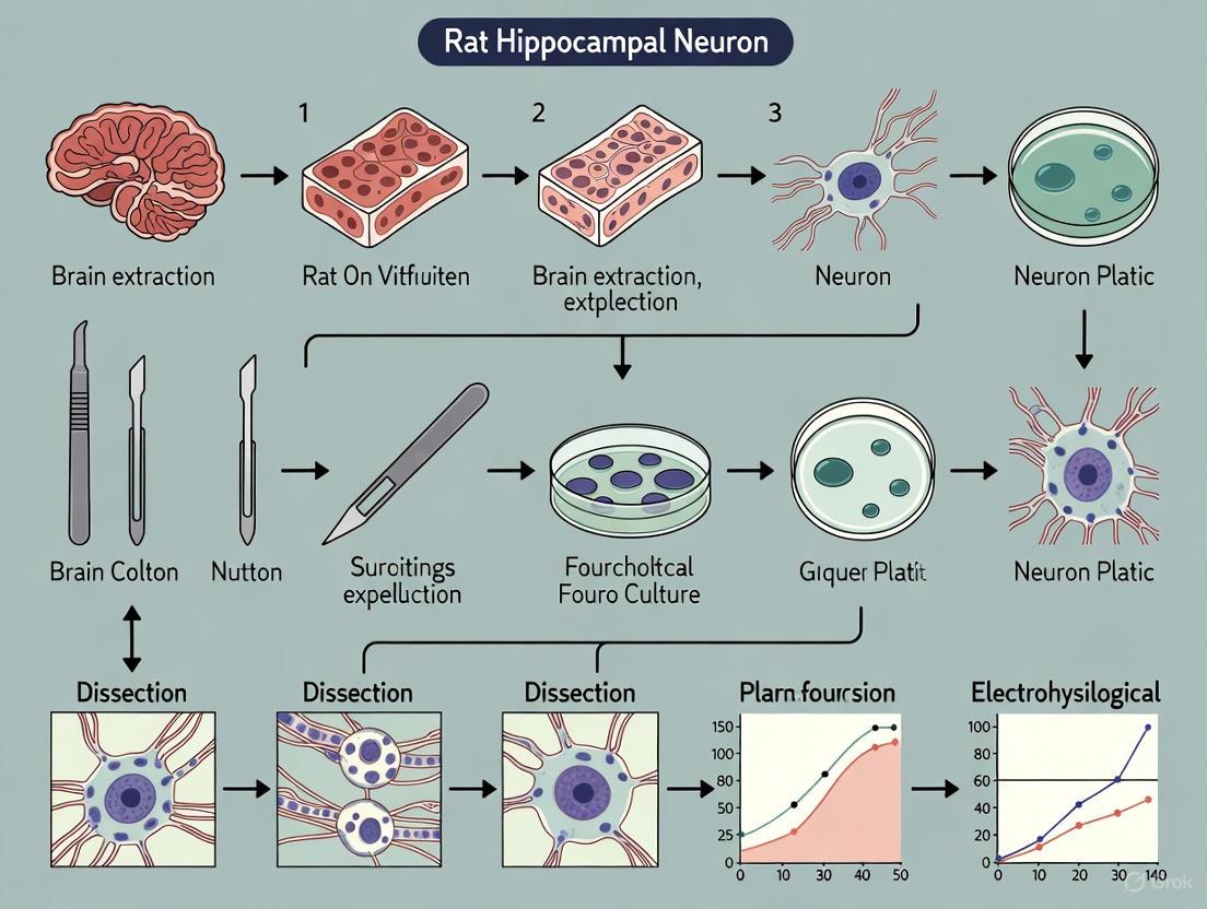
This article provides a comprehensive, step-by-step guide for the successful dissection and culture of primary rat hippocampal neurons, a cornerstone model for neuroscience and drug discovery research.
A Step-by-Step Guide to Rat Hippocampal Neuron Dissection and Culture: Optimized Protocols for Researchers
Abstract
This article provides a comprehensive, step-by-step guide for the successful dissection and culture of primary rat hippocampal neurons, a cornerstone model for neuroscience and drug discovery research. It covers fundamental neurobiology, detailed methodological protocols for both embryonic (E17-E18) and postnatal (P1-P2) tissue, common troubleshooting and optimization strategies, and essential validation techniques. Designed for researchers and drug development professionals, this guide integrates classic methods with recent adaptations to enhance neuronal yield, viability, and culture purity, supporting robust in vitro studies of neuronal function, development, and pathology.
Understanding the Hippocampus: Why it's a Premier Model for Neuronal Culture
The hippocampus, a structure located within the medial temporal lobe, plays an indispensable role in declarative learning and memory formation. As the brain's central hub for constructing cognitive maps and episodic memories, it enables navigation, contextual encoding, and the consolidation of long-term memories [1] [2]. Complex cognitive functions rely not on isolated brain regions but on synchronized network activity between the hippocampus and other areas such as the orbitofrontal cortex (OFC). During spatial learning, hippocampal CA1 and OFC exhibit synchronized neural rhythms, including theta (6-12 Hz) and gamma (30-90 Hz) oscillations, which are crucial for integrating path learning with reward acquisition [1]. Furthermore, hippocampal CA1 population activity is hypothesized to reflect internal predictive models that contain information about future events, allowing animals to use past experiences to guide future behavior [3] [4]. These models are formed through the integration of structured experiences, leading to more stable hippocampal neural ensembles that facilitate rapid learning of novel problems [3].
Disruptions in hippocampal function are implicated in a range of neurological disorders, including Alzheimer's disease, epilepsy, and intellectual disability disorders [2] [5]. Given its central role in cognition and pathology, the hippocampus represents a critical target for neuroscience research and therapeutic development. However, studying hippocampal processes in vivo presents significant challenges, including experimental accessibility, complex system interactions, and ethical constraints [6]. Therefore, establishing robust in vitro models of hippocampal neurons is essential for reducing experimental complexity and enabling detailed mechanistic studies of learning, memory, and neurodegenerative disease processes.
Key Hippocampal Functions and Rationale for Modeling
To effectively model hippocampal function in vitro, one must first understand the key cellular and network processes underlying its role in learning and memory. The following table summarizes core hippocampal functions and the corresponding rationale for their investigation using in vitro models.
Table 1: Core Hippocampal Functions and Rationale for In Vitro Modeling
| Hippocampal Function | Biological Basis | Rationale for In Vitro Modeling |
|---|---|---|
| Spatial Navigation & Cognitive Mapping | Activity of hippocampal place cells that fire at specific locations in an environment [7]. | Enables controlled study of cellular correlates of spatial memory and path integration mechanisms. |
| Memory Consolidation | Experience-dependent stabilization of place cell populations; increased CA1 representation stability across days [7]. | Permits investigation of synaptic and molecular changes during long-term memory formation. |
| Predictive Model Formation | Integration of structured experiences into stable CA1 ensemble activity patterns that predict solutions to novel problems [3] [4]. | Allows dissection of how neural ensembles organize new information relative to existing memories. |
| Inter-regional Synchronization | Theta-gamma phase-amplitude coupling between hippocampal CA1 and orbitofrontal cortex during goal-directed tasks [1]. | Facilitates analysis of network-level oscillations and synchrony in microcircuits. |
| Synaptic Plasticity | Behavioral timescale synaptic plasticity (BTSP) underlying formation and re-formation of place fields during learning [7]. | Provides a simplified system for studying fundamental plasticity mechanisms like LTP/LTD. |
Essential Reagents and Materials for Hippocampal Neuron Culture
Successful in vitro modeling of hippocampal function requires a carefully selected set of reagents and materials designed to maintain neuronal health, support process outgrowth, and recapitulate key aspects of the in vivo microenvironment. The following table catalogs essential components of the "Researcher's Toolkit" for hippocampal neuron dissection and culture.
Table 2: Research Reagent Solutions for Hippocampal Neuron Culture
| Reagent/Material | Function/Application | Example Specifications |
|---|---|---|
| Poly-D-Lysine (PDL) | Coats culture surfaces to promote neuronal adhesion. | 100 µg/mL in borate buffer [2] [8]. |
| Neurobasal/B27 Medium | Serum-free defined medium supporting long-term neuronal survival. | Neurobasal A supplemented with 2% B27 [2] [8]. |
| Papain | Proteolytic enzyme for gentle tissue dissociation. | 2 mg/mL in Hibernate-E or dissection medium [2]. |
| DNase | Degrades DNA released by damaged cells, reducing clumping. | Used in combination with papain [2]. |
| Hibernate-E/B27 Medium | Isotonic, low-temperature medium for tissue preservation during dissection. | Used for brain transport and storage [8]. |
| OptiPrep Density Gradient | Purifies neurons by removing cell debris, oligodendrocytes, and microglia. | Centrifugation at 800g for 15 minutes [2]. |
| Fibroblast Growth Factor 2 (FGF2) | Enhances neuronal viability and supports neurite regeneration in vitro. | 10 ng/mL in culture medium [2]. |
| L-Glutamine | Essential precursor for neurotransmitters and key metabolic substrate. | 0.5 mM in culture medium [2]. |
Protocols for Hippocampal Neuron Dissection and Culture
Adult Rat Hippocampal Neuron Isolation and Culture
This protocol adapts a method for the high-purity, high-viability isolation and long-term culture of adult rat hippocampal neurons, addressing the unique challenges posed by mature neuronal tissue [2].
Step-by-Step Workflow:
Preparation (3 days before dissection):
- Coat culture plates or coverslips with 100 µg/mL Poly-D-Lysine (PDL) solution overnight at room temperature.
- Aspirate PLL, rinse once with sterile, endotoxin-free water, and air-dry in a biosafety cabinet.
- One day before dissection, replace water with Neurobasal/B27 medium and incubate at 37°C.
Brain Dissection:
- Anesthetize an adult rat (e.g., 8-week-old Long-Evans or SD rat) deeply (e.g., sodium pentobarbital, 40 mg/kg, i.p.) [1] [2].
- Decapitate, disinfect the head with 70% ethanol, and rapidly open the cranium using cooled instruments.
- Gently remove the whole brain and place it in a chilled dish containing dissection medium (e.g., Hibernate-E/B27 or ice-cold HBSS). Complete this step within 3 minutes.
Hippocampal Isolation:
- Transfer the brain to a chilled dissection dish with a filter paper moistened with dissection medium.
- Using fine forceps, carefully separate the cerebral hemispheres and remove the meninges.
- Identify the hippocampi based on their distinctive curved structure beneath the cortex, and gently dissect them free. Complete this step within 1 minute per brain.
Tissue Digestion and Cell Dissociation:
- Slice the isolated hippocampi into thin sections.
- Incubate tissue slices with 2 mg/mL papain (with DNase) in dissection medium at 37°C for 30 minutes, agitating every 3 minutes.
- Terminate digestion by adding dissection medium with serum or inhibitors.
- Gently triturate the tissue 10-15 times with a fire-polished Pasteur pipette to create a single-cell suspension.
Neuron Purification:
- Layer the cell suspension onto a pre-prepared OptiPrep density gradient.
- Centrifuge at 800g for 15 minutes at 22°C.
- Carefully collect the neuron-enriched band (typically the fourth layer), transfer to a new tube, and wash with culture medium by centrifugation at 200g for 2 minutes.
Cell Seeding and Maintenance:
- Resuspend the purified neuronal pellet in complete culture medium (Neurobasal-A/B27 supplemented with 0.5 mM L-Glutamine and 10 ng/mL FGF2).
- Seed cells onto PDL-coated surfaces at a density of approximately ( 4.6 \times 10^4 ) cells/cm².
- Incubate at 37°C in 5% CO₂. Perform the first complete medium change at 4 hours post-seeding to remove non-adherent cells and debris.
- Thereafter, maintain cultures by replacing half the medium with fresh, pre-warmed medium every 2-3 days. For long-term cultures in flow-based systems, medium changes can be reduced to every 10 days [2].
Diagram 1: Workflow for Adult Rat Hippocampal Neuron Culture
Microfluidic 3D Hippocampal Network Construction
To better mimic the three-dimensional in vivo environment and study network-level properties, hippocampal neurons can be cultured within patterned microfluidic chips [6].
Step-by-Step Workflow:
Chip Fabrication and Preparation:
- Design a network-patterned microfluidic chip with microchannels (e.g., 100 µm width, 40 µm depth) using standard wet etching processes.
- Sterilize the chip and coat the channels with 100 µg/mL PLL for 30 minutes at 37°C.
Neuron Seeding in Microchannels:
- Isolate hippocampal neurons from neonatal SD rats (P0-P2) using the dissociation steps outlined in section 4.1.
- Prepare a cell suspension and seed 5,000-6,000 cells directly into the microfluidic chip's inlet.
- Allow cells to adhere within the microchannels for 4-6 hours before carefully adding maintenance medium.
Culture Maintenance and Monitoring:
- Replace half of the medium every 2 days.
- Monitor network formation over 7 days using immunofluorescence staining for neuronal markers (e.g., β-tubulin). Neurons should extend processes along the microchannels, forming a defined network.
Functional Validation:
- After 7 days in culture, assess network function using a Multi-Electrode Array (MEA) electrophysiology system.
- Detect and record spontaneous single-channel and multi-channel firing activity, indicating the development of synchronous network activity and functional connectivity [6].
In vitro models of hippocampal neurons, ranging from traditional dissociated cultures to advanced microfluidic 3D networks, provide indispensable tools for deciphering the cellular and molecular underpinnings of learning and memory. The protocols detailed herein enable researchers to probe the mechanisms of synaptic plasticity, network synchronization, and memory consolidation in a controlled reductionist environment. These models are particularly valuable for modeling neurological diseases, screening neuroactive compounds, and ultimately bridging the gap between molecular mechanisms and complex cognitive functions. By faithfully recreating key aspects of hippocampal circuitry in vitro, these methods will continue to drive discovery in neuroscience and therapeutic development.
The hippocampus contains a complex cellular architecture essential for learning, memory, and information processing. Its functionality arises from the precise interplay between principal excitatory neurons and local inhibitory interneurons. The major neuronal classes include pyramidal neurons, granule cells, and diverse interneuron populations, each possessing distinct morphological, molecular, and functional properties [9] [10] [11].
Table 1: Key Characteristics of Major Hippocampal Cell Types
| Cell Type | Major Subtypes / Classes | Key Molecular Markers | Primary Functions | Morphological Features |
|---|---|---|---|---|
| Pyramidal Neurons | AcD (Axon-carrying dendrite), Non-AcD [9] | c-Fos (activity-dependent) [9] | Spatial coding, memory formation, main excitatory output [9] | Apical and basal dendrites; axon from soma or basal dendrite [9] |
| Granule Cells (GCs) | Semilunar Granule Cells (SGCs) [10] | c-Fos (activity-dependent) [10] | Pattern separation, sparse encoding, memory engram formation [10] | Sparse dendrites; project to CA3 [10] |
| Interneurons | Parvalbumin (PV), Somatostatin (SST), Vasoactive Intestinal Peptide (VIP) [12] [11] | Parvalbumin, Somatostatin, VIP [12] [11] | Network inhibition, rhythm generation, control of excitation [11] | Diverse morphologies (e.g., basket, chandelier cells) [11] |
Pyramidal Neurons
Hippocampal pyramidal cells are the principal excitatory neurons, but they are not a uniform population. A key morphological distinction is based on the origin of the axon. Non-AcD cells have an axon emerging directly from the soma, while AcD cells (comprising ~50% of CA1 pyramidal neurons) have an axon originating from a basal dendrite [9]. This anatomical difference has significant functional consequences: the AcD largely evades perisomatic inhibition, creating a privileged input channel for action potential generation. Consequently, AcD cells are more strongly recruited during memory-related network oscillations characterized by strong inhibitory activity [9].
Granule Cells
Dentate gyrus granule cells (GCs) are the main excitatory neurons that receive cortical input via the perforant path and project to CA3. They are crucial for pattern separation, the process of making similar inputs more distinct [10]. A specialized subtype, semilunar granule cells (SGCs), constitutes about 3% of the DG projection neuron population [10]. SGCs are morphologically distinct from typical GCs, featuring a wider dendritic arbor, a greater soma width-to-length ratio, and more numerous primary dendrites. They also exhibit greater sustained firing and are disproportionately recruited into memory engrams, suggesting they possess unique excitability properties that bias their involvement in memory formation [10].
Interneurons
Interneurons are inhibitory neurons that form local circuits to control and coordinate the activity of principal cells. Their diversity is often categorized by molecular markers, which correlate with specific targeting patterns and functions [12] [11]:
- Parvalbumin (PV) Interneurons: Fast-spiking cells that primarily target the perisomatic region of pyramidal neurons, providing powerful, rapid inhibition critical for controlling spike timing and generating network oscillations [11].
- Somatostatin (SST) Interneurons: These often target the distal dendrites of pyramidal cells, regulating the integration of incoming synaptic signals [12] [11].
- Vasoactive Intestinal Peptide (VIP) Interneurons: Frequently target other interneurons, creating disinhibitory circuits that selectively enhance the activity of specific neuronal pathways [11]. Interneurons are generated in the ganglionic eminences and migrate long distances into the cortex during development, eventually integrating into circuits where they remain plastic throughout adulthood [12].
Protocols for Hippocampal Neuron Dissection and Culture
This section provides a detailed methodology for the dissection and culturing of primary hippocampal neurons from rats, a fundamental technique for investigating neuronal function and pathology in vitro [13] [14].
Reagent and Material Preparation
Table 2: Essential Reagents and Materials for Hippocampal Neuron Culture
| Category | Item | Function / Application |
|---|---|---|
| Culture Media & Supplements | Neurobasal or DME Medium | Base culture medium [13] [14] |
| N21-MAX Media Supplement / B-27 Supplement | Serum-free supplement to support neuronal growth and health [13] [14] | |
| L-Glutamine or GlutaMAX | Provides glutamine, essential for cell metabolism [13] [14] | |
| Antibiotic-Antimycotic (e.g., Penicillin/Streptomycin) | Prevents bacterial and fungal contamination [13] [14] | |
| Substrate Coating | Poly-D-Lysine / Poly-L-Lysine | Promotes neuronal adhesion to the culture surface [13] |
| Laminin | Enhances neurite outgrowth and cell adhesion [13] | |
| Dissection & Dissociation | Papain | Proteolytic enzyme for digesting postnatal tissue [13] |
| DNase I | Prevents cell clumping by digesting DNA released from damaged cells [13] | |
| Ovomucoid Protease Inhibitor | Halts enzymatic digestion to protect cell viability [13] | |
| Hanks' Balanced Salt Solution (HBSS) / PBS | Ionic and pH-balanced salt solution for tissue rinsing and dissection [13] [14] |
Step-by-Step Experimental Workflow
The following diagram illustrates the complete workflow for preparing primary hippocampal cultures, from plate coating to final plating of neurons.
Detailed Protocol
Coating and Preparation of Cell Culture Plates
Preparation of the culture plates should be done in a laminar flow cell culture hood [13].
- Poly-D-Lysine Coating: Dilute Poly-D-Lysine to 50 µg/mL in sterile dH₂O. Cover the well surfaces with the solution (e.g., 50 µL/well for a 96-well plate) and incubate for 1 hour in a 37°C, 5% CO₂ incubator. Aspirate the solution and wash the wells three times with sterile dH₂O. The plates can be sealed and stored at 2–8°C for up to two weeks at this stage [13].
- Laminin Coating: The day before harvesting hippocampi, dilute Laminin to 10 µg/mL in sterile PBS. Cover the Poly-D-Lysine-coated wells with the Laminin solution. Incubate the plates overnight at 2–8°C. On the day of dissection, aspirate the Laminin solution and wash the wells twice with sterile dH₂O. Aspirate all liquid before plating cells [13].
Dissection of Rat Hippocampi
All dissection tools should be sterilized via autoclaving. The initial dissection can be performed outside the hood, but all subsequent steps require aseptic technique within a laminar flow hood [13].
- Animal and Tissue Preparation: For embryonic neurons, asphyxiate a timed-pregnant rat (E17–E18) with CO₂ and perform a cesarean section to recover the embryos. Decapitate the embryos and place the heads in a dish with ice-cold PBS. For postnatal neurons (P1–P2), decapitate the pups directly [13] [14].
- Brain Extraction and Hippocampal Isolation: Place one head in a dissection dish with cold PBS. Under a dissecting microscope, use fine forceps and spring scissors to carefully cut through the skull and remove the whole brain. Transfer the brain to a new dish with cold PBS. Separate the cerebral hemispheres and remove the meninges. Locate the dark, C-shaped hippocampus in each hemisphere and remove it using spring scissors. Place the isolated hippocampi in a clean dish with cold PBS on ice [13] [14].
- Tissue Preparation: Using Vannas-Tübingen spring scissors, mince the hippocampi into small pieces (~2 mm²) [13].
Dissociation and Culture of Rat Hippocampal Neurons
From this point forward, all work must be conducted in a laminar flow hood [13].
- Tissue Dissociation:
- For Embryonic (E17-E18) Tissue: Transfer the tissue pieces to a 15 mL conical tube with 5 mL of DME or Neurobasal medium. Gently triturate the tissue ~10–15 times using a sterile, fire-polished Pasteur pipette until the solution appears homogenous. Proceed to Step 3 [13].
- For Postnatal (P1-P2) Tissue: Transfer the tissue to a 15 mL tube containing 5 mL of a pre-warmed enzymatic solution (20 U/mL Papain and 100 U/mL DNase I in EBSS). Incubate for 20–30 minutes in a 37°C incubator. After incubation, gently triturate the tissue with a fire-polished Pasteur pipette until homogenous [13].
- Cell Washing and Plating: Centrifuge the cell suspension at 200 × g for 5 minutes at room temperature. Decant the supernatant. For postnatal tissue, resuspend the pellet in 5 mL of EBSS containing Ovomucoid protease inhibitor (1 µg/mL) and centrifuge again at 200 × g for 4–6 minutes. Wash the cell pellet twice by resuspending in 10 mL of DME or Neurobasal medium, followed by centrifugation at 200 × g for 5 minutes [13].
- Cell Counting and Seeding: Resuspend the final cell pellet in pre-warmed culture media. Mix 10 µL of cell suspension with 10 µL of 0.4% Trypan blue and count the live (unstained) cells using a hemocytometer. Dilute the cell suspension to the desired seeding density with warmed culture media and plate the neurons onto the prepared, coated culture plates [13].
- Maintenance: Culture the neurons in a humidified 37°C incubator with 5% CO₂. Culture media may be partially refreshed every 5-7 days as needed [13].
Research Applications and Signaling Pathways
Primary hippocampal cultures and in vivo models enable researchers to investigate specific questions about neuronal function, plasticity, and response to injury.
Recruiting Pyramidal Cell Subtypes During Learning
Research using spatial learning tasks in mice has revealed that AcD and non-AcD pyramidal neurons in the CA1 region are differentially recruited during various stages of learning. This was quantified by analyzing the expression of the immediate early gene c-Fos, a marker of neuronal activity, in morphologically identified neurons at different training time points [9]. The findings suggest a dynamic and plastic involvement of distinct pyramidal cell subtypes in memory processes.
Granule Cell Spine Dynamics After Denervation
Organotypic slice cultures of the entorhinal cortex and hippocampus provide a powerful model to study how neurons respond to denervation, a consequence of brain injury. By transecting the entorhino-dentate projection and using time-lapse imaging of single, fluorescently labeled granule cells (GCs), researchers can visualize spine dynamics on denervated distal dendrites compared to non-denervated proximal dendrites [15]. While distal dendrites show an average spine loss of ~30% after denervation, individual GCs exhibit considerable heterogeneity in their responses, with some showing decreases and others increases in spine density [15].
The Scientist's Toolkit: Research Reagent Solutions
Table 3: Key Research Reagents for Cell-Type Specific Investigation
| Research Goal | Key Reagent / Tool | Specific Application |
|---|---|---|
| Labeling Active Ensembles | TRAP2 Mice [10] | Genetic labeling of neurons expressing the immediate early gene c-Fos during specific behaviors for engram identification. |
| Viral Tracing & Labeling | Adeno-associated virus (AAV) serotype 2 with hSyn promoter [15] | High-efficiency transduction of neurons for sparse or population-level expression of fluorescent reporters (e.g., tdTomato, EGFP). |
| Time-Lapse Imaging | Organotypic Slice Cultures (OTCs) [15] | Long-term, high-resolution imaging of dendritic spines and axonal structures in a preserved circuit environment. |
| Cell-Type Classification | Antibodies against Molecular Markers (e.g., Parvalbumin, Somatostatin) [11] | Immunohistochemical identification and visualization of specific interneuron subtypes in fixed tissue. |
| Enhancing Cell Culture | Recombinant Neurotrophic Factors (e.g., BDNF, IGF-I) [13] | Addition to culture media to enhance hippocampal neuron survival, growth, and synaptic development. |
The hippocampus, with its highly specialized cellular architecture and defined circuitry, is a critical focus for neuroscience research, particularly in the contexts of learning, memory, and neurodegenerative disease. Traditional bulk-tissue analysis often obscures the intricate molecular landscapes unique to its distinct subregions. This application note details how advanced spatial molecular mapping techniques are revolutionizing our understanding of hippocampal biology. We provide a validated, step-by-step protocol for the dissection and primary culture of rat hippocampal neurons, a foundational model for in vitro investigation. By integrating insights from spatial transcriptomics and proteomics with classical cell culture methodology, this guide empowers researchers to design more physiologically relevant studies for drug discovery and mechanistic research.
The hippocampus is not a uniform structure; it is composed of specialized subregions—including the dentate gyrus (DG), cornu ammonis (CA1-CA3), and subiculum—that form a tightly organized circuit essential for cognitive functions [16]. The molecular identity and function of cells within these subregions are dictated by their precise spatial location. Recent advances in spatially-resolved transcriptomics (SRT) and spatial proteomics have begun to map this complexity with unprecedented resolution.
Studies leveraging these technologies reveal that gene and protein expression patterns are exquisitely confined to specific hippocampal layers and subregions. For instance, spatial transcriptomics of the human hippocampus has identified distinct expression patterns for genes like PPFIA2 in the granule cell layer and PRKCG in pyramidal neuron layers [16]. Similarly, spatial proteomics in mouse models of traumatic brain injury (TBI) has uncovered subregion-specific protein abundance changes, such as the elevation of FN1 and LGALS3BP in the stratum moleculare, highlighting region-specific vulnerabilities [17] [18]. This spatial heterogeneity underscores the limitations of using homogenized tissue samples and emphasizes the need for targeted dissection and culture techniques that respect the innate architecture of the hippocampus.
Key Technologies for Spatial Molecular Mapping
The integration of high-resolution spatial mapping technologies provides a powerful framework for guiding targeted hippocampal research.
Spatially-Resolved Transcriptomics (SRT)
SRT techniques, such as the 10x Genomics Visium platform, allow for genome-wide expression profiling while retaining the histological context of tissue sections. A landmark 2024 study used SRT on human hippocampus to define 18 distinct spatial domains, identifying both known and novel marker genes for hippocampal subregions [16]. This approach enables the molecular delineation of areas that are difficult to distinguish by histology alone.
Spatial Proteomics
Spatial proteomics combines laser microdissection (LMD) with mass spectrometry to quantify protein abundance in specific tissue subregions. This is crucial for understanding post-translational events that transcriptomics cannot capture. A recent application in a mouse TBI model revealed dynamic, time-dependent protein alterations in specific hippocampal subregions, implicating disturbed glucose metabolism and activated cholesterol synthesis pathways in the recovery process [17] [18].
Automated Image Analysis for Neuronal Quantification
High-throughput, automated imaging and analysis pipelines are vital for quantifying neuronal distribution and morphology. Platforms combining whole-brain imaging systems like the Brain-wide Positioning System (BPS) with cell-localization algorithms such as NeuroGPS enable accurate stereological cell counting in three dimensions. This method surpasses traditional 2D counting, which is prone to error from cell overlap, and has been successfully used to map the brain-wide distribution of somatostatin-expressing neurons [19].
Protocol: Primary Rat Hippocampal Neuron Culture for Functional Studies
This protocol provides a method for culturing primary hippocampal neurons from embryonic (E17-E18) or postnatal (P1-P2) rats, creating a simplified in vitro system for investigating molecular mechanisms of neuronal development, synaptogenesis, and synaptic plasticity [20] [13].
Reagent and Material Preparation
- Coating Solutions: Prepare sterile solutions of Poly-D-Lysine (50 µg/mL in dH₂O) and Laminin I (10 µg/mL in PBS).
- Culture Media: Use high-glucose DME or Neurobasal medium, supplemented with a 1x solution like N21-MAX, 1x antibiotic-antimycotic, and 0.5 mM L-glutamine. Growth factors such as BDNF and IGF-I can be added to enhance culture health [13].
- Dissection Solution: Ice-cold, sterile Phosphate-Buffered Saline (PBS).
- Enzymatic Digestion Solution (for P1-P2 tissue): 20 U/mL Papain and 100 U/mL DNase I in EBSS.
Step-by-Step Procedure
A. Coating Culture Plates
- Cover the surface of culture plates with 50 µg/mL Poly-D-Lysine solution and incubate for 1 hour at 37°C.
- Aspirate the solution and wash the wells three times with sterile dH₂O.
- Cover the wells with 10 µg/mL Laminin I solution and incubate overnight at 2-8°C.
- Before use, aspirate the Laminin, wash twice with dH₂O, and aspirate completely [13].
B. Dissection and Hippocampal Isolation
- Euthanize and Decapitate: Asphyxiate a timed-pregnant rat (for E17-E18 embryos) or P1-P2 rat pups. Decapitate and place the heads in a dish of cold, sterile PBS.
- Remove the Brain: Using fine scissors and forceps, cut through the skull to expose and remove the whole brain. Place it in a fresh dish of cold PBS on ice.
- Separate Hemispheres: Under a dissecting microscope, separate the cerebral hemispheres along the median longitudinal fissure.
- Remove Meninges: Carefully peel away the meninges covering each hemisphere to reveal the hippocampal structure.
- Isolate Hippocampus: Identify the dark, C-shaped hippocampus on the mid-sagittal side of the brain. Using fine spring scissors, carefully free it from the surrounding tissue and transfer it to a new dish with cold PBS.
- Cut Tissue: Mince the isolated hippocampi into small pieces (~2 mm²) [13].
C. Tissue Dissociation and Plating
- For Embryonic (E17-E18) Tissue: Transfer the tissue pieces to a 15 mL tube with 5 mL of DME medium. Gently triturate 10-15 times using a fire-polished Pasteur pipette until the solution is homogenous [13].
- For Postnatal (P1-P2) Tissue:
- Transfer tissue to a tube containing pre-warmed enzyme solution (Papain/DNase I).
- Incubate for 20-30 minutes at 37°C.
- Gently triturate the tissue 10-15 times with a fire-polished Pasteur pipette [13].
- Centrifuge and Wash: Centrifuge the cell suspension at 200 × g for 5 minutes. Decant the supernatant.
- For postnatal tissue, resuspend the pellet in EBSS with an ovomucoid protease inhibitor and centrifuge again before proceeding.
- Wash the cells twice by resuspending in 10 mL of DME medium and centrifuging at 200 × g for 5 minutes.
- Resuspend and Count: Resuspend the final cell pellet in pre-warmed culture media. Mix 10 µL of cell suspension with 10 µL of 0.4% Trypan blue and count live cells using a hemocytometer.
- Plate Cells: Dilute the cell suspension to the desired density and plate onto the pre-coated culture plates. Maintain cultures in a 37°C, 5% CO₂ humidified incubator [13].
Seeding Densities for Different Culture Formats
Table 1: Recommended cell seeding densities for various culture vessels. Densities are based on protocols from R&D Systems [13].
| Culture Vessel | Surface Area | Seeding Density | Recommended Media Volume |
|---|---|---|---|
| 96-well plate | 0.3 cm² | 50,000 - 100,000 cells/well | 50 - 100 µL |
| 24-well plate | 2.0 cm² | 250,000 - 500,000 cells/well | 0.5 mL |
| 12-well plate | 4.0 cm² | 500,000 - 1,000,000 cells/well | 1 mL |
| 35 mm dish | 10.0 cm² | 1,500,000 - 2,500,000 cells/dish | 2 mL |
The Scientist's Toolkit: Essential Research Reagents
Table 2: Key reagents and their functions in primary hippocampal neuron culture and analysis.
| Reagent / Material | Function / Application | Example |
|---|---|---|
| Poly-D-Lysine | Synthetic polymer that coats culture surfaces to promote neuronal attachment. | Cultrex Poly-D-Lysine [13] |
| Laminin | Extracellular matrix protein that supports neurite outgrowth and cell differentiation. | Cultrex Mouse Laminin I [13] |
| Papain / DNase I | Enzyme combination used to digest extracellular matrix for tissue dissociation (critical for postnatal tissue). | Worthington Biochemical Corp. [13] |
| N21-MAX Supplement | A defined, serum-free supplement providing essential factors for long-term neuronal survival and growth. | R&D Systems [13] |
| FM Dyes (e.g., FM 1-43) | Styryl dyes that label recycling synaptic vesicles; used to study presynaptic function and plasticity. | [21] |
| Spatial Transcriptomics Kit | For genome-wide expression profiling while retaining spatial location in tissue sections. | 10x Genomics Visium [16] |
Data Integration: From Spatial Maps to Functional Insights
Integrating data from spatial mapping studies directly informs the design and interpretation of experiments using cultured neurons. The following workflow illustrates how a research program can bridge in vivo spatial data with in vitro functional validation.
For example, spatial proteomics identified MUG-1 and CD44 as upregulated across hippocampal subregions after TBI, suggesting shared molecular responses to injury [18]. Researchers can use this discovery to design functional studies in cultured hippocampal neurons, perhaps by overexpressing these proteins and using FM dye-based assays to quantify their impact on synaptic vesicle recycling [21]. This direct pipeline from spatial discovery to functional validation in a controlled culture system accelerates the identification of novel therapeutic targets.
Furthermore, more complex 3D co-culture models are emerging. A recent 2025 study detailed a three-dimensional compartmentalized system co-culturing basal forebrain cholinergic neurons (BFCNs) with primary hippocampal neurons. This model successfully recapitulated long-range cholinergic axon projection and age-dependent neuronal degeneration, providing a highly physiological platform for studying circuit-level mechanisms of neurodegeneration [22].
Spatial molecular mapping technologies are unveiling the intricate subregion-specific landscape of the hippocampus, providing a new depth of insight for neuroscience research. The protocol for primary hippocampal neuron culture, a cornerstone of in vitro neuroscience, provides a reductionist but powerful system to functionally validate discoveries from these spatial maps. By combining the guidance of in vivo spatial data with the controllability of in vitro culture models, researchers can deconstruct the complex molecular and cellular mechanisms of hippocampal function and pathology with greater precision, ultimately accelerating the drug discovery process for neurological and psychiatric disorders.
The isolation and culture of primary neurons from specific regions of the nervous system represent fundamental techniques in neuroscience research, enabling the investigation of neuronal function, development, and pathology in a controlled in vitro environment [14]. These region-specific cultures provide physiologically relevant data that closely mimic the in vivo environment, offering broad applicability for studying neurodegenerative disorders, pathological mechanisms, and therapeutic strategies [14]. The ability to explore complex intercellular interactions, including neuron-neuron connections, neuron-glial cell relationships, and synapse formation, makes primary cultured neurons an invaluable tool for experimental observation and analysis [14].
This application note provides a comprehensive comparison of three essential neuronal culture models: hippocampus and cortex as central nervous system (CNS) representatives, and dorsal root ganglia (DRG) as a peripheral nervous system (PNS) model. Each model possesses distinct characteristics and experimental advantages, requiring specialized methodologies for optimal results. The hippocampus is particularly valuable for studies of learning, memory, and synaptic plasticity, while cortical neurons offer insights into complex information processing, and DRG neurons serve as excellent models for sensory transduction and pain research [14]. The protocols outlined herein have been optimized to address the unique properties of each tissue type, focusing on key steps to enhance neuronal yield and viability while minimizing contamination with non-neuronal cells.
Comparative Analysis of Neuronal Culture Models
Table 1: Key Characteristics of Regional Neuronal Culture Models
| Parameter | Hippocampal Neurons | Cortical Neurons | DRG Neurons |
|---|---|---|---|
| Nervous System Region | Central Nervous System (CNS) | Central Nervous System (CNS) | Peripheral Nervous System (PNS) |
| Primary Functions | Learning, memory, spatial navigation | Complex information processing, integration | Sensory transduction, pain perception |
| Optimal Isolation Age | Embryonic Day 17-18 (E17-E18) or Postnatal Day 1-2 (P1-P2) [13] [14] | Embryonic Day 17-18 (E17-E18) [14] | Young adult (6-week-old) [14] |
| Major Neuronal Types | Pyramidal neurons, interneurons [13] | Glutamatergic projection neurons, GABAergic interneurons | Pseudounipolar sensory neurons |
| Glial Contamination Concerns | Moderate | Moderate | Low with proper dissection |
| Synapse Development | Forms substantial synaptic connections with dendritic spines [13] | Robust synaptogenesis | Specialized synaptic arrangements |
| Typical Applications | Synaptic plasticity, mechanisms of neurobiology [13] | Neurodevelopment, neurodegeneration research | Pain mechanisms, sensory biology, axon regeneration |
Table 2: Culture Medium Composition for Different Neuronal Types
| Component | Hippocampal Neurons | Cortical Neurons | DRG Neurons |
|---|---|---|---|
| Base Medium | Neurobasal or DME medium [13] | Neurobasal Plus Medium [14] | F-12 Medium [14] |
| Serum | Serum-free conditions [13] | Serum-free conditions | 10% Fetal Bovine Serum (FBS) [14] |
| Supplement | N21-MAX or B-27 [13] [14] | B-27 Supplement [14] | NGF (20 ng/mL) [14] |
| Antibiotics | Antibiotic-antimycotic [13] | Penicillin-Streptomycin [14] | Penicillin-Streptomycin [14] |
| Glutamine Source | L-glutamine or GlutaMAX [13] [14] | GlutaMAX [14] | Included in F-12 formulation |
| Growth Factors | Optional: BDNF, IGF-I [13] | Not typically added | Essential: Nerve Growth Factor (NGF) [14] |
Detailed Experimental Protocols
Substrate Coating Protocol
Proper substrate coating is essential for neuronal attachment, survival, and maturation across all neuronal culture types. The following procedure details the preparation of coated culture surfaces suitable for hippocampal, cortical, and DRG neurons.
- Poly-D-Lysine Coating Solution Preparation: Dilute Poly-D-Lysine solution with sterile distilled water to a final concentration of 50 µg/mL [13]. Alternatively, Poly-L-Lysine at 1 mg/mL can be used for mouse hippocampal neurons [23].
- Surface Coating Application: Cover the wells of culture plates with the diluted Poly-D-Lysine solution (e.g., 50 µL/well for a 96-well plate) [13]. Tilt the plates to ensure even coating of the entire surface area.
- Incubation and Washing: Incubate plates for 1 hour in a 37°C, 5% CO₂ humidified incubator [13]. After incubation, aspirate the solution and wash the wells three times with sterile distilled water to remove excess Poly-D-Lysine.
- Laminin Coating: Dilute Laminin solution with sterile PBS to a final concentration of 10 µg/mL [13]. Cover the Poly-D-Lysine-coated surfaces with the Laminin solution and incubate overnight at 2-8°C.
- Final Preparation: Aspirate the Laminin solution and wash twice with sterile distilled water before use [13]. Prepared plates can be sealed and stored at 2-8°C for up to two weeks.
For DRG neurons, additional extracellular matrix components may be beneficial, such as collagen coating, which can be prepared by combining Poly-D-Lysine (0.5 mg/mL stock), acetic acid (17 mM stock), and rat tail collagen (3 mg/mL stock) in specific ratios [24].
Hippocampal Neuron Isolation and Culture
Dissection of Rat Hippocampi
- Animal Preparation and Brain Extraction: Asphyxiate the pregnant rat with CO₂ and recover embryos via cesarean section using large surgical scissors and curved dissecting forceps [13]. Place the embryos in a 100 mm petri dish containing cold PBS and keep on ice. Remove embryos from their individual placenta sacs and wash with cold PBS. Decapitate each embryo at the head/neck junction using small surgical scissors. For postnatal (P1-P2) pups, decapitate directly with small surgical scissors [13].
- Brain Dissection and Hippocampal Isolation: Place the heads in a new 60 mm petri dish containing cold PBS. Stabilize the head using curved forceps and fine forceps, then cut through the skull with small surgical scissors, keeping cuts shallow to avoid damaging brain tissue [13]. Peel back the separated skull halves and remove the whole brain using curved forceps, placing it in a 60 mm petri dish with cold PBS on ice. Under a dissecting microscope, separate the cerebral hemispheres by cutting along the median longitudinal fissure with spring scissors, discarding any brain stem tissue [13].
- Hippocampal Exposure and Removal: Peel off the meninges covering each hemisphere using fine forceps and open the brain to reveal the mid-sagittal side [13]. Locate the hippocampus, which appears as a darker, c-shaped region, and remove it using spring scissors. Place the hippocampal tissue in a new 60 mm petri dish with cold PBS on ice. Cut the isolated hippocampi into smaller pieces (~2 mm²) using spring scissors to prepare for dissociation [13].
Dissociation and Plating
- Enzymatic Digestion (for Postnatal Tissue): For P1-P2 hippocampal tissue, prepare an enzyme solution containing 20 U/mL Papain and 100 U/mL DNase I in 5 mL of EBSS [13]. Warm the solution in a 37°C, 5% CO₂ incubator for 10 minutes, then transfer the tissue pieces to the enzyme solution and incubate for 20-30 minutes.
- Mechanical Dissociation: For embryonic hippocampi, transfer tissue pieces to a 15 mL conical tube with 5 mL of DME or Neurobasal medium [13]. Gently triturate the tissue with a fire-polished Pasteur pipette until the solution becomes homogeneous (approximately 10-15 times). For enzymatically digested tissue, perform trituration after the digestion step.
- Cell Washing and Counting: Centrifuge the cell suspension at 200 × g for 5 minutes at room temperature and decant the supernatant [13]. Resuspend cells in appropriate medium (10 mL of DME/Neurobasal for embryonic; 5 mL of EBSS with ovomucoid protease inhibitor for postnatal). Centrifuge again at 200 × g for 4-6 minutes, then wash cells twice with 10 mL of DME or Neurobasal medium. Resuspend the final cell pellet in warmed culture media and count live cells using trypan blue exclusion method [13].
- Plating and Maintenance: Dilute the cell suspension to the desired seeding density with warmed culture media and plate onto prepared culture plates [13]. Maintain cultures in a 37°C, 5% CO₂ humidified incubator, with periodic media changes as required. For long-term cultures, consider adding cytosine arabinoside (Ara-C) at 1-4 μM to inhibit glial cell proliferation after 2-4 days in vitro [25] [24].
Cortical Neuron Isolation and Culture
The protocol for cortical neuron isolation shares similarities with hippocampal isolation but requires attention to specific regional distinctions.
- Cortex Dissection: Follow the same initial steps for embryo extraction and brain removal as described for hippocampal dissection [14]. After removing the whole brain and separating the hemispheres, carefully remove the meninges. Position the hemispheres with the inner surface facing up and identify the cortical tissue, which constitutes the majority of the cerebral hemisphere. Carefully isolate the cortical tissue, avoiding inclusion of hippocampal or striatal structures.
- Tissue Dissociation and Plating: The dissociation process for cortical tissue is essentially identical to that described for hippocampal tissue, utilizing either enzymatic digestion or mechanical trituration based on developmental stage [14]. Plate cortical neurons at appropriate densities on coated surfaces and maintain in Neurobasal-based medium supplemented with B-27 and GlutaMAX [14].
Dorsal Root Ganglia Neuron Isolation and Culture
DRG neuron culture requires distinct approaches due to their peripheral location and unique biological characteristics.
- DRG Dissection: Sacrifice young adult rats (6-week-old) according to approved ethical guidelines [14]. Make a midline incision along the back and expose the vertebral column. Carefully open the vertebral canal to reveal the spinal cord with attached DRG. Identify DRG located in the intervertebral foramina and gently remove them using fine forceps and spring scissors. Remove surrounding connective tissue and place cleaned DRG in cold HBSS.
- Enzymatic and Mechanical Dissociation: Transfer DRG to a solution of 0.5% trypsin and 0.2% EDTA and incubate for 15-20 minutes at 37°C [14]. After enzymatic digestion, triturate the tissue with a fire-polished Pasteur pipette to achieve a single-cell suspension. Centrifuge at 200 × g for 5 minutes and resuspend in DRG-specific culture medium.
- DRG-Specific Culture Conditions: Plate DRG neurons on coated surfaces and maintain in F-12 medium supplemented with 10% FBS, penicillin-streptomycin, and 20 ng/mL nerve growth factor (NGF), which is essential for DRG neuron survival and maturation [14].
The Scientist's Toolkit: Essential Research Reagents
Table 3: Key Research Reagent Solutions for Neuronal Culture
| Reagent | Function | Application Notes |
|---|---|---|
| Poly-D-Lysine | Promotes neuronal attachment to plastic/glass surfaces [13] | Use at 50 µg/mL; requires water washes before use [13] |
| Laminin | Enhances neurite outgrowth and neuronal differentiation [13] | Use at 10 µg/mL in PBS; coat after poly-D-lysine [13] |
| Neurobasal Medium | Optimized base medium for CNS neurons [13] [14] [24] | Supports long-term survival with minimal glial growth |
| B-27 Supplement | Serum-free supplement for neuronal viability [14] [24] | Essential for hippocampal and cortical cultures; used at 1× concentration |
| Nerve Growth Factor (NGF) | Critical for DRG neuron survival and neurite outgrowth [14] | Use at 20 ng/mL for DRG cultures; not typically needed for CNS neurons |
| Papain/DNase I | Enzyme system for tissue dissociation [13] | Particularly important for postnatal tissue digestion |
| Cytosine Arabinoside (Ara-C) | Inhibits glial cell proliferation [25] [24] | Add at 1-4 μM after 2-4 days in vitro [25] [24] |
| GlutaMAX | Stable dipeptide form of L-glutamine [14] | Prevents glutamate accumulation and ammonia toxicity |
Experimental Workflow and Quality Control
Diagram 1: Primary Neuronal Culture Workflow. This flowchart illustrates the key steps in establishing primary neuronal cultures, with quality control checkpoints to ensure experimental reliability.
Troubleshooting and Optimization Strategies
Successful neuronal culture requires attention to potential pitfalls and implementation of optimized practices based on the specific neuronal population being studied.
- Low Cell Viability: If viability falls below 80%, consider reducing enzymatic digestion time, ensuring proper temperature control during dissection, and using ice-cold solutions throughout the dissection process [13] [14]. For DRG neurons from adult animals, more gentle digestion protocols may be necessary.
- Poor Neuronal Attachment: Inadequate substrate coating is a common cause of attachment issues [13]. Ensure proper preparation and washing of Poly-D-Lysine and Laminin coatings. Verify that coating solutions have not expired and are prepared at correct concentrations.
- Excessive Glial Contamination: The addition of anti-mitotic agents such as cytosine arabinoside (Ara-C) at 1-4 μM after 2-4 days in vitro can significantly reduce glial overgrowth without affecting neuronal health [25] [24]. For more pure neuronal cultures, immunopanning or density gradient separation methods may be employed.
- Inadequate Neurite Outgrowth: Optimize concentration of growth factors and coating substrates [13] [14]. For CNS neurons, ensure proper B-27 supplementation; for DRG neurons, verify NGF concentration and activity. Check that culture conditions (temperature, CO₂, humidity) are properly maintained.
- Regional Cross-Contamination: During dissection of hippocampal and cortical tissues, clearly identify anatomical landmarks to ensure tissue-specific isolation [13] [14]. Practice precise dissection techniques under high-quality microscopy to avoid inclusion of adjacent brain regions.
The selection of appropriate neuronal culture models—hippocampal, cortical, or DRG neurons—depends on specific research questions and experimental requirements. Each model offers unique advantages and requires specialized methodologies for optimal results. Hippocampal cultures provide excellent systems for studying synaptic plasticity and mechanisms underlying learning and memory [13]. Cortical neurons enable investigations of complex neural networks and information processing, while DRG cultures serve as valuable models for sensory biology and pain research [14].
The protocols detailed in this application note provide robust, reproducible methods for generating high-quality neuronal cultures from these distinct regions. By adhering to these optimized procedures and implementing appropriate quality control measures, researchers can establish reliable in vitro models for studying neuronal function, dysfunction, and therapeutic interventions across central and peripheral nervous system domains. These region-specific approaches enhance the physiological relevance of in vitro findings and facilitate more accurate translations to in vivo applications.
The isolation and culture of primary hippocampal neurons is a cornerstone technique in neuroscience, providing a fundamental model for investigating the cellular mechanisms of neurobiology, synaptic function, and the pathophysiology of neurological disorders [13] [26]. A critical first step in designing these studies is selecting the most appropriate neuronal source. The developmental age at which neurons are harvested—embryonic or postnatal—profoundly influences the cellular composition, physiological properties, and experimental outcomes of the culture system. This application note provides a structured comparison between embryonic (E17–E18) and postnatal (P1–P2) rat hippocampal neurons, offering detailed protocols and data-driven guidance to help researchers align their source selection with specific research objectives.
Section 1: Source Comparison and Key Considerations
The choice between embryonic and postnatal sources dictates the experimental timeline, the maturity of synaptic networks, and the complexity of the required isolation protocol. The following table summarizes the core quantitative and qualitative differences to inform this decision.
Table 1: Comparative Analysis of Embryonic (E17-E18) and Postnatal (P1-P2) Rat Hippocampal Neurons for Culture
| Feature | Embryonic (E17–E18) | Postnatal (P1–P2) |
|---|---|---|
| Developmental Stage | Peak of hippocampal neurogenesis [27] | Major phase of synaptogenesis and network formation [27] |
| Dissociation Protocol | Purely mechanical trituration [13] | Enzymatic (Papain) + mechanical trituration [13] [14] |
| Typical Cell Yield | High | Moderate [28] |
| Culture Purity & Glial Presence | Lower inherent glial contamination; can be maintained in serum-free conditions to minimize glial proliferation [26] | Higher inherent glial contamination; requires strategies like antimitotics to control glial overgrowth [26] |
| In Vitro Development Timeline | Synapses appear over days 4-7 in culture; mature networks form over weeks [26] | Neurons are more mature at plating; can form functional synapses more rapidly |
| Key Advantages | - Simplified, faster dissection and dissociation [13]- Superior viability and robustness to dissociation stress- High suitability for long-term studies of synaptogenesis and network development | - More mature synaptic physiology at the time of plating- May better model certain postnatal neurological diseases |
| Key Disadvantages | - Neurons are developmentally immature- Requires timed-pregnant dams, which involves more complex animal logistics | - Requires enzymatic digestion, adding complexity and potential toxicity [13]- Lower cell yield and viability due to extensive existing connections [28] |
| Ideal Research Applications | - Studies of neuronal polarization, axon/dendrite development, and synaptogenesis- Long-term culture (>2 weeks)- High-throughput screening | - Studies of synaptic function, plasticity, and mature network activity |
To visually guide the decision-making process, the following workflow diagram outlines the key selection criteria.
Section 2: Detailed Experimental Protocols
Protocol A: Culturing Embryonic (E17–E18) Rat Hippocampal Neurons
This protocol is optimized for the isolation of hippocampal neurons from E17–E18 rat embryos, leveraging mechanical dissociation for high cell viability [13] [26].
Reagents and Materials
Table 2: Essential Reagents and Materials for Embryonic Neuron Culture
| Item | Function | Example Catalog Number |
|---|---|---|
| Cultrex Poly-D-Lysine | Synthetic coating substrate for plate attachment | R&D Systems, #3439-200-01 [13] |
| Cultrex Mouse Laminin I | Natural extracellular matrix protein coating for neurite outgrowth | R&D Systems, #3400-010-02 [13] |
| Neurobasal Medium | Base serum-free medium optimized for neuronal health | ThermoFisher Scientific, #21103049 [13] |
| B-27 Supplement | Serum-free supplement providing essential hormones and nutrients | Not specified in sources |
| L-Glutamine | Essential amino acid for neuronal metabolism | Irvine Scientific, #9317 [13] |
| Antibiotic-Antimycotic | Prevents bacterial and fungal contamination | ThermoFisher Scientific, #15240062 [13] |
| Fire-polished Pasteur Pipette | Gentle trituration of tissue with minimal cell damage | Sterile [13] |
Step-by-Step Procedure
Coating Culture Plates (Day Before Dissection)
- Dilute and Apply Poly-D-Lysine: Dilute stock Poly-D-Lysine to 50 µg/mL in sterile dH₂O. Cover the entire surface of the culture plate (e.g., 50 µL/well for a 96-well plate) and incubate for 1 hour at 37°C [13].
- Wash and Seal: Aspirate the solution and wash the wells three times with sterile dH₂O. Aspirate completely, seal the plate with Parafilm, and store at 2–8°C for up to two weeks [13].
- Coat with Laminin: The day before dissection, dilute Laminin to 10 µg/mL in sterile PBS. Cover the Poly-D-Lysine-coated surfaces and incubate overnight at 2–8°C. Before use, aspirate the Laminin and wash twice with sterile dH₂O [13].
Dissection and Dissociation
- Dissect Hippocampi: Euthanize a timed-pregnant rat at E17–E18 according to approved animal protocols. Isolate embryos and decapitate. Dissect brains in cold PBS under a dissecting microscope. Isolate hippocampi from cerebral hemispheres, remove meninges, and collect tissue in cold PBS on ice [13] [14].
- Mechanically Dissociate Tissue: Transfer hippocampal pieces to a 15 mL tube containing 5 mL of DME or Neurobasal medium. Gently triturate the tissue 10–15 times using a fire-polished Pasteur pipette until the solution appears homogenous [13].
- Wash and Resuspend: Centrifuge the cell suspension at 200 × g for 5 minutes at room temperature. Decant the supernatant, resuspend the cell pellet in 10 mL of fresh DME or Neurobasal medium, and repeat the centrifugation step [13].
- Plate Cells: Resuspend the final pellet in pre-warmed complete culture medium (e.g., Neurobasal medium supplemented with B-27, L-Glutamine, and antibiotic-antimycotic). Count live cells using Trypan Blue exclusion and plate at the desired density on the pre-coated plates [13] [26].
The following diagram illustrates the complete experimental timeline for this protocol.
Protocol B: Culturing Postnatal (P1–P2) Rat Hippocampal Neurons
This protocol is customized for P1–P2 pups, requiring enzymatic digestion to dissociate the more established extracellular matrix and synaptic connections of postnatal tissue [13] [14].
Additional Reagents for Postnatal Protocol
- Papain: Proteolytic enzyme for tissue dissociation (Worthington Biochemical Corp., #LK003176) [13].
- DNase I: Prevents cell clumping by digesting DNA released from damaged cells (Worthington Biochemical Corp., #LK003170) [13].
- Ovomucoid Protease Inhibitor: Neutralizes papain activity after digestion to prevent over-digestion (Worthington Biochemical Corp., #LK003182) [13].
Step-by-Step Procedure
Coating Culture Plates
- The coating procedure is identical to the embryonic protocol (Section 2.1.2).
Dissection and Enzymatic Dissociation
- Dissect Hippocampi: Anesthetize and decapitate P1–P2 rat pups. Dissect and collect hippocampi in cold PBS on ice, as in the embryonic protocol [13].
- Prepare Enzyme Solution: Mix 20 U/mL Papain and 100 U/mL DNase I in 5 mL of EBSS. Warm the solution for 10 minutes in a 37°C, 5% CO₂ incubator [13].
- Digest Tissue: Transfer the hippocampal tissue to the warmed enzyme solution. Incubate for 20–30 minutes in the 37°C incubator [13].
- Triturate and Wash: Gently triturate the digested tissue 10–15 times with a fire-polished Pasteur pipette. Centrifuge at 200 × g for 5 minutes. Decant the supernatant [13].
- Inactivate Enzymes: Resuspend the cell pellet in 5 mL of EBSS containing 1 µg/mL Ovomucoid protease inhibitor. Centrifuge again at 200 × g for 4–6 minutes [13].
- Final Wash and Plate: Wash the cells twice by resuspending in 10 mL of DME or Neurobasal medium and centrifuging at 200 × g for 5 minutes. Resuspend the final pellet in complete culture medium, count cells, and plate on pre-coated plates [13].
Section 3: The Scientist's Toolkit
Table 3: Research Reagent Solutions for Hippocampal Neuron Culture
| Research Reagent | Function in Protocol | Specific Application Note |
|---|---|---|
| Poly-D-Lysine | Synthetic polymer that provides a positively charged surface for neuronal attachment. | Essential for both embryonic and postnatal protocols. Use at 50 µg/mL for coating [13]. |
| Laminin | Natural extracellular matrix protein that promotes neurite outgrowth and neuronal survival. | Applied after Poly-D-Lysine coating at 10 µg/mL to enhance long-term health and process formation [13]. |
| Neurobasal Medium / B-27 | Defined, serum-free system that supports neuronal growth while suppressing glial proliferation. | The gold standard for long-term hippocampal cultures. B-27 provides antioxidants, hormones, and essential nutrients [26]. |
| Papain / DNase I | Enzyme combination for digesting extracellular matrix in more mature tissue. | Critical for dissociating postnatal (P1-P2) tissue. DNase I reduces viscosity from released DNA, improving cell yield [13] [14]. |
| Recombinant Neurotrophic Factors (BDNF, IGF-I) | Enhance neuronal survival, synaptogenesis, and overall health of the culture. | Optional additives. For example, BDNF can be added to the culture medium to further support synaptic development and plasticity [13]. |
The decision to use embryonic or postnatal rat hippocampal neurons is fundamental and should be driven by the specific biological question under investigation. Embryonic (E17–E18) neurons, with their simpler isolation and high viability, are the preferred choice for most applications, particularly studies of de novo neuronal development, synaptogenesis, and for experiments requiring long-term culture or high cell yield. Postnatal (P1–P2) neurons, while more technically challenging to isolate, offer a model of more mature circuitry and are potentially better suited for investigating the physiology of established synapses or modeling certain postnatal neurological conditions. By applying the comparative data, detailed protocols, and decision framework provided herein, researchers can make an informed, strategic choice that will significantly enhance the rigor and reproducibility of their neuroscience research.
A Detailed Protocol: From Dissection to Mature Hippocampal Cultures
Essential Reagents and Sterilized Tools Checklist
The isolation and culture of primary rat hippocampal neurons is a fundamental technique in neuroscience research, providing a physiologically relevant in vitro model for studying neuronal development, synaptogenesis, and the mechanisms underlying neurological disorders [14]. This application note provides a detailed, step-by-step guide for the dissection and culture of rat hippocampal neurons, with a specific focus on the essential reagents and sterilized tools required for success. The protocol is designed to support researchers, scientists, and drug development professionals in establishing robust and reproducible neuronal cultures, which are critical for generating reliable data in fields ranging from basic neurobiology to preclinical drug screening [14].
Essential Reagents and Solutions
A successful neuronal culture begins with the preparation of high-quality, properly formulated reagents. Using validated reagents from reputable suppliers is crucial for maintaining consistency and ensuring the health of the primary neurons [29]. The following table summarizes the core reagents required for the dissection, dissociation, and culture of rat hippocampal neurons.
Table 1: Essential Reagents for Rat Hippocampal Neuron Culture
| Reagent Category | Specific Reagents | Function and Application |
|---|---|---|
| Basal Media | Neurobasal Medium, DME (high glucose, no L-glutamine) [13] [30] | Serves as the nutrient foundation for culturing and maintaining neurons. |
| Media Supplements | B-27 Supplement, N21-MAX Media Supplement, L-Glutamine (200 mM), Antibiotic-Antimycotic (100X) [13] [14] [30] | Provides essential hormones, antioxidants, and nutrients for neuronal survival and growth; prevents contamination. |
| Dissection & Dissociation | Hanks' Balanced Salt Solution (HBSS) or Dulbecco's Phosphate-Buffered Saline (DPBS), Papain, DNase I, Ovomucoid Protease Inhibitor [13] [14] [31] | Provides an ionic and pH-balanced environment for tissue dissection; enzymatically digests tissue to dissociate individual cells. |
| Substrate Coating | Poly-D-Lysine or Poly-L-Lysine, Mouse Laminin I [13] [31] [30] | Coats culture surfaces to promote neuronal attachment and neurite outgrowth. |
| Optional Growth Factors | Recombinant Human BDNF, Recombinant Human IGF-I [13] | Enhances neuronal survival, maturation, and synaptic plasticity when added to the culture medium. |
Reagent Preparation Notes
- Coating Solutions: Dilute Poly-D-Lysine to a final concentration of 50 µg/mL in sterile distilled water. Dilute Laminin to 10 µg/mL in sterile PBS immediately before use [13].
- Complete Culture Medium: For hippocampal neurons, Neurobasal medium should be supplemented with 1x B-27, 0.5 mM L-Glutamine, and 1x antibiotic-antimycotic [14] [30]. For embryonic neuronal plating, the addition of 25 µM glutamic acid is recommended [30].
Sterilized Tools and Laboratory Equipment
The dissection and culture of primary neurons require specialized tools and equipment to ensure aseptic conditions and tissue viability. All dissection tools must be sterilized via autoclaving before use [13].
Table 2: Sterilized Tools and Equipment Checklist
| Item Type | Specific Tools/Equipment | Purpose |
|---|---|---|
| Dissection Tools | #5 Fine Forceps (straight), #7 Forceps (curved), Vannas-Tübingen Spring Scissors, Small Surgical Scissors, Large Surgical Scissors, Curved Dissecting Forceps [13] [14] | Fine manipulation, cutting, and isolation of hippocampal tissue from the brain. |
| Lab Equipment | Laminar Flow Cell Culture Hood, 37°C CO2 Incubator, Centrifuge, 37°C Water Bath, Dissecting Microscope, Inverted Microscope, Analytical Balance [13] [32] | Maintains sterility; provides controlled environment for cell growth; enables tissue visualization and processing. |
| Consumables & Supplies | Fire-polished Pasteur Pipettes, 15 mL Conical Tubes, Cell Culture Plates/Dishes, Petri Dishes (60 mm & 100 mm), Hemocytometer [13] [31] | Used for gentle trituration of tissue, sample containment, cell counting, and culturing. |
Step-by-Step Protocol for Rat Hippocampal Neuron Culture
Coating and Preparation of Cell Culture Plates
Note: Start this process the day before dissection.
- Poly-D-Lysine Coating: Cover the surface of culture wells with 50 µg/mL Poly-D-Lysine solution. Incubate for 1 hour in a 37°C incubator [13].
- Rinsing: Aspirate the solution and wash the wells three times with sterile distilled water. Aspirate completely [13].
- Laminin Coating: Cover the Poly-D-Lysine-coated wells with 10 µg/mL Laminin solution. Incubate overnight at 2–8°C [13].
- Final Preparation: On the day of dissection, aspirate the Laminin, wash wells twice with sterile dH₂O, and aspirate completely. The plates are now ready for plating cells [13].
Dissection of Rat Hippocampi
This protocol is suitable for embryonic (E17–E18) or postnatal (P1–P2) rats [13] [14].
- Euthanasia and Tissue Harvest: Asphyxiate the timed-pregnant rat with CO₂ and perform a cesarean section to recover embryos. Decapitate the embryos and place the heads in a Petri dish with cold PBS. For postnatal pups, decapitate directly and place the heads in cold PBS. Keep all tissues on ice [13].
- Brain Extraction: Using sterilized tools, stabilize the head and cut through the skull with small surgical scissors, taking care to keep cuts shallow. Peel back the skull and remove the whole brain, transferring it to a new dish containing cold PBS [13].
- Hippocampal Isolation: Under a dissecting microscope, separate the cerebral hemispheres. Peel off the meninges covering each hemisphere. Identify the darker, C-shaped hippocampus and carefully remove it using spring scissors. Place the isolated hippocampi in cold PBS on ice [13] [14].
- Tissue Preparation: Using Vannas-Tübingen spring scissors, mince the hippocampi into small pieces (~2 mm²) [13].
Enzymatic Dissociation and Plating
Note: All steps from this point forward must be performed aseptically in a laminar flow hood.
- Enzymatic Digestion (Critical for Postnatal Tissue):
- For P1–P2 tissue, incubate the minced hippocampi in a pre-warmed solution of 20 U/mL Papain and 100 U/mL DNase I in EBSS for 20–30 minutes in a 37°C incubator [13].
- For embryonic (E17–E18) tissue, enzymatic digestion can be optional. Proceed directly to gentle trituration in DME or Neurobasal medium [13].
- Trituration: Gently triturate the tissue pieces 10–15 times using a fire-polished Pasteur pipette until the solution appears homogenous. This step is critical for cell viability and should be performed with patience [13] [31].
- Cell Washing and Counting:
- Centrifuge the cell suspension at 200 × g for 5 minutes. Decant the supernatant [13].
- Resuspend the cell pellet in 10 mL of DME or Neurobasal medium (for embryonic) or 5 mL of EBSS with ovomucoid protease inhibitor (for postnatal). Centrifuge again at 200 × g for 4–6 minutes [13].
- Wash the cell pellet twice more with culture medium [13].
- Resuspend the final cell pellet in complete Neurobasal/B-27 culture medium. Mix a small aliquot with Trypan Blue and count live cells using a hemocytometer [13].
- Plating: Dilute the cell suspension to the desired density with pre-warmed complete culture medium. Plate the neurons onto the prepared culture plates. For live imaging, a density of 250,000–300,000 cells per well of a 6-well plate is often used [31].
Maintenance of Neuronal Cultures
- Initial Feeding: For embryonic neurons cultured in glutamate-containing plating medium, replace the medium after 2–4 days with fresh, glutamate-free complete medium to avoid excitotoxicity [30].
- Long-Term Maintenance: Change half of the culture medium once or twice per week to maintain nutrient levels and metabolic waste removal [31]. Cultures can be maintained for several weeks, allowing for the study of mature neuronal networks and synapses.
Workflow Diagram
The following diagram illustrates the complete workflow for the isolation and culture of primary rat hippocampal neurons, from preparation to maintenance.
The success of primary neuronal cultures, such as those derived from rat hippocampus, is profoundly influenced by the initial preparation of the culture surface. Neurons are anchorage-dependent and highly sensitive cells that often require more than a standard plastic or glass surface to survive, attach, and develop healthy morphological extensions [33]. A properly coated substrate is not merely a passive attachment point but an active contributor to neuronal health, promoting robust adhesion, neurite outgrowth, and synaptic maturation [34] [35]. This application note details the combined use of two essential substrates—Poly-D-Lysine (PDL) and Laminin—to create an optimal environment for the dissection and culture of rat hippocampal neurons, a cornerstone technique in neuroscience research and drug development.
The Science of the Substrate: Poly-D-Lysine and Laminin
Poly-D-Lysine: The Foundational Layer
Poly-D-Lysine (PDL) is a synthetic polymer made from the D-enantiomer of the amino acid lysine. Its primary mechanism of action is electrostatic; it creates a uniform, positively charged surface that attracts the negatively charged components of the neuronal plasma membrane [33] [36]. This interaction provides the initial, robust adhesion necessary for cells to anchor to the dish. A key advantage of PDL over its counterpart, poly-L-lysine (PLL), is its resistance to enzymatic degradation by cellular proteases. Because it is composed of the "non-natural" D-lysine enantiomer, cells cannot easily break it down, making PDL the preferred choice for long-term culture protocols where coating stability is critical [33].
Laminin: The Biological Cue
Laminin is a large, natural glycoprotein and a major component of the basement membrane in vivo. It is a heterotrimeric protein consisting of one α, one β, and one γ chain, which assemble into various isoforms [37]. Unlike PDL, laminin promotes adhesion through specific biological interactions, offering binding domains for integrin receptors and other extracellular matrix (ECM) components on the cell surface [38] [37]. This does more than just help cells stick; it provides crucial signals that support cell survival, guide neurite outgrowth, and enhance functional maturation, including synapse formation [38] [13]. When used in combination, PDL provides a stable, non-degradable base layer, while laminin overlays a biologically relevant, instructive surface that mimics the natural neural environment.
Table 1: Key Characteristics of Coating Substrates
| Feature | Poly-D-Lysine (PDL) | Poly-L-Lysine (PLL) | Laminin |
|---|---|---|---|
| Chemical Nature | Synthetic polymer (D-lysine) | Synthetic polymer (L-lysine) | Natural glycoprotein |
| Mechanism | Electrostatic interaction | Electrostatic interaction | Integrin-mediated adhesion |
| Stability | Resistant to proteases; stable for long-term culture | Susceptible to cellular proteases; may degrade | Biologically active, but can be sensitive to handling |
| Primary Role | Provides strong, non-specific adhesion | Provides initial adhesion for short-term cultures | Provides bioactive signals for survival & maturation |
| Ideal Use Case | Long-term neuronal cultures, sensitive cells | Short-term imaging, transfection, budget-conscious labs | Enhancing differentiation, neurite outgrowth, and health |
Materials and Reagent Solutions
Research Reagent Toolkit
The following table lists the essential materials and reagents required for the coating procedure.
Table 2: Essential Materials and Reagents
| Item | Function/Description | Example Source/Reference |
|---|---|---|
| Poly-D-Lysine | Synthetic polymer for creating a positively charged adhesion surface. | Cultrex Poly-D-Lysine [13] |
| Laminin | Natural extracellular matrix protein providing bioactive signals for neuronal health. | Cultrex Mouse Laminin I [13] or recombinant human Laminin-521 [38] |
| Sterile dH₂O | Solvent for diluting PDL; used for rinsing coated surfaces. | N/A |
| Sterile PBS (1x) | Phosphate-buffered saline; solvent for diluting laminin and for rinsing. | N/A |
| Cell Culture Plates | Surface to be coated (e.g., 35 mm dishes, multi-well plates, glass-bottom dishes). | 35 mm PDL-coated FluoroDish [33] |
| Parafilm | Sealing material for storing coated plates prior to use. | [13] |
Step-by-Step Coating Protocol
This protocol is optimized for coating culture plates or glass coverslips to be used for the culture of rat hippocampal neurons [13].
Coating with Poly-D-Lysine
Workflow Overview
Detailed Procedure
- Preparation of PDL Solution: Dilute the stock Poly-D-Lysine solution with sterile distilled water to a final working concentration of 50 µg/mL [13]. Ensure the solution is mixed thoroughly.
- Application to Plate: Cover the entire surface of the culture plate wells with the diluted PDL solution. For a 96-well plate, use about 50 µL per well. Tilt the plate gently to ensure an even coating across the entire well surface [13].
- Incubation: Incubate the plates for 1 hour in a 37°C, 5% CO₂ humidified incubator [13].
- Washing: After incubation, carefully aspirate the PDL solution. Wash the coated wells three times with sterile distilled water to remove any unbound PDL. Aspirate completely to remove all liquid [13].
- Storage (Optional): At this stage, the PDL-coated plates can be wrapped with Parafilm to seal them and stored at 2–8°C for up to two weeks [13].
Subsequent Coating with Laminin
Note: Begin these steps the day before you plan to dissect and plate the hippocampal neurons.
- Preparation of Laminin Solution: Dilute the stock Laminin solution with sterile, cold PBS to a final working concentration of 10 µg/mL [13].
- Application to PDL-Coated Plate: Cover the wells of the pre-washed, PDL-coated plates with the diluted Laminin solution (e.g., 50 µL/well for a 96-well plate). Tilt the plates to ensure even coverage [13].
- Incubation: Incubate the plates overnight (approximately 16-24 hours) at 2–8°C (in a refrigerator) [13].
- Final Preparation: On the day of dissection, aspirate the Laminin solution. Wash the wells two times with sterile distilled water. Aspirate completely to remove all liquid. The plates are now ready for the immediate plating of dissociated hippocampal neurons [13].
Technical Considerations and Advanced Optimization
Coating for Long-Term Culture Stability
A common challenge in neuronal culture is maintaining cell adhesion and health over extended periods (e.g., >7 days). Conventional adsorbed PDL can sometimes lead to neuronal reaggregation over time [34] [35]. Recent research demonstrates that covalently grafting PDL to glass coverslips, as opposed to simple adsorption, significantly enhances neuronal maturation and network stability. One effective method involves using (3-glycidyloxypropyl)trimethoxysilane (GOPS) as a coupling agent, with PDL dissolved at a higher pH (e.g., pH 9.7). Neurons cultured on this grafted PDL (GPDL9) develop denser, more extended networks and exhibit enhanced synaptic activity compared to those on adsorbed PDL [34] [35].
Quantitative Data for Experimental Planning
Table 3: Summary of Coating Parameters from Literature Protocols
| Parameter | Protocol 1: R&D Systems [13] | Protocol 2: Frontiers [34] |
|---|---|---|
| PDL Concentration | 50 µg/mL | 1 - 40 µg/mL (20 µg/mL common) |
| PDL Solvent | Sterile dH₂O | Sterile ultra-pure water (pH 6 or pH 9.7) |
| PDL Incubation | 1 hour at 37°C | Variable (minutes to hours at RT or 37°C) |
| Laminin Concentration | 10 µg/mL | Not specified in results |
| Laminin Solvent | Sterile PBS | Not specified in results |
| Laminin Incubation | Overnight at 2-8°C | Not specified in results |
The meticulous preparation of culture surfaces with Poly-D-Lysine and Laminin is a critical first step in establishing reliable and physiologically relevant models of rat hippocampal neurons. This dual-coating strategy leverages the stable, electrostatic adhesion provided by PDL with the potent, biologically active signaling provided by Laminin. Adhering to the detailed protocol outlined herein will provide a solid foundation for successful neuronal cultures, enabling researchers to conduct high-quality investigations into neurodevelopment, synaptic function, and the mechanisms underlying neurological diseases and their potential treatments.
The isolation and culture of primary hippocampal neurons is a foundational technique in neuroscience, providing an essential in vitro model for investigating neuronal function, development, and the cellular mechanisms underlying neuropsychiatric disorders and neurodegeneration [13] [26]. The hippocampus is a preferred source for primary neuronal cultures due to its well-characterized architecture, relatively homogeneous cellular composition dominated by pyramidal neurons, and its pivotal role in learning and memory processes [13] [39]. This protocol provides a detailed, step-by-step guide for the dissection of rat hippocampi from two critical developmental stages: embryonic (E17-E18) and postnatal (P1-P2). Each stage offers distinct advantages; embryonic tissue yields a higher number of proliferating neuronal precursors ideal for long-term cultures, while postnatal dissection allows for the genotyping of pups prior to culture, thereby reducing animal usage [13] [40]. Mastering this initial dissection phase is crucial for obtaining a healthy, viable neuronal population, which forms the basis of all subsequent experimental findings.
Materials and Reagents
Research Reagent Solutions
The following table catalogues the essential reagents and materials required for the successful dissection and dissociation of rat hippocampi.
Table 1: Essential Reagents and Materials for Hippocampal Dissection and Dissociation
| Item | Function/Application | Notes/Specifications |
|---|---|---|
| Poly-D-Lysine [13] | Coats culture surfaces to promote neuronal adhesion. | Typically used at 50 µg/mL in sterile dH₂O. |
| Laminin [13] | Enhances neuronal attachment and outgrowth in combination with Poly-D-Lysine. | Used at 10 µg/mL in sterile PBS. |
| Neurobasal Medium [13] [26] [40] | A defined, serum-free medium optimized for neuronal survival. | Preferable for long-term cultures to minimize glial proliferation. |
| B-27 Supplement [26] [40] | Serum-free supplement providing essential hormones and nutrients for neurons. | Used with Neurobasal medium to create a supportive culture environment. |
| Papain [26] [14] | Proteolytic enzyme for digesting and dissociating postnatal hippocampal tissue. | Required for P1-P2 dissociations; often used with DNase I. |
| DNase I [13] [26] | Degrades DNA released by damaged cells, reducing viscosity during trituration. | Used in enzymatic digestion steps, particularly for postnatal tissue. |
| Hank's Balanced Salt Solution (HBSS) [14] [41] | Isotonic buffer for tissue dissection and washing; maintains physiological pH and ion balance. | Kept ice-cold during dissection to maintain tissue viability. |
| Trypan Blue [13] [26] | Dye exclusion test to count and assess the viability of isolated cells. | Stains non-viable cells blue; live cells remain unstained. |
Surgical Instruments and Equipment
- Dissecting Microscope: Essential for precise identification and isolation of the hippocampus [13] [26].
- Fine Forceps (#5 straight and #7 curved) [13] [14]: For handling delicate brain tissue and removing meninges.
- Spring Scissors (Vannas-Tübingen or similar) [13] [26]: For fine cuts within the brain tissue.
- Small Surgical Scissors: For decapitation and initial skull opening [13] [14].
- Laminar Flow Hood: All steps following tissue harvest must be conducted under sterile conditions [13].
- Humidified Incubator: Maintained at 37°C and 5% CO₂ [13].
- Centrifuge: For pelleting cells after trituration [13].
- Ice Bucket: To keep solutions and tissue cold throughout the dissection.
Detailed Step-by-Step Protocols
Pre-Dissection Preparation
Aseptic Technique and Solution Preparation All procedures, with the exception of the initial dissection, must be performed under sterile conditions in a laminar flow hood using aseptic technique to prevent bacterial, fungal, or mycoplasma contamination [13]. Pre-chill a sufficient volume of PBS or HBSS on ice before beginning. Coating of culture plates with poly-D-lysine and laminin should be completed in advance [13].
Embryonic (E17-E18) Hippocampal Dissection
The following workflow outlines the key stages of the embryonic dissection protocol:
Step 1: Embryo Extraction. Euthanize a timed-pregnant rat (E17-E18) via CO₂ asphyxiation, followed by cervical dislocation to ensure death [13] [26]. Lay the animal on its back and saturate the abdominal fur with 70% ethanol. Using large surgical scissors, open the peritoneal cavity. Identify the uterine horns containing the embryos, and carefully excise them. Place the uterine horns into a 100 mm petri dish filled with ice-cold PBS [13] [14].
Step 2: Embryo Isolation and Decapitation. Remove each embryo from its individual amniotic sac using fine forceps and wash with cold PBS [13]. Transfer the cleaned embryos to a new dish with cold PBS. Decapitate each embryo at the head/neck junction using small surgical scissors. Transfer the heads to a 60 mm petri dish containing fresh, ice-cold PBS [13] [41].
Step 3: Brain Extraction. Under a dissecting microscope, stabilize a single head with #7 curved forceps. Using small surgical scissors or fine forceps, make a shallow midline cut through the skull from the caudal (neck) side moving rostrally, taking care not to pierce the underlying brain [13]. Peel back the two halves of the skull to expose the brain. Use the curved forceps to gently scoop the brain out of the cranial cavity and transfer it to a new 60 mm dish with ice-cold PBS. Keep the dish on ice. Repeat for all heads.
Step 4: Hemisphere Separation and Meninges Removal. Place one brain in a clean dish with cold PBS. Under the dissecting microscope, use the fine spring scissors to cut along the median longitudinal fissure, separating the two cerebral hemispheres [13] [14]. Discard the brainstem and cerebellum. With the inner (mid-sagittal) surface of a hemisphere facing up, use #5 fine forceps to carefully grasp and peel away the meninges—the thin, vascular membrane covering the brain. Incomplete removal of the meninges will lead to excessive glial cell contamination in the final culture [14].
Step 5: Hippocampus Dissection. With the meninges removed and the hemisphere's inner surface exposed, identify the hippocampus. It appears as a distinct, darker, C-shaped or crescent-shaped structure lying in the posterior third of the hemisphere [13] [14] [41]. Using the fine spring scissors, carefully free the hippocampus from the surrounding cortex by cutting along its natural borders. Once isolated, transfer the hippocampus to a fresh 60 mm dish containing ice-cold PBS. Keep the dish on ice. Repeat for all hemispheres.
Postnatal (P1-P2) Hippocampal Dissection
Step 1: Pup Anesthesia and Decapitation. Place a P1-P2 rat pup on an ice pad for 2-3 minutes to induce deep hypothermia and anesthesia [14]. Confirm the absence of a nociceptive response by applying a toe pinch. Decapitate the pup swiftly using small surgical scissors [13].
Step 2: Brain Extraction and Hippocampus Isolation. The subsequent steps for brain removal, hemisphere separation, meninges removal, and hippocampus identification are identical to those described for embryonic tissue in Steps 3-5 above. The postnatal hippocampus is larger and more easily identifiable but requires extra care during meninges removal as the tissue is more fragile.
Tissue Dissociation and Plating
Table 2: Comparison of Dissociation Protocols for Embryonic vs. Postnatal Hippocampi
| Step | Embryonic (E17-E18) Tissue [13] [41] | Postnatal (P1-P2) Tissue [13] [26] |
|---|---|---|
| Enzymatic Digestion | Optional. Can proceed directly to mechanical trituration. | Mandatory. Incubate tissue in Papain (e.g., 20 U/mL) and DNase I (e.g., 100 U/mL) in EBSS at 37°C for 20-30 min [13]. |
| Mechanical Trituration | Transfer tissue to 5 mL DME/Neurobasal. Gently triturate 10-15x with fire-polished Pasteur pipette until solution is homogenous [13]. | After enzymatic digestion, gently triturate the tissue with a fire-polished Pasteur pipette until homogenous (~10-15x) [13]. |
| Centrifugation & Washing | Centrifuge at 200 × g for 5 min. Decant supernatant. Resuspend in 10 mL DME/Neurobasal and repeat centrifugation [13]. | Centrifuge at 200 × g for 5 min. Resuspend in EBSS with protease inhibitor (e.g., Ovomucoid). Centrifuge again, then wash cells with culture medium [13]. |
| Cell Counting & Plating | Resuspend final pellet in culture media. Mix with Trypan Blue, count live cells using a hemocytometer, and plate at desired density [13] [26]. |
The following diagram summarizes the post-dissection steps to create a viable neuronal culture:
Troubleshooting and Best Practices
Table 3: Troubleshooting Common Issues in Hippocampal Dissection and Culture
| Problem | Potential Cause | Recommended Solution |
|---|---|---|
| Low Cell Viability | Prolonged dissection time; excessive trituration; old or improperly prepared enzymes. | Keep tissue and solutions on ice at all times; limit total dissection time; use gentle trituration; aliquot and store enzymes correctly [14]. |
| High Glial Contamination | Incomplete meninges removal; use of serum-containing media; culture maintained too long without mitotic inhibitors. | Take meticulous care to remove every bit of meninges; use defined, serum-free media like Neurobasal/B-27 [26] [41]. |
| Poor Neuronal Adhesion | Culture surface not properly coated; coating solution expired. | Ensure culture vessels are thoroughly coated with Poly-D-Lysine and Laminin; prepare fresh coating solutions if needed [13]. |
| Unidentifiable Hippocampus | Lack of experience; brain orientation incorrect. | Practice on multiple brains; view instructional videos (e.g., supplementary videos in [26]); always position hemisphere with inner surface facing up. |
The dissection of the rat hippocampus is a critical first step in generating robust and physiologically relevant neuronal cultures. This guide has detailed the distinct protocols for embryonic (E17-E18) and postnatal (P1-P2) tissue, highlighting the unique requirements for each, such as the mandatory enzymatic digestion for postnatal pups [13]. Adherence to aseptic technique, meticulous attention to the speed and temperature of the dissection, and careful mechanical handling of the tissue are the most critical factors for success. A well-executed dissection lays the groundwork for high-quality cultures that will reliably develop extensive axonal and dendritic arbors and form functional synaptic connections, thereby providing a powerful model system for neuroscientific discovery and drug development [26] [42].
Tissue Dissociation - Enzymatic (Papain/DNase) and Mechanical Trituration
The Scientist's Toolkit: Research Reagent Solutions
Table 1: Essential Reagents for Papain-Based Tissue Dissociation
| Reagent | Function/Description | Typical Working Concentration |
|---|---|---|
| Papain [13] [43] [26] | Proteolytic enzyme that digests the extracellular matrix to loosen tissue integrity, thereby facilitating cell release. | 20 units/mL [13] [44] |
| DNase I [13] [43] [26] | Degrades DNA released from damaged cells, reducing viscosity and preventing cell clumping. | ~100 U/mL [13] or 0.005% [43] |
| L-Cysteine / DL-Cysteine [43] [26] | Acts as an activator for papain enzyme activity. | 1 mM (e.g., in pre-formulated vials) [43] or 0.2 mg/mL [26] |
| Ovomucoid Protease Inhibitor [13] [43] | Inactivates papain after digestion is complete, preventing excessive proteolysis and damage to cell surface proteins. | 1 mg/mL in EBSS [13] |
| Earle's Balanced Salt Solution (EBSS) [13] [43] | A balanced salt solution that provides an isotonic and physiologically buffered environment for the dissociation reaction. | N/A |
Table 2: Key Parameters for Dissociating Hippocampal Tissue of Different Ages
| Parameter | Embryonic (E17-E18) Tissue [13] [26] | Postnatal (P1-P2) Tissue [13] [14] | Adult Tissue [28] |
|---|---|---|---|
| Enzymatic Digestion | Optional [26] [28] | Required [13] | Required [28] |
| Papain Incubation Time | ~10-30 minutes [26] | 20-30 minutes [13] | 30-90 minutes [28] |
| Typical Yield (per hippocampus) | Varies with dissection | Varies with dissection | ~900,000 viable neurons [28] |
| Viability with Optimization | High (protocol-dependent) | High (protocol-dependent) | 40-80% (with FGF2) [28] |
| Critical Additional Steps | Gentle trituration often sufficient [13] | Requires inhibitor and density gradient [13] [43] | Essential density gradient to remove debris/inhibitors [28] |
Experimental Protocol: Detailed Methodology
The following diagram illustrates the complete workflow for dissociating rat hippocampal tissue, integrating both enzymatic and mechanical steps.
Part I: Reagent Preparation and Tissue Preprocessing
Preparation of Papain Solution [13] [43] [26]:
- Reconstitute a vial of papain (containing L-cysteine and EDTA) with 5 mL of pre-warmed, sterile EBSS to achieve a final concentration of 20 units/mL.
- Add 250 µL of a reconstituted DNase I solution to the papain solution for a final DNase concentration of approximately 0.005% (or 100 U/mL).
- Incubate the mixed enzyme solution in a 37°C water bath for about 10 minutes to ensure full activity and solubility. If the solution appears red or purple, equilibrate it with a 95% O₂/5% CO₂ gas mixture until it turns orange-red (pH ~7.2-7.4) [43].
Preparation of Ovomucoid Inhibitor Solution [13] [43]:
- Reconstitute the ovomucoid protease inhibitor vial with 32 mL of EBSS to create a stock solution.
- For use, mix 300 µL of this stock with 2.7 mL of EBSS and 150 µL of the saved DNase I solution.
Tissue Harvesting and Mincing [13] [14] [44]:
- Dissect hippocampi from rat pups (embryonic or postnatal) in ice-cold PBS or HBSS to maintain cell viability.
- Transfer the isolated hippocampi to a small dish and use fine spring scissors to cut them into small pieces of approximately 1-2 mm² [13].
Part II: Enzymatic Digestion and Mechanical Dissociation
Enzymatic Digestion [13] [43]:
- Transfer the minced hippocampal tissue to the tube containing the pre-warmed and activated papain-DNase enzyme solution.
- Incubate the tube for 20-30 minutes at 37°C in a humidified CO₂ incubator or on a shaking platform. The incubation time may be extended for more mature tissue [28].
- During incubation, gently agitate the tube to ensure even exposure.
Mechanical Trituration [13] [43] [24]:
- After digestion, carefully remove and discard most of the enzyme solution.
- Add 3-5 mL of trituration medium (typically preparation medium or EBSS with DNase I) to the tissue.
- Using a sterile, fire-polished glass Pasteur pipette, gently triturate the tissue by pipetting up and down 10-15 times. Avoid generating bubbles, as this reduces cell viability.
- The action should be smooth, at a rate of about 5 mL per second [43], until the solution appears cloudy and no large tissue fragments remain.
Part III: Cell Collection, Purification, and Plating
Halting Digestion and Washing [13] [43]:
- Allow any large, undissociated pieces to settle by gravity for a few minutes.
- Transfer the cloudy cell suspension to a new sterile tube.
- Centrifuge at 200-300 × g for 5-10 minutes at room temperature.
- Discard the supernatant and resuspend the cell pellet in the prepared ovomucoid inhibitor solution to neutralize any residual papain activity.
Density Gradient Purification (Critical for Postnatal/Adult Tissue) [43] [28]:
- Prepare a discontinuous density gradient by adding 5 mL of the stock ovomucoid inhibitor solution to a centrifuge tube.
- Carefully layer the cell suspension on top of this solution.
- Centrifuge at 70-100 × g for 6 minutes with the brake off.
- Intact cells will form a pellet at the bottom of the tube, while membrane fragments and debris will remain at the interface. Discard the supernatant and debris.
Final Resuspension and Plating [13] [26]:
- Resuspend the final cell pellet in pre-warmed, serum-free culture medium (e.g., Neurobasal medium supplemented with B27 and GlutaMAX).
- Mix a small aliquot of the cell suspension with Trypan Blue (e.g., 1:1 ratio) and count the live (unstained) cells using a hemocytometer [13].
- Dilute the cell suspension to the desired seeding density and plate onto culture vessels that have been pre-coated with poly-D-lysine and laminin.
This protocol details the steps for plating and maintaining primary rat hippocampal neurons in Neurobasal-based media, a critical phase following successful tissue dissection and dissociation. The use of this defined, serum-free system is essential for supporting the long-term survival and maturation of neurons while minimizing the growth of non-neuronal glial cells [30] [26]. Adherence to this protocol ensures the generation of high-purity, functionally active neuronal cultures suitable for a wide range of neuroscientific applications.
The diagram below illustrates the complete workflow for plating and maintaining hippocampal neuronal cultures.
Pre-Plating Preparation
Substrate Coating
Proper coating of culture vessels is essential for neuronal attachment and survival.
- Poly-D-Lysine (PDL) Coating: Dilute PDL stock to a final concentration of 50 µg/mL in sterile distilled water [13]. Cover the entire surface of the culture vessel (e.g., 50 µL/well for a 96-well plate) and incubate for 1 hour at 37°C. Aspirate the solution and wash the vessel three times with sterile dH₂O to remove any excess PDL [13].
- Laminin Coating: Dilute Laminin to 10 µg/mL in sterile PBS [13]. Aspirate the final PDL wash and cover the surface with the Laminin solution. Incubate the plates overnight at 2–8°C. Prior to plating, aspirate the Laminin, wash the wells twice with sterile dH₂O, and aspirate completely [13].
- Alternative: Poly-L-Lysine (PLL) can be used at a concentration of 0.1 mg/mL (100 µg/mL) and incubated for 12–16 hours at 37°C, followed by thorough washing with PBS [45] [46].
Cell Suspension and Counting
After enzymatic dissociation and trituration of the hippocampal tissue, a single-cell suspension is obtained.
- Resuspension: Following the final centrifugation step, resuspend the cell pellet in a suitable volume of pre-warmed, complete plating medium [13] [26].
- Cell Counting: Mix 10 µL of the cell suspension with 10 µL of 0.4% Trypan Blue [13]. Count the live (unstained) cells using a hemocytometer. Calculate the total viable cell concentration and adjust the suspension with complete plating medium to the desired seeding density.
Table 1: Recommended Seeding Densities for Rat Hippocampal Neurons
| Culture Vessel | Seeding Density (Cells/cm²) | Recommended Medium Volume | Primary Use |
|---|---|---|---|
| 96-well plate | 50,000 - 150,000 [13] | 50 - 100 µL [13] | High-throughput assays, immunocytochemistry |
| 24-well plate | 50,000 - 150,000 [13] | 0.3 - 0.5 mL | Immunocytochemistry, medium-scale treatments |
| Coverslips (12-18 mm) | 60,000 - 90,000 [30] [45] | 60 - 150 µL [30] | High-resolution microscopy |
| 35 mm dish | 50,000 - 1,000,000 [26] [46] | 2 mL | Biochemistry, protein analysis |
| 60 mm dish | 1,000,000 - 2,500,000 [26] | 4-5 mL | RNA extraction, large-scale protein analysis |
Plating Protocol
Plating Medium Formulation
The initial plating medium is critical for cell attachment and early survival. For embryonic neurons, the addition of glutamate is recommended [30].
Seeding Procedure
- Gently mix the diluted cell suspension to ensure even distribution.
- Pipette the appropriate volume of cell suspension into the center of each pre-coated well or onto the coated coverslip.
- Carefully transfer the culture vessels to a 37°C, 5% CO₂ humidified incubator.
- Avoid moving or disturbing the cultures for at least 18-24 hours to allow for proper cell attachment.
Culture Maintenance
Post-Plating Medium Exchange and Feeding Schedule
After the neurons have attached, the initial plating medium should be replaced or supplemented with maintenance medium that does not contain glutamate to prevent excitotoxicity [30].
- Initial Medium Exchange: For cultures plated with glutamate, carefully remove half to two-thirds of the plating medium after 2-4 days in vitro (DIV) and replace it with an equal volume of fresh, pre-warmed maintenance medium (e.g., Neurobasal/B-27 without glutamate) [30] [46].
- Subsequent Feeding: Thereafter, feed the cultures by replacing 50% of the medium with fresh maintenance medium every 3-4 days [26] [46]. For long-term cultures (beyond 2 weeks), complete medium changes every week may be performed [47] [46].
Table 2: Media Formulations for Different Stages and Ages
| Component | Plating Medium (Embryonic) | Maintenance Medium | Postnatal / Adult Neuron Medium |
|---|---|---|---|
| Base Medium | Neurobasal [30] | Neurobasal or Neurobasal-A [30] | Neurobasal-A or Neurobasal Plus [30] [47] |
| B-27 Supplement | 2% [26] | 2% [26] | 2% [45] |
| L-Glutamine | 0.5 mM [13] | 0.5 mM [13] | 0.5 mM [30] |
| Glutamic Acid | 25 µM [30] | - | - |
| Antibiotics | 1x [26] | 1x [26] | 1x [45] |
| Optional Additives | - | - | 5-10 ng/mL β-FGF [30] [28] |
The Scientist's Toolkit
Table 3: Essential Research Reagent Solutions
| Reagent / Material | Function / Purpose | Example Usage / Concentration |
|---|---|---|
| Neurobasal Media | A defined, serum-free basal medium optimized for the long-term survival of hippocampal and other CNS neurons [30]. | Serves as the base for both plating and maintenance media formulations [30] [26]. |
| B-27 Supplement | A serum-free supplement containing antioxidants, hormones, and fatty acids crucial for neuronal health; reduces glial overgrowth [30] [28]. | Used at a standard 2% (v/v) or 1x concentration in Neurobasal medium [26] [45]. |
| Poly-D-Lysine (PDL) | A synthetic polymer that coats culture surfaces, providing a positive charge to which neurons readily adhere [13]. | Coating solution at 50 µg/mL for 1 hour at 37°C [13]. |
| Laminin | An extracellular matrix protein that enhances neuronal attachment, spreading, and neurite outgrowth [13]. | Coating solution at 10 µg/mL, applied after PDL coating [13]. |
| L-Glutamine | An essential amino acid that serves as a building block for proteins and a precursor for neurotransmitters [13] [30]. | Supplemented at 0.5 mM in the culture medium [13] [30]. |
| Papain | A proteolytic enzyme used to gently digest the extracellular matrix in the hippocampal tissue, facilitating dissociation of individual cells [26] [45]. | Used at ~20 U/mL in EBSS or HBSS for 20-30 min at 37°C [13] [26]. |
| DNase I | An enzyme that degrades DNA released from lysed cells, preventing cell clumping and reducing viscosity during trituration [13] [46]. | Used at ~100 U/mL in conjunction with papain or in the trituration medium [13] [26]. |
Application Notes
This document provides adapted protocols for the dissection and culture of rat hippocampal neurons, specifically designed for research settings with limited resources. The modifications focus on maintaining high neuronal viability and purity while reducing costs and procedural complexity, enabling reliable neuroscience research without the need for expensive commercial kits or specialized equipment. These protocols are ideal for foundational studies on neuronal development, synaptogenesis, and neuropathological mechanisms in vitro.
Adapted Experimental Protocol
Reagent and Solution Preparation
Coating Solution (Alternative)
- Poly-D-Lysine (PDL) Working Solution: Dilute stock to 50 µg/mL in sterile distilled water [13].
- Laminin Working Solution: Dilute to 10 µg/mL in sterile phosphate-buffered saline (PBS) [13]. For cost-sensitive settings, this step can be optimized by determining the minimal effective concentration for specific applications.
Culture Media (Simplified Formulation)
- Base Medium: Neurobasal or DME (high glucose, no L-glutamine) [13] [14].
- Essential Supplements:
- Optional Growth Enhancers: Recombinant Human BDNF or IGF-I can be added for enhanced neuronal survival and maturation [13].
Tissue Dissociation Solutions
- For Embryonic Tissue (E17-E18): DME or Neurobasal medium alone is sufficient for mechanical trituration [13].
- For Postnatal Tissue (P1-P2): Requires enzymatic digestion with 20 U/mL Papain and 100 U/mL DNase I in EBSS, followed by inhibition with Ovomucoid protease inhibitor [13].
Step-by-Step Adapted Methodology
Coating of Culture Surfaces
- Cover culture plates with 50 µg/mL PDL solution and incubate for 1 hour at 37°C [13].
- Aspirate PDL, wash three times with sterile dH₂O, and air dry [13].
- For enhanced attachment, cover with 10 µg/mL Laminin and incubate overnight at 2–8°C [13].
- Aspirate Laminin, wash twice with sterile dH₂O, and use plates immediately for cell seeding.
Hippocampal Dissection from Rat Pups
- Tissue Source: Use embryonic Day 17–18 (E17–E18) or postnatal Day 1–2 (P1–P2) rats [13] [14]. Embryonic tissue provides higher neuronal yields and does not require enzymatic digestion, making it more cost-effective.
- Dissection Procedure:
- Decapitate pups and place heads in cold PBS [13].
- Isolate whole brain in cold PBS under dissecting microscope [13].
- Separate cerebral hemispheres and carefully remove meninges to improve neuronal purity [14].
- Identify and remove the C-shaped hippocampal structure from each hemisphere [13] [14].
- Cut isolated hippocampi into small pieces (~2 mm²) in cold PBS [13].
- Time-Saving Tip: Limit dissection time to 2–3 minutes per embryo to maintain neuronal health [14].
Tissue Dissociation and Cell Seeding
- For E17-E18 Tissue: Transfer pieces to 5 mL DME/Neurobasal medium and triturate gently 10–15 times with fire-polished Pasteur pipette until homogeneous [13].
- For P1-P2 Tissue: Digest tissue in Papain/DNase I solution at 37°C for 20–30 minutes before trituration [13].
- Centrifuge cell suspension at 200 × g for 5 minutes and discard supernatant [13].
- Wash cells twice with culture medium and resuspend in complete culture media [13].
- Count cells using Trypan Blue exclusion and plate at desired density on pre-coated surfaces [13].
Maintenance and Long-term Culture
- After 2 days in vitro (DIV), add 1 µM cytosine arabinoside for 48 hours to inhibit glial cell growth [48].
- Replace half of the culture medium twice weekly with fresh pre-warmed medium [48].
- For transfection studies, use Lipofectamine 2000 for young neurons (5–7 DIV) or calcium phosphate method for mature neurons (14–28 DIV) [48].
Research Reagent Solutions
Table 1: Essential Materials for Rat Hippocampal Neuron Culture
| Item | Function/Purpose | Notes for Resource-Limited Settings |
|---|---|---|
| Poly-D-Lysine | Promotes neuronal attachment to culture surfaces [13] | Can be reused if stored properly (2–8°C for up to 2 weeks) [13] |
| Laminin | Enhances neurite outgrowth and neuronal differentiation [13] | Determine minimal effective concentration to reduce costs |
| Neurobasal Medium | Optimized base medium for neuronal survival [13] [14] | DME high glucose is a suitable, often more affordable alternative [13] |
| B-27 Supplement | Provides essential hormones and nutrients for neuronal health [14] | N21-MAX is a cost-effective alternative [13] |
| Papain/DNase I | Enzymatic digestion of postnatal hippocampal tissue [13] | Not required for embryonic tissue (E17-E18), reducing cost and steps [13] |
| Cytosine Arabinoside | Inhibits glial cell proliferation [48] | Critical for improving neuronal purity; use at low concentration (1 µM) [48] |
Quantitative Protocol Data
Table 2: Comparative Analysis of Hippocampal Neuron Culture Conditions
| Parameter | Embryonic (E17-E18) | Postnatal (P1-P2) | Cost/Time Implications |
|---|---|---|---|
| Dissociation Method | Mechanical trituration only [13] | Enzymatic + mechanical dissociation [13] | Embryonic saves reagent costs and time |
| Neuronal Yield | Higher [14] | Lower | Embryonic more efficient for large-scale studies |
| Viability | High with rapid dissection [14] | Moderate | Proper technique critical for both |
| Culture Purity | High with meninges removal [14] | Moderate | Meninges removal essential for purity [14] |
| Synaptic Development | Mature synapses form over 2–3 weeks | Faster maturation | Embryonic preferred for developmental studies |
| Special Requirements | None | Papain, DNase I, protease inhibitor [13] | Postnatal requires additional reagents |
Protocol Workflow and Decision Pathway
Diagram 1: Hippocampal Neuron Culture Decision Pathway
This workflow illustrates the critical decision points in establishing primary hippocampal cultures, highlighting the cost-effective embryonic tissue pathway that bypasses enzymatic digestion requirements.
Technical Considerations for Implementation
Critical Success Factors
- Aseptic Technique: All procedures following tissue harvest must be conducted in a laminar flow hood to prevent contamination [13].
- Temperature Control: Maintain tissues and solutions on ice during dissection to preserve neuronal viability [13] [14].
- Trituration Control: Avoid excessive mechanical force during trituration to prevent cellular damage [13].
- Plating Density Optimization: Refer to established density tables for specific experimental applications to ensure proper neuronal development [13].
Troubleshooting Common Challenges
- Low Neuronal Viability: Limit total dissection time to under 1 hour and ensure rapid processing of tissue [14].
- Glial Contamination: Implement timely cytosine arabinoside treatment (2 DIV for 48 hours) to inhibit non-neuronal cell growth [48].
- Poor Neurite Outgrowth: Verify laminin coating quality and concentration; ensure proper supplement formulation in culture media [13].
- High Experimental Variance: Standardize dissection angles and tissue processing times across experiments [14].
These adapted protocols provide a foundation for establishing robust hippocampal neuron cultures while maximizing resource utilization, particularly beneficial for laboratories initiating neuronal culture work or operating with constrained budgets.
Primary hippocampal neurons cultured at low density represent a specialized and powerful in vitro model for neuroscientists investigating molecular dynamics at the single-cell level. Unlike high-density cultures that form dense, intertwined networks, low-density cultures provide unobstructed visualization of individual neurons, making them ideally suited for advanced imaging techniques such as single-particle tracking (SPT). SPT enables researchers to quantify the lateral mobility and trafficking pathways of individual molecules within living neurons, offering insights into fundamental processes like synaptic transmission, receptor diffusion, and cytoskeletal dynamics.
The primary challenge of low-density neuronal culture lies in maintaining long-term neuronal viability in the absence of the abundant trophic support naturally provided by neighboring cells in dense networks. This application note details optimized, reproducible protocols for establishing and maintaining ultra-low-density hippocampal cultures that remain healthy for extended periods (>3 months), enabling prolonged imaging and data collection for SPT studies [49]. These methodologies are framed within the broader context of a comprehensive guide to rat hippocampal neuron dissection and culture, providing researchers with the essential tools to generate high-quality, physiologically relevant data.
Key Concepts and Definitions
- Single-Particle Tracking (SPT): A high-resolution microscopy technique that involves labeling individual molecules (e.g., receptors, lipids) with fluorescent probes and tracking their movement over time and space within a living cell. The resulting trajectories provide quantitative data on diffusion coefficients, confinement, and directed transport.
- Low-Density Culture: A culture condition where neurons are plated at sufficiently sparse densities (e.g., ~2,000 neurons/cm²) to allow for the isolation and monitoring of individual cells without significant overlap of neurites from neighboring neurons [49].
- Sandwich Culture / Flipped-Culture System: A co-culture method where low-density neurons grown on a glass coverslip are suspended, face-down, over a feeder layer of high-density neurons or glial cells grown in a culture dish [50] [49]. This setup allows the low-density neurons to receive vital trophic factors from the feeder layer without physical contact, thereby supporting long-term survival.
- Autaptic Connections: In ultra-low-density cultures, a single neuron can form synapses onto itself. These "autapses" provide a simplified system for studying synaptic physiology and molecule dynamics at a single-neuron level [49].
- Trophic Support: Essential growth and survival factors secreted by cells. In neuronal cultures, glial cells or high-density neuron feeder layers are a critical source of these factors.
Quantitative Data for Low-Density Cultures
Successful experimental design for SPT relies on precise quantitative parameters. The following tables summarize critical data for establishing low-density hippocampal cultures.
Table 1: Low-Density Culture Seeding Parameters and Characteristics
| Parameter | Standard Low Density | Ultra-Low Density | High Density (for feeder layer) |
|---|---|---|---|
| Seeding Density | 250,000 - 300,000 cells/mL [31] | ~2,000 cells/cm² [49] | Not specified (Dense monolayer) |
| Well Plate Format (Example) | 1 mL/well in 6-well plate [31] | Varies to achieve target density | 24-well plate [49] |
| Culture Duration | Several weeks [50] | >3 months [49] | N/A |
| Key Feature | Isolated neurons with extensive arbors [50] | Ideal for autaptic studies; minimal network confounding [49] | Serves as a trophic support feeder layer |
Table 2: Key Reagent Solutions for Low-Density Culture and SPT
| Reagent / Material | Function / Purpose | Example Formulation / Notes |
|---|---|---|
| Poly-D-Lysine (PDL) | Substrate coating for neuronal adhesion | 50 µg/mL in sterile dH₂O [13] |
| Poly-L-Lysine (PLL) | Alternative substrate coating for adhesion | 1 mg/mL in Trizma buffer [31] |
| Laminin | Enhances neurite outgrowth and adhesion | 10 µg/mL in sterile PBS [13] |
| Neurobasal Medium | Serum-free basal medium for neurons | Prevents glial overgrowth [13] [14] |
| B-27 Supplement | Provides essential nutrients and hormones | Added to Neurobasal medium [14] [31] |
| GlutaMAX Supplement | Stable source of L-glutamine | Prevents glutamate degradation [14] |
| Papain | Proteolytic enzyme for tissue dissociation | 20 U/mL in EBSS [13] |
| DNase I | Prevents cell clumping during dissociation | 100 U/mL used with Papain [13] |
| Fire-polished Pasteur Pipettes | Gentle mechanical trituration of tissue | Narrowed, smooth opening to reduce shear stress [13] [31] |
Step-by-Step Experimental Protocols
Protocol 1: "Sandwich" Co-Culture Method for Long-Term Low-Density Growth
This protocol, adapted from established methods [50] [49], is ideal for long-term SPT experiments requiring neuron viability for over three weeks.
A. Preparation of Coated Coverslips
- Acid Wash Coverslips: Soak glass coverslips in concentrated HNO₃ overnight [31].
- Rigorous Washing: Wash coverslips extensively with autoclaved water (e.g., 2 liters over 5 changes) [31].
- Sterilize: Bake coverslips at 200°C for 2 hours to sterilize and remove acid traces [31].
- Coat with Poly-D-Lysine (PDL): Cover the coverslips with a filter-sterilized 50 µg/mL PDL solution in sterile dH₂O. Incubate for 1 hour at 37°C [13].
- Rinse and Coat with Laminin: Aspirate PDL, rinse wells 3x with sterile dH₂O. Then, cover the PDL-coated surface with a filter-sterilized 10 µg/mL Laminin solution in PBS. Incubate overnight at 2-8°C [13].
- Final Rinse: Before dissection, aspirate Laminin, rinse 2x with sterile dH₂O, and aspirate completely.
B. Dissection and Dissociation of Embryonic Rat Hippocampi Note: All procedures must be performed using aseptic technique. All solutions and materials in contact with tissue must be sterile [13].
- Setup: Prepare dissection tools by autoclaving. Pre-chill dissection medium (e.g., HBSS or DME) on ice [13] [14].
- Dissection: Sacrifice an E17-E18 timed-pregnant rat according to institutional animal care guidelines. Remove embryos and decapitate. Isolate whole brains in a cold dissection medium. Under a dissecting microscope, separate cerebral hemispheres, remove meninges, and identify the C-shaped hippocampus. Isolate hippocampi and place them in cold medium [13] [14].
- Enzymatic Dissociation: Transfer hippocampi to a tube containing pre-warmed Papain solution (20 U/mL in EBSS with 100 U/mL DNase I). Incubate for 20-30 minutes in a 37°C, 5% CO₂ incubator [13].
- Mechanical Trituration: Gently triturate the tissue 10-15 times using a series of fire-polished Pasteur pipettes with progressively narrower openings. This step is critical for cell viability—be gentle [13] [31].
- Cell Collection and Counting: Centrifuge the cell suspension at 200 × g for 5 minutes. Resuspend the pellet in culture medium. Mix a small aliquot with Trypan Blue and count live cells using a hemocytometer [13].
C. Establishment of the "Sandwich" Co-Culture
- Plate Low-Density Neurons: Dilute the cell suspension to an ultra-low density of ~2,000 cells/cm² in a serum-free neuronal culture medium (e.g., Neurobasal medium supplemented with B-27 and GlutaMAX). Plate this suspension onto the prepared, coated coverslips [49].
- Plate Feeder Layer Neurons: In a separate culture plate (e.g., 24-well plate), plate a high-density suspension of dissociated hippocampal neurons to form a confluent monolayer. This can be done concurrently [49].
- Assemble the Sandwich: Once neurons on both the coverslips and the feeder plate have adhered (after ~2-3 hours), carefully flip the coverslip containing the low-density neurons and place it face-down over the feeder layer in the well plate. The etched plastic bottom of the well plate can create a microspace conducive to neuron growth [49].
- Maintain Cultures: Culture the assembled system in a 37°C, 5% CO₂ humidified incubator. Change half of the medium with fresh, pre-warmed culture medium once a week [31].
Protocol 2: Sample Preparation for Single-Particle Tracking
Following the establishment of healthy low-density cultures, preparing them for SPT imaging requires specific labeling and mounting procedures.
- Molecular Labeling: Transfect or transduce neurons with an appropriate construct for expressing the protein of interest fused to a fluorescent tag (e.g., SNAP-tag, HaloTag, or a direct fluorescent protein fusion) suitable for SPT. Alternatively, use labeled antibodies or ligands for cell-surface targets.
- Mounting for Live-Cell Imaging: Prior to imaging, carefully flip the coverslip out of the co-culture dish and assemble it into a live-cell imaging chamber [50].
- Media and Environmental Control: Fill the chamber with a pre-warmed, CO₂-independent live-cell imaging medium. Maintain the chamber at a constant 37°C throughout the imaging session using a stage-top incubator.
- Image Acquisition: Use a TIRF (Total Internal Reflection Fluorescence) or HILO (Highly Inclined and Laminated Optical sheet) microscope with a sensitive EMCCD or sCMOS camera. Acquire movies with a high frame rate (e.g., 10-100 Hz) appropriate for the dynamics of the molecule being tracked.
Workflow and Logical Diagrams
The following diagram illustrates the integrated workflow for preparing low-density hippocampal cultures and applying them to single-particle tracking experiments.
Diagram 1: Integrated workflow for low-density hippocampal neuron culture and single-particle tracking application. The process is divided into two main phases: the establishment of long-term, low-density cultures using a "sandwich" co-culture system, followed by sample preparation and execution of the SPT experiment.
The Scientist's Toolkit
Table 3: Essential Research Reagent Solutions for Low-Density Culture
| Category | Item | Critical Function |
|---|---|---|
| Dissection & Dissociation | Papain Enzyme | Proteolytic digestion of extracellular matrix for tissue dissociation [13]. |
| DNase I | Prevents cell clumping by digesting DNA released from damaged cells [13]. | |
| Fire-polished Pasteur Pipettes | Enables gentle mechanical trituration with minimal shear stress [13] [31]. | |
| Substrate Coating | Poly-D-Lysine | Synthetic polymer coating that promotes neuronal attachment to the glass/plastic surface [13] [49]. |
| Laminin | Natural extracellular matrix protein that enhances neuronal survival and drives neurite outgrowth [13]. | |
| Cell Culture Medium | Neurobasal Medium | A serum-free formulation optimized for the long-term survival of postnatal neurons, minimizing glial growth [13] [14] [31]. |
| B-27 Supplement | A defined serum-free supplement providing hormones, antioxidants, and other essential nutrients for neuronal health [14] [31]. | |
| GlutaMAX | A stable dipeptide substitute for L-glutamine, reducing toxin accumulation and ensuring a consistent glutamine source [14]. | |
| SPT-Specific Reagents | Fluorescent Ligands/Antibodies | For labeling cell-surface receptors for SPT. |
| SNAP-tag/HaloTag Substrates | For specific, covalent labeling of intracellular proteins expressed as fusion constructs. | |
| CO₂-independent Medium | Maintains pH during live-cell imaging outside a CO₂ incubator. |
Troubleshooting Hippocampal Cultures: Ensuring High Viability and Purity
Common Pitfalls in Dissection and How to Avoid Them
The dissection of rat hippocampal neurons is a fundamental technique in neuroscience research, providing a critical model for studying neuronal function, development, and pathology in areas such as learning, memory, and neurodegenerative diseases. While embryonic rodent hippocampi have served as important tools in biomedical research for decades, the protocols for preparing primary neurons vary considerably across laboratories, often leading to conflicting results and a reproducibility crisis. This application note details the most common pitfalls encountered during this technically demanding procedure and provides evidence-based strategies to avoid them, framed within the context of a step-by-step guide for rat hippocampal neuron dissection and culture research.
Common Pitfalls and Evidence-Based Solutions
Inconsistent Tissue Dissection and Identification
The Pitfall: Imprecise identification and dissection of the hippocampus from surrounding brain structures lead to low neuronal yield, contamination with other cell types, and unreliable experimental outcomes. The complex anatomy of the rodent brain makes consistent isolation challenging, particularly for inexperienced researchers.
How to Avoid It:
- Use Microscopic Guidance: Perform dissection under a stereo microscope at room temperature for precise identification of brain structures [26]. Visual aids are invaluable; consult supplementary video files from established protocols that demonstrate the dissection step-by-step [26].
- Master Anatomical Landmarks: Identify the C-shaped, darker hippocampal structure located in the posterior third of the cerebral hemisphere. Position the brain hemispheres with the inner surface facing up for optimal visualization [14].
- Remove Meninges Completely: Carefully remove the meninges surrounding the brain using fine forceps, taking care to grasp only the meninges to avoid puncturing or damaging the underlying tissue. Incomplete removal significantly reduces neuron-specific purity [14].
Suboptimal Enzymatic Digestion and Cell Dissociation
The Pitfall: Over- or under-digestion of hippocampal tissue with proteolytic enzymes damages cell surface receptors, reduces viability, and decreases plating efficiency. The adult brain presents a particular challenge due to its multitudinous synapses and bundled axons.
How to Avoid It:
- Select the Appropriate Enzyme: Papain is consistently identified as the best protease for isolating viable neurons from hippocampal tissue [26] [28]. A screened comparison of six different proteases confirmed papain's superiority for neuronal survival after 4 days in culture [28].
- Optimize Digestion Parameters: Use a pre-warmed papain solution (37°C) and incubate the tissue for a controlled duration (e.g., 10 minutes). The digestion is complete when the tissue sinks to the bottom of the tube [26].
- Apply Gentle Mechanical Trituration: Following enzymatic digestion, triturate the tissue gently (4-5 times) using a fire-polished glass Pasteur pipette of decreasing diameters (starting from approximately 1 mm) to avoid generating bubbles and shearing cells [26] [51].
Low Cell Viability and Yield
The Pitfall: Neuronal death during the isolation process results in poor yield and unhealthy cultures that fail to mature properly. This is exacerbated by extended dissection times, improper osmolarity, and oxidative stress.
How to Avoid It:
- Control Dissection Time: Limit the dissection time per embryo to 2-3 minutes. The total dissection time for an entire litter should not exceed one hour to maintain neuronal health [14].
- Use Protective Media: Employ ice-cold, oxygenated dissection media such as Hibernate E or Hanks' Balanced Salt Solution (HBSS) supplemented with 1 mM sodium pyruvate and 10 mM HEPES to maintain physiological pH and reduce metabolic stress [26] [51].
- Implement Density Gradient Centrifugation: For adult neurons, use a density gradient (e.g., Percoll) to separate viable cells from considerable debris, which is inhibitory to sprouting and viability. This enrichment step can yield approximately 900,000 viable neurons per hippocampus from rats of any age [28].
Neuronal Culture Contamination and Non-Neuronal Cell Overgrowth
The Pitfall: Overgrowth of glial cells, such as astrocytes and microglia, in neuronal cultures alters the experimental environment and confounds the interpretation of results specific to neurons.
How to Avoid It:
- Use Serum-Free Media: Grow neurons in serum-free, defined media such as Neurobasal medium supplemented with B27. Serum promotes glial proliferation, whereas defined media like Neurobasal/B27 inhibit it [26] [51].
- Employ Antimitotics Judiciously: If necessary, add cytosine-β-D-arabinofuranoside (AraC) at 10 μM to the culture 24 hours after plating to prevent glial proliferation. Note that this step may not be required when using Neurobasal/B27 medium [51].
- Optimize Plating Density: Plate cells at an appropriate density (e.g., 5.0 x 10⁴/cm² for standard cultures or 8-10 x 10⁴/cm² for nucleofection) to support neuronal health without encouraging glial expansion [51].
Age-Specific Challenges in Neuronal Isolation
The Pitfall: Protocols optimized for embryonic tissue often fail when applied to postnatal or adult tissue due to increased cellular adhesion, myelination, and susceptibility to excitotoxicity.
How to Avoid It:
- Adjust Media for Mature Neurons: For postnatal cultures, consider combining the initial plating and growth advantages of Neurobasal-A with the long-term benefits of BrainPhys medium for maintenance. BrainPhys better recapitulates the neuronal milieu intérieur and reduces excitotoxicity in mature, active networks [52].
- Modify Enzymatic Treatment for Adults: The loose adhesion of cells in the embryonic hippocampus permits mechanical dissociation without enzymes, but adult brain tissue requires robust yet careful proteolytic treatment to extricate live cells from the complex three-dimensional network [28].
- Use Neurotrophic Factors: Supplementing culture medium with Fibroblast Growth Factor 2 (FGF2) can enhance the viability of adult hippocampal neurons at least 3-fold, independent of animal age [28].
Essential Research Reagent Solutions
Table 1: Key Reagents for Rat Hippocampal Neuron Dissection and Culture
| Reagent/Category | Specific Examples | Function & Importance |
|---|---|---|
| Basal Salts & Media | HBSS (Hank's Balanced Salt Solution), Hibernate E, Neurobasal, BrainPhys [26] [51] [52] | Maintains osmolarity, pH, and provides essential ions during dissection and culture; defined media support neuronal health and inhibit glial growth. |
| Enzymes | Papain, DNase I [26] [28] | Papain digests extracellular matrix for tissue dissociation; DNase I prevents cell clumping by digesting DNA released from damaged cells. |
| Supplements | B-27 Supplement, L-Glutamine, Nerve Growth Factor (NGF) [26] [51] [14] | Provides hormones, antioxidants, and essential nutrients for neuronal survival and maturation; NGF is critical for DRG neurons. |
| Coating Substrates | Poly-L-Lysine, Poly-D-Lysine, Laminin [26] [51] | Creates a positively charged, adhesive surface for neurons to attach and grow; Laminin enhances attachment for specific applications. |
| Protective Reagents | DL-Cysteine HCl (in papain buffer), FGF2 [26] [28] | Cysteine acts as an activator for papain; FGF2 is a neurotrophic factor that significantly boosts viability, especially for adult neurons. |
Standardized Workflow for Reliable Results
The following diagram summarizes the optimal workflow for rat hippocampal neuron culture, integrating the solutions to the common pitfalls discussed above.
Successful dissection and culture of rat hippocampal neurons require meticulous attention to technique and a deep understanding of potential failure points. By recognizing common pitfalls—ranging from inconsistent dissection and suboptimal enzymatic digestion to glial contamination and age-specific challenges—researchers can implement the targeted solutions and standardized protocols outlined in this document. Adherence to these evidence-based practices, including the use of defined media, appropriate enzymatic treatment, and careful anatomical dissection, will significantly enhance the reproducibility, viability, and purity of primary neuronal cultures. This reliability is fundamental for generating robust and translatable data in neuroscience and drug development research.
Optimizing Enzymatic Digestion to Minimize Neuronal Damage
The isolation and culture of primary neurons are fundamental techniques for investigating neuronal function, development, and pathology. These cultures provide physiologically relevant models that closely mimic the in vivo environment, offering valuable platforms for studying neurobiological mechanisms, neurodegenerative diseases, and preclinical drug development [14]. The process of enzymatic digestion during tissue dissociation represents a critical step that profoundly impacts neuronal yield, viability, and functionality. suboptimal digestion protocols can induce cellular stress, alter gene expression, and compromise synaptic integrity, thereby confounding experimental outcomes [53] [54]. This application note provides a comprehensive framework for optimizing enzymatic digestion protocols specifically for rat hippocampal neurons, with emphasis on preserving neuronal health and function for downstream applications.
The Impact of Digestion Methods on Neuronal Viability
Enzymatic digestion protocols significantly influence multiple aspects of neuronal health and functionality. Comparative studies between traditional trypsin-based methods and optimized gentle enzymatic approaches demonstrate substantial differences in both immediate and long-term culture outcomes.
Table 1: Comparative Analysis of Digestion Methods on Neuronal Cultures
| Parameter | Traditional Trypsin Method | Optimized Gentle Enzymatic Method |
|---|---|---|
| Cell Viability (Initial) | 83-92% [54] | 94-96% [54] |
| Cell Yield (Mouse Cortical) | ~2.25 × 10^6 cells/mL [54] | ~4.5 × 10^6 cells/mL [54] |
| Culture Purity (Day 1) | ~80% [54] | ~90% [54] |
| Dendritic Complexity | Reduced branching [54] | Intricately branched arbors [54] |
| Synaptic Protein Yield | Lower expression [54] | 33% higher yield [54] |
| Artifactual Gene Expression | Elevated IEGs, stress genes [53] | Minimal ex vivo activation [53] |
Beyond the parameters quantified in Table 1, researchers must consider the profound impact of digestion methods on transcriptional profiles. Enzymatic digestion without appropriate precautions induces an aberrant ex vivo gene expression signature, particularly in sensitive cell types like microglia. This signature includes immediate early genes (Fos, Jun), stress-induced genes (Hspa1a, Dusp1), and immune-signaling genes (Ccl3, Ccl4), which can substantially confound downstream analyses [53]. The mechanical dissociation alone, while preserving better transcriptional fidelity, typically yields fewer cells, creating a practical trade-off that researchers must navigate based on their experimental priorities [53].
Optimized Enzymatic Digestion Protocol for Rat Hippocampal Neurons
Reagent Preparation
Coating Solution Preparation:
- Prepare poly-D-lysine (PDL) at 50 µg/mL in sterile dH₂O [13]
- Prepare laminin at 10 µg/mL in sterile PBS [13]
- Aliquot papain enzyme solution: 20 U/mL papain with 100 U/mL DNase I in EBSS [13]
- Prepare culture media: Neurobasal medium supplemented with 1× B-27, 1× GlutaMAX, 1× antibiotic-antimycotic [13] [14]
- Optional: Add neurotrophic factors (BDNF, IGF-I) to enhance neuronal survival and maturation [13]
Step-by-Step Workflow
The following diagram illustrates the complete optimized workflow for the isolation and culture of rat hippocampal neurons:
Detailed Procedural Notes
Plate Coating and Preparation:
- Coating with poly-D-lysine should be performed for at least 1 hour at 37°C, followed by three washes with sterile dH₂O to remove excess coating solution [13]
- Laminin coating should be applied after PDL coating and incubated overnight at 2-8°C [13]
- Prepared plates can be sealed with Parafilm and stored at 2-8°C for up to two weeks before use [13]
Tissue Dissection and Processing:
- For embryonic tissue (E17-E18), dissect hippocampi in cold PBS under dissection microscope [13]
- Carefully remove meninges to minimize non-neuronal cell contamination [14]
- Cut isolated hippocampi into smaller pieces (~2 mm²) to facilitate enzymatic access [13]
- Limit total dissection time to maximize neuronal viability; for embryonic tissue, keep within 1 hour [14]
Enzymatic Digestion Optimization:
- Pre-warm enzyme solution to 37°C before adding tissue [13]
- For postnatal tissue (P1-P2), enzymatic digestion is essential; use 20 U/mL papain with 100 U/mL DNase I in EBSS [13]
- Digestion time should be carefully controlled (20-30 minutes) to balance tissue dissociation and neuronal stress [13]
- Consider adding transcriptional/translational inhibitors during digestion to prevent artifactual gene expression [53]
Mechanical Dissociation and Cell Washing:
- Use fire-polished Pasteur pipettes for gentle trituration to minimize mechanical damage [13] [14]
- Triturate until solution appears homogeneous (typically 10-15 times) [13]
- For postnatal tissue, resuspend initial pellet in EBSS containing ovomucoid protease inhibitor with BSA [13]
- Perform multiple washes with culture media to remove enzymes and cellular debris [13]
The Scientist's Toolkit: Essential Research Reagents
Table 2: Key Reagent Solutions for Optimal Neuronal Digestion and Culture
| Reagent | Function | Application Notes |
|---|---|---|
| Papain [13] | Proteolytic enzyme for tissue dissociation | Use at 20 U/mL with DNase I; more gentle than trypsin [54] |
| Poly-D-Lysine [13] | Substrate for cell adhesion | Promotes neuronal attachment at 50 µg/mL coating concentration |
| Laminin [13] | Extracellular matrix protein | Enhances neurite outgrowth at 10 µg/mL; apply after PDL coating |
| Neurobasal Medium [13] [14] | Optimized basal medium | Superior for neuronal culture compared to DMEM [13] |
| B-27 Supplement [14] | Serum-free supplement | Provides essential nutrients and antioxidants for neuronal health |
| DNase I [13] | Nuclease | Prevents cell clumping by digesting released DNA from damaged cells |
| Ovomucoid Protease Inhibitor [13] | Enzyme inactivation | Critical for stopping protease activity after digestion |
| Transcriptional Inhibitors [53] | Prevents artifactual gene expression | Essential for preserving in vivo transcriptional states |
Advanced Considerations for Method Selection
The decision between enzymatic and mechanical dissociation methods involves careful consideration of experimental priorities. The following diagram illustrates the key decision factors and their relationships:
Key Decision Factors:
Cell Yield Requirements: Enzymatic methods typically provide 2-fold higher cell yields compared to mechanical dissociation alone, making them preferable for experiments requiring large cell numbers [54].
Transcriptional Fidelity: Mechanical dissociation better preserves in vivo transcriptional profiles, while enzymatic digestion without inhibitors induces artifactual stress and activation genes [53].
Long-term Functionality: Optimized enzymatic protocols with appropriate inhibitors support excellent long-term neuronal health, as evidenced by sophisticated dendritic arborization and robust synaptic scaling [54].
Cell-type Specific Considerations: Microglia and other sensitive cell types show particularly strong artifactual responses to enzymatic digestion, requiring specialized protocols with transcriptional inhibitors [53].
Optimizing enzymatic digestion protocols is essential for successful neuronal culture experiments. By implementing the gentle enzymatic digestion methods, reagent solutions, and decision frameworks outlined in this application note, researchers can significantly improve neuronal yield, viability, and functional maturation while minimizing dissociation-induced artifacts. The optimized protocols balance the need for adequate cell yields with the preservation of neuronal health and transcriptional fidelity, enabling more reliable and physiologically relevant experimental outcomes in neuroscience research and drug development.
The isolation and culture of primary rat hippocampal neurons provide a fundamental model for investigating neurobiological mechanisms, synaptic function, and neurodegenerative diseases [13] [26]. A significant technical challenge in maintaining these cultures is preventing three primary contamination types: microbial (bacterial, fungal), glial (non-neuronal brain cells), and fibroblast (from meningeal tissues) overgrowth. Contamination compromises neuronal viability, synaptic development, and experimental reproducibility, potentially invalidating research outcomes [14] [26]. This application note provides a detailed, systematic protocol for establishing high-purity hippocampal neuronal cultures from embryonic rats (E17-E18), emphasizing key strategies to minimize contamination throughout dissection and culture processes. The methodology leverages aseptic technique, optimized dissection to exclude meningeal tissues, and serum-free culture conditions to suppress glial proliferation, ensuring robust and reliable results for downstream applications in neuroscience research and drug development [26].
Material and Reagent Solutions
Successful culture of high-purity hippocampal neurons requires specific reagents and materials to support neuronal health while inhibiting contamination.
Table 1: Essential Reagents for Hippocampal Neuron Culture and Contamination Control
| Reagent/Material | Function/Purpose | Key Considerations for Contamination Control |
|---|---|---|
| Poly-D-Lysine (PDL) or Poly-L-Lysine (PLL) [13] [31] [26] | Coats culture surfaces to promote neuronal adhesion. | Ensures a defined substrate that favors neuronal attachment over other cell types. |
| Laminin [13] | Enhances neuronal attachment and neurite outgrowth when used with PDL/PLL. | Further enriches for neurons when used in a combined coating strategy. |
| Neurobasal Medium [13] [14] [26] | A defined, serum-free formulation optimized for neuronal health. | Critical: The absence of serum inhibits the proliferation of glia and fibroblasts. |
| B-27 Supplement [14] [26] | Provides essential hormones, antioxidants, and other neuronal survival factors. | Supports long-term neuronal viability in serum-free conditions. |
| Antibiotic-Antimycotic [13] | Prevents bacterial (antibiotic) and fungal (antimycotic) growth. | Standard prophylactic measure against microbial contamination. |
| Papain [13] [26] | Enzyme for gentle tissue dissociation. | Prevents mechanical damage, preserving neuronal health and reducing non-neuronal cell release. |
| DNase I [13] [26] | Degrades DNA released from damaged cells, reducing clumping. | Improves cell suspension homogeneity and single-cell yield after dissociation. |
| Fire-polished Pasteur Pipettes [13] [31] | For gentle trituration of dissociated tissue. | Minimizes shear stress and cell death, which can stimulate glial growth. |
Step-by-Step Protocol for Hippocampal Neuron Culture
Preparation of Coated Culture Surfaces
Proper coating of culture vessels is the first critical step to ensure neuronal attachment and purity.
- Dilution: Dilute Poly-D-Lysine (PDL) or Poly-L-Lysine (PLL) to a working concentration of 50 µg/mL in sterile distilled water [13] [26].
- Coating: Apply the PDL/PLL solution to completely cover the culture surface (e.g., 50 µL/well for a 96-well plate). Ensure even coverage by tilting the plate [13].
- Incubation and Washing: Incubate the coated plates for 1 hour at 37°C. After incubation, aspirate the solution and wash the wells three times with sterile distilled water to remove any excess, unbound PDL/PLL [13].
- Optional Laminin Coating: For enhanced results, cover the PDL/PLL-coated surfaces with sterile Laminin diluted to 10 µg/mL in PBS. Incubate overnight at 2–8°C. Aspirate and wash twice with sterile water before use [13].
- Pre-conditioning: Immediately before plating cells, add the neuronal culture medium to the coated wells and place the plates in a 37°C, 5% CO₂ incubator to pre-condition the environment [31].
Dissection and Hippocampal Isolation
This phase requires precision to minimize tissue damage and exclude contaminating cell types.
- Aseptic Setup: Sterilize all dissection tools by autoclaving. Place cold, sterile PBS or HBSS in dissection dishes on ice. Work quickly to maintain tissue viability [13] [26].
- Embryo Extraction and Decapitation: Asphyxiate a timed-pregnant rat (E17-E18) with CO₂ and perform a cesarean section. Isolate embryos into cold PBS. Decapitate each embryo using small surgical scissors [13] [26].
- Brain Removal and Hemispheric Separation: Place the head in a dissection dish with cold PBS. Using fine forceps (#5 and #7) and small scissors, carefully cut through the skull and remove the whole brain. Under a dissecting microscope, separate the two cerebral hemispheres along the median longitudinal fissure [13] [14].
- Meninges Removal (Critical Step): Using fine #5 forceps, carefully peel away the meninges (the vascular membranes surrounding the brain). This is a crucial step to exclude fibroblasts [14] [26].
- Hippocampal Identification and Dissection: With the inner surface of the hemisphere facing up, identify the dark, C-shaped hippocampus in the posterior region. Use fine spring scissors (e.g., Vannas-Tübingen) to carefully dissect and remove the hippocampal tissue [13] [14].
- Tissue Preparation: Transfer the isolated hippocampi to a clean dish with cold PBS and cut them into small pieces (~2 mm²) using spring scissors [13].
Tissue Dissociation and Cell Plating
Gentle enzymatic and mechanical dissociation is key to achieving a healthy, single-cell suspension.
- Enzymatic Digestion: Transfer the hippocampal pieces to a 15 mL tube containing pre-warmed Papain solution (e.g., 20 U/mL Papain, 100 U/mL DNase I in EBSS). Incubate for 20-30 minutes in a 37°C incubator [13] [26].
- Trituration: After incubation, gently triturate the tissue using a fire-polished Pasteur pipette. Perform 10-15 slow up-and-down motions until the solution appears homogenous. Avoid generating bubbles [13] [31].
- Centrifugation and Washing: Centrifuge the cell suspension at 200 × g for 5 minutes. Decant the supernatant and resuspend the cell pellet in 10 mL of DMEM or Neurobasal medium to inactivate the enzymes. Centrifuge again and repeat the wash step [13] [26].
- Cell Counting and Seeding: Resuspend the final cell pellet in pre-warmed, complete neuronal culture medium (e.g., Neurobasal medium supplemented with B-27 and GlutaMAX). Mix a small aliquot with Trypan Blue and count live cells using a hemocytometer. Dilute the cell suspension to the desired density and plate the cells onto the pre-coated and pre-conditioned culture plates [13] [26].
Table 2: Recommended Seeding Densities for Different Culture Vessels
| Culture Vessel | Recommended Seeding Density | Reference |
|---|---|---|
| 96-well plate | Consult manufacturer's guide for PDL volume; density depends on application. | [13] |
| 12-well plate | 80,000 - 100,000 cells per well (on coverslips). | [31] |
| 6-well plate | 250,000 - 300,000 cells per well. | [31] |
Culture Maintenance
Proper maintenance is essential for long-term neuronal health and purity.
- Media Changes: Approximately 2-4 hours after plating, replace the entire medium with fresh, pre-warmed neuronal culture medium to remove cellular debris and non-adherent cells. Subsequently, change only half of the medium once or twice a week to maintain nutrient and growth factor levels while minimizing disturbance to the neurons [31] [26].
- Incubation: Maintain cultures in a humidified 37°C incubator with 5% CO₂. Neurons will develop extensive axonal and dendritic branching over 2-3 weeks, forming functional synapses [26].
Contamination Prevention Workflow
The following diagram summarizes the systematic approach to preventing the three main types of contamination in hippocampal neuron culture.
Troubleshooting Common Contamination Issues
Even with careful technique, issues can arise. The table below outlines common problems, their likely causes, and corrective actions.
Table 3: Troubleshooting Guide for Common Contamination Issues
| Problem | Potential Cause | Corrective Action |
|---|---|---|
| Widespread microbial growth | Failure in aseptic technique or contaminated reagents. | Discard culture. Review sterile technique. Aliquot and filter-sterilize reagents. Test media for contamination. |
| Progressive glial overgrowth | Serum present in media; incomplete removal of non-hippocampal brain regions. | Ensure strict use of serum-free media (Neurobasal/B-27). Improve dissection skill to isolate pure hippocampus. |
| Fibroblast contamination | Incomplete removal of meninges during dissection. | Practice meticulous meninges peeling under a dissecting microscope. Ensure meninges are fully stripped from hemispheres. |
| Poor neuronal adhesion | Improper coating of culture surfaces; low coating concentration. | Verify PDL/Laminin preparation, concentration, and coating duration. Ensure surfaces are thoroughly washed before plating. |
| Low neuronal viability | Over-digestion with enzymes; overly vigorous trituration. | Optimize papain incubation time. Use fire-polished pipettes and triturate gently and patiently. |
Establishing high-purity primary rat hippocampal neuron cultures is an achievable goal when a systematic approach to contamination prevention is implemented. The synergistic combination of rigorous aseptic technique, precise microdissection to remove meninges, and the use of defined, serum-free culture media forms the cornerstone of success. By adhering to this detailed protocol, researchers can consistently generate robust neuronal cultures with minimal glial, fibroblast, and microbial contamination, thereby providing a reliable and physiologically relevant model for advanced neuroscience research and drug screening applications.
Improving Neuronal Adhesion and Neurite Outgrowth
The study of neuronal development and function in vitro relies heavily on robust primary cultures where neurons successfully adhere to the substrate and extend neurites. These processes are fundamental for forming functional neural networks, making the enhancement of neuronal adhesion and neurite outgrowth a critical focus in neuroscience research, particularly in disease modeling and drug development. For researchers working with rat hippocampal neurons, achieving high-quality cultures requires careful optimization of culture conditions, substrate coatings, and an understanding of the molecular mechanisms that guide neuronal growth. This application note provides a consolidated, practical guide detailing proven methodologies to significantly improve the survival, adhesion, and morphological development of primary hippocampal neurons, enabling more reliable and physiologically relevant experimental outcomes.
Fundamental Protocol for Primary Hippocampal Neuron Culture
A successful neuronal culture begins with a precisely executed dissection and isolation procedure. The following protocol is optimized for rat hippocampal tissue and forms the foundation upon which specific adhesion and outgrowth enhancements can be built.
Materials and Reagent Preparation
Coating Solutions:
- Poly-D-Lysine (PDL) Solution: Dilute to 50 µg/mL in sterile distilled water [13].
- Laminin Solution: Dilute to 10 µg/mL in sterile phosphate-buffered saline (PBS) [13].
Culture Media:
- Basal Medium: Neurobasal or DME medium [13] [55] [31].
- Complete Culture Medium: Supplement basal medium with 1x B-27, 0.5 mM L-glutamine (or GlutaMAX), and 1x antibiotic-antimycotic [13] [14] [55]. Optional supplements include 1x N21-MAX [13] or growth factors like BDNF and IGF-I [13].
Step-by-Step Methodology
Substrate Coating (Perform 1-2 days before dissection):
- Cover culture surface with 50 µg/mL PDL solution to ensure even coverage [13].
- Incubate for 1 hour in a 37°C, 5% CO₂ humidified incubator [13].
- Aspirate PDL solution and wash the surface three times with sterile distilled water [13].
- Cover the PDL-coated surface with 10 µg/mL Laminin solution [13].
- Incubate overnight at 2-8°C [13].
- Aspirate Laminin immediately before plating cells and wash twice with sterile distilled water [13].
Hippocampal Dissection and Cell Dissociation:
- Dissection: Decapitate E17-E18 rat embryos or P1-P2 pups and isolate brains in cold PBS [13]. Under a dissecting microscope, separate cerebral hemispheres, remove meninges, and identify the C-shaped hippocampus for isolation [13] [14]. Cut isolated hippocampi into small (~2 mm²) pieces [13].
- Enzymatic Digestion (Critical for Postnatal Tissue): For P1-P2 tissue, incubate hippocampal pieces in a pre-warmed enzymatic solution containing 20 U/mL Papain and 100 U/mL DNase I in EBSS for 20-30 minutes at 37°C [13]. Embryonic tissue typically requires only mechanical dissociation [13].
- Mechanical Dissociation: Gently triturate the tissue 10-15 times using a fire-polished Pasteur pipette until the solution appears homogenous [13] [31].
- Cell Collection and Plating: Centrifuge the cell suspension at 200 × g for 5 minutes [13]. Resuspend the pellet in complete culture media, count live cells using Trypan Blue exclusion [13], and plate cells at the desired density on the pre-coated culture vessels [13] [31].
Table 1: Recommended Cell Seeding Densities for Hippocampal Neurons
| Culture Vessel | Recommended Seeding Density | Reference |
|---|---|---|
| 96-well plate | 50 µL/well of cell suspension | [13] |
| 12-well plate | 80,000 - 100,000 cells/well | [31] |
| 6-well plate | 250,000 - 300,000 cells/well | [31] |
Strategic Enhancement of Neuronal Adhesion
A stable and permissive substrate is the most critical factor for initial neuronal attachment and subsequent survival.
Optimized Substrate Coating
The combination of Poly-D-Lysine (PDL) and Laminin creates a highly adhesive and biologically active surface. PDL, a synthetic polymer, provides a strong cationic substrate that facilitates electrostatic interaction with the negatively charged neuronal membrane [13]. Laminin, an extracellular matrix (ECM) protein, engages specific integrin receptors on the neuronal surface, promoting not only stronger adhesion but also intracellular signaling that supports survival and growth [13] [56]. The sequential coating protocol ensures both molecules are properly presented to the cells.
Strategic Enhancement of Neurite Outgrowth
Once adherent, the extension and guidance of neurites are driven by a combination of biochemical and biophysical cues.
Biochemical and Engineering Approaches
Enhancing neurite outgrowth can be achieved through multiple complementary strategies, from medium supplementation to advanced biomaterials.
Table 2: Strategies for Enhancing Neurite Outgrowth
| Strategy | Key Reagents/Components | Mechanism of Action | Reported Effect |
|---|---|---|---|
| Medium Supplementation | BDNF, IGF-I [13] | Activation of tropomyosin receptor kinase (Trk) and insulin-like growth factor receptors, promoting neuronal survival and differentiation. | Improved neuronal health and complexity of neuritic arbors [13]. |
| Biomimetic Conducting Polymers | PEDOT-PC [57] | A cell membrane-mimicking polymer that resists non-specific protein binding, reduces immunogenic response, and allows efficient electrical communication. | "Large enhancement" in neurite outgrowth; enables efficient electrical stimulation [57]. |
| Peptide-Functionalized Surfaces | RGD peptide motif [58] | The RGD sequence in L1 Ig6 domain interacts with αvβ3 integrin on neuronal cells, activating pro-outgrowth signaling pathways. | Promoted neurite outgrowth from dorsal root ganglion cells [58]. |
Molecular Mechanisms of Neurite Outgrowth
The process of neurite extension is guided by the growth cone, which integrates signals from adhesion molecules and the extracellular environment.
The diagram above illustrates the core molecular pathway. Engagement of neuronal surface receptors (e.g., integrins) with ECM proteins like Laminin or with CAMs like L1 triggers intracellular signaling cascades [56] [58]. These signals lead to a reorganization of the actin cytoskeleton and microtubule dynamics within the growth cone, ultimately propelling neurite extension [58]. Notably, studies show that simultaneous blockade of N-CAM/L1 and integrin function dramatically inhibits neurite outgrowth on myotubes, highlighting the synergistic role of these systems [56]. The RGD motif in the L1 molecule provides a specific link between CAM and integrin pathways, promoting outgrowth via interaction with the αvβ3 integrin [58].
The Scientist's Toolkit
Table 3: Essential Research Reagent Solutions
| Reagent/Material | Function | Example |
|---|---|---|
| Poly-D-Lysine (PDL) | Synthetic coating polymer that provides a positively charged surface for electrostatic neuronal adhesion. | Cultrex Poly-D-Lysine [13] |
| Laminin | Native ECM protein that engages neuronal integrins, enhancing adhesion and promoting neurite outgrowth. | Cultrex Mouse Laminin I [13] |
| Neurobasal Medium | Optimized, serum-free basal medium designed for the long-term survival of postnatal neuronal cultures. | Neurobasal Medium [13] [55] [31] |
| B-27 Supplement | A defined serum-free supplement essential for neuron survival and growth, reducing the need for glial feeders. | B-27 Supplement [14] [55] |
| Papain | Proteolytic enzyme used for gentle digestion of postnatal hippocampal tissue to dissociate individual neurons. | Papain [13] [31] |
| Cytosine Arabinoside (Ara-C) | Antimitotic agent used to inhibit the proliferation of glial cells, thereby increasing neuronal purity in culture. | Cytosine Arabinoside [55] |
Robust neuronal adhesion and extensive neurite outgrowth are achievable through a methodical approach that combines a carefully executed dissection and culture protocol with strategic enhancements. The foundational practice of using PDL and Laminin as a substrate, coupled with a defined culture medium, ensures high neuronal viability and purity. For advanced applications, researchers can further promote neurite outgrowth by incorporating neurotrophic factors, utilizing innovative biomaterials like biomimetic conducting polymers, or leveraging specific peptide motifs such as RGD. By adhering to these detailed protocols and understanding the underlying molecular mechanisms, researchers can establish highly reproducible and physiologically relevant rat hippocampal neuron cultures, providing a powerful tool for neuroscientific discovery and drug development.
Addressing Low Cell Viability and Poor Long-Term Health
Achieving and maintaining high cell viability in primary rat hippocampal neuron cultures is a fundamental prerequisite for generating physiologically relevant data in neuroscience research. These cultures are a cornerstone model for investigating neuronal function, development, and pathology, serving applications from basic mechanism studies to preclinical drug efficacy and safety evaluation [14] [13]. However, the process is technically challenging, and low viability or poor long-term health can directly compromise experimental outcomes and reproducibility. This application note, framed within a broader thesis on hippocampal neuron dissection and culture, outlines the primary causes of culture failure and provides optimized, detailed protocols to enhance neuronal yield, purity, and long-term survival for researchers and drug development professionals.
Quantitative Data for Culture Optimization
Key parameters significantly influence the success of neuronal cultures. The tables below consolidate optimal conditions and common pitfalls based on established protocols.
Table 1: Optimal Conditions for Rat Hippocampal Neuron Culture
| Parameter | Optimal Condition | Notes and References |
|---|---|---|
| Developmental Stage | Embryonic Day 17-18 (E17-E18) or Postnatal Day 1-2 (P1-P2) | E18 is most common; P1-P2 requires enzymatic digestion [13] [31]. |
| Seeding Density | • 250,000 - 300,000 cells/mL (for 6-well plate, live imaging) [31]• 80,000 - 100,000 cells/0.5 mL (for 12-well plate) [31] | Cortical neurons may require higher density (500,000-600,000 cells/well for 6-well) [31]. |
| Coating Substrate | Sequential Poly-D-Lysine (50 µg/mL) and Laminin (10 µg/mL) | Incubate PDL for 1 hr at 37°C, then Laminin overnight at 2-8°C [13]. |
| Base Culture Medium | Neurobasal or DME medium | |
| Critical Supplements | • B-27 Supplement (2%)• L-Glutamine or GlutaMAX (0.5-1 mM)• Penicillin/Streptomycin (1x) | Serum-free conditions enhance neuronal purity [14] [26]. |
| Imaging Medium | Brainphys Imaging Medium | Superior for long-term viability and outgrowth in live imaging vs. Neurobasal [59]. |
Table 2: Common Pitfalls and Their Impact on Viability
| Pitfall | Impact on Culture | Recommended Solution |
|---|---|---|
| Prolonged Dissection Time | Increased neuronal stress and death. | Limit dissection to 2-3 minutes per embryo; complete entire process within 1 hour [14]. |
| Incomplete Meninges Removal | Reduced neuron-specific purity; glial contamination. | Carefully peel meninges using #5 fine forceps; a high-skill but crucial step [14]. |
| Overly Aggressive Trituration | Physical damage and shearing of neurons. | Use fire-polished Pasteur pipettes; triturate gently and patiently [31]. |
| Inadequate Coating | Poor neuronal attachment and survival. | Use a combination of PDL and Laminin for optimal adhesion [13]. |
| Suboptimal Culture Medium | Poor long-term health and susceptibility to phototoxicity. | For long-term or imaging studies, consider specialized media like Brainphys [59]. |
Detailed Experimental Protocols
Protocol: Coating of Culture Surfaces
This protocol ensures a proper substrate for neuron attachment and growth [13].
Materials:
- Cultrex Poly-D-Lysine (or Poly-L-Lysine)
- Cultrex Mouse Laminin I (or equivalent)
- Sterile dH₂O
- Sterile PBS
- Cell culture plates/coverslips
Procedure:
- Dilute Poly-D-Lysine in sterile dH₂O to a final concentration of 50 µg/mL.
- Cover the entire surface of the culture vessel (e.g., 50 µL/well for a 96-well plate) with the solution. Ensure even coating by tilting the plate.
- Incubate for 1 hour in a 37°C, 5% CO₂ humidified incubator.
- Aspirate the Poly-D-Lysine solution. Wash the wells three times with sterile dH₂O. Remove all liquid.
- Dilute Laminin in sterile PBS to a final concentration of 10 µg/mL.
- Cover the Poly-D-Lysine-coated surfaces with the Laminin solution.
- Incubate overnight at 2-8°C.
- The next day, before plating cells, aspirate Laminin, wash twice with sterile dH₂O, and aspirate completely. Plates can be used immediately.
Protocol: Dissection and Dissociation of Embryonic Rat Hippocampi
This protocol is optimized for E17-E18 rat embryos to maximize neuronal viability [14] [13] [26].
Materials:
- Pregnant Sprague-Dawley rat (E17-E18)
- Cold HBSS or Preparation Medium (HBSS, 1 mM sodium pyruvate, 10 mM HEPES, pH 7.2) on ice
- Dissection tools: Large surgical scissors, small surgical scissors, curved forceps (#7), fine forceps (#5 straight), Vannas-Tübingen spring scissors
- Dissection microscope
- Papain solution (e.g., 20 U/mL Papain, 100 U/mL DNase I in EBSS)
- Trituration medium (e.g., Preparation medium with DNase I) or Ovomucoid protease inhibitor solution (for postnatal tissue)
- Fire-polished glass Pasteur pipettes
Procedure:
- Euthanize and Extract Embryos: Euthanize the pregnant dam using CO₂ asphyxiation. Perform a cesarean section to extract the embryos and place them in a dish containing cold PBS or HBSS on ice [13].
- Decapitate and Remove Brain: Decapitate each embryo. Place the head in a 60 mm dish with cold PBS. Under a dissection microscope, stabilize the head and cut through the skull with small scissors, keeping cuts shallow to avoid damaging the brain. Peel back the skull and remove the whole brain into cold PBS on ice [13].
- Isolate Hippocampi: Transfer a brain to a new dish with cold PBS. Using spring scissors, separate the cerebral hemispheres along the median longitudinal fissure. With the inner surface facing up, identify the darker, C-shaped hippocampal structure. Carefully remove the meninges surrounding the brain using #5 fine forceps. Excise the hippocampus using spring scissors and transfer to a collection dish on ice containing cold PBS or preparation medium. Repeat for all brains [14] [13].
- Enzymatic Digestion (Critical for P1-P2): For postnatal (P1-P2) tissue, transfer the isolated hippocampi to a tube containing pre-warmed Papain solution (20 U/mL Papain, 100 U/mL DNase I). Incubate for 20-30 minutes in a 37°C incubator. For embryonic tissue, this step can be optional, and mechanical dissociation alone may suffice [13].
- Mechanical Dissociation (Trituration):
- Wash the tissue pieces twice with warm DME/Neurobasal medium or EBSS with inhibitor.
- Resuspend the tissue in 2-5 mL of trituration medium or culture medium.
- Using a fire-polished Pasteur pipette with a progressively narrower opening, gently triturate the tissue 10-15 times until the solution appears homogenous. Avoid generating bubbles. This is the most crucial step for cell viability [13] [31].
- Cell Collection and Counting:
- Pass the cell suspension through a cell strainer to remove any large clumps.
- Centrifuge the filtrate at 200 × g for 5 minutes at room temperature.
- Discard the supernatant and resuspend the cell pellet in an appropriate volume of pre-warmed culture media.
- Mix a small sample of cell suspension with Trypan Blue (0.4%) and count live cells using a hemocytometer. Dilute the cell suspension to the desired seeding density.
Protocol: Assessing Cell Viability
The Membrane Potential Cell Viability Assay (MPCVA) offers a direct method for determining viability based on membrane integrity [60].
Materials:
- FluoVolt Membrane Potential Kit
- Phosphate-buffered saline (PBS)
- Microplate reader or live-cell imaging system
Procedure:
- Plate neurons in a black-walled, clear-bottom 96-well plate at a density of 20,000-30,000 cells per well and culture as required.
- Following the kit instructions, load the cells with the FluoVolt dye.
- The dye's fluorescence intensity increases with membrane depolarization. A viable cell maintains a negative resting potential, while a dead cell with compromised membrane integrity is depolarized, showing higher fluorescence [60].
- Measure the fluorescence signal (Ex/Em: ~540/590 nm) using a microplate reader. For dynamic assessment, use live-cell imaging to track viability over time.
- This method correlates well with established viability assays and is compatible with live-cell imaging formats, allowing for longitudinal studies without fixing the culture [60].
Visualizing Critical Workflows
The following diagrams outline the logical workflow for the dissection and viability assessment protocols.
Diagram 1: Hippocampal Neuron Isolation Workflow. This chart outlines the critical steps from tissue dissection to cell plating, highlighting key points that directly impact final cell viability and culture purity.
Diagram 2: Viability Assay Principle. This chart illustrates the mechanism of the Membrane Potential Cell Viability Assay (MPCVA), linking the loss of membrane integrity in dead cells to a measurable increase in fluorescence signal.
The Scientist's Toolkit: Essential Research Reagents
Table 3: Key Research Reagent Solutions
| Reagent/Material | Function and Importance | Example Usage |
|---|---|---|
| Poly-D-Lysine (PDL) | Synthetic coating substrate that promotes neuronal attachment by interacting with negatively charged cell membranes. | Coating culture surfaces at 50 µg/mL for 1 hour [13]. |
| Laminin | Natural extracellular matrix protein that provides a bioactive surface, enhancing neuronal survival, adhesion, and neurite outgrowth. | Coating on top of PDL at 10 µg/mL overnight [13]. |
| Papain & DNase I | Enzyme combination for tissue dissociation. Papain breaks down proteins, while DNase I digests DNA released from damaged cells, preventing clumping. | Digesting P1-P2 hippocampal tissue (20 U/mL Papain, 100 U/mL DNase I) for 20-30 mins at 37°C [13]. |
| Neurobasal Medium | A specially formulated, serum-free medium designed to support the growth of primary neurons while minimizing glial cell proliferation. | Used as the base medium supplemented with B-27 and GlutaMAX [14] [26]. |
| B-27 Supplement | A serum-free supplement containing hormones, antioxidants, and other factors crucial for long-term survival of hippocampal neurons. | Added at 2% (v/v) to Neurobasal medium [14] [26]. |
| FluoVolt Dye | A fluorescent dye whose intensity changes rapidly in response to changes in membrane potential, allowing direct assessment of membrane integrity/viability [60]. | Used with a plate reader or live-cell imager to quantify viability after experimental treatments. |
Media Formulation and Supplement Optimization (e.g., B-27, BDNF, IGF-I)
Media formulation is a critical determinant of success in the cultivation of primary rat hippocampal neurons. The optimization of supplements such as B-27 and neurotrophic factors including BDNF and IGF-I supports neuronal survival, promotes axonal and dendritic outgrowth, and facilitates the formation of complex, functional synaptic networks. This application note provides a detailed, step-by-step protocol and quantitative data to guide researchers in establishing highly reproducible and healthy hippocampal neuron cultures suitable for a wide range of neuroscience applications, from electrophysiological studies to neuropharmacological screening.
Media and Supplement Composition
Basal Media and Standard Supplement Formulations
The foundation of a healthy neuronal culture lies in a defined, serum-free medium that minimizes the growth of non-neuronal glial cells. The following table summarizes the common basal media and a standard supplement formulation used for rat hippocampal neuron culture.
Table 1: Common Basal Media and Standard Serum-Free Supplementation
| Component | Final Concentration | Function/Purpose | Common Source/Reference |
|---|---|---|---|
| Neurobasal Medium | Base | A optimized basal medium designed for the long-term survival of central nervous system neurons. [26] | ThermoFisher Scientific [13] |
| B-27 Supplement | 2% (v/v) | A proprietary serum-free supplement containing hormones, antioxidants, and proteins crucial for neuronal survival and growth. [26] [31] | ThermoFisher Scientific [26] |
| L-Glutamine | 0.5 mM - 1 mM | Provides a essential precursor for neurotransmitters and protein synthesis. Often used as GlutaMAX, a stable dipeptide. [13] [26] | Irvine Scientific [13] |
| Antibiotic-Antimycotic | 1x | Prevents bacterial and fungal contamination. [13] | ThermoFisher Scientific [13] |
An alternative to Neurobasal/B-27 is the use of DME medium supplemented with N21-MAX, a defined supplement that also supports neuronal health without serum [13].
Optimization with Neurotrophic Factors
The addition of specific recombinant neurotrophic factors can further enhance neuronal maturation, synaptic density, and overall circuit health. The following table details two key factors for culture optimization.
Table 2: Neurotrophic Factors for Enhanced Culture Optimization
| Neurotrophic Factor | Typical Working Concentration | Primary Function in Culture | Key References |
|---|---|---|---|
| Recombinant Human BDNF | 10 - 50 ng/mL | Promotes neuron survival, stimulates dendritic and axonal arborization, and strengthens synaptic signaling. [13] | R&D Systems [13] |
| Recombinant Human IGF-I | 50 - 100 ng/mL | Enhances neuronal survival and works synergistically with other growth factors to support overall growth and metabolic health. [13] | R&D Systems [13] |
Experimental Protocols
Protocol: Preparation of Optimized Neuronal Culture Medium
This protocol describes the formulation of complete neuronal culture medium, with optional additions for enhanced performance.
Materials:
- Neurobasal Medium (ThermoFisher Scientific, Cat# 21103049)
- B-27 Supplement (50x)
- GlutaMAX Supplement (100x)
- Antibiotic-Antimycotic (100x)
- Recombinant Human BDNF (R&D Systems, Cat# 11166-BD)
- Recombinant Human IGF-I (R&D Systems, Cat# 291-G1)
- Sterile 0.1% Bovine Serum Albumin (BSA) in PBS (for growth factor stock dilution)
Procedure:
- Thaw all frozen supplements completely in a 37°C water bath and mix gently before use.
- Aseptically add the following components to a sterile bottle containing 500 mL of Neurobasal Medium:
- 20 mL of B-27 Supplement (50x) to achieve a final concentration of 1x.
- 5 mL of GlutaMAX Supplement (100x) to achieve a final concentration of 1x.
- 5 mL of Antibiotic-Antimycotic (100x) to achieve a final concentration of 1x.
- Mix the complete medium gently but thoroughly by inverting the bottle.
- For optimized medium, prepare stock solutions of BDNF and IGF-I at 100 µg/mL in sterile 0.1% BSA/PBS. Aliquot and store at -20°C.
- Add BDNF and IGF-I from stock solutions directly to the complete medium to achieve the desired final concentrations (e.g., 20 ng/mL BDNF and 50 ng/mL IGF-I). Mix gently.
- The complete medium is stable for up to two weeks when stored at 2-8°C, protected from light.
Protocol: Coating Culture Surfaces with Poly-D-Lysine and Laminin
Proper coating of culture surfaces is essential for neuron attachment and survival.
Materials:
- Cultrex Poly-D-Lysine (R&D Systems, Cat# 3439-200-01)
- Cultrex Mouse Laminin I (R&D Systems, Cat# 3400-010-02) or human-derived laminin [61]
- Sterile distilled water (dH₂O)
- Sterile PBS
Procedure:
- Dilute Poly-D-Lysine (PDL) in sterile dH₂O to a final concentration of 50 µg/mL [13].
- Add enough PDL solution to completely cover the culture surface (e.g., 50 µL/well for a 96-well plate). Ensure even coating.
- Incubate the plates for 1 hour in a 37°C incubator.
- Aspirate the PDL solution and wash the surface three times with sterile dH₂O. Remove all liquid.
- Dilute Laminin in sterile PBS to a final concentration of 10 µg/mL [13].
- Cover the PDL-coated surface with the Laminin solution.
- Incubate the plates overnight at 2-8°C.
- Aspirate the Laminin solution immediately before plating cells and wash twice with sterile dH₂O. Do not allow the surface to dry.
The Scientist's Toolkit
Table 3: Essential Research Reagent Solutions for Hippocampal Neuron Culture
| Item | Function/Application | Brief Explanation |
|---|---|---|
| Papain + DNase I | Enzymatic tissue dissociation | Proteolytic enzyme combination for breaking down extracellular matrix to free individual cells while DNase prevents cell clumping [13] [26]. |
| Fire-polished Pasteur Pipette | Mechanical trituration | A glass pipette with a smoothed, narrowed opening for gentle dissociation of tissue without damaging cells [13] [31]. |
| Ovomucoid Protease Inhibitor | Enzyme inactivation | Used to halt papain activity after digestion, protecting neuronal health [13]. |
| Poly-D-Lysine / Laminin | Substrate for cell adhesion | Synthetic polymer (PDL) provides initial cationic attachment, while biological laminin provides crucial bioactive cues for growth [13] [61]. |
Workflow and Signaling Pathways
Media Preparation and Plating Workflow
Trophic Factor Signaling Pathway
Validating Your Culture System: Functionality, Purity, and Physiological Relevance
The reliability of in vitro neuroscience research, particularly in studies utilizing primary rat hippocampal neurons, is fundamentally dependent on the cellular purity of the cultures. Non-neuronal cells, primarily glia, can significantly influence neuronal survival, synaptogenesis, and overall experimental outcomes. This application note provides a detailed, optimized protocol for the simultaneous immunostaining of neuronal markers (β-tubulin III, also known as Tubb3) and glial markers to accurately assess the purity of cultured neurons. Designed within the context of a comprehensive thesis on rat hippocampal neuron dissection and culture, this guide is tailored for researchers, scientists, and drug development professionals requiring rigorous validation of their in vitro models.
The Scientist's Toolkit: Research Reagent Solutions
The following table catalogues the essential reagents and materials required for the successful dissection, culture, and immunostaining of primary hippocampal neurons.
Table 1: Essential Research Reagents and Materials
| Item | Function/Description | Example Source/Catalog |
|---|---|---|
| Anti-β-tubulin III [2G10] | Mouse monoclonal primary antibody for specific neuronal labeling [62]. | Abcam, ab78078 |
| Anti-Iba1 | Rabbit polyclonal primary antibody for microglia identification [63]. | Multiple vendors |
| Anti-GFAP | Mouse or rabbit monoclonal primary antibody for astrocyte identification [64]. | Abcam, ab190288 |
| Fluorophore-conjugated Secondary Antibodies | Species-specific antibodies for detection (e.g., Alexa Fluor 488, 594) [62] [63]. | Invitrogen |
| Neurobasal Medium | Serum-free base medium for long-term neuronal culture maintenance [13] [65]. | ThermoFisher Scientific |
| B-27 Supplement | Defined supplement promoting neuronal survival and growth [13] [65]. | ThermoFisher Scientific |
| Poly-D-Lysine | Synthetic polymer for coating culture surfaces to enhance neuronal adhesion [13]. | R&D Systems, 3439-200-01 |
| Laminin | Extracellular matrix protein used with PDL for superior coating [13]. | R&D Systems, 3400-010-02 |
| Papain | Proteolytic enzyme for gentle tissue dissociation [65] [66]. | Worthington Biochemical |
| Paraformaldehyde (4%) | Common fixative for preserving cellular architecture for immunostaining [62] [65]. | Multiple vendors |
Quantitative Antibody Validation and Staining Parameters
To ensure reproducibility and specificity, the following table summarizes validated working concentrations for key antibodies used in this protocol, as derived from manufacturer data and peer-reviewed publications.
Table 2: Validated Antibody Dilutions for Immunostaining
| Antibody Target | Host Species | Clonality | Application | Dilution | Concentration | Observed Band/Pattern | Reference |
|---|---|---|---|---|---|---|---|
| β-tubulin III | Mouse | Monoclonal [2G10] | ICC/IF | 1:200 - 1:1000 | 1.0 - 0.2 µg/mL | Cytoplasmic, neuronal processes [62] | [62] |
| β-tubulin III | Chicken | Polyclonal | ICC/IF | 1:2000 | 0.15 µg/mL | Neuronal processes, cell bodies [67] | [67] |
| β-tubulin III | Mouse | Monoclonal [AA10] | WB | 1:10000 | - | ~50 kDa band [68] | [68] |
| Iba1 | Rabbit | Polyclonal | IHC-F | Not specified | - | Microglial cell bodies & processes [63] | [63] |
| GFAP | Mouse | Monoclonal [5C10] | IF | 1:500 | - | Astrocytic intermediate filaments [64] | [64] |
Experimental Workflow for Culture and Validation
The entire process, from hippocampal dissection to quantitative image analysis, is outlined in the following workflow diagram. This provides a visual guide to the multi-day procedure.
Detailed Experimental Protocols
Coating of Culture Surfaces
- Procedure:
- Dilute Poly-D-Lysine (PDL) to a final concentration of 50 µg/mL in sterile distilled water [13].
- Cover the surface of culture plates (e.g., 50 µL/well for a 96-well plate) and ensure even coating.
- Incubate for 1 hour in a 37°C, 5% CO₂ incubator.
- Aspirate the PDL solution and wash the wells three times with sterile dH₂O.
- Dilute Laminin to 10 µg/mL in sterile PBS.
- Cover the PDL-coated surfaces with the Laminin solution and incubate overnight at 2–8°C.
- Aspirate the Laminin solution, wash twice with sterile dH₂O, and allow to dry before use [13].
Primary Hippocampal Neuron Dissection and Culture
- Tissue Preparation:
- For E17-E18 Embryos: Euthanize a timed-pregnant rat and recover embryos via cesarean section. Decapitate embryos and place heads in cold PBS [13] [14].
- For P0-P2 Pups: Anesthetize pups on ice, decapitate, and isolate the brain [66].
- Under a dissecting microscope, carefully open the skull and remove the whole brain into cold PBS or HBSS [13] [14].
- Separate the cerebral hemispheres and remove the meninges to reduce glial contamination [14].
- Identify the dark, C-shaped hippocampus in the medial temporal lobe of each hemisphere and carefully dissect it out [13] [14].
- Dissociation and Plating:
- For embryonic tissue, mince hippocampi and transfer to a tube with DME or Neurobasal medium. Gently triturate 10-15 times with a fire-polished Pasteur pipette until the solution is homogenous [13].
- For postnatal tissue (P1-P2), enzymatic digestion is recommended. Incubate minced tissue in a solution of 20 U/mL Papain and 100 U/mL DNase I in EBSS at 37°C for 20-30 minutes before trituration [13].
- Centrifuge the cell suspension at 200 × g for 5 minutes and decant the supernatant.
- Resuspend the pellet in warm neuronal culture medium (e.g., Neurobasal medium supplemented with B-27 and GlutaMAX) [65] [14].
- Count live cells using Trypan Blue exclusion and a hemocytometer.
- Plate cells at the desired density (e.g., 0.6 – 0.65 × 10⁵ cells/well for a 6-well plate [65]) onto the pre-coated cultureware.
- Maintain cultures in a 37°C, 5% CO₂ humidified incubator, with a partial medium change every 5-7 days [65].
Immunofluorescence Staining Protocol
- Fixation:
- Permeabilization and Blocking:
- Permeabilize cells with 0.1% Triton X-100 in PBS for 5-10 minutes [62] [67].
- Prepare a blocking solution of 1% BSA, 10% normal serum (from the species of the secondary antibody), and 0.3M glycine in 0.1% PBS-Tween [62] [67].
- Incubate cells in blocking solution for 1 hour at room temperature to reduce non-specific binding.
- Antibody Incubation:
- Prepare primary antibody cocktails in blocking solution. For co-staining, use a combination of mouse anti-β-tubulin III (e.g., 1 µg/mL [62]) and rabbit anti-Iba1 (for microglia) or rabbit anti-GFAP (for astrocytes).
- Apply the primary antibody solution to the cells and incubate overnight at 4°C in a humidified chamber.
- The next day, wash the cells three times with PBS-Tween (0.1%) for 5 minutes each.
- Prepare secondary antibody cocktails in blocking solution using species-specific antibodies conjugated to different fluorophores (e.g., Alexa Fluor 488 for β-tubulin III and Alexa Fluor 594 for Iba1/GFAP), typically at a 1:1000 dilution [62].
- Incubate cells with the secondary antibody solution for 1 hour at room temperature, protected from light.
- Wash three times with PBS-Tween, followed by a final wash with PBS.
- Mounting and Imaging:
- Incubate cells with DAPI (1 µg/mL) for 5-10 minutes to label nuclei [67].
- After a final PBS wash, mount coverslips onto glass slides using a suitable anti-fade mounting medium.
- Acquire images using a high-content analyser, epifluorescence, or confocal microscope. Acquire Z-stacks for complex cultures and use maximum intensity projections for analysis [62] [67].
Data Analysis and Purity Calculation
To determine neuronal culture purity, acquire multiple, random fields of view from each culture condition. Manually or using automated image analysis software, count the total number of DAPI-positive nuclei. Then, count the number of β-tubulin III-positive neurons and the number of Iba1- or GFAP-positive glial cells.
Calculate purity using the following formula:
Neuronal Purity (%) = [Number of β-tubulin III+ cells / Total Number of DAPI+ cells] × 100
A high-purity neuronal culture should typically exceed 90% β-tubulin III-positive cells. Simultaneously, the percentage of glial cells can be calculated to confirm the low level of contamination. Representative images should document the staining, showing clear morphological distinction between neurons and glia.
This application note provides a standardized, reliable protocol for assessing the purity of primary rat hippocampal cultures. The use of the well-characterized β-tubulin III antibody offers a specific and robust marker for mature neurons [62] [68], while combining it with glial markers allows for a comprehensive quality control check. Key steps for success include meticulous dissection and meninges removal to minimize initial glial contamination [14], the use of serum-free medium supplemented with B-27 to selectively support neuronal growth [13] [65], and the application of antimitotics like Cytarabine (Ara-C) at DIV 3 to suppress the proliferation of any remaining glial cells [65]. By adhering to this detailed protocol, researchers can generate high-quality, reproducible data, bolstering the validity of their findings in fundamental neurobiological research and preclinical drug discovery.
The in vitro culture of primary rat hippocampal neurons is a cornerstone technique in neuroscience research, providing a physiologically relevant model for investigating neuronal development, synaptic function, and the mechanisms underlying neurodegenerative diseases [14] [26]. These cultures closely mimic the in vivo environment and allow for detailed observation of critical processes such as neurite outgrowth, dendritic spine formation, and synaptogenesis [14]. The ability to precisely control experimental conditions makes this system particularly valuable for studying the cellular and molecular mechanisms of neuronal health, plasticity, and pathology [20].
This application note provides a comprehensive, step-by-step guide for the dissection, culture, and morphological evaluation of rat hippocampal neurons, framed within the context of a broader thesis on neuronal development and health. We detail optimized protocols that ensure high neuronal viability, minimal non-neuronal contamination, and reproducible experimental outcomes suitable for a wide range of neuroscience applications including drug screening, toxicology studies, and mechanistic investigations of neuronal function [13] [14] [26].
The Scientist's Toolkit: Essential Reagents and Materials
Successful culture of hippocampal neurons depends on specialized reagents and materials that support neuronal survival, growth, and morphological development. The table below catalogues essential solutions and their specific functions in the culturing process [13] [26].
Table 1: Essential Reagents for Hippocampal Neuron Culture
| Reagent/Material | Function/Application | Example Formulation/Notes |
|---|---|---|
| Poly-D-Lysine (PDL) | Substrate coating promotes neuronal adhesion by providing a positively charged surface that interacts with cell membranes [13]. | 50 µg/mL in sterile dH₂O [13]. |
| Laminin | Enhances neurite outgrowth and neuronal differentiation through interactions with integrin receptors [13]. | 10 µg/mL in sterile PBS [13]. |
| Neurobasal Medium | Serum-free basal medium optimized for postnatal and embryonic neuronal culture, minimizing glial proliferation [13] [26] [31]. | Often used with B-27 supplement [26] [31]. |
| B-27 Supplement | Provides essential hormones, antioxidants, and other factors for long-term neuronal survival [26] [31]. | Typically used at 2% (v/v) [26] [31]. |
| Papain | Proteolytic enzyme for gentle tissue dissociation, helping to free individual neurons while preserving viability [14] [26] [31]. | Used with DNase I (e.g., 20 U/mL Papain, 100 U/mL DNase I) [13] [26]. |
| DNase I | Prevents cell clumping during dissociation by digesting DNA released from damaged cells [13] [26]. | Added to papain solution or trituration medium [13] [26]. |
| N21-MAX Supplement | Defined supplement for serum-free culture; can be used as an alternative to B-27 [13]. | Used at 1x concentration in DME or Neurobasal medium [13]. |
Hippocampal Neuron Dissection and Culture: A Step-by-Step Protocol
Coating and Preparation of Cell Culture Surfaces
Proper coating of culture surfaces is critical for neuronal attachment and survival. The following sequential procedure should be completed over two days prior to dissection [13]:
Day 1: Poly-D-Lysine Coating
- Dilute Poly-D-Lysine stock to a final concentration of 50 µg/mL in sterile distilled water [13].
- Apply sufficient volume to cover the entire surface of culture plates or coverslips (e.g., 50 µL/well for a 96-well plate) [13].
- Incubate for 1 hour in a 37°C, 5% CO₂ humidified incubator [13].
- Aspirate the solution and wash the wells three times with sterile distilled water to remove excess Poly-D-Lysine [13].
- Seal plates with Parafilm and store at 2–8°C for up to two weeks [13].
Day 2: Laminin Coating
Dissection of Rat Hippocampi
This protocol is suitable for E17–E18 embryonic rats or P1–P2 postnatal pups [13] [14]. All dissection tools should be autoclaved prior to use, and procedures should be performed with chilled solutions maintained on ice to maximize cell viability [13] [26].
Animal Euthanasia and Brain Extraction
- For embryonic tissue: Asphyxiate the timed-pregnant rat with CO₂ and perform a cesarean section to recover embryos. Decapitate embryos at the head/neck junction using small surgical scissors [13].
- For postnatal pups (P1-P2): Place pups on an ice pad to induce hypothermia, followed by isoflurane anesthesia. Decapitate using small surgical scissors [13] [14].
- Place heads in a petri dish containing cold PBS or HBSS. Under a dissecting microscope, stabilize a head with forceps and cut through the skull with small surgical scissors, taking care to keep cuts shallow to avoid damaging the brain [13] [14].
- Peel back the skull and remove the whole brain, transferring it to a new dish containing cold PBS or HBSS on ice [13] [26].
Hippocampal Isolation
- Transfer a brain to a clean dish with cold PBS. Under a dissecting microscope, use fine spring scissors (e.g., Vannas-Tübingen) to cut along the median longitudinal fissure, separating the cerebral hemispheres [13] [26].
- Using fine forceps (#5), carefully peel away the meninges covering each hemisphere. Incomplete removal of meninges can reduce neuronal purity [14].
- Identify the darker, C-shaped hippocampal structure located in the posterior portion of the hemisphere [13] [14].
- Carefully remove the hippocampus using spring scissors and transfer to a collection dish with cold PBS on ice [13] [26].
- Using spring scissors, mince the isolated hippocampi into small pieces (approximately 2 mm²) to facilitate enzymatic digestion [13].
Tissue Dissociation and Cell Plating
The dissociation process varies slightly between embryonic and postnatal tissue, with postnatal tissue requiring more rigorous enzymatic digestion [13].
Enzymatic Digestion and Mechanical Dissociation
- For Embryonic Tissue (E17-E18):
- For Postnatal Tissue (P1-P2):
- Prepare an enzyme solution containing 20 U/mL Papain and 100 U/mL DNase I in EBSS. Warm to 37°C [13].
- Transfer tissue to the enzyme solution and incubate for 20–30 minutes in a 37°C, 5% CO₂ incubator [13].
- Gently triturate with a fire-polished Pasteur pipette until homogenous [13].
- Centrifuge at 200 × g for 5 minutes and decant the supernatant [13].
- Resuspend the cell pellet in 5 mL of EBSS containing 1 µg/mL Ovomucoid protease inhibitor with BSA [13].
- Centrifuge again at 200 × g for 4–6 minutes and decant the supernatant [13].
Cell Counting and Plating
- Wash cells twice with 10 mL of DME or Neurobasal medium, centrifuging at 200 × g for 5 minutes between washes [13].
- Resuspend the final cell pellet in pre-warmed culture media [13].
- Mix 10 µL of cell suspension with 10 µL of 0.4% Trypan blue and count live cells using a hemocytometer [13] [26].
- Dilute the cell suspension to the desired seeding density with culture media and plate onto the prepared culture surfaces [13].
- Plate neurons at appropriate densities for specific applications (see Table 2) [13] [31].
- Culture neurons in a 37°C, 5% CO₂ humidified incubator [13].
Table 2: Recommended Cell Seeding Densities for Various Applications
| Application / Culture Vessel | Recommended Seeding Density | Notes |
|---|---|---|
| General Hippocampal Culture (6-well plate) | 250,000 - 300,000 cells/mL/well [31] | Suitable for most biochemical and morphological analyses. |
| Live Imaging (12-well plate) | 80,000 - 100,000 cells/0.5 mL/well [31] | Lower density for improved visualization of individual neurons. |
| Cortical Neuron Culture (12-well plate) | 160,000 - 200,000 cells/well [31] | Cortical neurons are often plated at higher densities. |
Experimental Workflow for Evaluating Neuronal Morphology
The diagram below outlines the complete experimental workflow for culturing hippocampal neurons and evaluating their health and morphology, integrating the key procedures detailed in this protocol.
Culture Maintenance and Assessment of Neuronal Health
Long-Term Culture Maintenance
Following plating, hippocampal neurons require specific maintenance conditions to support maturation and long-term viability:
- Media Changes: The day after plating (Day 1 in vitro - DIV1), replace the plating media with fresh serum-free maintenance media (e.g., NM0 with B-27) [31]. Subsequently, change half of the culture media twice per week to replenish nutrients and remove metabolic waste without disturbing the developing neuronal network [31].
- Feeder Cells or Mitotic Inhibitors: To control the proliferation of non-neuronal cells such as glia, some protocols recommend the use of mitotic inhibitors like cytosine β-D-arabinofuranoside (Ara-C) [26]. Alternatively, culturing neurons on a feeder layer of glial cells can provide trophic support [26].
Quantitative Analysis of Neurite Outgrowth and Spine Morphology
Robust quantification of neuronal morphology is essential for evaluating experimental manipulations, drug effects, or disease phenotypes. Key morphological parameters and their functional significance are summarized below.
Table 3: Key Morphological Parameters for Evaluating Neuronal Health
| Morphological Parameter | Functional Significance | Comparative Notes (Human vs. Mouse) |
|---|---|---|
| Neurite Outgrowth | Indicator of neuronal differentiation, health, and regenerative capacity. Measured as total neurite length or number of branches [69]. | N/A |
| Dendritic Spine Density | Reflects the number of potential excitatory synaptic connections. Lower density often indicates synaptic pathology [70]. | Human hippocampal spines show lower density but larger volume compared to mouse [70]. |
| Spine Head Volume | Correlates with synaptic strength, PSD size, and receptor number [70]. | Spine volume is generally larger in humans than in mice for homologous regions [70]. |
| Spine Neck Length | Influences biochemical and electrical isolation of the spine from the dendrite; longer necks provide greater isolation [70]. | Human spines can exhibit longer necks than mouse counterparts, affecting synaptic computation [70]. |
The protocols detailed in this application note provide a robust framework for establishing high-quality cultures of rat hippocampal neurons, enabling rigorous investigation of neurite outgrowth, spine development, and synaptic organization. The reliability of this culture system makes it an indispensable tool for modeling neurological diseases, screening neuroactive compounds, and elucidating fundamental mechanisms of neuronal biology [14] [26]. By adhering to these standardized procedures, researchers can generate reproducible and physiologically relevant data, thereby enhancing our understanding of neuronal health and morphology in both normal and pathological states.
Functional validation is a critical step in neuroscience research, confirming that cultured neurons not only survive but also mature and operate with the complex electrophysiological properties and synaptic plasticity characteristic of a functional network. Rat hippocampal neurons represent a premier in vitro model system for investigating the cellular mechanisms underlying learning, memory, and neurobiological function. These cultures are predominantly composed of pyramidal neurons, which form extensive dendritic arbors, spines, and robust synaptic connections, making them ideal for studying synaptic transmission and plasticity. This application note provides a detailed, step-by-step guide for the dissection and culture of rat hippocampal neurons, followed by core methodologies for their functional validation through electrophysiology and synaptic plasticity assays, framed within the context of a broader research thesis.
Primary Rat Hippocampal Neuron Culture: A Step-by-Step Protocol
The foundation of any successful functional study is a healthy, high-density neuronal culture. The following protocol is adapted for both embryonic (E17–E18) and postnatal (P1–P2) rat pups [13]. Note that enzymatic digestion is required for postnatal tissue to yield a healthy neuron population.
Reagent Preparation
Research Reagent Solutions
| Item | Function | Example Catalog Number |
|---|---|---|
| Cultrex Poly-D-Lysine | Coats culture surfaces to promote neuronal adhesion. | 3439-200-01 [13] |
| Cultrex Mouse Laminin I | Provides a supportive substrate for neurite outgrowth and network formation. | 3400-010-02 [13] |
| N21-MAX Media Supplement | A defined, serum-free supplement optimized for neuronal growth and longevity. | AR008 [13] |
| Papain | Proteolytic enzyme for digesting postnatal hippocampal tissue. | LK003176 [13] |
| DNase I | Prevents cell clumping by digesting DNA released from damaged cells during dissociation. | LK003170 [13] |
| Ovomucoid Protease Inhibitor | Halts papain activity after tissue digestion to prevent over-digestion. | LK003182 [13] |
| Recombinant Human BDNF | Optional additive to enhance neuronal survival and synaptic development. | 11166-BD [13] |
| Recombinant Human IGF-I | Optional additive to support neuronal growth and maturation. | 291-G1 [13] |
Culture Media Formulation: Combine high-glucose DME medium (or Neurobasal medium) with 1x N21-MAX Media Supplement, 1x antibiotic-antimycotic, and 0.5 mM L-glutamine. BDNF and IGF-I can be added as optional growth factors [13].
Coating and Preparation of Cell Culture Plates
This process should be completed in a laminar flow hood the day before dissection.
- Poly-D-Lysine Coating: Dilute Poly-D-Lysine to 50 µg/mL in sterile dH₂O. Cover the well surfaces of culture plates (e.g., 50 µL/well for a 96-well plate) and ensure even coating by tilting. Incubate for 1 hour at 37°C in a 5% CO₂ incubator [13].
- Washing: Aspirate the Poly-D-Lysine solution and wash the wells three times with sterile dH₂O. Remove all liquid [13].
- Laminin Coating: Dilute Laminin I to 10 µg/mL in sterile PBS. Cover the Poly-D-Lysine-coated surfaces with this solution and incubate overnight at 2–8°C [13].
- Final Preparation: On the day of dissection, aspirate the Laminin solution, wash the wells twice with sterile dH₂O, and aspirate completely. The plates are now ready for plating neurons [13].
Dissection of Rat Hippocampi
All dissection tools must be sterilized via autoclaving. Keep solutions cold and work quickly to maintain tissue viability.
- Decapitation: Decapitate P1–P2 rat pups at the head/neck junction using small surgical scissors [13].
- Craniotomy: Place the head in a petri dish with cold PBS. Stabilize with forceps and use small scissors to cut through the skull along the midline, taking care not to damage the brain. Peel back the skull halves [13].
- Brain Extraction: Gently remove the whole brain using curved forceps and place it in a new dish with cold PBS. Keep on ice [13].
- Hemisphere Separation: Under a dissecting microscope, use fine spring scissors to cut along the median longitudinal fissure, separating the two cerebral hemispheres [13].
- Meninges Removal: Using fine forceps (#5), carefully peel away the meninges covering each hemisphere to expose the hippocampal structure [13].
- Hippocampus Isolation: Identify the dark, C-shaped hippocampus on the mid-sagittal side of each hemisphere. Use spring scissors to dissect it free and transfer it to a clean dish with cold PBS [13].
- Tissue Preparation: Mince the isolated hippocampi into small pieces (~2 mm²) with spring scissors [13].
Dissociation and Plating
From this point forward, all work must be performed in a laminar flow hood using sterile technique.
- Enzymatic Digestion (for P1–P2 tissue): Incubate the tissue pieces in 5 mL of EBSS containing 20 U/mL Papain and 100 U/mL DNase I for 20–30 minutes in a 37°C incubator [13].
- Trituration: Gently triturate the tissue 10–15 times using a fire-polished Pasteur pipette until the solution appears homogenous [13].
- Washing: Centrifuge the cell suspension at 200 × g for 5 minutes. Decant the supernatant. Resuspend the cell pellet in 5 mL of EBSS containing 1 µg/mL Ovomucoid protease inhibitor [13].
- Final Wash: Centrifuge again at 200 × g for 4–6 minutes. Decant the supernatant. Wash the cells twice by resuspending in 10 mL of DME medium, centrifuging at 200 × g for 5 minutes, and decanting [13].
- Cell Counting and Plating: Resuspend the final cell pellet in pre-warmed culture media. Mix 10 µL of cell suspension with 10 µL of 0.4% Trypan blue and count live cells using a hemocytometer. Dilute the cell suspension to the desired density and plate onto the prepared culture plates [13].
Electrophysiological Assays for Functional Validation
Electrophysiology is the gold standard for assessing neuronal function, allowing direct measurement of electrical properties and synaptic transmission.
Whole-Cell Patch-Clamp Recording
This technique provides high-resolution data from individual neurons in culture, ideal for quantifying synaptic currents and intrinsic excitability.
Protocol Summary:
- Setup: Place a cultured coverslip in a recording chamber on an inverted microscope and continuously perfuse with oxygenated artificial cerebrospinal fluid (aCSF) at room temperature [71].
- Electrode Fabrication: Pull borosilicate glass capillaries to create recording pipettes with a resistance of 3-6 MΩ when filled with an intracellular solution (typically containing potassium gluconate or cesium methanesulfonate, EGTA, HEPES, and ATP-Mg).
- Neuron Selection: Identify healthy, phase-bright neurons under optical magnification (e.g., 40x).
- Gigaseal Formation and Break-in: Approach the neuron with positive pressure, touch the soma, release pressure to form a gigaohm seal, and then apply brief suction or a voltage zap to rupture the membrane patch, achieving whole-cell configuration.
- Recording: Record postsynaptic currents in voltage-clamp mode. To isolate excitatory transmission, clamp the neuron at -70 mV (near the reversal potential for GABAₐ receptor-mediated currents). To isolate inhibitory transmission, clamp at 0 mV (near the reversal potential for AMPA receptor-mediated currents) [71].
Key Measurements:
- Spontaneous Postsynaptic Currents (sPSCs): Record both spontaneous excitatory (sEPSCs) and inhibitory (sIPSCs) postsynaptic currents to assess baseline network activity and synaptic drive.
- Miniature Postsynaptic Currents (mPSCs): Record in the presence of tetrodotoxin (TTX) to block action potentials. This isolates quantal release events, providing information about presynaptic function and postsynaptic receptor density.
A study on ultrasound stimulation, for example, used this method to show that Low-Intensity Pulsed Ultrasound (LIPUS) significantly increases the frequency and amplitude of excitatory postsynaptic currents (EPSCs) in high-density cultures, indicating enhanced glutamatergic synaptic transmission [71].
Microelectrode Array (MEA) Recording
MEAs allow long-term, non-invasive recording of network-level activity from multiple neurons simultaneously.
Protocol Summary:
- Culture Preparation: Plate neurons directly on commercially available or custom-fabricated MEAs that contain a grid of embedded extracellular electrodes [72].
- Signal Acquisition: Connect the MEA to a high-gain amplifier and data acquisition system. Record extracellular field potentials or action potentials (spikes) from multiple electrodes at once.
- Data Analysis: Use specialized software to extract parameters such as spike rate, burst frequency and duration, and network synchronization.
Flexible, penetrating MEAs, as described for retinal studies, offer advantages for coupling to cells and can be used in various tissue preparations, though they are more complex to fabricate [72].
Synaptic Plasticity Assays
Synaptic plasticity, the activity-dependent change in synaptic strength, is the primary cellular model for learning and memory. The following assays can be induced and measured in cultured hippocampal neurons.
Chemically-Induced Long-Term Potentiation (cLTP)
Chemical LTP uses pharmacological agents to mimic the biochemical events of electrophysiologically-induced LTP.
Protocol Summary:
- Baseline Recording: Record baseline synaptic activity (e.g., mEPSC frequency) for 10-15 minutes.
- Induction: Briefly perfuse the culture with a cLTP induction solution containing: 200 µM Glycine (to activate NMDA receptors), 0 Mg²⁺ (to relieve the Mg²⁺ block of NMDARs), and sometimes a GABAA receptor antagonist (e.g., picrotoxin) to reduce inhibition.
- Wash and Post-Recording: Wash with standard aCSF and record synaptic activity for 30-60 minutes post-induction to measure the sustained increase in synaptic strength.
- Validation: Quantify the change in mEPSC frequency and amplitude, which reflects increased presynaptic release probability and postsynaptic responsiveness, respectively. Immunofluorescent labeling for synaptic proteins like GluA1 or PSD-95 before and after cLTP can provide structural correlates [20].
Ultrasound-Induced Synaptic Plasticity
Emerging evidence shows that low-intensity ultrasound can modulate synaptic function, offering a non-invasive tool for plasticity induction.
Protocol Summary:
- Stimulation: Apply Low-Intensity Pulsed Ultrasound (LIPUS) to cultured hippocampal neurons. The effect is intensity-dependent and requires a high-density, synaptically connected network [71].
- Recording: Simultaneously record postsynaptic currents via patch-clamp and image intracellular calcium levels to resolve network dynamics [71].
- Mechanistic Insight: Studies show LIPUS-evoked EPSCs require extracellular calcium influx and action potential firing. The recruited recurrent excitatory network activity can last for tens to hundreds of seconds, indicating a form of short-term plasticity or sustained potentiation [71].
Quantitative Analysis and Data Validation
Robust quantitative analysis is essential for validating functional data. The parameters below should be compared between experimental conditions (e.g., control vs. genetically modified, drug-treated vs. untreated).
Electrophysiological and Plasticity Parameters
| Parameter | Description | Functional Significance | Typical Measurement Method |
|---|---|---|---|
| Resting Membrane Potential | Voltage difference across the membrane at rest. | Indicator of neuronal health and ion channel function. | Current-clamp (I=0). |
| Input Resistance | Resistance to current flow into the cell. | Reflects ion channel density and open state; changes with maturation or pathology. | Voltage response to a small hyperpolarizing current step. |
| Action Potential (AP) Threshold | Membrane potential at which an AP is initiated. | Measures neuronal excitability. | Current-clamp recording. |
| sEPSC/mEPSC Frequency | Rate of spontaneous excitatory events. | Indicator of presynaptic release probability and number of functional release sites. | Patch-clamp recording (Vhold = -70 mV). |
| sEPSC/mEPSC Amplitude | Average size of excitatory events. | Reflects postsynaptic receptor density and responsiveness. | Patch-clamp recording (Vhold = -70 mV). |
| LTP/LTD Magnitude | Percent change in synaptic strength post-induction. | Direct measure of synaptic plasticity. | Normalized change in EPSC slope or mEPSC frequency. |
| Spike Rate | Frequency of action potentials. | Measure of overall network activity and excitability. | MEA or cell-attached recording. |
| Burst Duration | Length of high-frequency spike clusters. | Indicator of network maturation and synchronization. | MEA recording. |
Data Validation in Analysis: As with any data-driven research, ensuring the quality and integrity of electrophysiological data is paramount. Automated data validation tools (e.g., Great Expectations, Pandera) can be integrated into analysis pipelines to check for consistency in data types, value ranges (e.g., excluding non-physiological membrane potentials), and completeness of datasets before statistical testing, thereby reducing manual effort and error [73] [74].
The integrated framework of precise hippocampal neuron culture, rigorous electrophysiological recording, and targeted plasticity assays provides a powerful platform for functional validation. By following the detailed protocols for dissection, culture, and functional assessment outlined in this application note, researchers can reliably generate high-quality data to probe the molecular and cellular mechanisms of neuronal function, synaptic plasticity, and their alterations in disease models. This systematic approach is indispensable for basic neuroscience research and for the preclinical evaluation of potential neurotherapeutics.
The isolation and culture of primary rat hippocampal neurons are fundamental techniques in neuroscience, providing invaluable in vitro models for investigating neuronal function, development, and pathology [13] [14]. These cultures are crucial for studying mechanisms underlying learning and memory, synaptogenesis, and various neurobiological processes [13] [52]. Two predominant methodological frameworks have emerged: the Banker-style "sandwich" co-culture system and the Mixed Culture Method using glia-conditioned media or specialized media formulations. This analysis provides a detailed, comparative examination of these two approaches, offering structured protocols and quantitative comparisons to guide researchers in selecting and implementing the most appropriate method for their specific experimental requirements in rat hippocampal studies.
Theoretical Foundations and Methodological Principles
Banker-Style "Sandwich" Co-culture System
The Banker method, named for its pioneering developer, involves growing neurons on coverslips placed above a feeder layer of glial cells, creating a "sandwich" configuration [52]. This system allows for soluble factors released by glia to support neuronal health and maturation while maintaining physical separation between the two cell types. This physical separation is particularly advantageous for experiments requiring clear attribution of observed effects to neurons alone, or for studies involving co-culture of neurons and glia from different genetic backgrounds.
Mixed Culture Method
The Mixed Culture Method encompasses approaches where neurons are cultured directly in specialized media that support neuronal survival without requiring constant glial contact. This includes the use of glia-conditioned media (where media is pre-incubated with glial cells then transferred to neuronal cultures) or chemically defined media such as Neurobasal supplemented with B-27 [13] [52]. The development of Neurobasal medium represented a significant advancement, as it was specifically formulated with optimized concentrations of components to promote neuron survival while lacking some excitatory amino acids that can be toxic to neurons [52]. More recently, BrainPhys medium has been developed to better recapitulate the in vivo neuronal milieu by adjusting concentrations of inorganic salts, neuroactive amino acids, and energetic substrates, which better supports physiological neuronal activity [52].
Comparative Methodological Analysis
Table 1: Core Methodological Differences Between Banker and Mixed Culture Approaches
| Parameter | Banker-Style Co-culture | Mixed Culture Method |
|---|---|---|
| Spatial Configuration | Neurons on coverslips physically separated from glial feeder layer | Neurons plated directly on coated culture vessels |
| Glial Influence | Continuous, via diffusible factors from living glial cells | Limited to conditioned media components or absent in defined media |
| Experimental Complexity | High (requires preparation of two cell types) | Lower (requires only neuronal preparation) |
| Purity of Neuronal Population | High (physical separation allows selective analysis) | High with cytostatic agents (e.g., Ara-C) |
| Maturity & Network Formation | Excellent, mimics physiological neuron-glia interactions | Good, enhanced by advanced media (BrainPhys) |
| Throughput & Scalability | Lower | Higher |
| Suitability for Electrophysiology | Excellent, coverslips easy to manipulate | Excellent with proper plating surfaces |
Table 2: Quantitative Comparison of Culture Outcomes Based on Experimental Data
| Culture Characteristic | Banker-Style Co-culture | Mixed Culture with Neurobasal/B-27 | Mixed Culture with BrainPhys |
|---|---|---|---|
| Long-term Survival (>3 weeks) | Excellent [52] | Good [52] | Enhanced [52] |
| Spontaneous Activity | High, physiological patterns | Moderate | High, more physiological [52] |
| Synaptic Density (puncta/µm) | High | Moderate | High (comparable to Banker) [52] |
| Ratio of Excitatory/Inhibitory Neurons | Consistent with in vivo [52] | Slightly variable | Consistent with in vivo [52] |
| Neuronal Maturation Rate | Slightly accelerated | Standard | Optimized [52] |
Detailed Experimental Protocols
Universal Initial Steps: Hippocampal Dissection and Dissociation
The following initial steps are common to both culture methods and are critical for obtaining healthy neuronal cultures.
4.1.1 Hippocampal Dissection from Embryonic Rats (E17-E18)
- Animal Sacrifice and Embryo Extraction: Asphyxiate the timed-pregnant rat with CO₂ and perform cesarean section using large surgical scissors and curved dissecting forceps. Place embryos in a 100 mm petri dish containing cold PBS on ice [13].
- Decapitation and Brain Removal: Decapitate each embryo at the head/neck junction using small surgical scissors. Place heads in a new 60 mm petri dish with cold PBS. Stabilize the head using curved forceps and carefully cut through the skull with small surgical scissors, keeping cuts shallow to avoid damaging brain tissue. Peel back the skull and remove the whole brain using curved forceps, placing it in a fresh 60 mm petri dish with cold PBS on ice [13].
- Hippocampal Isolation: Under a dissecting microscope, cut the brain with Vannas-Tübingen spring scissors along the median longitudinal fissure to separate hemispheres. Peel off the meninges covering each hemisphere using #5 fine forceps. Identify the darker, C-shaped hippocampal structure and remove it using spring scissors. Place hippocampal tissue in a new dish with cold PBS on ice [13].
- Tissue Processing: Cut isolated hippocampi into smaller pieces (~2 mm²) using spring scissors. For enzymatic dissociation (particularly important for postnatal tissue), prepare a solution of 20 U/mL Papain and 100 U/mL DNase I in EBSS and warm to 37°C. Transfer tissue to the enzyme solution and incubate for 20-30 minutes at 37°C [13].
4.1.2 Cell Dissociation and Plating Preparation
- Mechanical Dissociation: Gently triturate the tissue with a fire-polished Pasteur pipette until the solution appears homogenous (approximately 10-15 times) [13].
- Cell Washing and Counting: Centrifuge at 200 × g for 5 minutes at room temperature. Decant the solution and resuspend cells in appropriate medium (DME/Neurobasal for embryonic; EBSS with ovomucoid protease inhibitor for postnatal). Wash cells twice with DME/Neurobasal medium, centrifuging at 200 × g for 5 minutes between washes. Resuspend in culture media and count live cells using Trypan blue exclusion [13].
- Substrate Coating (for both methods): Dilute Poly-D-Lysine to 50 µg/mL in sterile dH₂O and coat culture surfaces. Incubate for 1 hour at 37°C, aspirate, and wash three times with sterile dH₂O. Dilute Laminin to 10 µg/mL in sterile PBS and coat the poly-D-lysine-treated surfaces. Incubate overnight at 2-8°C. Aspirate laminin solution and wash twice with sterile dH₂O before plating cells [13].
Banker-Style Co-culture Protocol
4.2.1 Glial Feeder Layer Preparation
- Isolate cortical glial cells from P1-P2 rat pups using similar dissection and dissociation protocols as for hippocampal neurons.
- Culture glial cells in T75 flasks in DMEM supplemented with 10% FBS and 1× antibiotic-antimycotic until confluent.
- Plate glial cells on coverslips or directly on culture dish surfaces at high density (approximately 50,000 cells/cm²) and allow to form a confluent monolayer.
4.2.2 Neuronal Plating and "Sandwich" Configuration
- Plate dissociated hippocampal neurons on poly-D-lysine/laminin-coated coverslips at desired density (e.g., 50,000-70,000 cells per 18mm coverslip).
- Once neurons have attached (after 2-4 hours), place the neuron-seeded coverslips facedown over the glial feeder layer in the same culture dish, creating the "sandwich" configuration where neurons and glia share medium but remain physically separated.
- Maintain cultures in neuronal culture medium (e.g., Neurobasal-based medium) with half-medium changes performed carefully every 3-4 days.
Mixed Culture Method Protocol
4.3.1 Using Glia-Conditioned Media
- Culture glial cells separately to near-confluence in T75 flasks.
- Replace medium with neuronal culture medium (Neurobasal-based) and condition for 24 hours.
- Collect glia-conditioned medium, centrifuge to remove any cells or debris, and supplement with additional B-27 supplement and glutamine before use for neuronal culture.
- Plate neurons directly on coated culture vessels at desired density in the glia-conditioned medium.
4.3.2 Using Defined Media (Neurobasal/B-27 or BrainPhys)
- Plate dissociated hippocampal neurons directly on poly-D-lysine/laminin-coated culture vessels at desired density in complete neuronal culture medium.
- For embryonic hippocampal neurons, use culture medium composed of Neurobasal or DME medium, supplemented with 1× N21-MAX Media Supplement, 1× antibiotic-antimycotic, and 0.5 mM L-glutamine [13].
- Optional growth factors such as Recombinant Human BDNF and IGF-I can be added to enhance hippocampal cell culture [13].
- After 4-24 hours, add cytostatic agents such as cytosine arabinoside (Ara-C, 1-5 µM) to inhibit glial proliferation if high neuronal purity is desired.
- For enhanced physiological activity, consider transitioning cultures to BrainPhys medium after 5-7 days in vitro [52].
The Scientist's Toolkit: Essential Research Reagents
Table 3: Key Reagent Solutions for Rat Hippocampal Neuron Culture
| Reagent/Catalog | Function in Protocol | Application Notes |
|---|---|---|
| Cultrex Poly-D-Lysine [13] | Substrate coating promotes neuronal attachment | Dilute to 50 µg/mL in sterile dH₂O; critical for initial cell adhesion |
| Cultrex Mouse Laminin I [13] | Enhances neurite outgrowth and survival | Dilute to 10 µg/mL in PBS; applied after poly-D-lysine coating |
| Neurobasal Medium [13] | Optimized basal medium for neuronal culture | Lacks excitatory amino acids that can be neurotoxic |
| B-27 Supplement [45] | Serum-free supplement supporting neuronal health | Contains antioxidants, hormones, and other neuronal survival factors |
| N21-MAX Media Supplement [13] | Defined supplement for serum-free culture | Used as an alternative to B-27 in some protocols |
| Papain [13] | Proteolytic enzyme for tissue dissociation | Essential for digesting postnatal tissue (P1-P2); use at 20 U/mL |
| DNase I [13] | Prevents cell clumping during dissociation | Used alongside papain at 100 U/mL |
| Cytosine Arabinoside (Ara-C) | Cytostatic agent inhibits glial proliferation | Added 4-24 hours after plating (1-5 µM) in mixed cultures |
Experimental Workflow and Decision Pathway
The following diagram illustrates the key decision points and procedural workflows for selecting and implementing either the Banker or Mixed Culture method:
Technical Considerations and Troubleshooting
Age-Dependent Protocol Variations
The developmental stage of the source tissue significantly impacts protocol requirements. Embryonic tissue (E17-E18) requires gentler enzymatic treatment and primarily relies on mechanical trituration for dissociation [13]. In contrast, postnatal tissue (P1-P2) necessitates more rigorous enzymatic digestion with papain and DNase I before trituration, with extra washing steps involving ovomucoid protease inhibitor to neutralize enzymatic activity [13]. Postnatal cultures may exhibit different maturation timelines and network formation characteristics compared to embryonic cultures.
Optimization of Cell Density
Appropriate cell plating density is critical for proper network development and neuronal survival. While optimal density varies by specific application, general guidelines include:
- High-density cultures (≥100,000 cells/cm²): Promote rapid network formation and are suitable for biochemical analyses, but may complicate single-cell imaging.
- Medium-density cultures (50,000-70,000 cells/cm²): Ideal for most electrophysiological and morphological studies, balancing network effects with single-cell resolution.
- Low-density cultures (≤30,000 cells/cm²): Best for single-cell imaging and morphological analysis, but may require additional trophic support.
Culture Maintenance and Quality Assessment
For both Banker and Mixed Culture methods, regular half-medium changes should be performed every 3-4 days using pre-warmed medium equilibrated in a CO₂ incubator. Cultures should be routinely monitored using phase-contrast microscopy for signs of health (phase-bright somas, extensive neurite outgrowth) and potential contamination.
Immunocytochemical markers for validation typically include:
- Neuronal markers: MAP2, β-III-tubulin (neurites and somata)
- Synaptic markers: PSD-95 (excitatory postsynapses), VGAT (inhibitory vesicles), synapsin (presynaptic terminals)
- Glial markers: GFAP (astrocytes), Iba1 (microglia) - for purity assessment
Both Banker-style co-culture and Mixed Culture methods provide robust platforms for studying rat hippocampal neurons in vitro, yet each offers distinct advantages suited to different experimental priorities. The Banker system excels in recapitulating physiological neuron-glia interactions and supporting long-term network maturity, making it ideal for physiological and mechanistic studies requiring high biological fidelity. The Mixed Culture approach offers greater simplicity, scalability, and experimental control, particularly beneficial for high-throughput applications, genetic manipulation studies, and biochemical analyses. Recent advancements in defined media formulations, particularly BrainPhys, have significantly enhanced the physiological relevance of Mixed Culture systems, narrowing the performance gap between these methodologies. Selection between these approaches should be guided by specific experimental requirements, technical constraints, and the particular aspects of neuronal biology under investigation.
This application note provides a structured framework for benchmarking in vitro cultures of rat hippocampal neurons against their in vivo counterparts using transcriptomic and proteomic approaches. The hippocampus possesses a distinct spatial molecular architecture, where subregions like CA1, CA3, and the dentate gyrus (DG), along with strata such as Stratum Oriens (SO) and Stratum Pyramidale (SP), exhibit unique gene and protein expression profiles. We detail protocols for the dissection and culture of rat hippocampal neurons, and guide researchers on leveraging public multi-omics datasets to validate that their in vitro models accurately recapitulate the molecular and functional signatures of the native in vivo state. This process is critical for ensuring the physiological relevance of data generated in in vitro systems for the study of neurological diseases and drug development.
The hippocampus is a functionally and morphologically complex brain structure, making it a critical focus for neuroscience research and neuropharmacology. Its utility as a model system is enhanced by a detailed understanding of its in vivo molecular architecture. The hippocampus is not a homogeneous tissue; it contains subregions and layers, each with specialized cell types and functions. Recent advances in spatial transcriptomics and proteomics have begun to map this complexity with unprecedented resolution.
Integrated transcriptomic and proteomic studies of the mouse hippocampus reveal that subregions (CA1, CA3, DG) and individual strata within CA1 (SO, SP, Stratum Radiatum - SR, Stratum Lacunosum-Moleculare - SLM) possess distinct molecular identities [75]. These spatially defined molecular signatures are fundamental to hippocampal function. For instance, benchmarking an in vitro model requires demonstrating that it expresses key region-specific markers. Furthermore, proteins and their corresponding mRNAs are often differentially localized due to mechanisms like mRNA trafficking and local translation, particularly in synaptic compartments [75]. Therefore, a robust benchmarking strategy should incorporate both transcriptomic and proteomic analyses to capture the full complexity of the in vivo state.
Key Findings from In Vivo Hippocampal Omics Profiling
Spatial Molecular Diversity of Hippocampal Subregions
Deep spatial profiling of the mouse hippocampus has identified thousands of locally enriched mRNAs and proteins. The table below summarizes representative enriched molecules in key hippocampal subregions, which can serve as benchmarks for in vitro cultures [75].
Table 1: Key Molecular Markers for Major Hippocampal Subregions
| Subregion | Enriched Transcripts | Enriched Proteins | Associated Biological Processes (GO Terms) |
|---|---|---|---|
| CA1 | Fibcd1, Homer2 |
SHANK3, GRIN3A, GABRA3 |
Postsynaptic density, Synaptic signaling, Serotonin receptor signaling |
| CA3 | Bok, Nectin3 |
KCNQ2, KCNC1 |
Axonal transport, Myelination, Regulation of neuronal excitability |
| Dentate Gyrus (DG) | Prox1, Calb2, Ctnnb1 |
MECP2 |
Nuclear transcription, Gene expression regulation, Wnt signaling pathway |
Compartment-Specific Signatures within CA1
The CA1 region itself is highly laminated, with each stratum exhibiting a unique molecular profile driven by its specific neuronal compartments and interneuron populations.
Table 2: Stratum-Specific Molecular Profiles in the CA1 Hippocampus
| CA1 Stratum | Key Cellular Components | Representative Enriched Markers | Functional Specialization |
|---|---|---|---|
| Stratum Oriens (SO) | Basal dendrites, Interneurons | Trhde, Erbb4, Chrm2, Mobp (myelin) |
Axonal transport, Myelination, Local inhibitory microcircuits |
| Stratum Pyramidale (SP) | Pyramidal cell bodies | Cck, Pvalb |
Somatic integration, Perisomatic inhibition |
| Stratum Radiatum (SR) | Apical dendrites | Map2, Lrrtm1 |
Dendritic structure, Excitatory synaptic plasticity (Schaffer collateral inputs) |
| Stratum Lacunosum-Moleculare (SLM) | Distal apical tufts | Ndnf, Adgrl2 |
Cortico-hippocampal integration (Temporoammonic pathway inputs) |
Experimental Protocols
Protocol for Primary Rat Hippocampal Neuron Culture
This protocol is adapted from established methods for dissociating and culturing hippocampal neurons from embryonic (E17-E18) or postnatal (P1-P2) rat pups [13].
Supplies and Reagent Preparation
Research Reagent Solutions
| Reagent/Material | Function | Example (Supplier Catalog #) |
|---|---|---|
| Cultrex Poly-D-Lysine | Substrate coating for neuronal attachment | R&D Systems, #3439-200-01 [13] |
| Cultrex Mouse Laminin I | Substrate coating to promote neurite outgrowth | R&D Systems, #3400-010-02 [13] |
| Neurobasal Medium | Base culture medium | ThermoFisher Scientific, #21103049 [13] |
| B-27 Supplement | Serum-free supplement for neuronal survival | Included in N21-MAX [13] |
| N21-MAX Media Supplement | Defined supplement for long-term neuron culture | R&D Systems, #AR008 [13] |
| Papain | Proteolytic enzyme for tissue dissociation (P1-P2 pups) | Worthington Biochemical, #LK003176 [13] |
| DNase I | Prevents cell clumping during dissociation | Worthington Biochemical, #LK003170 [13] |
| Fire-polished Pasteur Pipette | Gentle mechanical trituration of tissue | Sterile [13] |
Culture Media Formulation:
- 1x N21-MAX Media Supplement
- 1x Antibiotic-Antimycotic
- 0.5 mM L-Glutamine
- in Neurobasal or high-glucose DME medium [13]
- Optional: Add recombinant neurotrophic factors like BDNF or IGF-I to enhance culture health.
Step-by-Step Procedure
A. Coating Culture Plates (Day before dissection)
- Dilute Poly-D-Lysine in sterile dH₂O to 50 µg/mL and cover the culture surface.
- Incubate for 1 hour at 37°C.
- Aspirate the solution and wash the wells 3x with sterile dH₂O.
- Dilute Laminin in sterile PBS to 10 µg/mL and cover the Poly-D-Lysine-coated surface.
- Incubate overnight at 2-8°C.
- On the day of dissection, aspirate Laminin, wash 2x with sterile dH₂O, and aspirate completely before plating cells [13].
B. Dissection of Rat Hippocampi
- Sacrifice and Brain Extraction: Asphyxiate the timed-pregnant rat (E17-E18) with CO₂ and perform a cesarean section to recover embryos. Decapitate embryos and place heads in cold PBS. For postnatal pups (P1-P2), decapitate directly [13].
- Craniotomy and Brain Removal: Using fine forceps (#5 and #7) and small scissors, cut through the skull shallowly, peel it back, and remove the whole brain into a dish of cold PBS. Keep the dish on ice [13].
- Hippocampal Isolation: Under a dissecting microscope, separate the cerebral hemispheres. Peel off the meninges to reveal the hippocampus, which appears as a darker, C-shaped structure. Remove it using fine spring scissors (e.g., Vannas-Tübingen) and place it in cold PBS. Keep the tissue on ice throughout [13] [14].
C. Dissociation and Plating
- For Embryonic (E17-E18) Tissue:
- Transfer the cleaned hippocampi to a 15 mL tube with 5 mL of DME/Neurobasal medium.
- Gently triturate the tissue ~10-15 times using a fire-polished Pasteur pipette until the solution is homogenous.
- Proceed to centrifugation and washing [13].
- For Postnatal (P1-P2) Tissue (requires enzymatic digestion):
- Incubate the hippocampal pieces in a pre-warmed solution of 20 U/mL Papain and 100 U/mL DNase I in EBSS for 20-30 minutes at 37°C.
- Gently triturate with a fire-polished pipette until homogenous.
- Centrifuge and wash the cells with EBSS containing an ovomucoid protease inhibitor [13].
- Cell Counting and Plating:
- Centrifuge the cell suspension at 200 x g for 5 minutes. Decant the supernatant.
- Resuspend the cell pellet in pre-warmed culture media.
- Mix a small aliquot with Trypan Blue and count live cells using a hemocytometer.
- Dilute the cell suspension to the desired density (e.g., 50,000 - 100,000 cells/cm²) and plate onto the prepared, coated culture vessels [13].
- Maintenance: Culture neurons in a humidified 37°C, 5% CO₂ incubator. Perform a half-medium change every 3-4 days.
Workflow for Molecular Benchmarking of Cultures
The following diagram illustrates the integrated experimental and computational workflow for validating in vitro cultures against in vivo benchmarks.
The Scientist's Toolkit: Essential Research Reagents
A successful hippocampal neuron culture and benchmarking experiment relies on key reagents and tools. The table below details essential solutions and their functions.
Table 3: Essential Reagents for Hippocampal Culture & Omics Benchmarking
| Category | Item | Critical Function | Application Notes |
|---|---|---|---|
| Cell Culture | Poly-D-Lysine / Laminin | Promotes neuronal adhesion and neurite outgrowth | Sequential coating is optimal [13]. |
| Neurobasal Medium & B-27/N21 | Provides defined, serum-free environment for mature neurons | Supports low glial cell proliferation [13] [14]. | |
| Papain / DNase I | Enzymatic dissociation of postnatal hippocampal tissue | Essential for P1-P2 pups; optional for E18 [13]. | |
| Molecular Assay | RNA Isolation Kit (e.g., miRNeasy) | Extracts high-quality total RNA, incl. small RNAs | Quality (RIN > 8.5) is critical for RNA-seq. |
| Protein Lysis Buffer (RIPA) | Extracts total protein for LC-MS/MS or WB | Include protease/phosphatase inhibitors. | |
| Data Analysis | R/Bioconductor Packages (e.g., DESeq2, Limma) | Statistical analysis of differential expression | Standard for bulk RNA-seq and proteomics data. |
| Spatial Transcriptomics Tools (e.g., Seurat, Giotto) | Analysis and integration of SRT data | Needed if using spatial transcriptomics data [76]. |
Analysis of Molecular Signaling Pathways
Interpreting omics data involves mapping molecular changes onto biologically relevant pathways. Chronic neuronal hyperactivity, a state relevant to epilepsy and Alzheimer's disease, induces specific proteomic changes that can serve as a functional benchmark.
Table 4: Key Pathways Altered by Chronic Neuronal Hyperactivity
| Pathway / Biological Process | Regulation in Hyperactivity | Key Associated Molecules | Functional Implication |
|---|---|---|---|
| Protein Translation & Phosphorylation | Upregulated | Eukaryotic translation factors | Increased protein synthesis demand [77]. |
| Alzheimer's Disease Pathway | Upregulated | Proteins linked to amyloid pathology | Mimics early AD-like molecular state [77]. |
| Glutamatergic & GABAergic Synapses | Downregulated | Synaptic vesicle proteins, neurotransmitter receptors | Disrupted excitatory/inhibitory balance [77]. |
| Mitochondrial Energy Metabolism | Downregulated (in toxicity models) | Oxidative phosphorylation complex proteins | Impaired ATP synthesis, links to cognitive decline [78]. |
The core signaling relationships underlying this hyperactivity phenotype can be summarized as follows:
Systematically benchmarking primary hippocampal neuron cultures against in vivo molecular states is no longer optional but a necessity for rigorous, reproducible neuroscience and drug discovery research. The protocols and frameworks provided here—from detailed dissection and culture techniques to the use of spatially resolved omics data for validation—empower researchers to critically evaluate their model systems. By ensuring that in vitro cultures faithfully reflect the molecular diversity and functional specializations of the native hippocampus, we can significantly increase the translational potential of findings related to learning and memory, and the development of novel therapeutics for neurodegenerative and neuropsychiatric disorders.
Conclusion
Mastering the dissection and culture of rat hippocampal neurons provides an invaluable, physiologically relevant model for probing the mechanisms of neurobiology, neurodegenerative diseases, and potential therapeutic interventions. By integrating a solid foundational understanding with a robust, optimized methodological protocol, researchers can reliably generate high-purity, functional neuronal cultures. The continued adaptation of these protocols, informed by emerging spatial transcriptomic and proteomic data, ensures that this foundational technique will remain at the forefront of neuroscience discovery. Future directions include further refining defined, serum-free culture conditions, integrating co-culture systems with other cell types, and leveraging this model for high-content screening in drug development pipelines.
References
- 1. Synchronized neural rhythms in rat hippocampal CA1 ... [https://pmc.ncbi.nlm.nih.gov/articles/PMC11955881/]
- 2. CN104818251A - 成年大鼠海马神经元体外分离培养方法 [https://patents.google.com/patent/CN104818251A/zh]
- 3. Emergence of a predictive model in the hippocampus - PMC [https://pmc.ncbi.nlm.nih.gov/articles/PMC10293047/]
- 4. Article Emergence of a predictive model in the hippocampus [https://www.sciencedirect.com/science/article/pii/S0896627323002076]
- 5. 熊志奇 - 药学院- 华东理工大学 [https://pharmacy.ecust.edu.cn/2008/1204/c2894a29783/page.htm]
- 6. 利用微流控技术体外构建三维海马神经元网络的初步研究 [https://pmc.ncbi.nlm.nih.gov/articles/PMC8337599/]
- 7. Formation of an expanding memory representation in the ... [https://www.nature.com/articles/s41593-025-01986-3]
- 8. 使用海马神经元培养物检测人血清中自身抗体的检测方法 [https://www.jove.com/cn/t/30865/an-assay-to-detect-autoantibodies-human-serum-using-hippocampal/]
- 9. Variation in the Involvement of Hippocampal Pyramidal ... [https://www.eneuro.org/content/12/11/ENEURO.0326-25.2025]
- 10. Cellular and circuit features distinguish dentate gyrus ... [https://pmc.ncbi.nlm.nih.gov/articles/PMC11370351/]
- 11. What Are Interneurons And Their Functions? [https://www.simplypsychology.org/interneuron-function.html]
- 12. Interneuron diversity: how form becomes function - PMC [https://pmc.ncbi.nlm.nih.gov/articles/PMC11751130/]
- 13. Protocol for Culturing Rat Hippocampal Neurons [https://www.rndsystems.com/resources/protocols/protocol-culturing-rat-hippocampal-neurons]
- 14. Optimized Protocols for the Isolation and Culture of Primary ... [https://www.integrmed.org/journal/view.php?number=64]
- 15. Time-lapse imaging of identified granule cells in the mouse ... [https://www.frontiersin.org/journals/neuroanatomy/articles/10.3389/fnana.2024.1513511/full]
- 16. An integrated single-nucleus and spatial ... | Nature Neuroscience [https://www.nature.com/articles/s41593-025-02022-0?error=cookies_not_supported&code=e9166942-fb6f-445c-9815-1e4b16a47ae8]
- 17. dynamic Mapping changes in molecular subregions... hippocampal [https://pubmed.ncbi.nlm.nih.gov/38735925/]
- 18. dynamic Mapping changes in molecular subregions... hippocampal [https://clinicalproteomicsjournal.biomedcentral.com/articles/10.1186/s12014-024-09485-6]
- 19. A platform for stereological quantitative analysis of the ... [https://www.nature.com/articles/s41598-017-14699-w]
- 20. Protocol for the culturing of primary hippocampal mouse ... [https://www.sciencedirect.com/science/article/pii/S2666166724001564]
- 21. A quantitative analytic pipeline for evaluating neuronal ... [https://pmc.ncbi.nlm.nih.gov/articles/PMC3437259/]
- 22. A Simple Three-Dimensional Compartmentalized Co ... [https://pubmed.ncbi.nlm.nih.gov/41007382/]
- 23. Dissociated mouse hippocampal neuron culture and ... [https://bio-protocol.org/exchange/minidetail?id=16123701&type=30]
- 24. Dissociating and Culturing Neurons from Hippocampal ... [https://www.jove.com/t/31066/dissociating-and-culturing-neurons-from-hippocampal-tissue-samples]
- 25. Primary Hippocampal Neuron Culture [https://bio-protocol.org/exchange/minidetail?id=10582249&type=30]
- 26. Culturing primary neurons from rat hippocampus and cortex [https://pmc.ncbi.nlm.nih.gov/articles/PMC7363313/]
- 27. Changing numbers of neuronal and non-neuronal cells ... [https://pmc.ncbi.nlm.nih.gov/articles/PMC2729028/]
- 28. Isolation and culture of adult rat hippocampal neurons [https://www.sciencedirect.com/science/article/abs/pii/S0165027096001367]
- 29. 10 Best Practices for Working with Neuroscience Reagents [https://blog.mblintl.com/10-best-practices-for-working-with-neuroscience-reagents]
- 30. Plating Primary Hippocampal | Cortical Neurons [https://www.thermofisher.com/us/en/home/references/protocols/cell-culture/primary-cell-protocols/plating-primary-hippocampal-cortical-neurons.html]
- 31. Culture of mouse/rat primary neurons [https://bio-protocol.org/exchange/preprintdetail?id=1074&type=3]
- 32. Small Lab Equipment Checklist: Essential Guide for 2025 [https://labequipmentdirect.com/blogs/lab-setup-and-planning/small-lab-equipment-checklist?srsltid=AfmBOorj9m3QUU8ui2CNtvZiEalZ9SztzWLEA9JKT0TXvxgOT0WlTtR_]
- 33. Poly-D-Lysine Coated Culture Dishes: Long-Term Support ... [https://www.wpiinc.com/blog/post/Poly-D-Lysine-Coated-Culture-Dishes-Long-Term-Support-for-Neuronal-Cultures?srsltid=AfmBOorvi2mEKz1YervNpkI-RJFJxJo86HKyA-ia94fCJaQmDSs2txS4]
- 34. A simple method for poly-D-lysine coating to enhance ... [https://www.frontiersin.org/journals/cellular-neuroscience/articles/10.3389/fncel.2023.1212097/full]
- 35. A simple method for poly-D-lysine coating to enhance ... [https://pmc.ncbi.nlm.nih.gov/articles/PMC10320290/]
- 36. I'm Sticking With You: Four Coatings To Help Cells ... [https://bitesizebio.com/13452/im-sticking-with-you-four-coatings-to-help-cells-stick-to-microscopy-slides/]
- 37. Laminin - an overview | ScienceDirect Topics [https://www.sciencedirect.com/topics/neuroscience/laminin]
- 38. Why stem cells prefer full-length laminin-521 [https://www.bio-connect.nl/news/why-stem-cells-prefer-full-length-laminin-521/]
- 39. Morphometrical study of rat hippocampus. Light- ... [https://pubmed.ncbi.nlm.nih.gov/8946785/]
- 40. Procedures for Culturing and Genetically Manipulating Murine ... [https://pmc.ncbi.nlm.nih.gov/articles/PMC7204911/]
- 41. Preparation of Primary Cultures of Embryonic Rat ... [https://en.bio-protocol.org/en/bpdetail?id=2551&type=0]
- 42. Culturing hippocampal neurons - PubMed - NIH [https://pubmed.ncbi.nlm.nih.gov/17406484/]
- 43. Papain Dissociation System - Worthington Enzyme Manual [https://www.worthington-biochem.com/products/papain-dissociation-system/manual]
- 44. How to Prepare a Single-Cell Suspension from Mouse ... [https://www.stemcell.com/how-to-prepare-a-single-cell-suspension-from-mouse-brain-tissue-protocol.html]
- 45. Protocol for the culturing of primary hippocampal mouse ... [https://pmc.ncbi.nlm.nih.gov/articles/PMC11017351/]
- 46. A Modified Technique for Culturing Primary Fetal Rat ... [https://pmc.ncbi.nlm.nih.gov/articles/PMC3486623/]
- 47. Neurobasal™ Plus Medium - FAQs [https://www.thermofisher.com/store/v3/products/faqs/A3582901]
- 48. Rat hippocampal neuron culture and transfection [https://bio-protocol.org/exchange/minidetail?id=2684547&type=30]
- 49. A simplified method for ultra-low density, long-term primary ... [https://experts.arizona.edu/en/publications/a-simplified-method-for-ultra-low-density-long-term-primary-hippo]
- 50. Low-Density Primary Hippocampal Neuron Culture [https://pubmed.ncbi.nlm.nih.gov/28448017/]
- 51. Isolation and Culture of Rat Embryonic Neural Cells [https://pmc.ncbi.nlm.nih.gov/articles/PMC3466945/]
- 52. Procedures for Culturing and Genetically Manipulating ... [https://www.frontiersin.org/journals/synaptic-neuroscience/articles/10.3389/fnsyn.2020.00019/full]
- 53. Dissection of artifactual and confounding glial signatures ... [https://www.nature.com/articles/s41593-022-01022-8]
- 54. An Efficient Method for the Isolation of Highly Functional ... [https://www.thermofisher.com/us/en/home/life-science/protein-biology/protein-biology-learning-center/protein-biology-resource-library/protein-biology-application-notes/efficient-method-isolation-highly-functional-primary-neurons.html]
- 55. Generation of Functional Mouse Hippocampal Neurons [https://bio-protocol.org/en/bpdetail?id=3702&type=0]
- 56. Neurite outgrowth on muscle cell surfaces involves ... [https://pmc.ncbi.nlm.nih.gov/articles/PMC304693/]
- 57. Large enhancement in neurite outgrowth on a cell ... [https://www.nature.com/articles/ncomms5523]
- 58. The Arg-Gly-Asp Motif in the Cell Adhesion Molecule L1 ... [https://pmc.ncbi.nlm.nih.gov/articles/PMC25251/]
- 59. Optimizing the in vitro neuronal microenvironment to ... [https://pubmed.ncbi.nlm.nih.gov/40999471/]
- 60. A Promising Method for the Determination of Cell Viability [https://www.mdpi.com/2073-4409/11/15/2314]
- 61. Optimizing the in vitro neuronal microenvironment to mitigate ... [https://stemcellres.biomedcentral.com/articles/10.1186/s13287-025-04591-0]
- 62. Anti-beta III Tubulin antibody [2G10] - Neuronal Marker [https://www.abcam.com/en-us/products/primary-antibodies/beta-iii-tubulin-antibody-2g10-neuronal-marker-ab78078]
- 63. Example Staining for Microglia and Neurons [https://pmc.ncbi.nlm.nih.gov/articles/PMC4542696/]
- 64. Immunofluorescence protocol for localizing protein targets ... [https://pmc.ncbi.nlm.nih.gov/articles/PMC10432796/]
- 65. Regulation of hippocampal excitatory synapse development ... [https://pmc.ncbi.nlm.nih.gov/articles/PMC11838441/]
- 66. A Simple Three-Dimensional Compartmentalized Co ... [https://www.mdpi.com/2079-7737/14/9/1238]
- 67. Anti-βIII Tubulin antibody ValidAb TM [https://hellobio.com/anti-beta-iii-tubulin-chicken-antibody-validab.html]
- 68. Anti-Tubulin, beta III Antibody (AA10) | 2020-TUB [https://www.antibodiesinc.com/products/anti-tubulin-beta-iii-antibody-2020-tub?srsltid=AfmBOopw3xL35DxVGojnPNQ-WE9xSciRZhUhskdXoZuU0hFkL9iOq3W2]
- 69. Restoration neurite growth by removing the blockage of ... [https://www.sciencedirect.com/science/article/pii/S1878747925002557]
- 70. Principles for Dendritic Spine Size and Density in Human and ... [https://pmc.ncbi.nlm.nih.gov/articles/PMC12108034/]
- 71. Low Intensity Pulsed Ultrasound Activates Excitatory ... [https://pubmed.ncbi.nlm.nih.gov/40393896/]
- 72. Intraretinal Electrophysiology and Resistivity Profiles of WT ... [https://www.mdpi.com/1424-8220/25/12/3765]
- 73. The data validation landscape in 2025 [https://aeturrell.com/blog/posts/the-data-validation-landscape-in-2025/]
- 74. The Best 10 Data Validation Best Practices in 2025 [https://numerous.ai/blog/data-validation-best-practices]
- 75. An integrated transcriptomic and proteomic map of the ... [https://www.nature.com/articles/s41467-025-63119-5]
- 76. An integrated single-nucleus and spatial transcriptomics ... [https://www.nature.com/articles/s41593-025-02022-0]
- 77. Chronic optogenetic activation of hippocampal CA1 ... [https://pmc.ncbi.nlm.nih.gov/articles/PMC12570377/]
- 78. DIA proteomic analysis revealed the molecular ... [https://www.sciencedirect.com/science/article/abs/pii/S0946672X25001920]