A Robust PCR Protocol for Mycoplasma Detection in Neuronal Cultures: From Foundational Principles to Advanced Validation
Mycoplasma contamination poses a significant threat to the integrity of neuronal culture research, potentially altering cellular metabolism, gene expression, and viability, leading to unreliable data.
A Robust PCR Protocol for Mycoplasma Detection in Neuronal Cultures: From Foundational Principles to Advanced Validation
Abstract
Mycoplasma contamination poses a significant threat to the integrity of neuronal culture research, potentially altering cellular metabolism, gene expression, and viability, leading to unreliable data. This article provides a comprehensive guide for researchers and drug development professionals on implementing PCR-based mycoplasma detection. It covers the foundational knowledge of why neuronal cultures are particularly vulnerable, details step-by-step methodological protocols including sample preparation from dense cultures and primer design, and offers extensive troubleshooting for common issues like inhibition and false positives. Furthermore, it explores advanced validation strategies to ensure compliance with regulatory standards for cell therapies and compares PCR performance against other detection methodologies like NGS and culture-based assays, empowering labs to establish a reliable and rapid quality control system.
Why Mycoplasma Poses a Unique Threat to Neuronal Culture Research
The Stealthy Nature of Mycoplasma Contamination and Its Prevalence in Cell Cultures
Mycoplasma contamination represents a pervasive and often undetected problem in cell culture laboratories, with profound implications for research validity and biopharmaceutical safety. These minute bacteria, lacking cell walls, evade standard detection methods and significantly alter cell physiology, potentially rendering experimental data worthless. This application note details the stealth characteristics of mycoplasma contamination, provides global prevalence statistics, and outlines a robust, standardized PCR protocol specifically validated for the detection of mycoplasma in neuronal cell cultures. The implementation of this protocol is essential for maintaining data integrity in neuroscience research and drug development.
The Stealth Contaminant: Understanding Mycoplasma
Mycoplasmas, belonging to the class Mollicutes, are the smallest self-replicating organisms, characterized by extremely small genomes and a complete lack of a cell wall [1] [2]. This fundamental biological characteristic is the origin of their stealthy nature.
Key Stealth Characteristics
- Filter-Passive: Due to their small size (often <1.0 µm) and plasticity, mycoplasmas can pass through standard 0.22 µm filters used for sterilizing cell culture media [2] [3].
- Antibiotic Resistance: They are naturally resistant to common cell culture antibiotics like penicillin and streptomycin that target cell wall synthesis [4].
- Visual Elusiveness: Contamination typically does not cause turbidity in culture media or overt morphological changes in host cells under routine observation, allowing it to go unnoticed [1] [2].
- Metabolic Interference: Mycoplasmas deplete nutrients from the medium and release metabolites, leading to subtle but significant alterations in the host cell's environment [1].
Impact on Host Cell Physiology
Mycoplasma contamination induces a wide range of covert yet drastic effects on infected cells, which is particularly critical in sensitive neuronal cultures. Table: Documented Effects of Mycoplasma Contamination on Cultured Cells
| Affected System | Specific Effects | Consequence for Research |
|---|---|---|
| Genetic & Metabolic | Chromosomal aberrations [4], disruption of nucleic acid synthesis [4] [3], alteration of gene expression profiles [4] | Erroneous data in genomics, transcriptomics, and metabolic studies |
| Cell Signaling & Function | Interference with signal transduction [1], induction of oxidative stress [1], changes in membrane antigenicity [3] | Compromised studies on receptor function, neuronal signaling, and differentiation |
| Proliferation & Viability | Inhibition of cell growth and metabolism [4] [3], promotion of cell death [4] | Misinterpretation of experimental treatments affecting cell survival and growth |
| Virus Production | Affects virus production and efficacy in infection models [4] | Invalidates data from virology and neurotropic virus research |
The following diagram illustrates how mycoplasma contamination leads to detrimental outcomes for cell-based research.
Prevalence and Economic Impact
Mycoplasma contamination is a widespread global issue with significant economic consequences for research and industry.
Contamination Statistics
Table: Documented Prevalence of Mycoplasma Contamination in Cell Cultures
| Context | Reported Contamination Rate | Source / Reference |
|---|---|---|
| Continuous Cell Lines (Global Avg.) | 15% - 35% | [5] [4] |
| Primary Cell Cultures | At least 1% | [6] [4] |
| Global Estimate (Plausible Average) | 25% - 50% worldwide | [2] |
Market Response
The significant risk posed by contamination has fueled a growing market for testing solutions. The global mycoplasma testing market was valued at approximately USD 926.5 million in 2024 and is projected to grow to USD 1,392.0 million by 2032, reflecting a compound annual growth rate (CAGR) of 5.3% [5]. This growth is driven by stringent regulatory requirements, rising R&D activities in biopharmaceuticals, and increased awareness of contamination issues [5] [6].
Standardized PCR Detection Protocol for Neuronal Cultures
While culture-based methods remain a regulatory gold standard, they are slow, requiring up to 4-5 weeks [2]. PCR-based methods offer a rapid, sensitive, and specific alternative, ideal for routine screening.
Protocol: Universal PCR for Mycoplasma Detection
This protocol is adapted from a 2023 study that established a universal, cost-effective PCR method designed to cover >90% of all species in the class Mollicutes [1].
Principle
The assay uses a four-primer PCR system targeting ultra-conserved sequences in the 16S rRNA gene of mycoplasmas. This approach is designed to detect a broad spectrum of mycoplasma species while including an internal control by simultaneously amplifying a eukaryotic gene to confirm the presence of amplifiable host DNA [1].
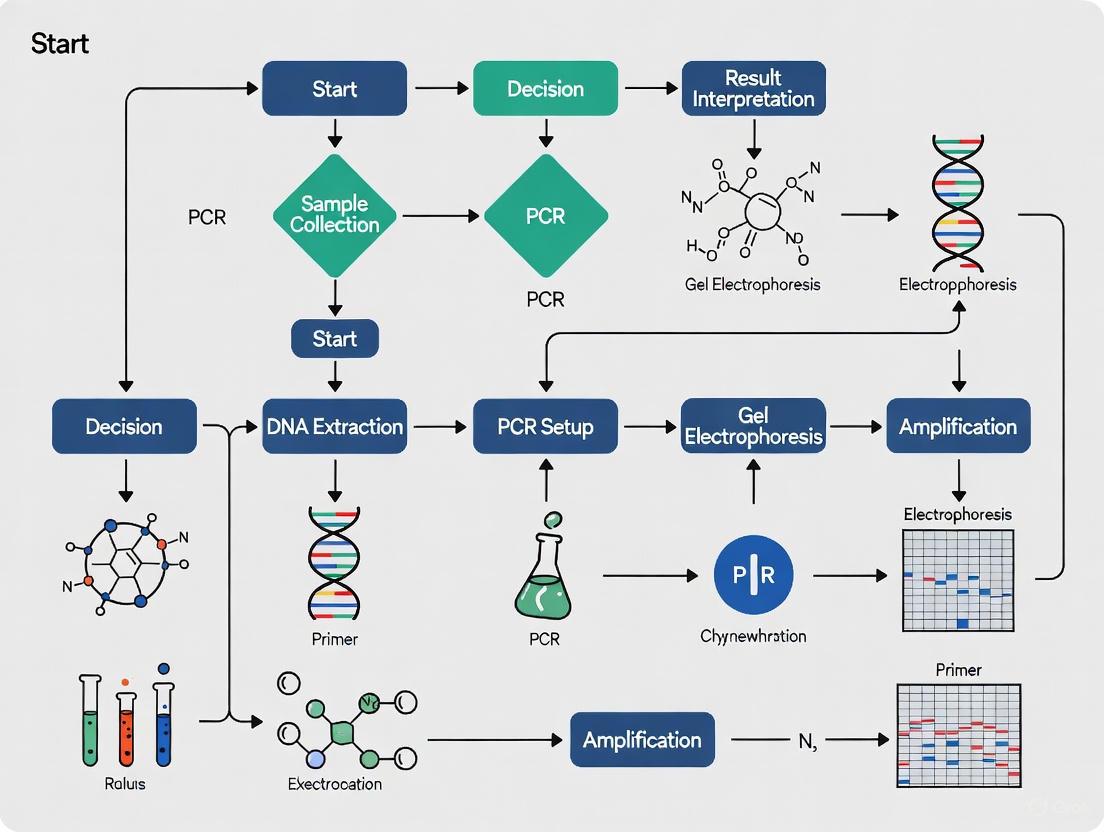
The entire testing process, from sample collection to result interpretation, is visualized below.
Materials and Reagents
Table: Essential Research Reagent Solutions for Mycoplasma PCR Detection
| Item | Function / Description | Example / Specification |
|---|---|---|
| DNA Extraction Kit | Isolates genomic DNA from both host cells and potential contaminants. | QIAamp DNA Mini Kit (Qiagen) or similar [7] [8] |
| PCR Master Mix | Contains Taq polymerase, dNTPs, and buffers necessary for DNA amplification. | Standard 10X PCR buffer, MgCl2, dNTPs [3] |
| Mycoplasma-Specific Primers | Targets ultra-conserved 16S rRNA regions for broad-spectrum detection. | Custom sequences covering Mollicutes [1] |
| Eukaryotic Control Primers | Amplifies a constitutive host gene; serves as an internal positive control. | Targets a conserved mammalian gene (e.g., Uc48-primer) [1] |
| Agarose Gel System | For visualizing PCR amplicons post-amplification. | Standard agarose, ethidium bromide or safer alternative, DNA ladder |
| Positive Control DNA | Confirms PCR is functioning correctly. | Genomic DNA from a known mycoplasma species (e.g., M. orale) [1] |
Step-by-Step Procedure
- Sample Collection: For neuronal cultures, collect 100-200 µL of supernatant or a small pellet of cells. Using supernatant is less invasive and allows for continuous monitoring of the same culture.
- DNA Extraction: Extract total DNA using a commercial kit or a simple boiling method [3]. For the boiling method, incubate the sample at 95°C for 10 minutes, then centrifuge at 12,000 x g for 10 minutes. Use the supernatant as the PCR template.
- PCR Reaction Setup:
- Prepare a master mix for the number of reactions needed (include extra for pipetting error). Each reaction should contain [1] [3]:
- 1X PCR Buffer
- 2.0-2.5 mM MgCl₂
- 200 µM of each dNTP
- 0.4-0.5 µM of each mycoplasma-specific primer
- 0.4-0.5 µM of each eukaryotic control primer
- 0.5-1.0 U of Thermostable DNA Polymerase
- 5 µL of template DNA
- Nuclease-free water to a final volume of 25 µL.
- Prepare a master mix for the number of reactions needed (include extra for pipetting error). Each reaction should contain [1] [3]:
- PCR Amplification: Run the PCR using the following cycling conditions [1] [3]:
- Initial Denaturation: 95°C for 3-5 minutes
- 35-40 Cycles of:
- Denaturation: 93-95°C for 20-30 seconds
- Annealing: 60°C for 20-30 seconds
- Extension: 72°C for 30-60 seconds
- Final Extension: 72°C for 5 minutes
- Hold: 4°C
- Analysis of PCR Products:
- Separate the PCR products by agarose gel electrophoresis (e.g., 2% agarose).
- Visualize the gel under UV light after staining with ethidium bromide or a safer alternative.
- Interpretation:
- Mycoplasma Positive: Presence of a band at 166-191 bp (mycoplasma-specific amplicon).
- Internal Control Valid: Presence of a band at 105 bp (eukaryotic control amplicon).
- Mycoplasma Negative: Absence of the 166-191 bp band, but presence of the 105 bp control band.
- Test Invalid: Absence of both bands, indicating PCR failure or insufficient DNA.
Assay Performance and Validation
- Specificity and Coverage: The described primer design can theoretically cover 92% of all species across the six orders of the class Mollicutes [1].
- Sensitivity (Limit of Detection): The protocol can detect as low as 6.3 pg of mycoplasma genomic DNA, equivalent to approximately 8.21 x 10³ genomic copies [1]. Other optimized PCR assays report sensitivity down to 10 genomic copies [3].
- Quality Control: Always include a no-template control (NTC) with water to check for contamination, and a positive control with known mycoplasma DNA to confirm assay performance.
Best Practices for Prevention and Routine Screening
Prevention is the most effective strategy against mycoplasma contamination.
- Routine Screening: Implement a mandatory testing regime for all new cell lines entering the laboratory and for working stocks at regular intervals (e.g., every 1-2 months) [9] [4].
- Aseptic Technique: Work meticulously in a certified laminar flow hood, use personal protective equipment, and avoid recycling pipette tips [4].
- Quarantine New Lines: Always quarantine and test new cell lines before integrating them into your main cell culture space [4].
- Use Mycoplasma-Free Reagents: Source sera, media, and reagents from suppliers that provide mycoplasma testing certificates.
- Avoid Indiscriminate Antibiotic Use: Routine use of antibiotics can mask low-level contamination, allowing it to spread covertly [4].
- Cell Banking: Establish a master cell bank that has been thoroughly tested and is free of mycoplasma. Use this to create working banks, minimizing the continuous use of passaged cells [4].
The stealthy nature of mycoplasma contamination, characterized by its lack of visual cues and profound impact on cell physiology, poses a severe threat to the integrity of research, particularly in the sensitive field of neuroscience. With prevalence rates remaining high globally, proactive and routine monitoring is not optional but essential. The standardized PCR protocol detailed herein provides researchers with a reliable, cost-effective, and rapid tool for safeguarding neuronal cultures. Integrating this protocol into a comprehensive quality control program, alongside strict aseptic techniques, is fundamental for ensuring the generation of robust, reproducible, and trustworthy scientific data in both academic research and drug development.
Maintaining healthy neuronal cultures is fundamental to neuroscience research and drug development. Contaminants, such as mycoplasma, can significantly alter key cellular functions, compromising experimental integrity. This application note details the profound impacts of mycoplasma contamination and other stressors on neuronal metabolism, viability, and gene expression. Furthermore, it provides a validated protocol for detecting mycoplasma in neuronal cultures using PCR, a critical quality control step to ensure reliable research outcomes.
Documented Impacts on Neuronal Biology
External stressors and contaminants can induce specific, measurable changes in neuronal cell biology. The tables below summarize key quantitative findings from recent studies.
Table 1: Transcriptomic Changes in the Ageing Human Prefrontal Cortex (Single-Cell RNA-seq) [10]
| Cell Type | Key Transcriptomic Changes | Functional Implications |
|---|---|---|
| Excitatory Neurons (L2/3) | 1,273 genes downregulated; 201 genes upregulated. Highest number of changes. | Potential widespread disruption of neuronal communication and maintenance. |
| All Non-Endothelial Cells | Widespread downregulation of 124 common "housekeeping" genes (e.g., HSPA8, VAMP2, TUBA1A, TUBB3, CALM2). | Compromised essential functions: protein folding, cytoskeletal integrity, vesicle transport, and calcium signaling. |
| IN-SST & IN-VIP Neurons | Significant decrease in SST (-2.63 fold) and VIP (-1.46 fold) expression. Increased transcriptional variability (IN-SST). | Loss of inhibitory neuron identity and disrupted network balance in the aged brain. |
Table 2: Metabolic Remodeling During Human iPSC to Cortical Neuron Differentiation [11]
| Metabolic Parameter | iPSCs (Baseline) | Induced Neurons (Day 14) | Functional Significance |
|---|---|---|---|
| Oxidative Phosphorylation | Low | Enhanced | Supports increased energetic demands of mature neurons. |
| Mitochondrial Content/Respiratory Capacity | Low | Increased | Facilitates efficient ATP production via OXPHOS. |
| Glycolytic Activity | High (proliferative state) | Maintained but specialized | Supports localized energy needs (e.g., growth cones) and biosynthesis. |
| NAD(P)H State (FLIM) | More free | More enzyme-bound | Shift towards oxidative metabolism. |
| Glucose Utilization | - | Enhanced PPP and glutathione labeling | Supports antioxidant defense and biosynthetic pathways. |
Beyond transcriptional and metabolic shifts, inflammatory conditions can trigger pathogenic pathways. In multiple sclerosis models, neuronal induction of the immunoproteasome subunit PSMB8 impairs proteasome activity, leading to accumulation of the metabolic regulator PFKFB3 [12]. This forces a pathological metabolic switch to glycolysis, reduces pentose phosphate pathway activity, and ultimately drives oxidative injury and ferroptosis, a form of iron-dependent cell death [12].
Protocol: Mycoplasma Detection in Neuronal Cultures via PCR
Mycoplasma contamination is a pervasive issue that can alter cell metabolism and gene expression, mimicking disease states and generating misleading data [2]. This protocol outlines a rapid and sensitive PCR-based method for its detection.
Principle
This method targets the 16S rRNA gene region, which is highly conserved across Mollicutes (the class containing common mycoplasma species) but distinct from other bacteria. This allows for broad-spectrum detection of over 60 mycoplasma species with high specificity and sensitivity, capable of detecting as few as a handful of genome copies [2].
Research Reagent Solutions
Table 3: Essential Materials for PCR-Based Mycoplasma Detection
| Item | Function / Key characteristic | Commercial Example(s) |
|---|---|---|
| DNA Extraction Kit | Isolates genomic DNA (both host and contaminant) from cell culture supernatant. | AllPrep DNA/RNA Mini Kit (Qiagen) |
| Mycoplasma PCR Assay Kit | Contains pre-optimized primers, probes, and master mix for specific amplification of mycoplasma DNA. | ATCC Universal Mycoplasma Detection Kit; MycoSensor RT-PCR Assay Kit (Agilent) |
| Positive Control DNA | Contains a known segment of the mycoplasma 16S rRNA gene. Verifies assay performance. | Included in most commercial kits |
| Nuclease-Free Water | Used for preparing reaction mixes; free of enzymes that degrade nucleic acids. | Various suppliers |
| Real-Time PCR System | Instrument for amplifying and quantifying DNA in real-time, providing rapid and sensitive detection. | Instruments from Bio-Rad, Thermo Fisher, Roche |
Detailed Procedure
Sample Collection: Aspirate the culture medium from the neuronal culture. Collect a 100 µL - 1 mL aliquot of the cell-free culture supernatant. Avoid collecting cellular debris to minimize host DNA contamination. Process samples immediately or store at -80°C.
DNA Extraction: Purify total genomic DNA from the collected supernatant using a commercial DNA extraction kit, following the manufacturer's instructions. Include a known negative control (e.g., from a confirmed mycoplasma-free culture) and the provided positive control.
PCR Reaction Setup:
- Prepare the PCR master mix according to the kit's specifications. A typical 25 µL reaction includes:
- 12.5 µL of 2x Master Mix (containing polymerase, dNTPs, buffer)
- 1.0 µL of forward primer (10 µM)
- 1.0 µL of reverse primer (10 µM)
- 0.5 µL of probe (10 µM) [For real-time PCR]
- 5.0 µL of template DNA
- Nuclease-free water to 25 µL
- Pipette the mix into the PCR tubes/plate. Include a no-template control (NTC) with water instead of DNA to check for reagent contamination.
- Prepare the PCR master mix according to the kit's specifications. A typical 25 µL reaction includes:
PCR Amplification:
- Place the samples in a real-time PCR instrument and run the following cycling program:
- Initial Denaturation: 95°C for 2-5 minutes.
- 40-45 Cycles of:
- Denaturation: 95°C for 15-30 seconds.
- Annealing/Extension: 60°C for 1 minute (data collection step for real-time PCR).
- The specific temperatures and times may be optimized based on the kit and primer set.
- Place the samples in a real-time PCR instrument and run the following cycling program:
Result Analysis:
- Analyze the amplification curves. A sample is considered positive if the fluorescence curve crosses the threshold line within the cycle limit (typically before cycle 40).
- The positive control should show positive amplification, and the NTC and negative control should show no amplification.
Diagram 1: Mycoplasma detection workflow.
Advanced Research Applications
The principles of detecting molecular changes in neurons are also applied in clinical oncology research. For instance, in neuroblastoma, a cancer of neural crest origin, sensitive molecular techniques are crucial for detecting minimal residual disease (MRD), which predicts relapse [13] [14]. These methods quantify neuroblastoma-associated mRNAs (e.g., PHOX2B, TH, DBH) in bone marrow via reverse transcriptase-PCR (RT-PCR) [14]. Combining this molecular approach with automated immunofluorescence significantly improves detection sensitivity and provides protein-level information for selecting immunotherapies [13].
Diagram 2: Inflammation-induced neuronal degeneration pathway.
Mycoplasma contamination and other cellular stressors induce specific, detrimental changes in neuronal metabolism, gene expression, and viability. The provided PCR protocol offers researchers a robust tool to routinely screen neuronal cultures, thereby safeguarding data quality. Integrating this quality control with advanced assays for transcriptional and metabolic profiling, as illustrated, enables a more comprehensive understanding of neuronal health and disease mechanisms, ultimately accelerating reliable neuroscience and drug discovery.
In the context of mycoplasma detection in neuronal cultures using PCR, maintaining sample integrity is paramount. Contamination poses a significant threat to the reliability of experimental results, potentially leading to false positives, erroneous conclusions, and wasted resources. This application note details the primary sources of contamination—cross-contamination, reagents, and personnel—and provides validated protocols to mitigate these risks, with a specific focus on low-biomass applications like mycoplasma screening.
Contamination in laboratory settings can be categorized into several key types, each with distinct sources and impacts on experimental integrity. The table below summarizes the major contamination sources, their characteristics, and associated risks.
Table 1: Primary Sources and Impacts of Laboratory Contamination
| Contamination Source | Specific Examples | Impact on Experiments | Notable Pathogens/Contaminants |
|---|---|---|---|
| Personnel | Improper aseptic technique, shedding of skin cells, aerosol generation from talking/sneezing [15] [16] [17] | Introduction of microbial contaminants and foreign DNA; compromises sample purity [18] [17] | Human-derived microbes (Mycoplasma spp., M. tuberculosis, skin flora), human DNA [18] [2] |
| Reagents & Kits | Contaminated DNA extraction kits, PCR master mix, laboratory water, culture media [19] [15] [20] | High background contaminant DNA in low-biomass samples; skewed microbial community profiles [19] [17] | Bacterial DNA from kit reagents (e.g., Pseudomonas, Comamonadaceae, Burkholderiales) [19] |
| Cross-Contamination | Sample-to-sample transfer via pipettes, well-to-well leakage in plates, shared equipment without decontamination [21] [15] [17] | Carryover of amplification products (amplicons) or biological material between samples [21] [22] | Mycoplasma carryover from infected cultures, PCR amplicons [21] [2] |
| Environmental | Airborne dust and microbes, improperly maintained HEPA filters, contaminated work surfaces [15] [16] | Direct contamination of open samples and cell cultures [15] | Airborne fungal spores, bacteria, and environmental microbes [16] |
Experimental Protocols for Contamination Mitigation and Detection
Protocol: Aseptic Technique and Personnel Hygiene
Objective: To minimize contamination introduced by laboratory personnel during handling of neuronal cultures and PCR setup.
Materials:
- Personal Protective Equipment (PPE): Lab coat, gloves, safety goggles [16] [20]
- Dedicated lab shoes or shoe covers [20]
- Biological Safety Cabinet (BSC) or laminar flow hood with HEPA filtration [15] [20]
- Surface decontaminants (e.g., 70% ethanol, sodium hypochlorite solution) [17]
Procedure:
- Preparation: Remove jewelry and secure hair. Don a clean lab coat and dedicated lab shoes. Wash hands thoroughly [16].
- PPE and Workspace: Enter the BSC and decontaminate all surfaces and the interior with 70% ethanol. Put on gloves and safety goggles before handling any materials [15] [16].
- Sample Handling: Work deliberately and avoid rapid movements that can create aerosols. Do not talk or sneeze over open samples [15] [17].
- Glove Management: Change gloves if they become contaminated, and always when moving between different cell lines or reagent setups to prevent cross-contamination [20]. Decontaminate gloves with ethanol before touching surfaces inside the BSC [17].
- Post-Processing: Dispose of all waste appropriately. Decontaminate all surfaces and equipment with ethanol before and after use [15].
Protocol: Reagent and Kit Quality Control for Low-Biomass PCR
Objective: To identify and account for contaminating DNA present in laboratory reagents, which is critical for sensitive PCR-based mycoplasma detection.
Materials:
- DNA extraction kits (e.g., MoBio PowerWater Kit) [19]
- PCR reagents (master mix, primers, nuclease-free water)
- Sterile, DNA-free collection tubes
Procedure:
- Include Negative Controls: With every batch of DNA extractions, process multiple "blank" negative controls. These should consist of the same volume of sterile, DNA-free water or elution buffer used for your samples [19] [17].
- Sample Randomization: Randomize sample processing order to prevent confounding of biological variables with batch effects from specific reagent kits [19] [17].
- Reagent Screening: Periodically screen new lots of critical reagents (especially water and DNA extraction kits) by running them through the entire PCR process as a negative control. Select lots with the lowest background contamination [19].
- Analysis and Interpretation: Sequence the negative controls alongside your experimental samples. Bioinformatic analysis should be performed to identify and remove operational taxonomic units (OTUs) or sequences that are also present in the negative controls from the final dataset [19] [17].
Protocol: PCR Setup to Prevent Amplicon Cross-Contamination
Objective: To prevent carryover contamination of PCR amplicons into new reactions, a major source of false positives.
Materials:
- Dedicated pipettes and tips with aerosol filters
- Separate physical areas or rooms for pre- and post-PCR work
- UV light cabinet for decontaminating surfaces and equipment [17]
Procedure:
- Physical Separation: Physically separate the laboratory into distinct areas: one for reagent preparation (pre-PCR), one for sample and DNA extraction, and one for post-PCR analysis [15].
- Unidirectional Workflow: Establish a strict unidirectional workflow. Personnel and materials must move from pre-PCR to post-PCR areas, never in reverse [15] [20].
- Dedicated Equipment: Use dedicated pipettes, tips, and lab coats for each area. Use only aerosol-filter pipette tips in the pre-PCR and sample setup areas [15].
- No-Template Controls (NTCs): Always include NTCs in every PCR run. These reactions contain all PCR components except the DNA template, testing for contamination of the master mix or primers [15].
Protocol: Enhanced Mycoplasma Detection in Cell Cultures
Objective: To accurately detect mycoplasma contamination in neuronal cell cultures, minimizing false positives from host cell DNA.
Materials:
- Hoechst DNA stain
- Wheat Germ Agglutinin (WGA) membrane stain [23]
- Fluorescence microscope
- Antibiotics for mycoplasma elimination (e.g., BM-Cyclin)
Procedure:
- Cell Culture: Grow neuronal cells on sterile coverslips in a culture dish until ~70% confluent.
- Staining: Treat cells with a combination of Hoechst DNA stain and a fluorescent WGA membrane stain according to manufacturer protocols [23].
- Microscopy and Analysis: Visualize the cells under a fluorescence microscope. True mycoplasma contamination is identified by the colocalization of Hoechst DNA signal with the WGA membrane stain on the surface of the host cell plasma membrane [23].
- Interpretation: This co-localization method minimizes interference from false positive signals caused by degraded host cell DNA or cytoplasmic DNA fragments, which would not colocalize with the membrane [23].
The following workflow integrates these protocols into a comprehensive strategy for managing contamination in mycoplasma detection research.
The Scientist's Toolkit: Essential Reagents and Materials
The following table lists key materials and their specific functions in preventing and detecting contamination in mycoplasma research.
Table 2: Essential Research Reagent Solutions for Contamination Control
| Item | Specific Function/Application | Key Consideration |
|---|---|---|
| HEPA-Filtered Laminar Flow Hood/BSC | Provides a sterile, particulate-free workspace for handling cell cultures and reagents [15] [20]. | Must be certified regularly; UV light can be used for supplemental decontamination [17]. |
| Automated Liquid Handling System | Reduces human error and cross-contamination between samples during reagent dispensing [20]. | Enclosed hoods on these systems provide an additional contamination-free layer [20]. |
| PCR Kits with UDG Decontamination | Enzymatically degrades carryover PCR amplicons from previous reactions, preventing false positives. | A standard feature in many commercial master mixes. |
| Validated Mycoplasma Detection Kit (qPCR) | Rapidly and sensitively detects a wide spectrum of mycoplasma species (e.g., >60 species) common in cell cultures [2]. | Targets 16S rRNA genes; results in 2-5 hours vs. weeks for culture methods [2]. |
| DNA Decontamination Solution (e.g., Bleach) | Destroys contaminating DNA on surfaces and equipment; critical for pre-PCR areas [17]. | Note that ethanol kills cells but does not remove DNA; bleach or commercial DNA removal solutions are required [17]. |
| Hoechst & WGA Membrane Stain | Enables specific detection of mycoplasma via DNA and membrane colocalization, reducing false positives from host DNA [23]. | More accurate than DNA staining alone for visual confirmation of mycoplasma [23]. |
| Aerosol-Filter Pipette Tips | Prevents aerosolized samples and reagents from contaminating pipette shafts and subsequent samples. | Essential for all pipetting steps, especially in PCR setup and sample handling. |
Vigilant contamination control is a foundational element of reliable scientific research, especially in sensitive applications like PCR-based mycoplasma detection in neuronal cultures. By understanding the primary sources of contamination and rigorously implementing the detailed protocols for mitigation—focusing on personnel practices, reagent quality control, and physical workflow separation—researchers can safeguard the integrity of their experiments. The adoption of advanced detection methods, such as DNA-membrane colocalization, further ensures accurate and trustworthy results.
Mycoplasma contamination represents a pervasive and insidious threat in cell culture laboratories, with particularly severe implications for neuronal culture research and the development of cell-based therapies. These wall-less bacteria infect an estimated 15-35% of cell cultures, with rates reaching as high as 85% in certain laboratory settings [24]. The consequences of undetected mycoplasma contamination extend beyond mere inconvenience, potentially compromising scientific data integrity and posing significant safety risks for therapeutic applications.
In neuronal cultures, where subtle changes in gene expression, metabolism, and cellular function are frequently central to research outcomes, mycoplasma contamination can fundamentally alter experimental results while remaining undetectable by routine microscopy [25] [26]. This application note examines the consequences of mycoplasma contamination and provides detailed protocols for its detection using PCR-based methods, with specific consideration for neuronal culture systems.
Consequences of Mycoplasma Contamination
Impact on Research Data Integrity
Mycoplasma contamination induces a range of cellular alterations that can compromise the validity of experimental data, particularly in sensitive neuronal culture systems:
- Altered Cellular Metabolism: Mycoplasma compete with host cells for nutrients, leading to depleted media and altered metabolic profiles [2].
- Chromosomal Abnormalities: Contamination can induce chromosomal aberrations, potentially skewing genetic studies [24].
- Gene Expression Changes: Mycoplasma infection modifies host cell gene expression patterns, which can confound transcriptomic analyses [26].
- Interference with Cell Signaling: The metabolic changes induced by contamination can interfere with neuronal signaling pathways and synaptic function [24].
Risks to Therapeutic Applications
For neuronal cultures destined for therapeutic use, such as in advanced therapy medicinal products (ATMPs), mycoplasma contamination presents direct safety concerns:
- Patient Safety Risks: Contaminated cell therapies can cause immune reactions in recipients [27].
- Altered Cell Function: Mycoplasma contamination can modify proliferation characteristics and cellular function of therapeutic cells [27].
- Batch Failure: In biopharmaceutical manufacturing, contamination can necessitate disposal of entire production batches, with significant economic consequences [24].
Table 1: Documented Effects of Mycoplasma Contamination on Cell Cultures
| Effect Category | Specific Consequences | Impact on Neuronal Cultures |
|---|---|---|
| Metabolic Effects | Depletion of nutrients; altered nucleic acid synthesis | Compromised neuronal viability; altered metabolic activity |
| Genetic Effects | Chromosomal abnormalities; apoptosis induction | Aberrant neuronal development; cell death |
| Functional Effects | Altered gene expression; modified cell signaling | Impaired neuronal network formation; skewed electrophysiological data |
| Therapeutic Risks | Immune reactions; functional impairment | Potential adverse effects in cell transplantation |
Mycoplasma Detection Strategies
Comparison of Detection Methods
Multiple methodologies exist for mycoplasma detection, each with distinct advantages and limitations for research versus therapeutic applications:
Table 2: Mycoplasma Detection Method Comparison
| Method | Time Required | Sensitivity | Key Advantages | Key Limitations |
|---|---|---|---|---|
| Culture-Based | Up to 28 days [24] [2] | 10 CFU/mL [24] | Gold standard; required for regulatory compliance [2] | Prolonged incubation; fastidious growth requirements [24] |
| Indicator Cell Culture | 3-5 days [2] | Moderate | Detects non-cultivable species [2] | Less sensitive than culture method [2] |
| PCR/qPCR | 1.5-5 hours [24] [2] | 10-100 copies/reaction [28] | Rapid; high sensitivity; cost-effective [24] | Cannot differentiate live/dead mycoplasma [24] |
| Fluorescence Staining | 1-2 days | Low to moderate | Visual confirmation; works with fixed cells | Lower sensitivity; subjective interpretation |
PCR-Based Detection Advantages
For most research laboratories, PCR-based methods offer the optimal balance of sensitivity, speed, and practicality:
- Speed: Results can be obtained within hours rather than weeks [24] [2]
- Sensitivity: Capable of detecting low-level contamination (as few as 10 genome copies) [28]
- Broad Coverage: Well-designed primers can detect over 250 mycoplasma species [24]
- Compatibility: Suitable for both research quality control and GMP applications when properly validated [2]
Experimental Protocols
Commercial qPCR Detection Protocol
For laboratories requiring regulatory compliance or maximum sensitivity, commercial qPCR kits provide validated solutions:
Materials and Reagents
Table 3: Essential Research Reagents for Mycoplasma Detection
| Reagent/Equipment | Function | Example Specifications |
|---|---|---|
| qPCR Master Mix | Amplification of target DNA | Contains DNA polymerase, dNTPs, buffer |
| Mycoplasma Primers/Probes | Specific detection | Targets 16S rRNA genes; broad species coverage [24] |
| DNA Extraction Kit | Nucleic acid purification | Compatible with cell culture supernatant |
| Positive Control DNA | Assay validation | Contains target sequence for quality control |
| qPCR Instrument | Amplification and detection | Real-time fluorescence monitoring |
| Sterile Filter Tips | Contamination prevention | Aerosol barrier to prevent amplicon contamination |
Sample Collection and Processing
Sample Collection:
- Collect 100-200 µL of cell culture supernatant from dense neuronal cultures (80-100% confluent) [29]
- Include conditioned media from neuronal cultures as mycoplasma may be cell-associated
- Process samples in a dedicated pre-PCR area to prevent contamination
DNA Extraction:
- Use commercial DNA extraction kits following manufacturer's protocols
- Include extraction controls to monitor cross-contamination
- Elute DNA in nuclease-free water or TE buffer
qPCR Setup:
- Prepare reaction mix according to kit specifications (typically 20-25 µL total volume)
- Include no-template controls (NTC) and positive controls in each run
- Use at least two replicates per sample for reliability
Thermal Cycling:
- Initial denaturation: 95°C for 10 minutes
- 40 cycles of: 95°C for 15 seconds (denaturation) and 60°C for 60 seconds (annealing/extension) [24]
- Perform fluorescence acquisition during the annealing/extension step
Data Analysis:
- Determine Cq values using instrument software
- Samples with Cq values below the validated threshold (e.g., ≤35) are considered positive
- Compare with positive controls to verify assay performance
Laboratory-Developed PCR Protocol
For routine screening where regulatory compliance is not required, a laboratory-developed test based on published protocols offers a cost-effective alternative [29]:
Primer Design and Preparation
Primer Sequences: Utilize published primer sets targeting the 16S rRNA gene with broad mycoplasma specificity [29]:
- Forward primers: Myco-5-1 to Myco-5-6 (mix of 6 sequences)
- Reverse primers: Myco-3-1 to Myco-3-3 (mix of 3 sequences)
Primer Mix Preparation:
- Resuspend each primer to 100 µM stock concentration
- Prepare working primer mix by combining equal volumes of each forward primer (10 µM each final) and each reverse primer (10 µM each final)
Sample Preparation and PCR Amplification
Sample Processing:
- Collect 100 µL of neuronal culture supernatant
- Heat at 95°C for 5 minutes to denature proteins and release DNA
- Centrifuge at maximum speed for 2 minutes to pellet debris
- Transfer supernatant to fresh tube for PCR
PCR Reaction Setup:
- Prepare 25 µL reactions containing:
- 10× PCR Buffer: 2.5 µL
- 25 mM MgCl₂: 2.0 µL
- 10 mM dNTPs: 1.0 µL
- Forward primer mix: 1.0 µL
- Reverse primer mix: 1.0 µL
- Cell culture supernatant: 2.0 µL
- Taq polymerase: 0.2 µL
- Water: 15.3 µL
- Prepare 25 µL reactions containing:
Thermal Cycling Conditions:
- Initial denaturation: 95°C for 2 minutes
- 5 cycles of: 94°C for 30 seconds, 50°C for 30 seconds, 72°C for 35 seconds
- 30 cycles of: 94°C for 15 seconds, 56°C for 15 seconds, 72°C for 30 seconds
- Final extension: 72°C for 5 minutes
- Hold at 4°C
Amplicon Detection:
- Separate PCR products by agarose gel electrophoresis (1.5-2.0%)
- Visualize with DNA staining; positive samples show ~500 bp band
- Include molecular weight markers for size verification
Protocol Validation and Quality Control
Regardless of the method chosen, proper validation is essential:
- Analytical Sensitivity: Determine the limit of detection using serial dilutions of mycoplasma DNA
- Specificity Testing: Verify absence of cross-reactivity with neuronal DNA and common laboratory contaminants
- Reproducibility: Assess inter-assay and intra-assay variability
- Positive Control: Use a defined mycoplasma species (e.g., M. hyorhinis) as a control
Implementation Workflow
The following workflow outlines a comprehensive approach to mycoplasma management in neuronal culture research:
Mycoplasma contamination poses a significant threat to both the integrity of neuronal culture research and the safety of cell-based therapies. Implementation of robust, regularly scheduled detection protocols is essential for maintaining data quality and ensuring patient safety in therapeutic applications. PCR-based methods offer the most practical solution for most research laboratories, providing rapid, sensitive detection with broad species coverage.
The consequences of unchecked contamination—compromised data, invalid conclusions, and potential patient harm—far outweigh the investment in established detection methodologies. By incorporating the protocols outlined in this application note, researchers can significantly reduce the risks associated with mycoplasma contamination in neuronal culture systems.
Step-by-Step PCR Protocol for Neuronal Cultures: From Sample to Result
Within the framework of mycoplasma detection in neuronal cultures using PCR, the integrity of the sample is paramount. The process of collecting supernatant, the cell culture medium devoid of cells, is a critical preliminary step. The quality of this supernatant directly influences the sensitivity and accuracy of downstream molecular analyses, including PCR. Contaminated or poorly collected samples can lead to false negatives in detection assays, compromising research validity and drug development pipelines. This application note provides a detailed protocol for preparing supernatant from dense primary neuronal cultures, ensuring samples are optimally collected for reliable mycoplasma PCR screening.
Research Reagent Solutions
The following table details essential materials and their functions for the successful culture of primary neurons and subsequent supernatant collection.
Table 1: Key Research Reagents for Neuronal Culture and Supernatant Preparation
| Item | Function/Application |
|---|---|
| Neurobasal-A or Neurobasal Plus Medium | A serum-free medium specifically formulated for the long-term support of low-density neuronal cultures [30] [31]. |
| B-27 Supplement | A serum-free supplement essential for neuron survival and growth, reducing the need for glial feeder layers [30] [31]. |
| Hibernate-E Medium | A medium designed for the storage and shipment of live neurons; useful for stabilizing cultures prior to procedures [30]. |
| Poly-D-Lysine or Poly-L-Lysine | A synthetic polymer used to coat culture vessels, providing a charged surface that enhances neuronal attachment [30] [31]. |
| Papain | A proteolytic enzyme used for the gentle dissociation of neural tissue into individual cells for culture establishment [30] [31]. |
| Cytosine β-D-arabinofuranoside (Ara-C) | A mitotic inhibitor used to suppress the proliferation of glial cells in primary neuronal cultures, preserving neuronal purity [30]. |
| L-Glutamine | An essential amino acid supplement that supports neuronal health, particularly in the initial days of culture [31]. |
| Plasmocin | An antibiotic prophylactic used to prevent mycoplasma contamination in cell cultures [30]. |
Protocol for Establishing Dense Neuronal Cultures
The foundation of high-quality supernatant begins with healthy, dense, and pure neuronal cultures. The following methodology is adapted from established protocols for primary neuronal isolation and culture [30] [32] [31].
Preparation of Coated Culture Vessels
- Dilution: Prepare a working solution of Poly-L-Lysine (PLL) or Poly-D-Lysine (PDL) at 50-100 µg/mL in sterile distilled water or PBS [30] [31].
- Coating: Apply the working solution to completely cover the surface of the culture vessel (e.g., 150 µL/cm²) and incubate for 1 hour at room temperature.
- Rinsing and Storage: Aspirate the coating solution and rinse the surface thoroughly with sterile distilled water at least three times to remove any excess, toxic polymer. Allow the vessels to dry completely before use or store at 4°C for up to one week [31].
Isolation and Culturing of Primary Neurons
- Dissection and Collection: Dissect the desired brain region (e.g., cortex, hippocampus) from postnatal day 0-1 mouse pups or embryonic day 18 rats [30] [31]. Collect the tissue in ice-cold Hibernate-E or another collection medium supplemented with B-27.
- Enzymatic Dissociation: Incubate the tissue in an enzyme solution such as papain (1-2 mg/mL) in Hibernate-A calcium-free medium, often with added Dispase II and DNAse, at 30-37°C for 20-30 minutes [30] [31].
- Trituration and Stratification: Gently triturate the digested tissue 3-5 times using a fire-polished glass Pasteur pipette in a trituration medium containing serum to inactivate enzymes. Allow debris to settle and strain the cell suspension through a 70 µm mesh [30].
- Centrifugation and Resuspension: Centrifuge the cell suspension at 150-200 x g for 4-5 minutes. Aspirate the supernatant and gently resuspend the cell pellet in complete culture medium (Neurobasal-A supplemented with B-27, L-Glutamine, and Plasmocin) [30] [31].
- Seeding and Maintenance: Seed the cells at a high density of approximately 125,000 cells/cm² onto the PLL/PDL-coated vessels [30]. Maintain cultures in a humidified incubator at 37°C with 5% CO₂.
- Feeding Schedule: Perform a half-medium change every 2-3 days to replenish nutrients without fully disturbing the neuronal environment. At 3-4 days in vitro (DIV), add the mitotic inhibitor Ara-C (e.g., 5 µM) to the medium to curb glial cell overgrowth [30].
Table 2: Critical Parameters for Dense Neuronal Culture
| Parameter | Specification | Rationale |
|---|---|---|
| Cell Seeding Density | ~125,000 cells/cm² | Optimal for network formation and experimental yield [30]. |
| Culture Vessel Coating | Poly-L-Lysine (0.1 mg/mL) or Poly-D-Lysine (50 µg/mL) | Promotes strong neuronal attachment [30] [31]. |
| Base Culture Medium | Neurobasal-A / Neurobasal Plus | Optimized for neuronal health and minimal glial proliferation [30] [31]. |
| Key Supplement | B-27 (2%) | Provides essential hormones, antioxidants, and nutrients [30] [31]. |
| Mitotic Inhibition | Ara-C (5 µM) at 3-4 DIV | Selectively inhibits dividing glial cells, enhancing neuronal purity [30]. |
| Time to Maturity | ≥14 Days In Vitro (DIV) | Allows for development of spontaneous synaptic activity [30]. |
Protocol for Supernatant Collection
Collect supernatant from mature cultures (≥14 DIV) for mycoplasma testing.
- Visual Inspection: Prior to collection, visually inspect the cultures under a microscope to confirm healthy neuronal morphology (e.g., extensive neurite outgrowth, phase-bright somas) and the absence of visible contamination.
- Pre-collection Handling: Gently remove the culture vessel from the incubator and place it on a clean, level surface. Avoid agitating or swirling the medium, as this may dislodge cells or debris.
- Supernatant Aspiration: Using a sterile pipette, carefully aspirate the culture medium (supernatant) from the well, ensuring the pipette tip is positioned away from the neuronal cell layer at the bottom of the vessel. Critical Step: Avoid touching the bottom of the well to prevent collecting cellular debris.
- Clarification (Optional but Recommended): Transfer the collected supernatant to a sterile microcentrifuge tube. Centrifuge at 500 x g for 5-10 minutes at 4°C to pellet any residual floating cells or particulate matter.
- Aliquoting and Storage: Carefully transfer the clarified supernatant into new, sterile, labeled tubes. If the supernatant is not for immediate PCR analysis, aliquot it to avoid repeated freeze-thaw cycles and store at or below -20°C.
Workflow for Supernatant Preparation and Mycoplasma Detection
The following diagram illustrates the logical workflow from culture establishment to PCR analysis.
Workflow for Supernatant Preparation
Quality Control and Troubleshooting
Ensuring the quality of the neuronal culture is a prerequisite for meaningful supernatant analysis.
Table 3: Quality Control and Troubleshooting Guide
| Aspect | Quality Indicator | Potential Issue & Solution |
|---|---|---|
| Cell Viability | >90% viability post-isolation by trypan blue exclusion [30]. | Low Viability: Optimize dissection speed, enzyme concentration, and trituration force. |
| Neuronal Purity | High percentage of MAP2-positive neurons; minimal GFAP-positive astrocytes [31]. | High Glial Contamination: Ensure timely use and correct concentration of Ara-C. |
| Culture Health | Extensive neurite outgrowth and network formation by 7 DIV; phase-bright cell bodies [31]. | Poor Neurite Growth: Check coating efficiency, medium quality, and supplement freshness. |
| Supernatant Clarity | Clear, non-viscious liquid post-clarification centrifugation. | Cloudy Supernatant: Increase centrifugation force/duration; check for microbial contamination. |
| Mycoplasma Contamination | Negative result in routine PCR tests. | Positive PCR Result: Discard culture and reagents; decontaminate workspace; use fresh aliquots of Plasmocin [30] [23]. |
The reliability of mycoplasma detection in neuronal cultures via PCR is fundamentally dependent on the initial steps of culture preparation and supernatant collection. The protocols detailed herein, emphasizing high-density plating, strict aseptic technique, and careful supernatant handling, provide a robust framework for generating high-quality samples. By standardizing this preparatory phase, researchers can significantly enhance the fidelity of their diagnostic assays, thereby safeguarding the integrity of their neuroscientific research and drug development efforts.
Mycoplasma contamination represents one of the most significant and prevalent threats to cell culture integrity, particularly in sensitive neuronal culture systems [29]. These bacteria can persistently infect cultures while remaining undetected, altering cellular functions and compromising experimental results [29]. The primary challenge in molecular detection stems from the diversity of Mycoplasma species that can contaminate cultures, necessitating primer designs that can simultaneously identify multiple potential contaminants with high sensitivity and specificity.
This application note provides a detailed framework for designing and implementing a broad-spectrum PCR detection strategy for common Mycoplasma contaminants, with specific consideration for neuronal culture applications. The protocols outlined enable researchers to establish a cost-effective, routine testing methodology that surpasses the limitations of targeted commercial kits.
Primer Design Strategy for Broad-Spectrum Detection
Effective broad-spectrum detection requires moving beyond single-target primer pairs to a multiplex approach that accounts for genetic diversity across contaminant species. The fundamental principle involves using multiple forward and reverse primers in a single reaction mixture to amplify conserved but variable regions across different Mycoplasma species [29].
Core Primer Sequences
The following primer set, derived from Uphoff and Drexler, enables detection of a wide range of Mycoplasma species through targeted amplification of conserved genomic regions [29]. This multi-primer approach significantly increases the probability of detection compared to single primer pair assays.
Table 1: Broad-Spectrum Mycoplasma Primer Set
| Primer Name | Sequence (5' to 3') | Type | Final Concentration in Mix |
|---|---|---|---|
| Myco-5-1 | CGCCTGAGTAGTACGTTCGC | Forward | 10 µM each |
| Myco-5-2 | CGCCTGAGTAGTACGTACGC | Forward | 10 µM each |
| Myco-5-2 | TGCCTGAGTAGTACATTCGC | Forward | 10 µM each |
| Myco-5-2 | TGCCTGGGTAGTACATTCGC | Forward | 10 µM each |
| Myco-5-5 | CGCCTGGGTAGTACATTCGC | Forward | 10 µM each |
| Myco-5-6 | CGCCTGAGTAGTATGCTCGC | Forward | 10 µM each |
| Myco-3-1 | GCGGTGTGTACAAGACCCGA | Reverse | 10 µM each |
| Myco-3-2 | GCGGTGTGTACAAAACCCGA | Reverse | 10 µM each |
| Myco-3-3 | GCGGTGTGTACAAACCCCGA | Reverse | 10 µM each |
Primer Design Considerations
The selected primers exhibit several key features essential for successful broad-spectrum detection:
- Target Conservation: Primers target regions conserved across multiple Mycoplasma species while containing sufficient sequence variation to enable comprehensive detection through multiple primer combinations [29]
- Compatibility: All primers are designed to work under identical PCR conditions, allowing simultaneous use in a single reaction tube
- Amplicon Size: The approximately 500 bp amplicon is optimal for clear visualization on standard agarose gels while providing sufficient sequence for specific detection [29]
Experimental Protocol for Mycoplasma Detection
Sample Preparation
Proper sample collection is critical for detection sensitivity:
- Collect 100 µL of cell culture supernatant from a dense culture (80-100% confluent)
- Transfer to a 1.5 mL microcentrifuge tube
- Heat sample at 95°C for 5 minutes to denature proteins and release DNA
- Centrifuge for 2 minutes at maximum speed to pellet debris
- Use 2 µL of the supernatant as template in the PCR reaction [29]
PCR Reaction Setup
Table 2: PCR Reaction Master Mix
| Reagent | Volume (µL) | Final Concentration |
|---|---|---|
| 10x PCR Buffer | 2.5 | 1X |
| 25 mM MgCl₂ | 2.0 | 2.0 mM |
| 10 mM dNTPs | 1.0 | 0.4 mM each |
| Forward Primer Mix | 1.0 | 0.4 µM each |
| Reverse Primer Mix | 1.0 | 0.4 µM each |
| Cell Culture Supernatant | 2.0 | - |
| Taq Polymerase | 0.2 | 0.5 U |
| Water | 15.3 | - |
| Total Volume | 25.0 |
Note: Always include negative control (water) and positive control (known contaminated supernatant) when available [29]
Thermal Cycling Conditions
The PCR protocol employs a two-stage amplification to ensure both sensitivity and specificity:
Table 3: Thermal Cycling Parameters
| Step | Temperature | Time | Cycles |
|---|---|---|---|
| Initial Denaturation | 95°C | 2:00 min | 1 |
| Denaturation | 94°C | 0:30 sec | 5 |
| Annealing | 50°C | 0:30 sec | 5 |
| Extension | 72°C | 0:35 sec | 5 |
| Denaturation | 94°C | 0:15 sec | 30 |
| Annealing | 56°C | 0:15 sec | 30 |
| Extension | 72°C | 0:30 sec | 30 |
| Final Hold | 4°C | ∞ | 1 |
The initial 5 cycles with lower annealing temperature (50°C) facilitate primer binding across diverse Mycoplasma templates, while the subsequent 30 cycles with higher stringency (56°C) ensure specific amplification [29]. If signal intensity is weak, increasing to 35 cycles in the second stage may improve detection.
Post-Amplification Analysis
- Prepare a 1.5-2.0% agarose gel in 1X TAE buffer with appropriate DNA stain
- Load 5-10 µL of PCR product alongside a 500 bp DNA ladder
- Run gel at 80-100V until adequate separation is achieved
- Visualize under UV light - a band at approximately 500 bp indicates Mycoplasma contamination
Workflow Visualization
Mycoplasma Detection Workflow
Research Reagent Solutions
Table 4: Essential Research Reagents and Materials
| Item | Function/Application | Specifications/Alternatives |
|---|---|---|
| Primer Mixes | Core detection components for broad-spectrum identification | Custom synthesized, resuspended to 100 µM stock in Tris-HCl [29] |
| Taq Polymerase | DNA amplification | Thermostable DNA polymerase with standard buffer system [29] |
| dNTPs | Nucleotides for DNA synthesis | 10 mM mixture of dATP, dCTP, dGTP, dTTP [29] |
| MgCl₂ | Cofactor for polymerase activity | 25 mM stock solution, concentration requires optimization [29] |
| PCR Tubes | Reaction vessels | Thin-walled for optimal thermal transfer [29] |
| Agarose | Gel matrix for amplicon visualization | Standard molecular biology grade [29] |
| Thermal Cycler | Precise temperature cycling | Standard PCR instrument with programmable blocks [29] |
| Gel Electrophoresis System | Amplicon separation and visualization | Standard horizontal system with UV transillumination [29] |
Quality Control and Validation
Controls and Standards
Robust experimental design requires appropriate controls:
- Negative Control: Nuclease-free water instead of template - should yield no amplification
- Positive Control: Known contaminated culture supernatant - essential for validating primer performance
- Template Quality Control: Test primers on known Mycoplasma DNA if available
- Prevention Control: Include reagents and media tested during aliquoting [29]
Sensitivity and Specificity Assessment
While the described protocol provides detection limits sufficient for routine screening (approximately 100 CFU/mL based on similar broad-spectrum PCR methods [33]), laboratories should validate sensitivity using:
- Serial dilutions of known positive samples
- Cross-reactivity testing with common cell culture bacteria
- Comparison with commercial detection kits if available
Applications in Neuronal Culture Systems
The protocol is particularly valuable for neuronal culture research where:
- Cultures may be maintained for extended periods, increasing contamination risk
- Non-neuronal contaminants (e.g., Schwann cells, fibroblasts) complicate interpretation of morphological changes [34] [35]
- Experimental outcomes are highly sensitive to cellular stress induced by undetected contaminants
- Primary neuronal cultures cannot be easily replaced if compromised [34]
Troubleshooting Guide
Table 5: Common Issues and Solutions
| Problem | Potential Cause | Solution |
|---|---|---|
| Weak or no amplification | Inhibitors in sample | Dilute template or use column purification |
| Insensitive primer mix | Verify primer concentrations, increase to 35 cycles | |
| Suboptimal Mg²⁺ concentration | Titrate Mg²⁺ between 1.5-3.0 mM | |
| Multiple non-specific bands | Low annealing temperature | Increase second-stage annealing to 58°C |
| Primer dimer formation | Optimize primer concentrations, reduce cycles | |
| Inconsistent results | Variable sample quality | Always collect from high-density cultures (>80% confluent) |
| Contaminated reagents | Prepare fresh aliquots, use dedicated equipment |
This application note provides a comprehensive framework for implementing broad-spectrum Mycoplasma detection in neuronal culture systems. The multi-primer approach ensures superior detection capability across diverse contaminant species compared to single-target assays. By incorporating this protocol into routine laboratory practice—particularly for new cultures and regular maintenance checks—researchers can significantly reduce the risk of compromised experiments due to undetected contamination. The methodology offers an optimal balance of sensitivity, specificity, and cost-effectiveness for research environments.
DNA Extraction and Simplified Preparation Methods for Rapid Screening
Mycoplasma contamination represents a critical concern in the maintenance of neuronal cultures, potentially leading to altered cellular physiology, genetic instability, and unreliable research data [1]. Effective monitoring through rapid, sensitive, and reliable detection methods is therefore essential for ensuring the integrity of research, particularly in drug development where reproducibility is paramount. Polymerase chain reaction (PCR)-based detection has emerged as a superior alternative to traditional culture methods, offering significant advantages in speed, sensitivity, and specificity [1] [36]. The efficacy of any PCR-based screening protocol, however, is fundamentally dependent on the initial DNA extraction and preparation steps. This application note details optimized nucleic acid extraction methodologies and a standardized PCR protocol tailored for the rapid screening of mycoplasma contamination in neuronal cultures, providing researchers with a comprehensive framework for maintaining culture purity.
Comparative Analysis of DNA Extraction Methods
The selection of an appropriate DNA extraction method is crucial for balancing yield, purity, processing time, and cost. The table below summarizes the key characteristics of several viable methods for mycoplasma DNA extraction.
Table 1: Comparison of DNA Extraction Methods for Mycoplasma Screening
| Method | Principle | Processing Time | Relative DNA Yield | Key Advantages | Key Limitations |
|---|---|---|---|---|---|
| Magnetic Silica Beads (SHIFT-SP) [37] | Binding of DNA to silica-coated magnetic beads in presence of chaotropic salts at low pH. | 6-7 minutes | ~96% (at 1000 ng input) | Highest speed; automation compatible; high yield. | Requires optimization of pH and bead mixing. |
| Chelex-100 Boiling [38] | Cellular lysis and DNA release via heat in a chelating resin matrix. | < 30 minutes | Significantly higher than column-based methods | Rapid, cost-effective, excellent for downstream qPCR. | Lower DNA purity; no purification steps. |
| Spin-Column (Silica Membrane) [38] [39] | Binding of DNA to silica membrane in a column format. | 25-40 minutes | Lower than Chelex or SHIFT-SP | Standardized protocols; relatively pure DNA. | Costly; time-consuming; lower recovery. |
| Hotshot [39] | Simplified chemical and thermal lysis. | Minutes (fastest) | Lower sensitivity in LAMP assays | Extreme simplicity and low cost; minimal equipment. | Lower sensitivity and DNA purity. |
Guidance for Method Selection
- For Highest Speed and Yield in an Automated Workflow: The Magnetic Silica Bead (SHIFT-SP) method is ideal, especially when integrated into high-throughput screening systems [37].
- For Routine, Cost-Effective Manual Screening: The Chelex-100 Boiling method provides an excellent balance of speed, yield, and cost, making it suitable for most research laboratories [38].
- For Maximum DNA Purity (when required): Spin-Column methods remain a viable option, though they come with trade-offs in yield and time [38].
- For Absolute Minimal Resource Settings: The Hotshot method can be considered as a last resort for initial screening, with the understanding that sensitivity may be compromised [39].
Recommended Protocols for Mycoplasma Screening
Protocol A: Rapid High-Yield DNA Extraction using Optimized Magnetic Beads (SHIFT-SP)
This protocol is adapted from the SHIFT-SP method, which has been demonstrated to extract nearly all nucleic acid from a sample in under 7 minutes [37].
Workflow Diagram:
Materials:
- Lysis Binding Buffer (LBB): Guanidine-based buffer, pH adjusted to 4.1.
- Magnetic Silica Beads
- Wash Buffers (e.g., Ethanol-based)
- Elution Buffer (EB): 10 mM Tris-HCl, pH 8.5-9.0.
- Thermal Shaker & Magnetic Stand
Step-by-Step Procedure:
- Lysis: Resuspend the cell culture pellet (e.g., from neuronal cultures) in Lysis Binding Buffer (LBB) with a pH of 4.1. The low pH is critical for maximizing DNA binding efficiency by reducing electrostatic repulsion between the negatively charged DNA and silica beads [37].
- Binding: Add magnetic silica beads to the lysate. For optimal binding, use a "tip-based" mixing method, which involves repeatedly aspirating and dispensing the mixture with a pipette for 1-2 minutes at 62°C. This method exposes the beads to the entire sample more effectively than orbital shaking, achieving over 90% binding efficiency within minutes [37].
- Washing: Place the tube on a magnetic stand to capture the beads. Discard the supernatant. Wash the bead-bound DNA twice with a suitable wash buffer to remove proteins, salts, and other impurities.
- Elution: Remove wash buffer completely and elute the pure DNA in a small volume of Elution Buffer (e.g., 50 µL). A single elution step at an elevated temperature (e.g., 70°C) can enhance yield [37].
- The extracted DNA is now ready for use in the downstream PCR detection assay.
Protocol B: Standardized Four-Primer PCR for Mycoplasma Detection
This PCR protocol utilizes a four-primer system to simultaneously amplify a conserved region of the mycoplasma 16S rRNA gene and a eukaryotic control gene, providing both a contamination check and an internal positive control for the PCR reaction itself [1].
Workflow Diagram:
Principle: The assay employs two primer pairs:
- Myco-primers: Target ultra-conserved sequences in the 16S rRNA gene of mycoplasma, designed to cover 92% of all species in the class Mollicutes [1].
- Uc48-primers: Target a conserved eukaryotic sequence (e.g., human 48-related sequence), serving as an internal control to confirm the presence of amplifiable host cell DNA and successful PCR run [1].
Materials:
- Primers: Myco-primer mix and Uc48-primer mix.
- PCR Master Mix: containing DNA polymerase, dNTPs, and reaction buffer.
- Thermal Cycler
- Gel Electrophoresis System
Step-by-Step Procedure:
- Reaction Setup: Prepare a PCR master mix on ice containing:
- 1x PCR Buffer
- 200 µM of each dNTP
- 0.2 µM of Myco-primer pair
- 0.2 µM of Uc48-primer pair
- 1 U of DNA Polymerase
- Nuclease-free water
- Template Addition: Aliquot the master mix into PCR tubes and add 2-5 µL of the extracted DNA sample (from Protocol A or B). Include a no-template control (NTC) with water.
- PCR Amplification: Run the following thermocycling protocol:
- Initial Denaturation: 95°C for 5 minutes.
- 35-40 Cycles of:
- Denaturation: 95°C for 30 seconds.
- Annealing: 60°C for 30 seconds.
- Extension: 72°C for 1 minute.
- Final Extension: 72°C for 7 minutes.
- Analysis: Analyze the PCR products using agarose gel electrophoresis.
- A band at 166-191 bp indicates the presence of mycoplasma DNA.
- A band at 105 bp confirms the presence of eukaryotic DNA and a successful PCR reaction.
- Samples showing only the 105 bp band are considered negative for mycoplasma contamination.
The Scientist's Toolkit: Essential Research Reagents
Table 2: Key Reagent Solutions for Mycoplasma DNA Extraction and PCR
| Item | Function/Description | Application Notes |
|---|---|---|
| Chaotropic Lysis Binding Buffer (LBB) [37] | Denatures proteins and enables DNA binding to silica in the presence of high salt. Critical for magnetic bead and spin-column methods. | pH is critical; optimize to ~4.1 for maximum DNA binding efficiency. |
| Chelex-100 Resin [38] | Chelating resin that binds metal ions, protecting DNA from degradation during boiling lysis. | Core component of a rapid, cost-effective, and high-yield boiling extraction method. |
| Magnetic Silica Beads [37] | Solid phase for nucleic acid binding, enabling easy separation and washing via a magnetic stand. | Ideal for automation and high-throughput applications. Bead quantity affects yield. |
| Myco/Uc48 Primer Sets [1] | Primer pairs for simultaneous amplification of mycoplasma 16S rRNA and eukaryotic control genes. | Enables specific detection and provides an internal control for the PCR itself. |
| MycoTOOL PCR Kit [40] | Commercial real-time PCR kit for mycoplasma detection. | Validated for quality control, compliant with pharmacopeial guidelines, saves development time. |
The integrity of neuronal culture research is heavily dependent on the reliable exclusion of mycoplasma contamination. The combination of a rapid, high-yield DNA extraction method—such as the optimized magnetic bead protocol—with a highly specific and controlled four-primer PCR assay provides a robust solution for routine screening. The protocols and analyses detailed in this application note offer researchers a clear pathway to implementing a reliable, cost-effective, and rapid detection system, thereby safeguarding the quality of their scientific data and the validity of their research outcomes in drug development and basic neuroscience.
PCR Master Mix Setup and Thermal Cycler Conditions for Robust Amplification
Mycoplasma contamination is a critical concern in cell culture research, including the maintenance of neuronal cultures. These minute bacteria can profoundly alter cellular functions, metabolism, and gene expression, compromising research integrity and leading to erroneous conclusions [41]. Polymerase chain reaction (PCR) has emerged as a powerful method for mycoplasma detection due to its high sensitivity, specificity, and rapid turnaround time compared to traditional culture-based methods, which can require up to 28 days [41]. This application note provides detailed protocols for PCR master mix preparation and thermal cycler conditions, optimized within the context of a broader thesis on mycoplasma detection in neuronal cultures. The procedures are designed to ensure robust amplification, enabling reliable identification of mycoplasma contamination to safeguard the quality of neuronal research.
Method Selection and Principle
Conventional PCR vs. Advanced Methods
For standard endpoint detection of mycoplasma in neuronal cultures, conventional PCR remains a robust, cost-effective choice. This protocol focuses on a single-round PCR optimized for speed, capable of completion in under one hour [42]. This method targets conserved regions of the mycoplasma genome, such as the 16S rRNA gene or the P1 adhesin gene for Mycoplasma pneumoniae, ensuring specific amplification [43] [42]. For applications demanding even greater sensitivity and speed, such as frequent process monitoring, isothermal methods like Recombinase Polymerase Amplification (RPA) coupled with CRISPR/Cas12a offer a powerful alternative. The RPA-CRISPR/Cas12a system can achieve detection limits as low as 0.1 copies/µL in under 40 minutes without the need for thermal cycling [41].
The following diagram illustrates the complete experimental workflow for mycoplasma detection in neuronal cultures, from sample collection to result analysis.
Materials and Reagents
Research Reagent Solutions
The following table lists the essential materials and reagents required for the PCR-based detection of mycoplasma.
| Item | Function | Example/Note |
|---|---|---|
| PCR Master Mix | Provides core components for amplification (dNTPs, Taq polymerase, buffer, MgCl₂). | Use commercial mixes or prepare in-lab [43]. |
| Primers | Specifically bind to conserved mycoplasma DNA sequences for targeted amplification. | Target 16S rRNA, P1 adhesin, or CARDS toxin genes [43] [42]. |
| Nuclease-Free Water | Solvent for reactions, ensuring no enzymatic degradation of primers or templates. | Essential for reaction integrity. |
| DNA Template | The target genetic material from the sample for amplification. | Extracted from neuronal culture supernatant or cell pellet. |
| Positive Control | Contains a known segment of mycoplasma DNA; verifies reaction efficiency. | Plasmid with target insert or known positive sample. |
| Negative Control | Contains no DNA template; checks for master mix contamination. | Nuclease-free water. |
| Agarose Gel | Medium for electrophoretic separation of PCR amplicons by size. | For post-amplification visualization. |
Experimental Protocol
Nucleic Acid Extraction
Nucleic acids should be extracted from neuronal culture supernatant or from a cell pellet. Automated extraction systems are recommended for consistency and to minimize cross-contamination [43]. If samples are frozen or preserved in different solutions, a centrifugation step (13,000 × g for 10 minutes) to remove debris, followed by a wash in sterile saline, is advised before extraction to improve purity and yield [43]. Extracted DNA/RNA should be stored at -80°C if not used immediately.
Primer Design Considerations
Primers must be designed to target conserved genomic regions of mycoplasma. Common targets include:
- 16S rRNA gene: Highly conserved across mycoplasma species [44] [41].
- P1 adhesin gene: Used for detection and typing of Mycoplasma pneumoniae [42].
- CARDS toxin gene: A target for Mycoplasma pneumoniae detection [43].
Primer sequences should be checked for specificity using the NCBI BLAST tool, and primers should be designed to have similar melting temperatures (Tm) to function under a unified thermal cycling profile [43] [42]. The use of asymmetric primer ratios can be employed to favor the production of single-stranded DNA, which is beneficial for subsequent melting curve analysis [43].
PCR Master Mix Setup
Prepare the master mix in a sterile, nuclease-free environment. The following table provides a standardized recipe for a 20 µL reaction, which can be scaled according to the number of reactions needed.
| Component | Final Concentration | Volume per 20 µL Reaction |
|---|---|---|
| 2X PCR Master Mix | 1X | 10 µL |
| Forward Primer (10 µM) | 0.5 µM | 1 µL |
| Reverse Primer (10 µM) | 0.5 µM | 1 µL |
| Nuclease-Free Water | - | 7 µL |
| DNA Template | - | 1 µL |
| Total Volume | 20 µL |
Notes:
- Gently mix the master mix by pipetting or brief vortexing, followed by a quick spin.
- Aliquot the appropriate volume of master mix into each PCR tube or plate well before adding the DNA template.
- Include positive control (mycoplasma DNA) and negative control (nuclease-free water) in every run.
Thermal Cycler Conditions
The optimized thermal cycling conditions for rapid and robust amplification are summarized in the table below. These parameters are adapted from a rapid-cycle protocol that completes amplification in less than one hour [42].
| Step | Temperature | Time | Cycles |
|---|---|---|---|
| Initial Denaturation | 95°C | 30 s | 1 |
| Denaturation | 96°C | 1 s | 40 |
| Annealing/Extension | 70°C | 1 s | 40 |
| Final Extension | 72°C | 5 min | 1 |
| Final Hold | 4°C | ∞ | 1 |
This protocol utilizes very short hold times, which is feasible with modern thermal cyclers that have rapid ramp rates. The annealing and extension steps are combined into a single, brief step at 70°C.
Performance and Validation
Analytical Validation
When validated, PCR assays for mycoplasma detection should demonstrate high sensitivity and specificity.
- Sensitivity (Limit of Detection): A well-optimized assay can achieve a detection limit between 4.94 and 14.03 copies/µL [43]. Advanced methods like RPA-CRISPR/Cas12a can push sensitivity down to 0.1 copies/µL [41].
- Specificity: The assay must not cross-react with other non-target pathogens or host cell DNA. Testing against a panel of non-target organisms is essential for validation [43].
- Precision: The assay should show low intra-assay and inter-assay variability, with coefficient of variation (CV) values for melting temperature (Tm) ideally at ≤ 0.70% and ≤ 0.50%, respectively [43].
Troubleshooting Common Issues
- No Amplification in Positive Control: Check reagent integrity, thermal cycler calibration, and primer specificity.
- False Positives (Bands in Negative Control): Indicates contamination. Use dedicated pre- and post-PCR areas, change pipette tips, and use UV irradiation in workstations.
- Non-Specific Bands or High Background: Optimize annealing temperature, consider a hot-start polymerase, and ensure magnesium concentration is optimal. Re-assess primer design for potential secondary structures or dimer formation.
The robust PCR protocol detailed in this application note, covering master mix setup and rapid thermal cycling conditions, provides researchers with a reliable tool for the sensitive detection of mycoplasma contamination in neuronal cultures. Adherence to this protocol, combined with rigorous validation and routine monitoring, is fundamental to maintaining the integrity of cellular models and ensuring the validity of subsequent research findings in neuroscience and drug development.
In the context of a thesis focused on detecting mycoplasma contamination in neuronal cultures using PCR, analyzing the resulting gel electrophoresis is a critical step. Contamination can significantly impact the reliability of cellular research, leading to erroneous conclusions about cellular functions and responses [23]. The accurate interpretation of bands on an agarose gel confirms not only the success of the PCR assay but also the presence or absence of mycoplasma DNA, thereby validating the integrity of the culture system used for subsequent experiments.
Expected Results and Band Interpretation
The PCR detection of mycoplasma in neuronal cultures typically targets a conserved region of the 16S rRNA gene, which is common to many Mycoplasma and Acholeplasma species [45]. The table below outlines the potential results and their interpretations.
Table 1: Gel Electrophoresis Band Patterns and Interpretation for Mycoplasma Detection
| Lane Contents | Expected Band Size (bp) | Band Observation | Interpretation | Action Required |
|---|---|---|---|---|
| Test Sample (Cell culture supernatant) | ~200-500 (product size varies by specific primer set) | Single, distinct band at expected size | Positive for mycoplasma contamination [45] | Proceed with anti-mycoplasma treatment (e.g., Plasmocin or Plasmocure) [45] |
| Test Sample (Cell culture supernatant) | ~200-500 | No band | Negative for mycoplasma contamination [45] | Culture is considered clean; continue with regular monitoring. |
| Positive Control (Mycoplasma DNA) | ~200-500 | Single, distinct band at expected size | Assay is functioning correctly. | Validates the experiment. |
| Positive Control (Mycoplasma DNA) | ~200-500 | No band | Assay failure. | Repeate the experiment; check reagent integrity and thermal cycler conditions. |
| Negative Control (Nuclease-free water) | N/A | No band | No contamination of reagents. | Validates the experiment. |
| Negative Control (Nuclease-free water) | N/A | Faint or distinct band | Reagent contamination. | Experiment is invalid; discard reagents and repeat with new stocks. |
Detailed Experimental Protocol
Sample Preparation and DNA Isolation
This protocol is designed for use with 1 mL of cell culture supernatant from neuronal cultures.
- Collect Supernatant: Aseptically collect 1 mL of cell culture supernatant from the neuronal culture under test into a sterile microcentrifuge tube [45].
- Pellet Cells and Debris: Centrifuge the sample at 12,000 × g for 5 minutes to pellet any intact cells or large debris.
- Transfer Supernatant: Carefully transfer the clarified supernatant to a new sterile microcentrifuge tube, avoiding the pellet.
- Concentrate Mycoplasma (Optional): For increased sensitivity, the sample can be concentrated. Transfer up to a larger volume of supernatant (e.g., 5-10 mL) to an ultrafiltration device and centrifuge according to the manufacturer's instructions to a final volume of ~100 µL [45].
- DNA Extraction: Extract DNA from the 1 mL (or concentrated) sample using a commercial DNA extraction kit suitable for bacterial DNA. Elute the DNA in 50-100 µL of the provided elution buffer or nuclease-free water. The extracted DNA can be stored at -20°C until the PCR is performed.
PCR Amplification
This protocol utilizes an isothermal PCR, a method noted for its simplicity and sensitivity in mycoplasma detection kits [45].
Reaction Setup:
- Prepare a master mix on ice. For a single 25 µL reaction:
- 12.5 µL of proprietary Reaction Mix (containing primers, nucleotides, and enzymes) [45]
- 11.5 µL of Reaction Buffer
- 1.0 µL of extracted DNA sample
- Mix the contents gently by pipetting.
- For a valid run, include both a positive control (provided in kits) and a negative control (nuclease-free water) [45].
Amplification Conditions:
- Place the reaction tubes in a pre-heated thermal cycler, heat block, or water bath precisely set to 65°C [45].
- Incubate for 40 minutes for the isothermal amplification to occur.
- After amplification, hold the samples at 4°C or proceed directly to gel electrophoresis.
Gel Electrophoresis
This section details the standard protocol for visualizing PCR products.
Prepare Agarose Gel:
- Weigh out 2-3 grams of agarose powder and add it to 250 mL of 1X TAE buffer in a flask. The percentage of the gel (e.g., 1.5-2%) can be adjusted for optimal resolution of the expected product size.
- Heat the mixture in a microwave until the agarose is completely dissolved. Let it cool slightly.
- Add a DNA intercalating dye (e.g., ethidium bromide or a safer alternative) as per the manufacturer's instructions.
- Pour the gel into a casting tray with a comb inserted and allow it to solidify at room temperature.
Load and Run the Gel:
- Place the solidified gel into an electrophoresis chamber filled with 1X TAE buffer until the wells are submerged.
- Carefully remove the comb.
- Mix 5-10 µL of each PCR product with 6X loading dye. Load the mixture into the wells.
- Include a DNA ladder (e.g., 100 bp ladder) in one well for size determination.
- Close the lid and run the gel at 80-120 V for 45-60 minutes, or until the dye front has migrated sufficiently.
Visualization:
- Carefully transfer the gel to a UV transilluminator or a gel imaging system.
- Activate UV light to visualize the DNA bands. A single, distinct band at the expected size in the test sample lane indicates a positive result for mycoplasma contamination.
Troubleshooting Common Issues
Table 2: Troubleshooting Guide for Gel Electrophoresis of Mycoplasma PCR
| Problem | Potential Cause | Solution |
|---|---|---|
| No band in Positive Control | Degraded reagents, incorrect thermal cycler temperature. | Prepare fresh reaction mix, verify the temperature of the heat block is precisely 65°C [45]. |
| Bands in Negative Control | Contaminated reagents or aerosols. | Discard all reagents, clean workspace, and use new aliquots of all components. |
| Faint band in Test Sample | Low level of mycoplasma contamination. | Concentrate the sample by processing a larger volume of supernatant or continue culturing for 48 hours before re-testing [45]. |
| Diffuse or smeared bands | Degraded DNA, overloading of the gel, incorrect gel percentage. | Ensure fresh reagents and DNA, load less sample, adjust agarose concentration. |
| Multiple non-specific bands | Non-specific primer binding, incorrect annealing temperature. | Optimize PCR conditions, consider using a proprietary reaction mix designed for specific mycoplasma detection [45]. |
The Scientist's Toolkit: Essential Research Reagents
Table 3: Key Research Reagent Solutions for Mycoplasma PCR Detection
| Item | Function / Description | Example / Specification |
|---|---|---|
| MycoStrip Reaction Mix | Proprietary mix for isothermal PCR; contains primers targeting the 16S rRNA gene of common contaminating species [45]. | Designed to detect M. orale, M. hyorhinis, M. arginini, and others [45]. |
| DNA Extraction Kit | Isolates and purifies DNA from cell culture supernatant for use in PCR. | Kits optimized for bacterial DNA from small volumes (e.g., 1 mL). |
| Agarose | Polysaccharide used to create the gel matrix for separating DNA fragments by size. | Molecular biology grade, low EEO. |
| TAE Buffer | The electrolyte solution used for preparing and running agarose gels. | Typically used as a 1X working solution from a 50X stock. |
| DNA Ladder | A mixture of DNA fragments of known sizes used to estimate the size of PCR products. | 100 bp DNA ladder is commonly used. |
| Nucleic Acid Staining Dye | Intercalates with DNA to allow visualization under UV light. | Ethidium bromide, SYBR Safe, or GelRed. |
| Anti-Mycoplasma Reagents | Used for decontamination following a positive test result. | Plasmocin or Plasmocure [45]. |
Solving Common PCR Pitfalls and Enhancing Assay Performance
Addressing False Positives and Non-Specific Amplification in Complex Samples
Mycoplasma contamination poses a significant threat to the integrity of neuronal cell cultures, potentially altering cellular functions and compromising research outcomes. Polymerase chain reaction (PCR) has become a cornerstone technique for detecting these contaminants due to its sensitivity and speed [46] [24]. However, the complex nature of biological samples and the exquisite sensitivity of PCR make this method susceptible to false positives and non-specific amplification, which can lead to erroneous conclusions and costly containment measures.
These challenges are particularly acute when working with neuronal cultures, which often contain complex mixtures of cells, growth factors, and metabolic byproducts that can interfere with molecular assays. This application note provides detailed methodologies and optimization strategies to enhance the specificity and reliability of mycoplasma detection in these demanding sample types, framed within the context of a comprehensive thesis on PCR-based mycoplasma detection protocols.
False positives in PCR-based mycoplasma detection typically arise from several sources. Contamination during sample handling or reagent preparation represents the most frequent cause, where even minute quantities of foreign DNA can be amplified to detectable levels [47]. Cross-contamination between samples, carryover of amplicons from previous PCR reactions, and contamination from laboratory personnel or environment can all contribute to false signals.
Non-specific amplification occurs when primers bind to non-target sequences, leading to amplification of unintended products. This often happens when primer sequences exhibit partial complementarity to non-target DNA present in the sample [48] [49]. The composition of the sample matrix itself can also promote non-specific amplification; neuronal culture media often contains complex components like serum, lipids, and proteins that may interfere with polymerase activity or primer specificity.
Impact on Mycoplasma Detection in Neuronal Cultures
In the context of mycoplasma detection, false positives can trigger unnecessary quarantine procedures, disposal of valuable cell lines, and costly decontamination efforts. For neuronal cultures specifically, which often require extended cultivation periods and specialized media, the consequences are particularly severe. Research has demonstrated that different detection methods can yield discrepant results, with PCR occasionally producing false positives that must be distinguished from true contamination events [46].
Optimization Strategies for Enhanced Specificity
Primer Design and Selection
Careful primer design is fundamental to minimizing non-specific amplification. Primers should be 15-30 nucleotides in length with a GC content of 40-60% and melting temperatures (Tm) between 55-70°C, with less than 5°C difference between forward and reverse primers [49] [50]. The 3' ends of primers are particularly critical; they should avoid complementarity to prevent primer-dimer formation and should ideally terminate with a G or C base to enhance binding specificity through stronger hydrogen bonding [50] [51].
For mycoplasma detection specifically, targeting the 16S rRNA gene has proven effective due to its conservation across mycoplasma species while containing variable regions that enable discrimination from host cell DNA [46] [47]. When designing primers for this application, selecting sequences from hypervariable regions of the 16S rRNA gene can improve specificity, or alternatively, using conserved, species-specific genes may be appropriate depending on the detection requirements [47].
Reaction Component Optimization
Precise optimization of PCR reaction components dramatically impacts specificity and yield. The following components require particular attention:
Template DNA: The amount and quality of input DNA significantly affects amplification specificity. For mycoplasma detection in neuronal cultures, optimal template amounts typically range from 1-100 ng of DNA, depending on the source [50]. Excessive template can increase non-specific amplification, while insufficient template reduces sensitivity.
DNA Polymerase Selection: Choice of DNA polymerase influences fidelity, processivity, and resistance to inhibitors. Hot-start polymerases are particularly valuable for mycoplasma detection as they remain inactive until the initial denaturation step, preventing primer-dimer formation and non-specific extension during reaction setup [51]. For applications requiring high fidelity, polymerases with 3'→5' exonuclease proofreading activity are preferred.
Magnesium Concentration: Magnesium ions serve as essential cofactors for DNA polymerase activity. The optimal concentration typically ranges from 1.5-2.5 mM but should be empirically determined for each assay [50] [51]. Excessive magnesium can reduce specificity by stabilizing non-specific primer-template interactions, while insufficient magnesium compromises polymerase activity.
dNTPs: Deoxynucleoside triphosphates should be used at balanced concentrations of 0.2 mM each [50]. Higher concentrations may increase misincorporation rates, while lower concentrations can improve fidelity but reduce yield.
Table 1: Optimal Concentrations for PCR Components in a 50 µL Reaction
| Component | Stock Concentration | Final Concentration | Notes |
|---|---|---|---|
| PCR Buffer | 10X | 1X | Supplier-provided |
| dNTPs | 10 mM | 200 µM each | Balanced concentrations |
| MgCl₂ | 25 mM | 1.5-2.5 mM | Requires optimization |
| Primers | 20 µM | 0.1-1 µM each | Avoid >1 µM to prevent mispriming |
| Template DNA | Variable | 1-100 ng | Depends on complexity |
| DNA Polymerase | 5 U/µL | 1-2.5 U | Hot-start recommended |
Cycling Conditions and Thermal Parameters
Thermal cycling parameters significantly influence amplification specificity. The annealing temperature is particularly critical and should be optimized for each primer set, typically starting at 5°C below the calculated Tm of the primers [49] [51]. Temperature gradient PCR is valuable for empirically determining the optimal annealing temperature that maximizes specific product yield while minimizing non-specific amplification.
A modified three-step cycling protocol often enhances specificity compared to two-step protocols. For mycoplasma detection, the following parameters serve as a starting point for optimization:
- Initial Denaturation: 94-98°C for 1-5 minutes
- Denaturation: 94-98°C for 10-30 seconds
- Annealing: 55-65°C for 20-40 seconds
- Extension: 70-80°C for 30-60 seconds per kb
- Cycle Number: 30-40 cycles
- Final Extension: 70-80°C for 5-10 minutes [51]
Reducing the number of amplification cycles can minimize non-specific products, as excessive cycling tends to amplify low-level artifacts [48] [52].
Physical Separation and Contamination Control
Implementing rigorous contamination control measures is essential for preventing false positives in sensitive mycoplasma detection assays. Physical separation of pre- and post-amplification activities represents the most effective strategy [47]. Dedicated work areas, equipment, and consumables should be maintained for reaction setup, physically separated from areas where amplified products are handled.
Routine decontamination of workspaces with 10% bleach solution and UV irradiation helps destroy contaminating DNA [47]. The use of barrier tips prevents aerosol contamination, and dedicated laboratory coats and gloves should be worn in each work area. Enzymatic methods such as uracil DNA glycosylase (UDG) treatment can effectively prevent carryover contamination from previous amplifications; this approach incorporates dUTP in place of dTTP during PCR, allowing subsequent UDG treatment to selectively degrade pre-existing amplicons before amplification [50].
Advanced Techniques for Complex Samples
Blocker Strands for Specificity Enhancement
The addition of blocker strands (also called clamps) to PCR reactions provides a powerful method for suppressing amplification of non-target sequences [53]. These oligonucleotides bind specifically to non-target sequences and create both energetic and kinetic barriers to primer mishybridization. Blocker strands are particularly valuable for discriminating between closely related sequences, such as different mycoplasma species or host DNA contaminants.
Blocker oligonucleotides are designed to complement non-target sequences in the primer-binding region and typically incorporate modified bases such as locked nucleic acids (LNA) to enhance binding stability [53]. They are used at higher concentrations than primers (typically 20-fold molar excess) to ensure effective competition for binding sites. The 3' ends of blocker strands should be modified to prevent extension.
Error-Correcting Unique Molecular Identifiers
For absolute quantification of mycoplasma contamination levels, incorporating unique molecular identifiers (UMIs) provides a robust mechanism for correcting PCR amplification errors [52]. UMIs are random oligonucleotide sequences that label individual molecules before amplification, allowing bioinformatic identification and correction of errors introduced during amplification.
Recent advances in UMI design utilize homotrimeric nucleotide blocks that enable efficient error correction through a "majority vote" approach [52]. This strategy significantly improves counting accuracy by mitigating errors introduced during PCR amplification, which can be a substantial source of inaccuracy in molecular quantification. Implementation of this approach is particularly valuable for longitudinal studies tracking mycoplasma contamination levels in neuronal culture systems.
Chemical Enhancers and Additives
Including specific additives in PCR reactions can improve amplification efficiency and specificity, particularly for challenging samples like neuronal cultures that may contain PCR inhibitors. The table below summarizes effective additives and their applications:
Table 2: PCR Additives for Enhanced Specificity in Complex Samples
| Additive | Recommended Concentration | Mechanism | Application |
|---|---|---|---|
| DMSO | 1-10% | Lowers Tm, disrupts secondary structures | GC-rich templates |
| Formamide | 1.25-10% | Weakens base pairing, increases specificity | High GC content |
| BSA | 10-100 µg/mL | Binds inhibitors, stabilizes polymerase | Samples with contaminants |
| Betaine | 0.5-2.5 M | Equalizes Tm, reduces secondary structures | GC-rich regions |
| Non-ionic Detergents | 0.1-1% | Stabilizes enzymes, prevents aggregation | Complex samples |
Detailed Experimental Protocols
Protocol 1: Optimized PCR Setup for Mycoplasma Detection
Materials and Reagents:
- Sterile nuclease-free water
- 10X PCR buffer (supplied with enzyme)
- 25 mM MgCl₂ (if not in buffer)
- 10 mM dNTP mix
- 20 µM forward and reverse primers (mycoplasma-specific)
- Template DNA (sample from neuronal culture)
- Hot-start DNA polymerase (e.g., Taq)
- Positive control (mycoplasma DNA)
- Negative control (nuclease-free water)
Procedure:
- Reaction Setup: Prepare master mix on ice according to Table 3.
- Aliquot: Distribute master mix to individual PCR tubes.
- Add Template: Add template DNA to respective tubes, including controls.
- Thermal Cycling: Place tubes in thermal cycler and run optimized program:
- Initial denaturation: 95°C for 2 minutes
- 35 cycles of:
- Denaturation: 95°C for 30 seconds
- Annealing: 60°C for 30 seconds (optimize temperature)
- Extension: 72°C for 45 seconds
- Final extension: 72°C for 7 minutes
- Hold: 4°C indefinitely
- Analysis: Analyze products by agarose gel electrophoresis (2% gel) or qPCR.
Table 3: Master Mix for Mycoplasma Detection PCR
| Component | Volume for 1 Reaction (µL) | Volume for 10 Reactions (µL) | Final Concentration |
|---|---|---|---|
| Sterile Water | 34.5 | 379.5 | - |
| 10X PCR Buffer | 5 | 55 | 1X |
| MgCl₂ (25 mM) | 3 | 33 | 1.5 mM |
| dNTPs (10 mM) | 1 | 11 | 200 µM each |
| Forward Primer (20 µM) | 1 | 11 | 0.4 µM |
| Reverse Primer (20 µM) | 1 | 11 | 0.4 µM |
| DNA Polymerase (5 U/µL) | 0.5 | 5.5 | 2.5 U |
| Template DNA | 5 | 55 (add individually) | Variable |
| Total Volume | 50 | 550 | - |
Protocol 2: Blocker Strand Implementation
Materials:
- Blocker oligonucleotides (DNA/LNA chimeric, HPLC-purified)
- Standard PCR components as in Protocol 1
Blocker Design Considerations:
- Length: 16-20 nucleotides
- Target: Primer-binding region of non-target sequences
- Modifications: LNA bases near 3' end for enhanced specificity
- 3' End: Two floating bases or chemical modification to prevent extension
Procedure:
- Blocker Preparation: Resynthesize or dilute blocker oligonucleotides to 100 µM stock concentration.
- Master Mix Preparation: Prepare master mix as in Protocol 1, with modification:
- Include blocker strands at 20-fold molar excess relative to primers (final concentration 8 µM)
- Reduce water volume to accommodate blocker addition
- PCR Amplification: Follow thermal cycling parameters as in Protocol 1.
- Specificity Assessment: Compare results with and without blockers using gel electrophoresis or melt curve analysis.
Protocol 3: Sample Processing from Neuronal Cultures
Sample Collection:
- Conditioned Media: Collect 100-200 µL of conditioned media from neuronal cultures.
- Cell Pellet: Alternatively, harvest cells by gentle scraping and centrifugation (500 × g, 5 minutes).
DNA Extraction:
- Lysis: Resuspend cell pellet or conditioned media in 200 µL of lysis buffer (e.g., 10 mM Tris-HCl, 1 mM EDTA, 0.1% Triton X-100, 0.1 mg/mL proteinase K).
- Incubation: Incubate at 56°C for 60 minutes, then 95°C for 10 minutes to inactivate proteinase K.
- Purification: Use commercial DNA purification kit or phenol-chloroform extraction.
- Quantification: Measure DNA concentration by spectrophotometry.
Quality Assessment:
- Determine 260/280 ratio (ideal: 1.8-2.0)
- Verify absence of PCR inhibitors by spiking control DNA
Troubleshooting and Quality Control
Systematic Troubleshooting Approach
When false positives or non-specific amplification occur, a systematic approach to troubleshooting identifies the root cause:
Problem: False Positives in Negative Controls
- Potential Causes: Contaminated reagents, amplicon carryover, contaminated equipment
- Solutions: Prepare fresh reagents, implement UDG treatment, decontaminate workspaces with UV and bleach, use dedicated equipment
Problem: Non-specific Bands on Gel Electrophoresis
- Potential Causes: Suboptimal annealing temperature, excessive magnesium, primer concentration too high, insufficient primer specificity
- Solutions: Optimize annealing temperature gradient, titrate magnesium concentration (1.0-3.0 mM), reduce primer concentration (0.1-0.5 µM), redesign primers with stricter parameters
Problem: Reduced Amplification Efficiency
- Potential Causes: PCR inhibitors in sample, insufficient template quality, suboptimal reaction components
- Solutions: Implement additional purification steps, assess template quality, optimize Mg²⁺ and dNTP concentrations, include enhancers like BSA or DMSO
Quality Control Measures
Implementing rigorous quality control measures ensures consistent, reliable mycoplasma detection:
Controls:
- Negative Template Control (NTC): Water instead of template DNA - detects reagent contamination
- Positive Control: Known mycoplasma DNA - confirms assay functionality
- Inhibition Control: Sample spiked with known target - detects inhibition
- Extraction Control: Monitor DNA extraction efficiency
Validation:
- Analytical Sensitivity: Determine limit of detection using serial dilutions
- Specificity: Test against related non-target organisms
- Repeatability: Assess intra-assay variability
- Reproducibility: Determine inter-assay variability
The Scientist's Toolkit: Research Reagent Solutions
Table 4: Essential Reagents for Mycoplasma Detection in Neuronal Cultures
| Reagent/Category | Specific Examples | Function/Application | Key Considerations |
|---|---|---|---|
| DNA Polymerases | Hot-start Taq, Pfu, engineered high-fidelity enzymes | Amplification of target sequences | Select based on fidelity requirements, proofreading activity |
| Primer Design Tools | NCBI Primer-Blast, Primer3 | Bioinformatics design of specific primers | Check for cross-reactivity with host DNA |
| Sample Prep Kits | Mycoplasma DNA Sample Preparation Kit (ACROBiosystems OPA-E101) | DNA extraction and purification | Ensure compatibility with cell culture media components |
| Detection Kits | Mycoplasma Rapid Detection Kit (ACROBiosystems OPA-S102) | qPCR-based detection | Validated against regulatory standards (EP 2.6.7) |
| Blocking Oligos | DNA/LNA chimeric oligonucleotides | Suppress non-target amplification | LNA bases enhance binding specificity |
| PCR Additives | DMSO, BSA, betaine, formamide | Enhance specificity and yield | Optimize concentration for each application |
| dNTPs | dATP, dCTP, dGTP, dTTP (balanced) | Building blocks for DNA synthesis | For carryover prevention: dUTP instead of dTTP |
| Decontamination Reagents | UDG, 10% bleach, UV light | Prevent amplicon carryover | UDG effective for dUTP-containing amplicons |
Implementing these comprehensive strategies for addressing false positives and non-specific amplification significantly enhances the reliability of mycoplasma detection in neuronal cultures. The integration of careful primer design, reaction optimization, advanced techniques like blocker strands, and rigorous contamination control provides a robust framework for accurate pathogen detection. These protocols offer researchers a standardized approach that balances sensitivity with specificity, ensuring that mycoplasma contamination is accurately identified without unnecessary sacrifice of valuable neuronal culture resources. As PCR technologies continue to evolve, incorporating emerging methods such as error-correcting UMIs and digital PCR will further enhance detection capabilities for quality control in neuronal culture applications.
Overcoming PCR Inhibition from High Cell Density and Culture Media Components
The accurate detection of mycoplasma in neuronal cultures using PCR is a critical quality control step in biopharmaceutical research and drug development. However, the high cell density and complex composition of culture media often introduce substances that potently inhibit the PCR process, leading to false-negative results and compromising data integrity [54] [2]. PCR inhibitors interfere with amplification through multiple mechanisms, including interaction with DNA polymerase, degradation or sequestration of nucleic acids, and chelation of essential divalent cations like Mg²⁺ [54] [55]. Understanding and overcoming these inhibitory effects is therefore paramount for laboratories conducting mycoplasma surveillance. This application note details the mechanisms of PCR inhibition and provides optimized, practical protocols to ensure reliable mycoplasma detection in neuronal culture systems.
Mechanisms of PCR Inhibition in Cell Culture Systems
The complex matrix of cell culture systems introduces multiple potential inhibitors that can disrupt different stages of the PCR process. Understanding these mechanisms is the first step toward developing effective countermeasures.
Inhibition of DNA Polymerase Activity: Many components found in culture media, such as serum proteins and metabolic byproducts, can directly bind to the DNA polymerase enzyme, reducing its catalytic activity or preventing its binding to the DNA template [54]. Additionally, bacterial cell lysis during DNA extraction can release nucleases that degrade the polymerase or the nucleic acids themselves.
Interaction with Nucleic Acids: Inhibitors like polysaccharides from cell membranes or humic acids from contaminated reagents can coat the DNA template, preventing denaturation, primer annealing, or primer extension [54] [55]. Mycoplasma cells themselves, when present in high density, can contribute to this effect.
Fluorescence Quenching: Real-time PCR relies on accurate fluorescence measurements for quantification. Certain culture media components, such as phenol red pH indicator, can quench the fluorescence of the reporter dyes (e.g., SYBR Green, TaqMan probes), leading to skewed quantification cycles (Cq) and an underestimation of the target concentration [54].
The diagram below illustrates how common contaminants from cell culture systems interfere with the key components and processes of a PCR reaction.
Strategies to Overcome PCR Inhibition
A multi-faceted approach is most effective for mitigating PCR inhibition. The following table summarizes the primary strategies, their mechanisms of action, and key considerations for implementation.
Table 1: Overview of Strategies to Overcome PCR Inhibition
| Strategy | Mechanism of Action | Key Considerations |
|---|---|---|
| Sample Dilution | Reduces inhibitor concentration below a critical threshold. | Simple and cost-effective, but also dilutes the target DNA, potentially reducing sensitivity [55]. |
| Use of Inhibitor-Tolerant Polymerases | Engineered enzymes or enzyme blends with enhanced resistance to inhibitory substances. | Highly effective; can be a direct replacement in existing protocols [54] [56]. |
| Addition of PCR Enhancers | Chemical or protein additives that bind inhibitors or stabilize the reaction. | BSA and T4 gp32 are particularly effective; requires concentration optimization [55]. |
| Improved Nucleic Acid Extraction | Purification methods designed to remove inhibitors while retaining target DNA. | Critical step; silica-column based methods generally offer better purity than simple lysis [54]. |
Employing Inhibitor-Tolerant DNA Polymerases
A direct and powerful solution is to use DNA polymerases specifically engineered or identified for their resilience to inhibitors. Research has demonstrated that not all polymerases are equally susceptible, and directed evolution can create variants with superior performance. For instance, one study screened thousands of Taq polymerase variants and identified mutants like Taq C-66 (E818V) and Klentaq1 H101 (K738R) that showed significantly enhanced resistance to a wide range of inhibitors, including those found in blood, humic acid, and plant extracts (analogous to complex culture media components) [56]. These resistance traits were intrinsic to the purified enzyme, making them a robust solution for challenging applications.
Utilizing PCR Enhancers
The addition of enhancing agents to the PCR master mix is a proven method to counteract inhibition. These compounds work through various mechanisms, such as binding to inhibitors, stabilizing enzymes, or altering reaction dynamics.
Table 2: Common PCR Enhancers and Their Applications
| Enhancer | Recommended Concentration | Proposed Mechanism | Effectiveness for Mycoplasma PCR |
|---|---|---|---|
| Bovine Serum Albumin (BSA) | 0.1 - 0.5 μg/μL | Binds to inhibitors like polyphenols and bile salts, preventing their interaction with the polymerase [55]. | Moderate to High |
| T4 Gene 32 Protein (gp32) | 0.05 - 0.2 μg/μL | Binds single-stranded DNA, prevents renaturation, and can shield the reaction from inhibitors like humic acid [55]. | High (shown to eliminate false negatives in complex matrices) |
| Glycerol | 5-10% (v/v) | Stabilizes polymerase enzymes and can improve efficiency and specificity [55]. | Moderate |
| Dimethyl Sulfoxide (DMSO) | 3-10% (v/v) | Lowers nucleic acid melting temperature, aiding in denaturation of secondary structures [55]. | Low to Moderate |
| Tween-20 | 0.1-1% (v/v) | A non-ionic detergent that can counteract inhibitory effects on Taq DNA polymerase [55]. | Moderate |
Optimized Sample Preparation and Nucleic Acid Extraction
The sample preparation method is a critical determinant of PCR success. For neuronal cultures, a multi-step process is recommended to separate the target mycoplasma DNA from the bulk of cellular debris and media components.
- Sample Collection: Centrifuge a small volume of culture supernatant (1-2 mL) to pellet cells and debris. Mycoplasmas, being small, may remain in the supernatant initially but can be pelleted with higher g-forces or longer centrifugation times.
- Chemical Lysis: Resuspend the pellet or concentrate supernatant in a lysis buffer containing proteinase K to digest proteins and release nucleic acids.
- Column-Based Purification: Use a commercial silica-membrane column kit (e.g., DNeasy Ultra) to purify the DNA [57]. These kits are designed to remove salts, proteins, and other common inhibitors while efficiently binding DNA. Mechanical lysis (e.g., bead beating) may be incorporated for robust mycoplasma cell wall disruption.
- Inhibition Control: Always spike a known amount of control DNA (non-target sequence) into an aliquot of the sample post-extraction. Failure to amplify this control indicates the presence of residual inhibitors, validating the need for further dilution or purification [58].
Integrated Protocol for Mycoplasma Detection in Neuronal Cultures
The following workflow integrates the strategies above into a robust protocol for detecting mycoplasma in neuronal cultures, from sample preparation to data interpretation.
Materials and Reagents
- Sample: Neuronal cell culture supernatant.
- Lysis Buffer: Commercially available from DNA extraction kits or prepare with Tris-HCl, EDTA, SDS, and Proteinase K.
- Nucleic Acid Purification Kit: DNeasy Blood & Tissue Kit (Qiagen) or equivalent.
- PCR Master Mix Components:
- Inhibitor-Tolerant DNA Polymerase: e.g., OmniTaq, Taq C-66, or commercial equivalents [56].
- PCR Buffer: As supplied with the polymerase.
- Primers/Probes: Designed to target a conserved region of the mycoplasma 16S rRNA gene (e.g., forward: 5'-ACACCATGGGAGYTGGTAAT-3', reverse: 5'-CTTCWTCGACTTYCAGACCCAAGGCAT-3') for broad specificity [2] [59].
- Enhancers: Molecular-grade BSA or T4 gp32 protein.
- Internal Control: A known sequence of DNA not found in mycoplasma or the neuronal host cells.
- Real-Time PCR Instrument.
Step-by-Step Procedure
- Sample Pre-treatment: Transfer 1 mL of neuronal culture supernatant to a microcentrifuge tube. Centrifuge at 12,000 × g for 10 minutes to pellet cells and particulate matter. Carefully remove and retain ~900 μL of supernatant.
- Concentration and Lysis: Centrifuge the retained supernatant at 16,000 × g for 30 minutes to pellet mycoplasma cells. Discard the supernatant. Resuspend the pellet in 200 μL of lysis buffer containing proteinase K. Incubate at 56°C for 1-2 hours.
- DNA Purification: Follow the manufacturer's instructions for your selected silica-column purification kit. Elute the DNA in a final volume of 50-100 μL of nuclease-free water.
- PCR Master Mix Preparation: For each reaction, combine the following on ice:
- 10 μL of 2x PCR Master Mix (containing buffer, dNTPs, MgCl₂)
- 0.5 - 1.0 U of inhibitor-tolerant DNA polymerase
- BSA (final concentration 0.3 μg/μL) OR T4 gp32 (final concentration 0.2 μg/μL)
- Forward and reverse primers (final concentration 0.2 - 0.5 μM each)
- Probe (final concentration 0.1 - 0.2 μM) if using a TaqMan assay
- Internal control DNA (a pre-determined optimal amount)
- Nuclease-free water to a final volume of 15 μL (excluding template).
- Reaction Setup: Add 5 μL of the purified DNA sample (or a 1:10 dilution if inhibition is suspected) to 15 μL of the master mix, for a total reaction volume of 20 μL. Include negative controls (nuclease-free water) and positive controls (DNA from a known mycoplasma species).
- Real-Time PCR Amplification: Run the PCR using the following cycling parameters, optimized for your instrument and reagents:
- Initial Denaturation: 95°C for 5-10 minutes.
- 40-45 Cycles of:
- Denaturation: 95°C for 15-30 seconds.
- Annealing/Extension: 60°C for 45-60 seconds (acquire fluorescence).
- Data Analysis:
- Internal Control Validation: The internal control must amplify in all test samples. Failure indicates significant inhibition, and the sample should be re-tested at a higher dilution or re-purified.
- Result Interpretation: A sample is positive for mycoplasma if the target amplification curve crosses the threshold within the defined cycle limit (e.g., Cq < 40). Compare Cq values to the positive control and standard curve for semi-quantification.
The Scientist's Toolkit: Essential Research Reagents
Table 3: Key Reagent Solutions for Inhibitor-Resistant Mycoplasma PCR
| Reagent / Material | Function | Example Products / Notes |
|---|---|---|
| Inhibitor-Tolerant DNA Polymerase | Core enzyme for amplification, engineered for resilience to common inhibitors. | OmniTaq, Taq C-66 variant [56]; many commercial "robust" or "direct" PCR polymerases. |
| T4 Gene 32 Protein (gp32) | PCR enhancer that binds ssDNA and neutralizes inhibitors. | Roche T4 gp32; supplied as a recombinant protein in a storage buffer [55]. |
| Bovine Serum Albumin (BSA) | Additive that binds and sequesters a range of PCR inhibitors. | Molecular-grade, acetylated BSA is preferred to avoid enzyme interference. |
| Silica-Column DNA Purification Kit | Isolates high-purity DNA while removing contaminants and inhibitors. | DNeasy Blood & Tissue Kit (Qiagen); MycoTOOL (Roche) [2]. |
| Mycoplasma 16S rRNA Primers | Oligonucleotides for broad-range detection of mycoplasma species. | Target conserved regions; validate for inclusivity and exclusivity [2] [59]. |
| Internal Control DNA | Non-target DNA sequence spiked into reactions to detect inhibition. | Commercially available or custom-designed; must be amplified with a separate primer/probe set. |
Reliable mycoplasma detection in neuronal cultures is achievable by systematically addressing the challenge of PCR inhibition. The most robust strategy involves a combination of optimized sample preparation to remove inhibitors, the use of engineered inhibitor-tolerant DNA polymerases, and the strategic inclusion of PCR enhancers like BSA or T4 gp32. By implementing the integrated protocol and reagent solutions detailed in this application note, researchers and drug development professionals can significantly reduce the risk of false-negative results, ensuring the integrity of their cell cultures and the validity of their downstream research and production outcomes.
Optimizing Primer Concentration and Annealing Temperature for Specificity
Mycoplasma contamination poses a significant threat to the integrity of neuronal culture research, potentially altering cellular metabolism, gene expression, and neural signaling pathways [60] [1]. Polymerase chain reaction (PCR) has emerged as a powerful tool for detecting these covert contaminants, but its effectiveness hinges on precise optimization of reaction conditions, particularly primer concentration and annealing temperature [61]. This application note provides a detailed framework for establishing a highly specific and sensitive PCR-based mycoplasma detection protocol specifically validated for neuronal culture systems, incorporating both traditional endpoint PCR and quantitative real-time PCR (qPCR) approaches.
Theoretical Background
The Critical Role of Annealing Temperature
The annealing temperature (Ta) in PCR is a critical parameter that balances primer binding specificity with reaction efficiency. At a molecular level, this temperature represents the point where primers form stable hybrids with their complementary DNA sequences in the template [62]. Selecting an appropriate Ta is fundamentally guided by the primer's melting temperature (Tm), defined as the temperature at which 50% of the primer-DNA duplexes dissociate [62].
When the Ta is set too low, primers may tolerate mismatches and anneal to non-target sequences, leading to nonspecific amplification and primer-dimer artifacts that compete with the desired product amplification [61] [62]. Conversely, an excessively high Ta prevents stable primer-template hybridization, drastically reducing or eliminating amplification efficiency [63]. The ideal annealing temperature is typically 5°C below the calculated Tm of the primers, though empirical validation is always recommended [63].
Key Reaction Components Influencing Specificity
Magnesium Ion Concentration: As an essential cofactor for DNA polymerase, Mg²⁺ concentration directly influences enzymatic activity and reaction specificity. Elevated Mg²⁺ concentrations can stabilize nonspecific primer-template interactions, leading to spurious amplification products, while insufficient Mg²⁺ reduces polymerase processivity and overall yield [61]. Most PCR buffers require Mg²⁺ concentrations higher than those of dNTPs and primers to ensure efficient DNA synthesis, with typical working concentrations ranging from 1.5 to 2.5 mM [61].
PCR Additives: Various chemical additives can enhance amplification specificity, particularly for challenging templates. Dimethyl sulfoxide (DMSO) helps denature secondary structures in GC-rich regions, while bovine serum albumin (BSA) stabilizes DNA polymerase and prevents enzyme adhesion to reaction vessels [61]. Glycerol modifies reaction viscosity and can improve polymerase stability under suboptimal conditions [61].
Experimental Data and Comparison
Performance Metrics of Mycoplasma Detection Methods
Table 1: Comparative analysis of PCR-based mycoplasma detection methods
| Method | Detection Limit | Time to Result | Key Advantage | Species Coverage |
|---|---|---|---|---|
| Universal qPCR [60] | 10⁻¹ copies | 1-2 hours | Superior sensitivity | 143 Mycoplasma species |
| Basic ERA [60] | 10² copies | 1-2 hours | Simplified workflow | 143 Mycoplasma species |
| Conventional PCR [60] | 10¹ copies | 1-2 hours | Equipment accessibility | 143 Mycoplasma species |
| Four-primer PCR [1] | 6.3 pg DNA (~8.2×10³ copies) | ~2 hours | Internal eukaryotic control | 92% of Mycoplasmatota species |
| m16S qPCR [64] | 19 copies | ~2 hours | Sequencing-ready amplicon | Broad Mollicute detection |
Primer Design Strategies for Comprehensive Detection
Table 2: Published primer systems for universal mycoplasma detection
| Primer System | Target Region | Sequence Details | Coverage | Application |
|---|---|---|---|---|
| 16S-23S rRNA ISR Universal Primers [60] | 16S-23S intergenic spacer region | Multiple primer sets | 143 Mycoplasma species | PCR, ERA, qPCR |
| Ultra-conserved 16S rRNA Primers [1] | 16S ribosomal RNA | Four-primer system with eukaryotic control | 92% of Mycoplasmatota | Routine screening |
| U1/U8 Degenerate Primers [64] | 1.5 kb fragment of 16S rDNA | Degenerate primer design | Broad Mollicute class | qPCR with sequencing |
| Multi-Species Primer Mix [29] | Species-specific targets | 6 forward, 3 reverse primers | Major contaminant species | Endpoint PCR |
Recommended Protocols
Universal qPCR for Mycoplasma Detection in Neuronal Cultures
Principle: This protocol utilizes primer sets targeting the 16S-23S rRNA intergenic spacer region, achieving unparalleled sensitivity (10⁻¹ copies) and broad species coverage (143 Mycoplasma species) [60]. The qPCR format enables both detection and quantification of contamination levels.
Sample Preparation:
- Collect 100 μL of supernatant from dense neuronal cultures (80-100% confluent)
- Heat at 95°C for 5 minutes to denature proteins and release DNA
- Centrifuge at maximum speed for 2 minutes to pellet debris
- Transfer cleared supernatant to fresh tube for analysis [29]
Reaction Setup:
- 2.5 μL 10x PCR Buffer
- 2.0 μL 25 mM MgCl₂ (final concentration 2.0 mM)
- 1.0 μL 10 mM dNTPs
- 1.0 μL Forward primer mix (10 μM each)
- 1.0 μL Reverse primer mix (10 μM each)
- 2.0 μL Template supernatant
- 0.2 μL DNA polymerase (≥5 U/μL)
- 15.3 μL Nuclease-free water
- Total Reaction Volume: 25 μL [29]
Thermocycling Parameters:
- Initial Denaturation: 95°C for 2 minutes
- 5 Cycles of:
- Denaturation: 94°C for 30 seconds
- Annealing: 50°C for 30 seconds
- Extension: 72°C for 35 seconds
- 30-35 Cycles of:
- Denaturation: 94°C for 15 seconds
- Annealing: 56°C for 15 seconds
- Extension: 72°C for 30 seconds
- Final Extension: 72°C for 5 minutes
- Hold: 4°C [29]
Annealing Temperature Optimization Protocol
Gradient PCR Setup:
- Prepare master mix as described above
- Dispense equal volumes into PCR tubes
- Set thermocycler with a gradient across the block ranging from 5°C below to 5°C above the calculated Tm
- Run amplification protocol with gradient annealing step
- Analyze products by gel electrophoresis to determine temperature yielding strongest specific amplification with minimal background [62]
Tm Calculation Methods:
- Basic Rule: Ta = Tm - 5°C [63]
- Advanced Calculation: Ta Opt = 0.3 × (Tm of primer) + 0.7 × (Tm of product) - 14.9 [63]
- Online Tools: Utilize manufacturer calculators (ThermoFisher, NEB) that account for buffer composition effects on Tm [62] [65]
Specificity Verification and Troubleshooting
Control Reactions:
- Positive Control: Use DNA from known mycoplasma species (e.g., M. orale, A. laidlawii)
- Negative Control: Nuclease-free water instead of template
- Internal Control: Eukaryotic primers targeting neuronal housekeeping genes [1]
Problem Resolution:
- Nonspecific Bands: Increase annealing temperature in 2°C increments, reduce Mg²⁺ concentration, or add DMSO (3-5%)
- Low Yield: Decrease annealing temperature, increase Mg²⁺ concentration, or extend extension time
- Primer-Dimers: Reduce primer concentration, use hot-start polymerase, or optimize template quality [61]
Workflow and Optimization Pathways
The Scientist's Toolkit
Table 3: Essential research reagents for mycoplasma PCR detection
| Reagent/Category | Specific Examples | Function/Purpose |
|---|---|---|
| DNA Polymerases | Platinum SuperFi, Phusion, Q5 | High-fidelity amplification with varying buffer requirements |
| PCR Additives | DMSO, BSA, Glycerol | Enhance specificity, reduce secondary structure, stabilize enzymes |
| Primer Design Tools | NEB Tm Calculator, ThermoFisher Tm Calculator | Calculate melting temperatures and optimal annealing conditions |
| Positive Controls | M. orale, A. laidlawii, M. hyorhinis | Verify assay performance and sensitivity |
| Buffer Components | MgCl₂, dNTPs, Monovalent cations | Optimize reaction conditions for specific primer-template systems |
| Detection Kits | MycoAlert, Commercial qPCR kits | Alternative validated methods for comparison |
Establishing a rigorously optimized PCR protocol for mycoplasma detection in neuronal cultures requires systematic attention to primer design, annealing temperature, and reaction composition. The methods outlined herein, particularly those leveraging universal primers targeting the 16S-23S rRNA intergenic spacer region, provide researchers with a framework for achieving exceptional sensitivity and species coverage. By implementing the optimization strategies and verification protocols detailed in this application note, scientists can ensure the genetic integrity of their neuronal culture systems and generate reliable, reproducible data free from mycoplasma-associated artifacts.
In the realm of molecular biology for drug development, the accurate detection of mycoplasma contamination in neuronal cell cultures is a critical quality control step. Polymerase chain reaction (PCR) has become the gold standard for this purpose due to its rapidity and sensitivity [2]. However, the reliability of mycoplasma PCR is wholly dependent on the quality and integrity of the reaction components. Low yield in PCR can lead to false negatives, a dangerous outcome when screening for contaminants, potentially compromising entire research lines or biopharmaceutical production batches. This application note provides a detailed troubleshooting guide focused on template quality and reaction component integrity to help researchers, particularly those working with sensitive neuronal cultures, overcome the challenge of low PCR yield.
Core Principles and Impact on Mycoplasma Detection
The fundamental requirement for successful PCR is the precise and efficient amplification of a specific DNA target. In the context of mycoplasma screening, this often involves targeting conserved regions of the 16S rRNA gene to ensure broad detection across numerous species [1] [2]. When the yield is low, the risk of failing to detect a low-level contamination increases significantly.
A mycoplasma-positive neuronal culture can exhibit altered cell physiology, gene expression, and viability, leading to irreproducible and unreliable research data [1]. Therefore, a robust, high-yield PCR protocol is not merely a technical preference but a necessity for ensuring the validity of downstream scientific conclusions and the safety of biotherapeutic products.
Systematic Troubleshooting of Low Yield
Template DNA Quality and Quantity
The DNA template is the foundational component of any PCR, and its quality and quantity are paramount for success.
Table 1: Template-Related Causes and Solutions for Low PCR Yield
| Problem Cause | Impact on PCR | Recommended Solution |
|---|---|---|
| Poor Integrity | Degraded DNA appears as smears or leads to high background; inhibits amplification [66]. | Minimize shearing during isolation. Evaluate integrity via gel electrophoresis. Store DNA in molecular-grade water or TE buffer (pH 8.0) [66]. |
| Low Purity (Inhibitors) | Residual phenol, EDTA, or proteinase K can inhibit DNA polymerases [66] [67]. | Re-purify template DNA via ethanol precipitation (wash with 70% ethanol) or use commercial purification kits [66] [67]. |
| Insufficient Quantity | Low template copy number results in weak or no amplification. | Increase the amount of input DNA. If the template is scarce, increase the number of PCR cycles to 40 and use DNA polymerases with high sensitivity [66]. |
| Excess Quantity | Can paradoxically increase nonspecific background and reduce yield of the desired product [66] [67]. | Titrate the template amount. For a 50 µl reaction, use 0.1–1 µg of genomic DNA or 0.01–1 ng for plasmid/phage DNA [67]. |
| Complex Targets | GC-rich sequences or secondary structures in mycoplasma or neuronal genomic DNA prevent efficient denaturation. | Use DNA polymerases with high processivity. Add co-solvents like DMSO, glycerol, or formamide. Increase denaturation time/temperature [66]. |
Integrity of Reaction Components
The integrity and concentration of core PCR reagents are equally critical for achieving high yield.
Primers
- Design: Primers should be 20–30 nucleotides long with ~50% GC content. The 3' end should ideally have one or two Gs or Cs to enhance specificity [67].
- Quality and Concentration: Old or degraded primers can severely reduce yield. Primers should be aliquoted after resuspension and stored properly. The concentration should be optimized, typically between 0.1–1 µM. Excessive concentration promotes primer-dimer formation and nonspecific amplification [66] [67].
DNA Polymerase
- Selection: For routine amplification, hot-start DNA polymerases are recommended to prevent nonspecific amplification and primer degradation at room temperature [66]. For difficult templates (e.g., GC-rich), choose polymerases with high processivity and affinity [66].
- Quantity: Insufficient enzyme leads to low yield. Follow manufacturer recommendations and consider increasing the amount if additives (DMSO) or inhibitors are present [66].
dNTPs and Magnesium Ions (Mg²⁺)
- dNTP Quality and Concentration: Use high-quality, pure dNTPs. The recommended final concentration for each dNTP is 0.2–0.25 mM. Unbalanced dNTP concentrations increase error rates and can reduce yield [66] [67].
- Mg²⁺ Concentration Optimization: Mg²⁺ is a crucial cofactor for DNA polymerase. Insufficient Mg²⁺ reduces yield, while excess can cause nonspecific products. The optimal concentration (usually 1.5–4.0 mM) must be determined empirically for each primer-template system [67]. Note that Mg²⁺ binds to dNTPs, so the concentration should be adjusted if dNTP levels are altered.
Table 2: Optimizing Magnesium Chloride Concentration in a 50 µl Reaction
| Final [MgCl₂] (mM) | Volume of 50 mM MgCl₂ to Add (µl) |
|---|---|
| 1.5 | 1.5 |
| 2.0 | 2.0 |
| 2.5 | 2.5 |
| 4.0 | 4.0 |
Thermal Cycling Conditions
Suboptimal cycling parameters are a common source of low yield.
- Denaturation: Incomplete denaturation prevents primer binding. A typical denaturation step is 30 sec to 2 min at 94–95°C. For GC-rich templates, this can be extended to 3–4 minutes [66] [67].
- Annealing: The temperature is critical and is typically 3–5°C below the primer Tm. Low temperature causes high background; high temperature reduces yield. Optimize in 1–2°C increments using a gradient cycler [66] [67].
- Extension: The time must be sufficient for the polymerase to synthesize the amplicon. A common guideline is 1 minute per kilobase for standard polymerases [67]. A final extension of 5–15 minutes ensures product completion [66].
Recommended Protocols for Mycoplasma Detection in Neuronal Cultures
Sample Processing and DNA Extraction
- Harvesting: Collect supernatant and/or cell pellets from the neuronal culture under test.
- Lysis and Extraction: Extract total nucleic acids using a robust commercial kit (e.g., QIAamp DNA Mini Kit [68]). This co-extracts eukaryotic (neuronal) and potential prokaryotic (mycoplasma) DNA.
- Storage: Resuspend the purified DNA in molecular-grade water or TE buffer (pH 8.0) and store at -20°C to prevent degradation [66].
PCR Setup and Execution
The following protocol is adapted from a universal mycoplasma detection method [1] and general best practices [66] [67].
Workflow: Mycoplasma PCR Testing
Master Mix Preparation (for a 50 µl reaction):
- Nuclease-free H₂O: to 50 µl
- PCR Buffer (10X): 5 µl
- MgCl₂ (50 mM): 1.5-2.5 µl (See Table 2 for optimization)
- dNTP Mix (10 mM each): 1 µl
- Forward Primer (10 µM): 1 µl
- Reverse Primer (10 µM): 1 µl
- DNA Polymerase (Hot-Start): 0.5-1.25 U (follow manufacturer's instructions)
- Template DNA: 0.1-1 µg (in a volume of 1-5 µl)
Procedure:
- Assemble on Ice: Keep all reagents on ice during setup to prevent non-specific activity [67].
- Thermal Cycling:
- Initial Denaturation: 95°C for 3-5 minutes.
- Amplification (35 cycles):
- Denature: 95°C for 30 seconds.
- Anneal: 55-65°C (optimize based on primer Tm) for 30 seconds.
- Extend: 72°C for 1 minute (adjust for amplicon size).
- Final Extension: 72°C for 5-10 minutes.
- Hold: 4°C ∞.
- Analysis: Analyze 5-10 µl of the PCR product by standard agarose gel electrophoresis. Use an appropriate DNA ladder for sizing and approximate quantification [67].
The Scientist's Toolkit: Research Reagent Solutions
Table 3: Essential Reagents for Mycoplasma PCR in Cell Culture
| Reagent / Material | Function | Key Considerations |
|---|---|---|
| Hot-Start DNA Polymerase | Catalyzes DNA synthesis. "Hot-Start" prevents activity until high temperature is reached, improving specificity [66]. | Choose enzymes with high processivity for complex templates (e.g., GC-rich regions) and high fidelity for cloning applications [66]. |
| Universal Mycoplasma Primers | Targets conserved 16S rRNA regions for broad-spectrum detection of Mycoplasmatota [1] [2]. | Designed for >90% coverage of mycoplasma species. Must include a eukaryotic primer as an internal control for extraction and PCR [1]. |
| dNTP Mix | Building blocks for new DNA strands. | Use high-purity, nuclease-free dNTPs. Ensure equimolar concentrations to maintain polymerase fidelity [66] [67]. |
| MgCl₂ Solution | Essential cofactor for DNA polymerase activity. | Concentration is critical and must be optimized. Supplied separately from the buffer in many kits to allow for flexibility [67]. |
| DNA Purification Kit | Isolates high-quality template DNA from neuronal cell culture samples. | Removes PCR inhibitors like proteins and salts. Kits designed for genomic DNA are suitable [68]. |
| PCR Additives (DMSO, Glycerol) | Co-solvents that help denature DNA with complex secondary structures [66]. | Use at recommended concentrations (e.g., 2-5% v/v). Note that they may lower the effective annealing temperature [66] [67]. |
Achieving consistent, high-yield PCR for mycoplasma detection is a cornerstone of quality assurance in neuronal culture research and development. By methodically addressing the quality of the template DNA and the integrity and concentration of all reaction components, researchers can eliminate a significant source of experimental variability and false negatives. The protocols and troubleshooting guidance provided here offer a clear pathway to robust and reliable detection, thereby safeguarding the integrity of valuable cell cultures and the scientific discoveries that depend on them.
Mycoplasma contamination represents one of the most significant yet insidious challenges in cell culture research, with studies indicating that 15-35% of cell lines worldwide are infected, reaching rates as high as 85% in some laboratories [69] [24]. These minimal bacteria lack cell walls, are visually undetectable by light microscopy, and resistant to common antibiotics, making them difficult to identify and eradicate [69] [70]. In neuronal cultures, mycoplasma contamination can profoundly impact cellular physiology, inducing chromosomal abnormalities, altering gene expression, interfering with signal transduction, and even promoting apoptosis, thereby compromising experimental data validity [1] [24]. The implementation of robust detection methods with appropriate controls is therefore not merely optional but essential for research integrity, particularly when working with sensitive neuronal cultures that require precise environmental conditions.
Molecular detection methods, particularly polymerase chain reaction (PCR), have become the cornerstone of modern mycoplasma screening due to their high sensitivity, specificity, and rapid turnaround time compared to traditional culture methods that can require up to 28 days [1] [24]. However, the reliability of PCR-based detection hinges entirely on the proper implementation of three critical control types: positive controls, negative controls, and internal controls. These controls verify reagent integrity, exclude contamination, and confirm reaction efficiency, respectively. Without these safeguards, false negatives may allow contaminated neuronal cultures to yield misleading data, while false positives may lead to unnecessary destruction of valuable cell lines. This application note provides detailed protocols and strategic frameworks for implementing these essential controls in mycoplasma detection protocols specifically optimized for neuronal culture systems.
Principles of Control Design for Molecular Detection
The Three Essential Control Types
Positive controls are perhaps the most intuitively understood control type, consisting of known mycoplasma DNA sequences that verify the PCR reaction can successfully amplify target sequences under the established conditions. A true positive control should contain the specific target sequence in a molecular context that mimics the natural detection environment, confirming that all reagents are functioning properly and the thermal cycling parameters are correct. For neuronal cultures, where subtle physiological changes can significantly impact research outcomes, ensuring the detection system can identify low-level contamination is paramount [1].
Negative controls serve as the sentinels against false positives by detecting contamination introduced during reaction setup. These controls contain all PCR components except the template DNA, which is replaced with molecular-grade water or buffer. The complete absence of amplification in negative controls provides confidence that any signal detected in experimental samples originates from the sample itself rather than environmental contamination or carryover. For laboratories maintaining multiple neuronal lines, where cross-contamination represents a constant risk, rigorous negative controls are non-negotiable [69].
Internal controls (also known as amplification controls) represent the most sophisticated control type, co-amplified within the same reaction tube as the target sequence. These controls typically consist of conserved eukaryotic sequences that verify the presence of amplifiable DNA and the absence of PCR inhibitors in the sample extract. In neuronal culture applications, where samples may contain residual media components or biochemical compounds that could inhibit polymerase activity, internal controls provide critical validation that a negative result truly indicates absence of mycoplasma rather than reaction failure [1].
Strategic Implementation in a Testing Workflow
The strategic placement of these controls throughout the testing workflow—from sample preparation through amplification and analysis—ensures comprehensive quality assurance. For neuronal cultures specifically, the testing strategy should account for their unique characteristics, including potentially lower biomass, specialized media formulations, and extended culture periods that increase contamination risk. A well-designed control scheme not only validates individual test results but also monitors the long-term performance of the detection system, enabling trend analysis and continuous quality improvement in research operations.
Experimental Protocol: Controlled Mycoplasma Detection in Neuronal Cultures
Sample Preparation and DNA Extraction
Materials Required:
- Neuronal culture samples (recommended: 1-2 × 10^6 cells)
- DNA extraction kit (phenol/chloroform or commercial silica-based)
- Positive control DNA: M. orale, M. hyorhinis, or M. arginini (10-100 pg/µL)
- Internal control template: Eukaryotic genomic DNA (e.g., HEK293T)
- Molecular grade water (nuclease-free)
- Equipment: microcentrifuge, heating block, vortexer
Procedure:
- Harvest neuronal cells by gentle scraping or trypsinization, followed by centrifugation at 300 × g for 5 minutes.
- Wash cell pellet with 1× phosphate-buffered saline (PBS) to remove culture media components that may inhibit PCR.
- Extract genomic DNA using preferred method. For phenol/chloroform extraction:
- Resuspend cell pellet in 400 µL lysis buffer with proteinase K (0.2 mg/mL)
- Incubate at 56°C for 2 hours with occasional mixing
- Extract with equal volume phenol:chloroform:isoamyl alcohol (25:24:1)
- Precipitate DNA with 0.1 volume 3M sodium acetate and 2 volumes 100% ethanol
- Wash with 70% ethanol, air dry, and resuspend in 50 µL TE buffer or molecular grade water
- Quantify DNA concentration using spectrophotometry and adjust to working concentration of 10-100 ng/µL for PCR.
- Prepare control aliquots:
- Positive control: Dilute mycoplasma DNA to 10 pg/µL in TE buffer
- Internal control: Use eukaryotic DNA at 20 ng/µL
- Negative control: Molecular grade water
Troubleshooting Note: For neuronal cultures, which may have lower cell densities than transformed lines, ensure a minimum of 100 ng total DNA is used per reaction to guarantee adequate internal control signal.
Primer Design and Validation
The selection of appropriate primers is fundamental to successful mycoplasma detection. For comprehensive coverage, target the 16S rRNA gene with primers spanning ultra-conserved regions. Research indicates that properly designed primers can cover 92% of all Mycoplasmatota species, including relevant contaminants such as M. hyorhinis, M. arginini, and M. orale commonly found in cell culture [1].
Mycoplasma Detection Primers:
- Forward: 5'-ACA TCC TTC TCG TAA GGG AAT G-3'
- Reverse: 5'-CTT TCA TCC TCT GAC AAT CTC T-3'
- Amplicon size: 166-191 bp (species-dependent)
Internal Control Primers (Eukaryotic):
- Forward: 5'-GCC TTC CCT CGC TCG CTC C-3'
- Reverse: 5'-CGG CTC GCT CGG CCA CGG-3'
- Amplicon size: 105 bp
Validation Procedure:
- Test primer specificity using in silico analysis against NCBI database with BLAST.
- Verify amplification efficiency with serial dilutions of control DNA (efficiency should be 90-110%).
- Confirm absence of primer-dimer formation in no-template controls.
- Optimize primer concentration (typically 0.1-0.5 µM each) to balance sensitivity and specificity.
For neuronal cultures specifically, verify that internal control primers amplify the target sequence from your specific neuronal cell type (e.g., primary cortical neurons, neuroblastoma lines, or induced pluripotent stem cell-derived neurons).
PCR Reaction Setup and Cycling Conditions
Reaction Composition: Table 1: PCR Master Mix Components
| Component | Final Concentration | Volume per 25 µL Reaction |
|---|---|---|
| PCR Buffer (10X) | 1X | 2.5 µL |
| dNTP Mix (10 mM each) | 0.4 mM | 1.0 µL |
| MgCl₂ (25 mM) | 2.0 mM | 2.0 µL |
| Mycoplasma Forward Primer (10 µM) | 0.2 µM | 0.5 µL |
| Mycoplasma Reverse Primer (10 µM) | 0.2 µM | 0.5 µL |
| Eukaryotic Forward Primer (10 µM) | 0.1 µM | 0.25 µL |
| Eukaryotic Reverse Primer (10 µM) | 0.1 µM | 0.25 µL |
| DNA Polymerase (5 U/µL) | 1.25 U | 0.25 µL |
| Template DNA | 10-100 ng | 2.0 µL |
| Molecular Grade Water | - | 15.75 µL |
Sample Setup Scheme:
- Experimental samples: Neuronal culture DNA (2 µL containing 10-100 ng)
- Positive control: Mycoplasma DNA (2 µL containing 20 pg)
- Negative control: Molecular grade water (2 µL)
- Inhibition control: Neuronal culture DNA + mycoplasma DNA (spiked)
Cycling Parameters: Table 2: Thermal Cycler Conditions
| Step | Temperature | Time | Cycles |
|---|---|---|---|
| Initial Denaturation | 95°C | 3 minutes | 1 |
| Denaturation | 95°C | 30 seconds | |
| Annealing | 58°C | 30 seconds | 35 |
| Extension | 72°C | 45 seconds | |
| Final Extension | 72°C | 5 minutes | 1 |
| Hold | 4°C | ∞ | 1 |
Analysis and Interpretation
Following amplification, analyze 8-10 µL of each PCR reaction by electrophoresis on a 2% agarose gel stained with ethidium bromide or superior DNA intercalating dye.
Expected Results:
- Positive control: Bands at both 105 bp (internal control) and 166-191 bp (mycoplasma)
- Negative control: No bands (complete absence of amplification)
- Internal control: Band at 105 bp in all samples containing eukaryotic DNA
- Experimental samples: Interpretation as follows:
Table 3: Results Interpretation Guide
| Mycoplasma Band | Internal Control Band | Interpretation | Action |
|---|---|---|---|
| Present | Present | Positive for mycoplasma | Quarantine culture, begin eradication or discard |
| Absent | Present | Negative for mycoplasma | Culture is clean, maintain routine monitoring |
| Absent | Absent | Invalid result (PCR inhibition or failed extraction) | Repeat test with new DNA preparation |
| Present | Absent | Inconclusive | Repeat with fresh sample, check primer compatibility |
For neuronal cultures showing positive results, immediate action should be taken to prevent spread to other cultures, particularly in shared incubator spaces where mycoplasmas can easily aerosolize and contaminate neighboring plates [69].
Research Reagent Solutions
Table 4: Essential Research Reagents for Mycoplasma Detection
| Reagent/Category | Specific Examples | Function/Purpose |
|---|---|---|
| DNA Polymerase | AccuTaq LA, KlenTaq LA | Enzymatic amplification of target sequences with high fidelity [71] |
| Primer Sets | Custom 16S rRNA targets | Specific recognition and binding to mycoplasma and control sequences [1] |
| Positive Control DNA | M. orale, M. hyorhinis | Verification of PCR efficiency and detection capability |
| DNA Extraction Kits | Silica-membrane columns | Isolation of high-quality, inhibitor-free DNA from neuronal cultures |
| dNTP Mix | 10 mM each dATP, dCTP, dGTP, dTTP | Building blocks for DNA synthesis during amplification [71] |
| PCR Buffers | MgCl₂-containing optimized buffers | Maintenance of optimal pH, ionic strength for polymerase activity |
| Electrophoresis Reagents | Agarose, DNA stains, size markers | Visualization and confirmation of amplified products |
Workflow Visualization
Control Implementation Workflow
Performance Characterization
Table 5: Analytical Performance of Controlled Mycoplasma Detection
| Parameter | Performance Characteristics | Experimental Validation |
|---|---|---|
| Detection Limit | 6.3 pg mycoplasma DNA (~8.2×10³ genomic copies) | Serial dilution of M. orale DNA spiked into eukaryotic DNA [1] |
| Coverage | 92% of Mycoplasmatota species (198/216 species) | In silico analysis of primer matching to 16S rRNA database [1] |
| Internal Control | 105 bp eukaryotic amplicon in all valid samples | Co-amplification with mycoplasma targets [1] |
| Specificity | 97% on-target (226/233 strains) | Bioinformatics evaluation against database entries [1] |
| Critical Species Detected | A. laidlawii, M. arginini, M. fermentans, M. hyorhinis, M. orale | Functional validation with infected cell cultures [1] [69] |
The implementation of systematic controls—positive, negative, and internal—transforms mycoplasma detection from a presumptive test to a validated analytical method essential for reliable neuronal culture research. The protocol outlined here provides a framework for laboratories to establish robust screening practices that protect valuable neuronal cultures from the pervasive threat of mycoplasma contamination. Through consistent application of these controlled detection methods, researchers can ensure the physiological relevance of their neuronal models and the validity of their experimental data, ultimately advancing neuroscience research with greater confidence and reproducibility.
Validating Your PCR Assay and Comparing Modern Detection Technologies
Mycoplasma contamination poses a significant threat to the integrity of neuronal cell cultures, potentially altering cellular metabolism, gene expression, and viability, thereby compromising experimental results in neurological research and drug development [72]. Establishing a rigorously validated PCR detection assay is paramount for ensuring the reliability of data generated from these sophisticated in vitro models. This application note provides a detailed framework for determining the critical validation parameters—sensitivity, specificity, and limit of detection (LOD)—for a PCR-based mycoplasma assay specifically in the context of neuronal culture research. The protocols outlined herein are designed to meet the rigorous standards required for high-quality research and biopharmaceutical development.
Key Validation Parameters and Experimental Designs
Assay validation systematically establishes that the performance characteristics of a method are suitable for its intended purpose. The table below defines the core parameters and corresponding experimental strategies for a mycoplasma PCR assay.
Table 1: Core Validation Parameters and Experimental Approaches
| Parameter | Definition | Experimental Approach |
|---|---|---|
| Analytical Sensitivity | The lowest concentration of mycoplasma DNA that can be reliably detected. | Determination of the Limit of Detection (LOD) using serial dilutions of a known standard. |
| Diagnostic Sensitivity | The ability of the assay to correctly identify contaminated samples (true positive rate). | Comparison against a reference method using a panel of known positive and negative samples [73]. |
| Specificity | The ability of the assay to exclusively detect mycoplasma without cross-reacting with other organisms. | Testing against DNA from a panel of non-target bacteria, viruses, fungi, and host neuronal cell DNA [60] [74]. |
| LOD (Limit of Detection) | The smallest quantity of mycoplasma DNA that can be distinguished from zero with a defined confidence level (e.g., ≥95% hit rate). | Probit analysis of replicate measurements (e.g., n=24) from a dilution series of a quantitated DNA standard [74]. |
Establishing Sensitivity and Specificity
Diagnostic Performance from Clinical Data
While clinical sampling differs from cell culture, a recent large-scale comparative study highlights the profound impact of sampling and method selection on diagnostic sensitivity. The following data demonstrates how these factors influence the ability to detect Mycoplasma pneumoniae.
Table 2: Comparative Sensitivity and Specificity of PCR Detection from a Clinical Study (n=422 participants) [73]
| Sample Type | PCR Method | Sensitivity (%, 95% CI) | Specificity (%, 95% CI) |
|---|---|---|---|
| Nasopharyngeal Swab | Multiplex PCR | 74.9% (67.9 - 81.0) | 99.2% (97.0 - 99.9) |
| Oropharyngeal Swab | Smart Gene Myco | 96.2% (92.3 - 98.4) | 100.0% (98.5 - 100.0) |
| Reference Standard: RT-PCR on oropharyngeal residual extract |
This study underscores that optimal sensitivity requires not just a validated PCR method, but also an effective sampling strategy. For neuronal cultures, this translates to ensuring representative sampling of the culture supernatant and/or cell pellet.
Experimental Protocol: Determining Diagnostic Sensitivity and Specificity
This protocol outlines the procedure for establishing the diagnostic sensitivity and specificity of your in-house PCR assay against a reference method.
Materials:
- Panel of characterized samples: Known positive neuronal culture supernatants contaminated with various mycoplasma species (e.g., M. arginini, M. fermentans, M. hyorhinis, A. laidlawii) and known negative supernatants.
- Reference method: This could be a commercial FDA-approved kit, microbiological culture (the historical gold standard), or an alternative well-validated NAT method [74] [72].
Procedure:
- Sample Panel Preparation: Assemble a blinded panel of at least 20 positive and 30 negative samples. Positive samples should encompass a range of mycoplasma species relevant to cell culture contamination.
- Parallel Testing: Test the entire panel using both the candidate in-house PCR assay and the chosen reference method.
- Data Analysis: Construct a 2x2 contingency table to compare the results.
- True Positive (TP): Samples positive by both your assay and the reference.
- False Positive (FP): Samples positive by your assay but negative by the reference.
- True Negative (TN): Samples negative by both your assay and the reference.
- False Negative (FN): Samples negative by your assay but positive by the reference.
- Calculation:
- Diagnostic Sensitivity = TP / (TP + FN) × 100
- Diagnostic Specificity = TN / (TN + FP) × 100
Determining the Limit of Detection (LOD)
Experimental Protocol: Probing the Assay's Lower Limit
The LOD is the smallest amount of target that can be reliably detected. This protocol uses a plasmid DNA standard to determine the molecular LOD.
Materials:
- Quantified DNA standard: A plasmid containing the target mycoplasma sequence (e.g., a portion of the 16S rRNA gene). The concentration should be accurately determined by spectrophotometry and confirmed with digital PCR for ultimate precision [75] [74].
- Diluent: TE buffer or nuclease-free water. For a more challenging matrix-based LOD, use filtered supernatant from a confirmed mycoplasma-negative neuronal culture.
Procedure:
- Prepare Dilution Series: Perform a serial dilution of the plasmid standard to create concentrations covering a range expected around the LOD (e.g., 1, 5, 10, 20, 50 copies/µL).
- Run Replicate Reactions: For each dilution level, run a minimum of 24 replicate PCR reactions [74]. The high number of replicates is critical for a robust statistical analysis.
- Record Results: For each reaction, record a positive or negative signal based on a predetermined threshold cycle (Cq) cut-off.
- Calculate Hit Rate: For each dilution, calculate the percentage of positive replicates (Hit Rate = Number of positive replicates / Total replicates × 100).
- Determine LOD: The LOD is the lowest concentration at which the hit rate is ≥95%. Probit or logistic regression analysis can provide a more statistically rigorous LOD value.
Table 3: Exemplary LOD Determination Data from a Validation Study [74]
| Plasmid Copy Number per Reaction | Positive Replicates / Total Replicates | Hit Rate (%) |
|---|---|---|
| 50 | 24 / 24 | 100% |
| 10 | 24 / 24 | 100% |
| 2 | Some positive results observed | Variable |
| 1 | Some positive results observed | Variable |
Based on a 95% positivity cut-off, the molecular LOD for this assay was set at 10 copies per reaction [74].
The Scientist's Toolkit: Essential Reagents and Materials
Table 4: Key Research Reagent Solutions for Mycoplasma PCR Assay Validation
| Reagent / Material | Function / Application | Examples / Specifications |
|---|---|---|
| DNA Standards | Absolute quantification and LOD determination; calibration of standard curves. | Synthetic gBlocks, linearized plasmids with target sequence [75] [74]. |
| Reference Panels | Assessing diagnostic sensitivity and specificity. | Titered microbial stocks (e.g., 10 CFU/mL), commercially available panels of inactivated Mycoplasma DNA [74] [72]. |
| Primers & Probes | Specific amplification and detection of target mycoplasma DNA. | Hydrolysis probes (TaqMan), universal probes (Rainbow); target conserved regions (e.g., 16S-23S rRNA ISR, 16S rDNA) [60] [72]. |
| DNA Extraction Kits | Isolation of high-purity, inhibitor-free nucleic acids from complex samples. | Column-based purification (e.g., QIAamp DNA Mini Kit) with optional DNase treatment for RNA-based assays [73] [74]. |
| Inhibition Control | Detects PCR inhibitors in the sample to prevent false negatives. | Exogenous control RNA (e.g., human cell line RNA) spiked into the sample [74]. |
| dPCR Systems | Absolute nucleic acid quantification without a standard curve; ideal for standard characterization. | Droplet Digital PCR (ddPCR) for copy number determination of primary standards [75] [76]. |
Workflow for Comprehensive Assay Validation
The following diagram illustrates the logical sequence and decision points in a complete assay validation workflow.
Within the context of establishing a robust protocol for mycoplasma detection in neuronal cultures, the selection of a testing methodology is paramount. Mycoplasma contamination is a serious and frequent issue in cell culture laboratories, affecting up to 80% of some cell lines, and can drastically alter cellular metabolism, gene expression, and the reliability of scientific data [77] [2]. For researchers working with sensitive neuronal cultures, undetected contamination can compromise months of experimental work. The two primary methods for detection are traditional microbial culture and polymerase chain reaction (PCR)-based techniques. This application note provides a detailed comparison of these methods in terms of speed, sensitivity, and labor, and offers a validated protocol for mycoplasma detection in neuronal cultures using PCR.
Quantitative Method Comparison
The following table summarizes the core performance characteristics of culture-based and PCR-based methods for mycoplasma detection, synthesizing data from comparative studies.
Table 1: Performance Comparison of Mycoplasma Detection Methods
| Parameter | Microbial Culture | Conventional PCR | Real-Time PCR |
|---|---|---|---|
| Time to Result | 28 days to 5 weeks [2] | 2 to 5 hours [2] | 2 to 5 hours [2] |
| Sensitivity | 33.33% - 40% [78] [79] | 94.44% [78] | 100% [78] |
| Specificity | 100% (Assumed as gold standard) [78] | 100% [78] | 100% [78] |
| Limit of Detection | Varies by species; some are non-cultivable [80] | A handful of genome copies [2] | < 10 CFU/mL [81] [77] |
| Labor Intensity | High (requires media preparation, subculturing, and expert interpretation) [79] | Moderate (requires DNA extraction and PCR setup) | Moderate (requires DNA extraction and qPCR setup) |
| Key Advantage | Considered the historical "gold standard" [79] | Cost-effective; broad species detection [1] | Rapid, highly sensitive, and quantitative [78] [2] |
| Key Disadvantage | Very slow; cannot detect non-cultivable species [80] [2] | Risk of false positives from contamination; not quantitative [2] | Requires expensive instrumentation [2] |
Experimental Protocols
Detailed Protocol: Mycoplasma Detection via Four-Primer PCR
This protocol, adapted from a 2023 study, uses a four-primer PCR approach to simultaneously amplify a mycoplasma target and a eukaryotic internal control, ensuring the integrity of the entire process [1].
3.1.1 Principle The assay utilizes two primer pairs:
- Mycoplasma-specific primers: Target highly conserved regions of the 16S rRNA gene, providing coverage for 92% of all species in the class Mollicutes.
- Universal eukaryotic (Uc48) primers: Target a conserved eukaryotic sequence, generating a positive control band to confirm the presence of amplifiable genomic DNA and successful PCR conditions [1].
3.1.2 Sample Preparation
- Grow neuronal cultures in an antibiotic-free medium for at least 4-5 days without changing the medium to allow potential mycoplasma to proliferate [78] [79].
- Harvest cells: For adherent neuronal cultures, use a cell scraper to collect both cells and supernatant. The supernatant is a rich source of mycoplasma.
- Extract genomic DNA: Use a commercial DNA extraction kit. The ZiXpress-32 system with a magnetic bead-based kit has been used effectively [82]. Ensure the final DNA elution volume is appropriate for downstream PCR (e.g., 50-100 µL).
- Quantify DNA: Measure DNA concentration using a spectrophotometer and normalize to a working concentration (e.g., 50 ng/µL).
3.1.3 PCR Master Mix Setup Prepare a master mix for the number of reactions needed (include extra for pipetting error). The following is a per-reaction guide:
- 10.0 µL of 2X PCR Master Mix (contains DNA polymerase, dNTPs, MgCl₂)
- 0.5 µL of Forward Mycoplasma Primer (10 µM)
- 0.5 µL of Reverse Mycoplasma Primer (10 µM)
- 0.5 µL of Forward Eukaryotic (Uc48) Primer (10 µM)
- 0.5 µL of Reverse Eukaryotic (Uc48) Primer (10 µM)
- 2.0 µL of extracted DNA template (e.g., 100 ng)
- 6.0 µL of Nuclease-Free Water
- Total Volume: 20.0 µL
3.1.4 PCR Cycling Conditions Amplification is performed in a thermal cycler with the following profile:
- Initial Denaturation: 95°C for 5 minutes
- Amplification (35 cycles):
- Denaturation: 95°C for 30 seconds
- Annealing: 60°C for 30 seconds
- Extension: 72°C for 1 minute
- Final Extension: 72°C for 7 minutes
- Hold: 4°C ∞
3.1.5 Analysis of Results
- Gel Electrophoresis: Load 5-10 µL of the PCR product onto a 2-3% agarose gel containing a DNA intercalating dye.
- Visualization: Image the gel under UV light.
- Interpretation:
- A band at 166-191 bp indicates mycoplasma contamination.
- A band at 105 bp confirms the presence of eukaryotic DNA and a successful PCR reaction.
- The absence of the 105 bp band invalidates the test, suggesting PCR inhibition or failed DNA extraction.
Workflow Visualization
The logical workflow for the described protocol, from sample preparation to result interpretation, is outlined below.
The Scientist's Toolkit: Essential Research Reagents
The following table lists key reagents and kits essential for implementing the described mycoplasma detection protocols.
Table 2: Key Research Reagent Solutions for Mycoplasma Detection
| Item | Function / Description | Example Products / Suppliers |
|---|---|---|
| DNA Extraction Kit | Purifies genomic DNA from cell culture samples, a critical first step for PCR. | Magnetic bead-based kits (e.g., NKDNARNAprep-MAGBEAD [82]), Zymo Research kits. |
| PCR Master Mix | A pre-mixed solution containing Taq polymerase, dNTPs, MgCl₂, and buffer for robust PCR amplification. | Thermo Scientific DreamTaq Green, Promega GoTaq Flexi. |
| Mycoplasma Primers | Primers targeting conserved 16S rRNA regions for broad detection of Mollicutes species [1]. | Custom synthesized primers (see protocol 3.1.3 for sequences). |
| Agarose Gel Electrophoresis System | For separation and visualization of PCR amplicons to determine test results. | Systems from Bio-Rad, Thermo Fisher Scientific. |
| Validated Commercial Kits | Pre-optimized and validated kits for ease of use and regulatory compliance. | MycoSEQ (Life Technologies), MycoTOOL (Roche), VenorGEM (Minerva Biolabs) [80] [2]. |
| Cell Culture Media & Sera | For maintaining neuronal cultures; must be tested for mycoplasma or obtained from certified mycoplasma-free sources. | DMEM, F12, Fetal Bovine Serum (FBS) from Gibco, Sigma [78] [79]. |
The data and protocols presented demonstrate the clear superiority of PCR-based methods, particularly real-time PCR, over traditional culture for the routine monitoring of mycoplasma in neuronal cultures. The dramatic difference in time-to-result—from several weeks to a few hours—is a decisive factor for research efficiency and the safety of critical biological products like cell therapies [77] [2]. Furthermore, the significantly higher sensitivity of PCR ensures that low-level or non-cultivable contaminants are detected, safeguarding the integrity of sensitive research on neuronal models [78] [80].
While microbial culture retains its place as a compendial method for final product release in some regulatory contexts, its lengthy duration and lower sensitivity make it unsuitable for routine, in-process testing [80]. The PCR protocol detailed here, which includes an internal eukaryotic control, provides a reliable, cost-effective, and rapid solution that can be implemented as a standard operating procedure in any molecular biology lab. For drug development professionals and scientists, adopting such a robust PCR-based testing regime is essential for ensuring the quality of their cellular models, the validity of their data, and the safety of any resulting therapeutics.
Mycoplasma contamination represents a persistent and costly challenge in cell culture laboratories, compromising experimental data and the safety of biopharmaceutical products. It is estimated that mycoplasma is responsible for up to 60% of cell culture contamination [83]. Detection is particularly critical in sensitive neuronal cultures, where subtle functional changes can significantly impact research outcomes. Traditional detection methods, especially direct DNA staining, often struggle with accuracy due to interference from cytoplasmic DNA components within host cells. This application note compares polymerase chain reaction (PCR) and DNA staining methods, focusing on their capabilities to overcome this interference, and provides validated protocols for reliable mycoplasma detection in neuronal culture systems.
Technical Comparison: Mechanisms and Limitations
DNA Staining and Cytoplasmic Interference
Direct DNA staining with fluorochromes like Hoechst 33342 is a traditional mycoplasma detection method. It relies on visualizing extranuclear fluorescent spots indicating mycoplasma DNA on the host cell surface [84]. However, this method faces significant challenges:
- False Positives from Cytoplasmic DNA: Degraded host cell DNA, including micronuclei from cell division or nuclear fragmentation from apoptosis, produces small fluorescent spots indistinguishable from mycoplasma under microscopy [84].
- Limited Sensitivity: DNA staining reliably detects only heavily contaminated cultures (≥10⁶ CFU/mL), leaving low-level infections undetected [60].
- Interpretation Difficulties: Equivocal results are common, requiring experienced interpretation and often confirmatory testing [84].
Table 1: Limitations of Direct DNA Staining for Mycoplasma Detection
| Limitation | Impact on Detection |
|---|---|
| Low Sensitivity (>10⁶ CFU/mL) | Fails to detect low-level contaminations |
| Cytoplasmic DNA Interference | High false-positive rate; equivocal results |
| Subjectivity | Requires expert interpretation; low throughput |
| No Species Identification | Cannot identify the contaminating species |
Advanced Staining: Colocalization Approach
Recent research developed an enhanced staining method using colocalization of Hoechst 33342 (DNA dye) and Wheat Germ Agglutinin (WGA, membrane dye). Since mycoplasma parasitizes the cell surface, correlating blue (Hoechst) and green (WGA) fluorescence ensures signals originate from the plasma membrane, not cytoplasmic components [84]. This method effectively reduces false positives caused by cytoplasmic DNA and provides more reliable results than DNA staining alone [84] [23].
PCR-Based Detection: Overcoming Interference
PCR methods target specific mycoplasma genomic sequences (e.g., 16S rRNA or 16S-23S rRNA intergenic spacer region), completely bypassing interference from host cell cytoplasmic components [83] [85] [60]. Key advantages include:
- Exceptional Sensitivity: Detects as few as 5 mycoplasma genomes per microliter [85] or even 0.1 copies per reaction in optimized qPCR assays [60], far surpassing staining methods.
- High Specificity: Primers and TaqMan probes targeting conserved regions enable specific detection of over 160 mycoplasma species without cross-reactivity [83] [60].
- Rapid Results: Time-to-result is significantly reduced, often to under 3 hours, enabling quick decision-making [83] [85].
- Species Identification: Some assays can identify specific contaminating species, aiding in source tracing.
Table 2: Quantitative Comparison of Mycoplasma Detection Methods
| Parameter | Direct DNA Staining | DNA-Membrane Colocalization | Conventional PCR | qPCR |
|---|---|---|---|---|
| Sensitivity | ≥10⁶ CFU/mL [60] | Improved over DNA staining | 10¹ copies/reaction [60] | 10⁻¹ copies/reaction [60] |
| Time to Result | 1-2 days | 1-2 days | 1-2 hours [60] | <3 hours [83] |
| Species Coverage | Limited | Limited | 143 species [60] | >160 species [83] |
| Cytoplasmic DNA Interference | High | Effectively mitigated [84] | None | None |
| Ease of Interpretation | Subjective | Requires analysis | Objective (gel electrophoresis) | Objective (Cq value) |
Recommended Protocols
Protocol A: Colocalization Staining for Microscopy
This protocol mitigates cytoplasmic DNA interference for visual confirmation [84].
Research Reagent Solutions:
- Hoechst 33342: DNA-binding fluorescent dye (e.g., Cat. No. 561908).
- WGA Oregon Green 488: Wheat Germ Agglutinin conjugate for membrane staining (e.g., Cat. No. W6748).
- Cell Culture: B16, MDA-MB-231, or neuronal cultures on confocal dishes.
- Fixation/Washing: Phosphate-buffered saline (PBS).
Procedure:
- Culture and Plate Cells: Grow cells on sterile confocal dishes until 60-70% confluent.
- Stain Cell Membrane: Incubate cells with WGA Oregon Green 488 (diluted per manufacturer's instructions) for 15 minutes at 37°C.
- Stain DNA: Without washing, add Hoechst 33342 (final concentration 1 µg/mL) and incubate for an additional 15 minutes at 37°C in the dark.
- Wash: Gently wash cells twice with 1X PBS to remove unbound dye.
- Image: Capture images using a confocal microscope (e.g., 60x oil-immersion objective). Excite Hoechst at ~405 nm and WGA at ~488 nm.
- Analyze: Use image analysis software (e.g., ImageJ) to identify colocalized signals (blue Hoechst and green WGA) on the cell membrane, indicating genuine mycoplasma contamination.
Protocol B: qPCR for Sensitive Molecular Detection
This protocol uses qPCR for maximum sensitivity and specificity, ideal for neuronal cultures [83] [85] [60].
Research Reagent Solutions:
- Primers/Probes: Target the 16S rRNA gene or 16S-23S ISR. Commercial kits available (e.g., Venor QMP, MycoScope).
- DNA Extraction Kit: For purifying genomic DNA from culture supernatant (optional for some direct protocols).
- qPCR Master Mix: Contains DNA polymerase, dNTPs, buffer.
- Real-Time PCR Instrument.
Procedure:
- Sample Collection: Aseptically collect 100-500 µL of cell culture supernatant. Centrifuge at 3000g for 10 minutes to pellet mycoplasma cells if concentration is low.
- DNA Extraction (Optional): Use a commercial DNA extraction kit per manufacturer's instructions. Some kits allow direct testing of supernatants.
- Prepare qPCR Reaction: In a reaction tube, combine:
- 10-12 µL qPCR Master Mix
- 1-2 µL each of forward and reverse primer
- 0.5-1 µL TaqMan probe (if using)
- 5 µL template DNA
- Nuclease-free water to 20 µL total volume.
- Run qPCR: Place tubes in real-time cycler and run the program:
- 95°C for 5-10 minutes (initial denaturation/activation)
- 40 cycles of:
- 95°C for 15 seconds (denaturation)
- 54-60°C for 30-60 seconds (annealing/extension with fluorescence read)
- Analyze Results: Use instrument software to analyze amplification curves. A sample is positive if the Cq (quantification cycle) value is below a predetermined threshold. Include positive and negative controls in each run.
The Scientist's Toolkit
Table 3: Essential Research Reagents for Mycoplasma Detection
| Reagent/Category | Specific Examples | Function in Detection |
|---|---|---|
| Fluorescent Dyes (Staining) | Hoechst 33342 [84] | Binds AT-rich regions of DNA, staining host and mycoplasma nuclei. |
| WGA Oregon Green 488 [84] | Binds to cell membrane sugars, outlining cell surface for colocalization. | |
| PCR Components | 16S rRNA Primers [83] [60] | Amplifies conserved mycoplasma genomic region for high specificity. |
| TaqMan Probes [83] | Fluorescently-labeled probes for real-time, specific target detection in qPCR. | |
| Commercial Kits (qPCR) | Venor QMP (Minerva Biolabs) [83] | Validated kit for rapid (<3h) detection of >160 species, compliant with pharmacopeia. |
| MycoScope (AMSBIO) [85] | PCR kit for sensitive detection (<5 genomes/µL) of common contaminant species. | |
| Cell Culture Reagents | Myco-Off Mycoplasma Cleaner [84] | Antibiotic treatment used experimentally to confirm eradication of mycoplasma. |
| CultureOne Supplement [86] | Serum-free supplement used in neuronal cultures to control astrocyte expansion. |
Choosing the appropriate mycoplasma detection method is critical for ensuring the integrity of neuronal culture research.
- DNA-Membrane Colocalization Staining is a significant improvement over direct DNA staining, effectively mitigating cytoplasmic interference. It is suitable for labs with confocal microscopy capability needing visual confirmation.
- qPCR is the unequivocally recommended method for most applications, particularly in sensitive neuronal culture work. It offers supersensitive detection, definitive results, regulatory compliance, and the fastest turnaround time, completely eliminating concerns about cytoplasmic DNA interference.
For reliable results, incorporate regular mycoplasma screening into your cell culture maintenance schedule using a validated qPCR method. This practice is essential for protecting your research investments and ensuring the generation of robust, reproducible scientific data.
Mycoplasma contamination represents a persistent and serious challenge in maintaining the integrity of neuronal cultures, potentially altering cell physiology, gene expression, and data reliability [1]. Traditional detection methods, including microbiological culture and conventional PCR, present significant limitations for modern neuroscience research and biopharmaceutical applications. Culture methods require up to 28 days for results [87] [88], while standard PCR may lack sufficient sensitivity or suffer from cross-reactivity with non-target DNA [88] [1].
Advanced methodologies now combine biological enrichment with molecular detection to overcome these limitations. This article details two sophisticated approaches—hybrid PCR with broth enrichment and next-generation sequencing (NGS)-based detection—that offer researchers enhanced sensitivity, specificity, and speed for ensuring mycoplasma-free neuronal cultures.
Methodological Comparison & Selection Guide
The selection of an appropriate detection method depends on research requirements, including sensitivity, throughput, and timeframe. The following table compares key methodologies:
Table 1: Comparison of Mycoplasma Detection Methods
| Method | Principle | Time to Result | Approximate Sensitivity | Key Advantages | Key Limitations |
|---|---|---|---|---|---|
| Microbiological Culture [88] | Growth on specialized broth/agar | Up to 28 days | Varies by species | Detects viable organisms; considered historical "gold standard" | Time-consuming; cannot detect non-cultivable species |
| Conventional PCR [1] | Amplification of mycoplasma DNA | 1 day | ~1,000-2,500 CFU/mL [89] | Rapid; cost-effective; specific | Cannot differentiate viable/non-viable; may lack sensitivity for low-level contamination |
| Hybrid PCR with Enrichment [90] | Broth enrichment followed by qPCR | 3-6 days | <10 CFU/mL [81] [90] | Enhances sensitivity; detects viable contaminants; neutralizes inhibitors | Longer than standard PCR |
| NGS-Based Detection [88] | Sequencing of all DNA in sample | 1-3 days | Up to 100x more sensitive than PCR for some species [88] | Unbiased detection; no prior knowledge needed; identifies species | Higher cost; complex data analysis |
| Digital Droplet PCR (ddPCR) [8] | Absolute quantification via sample partitioning | 1 day | 2.9 copies/reaction [8] | Absolute quantification without standard curves; high precision | Specialized equipment required |
Hybrid PCR with Broth Enrichment: Protocol & Application
Principles and Workflow
The hybrid PCR method combines a brief biological enrichment step in a proprietary mycoplasma culture medium followed by real-time PCR analysis [90]. This approach significantly enhances detection sensitivity by amplifying viable mycoplasma cells to readily detectable levels while simultaneously neutralizing potential PCR inhibitors present in the sample matrix [90]. The method reliably differentiates between viable and non-viable contaminations, which is crucial for assessing actual risk in cell cultures and biological products [90].
Table 2: Key Advantages of Hybrid PCR with Enrichment
| Feature | Research Benefit |
|---|---|
| Enhanced Sensitivity | Detects contamination at levels as low as 0.05-0.25 CFU/mL after enrichment [87] |
| Viability Determination | Broth enrichment selectively amplifies living organisms, clarifying significance of positive results |
| Matrix Tolerance | Neutralizes inhibitory components in complex samples (e.g., culture media, reagents) [90] |
| Rapid Turnaround | Provides results in 3-6 days versus 28 days for compendial culture methods [90] |
Detailed Experimental Protocol
Sample Preparation and Enrichment
- Sample Collection: Aseptically collect at least 1.2 mL of supernatant from neuronal cultures maintained for at least 7 days without antibiotics [87] [89].
- Broth Enrichment: Inoculate the sample into proprietary mycoplasma enrichment broth (e.g., Bionique M-1500 system) [90].
- Incubation: Incubate at 37°C under appropriate atmospheric conditions (e.g., 5% CO₂ for mammalian cell-derived mycoplasmas) for 2-5 days to allow proliferation of viable mycoplasma cells [90].
Nucleic Acid Extraction and PCR
- DNA Extraction: Extract nucleic acids from enriched culture using magnetic bead-based protocols (e.g., TANBead Nucleic Acid Extraction Kit) or silica-membrane technology (e.g., QIAamp DNA Mini Kit) [88] [8].
- PCR Setup: Prepare real-time PCR reactions using mycoplasma-specific primers targeting conserved 16S rRNA regions. Include an internal control to monitor PCR inhibition and reaction efficiency [1] [89].
- Thermal Cycling: Perform amplification on a real-time PCR instrument with the following typical conditions: 95°C for 10 min, followed by 40 cycles of 94°C for 30 s and 60°C for 1 min [8].
Interpretation of Results
- Positive Result: Amplification curve crossing the threshold within the defined cycle number (e.g., Cq ≤38) [8].
- Negative Result: No amplification curve for mycoplasma target with proper internal control amplification.
- Inconclusive: Suppressed internal control amplification may indicate PCR inhibitors; sample requires re-testing with dilution or additional purification.
NGS-Based Detection: Protocol & Application
Principles and Workflow
Next-generation sequencing offers an unbiased approach for detecting mycoplasma contamination by sequencing all DNA present in a sample without prior knowledge of potential contaminants [88]. Two primary NGS approaches have been developed specifically for mycoplasma detection in biological products:
- Reference-Mapping Method: This involves a two-step alignment process to filter non-specific reads, followed by de novo assembly to reconstruct Mycoplasma-derived contigs [88].
- 16S rRNA-Based Metabarcoding: This method uses taxonomic profiling with quantitative resolution, leveraging tools like DADA2 and Qiime2 for analysis [88].
NGS-based methods have demonstrated up to 100-fold improvement in detection limits compared to conventional PCR for certain mycoplasma species, and they are particularly valuable for resolving issues of cross-reactivity in complex samples like combination vaccines [88].
Detailed Experimental Protocol
Sample Preparation and Library Construction
- DNA Extraction: Extract total DNA from 300 μL of neuronal culture supernatant using automated magnetic bead-based platforms (e.g., TANBead system) [88]. Elute in 80 μL of elution buffer.
- Library Preparation: Prepare sequencing libraries using kits compatible with your NGS platform. For targeted approaches, amplify the 16S rRNA gene using universal bacterial primers [88].
- Quality Control: Assess library quality and quantity using appropriate methods (e.g., Fragment Analyzer, qPCR).
Sequencing and Data Analysis
- Sequencing: Perform sequencing on an appropriate NGS platform (e.g., Illumina MiSeq) following manufacturer's protocols.
- Reference-Mapping Analysis:
- Perform initial alignment to a host genome (e.g., human, mouse) to remove host-derived sequences.
- Align unmapped reads to a comprehensive mycoplasma database.
- Perform de novo assembly of aligned reads to reconstruct mycoplasma contigs [88].
- Metabarcoding Analysis:
- Process raw sequences through DADA2 or similar pipeline to resolve amplicon sequence variants (ASVs).
- Assign taxonomy using reference databases (e.g., SILVA, Greengenes).
- Generate quantitative profiles of microbial composition [88].
Interpretation of Results
- Positive Identification: Detection of mycoplasma-derived contigs or ASVs with high confidence taxonomy assignments.
- Species Identification: Specific species identification based on sequence similarity to reference strains.
- Quantification: Relative abundance estimation of mycoplasma contamination in the sample.
The Scientist's Toolkit: Essential Research Reagents
Table 3: Essential Research Reagents for Advanced Mycoplasma Detection
| Reagent/Cell Line | Function/Application | Specific Examples & Notes |
|---|---|---|
| Permissive Cell Lines | Biological enrichment of mycoplasma | MDCK cells support efficient growth of most common mycoplasma species [87] |
| Mycoplasma Enrichment Broth | Selective amplification of viable mycoplasma | Proprietary media (e.g., Bionique) optimize growth of diverse species [90] |
| Broad-Range PCR Primers | Molecular detection of diverse species | Target ultra-conserved 16S rRNA regions; cover >90% of Mycoplasmatota species [1] |
| Internal Control Templates | Monitoring PCR inhibition & efficiency | Included in validated kits to distinguish true negatives from failed reactions [89] |
| Magnetic Bead DNA Extraction Kits | Nucleic acid purification | Automated systems (e.g., TANBead) provide consistent recovery [88] |
| Positive Control Strains | Assay validation & quality control | A. laidlawii, M. orale, M. fermentans, M. hyorhinis per regulatory guidelines [88] [89] |
| NGS Library Prep Kits | Preparation for sequencing | Platform-specific kits for whole genome or 16S rRNA sequencing [88] |
| Bioinformatics Tools | Analysis of NGS data | DADA2, Qiime2 for metabarcoding; custom pipelines for reference-mapping [88] |
Advanced detection methodologies combining biological enrichment with PCR or NGS-based approaches provide powerful tools for ensuring mycoplasma-free neuronal cultures. The hybrid PCR method offers an optimal balance of sensitivity, speed, and practicality for most research settings, while NGS-based detection provides an unparalleled comprehensive analysis for critical applications or troubleshooting. Implementation of these advanced protocols will significantly enhance the reliability and reproducibility of neuroscience research and therapeutic development involving neuronal culture systems.
Mycoplasma contamination represents a significant risk in biopharmaceutical manufacturing and cell-based research, particularly for sensitive applications such as neuronal culture studies. These cell wall-less bacteria can persistently infect cell cultures without causing overt turbidity, potentially compromising experimental results and product safety [91]. Compliance with international pharmacopoeia standards—the United States Pharmacopeia (USP), European Pharmacopoeia (EP), and Japanese Pharmacopoeia (JP)—is essential for ensuring the reliability and regulatory acceptance of mycoplasma testing data in clinical applications.
Recent regulatory revisions have significantly transformed the landscape of mycoplasma detection. The European Pharmacopoeia Commission has adopted a revised version of general chapter 2.6.7 "Mycoplasmas" (Edition 12.2), which will become effective in April 2026 [92] [93]. This update marks a pivotal milestone by formally recognizing nucleic acid amplification techniques (NAT) as fully equivalent to traditional culture methods, thereby enabling laboratories to implement more rapid and sensitive PCR-based detection systems [94]. Concurrently, harmonization efforts with USP <63>, USP <77> (draft), and JP 18
This application note provides detailed protocols and guidance for implementing PCR-based mycoplasma detection methods in neuronal culture systems while maintaining compliance with updated USP, EP, and JP requirements. By adopting these standardized approaches, researchers and drug development professionals can ensure robust contamination control while meeting evolving regulatory expectations.
Regulatory Framework Comparison
The recent revisions to major pharmacopoeia chapters have established a more harmonized global framework for mycoplasma testing, though important distinctions remain. Understanding these requirements is essential for designing compliant testing strategies for neuronal cultures and related clinical applications.
Table 1: Comparison of Mycoplasma Testing Requirements Across Major Pharmacopoeias
| Parameter | European Pharmacopoeia (EP 2.6.7) | United States Pharmacopeia (USP <63>) | Japanese Pharmacopoeia (JP 18 |
|---|---|---|---|
| Effective Date | April 2026 (Edition 12.2) [92] | Already effective (with draft <77> for NAT) [94] | Already effective [94] |
| NAT Method Status | Equivalent to culture methods [94] | Alternative method requiring validation [96] | Harmonized with EP for NAT [94] |
| Sensitivity Requirement | ≤10 CFU/mL or <100 GC/mL [92] | Comparable sensitivity to culture methods | Harmonized with EP requirements [94] |
| Detection Spectrum | Both culturable and non-culturable mycoplasmas [95] | Not explicitly defined for NAT | Likely harmonized with EP |
| Sample Requirements | Cells and supernatant whenever possible [95] | Not specified | Not specified |
| Reference Material GC:CFU Ratio | <10 [93] | Not specified | Likely harmonized with EP |
The EP 2.6.7 revision introduces several critical updates for nucleic acid testing methods. NAT now has its own dedicated regulatory framework and is considered equivalent to culture-based methods, provided it demonstrates a detection limit of ≤10 CFU/mL or <100 genomic copies (GC)/mL [95]. The introduction of genomic copies as a standardized unit represents a significant advancement optimized for molecular methods, enhancing comparability between NAT and culture results [94]. Furthermore, the revised chapter explicitly requires detection of both culturable and non-culturable mycoplasmas, addressing a previous limitation of culture-based methods [95].
For neuronal culture applications, the regulatory expectation specifies that samples should contain both cells and supernatant whenever possible, as mycoplasmas can adhere to or reside within cells [95]. This is particularly relevant for neuronal cultures where cell-associated mycoplasmas might escape detection if only supernatant is tested. The updated chapter also establishes more detailed specifications for reference materials, including a GC:CFU ratio of less than 10, which improves traceability and consistency between laboratories [93].
Experimental Protocol: qPCR-Based Mycoplasma Detection in Neuronal Cultures
Principle
This protocol describes a real-time quantitative PCR (qPCR) method with reverse transcription for comprehensive detection of mycoplasma contamination in neuronal cultures. The method targets both DNA and RNA to enhance analytical sensitivity and detection spectrum, aligning with updated EP 2.6.7, USP, and JP requirements [92] [94]. The inclusion of reverse transcriptase enables detection of RNA-containing mollicutes, providing broader coverage compared to DNA-only assays and ensuring identification of both culturable and non-culturable species that may contaminate neuronal cultures [95].
Scope and Applicability
This procedure applies to quality control testing of neuronal cultures used in research and advanced therapy medicinal product (ATMP) development. It is suitable for in-process testing, lot release, and cell bank characterization [97]. The protocol has been validated for various neuronal culture systems, including primary neuronal cultures, neuronal stem cells, and induced pluripotent stem cell (iPSC)-derived neuronal models.
Materials and Equipment
Research Reagent Solutions
Table 2: Essential Reagents and Materials for Mycoplasma Testing
| Item | Function | Example Products |
|---|---|---|
| qPCR Master Mix | Amplification of mycoplasma nucleic acids | Venor Mycoplasma qPCR [92], Microsart ATMP Mycoplasma [97] |
| Mycoplasma Standards | Assay validation and quantification | 100GC Mycoplasma Standards [92], Microsart Validation Standard [97] |
| Nucleic Acid Extraction Kit | Isolation of DNA/RNA from samples | Venor Mycoplasma Extraction Kit [95], Cyclus Bead Extraction [97] |
| Internal Control | Detection of inhibition | Included in Venor Mycoplasma qPCR [92] |
| Positive Controls | Verification of assay performance | Species-specific controls (e.g., M. hyorhinis, M. orale) [94] |
| Negative Control | Contamination monitoring | Nuclease-free water without template [95] |
Equipment
- Real-time PCR instrument capable of detecting FAM and ROX fluorescence channels [97]
- Magnetic bead extraction system (e.g., KingFisher Flex) or manual extraction setup [97]
- Centrifuge with cooling capability
- Laminar flow cabinet for sterile sample handling
- Freezers (-20°C ± 5°C for reagent storage)
Sample Collection and Preparation
Sample Collection: For neuronal cultures, harvest both cells and supernatant whenever possible. Mycoplasmas often adhere to cell membranes, and testing both fractions maximizes detection sensitivity [95].
- Collect 1-2 mL of culture supernatant
- Gently detach neuronal cells using enzyme-free dissociation solutions to preserve cell integrity
- Pellet cells by centrifugation at 300 × g for 5 minutes
- Resuspend cell pellet in 500 μL of fresh culture medium
Sample Processing: Process samples within 2 hours of collection or store at -70°C ± 10°C to prevent degradation of nucleic acids. Avoid repeated freeze-thaw cycles.
Nucleic Acid Extraction
Extraction Method Selection: Use validated extraction methods compatible with your sample matrix and downstream qPCR applications. Magnetic bead-based systems offer advantages for automated processing and consistent recovery [97].
Extraction Protocol (Cyclus Bead Extraction):
- Combine 200 μL of sample with 20 μL of proteinase K and 200 μL of lysis/binding buffer
- Incubate at 70°C ± 3°C for 10 minutes to complete lysis
- Add 150 μL of isopropanol and 50 μL of magnetic beads to the lysate
- Perform binding, washing, and elution steps according to manufacturer's instructions
- Elute nucleic acids in 50-100 μL of elution buffer [97]
Extraction Controls: Include both positive and negative extraction controls in each processing batch:
- Positive extraction control: Use 100GC Mycoplasma Standard spiked into sterile culture medium
- Negative extraction control: Use sterile culture medium only
qPCR Setup and Amplification
Reaction Preparation:
- Thaw all reagents completely and mix gently by inversion
- Prepare master mix according to manufacturer's instructions
- Include internal control to monitor for inhibition [95]
- Dispense 45 μL of master mix into each qPCR tube
- Add 5 μL of extracted template DNA
- Cap tubes and centrifuge briefly to collect contents
Amplification Parameters:
- Reverse transcription: 50°C for 10 minutes (for RNA-containing targets)
- Initial denaturation: 95°C for 2 minutes
- Amplification (45 cycles): 95°C for 15 seconds, 60°C for 60 seconds with fluorescence acquisition [92]
Controls in Each Run:
- Negative control: Nuclease-free water instead of template
- Positive control: 100GC Mycoplasma Standard
- Internal control: Verify absence of inhibition in samples
Result Interpretation and Acceptance Criteria
Control Validation:
- Negative control: No amplification signal
- Positive control: Amplification with Cq value within established range
- Internal control: No significant inhibition (Cq shift < 3 cycles compared to neat)
Sample Interpretation:
- Positive: Amplification curve with Cq value ≤ established cut-off (typically 35-40 cycles)
- Negative: No amplification curve or Cq value > cut-off
Compliance Documentation:
- Record all control results and sample Cq values
- Document any deviations from the protocol
- Include lot numbers of all reagents and controls
Figure 1: Experimental workflow for mycoplasma detection in neuronal cultures using qPCR, incorporating essential control points as required by updated pharmacopoeia standards.
Method Validation Requirements
Sensitivity and Limit of Detection
According to updated EP 2.6.7, NAT methods must demonstrate a detection limit of ≤10 CFU/mL or <100 genomic copies/mL [92]. This requirement aligns with USP and JP expectations for comparable sensitivity to culture methods [94]. Validation should include:
Limit of Detection (LOD) Determination:
- Prepare serial dilutions of quantified mycoplasma standards (e.g., 100GC Mycoplasma Standards)
- Test each dilution in at least 12 replicates
- Calculate LOD using statistical methods (e.g., probit analysis) where 95% of positive samples are detected [94]
Matrix-Specific LOD Verification:
- Spike mycoplasma standards into neuronal culture matrices
- Verify detection at <100 GC/mL in the presence of matrix components
- Document any matrix effects on assay sensitivity
Specificity and Detection Spectrum
The assay must detect a broad range of mycoplasma species, including all relevant regulatory strains:
Inclusivity Testing:
- Validate detection of at least the following EP-recommended species: A. laidlawii, M. fermentans, M. hyorhinis, M. orale, M. pneumoniae, and M. arginini [92]
- Demonstrate detection of >130 mollicute species for comprehensive coverage [95]
- Include species commonly found in neuronal culture contaminants
Exclusivity Testing:
- Verify no cross-reactivity with human genomic DNA
- Test against common bacterial contaminants in cell culture laboratories
- Confirm no amplification of neuronal cell DNA alone
Robustness and Reproducibility
Inter- and Intra-Assay Precision:
- Test positive controls and low-positive samples in multiple replicates across different runs
- Demonstrate %CV < 15% for Cq values in precision studies
- Include different operators, instruments, and reagent lots in validation
Inhibition Testing:
- Incorporate internal controls in each reaction to detect inhibition
- Demonstrate that common neuronal culture components (e.g., growth factors, lipids) do not inhibit amplification
- Establish maximum valid dilution for samples showing inhibition
Implementation Strategy
Transition Planning
With the updated EP 2.6.7 becoming effective in April 2026, laboratories should develop a structured transition plan [92] [95]:
- Gap Analysis: Compare current methods against new requirements
- Product Evaluation: Select commercially available kits that fully comply with updated standards
- Validation Timeline: Allow 6-12 months for complete method revalidation
- Personnel Training: Ensure staff competency in NAT techniques and data interpretation
Documentation and Compliance
Maintain comprehensive documentation to demonstrate regulatory compliance:
- Validation Protocol and Report: Document all validation activities and results
- Standard Operating Procedures: Detail the complete testing workflow
- Quality Control Records: Record all control results and reagent tracking
- Training Records: Document personnel competency assessments
Figure 2: Implementation strategy for achieving compliance with updated mycoplasma testing requirements, highlighting key stages from method selection to routine monitoring.
Troubleshooting and Technical Considerations
Common Challenges in Neuronal Cultures
Inhibition from Culture Components:
- Neuronal cultures often contain complex media components that may inhibit PCR
- Solution: Include internal control in every reaction to detect inhibition
- Alternative: Dilute sample or use purification methods to remove inhibitors [95]
Low Biomass Samples:
- Neuronal cultures may have low cell numbers, reducing target availability
- Solution: Concentrate samples by centrifugation or increase sample volume
- Alternative: Use digital PCR for absolute quantification in low-copy samples [95]
RNA Degradation in Differentiated Neurons:
- Mature neuronal cultures may have higher RNase activity
- Solution: Include RNA stabilizers during sample collection
- Alternative: Use DNA-only detection if comprehensively validated
Quality Control Tips
Reference Material Management:
- Use quantified standards with GC:CFU ratio <10 as required by EP [93]
- Establish proper storage conditions and stability testing
- Document traceability to reference materials
Prevention of Cross-Contamination:
- Implement unidirectional workflow from clean to dirty areas
- Use separate rooms for sample preparation and amplification
- Include multiple negative controls to monitor contamination
Implementation of PCR-based mycoplasma detection methods for neuronal cultures requires careful attention to updated regulatory requirements across USP, EP, and JP. The recent EP 2.6.7 revision (Edition 12.2) establishes a clear framework for NAT methods, formally recognizing their equivalence to traditional culture methods while specifying requirements for sensitivity, specificity, and validation [92] [94]. By following the protocols and guidance outlined in this document, researchers and quality control professionals can ensure compliant, robust detection of mycoplasma contamination in neuronal culture systems, thereby safeguarding both research integrity and product safety in clinical applications.
The harmonization effort across major pharmacopoeias presents an opportunity for laboratories to implement standardized approaches that meet global regulatory expectations. With the April 2026 effective date approaching, timely implementation of compliant testing strategies is essential for uninterrupted progression of neuronal culture research and therapy development [95] [93].
Conclusion
PCR has firmly established itself as an indispensable tool for the rapid and sensitive detection of mycoplasma in neuronal cultures, addressing the critical need for research integrity and safety in advanced therapeutic development. By integrating foundational knowledge with a robust, optimized protocol and a comprehensive troubleshooting framework, laboratories can effectively safeguard their valuable cultures. Looking forward, the field is moving towards even more sophisticated solutions, including hybrid PCR methods that combine enrichment for unparalleled sensitivity and next-generation sequencing for unbiased detection. The ongoing refinement of these PCR-based strategies will continue to be paramount in ensuring the reliability of neurological research and the successful translation of cell-based therapies from the bench to the clinic.
References
- 1. A PCR protocol to establish standards for routine mycoplasma ... [https://pmc.ncbi.nlm.nih.gov/articles/PMC10192841/]
- 2. Mycoplasma Detection in Cell Cultures [https://www.rapidmicrobiology.com/test-method/rapid-detection-of-mycoplasmas]
- 3. Development of a PCR assay to detect mycoplasma ... [https://pmc.ncbi.nlm.nih.gov/articles/PMC4367946/]
- 4. Mycoplasma Contamination [https://www.atcc.org/the-science/authentication/mycoplasma-contamination]
- 5. Mycoplasma Testing Market Size, Share [https://www.fortunebusinessinsights.com/mycoplasma-testing-market-108687]
- 6. Mycoplasma Testing Market Outlook 2027 [https://www.pharmiweb.com/press-release/2025-11-19/mycoplasma-testing-market-outlook-2027-key-developments-and-future-scope]
- 7. Real-Time PCR and Quantitative Culture for Mycoplasma ... [https://pmc.ncbi.nlm.nih.gov/articles/PMC6970502/]
- 8. Absolute quantification of Mycoplasma pneumoniae in ... [https://www.frontiersin.org/journals/microbiology/articles/10.3389/fmicb.2023.1177273/full]
- 9. Detection of Mycoplasma in cell cultures [https://www.nature.com/articles/nprot.2010.43]
- 10. Single-cell transcriptomic and genomic changes in the ... [https://www.nature.com/articles/s41586-025-09435-8]
- 11. Development of Cellular Energy Metabolism During ... [https://pmc.ncbi.nlm.nih.gov/articles/PMC12615542/]
- 12. The immunoproteasome disturbs neuronal metabolism and ... [https://www.sciencedirect.com/science/article/pii/S0092867425006166]
- 13. New combination method reliably detects hidden tumor cells [https://ccri.at/neuroblastoma-new-combination-method-reliably-detects-hidden-tumor-cells/]
- 14. Evaluation of Minimal Residual Disease in Patients with ... [https://pmc.ncbi.nlm.nih.gov/articles/PMC12227348/]
- 15. How Contamination Happens in Labs (and How to Prevent It) [https://www.msesupplies.com/blogs/news/how-contamination-happens-in-labs-and-how-to-prevent-it?srsltid=AfmBOoqr8Bni2YSHiuKHNz6eGpYqLHWkEUJUqR8lG0kPU817CNbcivWi]
- 16. Laboratory Contamination: Identifying and Mitigating Sources [https://www.kewaunee.in/blog/laboratory-contamination-identifying-and-mitigating-sources/]
- 17. Guidelines for preventing and reporting contamination in ... [https://www.nature.com/articles/s41564-025-02035-2]
- 18. Laboratory-associated infections and biosafety [https://www.sciencedirect.com/science/article/pii/S0755498225000107]
- 19. Tracking down the sources of experimental contamination in ... [https://pmc.ncbi.nlm.nih.gov/articles/PMC4311479/]
- 20. How to Avoid Contamination in Lab Samples [https://www.aurorabiomed.com/how-to-avoid-contamination-in-lab-samples/?srsltid=AfmBOopXFOCCc-6OJPKbkW-kdLhfoN0gw3A4aW-4ga6PJg3-g1wDDS0B]
- 21. Controlling Cross-Contamination in Research Facilities [https://dycem.com/blog/controlling-cross-contamination-in-research-facilities/]
- 22. How To Avoid Contamination in Lab [https://techsafety.com/blog/avoid-contamination-in-lab]
- 23. A new method for detecting mycoplasma in cell cultures ... [https://www.sciencedirect.com/science/article/pii/S2405580825002201]
- 24. Biopharma release and mycoplasma testing [https://www.news-medical.net/whitepaper/20251024/Biopharma-release-and-mycoplasma-testing.aspx]
- 25. Cell culture challenges: Contamination & prevention [https://greenelephantbiotech.com/blog/cell-culture-challenges-contamination-prevention/]
- 26. Cell Culture Contamination Troubleshooting [https://www.sigmaaldrich.com/US/en/technical-documents/technical-article/cell-culture-and-cell-culture-analysis/mammalian-cell-culture/cell-culture-troubleshooting-contamination?srsltid=AfmBOoqo_jhP5wu0WjXq9yWVyoCBFg2us33V5FzRCSt0I5dvRLqpA5R6]
- 27. Reduce the risk of mycoplasma contamination in cell cultures [https://www.labroots.com/trending/microbiology/25709/reduce-risk-mycoplasma-contamination-cell-cultures?srsltid=AfmBOooYZMaE1bPvBFC1tuIdozh7CuVoTVsfRm310z0H657OUjcIE0Or]
- 28. A one-pot, visual, multiplex microfluidic digital PCR chip for ... [https://pmc.ncbi.nlm.nih.gov/articles/PMC12640339/]
- 29. Homemade PCR Test for Mycoplasma Contamination [https://bitesizebio.com/23682/homemade-pcr-test-for-mycoplasma-contamination/]
- 30. Methods for shipping live primary cortical and hippocampal ... [https://pmc.ncbi.nlm.nih.gov/articles/PMC9794877/]
- 31. Isolation, Culture, and Characterization of Cortical and ... [https://www.thermofisher.com/us/en/home/references/protocols/neurobiology/neurobiology-protocols/isolation-culture-and-characterization-of-cortical-and-hippocampal-neurons.html]
- 32. Methods for culturing adult CNS neurons reveal a ... [https://www.sciencedirect.com/science/article/pii/S266723752200128X]
- 33. Broad-Spectrum Biosensor Capable of Detecting and ... [https://pmc.ncbi.nlm.nih.gov/articles/PMC3719611/]
- 34. General overview of neuronal cell culture - PMC [https://pmc.ncbi.nlm.nih.gov/articles/PMC4052554/]
- 35. A method utilizing differential culture and comparative RT ... [https://www.sciencedirect.com/science/article/abs/pii/S0165027002003485]
- 36. and biopharmaceutical quality Detecting mycoplasma [https://www.news-medical.net/whitepaper/20250724/Detecting-mycoplasma-and-biopharmaceutical-quality.aspx]
- 37. A rapid and high-yield method for nucleic acid extraction [https://www.nature.com/articles/s41598-025-95226-0]
- 38. Comparison and Optimization of DNA Extraction Methods ... [https://pmc.ncbi.nlm.nih.gov/articles/PMC11932244/]
- 39. Evaluation of different DNA extraction methods for the ... [https://www.sciencedirect.com/science/article/pii/S1056617125000844]
- 40. MycoTOOL Mycoplasma Real-Time PCR Kit [https://custombiotech.roche.com/global/en/products/cb/mycotool-mycoplasma-real-time-pcr-kit-3531932.html]
- 41. Rapid and sensitive Mycoplasma detection in antibody ... [https://www.sciencedirect.com/science/article/abs/pii/S0731708525002456]
- 42. Rapid-Cycle PCR for Detection and Typing of Mycoplasma ... [https://pmc.ncbi.nlm.nih.gov/articles/PMC87576/]
- 43. Development and clinical validation of a novel multiplex ... [https://www.nature.com/articles/s41598-025-16434-2]
- 44. Comparison of Two Mycoplasma genitalium Real-Time ... [https://pmc.ncbi.nlm.nih.gov/articles/PMC3067702/]
- 45. MycoStrip® | Mycoplasma Detection Kit [https://www.invivogen.com/mycostrip]
- 46. Detection of mycoplasma contamination in cell cultures by ... [https://pmc.ncbi.nlm.nih.gov/articles/PMC201282/]
- 47. Avoiding false positives with PCR | IDT [https://www.idtdna.com/page/support-and-education/decoded-plus/how-to-avoid-false-positives-in-pcr-and-what-to-do-if-you-get-them/]
- 48. Amplification of nonspecific products in quantitative ... [https://pmc.ncbi.nlm.nih.gov/articles/PMC5727009/]
- 49. Polymerase Chain Reaction: Basic Protocol Plus ... [https://pmc.ncbi.nlm.nih.gov/articles/PMC4846334/]
- 50. PCR Setup—Six Critical Components to Consider [https://www.thermofisher.com/us/en/home/life-science/cloning/cloning-learning-center/invitrogen-school-of-molecular-biology/pcr-education/pcr-reagents-enzymes/pcr-component-considerations.html]
- 51. Optimizing PCR: Proven Tips and Troubleshooting Tricks [https://www.the-scientist.com/optimizing-pcr-proven-tips-and-troubleshooting-tricks-71660]
- 52. Correcting PCR amplification errors in unique molecular ... [https://www.nature.com/articles/s41592-024-02168-y]
- 53. Error-suppression mechanism of PCR by blocker strands - PMC [https://pmc.ncbi.nlm.nih.gov/articles/PMC10111364/]
- 54. PCR inhibition in qPCR, dPCR and MPS—mechanisms ... [https://link.springer.com/article/10.1007/s00216-020-02490-2]
- 55. Evaluation of PCR-enhancing approaches to reduce ... [https://www.sciencedirect.com/science/article/abs/pii/S0048969724069250]
- 56. Live culture-based qPCR screening of Taq DNA ... [https://www.frontiersin.org/journals/bioengineering-and-biotechnology/articles/10.3389/fbioe.2025.1624735/full]
- 57. Genomic DNA Extraction Protocol using DNeasy® ... [https://www.protocols.io/view/genomic-dna-extraction-protocol-using-dneasy-ultra-yxmvmyopnv3p/v1]
- 58. Inhibition Controls for Qualitative Real-Time PCR Assays [https://pmc.ncbi.nlm.nih.gov/articles/PMC4042775/]
- 59. Isolation and Detection of Mycoplasma pneumoniae from ... [https://ijmm.ir/article-1-905-en.html]
- 60. Development and validation of universal PCR, basic ERA ... [https://www.sciencedirect.com/science/article/abs/pii/S0882401025003948]
- 61. PCR conditions | Primer annealing specificity | PCR buffers [https://www.qiagen.com/us/knowledge-and-support/knowledge-hub/bench-guide/pcr/introduction/pcr-conditions]
- 62. FAQ: How should I determine the appropriate annealing ... [https://www.neb.com/en-us/faqs/2023/07/27/how-should-i-determine-the-appropriate-annealing-temperature-for-my-reaction?srsltid=AfmBOopd8OXuhYy-MGeNnmf2RMIi-KznvWQv6qYx6a_tSdF-vWO5GyeC]
- 63. How to Calculate Annealing Temperature for PCR | IDT [https://www.idtdna.com/pages/support/faqs/how-do-you-calculate-the-annealing-temperature-for-pcr-]
- 64. Assessing mycoplasma contamination of cell cultures by ... [https://journals.plos.org/plosone/article?id=10.1371/journal.pone.0172358]
- 65. Tm Calculator for Primers [https://www.thermofisher.com/us/en/home/brands/thermo-scientific/molecular-biology/molecular-biology-learning-center/molecular-biology-resource-library/thermo-scientific-web-tools/tm-calculator.html]
- 66. PCR Troubleshooting Guide | Thermo Fisher Scientific - US [https://www.thermofisher.com/us/en/home/life-science/cloning/cloning-learning-center/invitrogen-school-of-molecular-biology/pcr-education/pcr-reagents-enzymes/pcr-troubleshooting.html]
- 67. Endpoint PCR Troubleshooting - Technical Support [https://www.biotechrabbit.com/support/technical-support/endpoint-pcr-troubleshooting.html]
- 68. Comparison of different detection methods for Mycoplasma ... [https://pmc.ncbi.nlm.nih.gov/articles/PMC7893926/]
- 69. Prevention and Detection of Mycoplasma Contamination in ... [https://pmc.ncbi.nlm.nih.gov/articles/PMC3584481/]
- 70. Mycoplasma Contamination: Prevention, Testing & Treatment [https://blog.cellsignal.com/mycoplasma-contamination]
- 71. End Point PCR Protocol for Long and Accurate DNA ... [https://www.sigmaaldrich.com/US/en/technical-documents/protocol/genomics/pcr/long-and-accurate-dna-amplification?srsltid=AfmBOooZLEpZVIOM9cyE8Hk78F5LbatHGac36y-m2OtyyOJ8Yw1NV2oE]
- 72. Development and Evaluation of a New qPCR Assay for the ... [https://www.mdpi.com/1467-3045/45/8/435]
- 73. PCR sensitivity for Mycoplasma pneumoniae detection in ... [https://pmc.ncbi.nlm.nih.gov/articles/PMC12345179/]
- 74. Assay Validation for Rapid Detection of Mycoplasma ... [https://www.bioprocessintl.com/assays/assay-validation-for-rapid-detection-of-mycoplasma-contamination]
- 75. Multiplexed Digital PCR Reference Gene Measurement for ... [https://pmc.ncbi.nlm.nih.gov/articles/PMC12523864/]
- 76. The Theoretical Basis of qPCR and ddPCR Copy Number ... [https://www.mdpi.com/2073-4441/17/3/381]
- 77. The Cell-culture Sleuth: How to Detect Mycoplasmas in ... [https://www.sartorius.com/en/knowledge/science-snippets/cell-culture-sleuth-detect-mycoplasma-1583166]
- 78. Real-time PCR assay is superior to other methods for the ... [https://pmc.ncbi.nlm.nih.gov/articles/PMC4960155/]
- 79. Sensitivity of biochemical test in comparison with other ... [https://pmc.ncbi.nlm.nih.gov/articles/PMC4158010/]
- 80. Comparison of Five Commercial Molecular Assays for ... [https://pmc.ncbi.nlm.nih.gov/articles/PMC9945500/]
- 81. A highly sensitive internally-controlled real-time PCR assay ... [https://www.sciencedirect.com/science/article/pii/S104510561930137X]
- 82. Comparison of Real-time Polymerase Chain Reaction and ... [https://pmc.ncbi.nlm.nih.gov/articles/PMC11332497/]
- 83. rapid and reliable mycoplasma detection based on PCR [https://www.rapidmicrobiology.com/news/unexplained-cell-culture-results-hidden-mycoplasma-contamination-could-be-the-cause]
- 84. A new method for detecting mycoplasma in cell cultures ... [https://pmc.ncbi.nlm.nih.gov/articles/PMC12270630/]
- 85. Sensitive detection of cell culture mycoplasma infection [https://pharmaceuticalmanufacturer.media/pharma-manufacturing-news/new-drug-development-approvals-regulatory-news/sensitive-detection-of-cell-culture-mycoplasma-infection/]
- 86. Primary Cell Cultures in Neurobiology: Optimized Protocol ... [https://www.mdpi.com/2073-4409/14/11/758]
- 87. Biological Enrichment of Mycoplasma Agents by Cocultivation ... [https://pmc.ncbi.nlm.nih.gov/articles/PMC2546620/]
- 88. Application of next-generation sequencing for detecting ... [https://www.frontiersin.org/journals/veterinary-science/articles/10.3389/fvets.2025.1657098/full]
- 89. Comparison Mycoplasma test kits for cell culture (research to ... [https://incelligence.de/en/cell-culture/cell-culture-contamination/mycoplasma-test-kits]
- 90. Real-Time PCR w/ Broth Enrichment (M-1500) [https://www.bionique.com/services/cell-culture-testing/m-1500/]
- 91. Biosafety Testing of Biologicals for Mycoplasma ... [https://www.bioprocessintl.com/analytical/biosafety-testing-of-biologicals-for-mycoplasma-contamination]
- 92. Contamination Control Compass Edition #10/2025 [https://news.minerva-biolabs.com/newsletter/2025/10/]
- 93. EPC adopts mycoplasmas general chapter and monographs ... [https://www.edqm.eu/en/-/epc-adopts-mycoplasmas-general-chapter-and-monographs-updated-to-incorporate-latest-analytical-developments]
- 94. New EP 2.6.7 Revision: Compliant Mycoplasma Testing [https://www.rapidmicrobiology.com/news/ep-2-6-7-revision-compliant-mycoplasma-testing-for-biopharma-and-atmps]
- 95. Update EP 2.6.7 – What will Change for You [https://news.minerva-biolabs.com/informative-articles/2025/11]
- 96. USP <63> Mycoplasma Tests: A New Regulation for ... [https://www.biopharminternational.com/view/usp-mycoplasma-tests-new-regulation-mycoplasma-testing]
- 97. Rapid Mycoplasma Detection [https://www.sartorius.com/en/products/microbiological-testing/rapid-mycoplasma-detection]