A Comprehensive Protocol for Mouse Stereotaxic Surgery and Intracranial Injection: From Fundamentals to Advanced Applications
This article provides a complete guide for performing mouse stereotaxic surgery for intracranial injection, a core technique for precise delivery of viral vectors, drugs, or implants into specific brain regions.
A Comprehensive Protocol for Mouse Stereotaxic Surgery and Intracranial Injection: From Fundamentals to Advanced Applications
Abstract
This article provides a complete guide for performing mouse stereotaxic surgery for intracranial injection, a core technique for precise delivery of viral vectors, drugs, or implants into specific brain regions. Tailored for researchers, scientists, and drug development professionals, the content spans from foundational principles and step-by-step methodological protocols to advanced troubleshooting, optimization strategies, and validation techniques. It also covers critical post-operative care and compares stereotaxic surgery with alternative drug delivery methods, serving as an essential resource for ensuring experimental reproducibility, animal welfare, and successful outcomes in preclinical neuroscience research.
Understanding Stereotaxic Surgery: Principles, Equipment, and Pre-operative Planning
Stereotaxic surgery, also known as stereotactic surgery, is a minimally invasive surgical technique that enables precise navigation and intervention within deep brain structures of small animals, such as mice, using a three-dimensional coordinate system. This methodology is fundamental to neuroscience research, allowing scientists to target specific brain regions with sub-millimeter accuracy for interventions including intracranial injections, device implantation, and lesion creation. The core principle involves using stereotaxic atlases, which are detailed anatomical maps of the brain, in conjunction with a stereotaxic frame that rigidly holds the animal's head in a standardized position. By referencing external cranial landmarks, such as bregma (the junction of the coronal and sagittal sutures) and lambda (the junction of the sagittal and lambdoid sutures), researchers can calculate the precise three-dimensional coordinates of any brain structure relative to these fixed points [1].
The technological evolution of stereotaxic systems has progressed from traditional frame-based apparatus to advanced frameless neuro-navigation systems that integrate real-time 3D imaging with robotic assistance. The global market for these systems is experiencing substantial growth, projected to surge from USD 28.54 billion in 2025 to USD 42.66 billion by 2035, reflecting a compound annual growth rate (CAGR) of 4.1% [2]. This growth is largely driven by the rising prevalence of neurological disorders and continuous technological innovations. Furthermore, the stereotaxic neuro-navigation system market specifically is expected to grow even more rapidly, from USD 840.7 million in 2024 to USD 3.10 billion by 2035, at a remarkable CAGR of 12.92% [3]. This expansion underscores the critical role stereotaxic techniques play in both basic neuroscience research and advanced therapeutic development.
Key Applications in Neuroscience Research
Stereotaxic surgery serves as a cornerstone technique for numerous neuroscience applications, enabling precise manipulation and measurement within the intact brain.
Table 1: Key Applications of Stereotaxic Surgery in Neuroscience
| Application Category | Specific Examples | Research Purpose |
|---|---|---|
| Intracranial Injection | Virus Delivery (e.g., AAV), Neurotoxins (e.g., 6-OHDA), Pharmacological Agents, Stem Cells | Gene manipulation, selective neuronal ablation, drug efficacy testing, cell therapy research [4] [5] |
| Device Implantation | Optical Fibers (optogenetics), Electrode Arrays (electrophysiology), High-Density Silicon Probes, Headbars | Neural circuit manipulation, recording neural activity in behaving animals, head-fixed microscopy [4] |
| Disease Modeling | Parkinson's Disease (6-OHDA), Neurodegenerative Disorders, Brain Tumors, Epilepsy | Creating animal models of human neurological diseases for pathophysiological studies and therapeutic screening [4] [2] |
| Therapeutic Intervention | Deep Brain Stimulation (DBS), Localized Drug Delivery | Investigating neuromodulation therapies, developing targeted treatment approaches [4] [2] |
The application of stereotaxic surgery extends beyond basic research into clinical therapeutics. In human medicine, stereotactic radiosurgery (SRS), such as Gamma Knife and CyberKnife procedures, delivers highly focused radiation to treat brain tumors and functional disorders like trigeminal neuralgia with minimal damage to surrounding tissue [6]. Advanced techniques like HyperArc, a specialized form of SRS, have demonstrated superior dosimetric characteristics compared to traditional Volumetric Modulated Arc Therapy (VMAT), providing improved target coverage (98.89% vs. 83.61%), better conformity, and enhanced organ-at-risk sparing in treating brain metastases [7].
Essential Materials and Reagents for Mouse Stereotaxic Surgery
Successful execution of mouse stereotaxic surgery requires careful preparation and access to specialized equipment and reagents.
Table 2: Research Reagent Solutions and Essential Materials for Stereotaxic Surgery
| Category | Specific Items | Function and Purpose |
|---|---|---|
| Anesthetics & Analgesics | Ketamine/Xylazine, Isoflurane, Buprenorphine, Meloxicam | Induction and maintenance of anesthesia; post-operative pain management [4] [5] |
| Surgical Supplies | Stereotaxic frame with attachments, Drill with bits, Hamilton Syringe or Micro4 Injector, Surgical tools (forceps, scissors, scalpel), Sutures, Surgical clips | Precise head fixation, skull drilling, controlled substance delivery, and surgical field preparation [4] |
| Skull Fixation & Repair | Metabond (dental acrylic), Dental Cement, Vetbond | Secure implant stability and skull repair post-surgery [4] |
| Injection Substances | Viruses (AAV), Neurotoxins (6-OHDA), Saline, Pharmacological Agents, | Experimental manipulation of neural circuits, targeted lesioning, and controlled substance delivery [4] [5] |
| Preparation & Sterilization | Betadine, 70% Ethanol, Sterile Saline, Hair Remover (Nair) | Surgical site preparation, instrument sterilization, and maintenance of aseptic technique [4] |
The materials used in stereotaxic devices have also evolved to enhance functionality. Currently, there is a strong preference for carbon fiber and titanium (68% of respondents) in device manufacturing due to their lightweight nature and non-magnetic properties, which reduce imaging artifacts during MRI/CT-guided procedures [2]. Regional variations exist in material preferences, with Western Europe showing greater interest in biodegradable polymers (55%) for disposable components to meet sustainability goals, while the U.S. continues to use high-grade stainless steel for its durability (73%) [2].
Detailed Experimental Protocol for Mouse Stereotaxic Intracranial Injection
This protocol provides a step-by-step methodology for performing stereotaxic intracranial injections in mice, a fundamental procedure in neuroscience research. The process can be visualized in the following workflow, which outlines the key stages from preparation to post-operative care.
Stereotaxic Injection Workflow
Pre-Surgical Preparation
- Drug and Virus Preparation: Draw up all necessary drugs (anesthetics, analgesics) in labeled syringes. If using 6-OHDA, retrieve an aliquot from the -20°C freezer and dilute with normal saline to the desired concentration (e.g., 5 μg/μL for MFB injections). For viral injections, obtain aliquots from the -80°C freezer, dilute to the desired titer with sterile saline, and keep on ice [4].
- Surgical Area Setup: Turn on the bead sterilizer, heating pad, and injection device (e.g., Hamilton syringe or Micro4). Set a clean, empty cage halfway onto a heating pad set to medium for postoperative recovery. Arrange sterile surgical tools (forceps, scalpel, small scissors, hemostat, surgical clips) on the stereotaxic apparatus. Dispense hair remover (Nair), Betadine, and 70% ethanol into separate small weigh boats for easy access during surgery [4].
- Animal Preparation: Administer pre-operative analgesics such as Buprenorphine (1 mg kg⁻¹, subcutaneous) one hour before surgery and Meloxicam (5 mg kg⁻¹, subcutaneous) half an hour before surgery [5]. Weigh the mouse and induce anesthesia with an intraperitoneal injection of ketamine/xylazine (40/10 mg kg⁻¹) or place the animal in an induction chamber with 4-5% isoflurane [4] [5].
Surgical Procedure
- Anesthesia and Positioning: Once the animal is deeply anesthetized (confirmed by absence of toe-pinch reflex), place it on the stereotaxic frame. Gently open its mouth and position the teeth over the bite bar, then slip the nose cone over the snout for continuous isoflurane delivery (1-2%). Apply lubricating ophthalmic ointment to both eyes to prevent corneal damage [4]. Adjust the ear bars symmetrically to hold the head firmly without movement. Confirm the absence of toe-pinch response before proceeding [4].
- Incision and Skull Exposure: Remove hair from the scalp using a hair remover and wipe clean. Disinfect the surgical site with alternating betadine and 70% ethanol scrubs (three times each) [4] [5]. Using a sterile scalpel, make a midline incision along the sagittal suture. Use small scissors to extend the incision to the desired length, and secure the skin open with surgical clips [4].
- Skull Leveling and Coordinate Calculation: Clear the skull surface of any tissue using a scalpel blade or curette. Place a drill with a fresh drill bit onto the stereotaxic arm. Lower the drill bit to touch Bregma and note the Z (dorsal-ventral) coordinate. Move the drill bit posterior to Lambda and note its Z coordinate. The skull is considered level if these coordinates differ by <0.05 mm; if not, adjust the head position [4]. To confirm lateral leveling, move the drill bit 2 mm left and right of Bregma, ensuring Z coordinates are equal [4].
- Drilling and Injection: Move the drill bit to the target coordinates (AP and ML relative to Bregma) and carefully drill through the skull without piercing the dura [4]. Before injecting, intentionally puncture the dura using a bent 32G needle [4]. Load the injection substance (virus, drug, or neurotoxin) into a Hamilton syringe or Micro4 injector system. Prime the injection system until a small bead of fluid appears at the needle tip, then wipe it away [4]. Lower the needle through the drill hole to the desired DV coordinate. Inject at a controlled rate (e.g., 100 nL/min for viral vectors). After injection, wait 5-10 minutes to allow for diffusion before slowly withdrawing the needle [4] [5].
Post-operative Care
- Wound Closure and Recovery: Suture the incision or use 3M Vetbond adhesive to close the skin [5]. Administer 1 mL sterile saline subcutaneously to prevent dehydration [4] [5]. Place the animal in a clean cage on a heating pad and monitor until fully recovered from anesthesia. Continue post-operative analgesia (e.g., Meloxicam) for 2-3 days and monitor the animal daily for signs of distress or complications [5].
Advanced Stereotaxic Atlas Systems and Technological Integration
The precision of stereotaxic surgery is fundamentally dependent on the quality and resolution of the reference brain atlases used for navigation. Recent advances in imaging technology have revolutionized these essential resources.
Next-Generation Digital Brain Atlases
Traditional 2D reference atlases, while useful, have significant limitations due to their discontinuous sections with intervals of hundreds of micrometers, which prevent observation of continuous anatomical changes and hinder accurate 3D reconstruction [8]. Newly developed atlases have overcome these limitations:
- Stereotaxic Topographic Atlas (STAM): This whole mouse brain dataset features Nissl-based cytoarchitecture with isotropic 1-μm resolution, achieved through continuous micro-optical sectioning tomography. It provides 3D topographies of 916 brain structures and enables arbitrary-angle slice image generation at single-cell resolution. The atlas is interoperable with widely used stereotaxic atlases, supporting cross-atlas navigation and spatial mapping across different atlas spaces [8].
- Duke Mouse Brain Atlas (DMBA): This multimodal resource combines 3D magnetic resonance histology (MRH) at 15-micrometer resolution with 3D light sheet microscopy of the same brains in a stereotaxic space. The DMBA includes diffusion tensor images that highlight unique cytoarchitecture and provides a common spatial framework to integrate data across molecular, structural, and functional studies [9].
- 3D Automated Onscreen Atlases: Modern digital atlases can be rotated to any angle and sectioned virtually at any desired plane, greatly facilitating the interpretation of brain sections that are not cut in standard cardinal planes. This capability is particularly valuable when histological sections are asymmetrical or not cut in the skull-flat position [1].
Integration of AI and Robotic Assistance
The field of stereotaxic surgery is rapidly evolving with the integration of advanced technologies:
- AI-Driven Surgical Navigation: 78% of industry stakeholders emphasize the need for AI-powered navigation systems and robotic-assisted surgery tools to improve precision and efficiency [2]. These systems enhance surgical planning and execution by providing real-time guidance and correction.
- Frameless Stereotactic Systems: There is increasing demand for frameless systems in hospital and specialty neurosurgery units due to their improved accuracy and patient comfort [2]. These systems are particularly valuable for complex procedures requiring maximal precision.
- Robotic Assistance: AI-powered robots are being increasingly deployed for procedures such as brain tumor biopsies and deep brain stimulation (DBS) for Parkinson's disease [2]. These systems translate virtual coordinates from advanced atlases into physical space with sub-millimeter accuracy.
Regional adoption of these advanced technologies varies significantly. In the U.S., 61% of neurosurgeons utilize real-time 3D imaging guidance systems, driven by complex brain surgeries, while only 28% in Japan have adopted robotic-assisted stereotactic systems, citing cost barriers and lack of clinical adoption [2]. This technological disparity highlights the varying rates of advancement across different research and clinical environments.
Troubleshooting and Technical Considerations
Successful stereotaxic surgery requires attention to numerous technical details and potential complications:
- Skull Flat Position: The standardized reference position for the skull ("skull-flat") is achieved when Bregma and Lambda are at the same vertical position. The coordinates for these landmarks are determined not necessarily exactly where the sutures cross, but where the lines of best fit would cross the midline suture [1].
- Dura Puncture: Before injecting, intentionally puncture the dura at the injection site using a 32G needle with a bent tip. A small bead of fluid (CSF or blood-tinged CSF) will typically appear in the hole, confirming successful puncture [4].
- Geometric Distortion Correction: Traditional histological processing outside the skull causes significant tissue distortion. The DMBA addresses this by mapping everything into a stereotaxic space with cranial landmarks from micro-CT, correcting the geometric distortion common in other methodologies [9].
- Regional Challenges: Researchers face different challenges based on location. In the U.S., 57% of manufacturers struggle with longer FDA approval timelines for new AI-assisted devices, while in Japan, 62% report slower demand growth due to an aging population and shrinking hospital budgets [2].
The future of stereotaxic surgery will be shaped by continued technological integration, with 76% of manufacturers planning to increase R&D spending on AI-driven surgical guidance systems [2]. Regional strategies will vary, focusing on high-tech AI-assisted systems in the U.S., sustainable solutions in Europe, and compact, cost-effective devices in Asia to address specific market needs [2].
Stereotaxic surgery in mice is a foundational technique in modern neuroscience research, enabling precise access to specific brain regions for intracranial injections of viruses, drugs, or tracers, and the placement of implants such as optical fibers or electrode arrays [10] [11]. The core principle of stereotaxy involves stabilizing an anesthetized mouse's head in a predefined position on a rigid frame and using a three-dimensional coordinate system to locate targeted structures within the brain [12]. The accuracy of this procedure is paramount, relying on the interplay of anatomical landmarks on the skull—primarily the bregma and lambda—and detailed brain atlases, with the Paxinos and Watson atlas being the most trusted source of accurate coordinates and anatomical information in laboratories throughout the world [12].
The reliability and repeatability of stereotaxic procedures are critical for generating valid animal models of neurological disorders such as Parkinson's disease, Epilepsy, and Cerebral ischemia, as well as for advanced studies involving stem cell transplantation or neural circuit manipulation [13] [12]. This application note details the essential surgical equipment and provides a standardized protocol for performing mouse stereotaxic surgery, specifically framed within the context of intracranial injection research for researchers, scientists, and drug development professionals.
Essential Equipment and Research Reagents
A successful stereotaxic surgery setup comprises integrated components that ensure stability, precision, and sterility. The following table details the key equipment and reagent solutions essential for intracranial injection research.
Table 1: Research Reagent Solutions and Essential Materials for Mouse Stereotaxic Surgery
| Item Category | Specific Examples / Models | Function & Application Notes |
|---|---|---|
| Stereotaxic Frame | WPI Ultra Precise Digital, Kopf Model 940, RWD Standard Manual or Digital [14] [15] [12] | Provides a stable, three-dimensional coordinate system for targeting specific brain regions. Digital models offer enhanced precision and ease of use [14] [12]. |
| Injectors & Micropipettes | Glass Syringes (for free-hand ICV) [16], Microinjection Robots [15] | Delivery of nano-liter volumes of viral vectors, drugs, or tracers directly into the brain parenchyma or ventricles. |
| Drill Systems | Surgical Drills compatible with stereotaxic instrument holders [15] [13] | Creates a small craniotomy in the skull to allow access for injections or implants. |
| Stereo Microscope | (Implied as essential for visualizing landmarks) | Provides magnification and illumination for clear identification of bregma and lambda, and visualization of the injection site. |
| Anesthesia System | Isoflurane vaporizer, RWD Animal Anesthesia Solutions [15] | Maintains the mouse in a stable surgical plane of anesthesia throughout the procedure. |
| Viral Vectors & Reagents | Lentivirus, Adeno-associated Virus (AAV), Tracers, Drugs [17] [12] | Experimental agents for gene expression manipulation, neural circuit tracing, or pharmacological studies. |
| Body Temperature Maintenance | Rodent Warmer System with homeothermic control [14] | Maintains core body temperature of the anesthetized animal, which is critical for physiological stability and recovery. |
Technical Specifications of Stereotaxic Frames
Selecting an appropriate stereotaxic frame is a critical decision that directly impacts the accuracy and repeatability of experimental outcomes. Frames are available in manual, digital, and motorized configurations, with varying levels of precision to suit different experimental needs. The following table provides a quantitative comparison of key specifications.
Table 2: Quantitative Comparison of Stereotaxic Frame System Components
| Feature | Manual Systems (Vernier Scale) | Digital / Ultra-Precise Systems | Motorized Systems |
|---|---|---|---|
| Typical Resolution | 100 microns (0.1 mm) [14] | 1 to 10 microns (0.001 - 0.01 mm) [14] | 10 microns (0.01 mm) [14] |
| Manipulator Travel | Up to 80 mm in all directions [14] [13] | Up to 80 mm in all directions [14] | Up to 80 mm in all directions [14] |
| Coordinate Readout | Manual reading of engraved scales [13] | Digital LED or Touchscreen Display [14] [12] | Digital Display with motor control |
| Key Advantages | Cost-effective, durable | Reduced human error, easy zeroing function, better for low-light conditions [14] [12] | Programmable coordinates, high throughput capability |
| Ideal For | Standard injections where ultimate precision is less critical | Highly placement-sensitive procedures (e.g., small nuclei), validation for publications [14] | High-volume labs or procedures requiring highly repeatable, programmable movements |
Modern stereotaxic instruments, such as the WPI Ultra Precise series, feature integrated warming bases to maintain rodent body temperature, and their manipulator arms offer up to 90° of angle adjustment in the anterior-posterior or medial-lateral planes, which is crucial for targeting certain brain structures [14]. The Kopf Model 940 is noted for its state-of-the-art digital linear positioning scales with 10-micron resolution and a detachable manipulator top assembly for ease of cleaning and storage [12].
Detailed Experimental Protocol for Intracranial Injection
This protocol describes the steps for performing stereotaxic intracranial injections in mice, applicable for delivering viral vectors (e.g., for optogenetics or chemogenetics) or drugs into targeted brain regions [10] [11] [17].
Pre-Surgical Preparation
- Animal Anesthesia: Induce and maintain anesthesia using an isoflurane vaporizer (e.g., 3-5% for induction, 1-2% for maintenance) or an injectable anesthetic. Ensure the absence of pedal reflexes before proceeding.
- Animal Positioning: Secure the mouse in the stereotaxic frame. Apply ophthalmic ointment to prevent corneal drying. Place the mouse's incisor teeth over the tooth bar, and gently secure the head using the nose clamp. Adjust the ear bars—independently if possible, to level the skull—and insert them into the ear canals without applying excessive force to avoid injury [14] [12].
- Scalp Preparation: Shave the scalp and disinfect the skin alternating between iodine and alcohol swabs. Administer a local analgesic (e.g., Lidocaine) subcutaneously. Make a midline incision (1-2 cm) to expose the skull.
- Landmark Identification and Coordinate Zeroing: Use a stereo microscope to clearly visualize the bregma (the intersection of the coronal and sagittal sutures) and the lambda (the intersection of the sagittal and lambdoid sutures). Ensure the skull is level by verifying that the dorsal-ventral (DV) coordinate of bregma and lambda are equal. Manually or digitally set the anteroposterior (AP), mediolateral (ML), and DV coordinates of bregma to zero. This establishes the reference point for all subsequent targeting [12].
Targeting and Injection Procedure
- Craniotomy: Calculate the target coordinates relative to bregma based on a mouse brain atlas [12]. Move the manipulator arm to the target AP and ML coordinates. Using a micro-drill, carefully perform a small craniotomy at the target site, taking care not to damage the underlying brain tissue.
- Injection System Setup: Load a glass micropipette or a Hamilton syringe with the injection solution (e.g., viral vector). Securely attach the injector to the probe holder on the manipulator arm. Connect the injector to a microprocessor-controlled syringe pump for precise volume delivery.
- Intracranial Injection: Move the injector tip to the target AP and ML coordinates, then lower it to the target DV coordinate. For the striatum, a common target, this may be approximately +1.0 mm AP, -2.0 mm ML, and -3.0 mm DV from bregma [17]. Infuse the solution at a slow, controlled rate (e.g., 50-100 nL/minute) to minimize tissue damage and backflow. After infusion, leave the injector in place for 5-10 minutes to allow for pressure dissipation before slowly retracting it.
Post-Surgical Care
- Closure: Suture the scalp incision or close it with tissue adhesive. Administer a systemic analgesic (e.g., Meloxicam) and saline for hydration subcutaneously.
- Recovery: Place the animal in a clean, warm cage on a heating pad until it fully recovers from anesthesia. Monitor the animal daily until healing is complete.
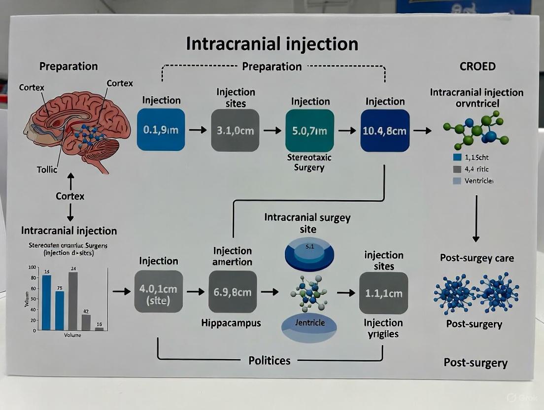
Figure 1: Stereotaxic Intracranial Injection Workflow. This diagram outlines the key stages of the surgical protocol, from animal preparation to post-operative recovery.
Discussion and Technical Notes
Validation of Technique and Equipment Calibration
The accuracy of stereotaxic surgery is not solely dependent on the equipment but also on the consistent application of the technique. Pilot studies using different strains, ages, or sexes of mice are recommended to verify atlas coordinates, as these factors can influence neuroanatomy [12]. Furthermore, the universal calibration of surgical instruments is a concept that enhances the versatility and safety of stereotactic procedures. Using universal dynamic registration hardware and software, standard surgical instruments like drills and screwdrivers can be adapted for real-time image-guided surgery, allowing for intraoperative monitoring of every step of the procedure [18]. Regular maintenance and calibration of stereotaxic instruments are critical to preserve their long-term accuracy and are services offered by reputable manufacturers [12].
Comparison with Alternative Techniques
While stereotaxic surgery is the gold standard for precise intracranial targeting, free-hand intracerebroventricular (ICV) injections serve as an alternative for specific applications. This technique relies on visual and tactile landmarks on the mouse head and does not require a stereotaxic frame [16]. It allows for rapid injections under brief anesthesia, which is beneficial for subsequent behavioral assessments. However, this method generally offers lower precision and reproducibility compared to frame-based stereotaxy and is typically reserved for targeting the larger ventricular spaces rather than specific parenchymal nuclei.
Troubleshooting Common Issues
- Inconsistent Results: Ensure the skull is perfectly level before zeroing coordinates. Verify the accuracy of bregma and lambda identification under the microscope.
- Backflow of Injectate: Confirm the injection rate is not too fast. Increasing the post-infusion wait time can significantly reduce backflow.
- Animal Morbidity: Maintain strict aseptic technique throughout the procedure. Provide adequate peri-operative analgesia and ensure the animal's body temperature is maintained during and after surgery using a homeothermic warming system [14].
This document provides detailed Application Notes and Protocols for the preparation and use of critical reagents in mouse stereotaxic surgery for intracranial injection. The procedures outlined are essential for research in neuroscience and drug development, focusing on the precise delivery of viral vectors or cells into specific brain regions. The protocol emphasizes rigorous pre-operative planning, aseptic technique, and comprehensive post-operative care to ensure animal welfare and experimental reproducibility. Adherence to these guidelines is crucial for achieving high survival rates, robust transgene expression, and valid experimental outcomes in studies employing optogenetics, chemogenetics, or disease modeling.
Research Reagent Solutions
The following table catalogues the essential materials and reagents required for successful mouse stereotaxic surgery and intracranial injection.
Table 1: Essential Reagents and Materials for Stereotaxic Intracranial Injection
| Reagent/Material | Specification/Function |
|---|---|
| Viral Vectors | Adeno-associated virus (AAV); common for gene delivery in the nervous system [19]. |
| Anesthetic Agent | Isoflurane; for induction and maintenance of surgical anesthesia via inhalation [20]. |
| Analgesic Agent | Buprenorphine HCl (0.05 mg/kg); administered subcutaneously for peri- and post-operative pain management [20]. |
| Stereotaxic Instrument | Kopf 1900 frame or equivalent; for precise, stable head fixation during surgery [20]. |
| Microinjection Syringe | Hamilton syringe with a 33-gauge needle; for accurate delivery of small volumes [20]. |
| Injection Controller | Micro4 controller (World Precision Instruments) or equivalent; to control injection flow rate (e.g., 100 nl/min) [20]. |
| Surgical Implants | Fiber-optic ferrules (e.g., 0.48 NA, Ø400 µm core); for concurrent optogenetics experiments [20]. |
| Cell Preparations | Glioma cells (e.g., 5×10^5 cells in 5 µl of DMEM) for tumor model studies [21]. |
This section summarizes critical quantitative parameters from established protocols to guide experimental design.
Table 2: Key Quantitative Parameters for Intracranial Injection in Mice
| Parameter | Typical Value/Range | Context and Purpose |
|---|---|---|
| Injection Volume | 400 nl [20] to 5 µl [21] | Volume depends on injectate (viral vector vs. cells) and target brain region. |
| Injection Flow Rate | 100 nl/min [20] | Slow, controlled flow minimizes tissue damage and backflow up the injection tract. |
| Post-Injection Pause | 5-10 minutes [20] | Allows for pressure equilibration and complete diffusion of the injectate before needle withdrawal. |
| Animal Age | 4 weeks old [21] | Common age for young adult mice in neuroscientific studies. |
| Post-op Recovery | 3 weeks [20] | Standard time to allow for maximal virally transduced gene expression before behavioral testing. |
| Sample Size | 7 mice per group [21] | Example sample size for an experiment; should be determined by power analysis. |
| Analgesic Dose | 0.05 mg/kg (Buprenorphine) [20] | Subcutaneous injection for perioperative analgesia. |
Experimental Protocols
Protocol 1: Pre-operative Anesthesia and Analgesia
Objective: To safely induce and maintain a surgical plane of anesthesia and provide pre-emptive analgesia for the mouse undergoing stereotaxic surgery. Background: Effective anesthesia is critical for animal welfare and procedural stability. Multimodal analgesia is a cornerstone of modern surgical practice, even in rodents, to minimize suffering and reduce confounding effects of post-operative pain [22].
Materials:
- Isoflurane vaporizer and induction chamber
- Medical oxygen (carrier gas)
- Buprenorphine hydrochloride (0.05 mg/kg)
- Sterile saline
- Heating pad
Procedure:
- Anesthetic Induction: Place the mouse in an induction chamber and deliver 4-5% isoflurane in oxygen until the mouse loses its righting reflex (approximately 1-2 minutes).
- Anesthetic Maintenance: Transfer the mouse to the stereotaxic frame and secure its head using ear bars and a nose cone. Maintain anesthesia with 1-2% isoflurane in oxygen for the duration of the surgery.
- Analgesic Administration: Administer buprenorphine hydrochloride (0.05 mg/kg) subcutaneously prior to the first surgical incision [20]. Pre-emptive analgesia is crucial for mitigating severe postoperative pain [22].
- Physiological Monitoring: Continuously monitor respiratory rate and depth of anesthesia. Apply ophthalmic ointment to prevent corneal drying and place the animal on a regulated heating pad to maintain body temperature at 37°C.
Protocol 2: Viral Vector Preparation and Intracranial Injection
Objective: To prepare a viral vector and perform a precise, sterile microinjection into a targeted brain region of the mouse. Background: Intracranial injection of viral vectors enables targeted gene expression in the brain [19]. This protocol is generalizable for injections into various structures like the midbrain, striatum [19], or arcuate nucleus (ARC) [20].
Materials:
- Purified and titered viral vector (e.g., AAV) aliquots, kept on dry ice until use
- Stereotaxic frame (e.g., Kopf 1900) [20]
- Hamilton syringe with a 33-gauge needle [20]
- Microinjection pump (e.g., Micro4 controller, World Precision Instruments) [20]
- Drill with fine burr
- Sterile cotton-tipped applicators and saline
Procedure:
- Viral Vector Thawing: Briefly centrifuge the viral aliquot. Thaw it rapidly in your hand or at room temperature and immediately place it on wet ice. Keep the virus on ice for the duration of the loading and injection procedure. Avoid repeated freeze-thaw cycles.
- Stereotaxic Alignment: After anesthetic induction and scalp incision, identify Bregma. Use the stereotaxic manipulator to align the injection syringe needle precisely at Bregma. Zero the digital readouts for Anterior/Posterior (A/P), Medial/Lateral (M/L), and Dorsal/Ventral (D/V) coordinates.
- Craniotomy: Move the needle to the target A/P and M/L coordinates. Using a drill, create a small craniotomy (~0.5 mm diameter) at the target site.
- Syringe Loading: Carefully load the Hamilton syringe with the chilled viral vector, ensuring no air bubbles are present in the system.
- Brain Targeting: Lower the syringe needle to the target D/V coordinate at a controlled speed.
- Microinjection: Initiate injection at a flow rate of 100 nl/min [20]. The total injection volume is typically 400 nl for viral vectors [20], though volumes can vary (e.g., 5 µl for cell injections) [21].
- Diffusion Period: Once the full volume is delivered, leave the needle in place for an additional 5-10 minutes [20]. This critical step allows for fluid pressure to normalize and the injectate to diffuse away from the needle tip, preventing backflow up the injection tract.
- Needle Withdrawal: After the diffusion period, slowly retract the needle from the brain over several minutes.
Protocol 3: Post-operative Care and Monitoring
Objective: To ensure humane recovery and well-being of the animal after surgery, maximizing the validity of experimental results. Background: Severe postoperative pain can lead to prolonged recovery, stress, and data variability. A personalized, evidence-based approach to post-operative care is essential, particularly for high-risk procedures [22].
Materials:
- Warm recovery chamber or heating pad
- Soft, sterile diet (e.g., DietGel)
- Saline for subcutaneous injection (if dehydrated)
- Scale for daily weight monitoring
Procedure:
- Recovery Environment: Place the mouse in a clean, warm, and quiet recovery cage. Provide a heat source until the animal is fully ambulatory.
- Post-operative Analgesia: Continue analgesic administration for at least 48-72 hours post-surgery. Buprenorphine can be re-administered every 8-12 hours as needed. Monitor the animal for signs of pain or distress (e.g., hunched posture, piloerection, reduced mobility).
- Health Monitoring: Weigh the animal daily until it returns to its pre-surgical weight. Check the surgical incision for signs of infection or dehiscence. Offer softened food and hydrating gels on the cage floor to facilitate easy access.
- Experimental Timeline: Allow for an adequate recovery and viral expression period, typically 3 weeks for AAVs [20], before commencing behavioral assays or terminal experiments.
Workflow and Signaling Visualization
Within the context of a mouse stereotaxic surgery protocol for intracranial injection research, the dual principles of aseptic technique and comprehensive animal welfare are not merely ethical obligations but fundamental scientific necessities. Successful surgical outcomes in research animals require the same rigorous techniques and procedures as in any veterinary practice [23]. Adherence to a standardized protocol for surgical site preparation and perioperative care ensures that experimental results are reproducible and valid, while simultaneously minimizing animal pain, distress, and the risk of confounding factors such as surgical site infections (SSIs). This application note provides a detailed framework for integrating these critical components into a single, cohesive protocol for mouse stereotaxic intracranial surgery.
Core Principles and Definitions
The Foundation of Aseptic Technique
The primary goal of aseptic technique is to reduce microbial contamination to the lowest practical level [23]. This objective is not achieved by any single practice or piece of equipment but is dependent on the combination of numerous practices and the cooperation of all personnel within the operating area. According to the Centers for Disease Control and Prevention (CDC), standard precautions form the minimum infection prevention practices that apply to all patient care, regardless of the suspected infection status [24]. In a surgical context, this translates to practices designed to protect both the animal and the integrity of the research data.
Key definitions include:
- Aseptic Technique: Practices and procedures used to reduce microbial contamination to the lowest possible level [23].
- Survival Surgery: Any operative procedure after which the animal recovers from anesthesia. All survival surgeries, whether minor or major, must be performed using aseptic surgical techniques [23].
- Major Surgical Procedure: Any intervention that penetrates and exposes a body cavity or produces permanent impairment of physical or physiological functions. Stereotaxic intracranial injection that involves a craniotomy is classified as a major survival surgery [23].
Animal Welfare and the 3Rs
Animal welfare in research is guided by the principle of the 3Rs: Replacement, Reduction, and Refinement. The protocols described herein directly address Refinement by minimizing pain and distress. Key considerations include:
- Justification of Multiple Surgeries: A single animal may not undergo more than one major survival surgery unless the procedures are interrelated components of a single IACUC-approved research project and are scientifically justified. Cost savings alone is not an acceptable justification [23].
- Personnel Training: Regardless of an individual’s educational background, all personnel must be trained thoroughly in aseptic technique, anesthesia, tissue handling, and post-surgical care, including the recognition and alleviation of pain [23].
Comprehensive Pre-Operative Preparation
Pre-Surgical Planning and Animal Acclimatization
Adequate preparation is critical for procedural success and animal well-being. Researchers should bring the animals to the surgery room ahead of time to allow for acclimatization [25]. All procedures must be performed in accordance with an IACUC-approved protocol, and all necessary anesthetics and analgesics should be acquired and handled according to institutional rules [25] [23].
Surgical Area and Instrument Preparation
The surgical area must be designed and managed to minimize contamination. Key requirements include [23]:
- Separation of animal preparation, operating, and recovery areas.
- Minimization of personnel traffic through the surgery area.
- All surfaces must be non-porous and easily sanitized.
- A regular room-cleaning and disinfection schedule must be established.
Surgical instruments, including scalpel handles, forceps, and drill bits, must be sterilized prior to the procedure using an autoclave or a glass bead sterilizer [25] [5]. The stereotaxic instrument and surrounding area should be disinfected with 70% ethanol [25] [5].
Anesthesia and Analgesia Regimen
A pre-emptive and multi-modal approach to anesthesia and analgesia is essential for animal welfare. The following table summarizes a common regimen derived from the protocols.
Table 1: Pre-Operative Anesthesia and Analgesia Regimen
| Step | Agent | Dosage and Route | Timing | Purpose | Citation |
|---|---|---|---|---|---|
| 1 | Buprenorphine (slow-release) | 1 mg kg⁻¹ (subcutaneous) | 1 hour before surgery | Pre-emptive analgesia | [5] |
| 2 | Meloxicam | 5 mg kg⁻¹ (subcutaneous) | 30 minutes before surgery; continued for 3 post-op days | Anti-inflammatory and analgesic | [5] |
| 3 | Ketamine/Xylazine/Acepromazine mixture | 0.75-1.5 ml kg⁻¹ (intraperitoneal) | After anesthetic induction | General anesthesia | [5] |
| 4 | Local Anesthetic (e.g., Lidocaine) | Applied topically | After head shaving, before incision | Local pain control | [26] |
Surgical Site Preparation Protocol
The surgical site preparation is a multi-step process designed to achieve asepsis. The following workflow diagram illustrates the sequence of key activities.
Step-by-Step Aseptic Preparation
- Head Shaving: Once the mouse is deeply anesthetized (confirmed by the absence of a pedal reflex), the hair on the head should be closely shaved using an electric hair shaver [5] [26].
- Skin Disinfection: This is a critical step for eliminating skin flora. Using sterile cotton swabs, the shaved scalp should be disinfected alternately with betadine (povidone-iodine) and 70% ethanol. This alternating scrub should be performed three times to ensure thorough asepsis [5] [26].
- Sterile Draping: After skin preparation, the area should be draped with sterile materials to maintain a sterile field around the incision site [23].
- Incision and Skull Exposure: Using a sterile surgical blade, a midline incision is made over the skull, and the skin is retracted to expose the skull surface [5].
- Skull Surface Preparation: Any residual tissue on the skull bone should be gently removed. The skull can then be cleaned with a cotton swab dipped in a freshly prepared solution of 3% H2O2 in sterile water or with sterile water alone to dry and etch the bone surface, aiding in the visualization of cranial landmarks like Bregma and Lambda [25] [27].
Essential Materials and Reagent Solutions
The following table details key reagents and materials required for effective aseptic preparation and animal welfare during stereotaxic surgery.
Table 2: Research Reagent Solutions for Asepsis and Welfare
| Category | Item | Function and Application | Citation |
|---|---|---|---|
| Disinfectants | 70% Ethanol | Disinfection of surgical area, instruments, and skin (alternating with betadine). | [25] [26] [5] |
| Betadine (Povidone-Iodine) | Broad-spectrum antiseptic for skin disinfection prior to incision. | [26] [5] | |
| 3% Hydrogen Peroxide (H2O2) | Skull cleaning and etching to visualize Bregma and Lambda; must be freshly prepared. | [25] | |
| Anesthetics & Analgesics | Ketamine/Xylazine | Injectable combination for general anesthesia. | [26] [5] |
| Isoflurane | Inhalant anesthetic for induction and maintenance of anesthesia. | [25] [26] | |
| Buprenorphine | Opioid analgesic for pre- and post-operative pain relief. | [25] [5] | |
| Meloxicam | Non-steroidal anti-inflammatory drug (NSAID) for post-operative analgesia. | [5] | |
| Animal Support | Lubricating Eye Ointment | Prevents corneal drying during anesthesia. | [26] [27] [5] |
| Sterile Saline (Lactated Ringer's) | Subcutaneous or intraperitoneal fluid for re-hydration during/after surgery. | [5] | |
| Heating Pad | Maintains body temperature during surgery and recovery; must be thermostatically controlled. | [26] [23] [5] |
Intra-Operative and Post-Operative Welfare
Intra-Operative Support
During the procedure, several measures are crucial for supporting the animal's physiological state:
- Body Temperature Maintenance: A thermostatically controlled heating pad or a circulating water blanket must be used to prevent hypothermia, as animals lose the ability to thermoregulate under anesthesia. Human heating pads or heat lamps are prohibited due to the risk of thermal injury [23].
- Eye Care: Sterile ophthalmic ointment (e.g., Lubrigel) must be applied to both eyes to prevent corneal drying [26] [23] [5].
- Hydration: Supplemental fluids such as sterile saline or Lactated Ringer's Solution should be administered subcutaneously (e.g., 1 ml) to maintain hydration [23] [5].
Post-Operative Care and Monitoring
Careful monitoring and support after surgery are imperative for recovery. Key steps include:
- Analgesia: Continue administering analgesics as prescribed in the approved protocol (e.g., Meloxicam for three consecutive days) [5].
- Monitoring: Animals should be monitored until they fully recover from anesthesia, placed in a clean cage on a heating pad, and observed for at least three days post-operatively [5]. Post-operative records must be maintained, detailing all care provided, any complications, and the administration of all medications [23].
- Wound Care: The incision should be monitored for signs of infection or dehiscence. Skin sutures or staples must be removed 10-14 days after surgery once the incision has healed [23].
- Nutritional Support: Providing moistened food pellets on the cage floor can encourage feeding and aid recovery [23].
A rigorous, integrated protocol for surgical site preparation that places aseptic technique and animal welfare on equal footing is a cornerstone of ethical and scientifically valid intracranial injection research. By adhering to the detailed procedures outlined in this application note—from pre-operative planning and meticulous skin disinfection to comprehensive intra-operative support and post-operative care—researchers can significantly improve animal well-being, minimize experimental variables, and ensure the generation of robust, reproducible data. This approach not only fulfills regulatory and ethical obligations but also enhances the overall quality and reliability of preclinical neuroscience research.
Stereotaxic surgery, a cornerstone technique in modern neuroscience research, enables precise targeting of specific brain structures for applications ranging from viral vector injections to electrode implantations. The foundation of this technique rests upon a three-dimensional Cartesian coordinate system, where cranial landmarks—primarily bregma and lambda—serve as the critical reference points for navigation [28]. The reliability of any stereotaxic procedure is therefore directly contingent upon the accurate identification and alignment of these landmarks. Despite its fundamental importance, a significant challenge persists across laboratories: the specific procedure for measuring bregma is not uniformly applied, and renowned atlases like Paxinos and Franklin often lack explicit instructions for its determination [28]. This protocol outlines a detailed methodology for utilizing stereotaxic atlases, emphasizing the correct setup from bregma and lambda to achieve highly precise and reproducible intracranial injections in the mouse brain, framed within the context of a thesis on stereotaxic surgery for intracranial injection research.
The Stereotaxic Coordinate System: Establishing the Planes of Navigation
The stereotaxic apparatus allows for movement along three primary axes: Anteroposterior (AP), Mediolateral (ML), and Dorsoventral (DV). The origin point (0,0,0) for this coordinate system is typically set at bregma, the point of intersection between the sagittal suture and the coronal suture [28]. A second landmark, lambda, which is the junction of the sagittal and lambdoid sutures, is used in conjunction with bregma to define the horizontal plane.
The core principle is to align the skull such that the bregma and lambda points are at the same dorsal-ventral height [5]. This alignment establishes a standardized horizontal plane, which is crucial because all coordinates provided in stereotaxic atlases assume this plane is level. Discrepancies in this alignment are a major source of stereotaxic error, as even minor deviations in the angle of the skull can lead to missed targets [28]. The following workflow diagram illustrates the critical steps for establishing this coordinate system.
Stereotaxic Setup Workflow
Modern Stereotaxic Atlases and Tools: From 2D Histology to 3D Digital Guides
The selection of an appropriate stereotaxic atlas is paramount for experimental success. While traditional 2D histology-based atlases like the Paxinos and Franklin atlas are widely used, they possess limitations, including tissue distortion from fixation and an inability to visualize oblique needle paths [29] [30]. The field is now advancing with 3D digital atlases that offer superior accuracy and planning capabilities.
Table 1: Comparison of Stereotaxic Atlas Modalities
| Atlas Type | Key Features | Advantages | Limitations |
|---|---|---|---|
| Traditional 2D (e.g., Paxinos & Franklin) | Histology-based coronal sections; Bregma-referenced coordinates [29]. | Widely available and accepted; Excellent histological detail. | Limited slice orientations; Potential tissue shrinkage; Difficult to plan complex trajectories [29]. |
| 3D CT/MRI Hybrid (e.g., AtlasGuide) | Co-registered CT (skull) and MRI (brain) images; Multiple developmental ages [29] [30]. | Enables 3D visualization and oblique path planning; Dynamic reorientation to subject's skull [29]. | Requires software and computational resources; May have lower cellular resolution than histology. |
| High-Resolution Cytoarchitecture (e.g., STAM) | Isotropic 1-μm resolution; Based on micro-optical sectioning tomography [8]. | Single-cell resolution; Precise 3D topography of 916 structures [8]. | Very new resource; Large dataset size may require significant computing power. |
| Waxholm Space Rat Atlas | Standardized volumetric space (NIfTI); Integration with data analysis tools [31]. | Facilitates data sharing and integration; Includes spatial coordinates of bregma/lambda [31]. | Developed for rat brain, though similar frameworks exist for mouse. |
Software tools like AtlasGuide have been developed to leverage these 3D atlases fully. A key feature is the dynamic reorientation function, which calculates the angle between the ideal bregma-lambda vector in the atlas and the actual vector measured from the experimental mouse [29]. The software then applies a rotation matrix to the 3D atlas data, matching it to the subject's unique skull orientation and eliminating the need for perfect manual alignment [29]. This significantly enhances targeting precision.
Detailed Experimental Protocol: Mouse Stereotaxic Intracranial Injection
This protocol provides a step-by-step methodology for intracranial stereotaxic injection, incorporating best practices for precise targeting from bregma and lambda [5] [32].
A. Pre-Surgical Preparation
- Anesthesia and Analgesia: Administer pre-operative analgesics (e.g., Buprenorphine, 1 mg/kg, subcutaneous) one hour before surgery. Anesthetize the mouse using an intraperitoneal injection of a ketamine/xylazine/acepromazine mixture (e.g., 0.75-1.5 ml/kg) [5]. Confirm deep anesthesia by the absence of a pedal reflex (toe pinch).
- Animal Positioning: Place the mouse in the stereotaxic instrument. Secure the head using tooth and ear bars. Apply lubricating ophthalmic ointment to prevent corneal drying.
- Aseptic Preparation: Shave the scalp and disinfect the surgical site by alternating betadine and 70% ethanol scrubs three times [5].
- Skull Exposure: Using sterile instruments, make a midline incision over the skull and gently retract the skin to expose the skull surface. Clear any connective tissue to visualize bregma and lambda sutures clearly.
B. Establishing the Stereotaxic Coordinate System
- Zero at Bregma: Lower the injection needle (e.g., a Hamilton syringe) until the tip is precisely positioned on the bregma point. Set this position as the origin for all three axes: AP=0, ML=0, DV=0 [5].
- Level the Skull: Move the needle tip to lambda and read the dorsal-ventral (DV) coordinate. Adjust the angle of the animal's head in the stereotaxic frame until the DV reading at lambda is identical to the DV reading that was recorded at bregma [5]. This critical step ensures the skull is level in the horizontal plane.
- Re-zero at Bregma: Return the needle to bregma and confirm that the coordinates are still (0,0,0). Re-adjust if necessary.
C. Targeting and Injection
- Calculate Target Coordinates: Using your chosen stereotaxic atlas (e.g., Paxinos & Franklin), identify the Anteroposterior (AP), Mediolateral (ML), and Dorsoventral (DV) coordinates for your brain region of interest relative to bregma. For example, intracerebroventricular (ICV) injections may use: AP = -0.3 mm, ML = -1.0 mm, DV = -3.0 mm [5].
- Move to Target: Move the needle to the calculated AP and ML coordinates. Drill a small craniotomy at this location.
- Inject: Slowly lower the needle to the target DV coordinate. Initiate the injection using a pump at a controlled volume and speed (e.g., 50-100 nL/min). After injection, leave the needle in place for at least 5 minutes to allow for diffusion and prevent backflow [5].
- Post-Injection: Gently withdraw the syringe, suture the incision, and apply tissue adhesive. Administer subcutaneous saline for rehydration and place the animal in a clean, warm cage for recovery, monitoring until it is ambulatory.
Advanced Applications and Considerations
A. Atlas Reorientation Logic
The mathematical principle behind software-assisted reorientation, as used in AtlasGuide, involves calculating the rotation needed to align the atlas data with the experimental subject. The software uses the bregma-lambda vector from the atlas (ṽ1) and the measured vector from the mouse (ṽ2) to compute a rotation matrix [R] [29]. This matrix dynamically reorients the 3D atlas, compensating for any tilt in the animal's head and providing more accurate coordinates for the underlying brain structures.
Atlas Reorientation Process
B. Targeting in Developing Brains
Stereotaxic surgery in neonatal rodents presents unique challenges due to rapid and non-proportional brain growth, which causes brain structures to change position relative to skull landmarks [33]. For postnatal research, specialized atlases are required. The series of atlases of the developing rat brain in stereotaxic coordinates by Khazipov et al. provides reference points for ages P0 through P21, which is crucial for targeted interventions during early development [33].
The Scientist's Toolkit: Essential Research Reagents and Materials
Table 2: Essential Materials for Stereotaxic Intracranial Injections
| Item | Function / Application | Example / Specification |
|---|---|---|
| Stereotaxic Apparatus | Provides a stable frame for precise 3D navigation and head fixation. | Digital lab standard device with micromanipulators. |
| Microsyringe | For precise delivery of nanoliter volumes into the brain. | Hamilton syringe (e.g., 10 μL) with a blunt-ended needle. |
| Anesthetic Agents | To induce and maintain a surgical plane of anesthesia. | Ketamine/Xylazine mixture; Isoflurane vaporizer. |
| Analgesics | For pre- and post-operative pain management. | Buprenorphine (slow-release), Meloxicam [5]. |
| Viral Vectors | To deliver genetic material for gene expression manipulation or tracing. | Adeno-associated virus (AAV) serotypes [32]. |
| Stereotaxic Atlas | Reference for determining 3D coordinates of brain regions. | Paxinos & Franklin (Mouse Brain); AtlasGuide Software [29] [30]. |
| Drill | To perform a craniotomy through the skull bone. | High-speed micro-drill with fine tips (e.g., 0.5 mm). |
Precise stereotaxic targeting, anchored by the correct measurement and alignment of bregma and lambda, is a non-negotiable prerequisite for rigorous and reproducible neuroscience research. While traditional 2D atlases remain useful, the adoption of 3D digital atlases and guidance software like AtlasGuide represents a significant advancement, mitigating common sources of error and enabling complex surgical planning. By adhering to the detailed protocols outlined herein and leveraging modern tools, researchers can significantly enhance the accuracy of their intracranial injections, thereby strengthening the validity of their findings in the context of drug development and basic neurological research.
Step-by-Step Stereotaxic Intracranial Injection Protocol
Within the precise domain of mouse stereotaxic surgery for intracranial injection research, achieving and maintaining a proper plane of anesthesia is a critical determinant of experimental success and animal welfare. A safe and effective anesthetic regimen ensures immobility, controls pain, and provides stable physiological conditions, thereby enabling the accurate targeting of specific brain regions such as the striatum or ARC nucleus [4] [20]. This application note details protocols for using ketamine/xylazine and isoflurane, two common anesthetic approaches, framing them within the context of a balanced anesthesia strategy to optimize outcomes in stereotaxic procedures.
Anesthetic Agent Properties and Selection
Selecting an appropriate anesthetic regimen requires a thorough understanding of the pharmacological properties of available agents. The table below summarizes the key characteristics of ketamine, xylazine, and isoflurane, which are foundational to their use in rodent surgery.
Table 1: Properties of Common Anesthetic Agents Used in Mouse Stereotaxic Surgery
| Anesthetic Agent | Mechanism of Action | Advantages | Disadvantages/Risks |
|---|---|---|---|
| Ketamine | N-methyl-D-aspartate (NMDA) receptor antagonist [34] | Provides potent analgesia; minimal depression of the cardiovascular system [34] | Can cause tremors and ataxia when used as a premedicant before isoflurane induction [34] |
| Xylazine | α2 adrenergic receptor agonist [34] | Provides sedation, muscle relaxation, and analgesia [34] | Can cause bradycardia and respiratory depression; effects are reversible with alpha-2 antagonists [34] |
| Isoflurane | Potent inhalant anesthetic; precise mechanism not fully defined | Rapid induction and recovery; easy titration of anesthetic depth [34] | Dose-dependent cardiovascular and respiratory depression; can be aversive to animals when used without sedation [34] |
Balanced Anesthesia Protocol for Stereotaxic Surgery
A balanced, or multimodal, anesthesia protocol combines drugs to capitalize on their benefits while mitigating their individual drawbacks. This approach leads to a smoother induction, reduced stress for the animal, and a lower required dose of inhalant anesthetic, which minimizes associated side effects [34]. The following integrated protocol is adapted for mouse stereotaxic intracranial injection surgery.
Pre-Anesthetic Preparation and Premedication
- Fasting: While not always mandatory for mice, consult your institution's Animal Protocol for specific guidance.
- Preemptive Analgesia: Administer analgesics such as Buprenorphine (0.05 mg/kg SC) or Meloxicam (5 mg/kg SC) 30-60 minutes before the initial incision to manage postoperative pain [20] [5].
- Premedication: Administer xylazine (4 mg/kg IP) approximately 5 minutes before induction with isoflurane. Studies show this significantly reduces the stress of isoflurane induction and lowers the minimum alveolar concentration (MAC) of isoflurane required for a surgical plane [34]. In contrast, premedication with ketamine alone may cause tremors and ataxia; therefore, if ketamine is used in a combination, it is often administered after isoflurane induction in a balanced protocol [34]. A common injectable combination for initial anesthesia is ketamine/xylazine at 40/10 mg/kg IP [4].
Induction and Maintenance with Isoflurane
- Induction: Place the premedicated mouse in an induction chamber and induce anesthesia with 4.0% isoflurane delivered in oxygen [34]. Premedication with xylazine makes this process less aversive for the animal.
- Secure the Animal: Once the mouse loses consciousness, quickly transfer it to the stereotaxic frame. Secure its head using the bite bar and ear bars, and maintain anesthesia by delivering 1.0-2.0% isoflurane via a nose cone [4]. The exact concentration must be determined based on the animal's response.
- Monitor Anesthetic Depth: Continuously assess the plane of anesthesia by checking for the absence of a pedal reflex (response to a firm toe pinch) [4] [35]. Adjust the isoflurane concentration (typically between 0.6-1.5%) to maintain the surgical plane, checking responsiveness every 5-10 minutes [4] [34].
Intraoperative Supportive Care
- Eye Lubrication: Apply ophthalmic ointment to both eyes to prevent corneal desiccation during anesthesia [4] [35].
- Thermal Support: Rodents are highly susceptible to hypothermia under anesthesia. Actively maintain body temperature using a calibrated heating pad or a circulating warm water blanket for the procedure's duration [4] [36] [35].
- Hydration: Administer sterile saline (e.g., 1 mL SQ or IP) to maintain hydration, especially during prolonged surgeries [4] [5].
The following workflow diagram summarizes the key decision points and steps in the balanced anesthesia protocol for stereotaxic surgery.
Monitoring and Troubleshooting
Vigilant monitoring is essential for detecting and correcting deviations from normal physiological parameters.
Table 2: Intraoperative Monitoring Parameters and Corrective Actions
| Parameter | Target / Normal Finding | Deviation | Potential Corrective Action |
|---|---|---|---|
| Anesthetic Depth | Absence of pedal reflex (toe pinch) [35] | Positive reflex (movement) | Gradually increase isoflurane concentration by 0.25-0.5% [4] |
| Respiratory Rate & Effort | Regular, unlabored breathing [35] | Depressed, irregular, or shallow breathing | Reduce isoflurane concentration; ensure airway patency [34] |
| Body Temperature | ~37°C (Prevent hypothermia) [36] | Hypothermia (<35°C) | Increase efficacy of active warming (e.g., heating pad) [36] [35] |
| Mucous Membrane Color | Pink [35] | Pale, blue (cyanotic), or dark red | Check for respiratory obstruction; ensure oxygen supply [35] |
The Scientist's Toolkit: Essential Materials
Successful execution of a stereotaxic surgery under anesthesia requires the following key reagents and equipment.
Table 3: Key Research Reagent Solutions and Materials for Anesthesia in Stereotaxic Surgery
| Item | Function / Application | Example / Specification |
|---|---|---|
| Isoflurane | Primary inhalant anesthetic for induction and maintenance [34] | Liquid for use with a calibrated vaporizer and oxygen carrier gas [35] |
| Ketamine & Xylazine | Injectable agents for premedication or initial anesthesia [4] [34] | Ketamine (100 mg/mL), Xylazine (20 mg/mL); often diluted and combined for IP injection [34] |
| Buprenorphine | Preemptive and postoperative analgesic to manage pain [4] [20] | Typically administered subcutaneously (e.g., 0.05 mg/kg SR or 0.1 mg/kg) [4] [20] |
| Calibrated Vaporizer | Precisely delivers a specific concentration of inhalant anesthetic to the animal [35] | Device must be calibrated annually for accuracy and safety [35] |
| Active Warming System | Prevents anesthesia-induced hypothermia, improving survival and recovery [36] [35] | Circulating water blanket, thermal pad, or custom heat bed with temperature control [36] [35] |
| Ophthalmic Ointment | Prevents corneal drying and damage during anesthesia [4] [35] | Petroleum-based ophthalmic ointment |
A meticulously planned and executed anesthetic protocol is the cornerstone of ethical and successful mouse stereotaxic surgery. The balanced approach, which leverages the synergistic effects of xylazine premedication and isoflurane maintenance, offers significant benefits. These include a less stressful induction for the animal, a reduced requirement for isoflurane, and enhanced intraoperative stability [34]. When combined with rigorous monitoring and proactive supportive care—especially the prevention of hypothermia [36]—this protocol provides a robust framework for ensuring animal welfare and the collection of highly reproducible scientific data in intracranial injection research.
Within the rigorous framework of mouse stereotaxic surgery for intracranial injection research, the initial steps of secure head fixation and skull leveling are undeniably foundational. The precision required to accurately target specific brain regions—be it for viral vector delivery, drug administration, or device implantation—is entirely contingent upon a stable and correctly aligned cranial platform. Inaccuracies at this stage propagate through the entire procedure, compromising data integrity and experimental reproducibility. This protocol details the critical methodologies for achieving rigid head fixation in the stereotaxic frame and systematically ensuring the skull is positioned in a true horizontal plane, thereby establishing the bedrock for successful and reliable neuroscientific investigations.
The Scientist's Toolkit: Essential Materials for Head Fixation and Leveling
The following table catalogues the essential reagents and equipment required for the procedures outlined in this application note.
Table 1: Key Research Reagent Solutions and Essential Materials
| Item Name | Function/Application |
|---|---|
| Stereotaxic Frame | Provides the rigid framework for immobilizing the mouse skull during surgery [4]. |
| Non-Rupture Ear Bars | Paired components that gently secure the mouse's head by engaging the auditory canals, ensuring symmetric lateral fixation [37]. |
| Bite Bar | Stabilizes the head in the anteroposterior (AP) axis and, when adjustable, helps control the pitch of the skull [4]. |
| Isoflurane Anesthesia System | Delivers inhaled gas anesthetic (e.g., 2-2.5% for maintenance) for stable, prolonged unconsciousness, preventing movement during leveling and surgery [38] [37]. |
| Heating Pad | Maintains the animal's body temperature at approximately 39°C during anesthesia to prevent hypothermia [37]. |
| Digital Vernier Scale / Readout | Provides high-precision digital measurements of stereotaxic coordinates for accurate positioning and leveling [37]. |
| Surgical Drill | Used with a fine drill bit to create precise burr holes in the skull for injections or implants after leveling is complete [4] [38]. |
| Ophthalmic Ointment | Prevents corneal damage and drying during anesthesia [4] [37]. |
| Analgesics (e.g., Buprenorphine) | Administered pre-emptively (e.g., 0.05-0.1 mg/kg) to manage postoperative pain, a critical animal welfare consideration [39] [38]. |
Quantitative Specifications for a Level Skull
Achieving a level skull is a quantitative process, defined by specific tolerance thresholds between key cranial landmarks. The following table summarizes the core coordinate targets and acceptance criteria.
Table 2: Quantitative Coordinates and Leveling Tolerances
| Parameter | Target Landmarks | Acceptance Criteria | Citation |
|---|---|---|---|
| A/P (Anteroposterior) Levelness | Bregma and Lambda Dorsal Height | The dorsal-ventral (DV) coordinate difference between Bregma and Lambda should be < 0.05 mm. | [4] |
| M/L (Mediolateral) Levelness | Bregma and Points 2 mm Lateral | The DV coordinate difference between points 2 mm to the left and right of Bregma should be identical. | [4] |
| Injection Depth Zeroing | Skull Surface at Bregma | The DV coordinate at the skull surface of Bregma is defined as zero (0.00 mm) for subsequent depth measurements. | [5] |
Experimental Protocol: Head Fixation and Skull Leveling
This detailed methodology guides the researcher from animal preparation through the final verification of a level skull.
Animal Preparation and Anesthesia
- Analgesia: At least 20 minutes prior to surgery, administer a subcutaneous injection of a sustained-release analgesic such as Buprenorphine at a dose of 50 µg/kg [37]. Some protocols administer this up to an hour before surgery [5].
- Anesthesia Induction: Place the mouse in an induction chamber saturated with 5% isoflurane delivered in oxygen (flow rate of 1.2 L/min) until the animal loses consciousness [37].
- Head Fixation in Frame:
- Ear Bar Placement: Carefully insert the blunt, non-rupture ear bars into the external auditory meatus. Adjust them symmetrically until the head is firmly and immovably seated. The head should not shift when gentle downward pressure is applied [4].
Surgical Exposure and Initial Skull Preparation
- Scalp Preparation: Remove hair from the surgical site using clippers or a depilatory cream. Disinfect the scalp with alternating scrubs of betadine and 70% ethanol, repeated three times [4] [37].
- Incision: Using a sterile scalpel, make a midline incision along the sagittal suture, from the level of the eyes to the back of the skull. Use surgical clips to retract the skin and expose the skull [4] [38].
- Skull Cleaning: Use the rounded edge of a scalpel blade or a small tool to gently scrape the surface of the skull, clearing away any connective tissue or fascia to clearly visualize Bregma (the junction of the sagittal and coronal sutures) and Lambda (the junction of the sagittal and lambdoid sutures) [4].
Systematic Skull Leveling Procedure
This process ensures the skull surface is flat in both the anteroposterior (A/P) and mediolateral (M/L) planes. Use a dissecting microscope for all steps.
Diagram 1: Skull leveling workflow and decision process.
Anteroposterior (A/P) Leveling:
- Lower a drill bit or sterile needle attached to the stereotaxic arm until it just touches the skull at Bregma. Note the Dorsal-Ventral (Z) coordinate precisely [4] [5].
- Lift the tool, move it posteriorly to Lambda, and lower it to touch the skull. Note the Z coordinate at Lambda [4].
- Calculate the difference between the two Z coordinates. The skull is considered level in the A/P plane if this difference is less than 0.05 mm [4].
- If the difference exceeds this tolerance, adjust the height of the bite bar to tilt the skull and re-measure the coordinates at Bregma and Lambda iteratively until the criterion is met [38].
Mediolateral (M/L) Leveling:
- Return the tool to Bregma and zero the X, Y, and Z coordinates [5].
- Move the tool 2.0 mm to the left of Bregma, lower it to the skull surface, and note the Z coordinate.
- Lift the tool, move it 2.0 mm to the right of Bregma, lower it to the skull, and note the Z coordinate [4].
- The Z coordinates on the left and right must be identical. A difference indicates head tilt.
- If a difference is found, re-check the symmetry and depth of the ear bars, ensuring the head is perfectly centered. Adjust the ear bars and re-measure until the left and right Z coordinates are equal [4].
Once both A/P and M/L leveling are complete, the skull is correctly positioned for accurate navigation to target brain coordinates. The coordinates for the target region can now be calculated relative to the defined zero point at Bregma [5].
In mouse stereotaxic surgery for intracranial injection research, the creation of a precise burr hole is a critical step that enables access to the brain for the delivery of viral vectors, drugs, or other therapeutic agents. This procedure requires meticulous planning and execution to ensure accurate targeting while minimizing damage to underlying neural tissue and surrounding vasculature. The burr hole serves as the primary portal through which all intracranial manipulations occur, making its size, location, and construction fundamental to experimental success. Within the broader context of a stereotaxic surgery protocol, burr hole creation bridges the gap between superficial surgical exposure and deep brain targeting, requiring integration of anatomical knowledge, precision instrumentation, and refined technical skill. The precision of this step directly influences the reliability and reproducibility of research outcomes in neuroscience and drug development studies [40] [41].
Essential Concepts and Definitions
Stereotaxic Surgery: A minimally invasive neurosurgical technique that enables precise targeting of specific brain structures using a three-dimensional coordinate system based on cranial landmarks. This approach allows researchers to accurately deliver injections to discrete brain regions with minimal tissue disruption [42] [43].
Bregma: The anatomical point on the skull where the coronal and sagittal sutures intersect. This landmark serves as the primary reference point (zero point) for establishing the stereotaxic coordinate system in mouse surgery. Accurate identification of bregma is crucial as all subsequent target coordinates are calculated relative to this point [41] [43].
Lambda: The point where the sutures of the parietal and occipital bones converge. This landmark provides an important secondary reference point for verifying head position and coordinate accuracy, particularly for targets in the posterior regions of the brain [43].
Burr Hole: A small opening created in the skull bone to provide access to the underlying brain tissue for injections, implant placement, or other experimental procedures. The optimal burr hole is just large enough to accommodate the injection needle without unnecessary damage to the skull or underlying tissue [40] [43].
Brain Atlases: Reference publications containing detailed maps of brain anatomy with corresponding stereotaxic coordinates. The most widely used authority is Paxinos and Franklin's The Mouse Brain in Stereotaxic Coordinates, which provides comprehensive coronal, sagittal, and horizontal diagrams with over 800 identified structures [44] [45].
Materials and Instrumentation
Table 1: Essential Equipment for Stereotaxic Surgery and Burr Hole Creation
| Equipment Category | Specific Items | Purpose and Specifications |
|---|---|---|
| Stereotaxic Frame | Mouse adapter, ear bars, mouth bar | Secure and stable head fixation using non-traumatic ear bars [41] [43] |
| Drilling System | High-speed micro drill (e.g., Stoelting), carbide burrs (0.5-1.0 mm) | Create precise craniotomy with minimal vibration and thermal damage [40] [41] |
| Surgical Instruments | Scalpel handle (#10 blade), tissue forceps (Graefe), wound retractors, spring scissors (Vannas), hemostats | Tissue dissection, hemostasis, and surgical site exposure [43] |
| Anesthesia System | Isoflurane vaporizer (4% induction, 1-2% maintenance), oxygen supply (0.5 L/min), induction chamber | Maintain surgical plane of anesthesia while preserving physiological functions [43] |
| Stereotaxic Navigation | Micromanipulator (Kopf), electrode holder, pneumatic PicoPump | Precise coordinate targeting and controlled fluid delivery [43] |
| Monitoring Equipment | Heating pad (Stoelting), thermoregulation system | Maintain mouse body temperature at physiological levels during surgery [43] |
Table 2: Key Consumables and Reagents
| Material Type | Specific Examples | Application Notes |
|---|---|---|
| Anesthetics | Isoflurane, oxygen | Preferred over injectables for rapid induction and recovery [43] |
| Analgesics | Meloxicam SR | Pre- and post-operative pain management [43] |
| Injection Materials | AAV vectors, fluorescent tracers (Cholera toxin subunit-b), fluorescent microspheres | Neural tracing, gene expression manipulation [43] |
| Sterile Supplies | Saline, cotton applicators, non-fenestrated drapes, surgical gloves | Maintain aseptic technique throughout procedure [43] |
| Suture Materials | 5-0 polypropylene | Wound closure with minimal tissue reaction [43] |
Preoperative Planning Procedures
Anatomical Landmark Identification
The foundation of accurate stereotaxic surgery begins with proper identification of cranial landmarks. The mouse must be securely positioned in the stereotaxic frame using non-traumatic ear bars and a mouth bar to eliminate head movement. The surgical site should be shaved and disinfected using alternating alcohol and betadine pads. Using a surgical microscope under 5-40x magnification, the sagittal suture should be visually identified running along the midline of the skull. Bregma is located as the intersection point between the coronal and sagittal sutures, while lambda is identified as the convergence point of the sagittal and lambdoid sutures. Verification of proper head alignment is confirmed by ensuring the dorsal-ventral coordinates of bregma and lambda do not differ by more than 0.05 mm [41] [43].
Coordinate Calculation and Target Planning
Using the Paxinos and Franklin mouse brain atlas as a reference, the target brain structure coordinates are calculated relative to bregma. The atlas provides detailed diagrams spaced at approximately 120 µm intervals, allowing for precise targeting of over 800 identifiable structures. Modern digital versions, such as the Mouse Brain Atlas by Matt Gaidica, offer interactive coordinate planning and visualization. When planning the burr hole location, consider the surgical trajectory to avoid major blood vessels and ventricles. For targets in the caudal brainstem or upper cervical cord, the cisterna magna approach provides an alternative method that bypasses the challenges of remote targeting from skull landmarks [44] [45] [43].
Surgical Protocol: Burr Hole Creation
Surgical Approach and Skull Exposure
After anesthesia induction and scalp preparation, a midline incision (approximately 1.5-2 cm) is made using a #10 surgical blade, extending from the level of the eyes to the posterior skull. The skin is gently retracted using wound hooks, and the underlying connective tissue is carefully dissected to fully expose the skull surface. The periosteum should be thoroughly cleared using a combination of blunt dissection and cotton-tipped applicators, providing a clean visual field for landmark identification. Hemostasis is maintained through gentle pressure with sterile cotton applicators [40] [43].
Drilling Technique and Optimization
The drill should be held perpendicular to the skull surface at the predetermined coordinates. Initial contact should be made with the drill bit at low speed, using a gentle pecking motion rather than continuous pressure. Intermittent drilling with saline irrigation prevents thermal injury to the underlying cortex. The burr hole diameter should be precisely calibrated to the injection needle (typically 0.5-1.0 mm) to minimize cerebrospinal fluid leakage and brain surface exposure. Drilling should cease immediately upon breakthrough, characterized by an audible change and visible dura mater. The bone dust collected during drilling can be preserved in sterile saline for potential skull reconstruction [40] [46] [43].
Special Considerations for Caudal Brainstem Targets
For targets in the medulla oblongata or upper cervical cord, the standard dorsal approach through the skull presents specific challenges due to the anatomical position of the cerebellum and the slanting occipital bone. In these cases, the cisterna magna approach provides direct visualization of brainstem landmarks. This technique requires anteroflexing the head and carefully navigating between the paired bellies of the longus capitis muscle to expose the dorsal surface of the caudal brainstem. The obex, where the central canal opens into the fourth ventricle, serves as the zero point for coordinate measurement in this approach [43].
Technical Considerations and Troubleshooting
Table 3: Common Burr Hole Creation Challenges and Solutions
| Technical Challenge | Potential Consequences | Recommended Solutions |
|---|---|---|
| Inaccurate landmark identification | Coordinate miscalculation, target miss | Verify head alignment (bregma/lambda difference <0.05 mm), use surgical microscope [43] |
| Excessive drilling pressure | Skull fracture, underlying cortical damage | Use pecking motion, intermittent drilling, high-speed drill to reduce required force [40] |
| Thermal injury from drilling | Cortical necrosis, inflammatory response | Implement saline irrigation, use sharp drill bits, limit continuous drilling duration [40] |
| Dural perforation or tear | CSF leakage, cortical dehydration, infection | Use fine forceps (Dumont #5/45) for dural manipulation, consider cisterna magna approach for caudal targets [43] |
| Inadequate hemostasis | Obscured surgical field, subdural hematoma | Apply gentle pressure with sterile cotton applicators, use bone wax for skull bleeding [43] |
| Burr hole size miscalibration | Needle drag, CSF leakage, or insufficient access | Match drill bit to needle gauge (typically 0.5-1.0 mm), create minimal necessary access [40] [43] |
Postoperative Management and Validation
Following burr hole creation and subsequent intracranial injection, the surgical site should be thoroughly irrigated with sterile saline to remove any bone debris or blood. The dura should remain intact unless specifically required by the experimental protocol. The scalp incision is closed in layers using 5-0 polypropylene suture, with particular attention to achieving watertight closure of the skin. Postoperative analgesia (Meloxicam SR) should be administered, and the animal monitored closely during recovery until ambulatory. For validation of injection accuracy, histological verification should be performed post-sacrifice. For functional studies, appropriate behavioral testing or physiological measurements should be implemented according to experimental objectives [40] [43].
Advanced surgical navigation technologies, while more common in human neurosurgery, provide conceptual frameworks for precision validation. These systems allow for intraoperative verification of target accuracy and can detect complications such as bleeding. While not typically used in mouse models due to scale constraints, the principles of real-time feedback and accuracy verification should be incorporated through alternative validation methods [42] [47].
In the field of neuroscientific research, particularly in mouse stereotaxic surgery for intracranial injection, the precision of the injection system is a critical determinant of experimental success. The process of loading the delivery instrument—whether a Hamilton syringe or a Nanoject injector—with a therapeutic or investigative agent (such as a virus, drug, or cells) is a fundamental yet delicate step. An improperly loaded system can introduce air bubbles, leading to inaccurate dosing, unpredictable diffusion, and potential damage to the target brain tissue. This application note provides detailed protocols and technical data to guide researchers in achieving bubble-free, precise loading of Hamilton syringes and Nanoject injectors, ensuring the reliability and reproducibility of stereotaxic intracranial injections.
The Scientist's Toolkit: Essential Materials and Reagents
The following table details the key materials required for the loading and injection procedures described in this note.
Table 1: Key Research Reagent Solutions and Materials for Intracranial Injection
| Item | Function/Description |
|---|---|
| Hamilton Syringe [48] [49] | A precision microsyringe, often with a small gauge needle, designed for accurate dispensing of volumes in the microliter range. Essential for both direct intracranial injections and loading into cannula systems. |
| Glass Capillary [27] | A fine, pulled glass tip used with micropipette injection pumps for highly precise delivery. It is filled with the injection solution and is critical for minimizing tissue damage. |
| Cell/Drug Solution | The substance to be injected, which can include viruses, cells, drugs, or dyes, prepared in a sterile, compatible buffer [27]. |
| Mineral Oil [27] | Used to fill the glass capillary and the connected tubing system in a cannula setup to create a hydraulically continuous, bubble-free fluid path for the drug solution. |
| Phosphate Buffered Saline (PBS) / Culture Medium [49] | Aqueous solutions used to flush and prime microfluidic systems or to dilute concentrates, ensuring a sterile and physiologically compatible environment. |
| Aqueous Ethanol (70%) [49] | Used to disinfect and pre-wet the surfaces of devices like lab-on-a-chip platforms, which aids in removing air bubbles by reducing surface hydrophobicity. |
| Dental Cement & Biological Glue [27] | Used to securely fix an implanted guide cannula to the skull, creating a permanent port for repeated intracranial administrations. |
Technical Data and Specifications
Selecting the appropriate apparatus and parameters is foundational to a successful injection. The quantitative data below serves as a guide for system setup.
Table 2: Key Technical Data for Loading and Injection Parameters
| Parameter | Typical Specification | Application Context |
|---|---|---|
| Injection Volume | 0.2 µL to 10 µL [48] | Common range for Hamilton syringes in gel loading and intracranial applications. |
| Needle Gauge | Small gauge (e.g., 26s - 33s) | Designed for minimal tissue disruption during brain injections [48]. |
| Post-Injection Diffusion Wait Time | 5 - 20 minutes [5] [27] | Critical period after injection to allow for proper diffusion of the solution into the brain tissue before withdrawing the needle. |
| Injection Speed | Controlled, slow rate (e.g., 0.01 mm/s for needle movement) [27] | Minimizes backflow and leakage of the solution along the injection tract. |
| Pre-Injection Pause | ~1 minute [27] | A brief pause after the needle is positioned at the target site to allow tissue pressure to equilibrate. |
Protocols for Bubble-Free System Loading
Air bubbles are the most common source of error in micro-injections, causing volume inaccuracies and potential clogging. The following protocols outline steps to eliminate them.
Protocol 1: Loading a Hamilton Syringe for Direct Injection
This protocol is adapted from general principles of bubble-free fluid handling for precise biological applications [49].
- Preparation: Ensure the Hamilton syringe and needle are clean and sterile.
- Draw the Solution: Immerse the needle tip completely into the solution to be injected. Slowly retract the plunger to draw the required volume into the syringe barrel. Avoid drawing the solution too rapidly.
- Bubble Elimination: Hold the syringe vertically with the needle pointing upwards. Gently tap the side of the syringe barrel to dislodge any air bubbles, allowing them to rise to the top.
- Purge Air: Slowly depress the plunger to expel the air bubbles and a small amount of solution from the needle hub, ensuring the entire fluid path is filled with liquid.
- Inspection: Visually inspect the syringe and needle to confirm the absence of bubbles before mounting it onto the stereotaxic instrument.
Protocol 2: Priming a Cannula System for Repeated Administration
For chronic experiments requiring multiple injections, a guide cannula is implanted and connected to a reservoir via tubing. This system must be meticulously primed [27].
- Fill with Mineral Oil: Using a syringe and PE tube, fill the entire system—including the guide cannula, tubing, and injection needle—with mineral oil. Use a glue gun to seal connections and prevent leaks.
- Eliminate Bubbles: Perform a suction procedure with the syringe pump to draw mineral oil through the system, ensuring all air bubbles are purged.
- Load the Drug: With the system filled with oil, execute the syringe pump's suction procedure again to draw the drug solution from a reservoir into the tip of the injection cannula. This creates a clean interface between the oil and the drug solution without introducing air.
- Connect and Inject: Remove the dummy cannula from the guide cannula implanted in the animal. Insert the now-primed injection cannula. After a brief rest period of about 1 minute, execute the injection.
Integrated Workflow for Stereotaxic Intracranial Injection
The diagram below illustrates the complete logical workflow for a stereotaxic intracranial injection procedure, integrating the loading of the injection system with the surgical steps [5] [27].
Critical Technical Considerations for Loading and Injection
Beyond the basic steps, several technical considerations are vital for ensuring the fidelity of the procedure and the validity of the resulting data.
Precision of Stereotaxic Coordinates: The absolute reliance on stereotaxic coordinates demands rigorous calibration. Before loading the injection system, the skull must be leveled with high precision by confirming the dorsal-ventral (DV) coordinates at both Bregma and Lambda. A difference of less than 0.03 mm is often considered acceptable for a horizontal skull position [27].
Injection Speed and Volume Control: The use of a micro-syringe pump is non-negotiable for modern stereotaxic surgery. It allows for the precise digital control of injection speed and volume. A slow, controlled injection speed is crucial to prevent backflow of the solution and to minimize trauma to the surrounding tissue [27].
The Importance of the Diffusion Period: The waiting period of 5 to 20 minutes after the injection is complete, before withdrawing the needle, is a critical step for allowing the injected solution to diffuse adequately into the interstitial space of the brain tissue. This practice significantly reduces the volume of solution that may leak back up the injection tract [5] [27].
Aseptic Technique and Post-Operative Care: Maintaining sterility throughout the procedure is paramount to prevent infection, which can confound experimental results. This includes autoclaving surgical tools and using disinfectants. Post-operative care, including the administration of analgesics (e.g., Buprenorphine, Meloxicam) and antibiotics, is essential for animal welfare and data quality [5].
In the field of basic brain science research, intracranial stereotaxic injection is the most direct method for achieving precise delivery of substances to target brain regions. This technique is a cornerstone of most animal experiments, enabling the injection of viruses, cells, protein molecules, drugs, and labeled dyes [27]. The precise control of infusion parameters—specifically flow rate, injection volume, and dwell time—is critical for maximizing delivery accuracy, minimizing tissue damage, and ensuring experimental reproducibility. These parameters directly influence the distribution of the injected substance and the extent of backflow along the needle tract, which can compromise targeting specificity. Within the broader context of a mouse stereotaxic surgery protocol, mastering these variables is fundamental to the success of intracranial injection research for neurological disease modeling, advanced brain function studies, and drug development.
Key Infusion Parameters and Recommendations
The following table summarizes the core quantitative parameters for intracranial injection in rodents, as established in current protocols and research. Adherence to these guidelines is essential for maintaining tissue integrity and achieving predictable substance distribution.
Table 1: Key Infusion Parameters for Mouse Intracranial Injection
| Parameter | Recommended Value | Additional Context & Notes |
|---|---|---|
| Injection Volume | 1-2 µL (for standard bolus) | Maximum recommended volume for a single intracranial injection in mice is 0.15-0.2 mL [50]. Smaller volumes (e.g., 2.5 µL) are used for specific targets like the lateral ventricle [51]. |
| Flow Rate | 0.1 - 0.5 µL/min | A slow infusion speed is critical. A common rate for ventricular injection is 0.5 µL/min [51]. For very precise control, an even slower rate of 0.01 mm/s for needle movement is suggested to reduce leakage [27]. |
| Dwell Time | Pre-injection: ~1 minutePost-injection: 15-20 minutes | After the needle is positioned, wait ~1 minute to balance air pressure before starting infusion [27]. After infusion is complete, let the needle remain in place for 15-20 minutes to allow for tissue absorption and to reduce backflow [27]. |
| Needle Gauge | 33-gauge | Example from a lateral ventricle injection study, indicating the use of fine needles for minimal tissue disruption [51]. |
Detailed Experimental Protocols
Standard Protocol for Single Cranial Administration
This protocol outlines the core steps for a one-time stereotaxic injection, which is suitable for many applications including viral vector delivery, tracer studies, and single-dose drug administration [27].
The Scientist's Toolkit: Essential Materials for Single Administration
- Stereotaxic Apparatus: For precise, three-dimensional positioning of the injection needle.
- Laboratory Animal Anesthesia Machine: Typically using isoflurane, to maintain the animal at a surgical plane of anesthesia.
- Micro syringe Pump: Allows for accurate, computer-controlled delivery of very small volumes at defined flow rates.
- Micropipette Puller: For creating fine-tipped glass capillaries used for injection.
- Micro drill: For performing a carefully controlled craniotomy.
Step-by-Step Procedure:
- Animal Anesthesia and Preparation: Induce and maintain anesthesia (e.g., with isoflurane). Place the mouse in the stereotaxic frame using ear bars and a nose clip. Apply ophthalmic ointment to prevent corneal drying and shave the scalp [27].
- Skull Leveling and Targeting: Using the micro drill tip, identify the Bregma and Lambda sutures. Adjust the skull in the horizontal plane until the height difference (DV value) between Bregma and Lambda is less than 0.03 mm [27].
- Craniotomy: Move the micro drill above the target coordinates and drill a small hole through the skull, taking care to stop at the dura mater [27].
- Needle Preparation and Loading: Pull a glass capillary using a micropipette puller. Fill it with mineral oil to eliminate air bubbles, then load it with the substance to be injected [27].
- Lowering the Needle and Substance Infusion:
- Fix the loaded capillary into the micropipette pump on the stereotaxic arm.
- Lower the needle slowly to the predetermined depth in the target brain region.
- Critical Step: Upon reaching the target, pause for 1 minute to allow tissue pressure to equilibrate [27].
- Initiate infusion using the pre-set flow rate (e.g., 0.1-0.5 µL/min) and volume (e.g., 1-2 µL).
- Dwell Time and Needle Withdrawal:
- Critical Step: After the infusion is complete, let the needle remain in place for 15-20 minutes. This allows the injected substance to be absorbed by the tissue and significantly reduces reflux up the needle track [27].
- Withdraw the needle very slowly, ideally at a controlled speed of 0.01 mm/s [27].
- Suture and Postoperative Care: Suture the scalp, apply antibiotic ointment to the wound, and provide postoperative analgesia and warmth until the animal fully recovers [27].
Protocol for Chronic Cannula Systems for Multiple Administrations
For studies requiring repeated substance delivery to the same brain region over days or weeks, the implantation of a guide cannula is the preferred method. This avoids the need for repeated surgeries and ensures consistent targeting [27].
The Scientist's Toolkit: Essential Materials for Cannula Systems
- Guide Cannula: A permanent guide tube cemented to the skull, providing a port for repeated injections.
- Dummy Cannula: Keeps the guide cannula sealed between injections.
- Internal Injection Cannula: A thinner needle that extends from the guide cannula to the injection site, connected via PE tubing to a syringe pump.
- Dental Acrylic & Sterile Screws: Used to securely anchor the guide cannula to the skull.
- Micro syringe Pump: For controlled infusion during each dosing session.
Step-by-Step Procedure:
- Anesthesia, Skull Preparation, and Leveling: Follow steps 1-3 from the single administration protocol [27].
- Drilling and Screw Implantation: Drill the main hole for the cannula and additional small holes in the surrounding skull. Insert sterile skull screws to serve as anchors for the dental cement [27].
- Cannula Implantation: Lower the guide cannula to the target brain region. Apply biological glue to the skull opening, then carefully build up layers of dental cement around the cannula and skull screws to create a stable, sealed head cap [27].
- Drug Delivery Session:
- Connect an oil-filled syringe to the injection cannula via PE tubing and load the substance into the tip of the cannula.
- Remove the dummy cannula from the guide.
- Gently insert the injection cannula into the guide cannula and secure it.
- Let the animal acclimatize for 1 minute [27].
- Initiate infusion using the syringe pump at the desired flow rate and volume.
- After infusion, employ a post-injection dwell time of 1-5 minutes before carefully removing the internal injection cannula and replacing the dummy cannula [27].
The following workflow diagram illustrates the key decision points and procedures for both single and repeated intracranial injection methods.
Critical Experimental Considerations
Anesthesia and Physiological Monitoring
The choice and management of anesthesia are critical variables that can directly influence infusion outcome and data interpretation. Isoflurane is a commonly used anesthetic for stereotaxic surgery [52] [51]. However, it induces peripheral vasodilation and can cause significant hypothermia in rodents, which is associated with cardiac arrhythmias, vulnerability to infection, and prolonged recovery time [52]. The use of an active warming pad system to maintain the animal's body temperature at approximately 37-40 °C throughout the procedure is highly recommended, as it has been shown to notably improve survival rates and postoperative recovery [52]. Furthermore, the depth of anesthesia can affect brain physiology and solute distribution. Studies on cerebrospinal fluid (CSF) dynamics have shown that tracer clearance from the ventricles is more efficient under awake and low-dose isoflurane conditions compared to high-dose isoflurane, which causes greater tracer retention [51]. Researchers must therefore standardize and report their anesthesia protocols meticulously.
Substance Formulation and Quality
The properties of the injected solution are as important as the mechanical parameters of the infusion. For parenteral administration, including intracranial injection, substances should ideally be sterile, isotonic (the same solute concentration as blood), and at a physiologic pH (6.8-7.2) to minimize tissue irritation and neuronal damage [50]. If the solution's pH is outside this range, it should be buffered appropriately. Solutions that are not commercially manufactured and pre-sterilized must be prepared under aseptic conditions, typically in a laminar flow hood or biosafety cabinet, and filtered through a 0.2-micron filter to ensure sterility [50]. For novel formulations, such as nanoparticles or hydrogels used in convection-enhanced delivery (CED), the physical properties like viscosity and particle size must be optimized to promote flow and distribution within the brain parenchyma [53].
Troubleshooting and Optimization
Even with a meticulous protocol, challenges can arise. The following table addresses common issues and provides evidence-based solutions.
Table 2: Troubleshooting Common Intracranial Injection Issues
| Problem | Potential Cause | Recommended Solution |
|---|---|---|
| Backflow/Leakage | High flow rate, large volume, insufficient dwell time, incorrect needle size. | Optimize parameters: Slow the flow rate (0.1 µL/min), reduce volume if possible, and ensure a 15-20 minute post-infusion dwell time [27]. Withdraw the needle slowly (0.01 mm/s) [27]. |
| High Mortality Rate | Hypothermia from prolonged isoflurane anesthesia, surgical stress. | Use an active warming pad set to 40 °C to maintain body temperature throughout surgery, which has been shown to dramatically improve survival [52]. |
| Low Experimental Reproducibility | Inconsistent skull leveling, variable infusion parameters. | Strictly enforce the skull leveling criterion (Bregma-Lambda DV difference < 0.03 mm) [27]. Use a microprocessor-controlled syringe pump for consistent flow rates and volumes across all animals. |
| Poor Substance Distribution | Incorrect formulation properties, backflow, rapid clearance. | For ventricular injections, consider the animal's state; clearance is faster in awake states [51]. For parenchymal delivery, ensure proper dwell time and explore formulation strategies (e.g., viscosity modifiers) for CED [53]. |
The precise control of flow rate, volume, and dwell time during intracranial injection is not merely a technical detail but a fundamental determinant of experimental success. Adherence to the established parameters—slow flow rates (0.1-0.5 µL/min), small volumes (1-2 µL for most mouse applications), and adequate pre- and post-infusion dwell times (~1 min and 15-20 min, respectively)—is essential for maximizing target engagement, minimizing tissue trauma, and ensuring the reliability and interpretability of research data. By integrating these optimized infusion parameters with rigorous surgical technique, careful physiological monitoring, and appropriate substance formulation, researchers can significantly enhance the validity and impact of their stereotaxic studies in mouse models of brain function and disease.
This application note provides detailed protocols for the perioperative care of mice following stereotaxic surgery for intracranial injection, a critical procedure in neuroscience research. The focus is on standardized methods for wound closure, effective analgesia, and systematic post-operative monitoring to ensure animal welfare, minimize experimental variability, and enhance data reproducibility. The recommendations are framed within the context of a broader thesis on mouse stereotaxic surgery, integrating evidence-based practices from clinical neurosurgery and tailored pre-clinical studies to support researchers and drug development professionals.
Experimental Protocols & Workflows
Standardized Protocol for Wound Closure and Post-operative Care
The following workflow outlines the key stages for managing a mouse from the end of an intracranial injection procedure through the initial recovery phase. Adherence to this protocol is crucial for animal well-being and data consistency.
Procedure Details:
- Wound Irrigation: After removing the stereotaxic arm and ensuring hemostasis, gently irrigate the surgical wound on the skull with sterile saline to remove any debris or blood clots [54].
- Layered Wound Closure: If a significant muscle layer (e.g., temporalis) was incised, approximate it using absorbable sutures (e.g., Vicryl 5-0) to eliminate dead space and reduce tension on the skin. For the skin, use simple interrupted stitches or wound clips (e.g., Nylon 6-0, Autoclips) to ensure secure closure [55] [56].
- Initial Recovery: Place the animal in a clean, single-housed cage placed on a warming pad (or under a heat lamp) in a quiet environment. Monitor the animal every 15 minutes until it is ambulatory, typically for the first 1-2 hours post-anesthesia.
Post-operative Analgesia Management Pathway
Effective pain management is paramount for animal welfare and scientific rigor. This pathway supports decision-making for post-operative analgesia, based on a multimodal approach.
Procedure Details:
- Pain Assessment: Utilize a validated tool like the Mouse Grimace Scale (MGS) to score pain at least twice daily for the first 72 hours. Signs include orbital tightening, nose/cheek bulge, and ear position change.
- Pre-emptive and Base Analgesia: Administer a non-steroidal anti-inflammatory drug (NSAID) such as Meloxicam (5 mg/kg SC) or Ketorolac (0.3 mg/kg SC) subcutaneously prior to or immediately following surgery. NSAIDs are effective for somatic pain originating from cutaneous and deep tissues like the scalp and pericranial muscles [57] [58].
- Scheduled Dosing and Escalation: Continue NSAIDs for a minimum of 48-72 hours post-operatively on a scheduled basis. If pain is not adequately controlled (e.g., MGS score remains high), escalate therapy by adding a opioid such as Buprenorphine (0.05-0.1 mg/kg SC every 6-12 hours), which was used for perioperative analgesia in a mouse TBI model [55]. A multimodal regimen combining different drug classes (NSAIDs and opioids) provides superior analgesia compared to intermittent administration of a single agent [57] [59].
- Monitoring and Weaning: Closely monitor for potential side effects of analgesics, such as sedation or respiratory depression, though IV-PCA with fentanyl and ketorolac in humans did not show increased respiratory depression or miosis [57]. Wean analgesics after 72 hours if no signs of pain are present.
Post-operative Monitoring and Intervention Workflow
Systematic post-operative monitoring is essential for the early detection of complications. This workflow guides the daily assessment of recovered mice.
Procedure Details:
- General Health and Behavior: Record body weight daily until it returns to pre-surgical levels. Monitor for normal activity, grooming, and nesting behavior. A hunched posture, piloerection, or lethargy can indicate pain or distress.
- Wound Assessment: Inspect the incision site daily for signs of excessive inflammation (redness, swelling), dehiscence (wound opening), or infection (pus). The wound should be clean and dry [55] [54].
- Neurological Assessment: For models involving brain injury, conduct simple neurological checks tailored to your study (e.g., observing gait, limb strength, or circling behavior) [55].
- Intervention: Any abnormal findings, such as weight loss exceeding 15-20%, wound dehiscence, or severe neurological deficit, warrant immediate intervention (e.g., analgesic escalation, fluid therapy, wound cleaning) and consultation with a veterinarian.
Efficacy of Analgesic Regimens in Neurosurgery
Table 1: Analgesic efficacy and dosing based on clinical and pre-clinical studies. Mouse dosing is extrapolated from clinical evidence and standard laboratory practice.
| Analgesic Regimen | Reported Efficacy (in humans) | Proposed Mouse Dosage & Route | Key Findings & Considerations |
|---|---|---|---|
| NSAIDs (e.g., Ketorolac) | Significantly reduced pain scores at 12h and 24h post-craniotomy compared to control [58]. | Ketorolac: 0.3 mg/kg SC BID [57]. Meloxicam: 5 mg/kg SC SID. | High-certainty evidence for moderate pain reduction at 24h [58]. First-line for somatic pain. |
| Opioids (e.g., Buprenorphine) | IV-PCA with Fentanyl provided superior analgesia vs. intermittent injection [57]. | Buprenorphine: 0.05-0.1 mg/kg SC every 6-12 hours. Fentanyl: 0.2 µg/kg/hr (via pump) [57]. | Effective for moderate-severe pain. Use in a multimodal regimen to minimize side effects [59]. |
| Local Anesthetic Block (e.g., Ropivacaine) | Scalp block significantly reduced pain scores at 6h post-op and opioid consumption [58] [59]. | Ropivacaine/Bupivacaine: 0.5-1%, infiltrate at incision site (max 10 mg/kg). | Provides excellent pre-emptive and localized analgesia. Effect may be of shorter duration [58]. |
| Multimodal (NSAID + Opioid) | IV-PCA (Fentanyl+Ketorolac) resulted in significantly lower pain scores at 4h and 24h vs. intermittent dosing [57]. | Ketorolac (0.3 mg/kg SC) + Buprenorphine (0.05 mg/kg SC). | Superior, consistent pain control without increased adverse events in a clinical cohort [57]. Recommended for major procedures. |
Key Parameters for Post-operative Monitoring
Table 2: Schedule and criteria for post-operative monitoring of mice after stereotaxic surgery.
| Parameter | Frequency (Post-operative) | Normal Findings | Abnormal Findings (Action Required) |
|---|---|---|---|
| Body Weight | Daily for 7 days, then 2-3 times/week. | Stable or return to pre-surgical weight within 3-5 days. | Loss of >15% body weight (Provide supplemental diet, hydration support). |
| Wound Condition | Daily until healed (≈10-14 days). | Clean, dry, and closed incision. Mild, localized scabbing. | Dehiscence, significant swelling, redness, or purulent exudate (Clean wound, consult veterinarian). |
| Pain Score (MGS) | At least every 12h for first 72h. | Score of 0-1 (no to minimal pain). | Sustained score of ≥2 (Re-assess and escalate analgesia regimen). |
| Behavior & Activity | Daily. | Normal ambulation, grooming, nesting, and drinking/eating. | Hunched posture, lethargy, isolation, excessive scratching at wound (Investigate cause, provide supportive care). |
| Suture/Autoclip Retention | Daily. | Clips/sutures remain secure. | Premature loss of clips/sutures (Monitor wound closely for dehiscence). |
The Scientist's Toolkit: Research Reagent Solutions
Table 3: Essential materials and reagents for wound closure and post-operative care in mouse stereotaxic surgery.
| Item | Function/Application | Examples & Notes |
|---|---|---|
| Absorbable Suture | Closure of underlying muscle layers. | Polyglactin 910 (Vicryl) 5-0 or 6-0: Degrades in ~3-4 weeks, eliminates need for removal. |
| Non-Absorbable Suture / Clips | Closure of skin incision. | Nylon (Ethilon) 6-0 / Autoclips: Provide strong skin apposition. Must be removed after 10-14 days. |
| Local Anesthetic | Pre-emptive or intraoperative analgesia via infiltration. | Bupivacaine HCl (0.25-0.5%): Long-acting (4-6 hours). Infiltrate subcutaneously around the incision site. |
| Systemic Analgesics | Management of post-operative pain. | Meloxicam: NSAID, longer half-life in mice. Buprenorphine: Opioid partial agonist. Ketorolac: NSAID, potent analgesic. |
| Antiseptic Solution | Pre-operative skin preparation and post-operative wound cleaning. | Povidone-Iodine / Chlorhexidine: Used to scrub the surgical site pre-operatively. |
| Warming Pad/Lamp | Post-operative thermoregulation. | Prevents hypothermia during recovery from anesthesia, which is critical for survival and well-being. |
| Moist Diet Gel | Nutritional and hydration support. | DietGel Recovery / Critical Care: Placed on cage floor to facilitate easy access and promote intake. |
Troubleshooting Common Issues and Optimizing Surgical Outcomes
Within the framework of a comprehensive thesis on mouse stereotaxic surgery protocols for intracranial injection research, mastering specific technical challenges is paramount to experimental success. Stereotaxic surgery is a minimally invasive technique that enables researchers to precisely target specific brain regions in vivo for applications ranging from viral vector delivery and lesion studies to the implantation of recording devices [60]. Despite its widespread use in neuroscience and drug development, the reliability and reproducibility of data generated through these procedures can be severely compromised by difficulties in three critical areas: achieving a perfectly level skull, controlling bleeding from the skull or dura, and consistently piercing the dura mater without damaging underlying neural tissue. This application note addresses these three pivotal challenges by synthesizing current, refined methodologies. We provide structured quantitative data, detailed protocols, and visual workflows to enhance surgical precision, improve animal welfare, and ensure the integrity of research outcomes in accordance with the 3Rs principle (Replacement, Reduction, and Refinement) [61].
The Scientist's Toolkit: Essential Materials and Reagents
The following table catalogs the essential materials required to effectively implement the protocols described in this note.
Table 1: Key Research Reagent Solutions for Stereotaxic Surgery
| Item | Function/Application | Key Considerations |
|---|---|---|
| Stereotaxic Frame | Secures the animal's head in a fixed, stable position [60]. | Must include adjustable ear bars and an incisor bar. |
| Anaesthetics (e.g., Ketamine/Xylazine, Isoflurane) [4] | Induces and maintains a surgical plane of anesthesia. | Isoflurane allows for rapid adjustment of anesthesia depth [4]. |
| Analgesics (e.g., Buprenorphine) [4] | Manages post-operative pain. | Essential for animal welfare and protocol refinement [61]. |
| Drill & Drill Bits | Creates a craniotomy (hole in the skull) for access to the brain [4]. | Small drill bits (e.g., 0.6 mm) are needed for mouse surgery to minimize damage [26]. |
| Skull Screws | Provides an anchor for the dental cement head-cap [4]. | Crucial for the long-term stability of chronic implants. |
| Dental Acrylic/Cement | Forms a permanent, stable head-cap to secure implants to the skull [4]. | |
| Bent-Tip Needle (e.g., 32G) or Microcurette [4] [26] | Used to pierce or gently tear the dura mater without plunging into the brain. | A bent tip helps avoid damage to underlying tissue. |
| Hemostatic Agents (e.g., Gelfoam, bone wax) | Controls bleeding from bone or dura [62]. | Applying pressure with a cotton swab is a primary method [62]. |
| Surgical Tools (Forceps, Scissors, Scalpel) [4] | For soft tissue dissection and incision. | Must be sterile and dedicated to surgery. |
Quantitative Data and Comparative Analysis
Skull Leveling Tolerance and Anatomical Coordinates
Achieving a flat skull plane is the foundational step for accurate stereotaxic targeting. The consensus from established protocols indicates a very low tolerance for vertical deviation between the cranial landmarks, bregma and lambda.
Table 2: Skull Leveling Tolerances and Reference Coordinates
| Parameter | Target Value / Tolerance | Protocol Reference |
|---|---|---|
| Bregma-Lambda Z-axis Difference | < 0.05 mm | [4] |
| Left-Right Skull Balance (2mm lateral) | Equal Z-coordinates | [4] |
| Example SCN Lesion Coordinates (from Bregma) | AP: +0.2 mm caudal; ML: ±0.23 mm; DV: -5.9 mm | [62] |
| Typical Craniotomy Size for 200µm Fiber | Single drill hole | [4] |
| Typical Craniotomy Size for 400µm Fiber/Larger Implants | "Cloverleaf" pattern (0.2 mm offsets) | [4] |
Hemostasis and Dura Piercing Techniques
Effective management of bleeding and dura piercing are sequential steps that ensure clear access and minimize tissue damage.
Table 3: Methods for Hemostasis and Dura Management
| Challenge | Recommended Technique | Notes & Rationale |
|---|---|---|
| Skull Bleeding | Apply pressure with a cotton swab [62]. | Direct mechanical pressure is the first-line intervention. |
| Dura Piercing | Use a bent-tip needle (32G) [4] or a microcurette [26]. | The bent tip prevents penetrating too deeply into the brain parenchyma. |
| Post-Piercing Sign | Appearance of a small bead of CSF [4]. | Confirms successful penetration of the dura and arachnoid mater. |
Detailed Experimental Protocols
Protocol 1: Skull Leveling for a Flat Skull Position
The flat skull position ensures that the coordinate system of the stereotaxic atlas aligns with the animal's brain. This is the most critical step for targeting accuracy [61].
Materials: Stereotaxic frame, anaesthetized mouse with head fixed, drill with sterile drill bit, surgical microscope.
- Expose the Skull: Make a midline incision and retract the skin. Gently scrape the skull clean of periosteal tissue using a scalpel blade to clearly expose the sutures [4].
- Identify Landmarks: Using a dissecting microscope, identify bregma (the intersection of the sagittal and coronal sutures) and lambda (the intersection of the sagittal and lambdoid sutures) [62].
- Balance Anterior-Posterior (AP):
- Lower the drill tip until it just touches the skull at bregma. Record the dorsal-ventral (DV or Z) coordinate.
- Lift the drill bit, move it posteriorly to lambda, and lower it to touch the skull. Record the DV coordinate.
- Adjustment: If the difference between the two Z-coordinates is >0.05 mm, adjust the height of the incisor bar until bregma and lambda are level [4].
- Balance Medial-Lateral (ML):
- Return to bregma. Move the drill bit 2.0 mm to the left, lower it to the skull, and record the Z-coordinate.
- Lift and move the drill bit 2.0 mm to the right of bregma, lower it, and record the Z-coordinate.
- Adjustment: If the coordinates differ, adjust the symmetry of the ear bars until the left and right sides are level [4].
- Verify and Proceed: Once the skull is flat in both AP and ML axes, set the coordinates at bregma to zero. You can now navigate to your target coordinates with high confidence.
Protocol 2: Achieving Hemostasis and Piercing the Dura
This protocol follows the creation of the craniotomy and precedes the intracranial injection or implant placement.
Materials: Sterile cotton swabs, Gelfoam or similar hemostatic agent, 32G needle with a bent tip or a microcurette, surgical microscope.
- Address Craniotomy Bleeding: After drilling the craniotomy, if bleeding occurs from the bone edges, apply gentle pressure to the site using a sterile cotton swab [62]. If simple pressure is insufficient, a small piece of hemostatic agent like Gelfoam can be applied.
- Clear the Surgical Field: Ensure the craniotomy site is free of blood and bone debris to have a clear view of the underlying dura mater.
- Pierce the Dura:
- Under high magnification of the surgical microscope, take a 32G needle whose tip has been intentionally bent or a fine microcurette.
- Gently puncture the dura at the center of the craniotomy site. Avoid a stabbing motion; a controlled, tearing or lifting motion is preferable.
- Confirmation: A successful puncture is confirmed by the appearance of a small bead of clear cerebrospinal fluid (CSF) in the hole [4]. In some cases, the CSF may be slightly blood-tinged, which is also acceptable.
- Final Check: Ensure the dura is opened widely enough to allow your injection needle or implant to pass through without snagging. The pathway into the brain is now clear.
Visualization of Workflows
The following diagrams summarize the logical workflows for the skull leveling and overall surgical challenge protocols.
Skull Leveling Workflow
Surgical Challenge Management
Stereotaxic intracranial injection is a fundamental technique in neuroscience research, enabling precise delivery of substances into the brain of live mice [25]. While this method provides unparalleled access to specific brain regions, its success is heavily dependent on effective management of post-operative complications [63] [64]. Infections can compromise both animal welfare and experimental outcomes by introducing uncontrolled variables, while unmanaged pain represents a significant ethical concern and source of data confounds [63]. Post-operative weight loss serves as a crucial indicator of overall animal health and recovery status [64]. This protocol outlines evidence-based strategies for preventing and managing these key complications, incorporating refinements that align with the 3Rs principle (Replacement, Reduction, and Refinement) to enhance both animal welfare and data quality [63] [64].
The Scientist's Toolkit: Essential Research Reagents and Materials
The following table details critical materials required for implementing effective post-operative care in mouse stereotaxic surgery.
Table 1: Essential Materials for Post-operative Complication Management
| Category | Item | Specific Examples | Function and Application |
|---|---|---|---|
| Analgesics | Pre-operative Analgesic | Buprenorphine SR (1 mg/kg) [5] | Provides extended post-operative pain relief. |
| Post-operative NSAID | Meloxicam (5 mg/kg) [5] | Reduces inflammation and pain for several days. | |
| Anesthetics | Injectable Anesthetic | Ketamine/Xylazine mixture [4] [5] [26] | Induces and maintains surgical anesthesia. |
| Inhalable Anesthetic | Isoflurane [4] [26] | Allows for precise control of anesthetic depth. | |
| Asepsis & Surgery | Skin Antiseptics | Betadine and 70% ethanol [4] [5] [26] | Used alternately to disinfect the surgical site. |
| Surgical Tool Sterilizer | Glass bead sterilizer [4] [5] | Provides rapid sterilization of instruments between animals. | |
| Sterile Drills & Drill Bits | --- | Creates clean craniotomies without introducing contaminants. | |
| Supportive Care | Heating Pad | Thermostatically controlled pad [63] [4] | Prevents hypothermia during and after surgery. |
| Eye Ointment | Lubricating ophthalmic ointment [63] [26] | Protects corneas from desiccation during anesthesia. | |
| Hydration Support | Sterile saline (1 mL, subcutaneous) [4] [5] | Rehydrates the animal post-operatively. |
Complication 1: Surgical Site Infection
Pathophysiology and Risk Factors
Surgical site infections introduce significant experimental confounds by triggering a localized inflammatory response that can extend to underlying brain tissue, potentially altering the very neural circuits and processes under investigation [63]. The primary risk factors in rodent stereotaxic surgery include contamination from non-sterile instruments or implants, inadequate preparation of the surgical site, and breaches in aseptic technique during the procedure [63]. Furthermore, the implantation of foreign materials like cannulas or devices creates surfaces that can facilitate biofilm formation, making established infections particularly difficult to eradicate [64].
Detailed Prevention Protocol
A multi-layered approach to asepsis is critical for infection prevention.
- Pre-surgical Setup: Designate separate "dirty" (animal preparation) and "clean" (surgical) areas to prevent cross-contamination [63]. Sterilize all surgical instruments (forceps, scalpel handles, drills) using an autoclave or a glass bead sterilizer (170°C for 30 minutes) prior to the procedure [63] [5]. For long-term implants, consider miniaturizing devices to reduce the foreign body burden and improve tissue integration [64].
- Surgeon Preparation: Perform a thorough surgical hand wash. Don a sterile gown, mask, and sterile gloves with the assistance of a non-sterile assistant to maintain sterility [63].
- Animal Preparation: After inducing anesthesia, shave the scalp and gently clean the paws and tail with an iodine or chlorhexidine scrub solution [63]. Transport the animal to the clean surgical area and secure it in the stereotaxic frame. Apply a ophthalmic ointment to prevent corneal damage. Disinfect the surgical site on the scalp by scrubbing alternately with iodinated foam (e.g., Vetedine Scrub) and 70% ethanol, repeated three times. Finally, apply a iodinated solution (e.g., Vetedine Solution) and allow it to air dry [63] [4].
Monitoring and Intervention Strategies
Monitor the animal daily for 7-10 days post-surgery. Key signs of infection include redness, swelling, or purulent discharge at the incision site, as well as lethargy or hunched posture. The implementation of a customized welfare assessment scoresheet that includes specific infection indicators can standardize and improve monitoring [64]. If infection is suspected, consult with a veterinarian promptly. Treatment may require antibiotic therapy based on culture and sensitivity testing.
Complication 2: Post-operative Pain
Neurobiology of Pain and Impact on Research
Post-operative pain activates the stress axis, leading to elevated levels of corticosteroids and catecholamines that can profoundly affect neuronal activity, neurogenesis, and animal behavior, thereby confounding experimental results [63]. Uncontrolled pain is also a major animal welfare concern and can lead to secondary complications such as reduced mobility, anorexia, and delayed healing.
Detailed Analgesia Protocol
A multi-modal analgesic regimen, targeting different pain pathways, is recommended for optimal pain control.
- Pre-emptive Analgesia: Administer a long-acting analgesic such as Buprenorphine-SR (sustained-release) at a dose of 1 mg/kg subcutaneously approximately one hour before the skin incision is made [5]. This ensures therapeutic drug levels are present at the time of the surgical insult, which helps to prevent the establishment of central sensitization and wind-up pain.
- Post-operative Analgesia: In addition to pre-emptive dosing, a non-steroidal anti-inflammatory drug (NSAID) such as Meloxicam (5 mg/kg) should be administered subcutaneously 30 minutes before surgery and then continued once daily for at least 2-3 consecutive post-operative days to manage inflammation and pain [5]. For surgeries involving significant tissue trauma, the combination of Buprenorphine (standard formulation, requiring twice-daily dosing) and Meloxicam may be more appropriate.
Pain Assessment and Management Workflow
Systematic pain assessment is essential for ensuring animal welfare and data quality. The following diagram outlines the key decision points in a robust post-operative pain management plan.
Complication 3: Post-operative Weight Loss
Physiological Basis and Significance
Post-operative weight loss is a sensitive, non-specific indicator of animal well-being that reflects the combined effects of surgical stress, pain, and reduced food and water intake [64]. Monitoring weight provides an objective measure of recovery. Mice typically lose 5-10% of body weight after major surgery but should regain it within 3-5 days with proper supportive care. Persistent weight loss exceeding 10-15% is a serious welfare concern and a common humane endpoint criterion.
Detailed Monitoring and Support Protocol
A standardized protocol for monitoring and supporting recovery is essential.
- Pre-surgical Baseline: Weigh the animal immediately before surgery to establish a baseline and to accurately calculate anesthetic and analgesic doses [63].
- Post-operative Weighing: Weigh the animal daily at approximately the same time each day for at least one week post-surgery, or until body weight has stabilized and returned to pre-surgical levels [64].
- Supportive Measures: Provide softened, highly palatable food (e.g., hydrated diet gels, mashed pellets) on the cage floor to encourage eating. Administer 1 mL of warm, sterile saline subcutaneously after surgery to counter dehydration and support circulation [4] [5]. Ensure ready access to water.
Data-Driven Decision Matrix for Weight Loss
Systematic tracking of body weight enables data-driven decisions about animal care and experimental continuity. The table below provides clear criteria for intervention based on quantitative weight loss data.
Table 2: Intervention Guidelines Based on Post-operative Weight Loss
| Weight Loss Percentage | Clinical Significance | Recommended Actions |
|---|---|---|
| < 10% | Expected post-operative range. | Continue standard monitoring and supportive care (soft food, hydration support). |
| 10% - 15% | Moderate concern. Requires intervention. | Intensify supportive care: provide supplemental nutrition (e.g., high-fat gels), consider fluid therapy, and assess pain management regimen. |
| > 15% | Severe concern. Humane endpoint may be reached. | Immediate veterinary consultation. Euthanasia should be strongly considered if weight loss is progressive or the animal shows other signs of distress. |
Integrated Post-operative Workflow
Success in managing post-operative complications relies on a seamless, integrated workflow that spans the entire peri-operative period. The following diagram synthesizes the key steps for infection control, pain management, and recovery support into a single, comprehensive visual protocol.
Effective management of infection, pain, and weight loss is not merely an ethical obligation but a scientific necessity in mouse stereotaxic surgery. The protocols outlined herein, grounded in current literature and the 3Rs principle, provide a comprehensive framework for minimizing these complications. By implementing rigorous aseptic techniques, a multi-modal analgesic regimen, and systematic post-operative monitoring, researchers can significantly enhance animal welfare, reduce experimental variability, and ensure the generation of robust and reproducible scientific data.
Targeted gene delivery to the brain is a critical tool for neuroscience research and has significant potential for treating human diseases. Intracranial injection via stereotaxic surgery remains a fundamental technique for achieving precise, localized transduction in the mouse brain. The efficiency of this process is not governed by a single factor but by the critical interplay of three key parameters: viral titer, injection volume, and injection speed. Optimizing these parameters is essential for maximizing transgene expression while minimizing tissue damage and off-target effects. This protocol provides a detailed, evidence-based framework for researchers to standardize and enhance their viral vector delivery methods within the context of a broader mouse stereotaxic surgery thesis.
The choice of administration route profoundly influences the biodistribution and efficiency of viral vector transduction. Table 1 summarizes quantitative findings from a comparative study of adeno-associated virus serotype 9 (AAV9) delivery, highlighting the trade-offs between different methods.
Table 1: Comparative Analysis of AAV9 Administration Routes in a Murine Model
| Administration Route | CNS Transduction Efficiency | Peripheral Organ Transduction | Key Target Areas | Quantitative GFP Findings |
|---|---|---|---|---|
| Intracerebroventricular (ICV) | Robust | Limited | Hippocampus, Fimbria | High-dose ICV resulted in robust GFP expressions [65] |
| Intra-arterial (IA) | Moderate | Moderate | CNS and peripheral tissues | Facilitated moderate transduction in both CNS and peripheral tissues [65] |
| Intravenous (IV) | Limited | Robust | Liver, Lungs | Limited CNS penetration with robust peripheral organ expression [65] |
| FUS-BBBO with engineered AAVs | High (at targeted site) | Significantly Reduced | Specific brain regions (e.g., Hippocampus) | >10-fold improvement in targeting specificity vs. wild-type AAV9 [66] |
For standard intracranial injections, the intracerebroventricular (ICV) route provides robust transduction in periventricular structures like the hippocampus [65]. In contrast, systemic routes such as intravenous (IV) administration lead to widespread peripheral transduction with limited blood-brain barrier penetration [65]. Emerging techniques like Focused Ultrasound Blood-Brain Barrier Opening (FUS-BBBO) combined with engineered AAV vectors demonstrate remarkable potential, enabling non-invasive, site-specific neuronal transduction with a more than ten-fold improvement in targeting specificity while reducing peripheral organ transduction [66].
Experimental Workflow for Stereotaxic Intracranial Injection
The following workflow outlines the key stages for performing a stereotaxic intracranial injection, from pre-surgical preparation to post-operative care.
Step-by-Step Stereotaxic Injection Protocol
Pre-Surgical Preparation
- Anesthesia: Induce anesthesia with an intraperitoneal injection of a ketamine (37.6 mg ml−1)/xylazine (1.92 mg ml−1) mixture at 0.75-1.5 ml kg−1 [5]. Confirm deep anesthesia by the absence of a pedal reflex (toe pinch) [5].
- Analgesia: Administer pre-emptive analgesia such as Buprenorphine (1 mg kg−1, subcutaneous) one hour before surgery and Meloxicam (5 mg kg−1, subcutaneous) for three consecutive post-operative days [5].
- Animal Preparation: Secure the mouse in a stereotaxic apparatus with a heating pad. Shave the head, disinfect the skin alternately with betadine and 70% ethanol, and apply lubricant eye ointment [5] [26].
- Surgical Field: Make a midline incision to expose the skull and clean the surface with a small amount of 3% hydrogen peroxide to define Bregma and Lambda landmarks clearly [5] [67].
Stereotaxic Coordinates and Viral Injection
- Coordinate Registration: Lower the injection needle tip to locate Bregma and zero the stereotaxic instrument. Verify the dorsal-ventral coordinate by moving the needle to Lambda; the head angle should be adjusted until the Bregma and Lambda readings are identical, ensuring a flat skull position [5].
- Viral Vector Preparation: Thaw the viral aliquot on ice. Pull borosilicate glass capillaries to a tip diameter of 15-20 μm using a micropipette puller [67]. Backfill the capillary with mineral oil and front-load with the viral solution.
- Injection Procedure: Calculate the target coordinates relative to Bregma. Drill a small craniotomy at the target site. Lower the needle to the desired dorsal-ventral coordinate. Initiate the injection at a controlled speed of 50 nL/min for a typical 100-500 nL volume [26]. Upon completion, wait for at least 5 minutes to allow for diffusion before slowly withdrawing the syringe [5].
Post-Injection and Recovery
- Surgical Closure: Suture the incision and apply tissue adhesive [5].
- Supportive Care: Administer 1 ml of sterile saline subcutaneously to prevent dehydration [5].
- Post-operative Monitoring: Place the mouse in a clean cage on a heating pad and monitor until fully recovered. Continue analgesia and monitoring for at least 3 days [5].
Decision Pathway for Route and Vector Selection
Selecting the appropriate delivery strategy depends on the experimental requirements for transduction spread and specificity. The following diagram outlines the logical decision-making process.
The Scientist's Toolkit: Essential Research Reagents and Materials
Successful viral vector delivery relies on the precise use of specialized reagents and equipment. Table 2 catalogs the essential components for stereotaxic surgery and viral transduction.
Table 2: Key Research Reagent Solutions for Stereotaxic Viral Vector Delivery
| Category | Item | Function & Application Notes |
|---|---|---|
| Viral Vectors | AAV serotypes (e.g., AAV9) [65] [66] | Gene delivery vehicle; AAV9 shows superior CNS transduction. Engineered variants can enhance FUS-BBBO efficiency [66]. |
| Lentiviral-GFP Vectors [68] | For genetic modification of T cells or other dividing cells; used with a defined Multiplicity of Infection (MOI). | |
| Anesthesia & Analgesia | Ketamine/Xylazine/Acepromazine Mixture [5] | General anesthesia for surgical-level immobilization. |
| Buprenorphine SR [5] | Pre-operative analgesic for sustained pain management. | |
| Meloxicam [5] | Non-steroidal anti-inflammatory drug for post-operative care. | |
| Surgical Materials | Borosilicate Glass Capillaries [26] [67] | For precise intracranial injection; pulled to tip diameters of 15-20 μm to minimize tissue damage [67]. |
| Nanoject II/III Auto-Nanoliter Injector [26] | Provides highly accurate, automated control over injection volume and speed. | |
| Stereotaxic Apparatus with Cannula Holder [5] [26] | Provides rigid head fixation and precise 3D coordinate targeting. | |
| Specialized Equipment | Transduction Boosting Device (TransB) [68] | A closed-system platform using hollow fibers to enhance cell-virus interactions, improving T cell transduction efficiency and scalability. |
| Microbubbles with FUS System [66] | Enables non-invasive, site-specific blood-brain barrier opening for targeted IV vector delivery. |
Advanced Optimization Strategies
Engineering Vectors for Enhanced Targeting
Recent advances move beyond conventional serotypes. High-throughput in vivo selection can engineer novel AAV capsids with enhanced properties. For instance, screening a library of AAV9 variants with 7-mer peptide insertions has yielded mutants that, when combined with FUS-BBBO, provide a more than ten-fold improvement in neuronal transduction specificity at the targeted site while significantly reducing off-target transduction in peripheral organs [66].
Optimizing Transduction for Cell Therapy Manufacturing
For ex vivo applications like T-cell therapy manufacturing, the Transduction Boosting Device (TransB) represents a significant innovation. This automated, closed-system platform uses hollow fibers to enhance cell-virus interactions. Compared to static 24-well plate methods, TransB achieves an average 0.5-fold increase in transduction efficiency, a 3-fold reduction in viral vector consumption, and a 1-fold decrease in processing time, addressing key challenges of scalability and cost [68].
Concluding Remarks
Optimizing viral vector delivery for high transduction is a multidimensional challenge. This protocol establishes that successful outcomes depend on the synergistic optimization of titer, volume, and injection speed, guided by the specific experimental goal. The foundational technique of stereotaxic intracranial injection remains indispensable for precise brain region targeting. Furthermore, emerging technologies like FUS-BBBO with engineered AAVs and advanced ex vivo platforms like TransB are pushing the boundaries of efficiency, specificity, and scalability. By adhering to these detailed protocols and considering the full spectrum of available tools, researchers can significantly enhance the reliability and impact of their gene delivery experiments.
In the realm of neuroscience research utilizing mouse stereotaxic surgery for intracranial injection, the precision of delivery is paramount. The core challenge lies in reconciling the theoretical coordinates derived from standardized brain atlases with the anatomical reality of the individual subject. Even minor discrepancies can lead to off-target injections, compromised data, and ineffective therapeutic outcomes in drug development research. This Application Note details a refined protocol integrating two critical methodologies: the pre-testing of stereotaxic coordinates with dye tracers and the use of finely-pulled glass micropipettes. The systematic pre-validation of injection sites using dyes like Trypan Blue provides a direct visual confirmation of targeting accuracy before committing crucial viral vectors or compounds, thereby enhancing the reliability and reproducibility of intracranial interventions [69] [70].
The Critical Need for Coordinate Validation
The widespread use of stereotaxic atlases, such as those by Paxinos and Franklin, has standardized targeting in rodent brains. However, even these renowned resources exhibit inconsistencies in landmark descriptions and do not fully account for inter-animal variability influenced by factors like strain, age, and sex [71]. The Bregma point, the most common origin for the stereotaxic coordinate system, is subject to varying measurement techniques across laboratories, introducing a potential source of significant error [71]. These challenges underscore that atlas coordinates are a starting point, not an absolute guarantee. Pre-testing with dye provides an empirical method to bridge this gap, offering a direct means to calibrate and confirm the actual injection site for a given experimental setup, thus ensuring that therapeutics or research compounds are delivered to the intended micro-anatomy with sub-millimeter precision [69].
Dye Pre-Testing: Principles and Quantitative Analysis
The principle of dye pre-testing is to use an inert, traceable molecule to simulate the delivery of an active agent. By injecting a dye solution using the same coordinates, volume, and flow rate intended for the actual experiment, researchers can subsequently visualize the distribution and validate the targeting before a single valuable experimental sample is used.
Recent studies have quantitatively mapped the precision achievable with meticulous injection techniques. The table below summarizes key quantitative findings on dye distribution from relevant research:
Table 1: Quantitative Data on Dye Distribution for Precision Injection Validation
| Injection Parameter | Experimental Findings | Significance for Pre-Testing |
|---|---|---|
| Spatial Precision | Sub-millimetre precision (0.4 to 1.5 mm diameter) achievable [69]. | Confirms the ability to confine delivery to small, specific brain anatomy. |
| Injection Volume | 1 μL volume used to fully replace mouse cochlear perilymph (est. volume 0.62 μL) [70]. | Guides volume selection to ensure adequate distribution without excessive diffusion. |
| Flow Rate | 300 nL/min provided a balance between speed and avoiding tissue damage [70]. | Recommends a slow, controlled flow rate for precise delivery. |
| Dye Characteristics | Trypan Blue recommended for its low tissue interaction and non-uptake by cells [70]. | Suggests an ideal tracer dye that reveals distribution without confounding biological effects. |
The choice of dye is critical. Trypan Blue has been identified as a superior tracer for these studies because it interacts minimally with tissue, is not taken up by cells (unlike AM1-43 or Methylene Blue), and does not adhere excessively to tissue, providing a clearer representation of the fluid distribution [70].
The Glass Micropipette: A Scientific Tool
The glass micropipette is more than a simple conduit; it is a precision-engineered extension of the researcher's toolkit. Its fabrication is both a science and an art, essential for applications requiring minimal tissue damage and precise fluid control, such as intracellular recording, patch clamping, and microinjection [72] [73].
Fabrication and Pulling Parameters
Micropipettes are manufactured from thin-walled borosilicate glass capillary tubes using a programmable pipette puller. The pulling process involves heating the glass until it softens and then applying a controlled force to pull it apart, creating two fine-tipped pipettes. The shape, taper, and tip diameter are controlled by adjusting the puller's parameters [72] [73].
Table 2: Micropipette Puller Parameters and Their Effects on Tip Geometry
| Parameter | Effect of Increasing Parameter | Effect of Decreasing Parameter |
|---|---|---|
| Heat | Longer Taper | Shorter Taper |
| Force | Smaller Tips, Longer Taper | Larger Tips, Shorter Taper |
| Distance | Smaller Tips | Larger Tips |
| Delay | Shorter Taper | Longer Taper |
For intracranial injections in mice, the objective is to create a pipette with a small tip opening (approximately 1 μm) and a narrow shaft to facilitate smooth penetration of the brain tissue and minimize damage [73]. The tip must be fine enough for precision but large enough to avoid clogging and allow controlled flow. Thin-walled borosilicate glass with an internal filament is often used for its electrical properties and ease of filling [73].
The Researcher's Toolkit
Table 3: Essential Research Reagent Solutions for Stereotaxic Dye Injection
| Item | Function & Specification | Experimental Notes |
|---|---|---|
| Glass Capillaries | Thin-walled borosilicate, 1.0 mm OD, with internal filament [73] [70]. | Facilitates smooth, consistent pulling and easy backfilling of solutions. |
| Programmable Puller | Microprocessor-controlled multi-stage puller (e.g., Sutter P-97, WPI PUL-1000) [72] [70]. | Ensures reproducibility and fine control over tip geometry. |
| Tracer Dye | Trypan Blue (4.6 mM) [70]. | Low tissue interaction provides a clear distribution profile. |
| Microinjection Pump | UltraMicroPump with microprocessor controller (e.g., WPI UMP3) [70]. | Guarantees precise, pulse-free control of injection volume and flow rate. |
| Gas-Tight Syringe | 10 μL syringe (e.g., Hamilton) [70]. | Couples to the glass micropipette for delivering the injection volume. |
Integrated Experimental Protocol
This section provides a detailed, step-by-step methodology for performing a dye pre-test to validate stereotaxic coordinates for intracranial injection in mice.
The following diagram illustrates the integrated experimental workflow, from pipette preparation to data analysis.
Step-by-Step Methodology
Part A: Micropipette and System Preparation
- Pull Micropipettes: Using a programmable puller (e.g., Sutter P-97 or WPI PUL-1000) and thin-walled borosilicate glass capillaries (1.0 mm OD, with filament), execute a programmed pulling sequence. The goal is a pipette with a final tip inner diameter of approximately 25 μm [70]. The pipette may be beveled to achieve the desired resistance.
- Prepare Dye Solution: Prepare a sterile solution of 4.6 mM Trypan Blue in phosphate-buffered saline (PBS) [70].
- Backfill the Micropipette: Using a fine-gauge needle or microloader tip, carefully backfill the pulled micropipette with 2-4 μL of the Trypan Blue solution, ensuring the tip is filled and no air bubbles are trapped.
- Assemble Injection System: Mount a 10 μL gas-tight syringe onto a microinjection pump (e.g., WPI UMP3). Fill the syringe with PBS. Couple the backfilled micropipette to the syringe. Use the pump controller to draw a 1 μL air gap into the pipette tip, which helps to isolate the dye from the PBS and limits fluid compression, ensuring a highly reproducible injection volume [70].
Part B: Stereotaxic Surgery and Dye Injection
- Animal Preparation: Anesthetize the mouse and securely place it in the stereotaxic frame with a heating pad. Ensure the head is level and stable using ear bars and a nose clamp. Apply ophthalmic ointment and shave/swab the scalp.
- Surgical Exposure: Make a midline incision on the scalp to expose the skull. Gently clean the skull surface. Clearly identify the Bregma and Lambda sutures under the stereotaxic microscope [71].
- Coordinate System Alignment: Lower the tip of the dye-filled micropipette directly onto the Bregma point. Set this position as the zero point (origin) for the anteroposterior (AP), mediolateral (ML), and dorsoventral (DV) coordinates [71].
- Navigate to Target: Using the stereotaxic manipulator, move the micropipette to the precise AP and ML coordinates of the target brain region. Mark the skull and perform a small craniotomy at this location.
- Execute Dye Injection: Slowly lower the micropipette to the target DV coordinate. Initiate the injection using the pre-programmed pump settings: Volume: 1.0 μL, Flow Rate: 300 nL/min [70]. After injection is complete, leave the pipette in place for 1-2 minutes to prevent backflow up the pipette tract before slowly retracting it.
Part C: Analysis and Validation
- Tissue Processing and Imaging: After a predetermined survival time (e.g., 15-30 minutes), transcardially perfuse the mouse with PBS followed by 4% paraformaldehyde (PFA). Extract the brain, post-fix in PFA, and then section it using a vibratome or cryostat.
- Data Analysis: Image the brain sections under a microscope. Measure the intensity and distribution of the dye spot. The center of the dye spread should be compared to the intended target coordinates. Any consistent deviation should be used to adjust the final coordinates for subsequent experimental injections.
Integrating dye-based pre-testing and the use of precision glass micropipettes into a mouse stereotaxic surgery protocol provides a robust method for verifying injection accuracy. This approach directly addresses the limitations of brain atlases and biological variability, offering researchers in neuroscience and drug development an empirical tool for quality control. By adopting this protocol, scientists can significantly enhance the reliability, precision, and reproducibility of their intracranial injection studies, thereby strengthening the validity of their research findings.
Stereotaxic surgery in mice is a cornerstone technique in neuroscience research, enabling precise access to specific brain regions for both intervention and recording. While intracranial injections and device implantation have historically been performed as separate procedures, advanced applications are increasingly focused on their integration. Combining viral vector injections for optogenetics with the simultaneous implantation of recording or stimulation devices, such as EEG electrodes or optical fibers, allows for more efficient and robust investigation of neural circuits. This integrated approach, framed within the broader context of a thesis on stereotaxic protocols, minimizes the need for multiple survival surgeries, thereby reducing animal stress, surgical complications, and potential inflammatory responses that can confound experimental results [74]. This article provides detailed application notes and protocols for these sophisticated methodologies, designed for researchers, scientists, and drug development professionals.
The traditional workflow for optogenetics involves an initial surgery for viral vector injection, a waiting period of 1-2 weeks for opsin expression, and then a second surgery for device implantation [74]. This dual-process increases the risk of tissue damage, immune response, and misalignment between the injection and implantation sites. Recent innovations, such as 3D-printed multimodal probes, now allow for viral delivery and device implantation in a single surgical session [74]. Similarly, integrating drug or viral injections with EEG array implantation during the same surgery enables researchers to immediately monitor neural activity following a manipulation. These advanced applications require a high degree of surgical skill and meticulous planning but offer significant payoffs in data quality, experimental throughput, and animal welfare.
Integrated Experimental Workflows
Combining injections with device implantation streamlines complex experimental designs. The following workflows contrast traditional and modern approaches.
Figure 1: A comparison of traditional and integrated surgical workflows for optogenetics and EEG. The integrated approach reduces the number of survival surgeries, minimizing tissue damage and improving experimental alignment [74].
Quantitative Data and Device Specifications
Successful integration of injection and implantation relies on precise equipment and an understanding of biological requirements. The following tables summarize key quantitative data for optogenetics and surgical coordinates.
Table 1: Key Specifications for Integrated Optogenetic Devices
| Parameter | Typical Value/Range | Importance & Context |
|---|---|---|
| Minimum Light Intensity | 1 mW/mm² | Sufficient to stimulate Channelrhodopsin-2 (ChR2) expressing neurons [74]. |
| Optimal Wavelength (ChR2) | ~465 nm | Matches the peak response spectrum of the ChR2 opsin [74]. |
| Maximum Temperature Increase | ΔT < 2 °C | Critical limit to prevent heat-induced tissue damage during light stimulation [74]. |
| Optical Fiber Diameters | 200 μm (single hole), 400 μm (cloverleaf) | Determines the size of the craniotomy and potential tissue displacement [4]. |
| Injection Wait Time Post-Implant | ≥ 5 minutes | Allows for drug/viral vector diffusion; reduces backflow upon syringe withdrawal [5]. |
Table 2: Example Stereotaxic Coordinates for Mouse Brain Targets
| Target Brain Region | Anteroposterior (AP) | Mediolateral (ML) | Dorsoventral (DV) | Common Application |
|---|---|---|---|---|
| Subthalamic Nucleus (STN) | Varies by study | Varies by study | Varies by study | Optogenetic stimulation for motor control studies [74]. |
| Hippocampus | -1.6 mm | ±1.8 mm | -1.8 mm | EEG depth electrode implantation [75]. |
| Intracerebroventricular | -0.3 mm | ±1.0 mm | -3.0 mm | Drug delivery [5]. |
| Cortical EEG Electrodes | -0.1 mm, -2.3 mm | ±1.8 mm | Subdural | Surface recording of neural oscillations [75]. |
Detailed Integrated Protocol: Optogenetic Probe with Viral Injection
This protocol describes the simultaneous implantation of a 3D-printed optogenetic probe integrated with a microfluidic tube for viral vector injection, targeting the subthalamic nucleus (STN) [74].
The Scientist's Toolkit: Research Reagent Solutions
| Item | Function & Specification |
|---|---|
| 3D-Printed Multimodal Probe | Combines a μLED for light delivery and a microfluidic channel for viral/drug injection [74]. |
| Viral Vector (e.g., AAV) | Delivers opsin gene (e.g., ChR2(H134R)) to target neurons; aliquoted at -80°C and diluted with sterile saline [4] [74]. |
| Anesthetics | Ketamine/Xylazine (e.g., 40/10 mg/kg IP) for initial induction; Isoflurane (0.6-1.5%) for maintenance during surgery [4]. |
| Analgesics | Buprenorphine (slow-release, 1 mg/kg SC) and Meloxicam (5 mg/kg SC) for pre- and post-operative pain management [4] [5]. |
| Skin Prep Solutions | Betadine and 70% Ethanol, applied alternately 3 times for aseptic preparation of the surgical site [4] [5]. |
| Skull Etching & Adhesion | Metabond and Dental Acrylic to securely anchor the implant to the skull [4] [75]. |
| Stereotaxic Injector | Micro4 injector system or Hamilton Syringe Pump for precise delivery of viral vectors [4]. |
Step-by-Step Methodology
Pre-Surgical Preparation:
- Draw up pre-operative analgesics (Buprenorphine, Meloxicam) and anesthetics.
- Retrieve the viral vector aliquot from the -80°C freezer and dilute to the desired titer with sterile normal saline. Keep on ice and protected from light [4].
- Turn on the stereotaxic frame, heating pad, bead sterilizer, and injection pump. Set the injection speed and volume on the pump controller [5].
Mouse Anesthesia and Positioning:
- Induce anesthesia with an intraperitoneal injection of Ketamine/Xylazine. Place the mouse in a stereotaxic frame, securing the head with ear bars and a bite bar. Maintain anesthesia with 0.6-1.0% isoflurane delivered via a nose cone [4].
- Apply lubricating ophthalmic ointment to prevent corneal drying. Confirm depth of anesthesia by absence of toe-pinch reflex.
Surgical Site Preparation:
- Shave the scalp and apply a hair removal cream. Clean the exposed skin by scrubbing alternately with Betadine and 70% ethanol, repeating three times [4].
- Make a midline sagittal incision (1-2 cm) with a sterile scalpel blade. Use surgical clips to retract the skin and expose the skull.
Skull Leveling and Targeting:
- Clear the skull of connective tissue using a scalpel blade or curette.
- Using a dissecting microscope and the stereotaxic arm, lower a drill bit to touch the Bregma landmark. Note the dorsal-ventral (DV) coordinate. Move the drill bit to the Lambda landmark and note its DV coordinate. Adjust the angle of the head in the frame until the difference between Bregma and Lambda is less than 0.05 mm, ensuring a flat skull surface [4].
- Move the drill to the target coordinates for the STN (or other region), and mark the location.
Craniotomy and Dura Puncture:
- Drill a small hole at the marked coordinates. For larger devices like a 400 μm fiber or a probe, a "cloverleaf" pattern of overlapping holes may be necessary [4].
- Intentionally puncture the dura mater at the injection site using a bent 32G needle. A small bead of cerebrospinal fluid will appear, indicating a clear path into the brain [4].
Device Implantation and Viral Injection:
- Attach the pre-filled 3D-printed optogenetic probe to the stereotaxic arm and microfluidic injector.
- Carefully lower the integrated device through the craniotomy to the target DV coordinate for the STN.
- Inject the viral vector at a slow, controlled rate (e.g., 50 nL/min). After the injection is complete, leave the device in place for at least 5-10 minutes to allow for diffusion and prevent backflow [74] [5].
Device Securing and Wound Closure:
- After the diffusion period, slowly retract the injection needle (if separate) but leave the implanted device in place.
- Secure the device to the skull using skull screws as anchors, followed by layers of Metabond and dental acrylic, ensuring the entire implant base is covered and stable [4] [75].
- Suture the skin incision around the implant or seal it with surgical adhesive [5].
Post-Operative Care:
Integrated Protocol: EEG Array Implantation with Intracranial Injection
This protocol outlines the steps for implanting a cortical EEG array and a hippocampal depth electrode, combined with an intracranial injection (e.g., of a drug or virus).
Workflow for Simultaneous EEG and Injection Surgery
The integration of recording and injection modalities requires careful sequencing to ensure both the stability of the implant and the efficacy of the delivered substance.
Figure 2: Sequential workflow for combining an intracranial injection with the implantation of a multi-electrode EEG array. This approach allows for immediate electrophysiological recording following a pharmacological or genetic manipulation [4] [75].
Key Surgical Steps
- Coordinate Setting and Craniotomies: After standard anesthesia, scalp incision, and skull leveling, drill holes for all components. This includes holes for cortical screw electrodes (e.g., at AP -0.1 mm, ML ±1.8 mm), a hole for the hippocampal depth electrode (e.g., AP -1.6 mm, ML ±1.8 mm, DV -1.8 mm), a hole for the injection, and an additional hole in the frontal or posterior skull for a ground/reference screw [75].
- Injection and Electrode Placement: Perform the intracranial injection at the target site first, following the injection and wait protocol described in Section 4.2. Subsequently, implant the cortical screw electrodes subdurally and lower the hippocampal depth electrode to its target DV coordinate [75].
- Secure the Assembly: Connect all electrodes to the pedestal. Carefully apply Metabond to the skull surface around the screws and base of the pedestal, followed by dental acrylic to form a robust, insulated head cap that secures the entire assembly [75].
Troubleshooting and Technical Notes
- Skull Leveling: Inaccurate leveling is a major source of coordinate error. Always confirm that Bregma and Lambda are within 0.05 mm in the DV axis, and re-check after any adjustment to the ear bars or bite bar [4].
- Dura Puncture: Failure to properly puncture the dura will result in dimpling of the brain tissue and an inaccurate injection. The bent 32G needle technique is highly reliable for creating a clean opening without damaging the underlying cortex [4].
- Minimizing Backflow: The post-injection wait period is critical. For viscous solutions like viral vectors, waiting at least 5 minutes, and sometimes up to 10 minutes, before slowly retracting the syringe significantly reduces backflow up the injection tract [5].
- Biocompatibility and Inflammation: Integrated devices, particularly those with fluidic channels, should be designed for minimal footprint. Studies show that 3D-printed probes can reduce astrocytic (GFAP) and microglial (ED1) activation, improving signal quality over the long term [74].
Validating Injection Accuracy and Comparing Drug Delivery Methods
In mouse stereotaxic surgery research for intracranial injection, post-mortem validation is a critical final step that confirms the accuracy of the surgical intervention and the validity of the collected data. These techniques provide the definitive proof required to ensure that experimental manipulations—whether drug delivery, viral vector expression, or electrode placement—were precisely targeted as intended. The integration of perfusion fixation and subsequent histological analysis forms the cornerstone of this validation process, bridging the gap between in vivo experimental procedures and ex vivo confirmation.
The critical importance of these techniques is underscored by studies reporting that without rigorous validation, a significant proportion of stereotaxic interventions may miss their intended targets. One investigation found that only about 30% of electrodes were accurately placed within the targeted subnucleus structure despite identical coordinates being used across animals [76]. This highlights the necessity of robust validation protocols to ensure scientific rigor and reproducibility in intracranial injection research.
Perfusion Fixation Fundamentals
Perfusion fixation is the gold standard method for preserving tissue architecture for histological examination in mouse models. This process involves vascular delivery of fixatives, most commonly paraformaldehyde (PFA), which rapidly crosslinks proteins to maintain cellular integrity and prevent decomposition [77] [78]. Compared to immersion fixation, where tissue is simply submerged in fixative solution, perfusion fixation provides superior preservation quality with fewer artifacts, more uniform fixation throughout the tissue, and better protection against hypoxia-induced changes [77] [78].
The fundamental principle of perfusion fixation centers on replacing blood with a physiological rinse solution followed by a fixative, delivered through the vascular system at controlled pressures. This approach ensures rapid and uniform fixation throughout the brain tissue, which is particularly crucial for preserving delicate neuronal structures and molecular epitopes for immunohistochemical analysis [78]. Properly executed perfusion fixation maintains the spatial relationships between cells and extracellular components in a state closely resembling their living configuration, allowing for meaningful histological assessment.
Optimization of Perfusion Parameters
Achieving optimal tissue preservation requires careful control of perfusion parameters, with perfusion pressure being particularly critical. Research specifically investigating perfusion pressures for brain fixation has identified an optimal range of 125-150 mmHg for simultaneous preservation of both vascular integrity and tissue morphology [78].
Table 1: Effects of Perfusion Pressure on Tissue and Vascular Integrity
| Perfusion Pressure | Tissue Integrity | Vascular Integrity | BBB Permeability |
|---|---|---|---|
| 50 mmHg | Numerous neuronal artifacts, collapsed vessels | Microvasospasms, microclots | Low Evans blue extravasation |
| 125-150 mmHg | Optimal preservation, minimal artifacts | Maintained vessel structure, minimal spasms | Moderate, intact BBB |
| 300 mmHg | Good cellular preservation | Vessel dilation, microvasospasms | Significant disruption |
Pressures significantly below the physiological systolic blood pressure result in incomplete fixation and numerous artifacts, including collapsed parenchymal vessels and the formation of microvasospasms and microclots [78]. Conversely, pressures exceeding physiological levels (300 mmHg) cause vascular damage, including blood-brain barrier disruption, significant Evans blue extravasation, and artificial vessel dilation [78]. These findings demonstrate that the optimal perfusion pressure represents a balance between achieving thorough fixation and maintaining physiological tissue structure.
Histological Processing and Analysis
Following successful perfusion fixation, brain tissue undergoes a series of processing steps to enable microscopic evaluation. This typically includes cryoprotection in sucrose solutions, sectioning using a cryostat or microtome, and staining with various histological or immunohistochemical markers to visualize cellular structures and confirm injection sites [79] [76].
Traditional histological validation relies on identifying the injection track or electrode trace on two-dimensional sections and mapping these locations onto corresponding plates from a stereotaxic brain atlas [76]. For viral vector injections, confirmation often involves visualizing fluorescent tags (e.g., GFP or RFP) expressed by the delivered construct [79]. Common staining techniques include Nissl staining for cytoarchitectural boundaries, immunohistochemistry for specific cellular markers, and specialized stains tailored to the experimental intervention.
Table 2: Histological Validation Methods for Different Intervention Types
| Intervention Type | Primary Validation Method | Complementary Techniques |
|---|---|---|
| Viral vector injection | Fluorescence microscopy for GFP/RFP tags | Immunohistochemistry for transgene expression |
| Drug infusion | Tracer co-infusion (e.g., fluorescent dyes) | Lesion assessment, receptor autoradiography |
| DBS/electrode implantation | Electrode trace identification | Gilal fibrillary acidic protein (GFAP) for reactive astrocytes |
| Cell transplantation | Cell-specific markers (e.g., HuNu for human cells) | Track geometry analysis |
While traditional histology remains widely used, several limitations affect its accuracy and objectivity. The process relies on manual alignment of histological sections with stereotaxic atlases, which introduces subjectivity [76]. Sectioning artifacts, tissue shrinkage, and distortion during processing can further compromise spatial accuracy. Additionally, conventional 2D assessment provides incomplete information about the three-dimensional distribution of interventions and cannot fully evaluate the entire trajectory path [76].
Advanced Imaging-Based Validation Techniques
Advanced imaging modalities offer powerful alternatives or complements to traditional histology for validating stereotaxic targeting. Multi-modal imaging approaches combining post-operative MRI and CT scanning provide non-destructive, three-dimensional assessment of targeting accuracy while also identifying potential adverse effects like hemorrhage or vascular damage [76].
The fundamental workflow for image-based validation involves several key steps: (1) acquisition of multi-modal post-operative images (MRI for soft tissue and CT for implants); (2) co-registration of these images with each other and with a standard stereotaxic atlas; (3) 3D reconstruction of the surgical trajectory; (4) quantitative assessment of targeting accuracy; and (5) documentation of potential surgical complications [76]. This approach enables researchers to objectively quantify targeting errors in three dimensions and make informed decisions about including or excluding animals based on actual rather than presumed targeting accuracy.
Diagram 1: Image-based validation workflow for objective 3D assessment of targeting accuracy.
Compared to traditional histology, imaging-based validation offers several significant advantages. It provides objective, quantifiable 3D localization data, enables assessment of the entire trajectory rather than just endpoint, allows early identification of off-target cases in longitudinal studies, and facilitates detection of surgical complications like intracerebral hemorrhage [76]. This approach shifts validation from invasive, endpoint 2D assessment to non-destructive 3D analysis that can potentially be performed in vivo.
Integrated Validation Protocol
This section provides a detailed, practical protocol for post-mortem validation of mouse stereotaxic intracranial injections, integrating both perfusion fixation and histological verification.
Perfusion Fixation Protocol
Materials Required:
- Perfusion pump or gravity-fed system
- 4% Paraformaldehyde (PFA) in 0.1M phosphate buffer
- Physiological saline (0.9% NaCl)
- Surgical instruments (scissors, forceps, hemostats)
- Perfusion needles (22-25 gauge)
- Ice bucket
Procedure:
- Deeply anesthetize the mouse according to approved institutional protocols.
- Secure the mouse in supine position and open the thoracic cavity using midline incision.
- Carefully expose the heart and ascending aorta while avoiding excessive tissue damage.
- Insert perfusion needle into the left ventricle and advance into the ascending aorta, clamping securely.
- Immediately make an incision in the right atrium to create an outflow path.
- Initiate perfusion with ice-cold physiological saline at pressure of 125-150 mmHg [78].
- Observe clearing of liver and outflow from right atrium (typically 50-100 ml saline).
- Switch to 4% PFA fixative without introducing air bubbles.
- Perfuse with 4% PFA at same pressure until rigidity develops (approximately 100-200 ml).
- Carefully remove the brain from the skull, taking care not to compress or damage tissue.
- Post-fix the brain in 4% PFA for 4-24 hours at 4°C depending on experimental needs.
- Transfer to 30% sucrose in 0.1M phosphate buffer for cryoprotection until tissue sinks.
Histological Verification Protocol
Materials Required:
- Cryostat or vibrating microtome
- Superfrost plus slides
- Primary antibodies (as required)
- Fluorescent mounting medium
- Nissl stain components
Procedure:
- Embed the fixed brain in optimal cutting temperature (OCT) compound.
- Section the brain in coronal plane at 20-50 μm thickness using cryostat.
- Collect serial sections throughout the region of interest.
- For fluorescent visualization of viral vectors, mount sections directly with fluorescent mounting medium.
- For immunohistochemical confirmation:
- Perform antigen retrieval if required
- Block with appropriate serum
- Incubate with primary antibody (e.g., anti-GFP for AAV vectors)
- Incubate with fluorescently-labeled secondary antibody
- Counterstain with DAPI if desired
- For Nissl staining, follow standard cresyl violet or thionin protocols.
- Image sections using fluorescence or brightfield microscopy.
- Map injection sites onto corresponding stereotaxic atlas plates.
Targeting Accuracy Assessment
To quantitatively assess targeting accuracy:
- Identify the center of the injection site or electrode tip on histological sections.
- Note the anterior-posterior, medial-lateral, and dorsal-ventral coordinates.
- Compare these coordinates with the intended target coordinates.
- Calculate the Euclidean distance error: √((ΔAP)² + (ΔML)² + (ΔDV)²)
- Classify injections as on-target (within predetermined radius, typically 0.2-0.5 mm) or off-target.
Research Reagent Solutions
Table 3: Essential Materials and Reagents for Post-mortem Validation
| Reagent/Material | Function | Application Notes |
|---|---|---|
| Paraformaldehyde (4%) | Protein cross-linking fixative | Freshly prepared or aliquoted, stored at -20°C |
| Phosphate buffered saline | Physiological rinse | Prevents osmotic damage during initial perfusion |
| Sucrose (30%) | Cryoprotectant | Prevents ice crystal formation during freezing |
| Primary antibodies (e.g., anti-GFP) | Antigen detection | Validate viral expression; species-specific |
| Fluorescent secondary antibodies | Signal amplification | Multiple fluorophore options available |
| Cresyl violet | Nissl staining | Labels neuronal cell bodies for cytoarchitecture |
| DAPI | Nuclear counterstain | Blue fluorescent DNA label for orientation |
| OCT compound | Tissue embedding medium | Optimal for cryostat sectioning |
Discussion and Technical Considerations
Successful post-mortem validation requires careful consideration of several technical factors. The post-mortem interval before fixation is critical—shorter intervals yield superior preservation of cellular integrity and molecular targets [80]. For techniques requiring high-quality RNA or protein preservation, rapid processing is essential. When combining multiple validation methods (e.g., histology with ex vivo MRI), protocol compatibility must be considered in the experimental design phase.
Traditional histological validation faces challenges including inter-animal anatomical variability, which can lead to targeting errors even with identical stereotaxic coordinates [76]. The skill and experience of the researcher also influences validation outcomes, particularly for manual alignment of sections with atlas plates. These limitations highlight the value of implementing objective, quantitative validation methods such as the imaging-based approaches previously discussed.
Recent methodological advances offer promising directions for improving validation techniques. Image registration algorithms that automatically align experimental data with standard atlases can reduce subjectivity [76]. Tissue clearing techniques enable three-dimensional visualization of entire injection sites without sectioning artifacts. Additionally, multi-modal approaches that combine the molecular specificity of histology with the spatial precision of imaging provide comprehensive validation solutions [76] [80].
Implementation of robust validation protocols directly supports the 3Rs principles (Replacement, Reduction, and Refinement) in animal research by ensuring that only data from accurately targeted interventions contribute to experimental results [61] [81]. This improves data quality while potentially reducing the number of animals required to achieve statistical power. Furthermore, comprehensive validation enhances reproducibility across laboratories—a critical concern in neuroscience research employing stereotaxic techniques.
Post-mortem validation through perfusion fixation and histological analysis remains an indispensable component of rigorous stereotaxic surgery research in mice. These techniques provide the definitive confirmation that experimental manipulations reached their intended targets, lending credibility and interpretability to resulting data. While traditional histological methods continue to offer valuable information, emerging imaging-based approaches provide complementary three-dimensional, quantitative assessment of targeting accuracy.
The integration of optimized perfusion protocols with appropriate histological or imaging validation creates a powerful framework for verifying stereotaxic targeting. By implementing these techniques with attention to technical details and methodological limitations, researchers can significantly enhance the quality, reproducibility, and scientific value of their intracranial injection studies. As stereotaxic techniques continue to evolve in sophistication, parallel advances in validation methodologies will remain essential to ensure accurate interpretation of experimental outcomes.
This application note details comprehensive methodologies for the functional validation of transgenes delivered via stereotaxic intracranial injection in mice. The protocol is designed for researchers and drug development professionals requiring robust assessment of transgene expression and its corresponding functional outcomes through behavioral and electrophysiological analyses. Intracranial injection coupled with functional validation represents a cornerstone technique in neuroscience research, enabling precise investigation of neural circuits, gene function, and therapeutic potential of biological compounds. The integrated approaches described herein ensure thorough characterization from molecular expression to systems-level functional consequences, providing a complete framework for preclinical research.
Stereotaxic Surgery Protocol for Intracranial Injection
The following section details the critical steps for performing stereotaxic surgery to deliver transgenes intracranially in mouse models. This protocol is adapted from established methodologies [82] [11] [20] and must be performed with strict adherence to aseptic techniques and institutional animal care guidelines.
Preoperative Preparation
- Anesthesia: Induce and maintain anesthesia using isoflurane (3-5% for induction, 1-2% for maintenance) delivered via precision vaporizer. Ensure adequate anesthesia depth by confirming absence of pedal reflex.
- Analgesia: Administer preoperative subcutaneous injection of buprenorphine hydrochloride (0.05 mg/kg) for analgesia [20].
- Animal Positioning: Secure the mouse in a stereotaxic frame (e.g., Kopf 1900) using ear bars and a nose cone for continuous anesthetic delivery. Apply ophthalmic ointment to prevent corneal drying.
- Surgical Site Preparation: Shave the scalp and disinfect sequentially with iodine and 70% ethanol solutions. Maintain body temperature at 37°C using a heating pad throughout the procedure.
Surgical Procedure and Injection
- Incision: Make a midline scalp incision (approximately 1.5-2 cm) to expose the skull. Gently clear periosteal tissue to visualize bregma and lambda landmarks.
- Coordinate Calculation: Identify target coordinates based on the Mouse Brain Atlas [20]. Adjust coordinates for any angled approaches (e.g., ∼12.5° angle from midline for ARC targeting) [20].
- Burr Hole Drilling: Drill small burr holes at calculated coordinates using a precision drill, taking care not to damage the underlying dura mater or brain tissue.
- Viral Injection: Load the viral vector (e.g., AAV) into a Hamilton syringe with a 33-gauge needle. Lower the needle to the target depth and perform microinjection at a flow rate of 100 nl/min [20]. For the AgRP in the ARC, a typical injection volume is 400 nl [20].
- Needle Withdrawal: Following injection completion, pause for 5 minutes to allow for pressure dissipation and viral diffusion, then slowly withdraw the needle over several minutes to prevent backflow.
Implant Placement and Closure
- Implant Integration: For optogenetics or fiber photometry experiments, implant a fiber-optic ferrule (e.g., 0.48 NA, Ø400 μm core) using the same stereotaxic coordinates immediately following viral injection [20].
- Closure: Secure the implant to the skull using dental acrylic applied to exposed skull surface. Suture the surgical incision in layers.
- Recovery: Monitor animals closely during recovery in a warmed, clean environment until fully ambulatory. Continue postoperative analgesia for 48-72 hours as approved by veterinary staff.
Postoperative Care and Validation
- Allow 3 weeks for recovery, viral expression maximalization, and animal acclimation to handling [20].
- Verify viral expression post hoc in all animals through histological methods. Exclude from analysis any data from animals with ectopic viral expression outside the targeted neuroanatomical area [20].
Assessing Transgene Expression
Validation of successful transgene expression is crucial before proceeding to functional assays. The following methods provide molecular confirmation at transcriptional and translational levels.
RNA-Based Expression Analysis
Reverse transcription-quantitative real-time polymerase chain reaction (RT-qPCR) provides sensitive quantification of transgene expression. Careful normalization is essential for accurate interpretation.
- RNA Extraction: Extract total RNA from brain tissue samples using QIAzol Lysis Reagent and homogenization. Assess RNA quality using RNA Integrity Number (RIN) >8.0 [83].
- DNase Treatment: Remove genomic DNA contamination using RNase-Free DNase treatment (37°C for 30 minutes) followed by inactivation (65°C for 10 minutes) [83].
- cDNA Synthesis: Reverse transcribe RNA to cDNA using reverse transcriptase with T7-linked oligo(dT) or random hexamer primers.
- qPCR Amplification: Perform qPCR with primers specifically targeting the transgene. Normalization is critical for accurate expression quantification [83].
The following table compares normalization methods for gene expression studies:
Table 1: Comparison of Normalization Methods for Gene Expression Analysis
| Method | Principle | Advantages | Limitations | Suitable for Transgene Studies |
|---|---|---|---|---|
| Reference Genes | Uses geometric mean of stable endogenous genes [83] | Well-established; familiar to researchers | Requires validation of stability; susceptible to experimental conditions [83] | Moderate (stability must be confirmed) |
| NORMA-Gene | Algorithm using least squares regression without reference genes [83] | Reduces variance effectively; requires fewer resources [83] | Requires expression data of at least five genes [83] | High (especially for novel transgenes) |
NORMA-Gene has demonstrated superior variance reduction compared to reference gene methods in recent studies and is particularly valuable when suitable reference genes haven't been validated [83].
Protein-Based Expression Analysis
- Immunohistochemistry: Confirm transgene expression localization and cellular specificity in fixed brain sections using primary antibodies against the transgene product and appropriate fluorescent or enzymatic secondary detection.
- Western Blot: Quantify transgene expression levels in tissue lysates electrophoretically separated and probed with transgene-specific antibodies.
Behavioral Outcome Assessments
Behavioral paradigms evaluate the functional consequences of transgene expression in awake, behaving animals. The selection of tests should align with the predicted transgene function and targeted neural circuitry.
Table 2: Behavioral Assays for Functional Validation
| Behavioral Paradigm | Measured Parameters | Equipment | Typical Experimental Timeline | Key Transgene Applications |
|---|---|---|---|---|
| Open Field Test | Locomotor activity, anxiety-like behavior (time in center) | Activity monitoring system, video tracking | 10-60 minute session | Neuromodulators, neurodevelopmental genes |
| Fear Conditioning | Associative learning, memory consolidation | Conditioning chambers with shock generators | 2-3 days (acquisition, retention) | Learning and memory-related genes |
| Social Interaction | Social preference, novelty recognition | Three-chamber apparatus, video tracking | 10-30 minute session | Social behavior genes (e.g., oxytocin, vasopressin) |
| Operant Conditioning | Reward-seeking, motivation | Operant chambers with levers/ports | Multiple sessions (days-weeks) | Reward pathway genes, dopamine receptors |
Electrophysiological Outcome Assessments
Electrophysiological techniques provide direct measurement of neuronal function and connectivity following transgene expression.
In Vitro Electrophysiology
- Brain Slice Preparation: Prepare acute brain slices (300-400 μm) in ice-cold artificial cerebrospinal fluid (aCSF) using a vibratome. Maintain slices in oxygenated (95% O₂/5% CO₂) aCSF at room temperature for recovery.
- Whole-Cell Patch Clamp Recording: Target fluorescently identified transgene-expressing neurons under differential interference contrast (DIC) and fluorescence microscopy. Record action potentials, synaptic currents, and intrinsic membrane properties in current-clamp or voltage-clamp configurations.
In Vivo Electrophysiology
- Extracellular Recording: Implant recording electrodes in target regions simultaneously with viral injection or during a separate surgery. Record single-unit or multi-unit activity in awake, behaving animals.
- Local Field Potential (LFP) Recording: Measure population-level neural oscillations using implanted macroelectrodes. Correlate LFP patterns with specific behavioral states.
Integrated Experimental Workflow
The following diagram illustrates the comprehensive timeline and decision points for functional validation of transgene expression:
Integrated Workflow for Transgene Functional Validation
The Scientist's Toolkit: Essential Research Reagents and Materials
Successful implementation of this functional validation pipeline requires specific reagents and equipment. The following table details essential components:
Table 3: Essential Research Reagents and Materials for Functional Validation Studies
| Category | Specific Items | Function/Purpose | Example Specifications |
|---|---|---|---|
| Surgical Equipment | Stereotaxic frame | Precise head stabilization and coordinate targeting | Kopf 1900 [20] |
| Microinjection system | Controlled viral delivery | Hamilton syringe, 33-gauge needle, Micro4 controller [20] | |
| Viral Vectors | Adeno-associated virus (AAV) | Efficient neuronal transduction | AAV serotypes 2/5, 2/8, or 2/9 for CNS |
| Molecular Biology | RT-qPCR reagents | Transgene expression quantification | Primers, probes, reverse transcriptase, polymerase [83] |
| Normalization reagents | Accurate expression analysis | Reference genes or NORMA-Gene algorithm [83] | |
| Behavioral Equipment | Open field apparatus | Locomotor and anxiety assessment | 40×40 cm arena, video tracking system |
| Fear conditioning system | Learning and memory assessment | Conditioning chambers with shock generators | |
| Electrophysiology | Patch-clamp setup | Cellular-level neuronal activity | Amplifier, micromanipulators, vibration isolation |
| In vivo recording system | Network-level activity in behaving animals | Implantable electrodes, data acquisition system | |
| Histology | Antibodies | Transgene localization and validation | Primary and secondary antibodies for target protein |
| Microscopy | Cellular resolution imaging | Confocal or epifluorescence microscope |
This application note provides a comprehensive framework for assessing transgene expression and its functional outcomes following stereotaxic intracranial injection. The integrated approach combining molecular validation with behavioral and electrophysiological analyses ensures robust characterization of transgene effects. Adherence to this protocol, including proper surgical techniques, appropriate normalization methods for expression analysis, and selection of behaviorally relevant functional assays, will generate reliable, reproducible data for neuroscience research and drug development. The 3-week post-surgical recovery period is critical for maximizing transgene expression and allowing animals to fully recover before functional testing, ultimately strengthening experimental validity [20].
The blood-brain barrier (BBB) represents the most significant challenge for the development of therapeutics for neurological disorders. This highly selective semipermeable barrier, formed by specialized endothelial cells lined with tight junctions, astrocytes, and pericytes, prevents up to 98% of all small-molecule therapeutics and essentially 100% of all unmodified large-molecule therapeutics from entering the brain parenchyma [84]. In the context of mouse models for neurological research, two primary strategies have emerged to overcome this obstacle: systemic delivery, which relies on the circulatory system to transport substances throughout the body, and stereotaxic intracranial injection, which bypasses the BBB entirely by delivering agents directly to specific brain regions. This application note provides a detailed comparison of these approaches within the framework of mouse stereotaxic surgery protocols, offering researchers guidance on method selection, implementation, and optimization for preclinical studies.
The Blood-Brain Barrier Challenge
BBB Structure and Function
The BBB is a sophisticated biological interface that maintains CNS homeostasis through multiple specialized mechanisms. Brain endothelial cells are interconnected by tight junctions (TJs) and adherens junctions (AJs), which collectively restrict paracellular diffusion [85]. Key TJ proteins include claudins-3, -5, and -12, occludin, and zonula occludens (ZO-1 and ZO-2) proteins [85]. These specialized structures create high transendothelial electrical resistance (TEER), significantly limiting molecule permeability compared to peripheral capillaries [85].
Table 1: Key Components of the Blood-Brain Barrier
| Component | Function | Significance for Drug Delivery |
|---|---|---|
| Endothelial Cells | Form the vessel wall; primary barrier | Express transporters and receptors that can be leveraged for delivery |
| Tight Junctions | Seal paracellular spaces | Restrict passage of most molecules >500 Da [85] |
| Adherens Junctions | Provide structural support | Contribute to barrier integrity |
| Pericytes | Cover ~30% of BEC monolayer [85] | Regulate blood flow, BBB integrity, and clearance |
| Astrocyte End-Feet | Ensheath vessels | Secret protective factors and maintain ionic homeostasis |
| P-glycoprotein (P-gp) | ATP-binding cassette (ABC) efflux transporter | Actively pumps many drugs back into circulation [85] |
Implications for Neurotherapeutics
The restrictive nature of the BBB has profound implications for drug development. For Alzheimer's disease (AD), BBB dysfunction is now recognized as a third core pathology alongside Aβ plaques and hyperphosphorylated tau, with cerebrovascular dysfunction appearing in preclinical stages [85]. In neuro-oncology, the limited tissue accumulation of chemotherapeutics for CNS metastases is 85% less intracranially compared with penetration for extracranial neoplasms [84]. Even when chemotherapeutics like temozolomide for glioblastoma multiforme (GBM) do cross the BBB, brain serum levels peak at only 17-20% of blood concentrations [84].
Delivery Methodologies: A Comparative Analysis
Systemic Delivery Approaches
Systemic administration (intravenous, oral, intraperitoneal) relies on the circulatory system to distribute substances throughout the body. For a drug to reach the brain via this route, it must possess favorable properties for passive diffusion: small molecular weight, uncharged at physiological pH, and lipid solubility without increasing plasma protein binding [84].
Strategies to Enhance Systemic Delivery
- Chemical Modification: Lipidization adds lipid groups to drug molecules to increase permeability, though this approach increases nonspecific distribution and potential systemic toxicity [86].
- Receptor-Mediated Transcytosis (RMT): Leverages endogenous transport systems like the transferrin receptor (TfR) or human insulin receptor (HIR) to shuttle therapeutics across the BBB [87].
- Viral Vector Systems: Engineered adeno-associated viruses (AAVs) with modified capsids can cross the BBB. The AAV9P31 vector has demonstrated widespread CNS transduction in mice after intravenous administration, though this capability does not translate to rats or non-human primates [88].
- Focused Ultrasound (FUS) with Microbubbles: A non-invasive technique where focused ultrasound energy, combined with intravascular microbubbles, temporarily disrupts the BBB in targeted locations. Magnetic resonance-guided FUS (MRgFUS) allows for real-time targeting and monitoring [84] [87].
Stereotaxic Intracranial Injection
Stereotaxic injection bypasses the BBB entirely by delivering agents directly into specific brain regions using a three-dimensional coordinate system. This approach is widely used for injecting viruses, cells, protein molecules, drugs, and labeled dye probes in mouse models [27].
Table 2: Stereotaxic Injection Models for Neurological Research
| Injected Substance | Application in Mouse Models | Key Characteristics |
|---|---|---|
| Toxins (e.g., 6-OHDA) | Model Parkinson's disease by inducing dopaminergic neuron loss | Guarantees high degree of nigrostriatal dopaminergic cell loss and behavioral phenotypes [25] |
| Preformed Fibrils (PFFs) | ||
| Model protein aggregation in Parkinson's and Alzheimer's | Develops slowly over several months, recapitulating progressive nature [25] | |
| Viral Vectors (e.g., AAVs) | Gene overexpression or knock-down | Highly flexible; allows targeting of various brain areas, cell types, and expression levels [25] |
| Therapeutic Compounds | Direct drug delivery to target sites | Bypasses BBB; allows use of otherwise toxic substances not suitable for systemic administration [89] |
Stereotaxic Intracranial Injection Protocol for Mice
This protocol outlines the steps for performing stereotaxic surgery in mice for intracranial injections, applicable to virus, drug, or cell delivery into targeted brain regions.
Preoperative Preparation
Duration: Approximately 1 hour [25]
- Anesthesia: Bring animals to the surgery room for acclimatization. Anesthetize using an approved method:
- Analgesia: Administer preoperative analgesia such as buprenorphine (0.1 mg/kg subcutaneously) [88].
- Stereotaxic Instrument Setup: Disinfect the surgical area and stereotaxic instrument with 70% ethanol. Cover the adjacent area with clean paper towels or a sterile absorbent sheet [25].
- Surgical Tools Sterilization: Sterilize instruments (scalpel, forceps, drill, etc.) using an autoclave or glass bead sterilizer [25].
- Injection System Preparation:
- Assemble a Hamilton glass syringe and glass capillary [25].
- Carefully break the glass capillary to create a blunt tip using a ceramic tile, ensuring the needle diameter is >50 μm and does not bend easily [25].
- Seal the glass capillary to the Hamilton syringe using shrink tubing [25].
- Fill the capillary with mineral oil and perform test emptying to ensure no bubbles are present [27].
Surgical Procedure
Duration: 30 minutes to 4 hours per animal, depending on the model [25]
- Animal Positioning and Fixation:
- Skull Exposure and Leveling:
- Shave the scalp and disinfect the skin with iodixanol [25].
- Make a midline incision (1-2 cm) to expose the skull.
- Clean the skull with a disinfectant such as 3% H₂O₂ in sterile H₂O to remove periosteum [25].
- Critical Step: Level the skull by setting the bregma point as zero. Move the drill to the lambda point; the dorsal-ventral (DV) reading difference should be <0.03 mm. Adjust the ear bars until this tolerance is achieved [27].
- Drilling and Injection:
- Move the drill to the target coordinates relative to bregma.
- Turn on the cranial drill and carefully drill through the skull until the dura mater is reached [27].
- Fill the prepared glass capillary with the solution to be injected [27].
- Lower the capillary to the target brain region at an appropriate speed (e.g., 0.01 mm/s to minimize tissue damage) [27].
- Injection Parameters:
- Wait 1 minute after reaching the target site to balance air pressure [27].
- Inject at a slow, consistent speed (e.g., 0.2 μL/min for striatum) [88] to allow for adequate absorption and minimize backflow.
- After injection, wait 15-20 minutes before slowly withdrawing the capillary to prevent reagent leakage along the needle track [27].
- Closure and Recovery:
- Suture the scalp with sterile sutures or apply tissue adhesive.
- Apply antibiotic ointment (e.g., erythromycin) to prevent infection [27].
- Keep the animal warm and monitor until fully recovered from anesthesia.
- Provide postoperative analgesia (e.g., buprenorphine) and antibiotics if approved in the ethical permit [25] [27].
Figure 1: Stereotaxic Intracranial Injection Workflow for Mice
Comparative Data Analysis
Table 3: Quantitative Comparison of Delivery Methods for Mouse Brain Research
| Parameter | Systemic Delivery | Stereotaxic Injection |
|---|---|---|
| BBB Penetration | Limited (depends on drug properties) | Complete bypass |
| Therapeutic Concentration in Brain | Highly variable; often subtherapeutic | Can be precisely controlled at target site |
| Spatial Precision | Diffuse, whole-brain exposure | Highly precise (millimeter-scale targeting) |
| Invasiveness | Minimally invasive | Invasive surgery requiring recovery |
| Systemic Exposure | High, potential for off-target effects | Minimal, localized delivery |
| Suitability for Large Molecules | Poor without enhancement strategies | Excellent (viruses, proteins, antibodies) |
| Technical Complexity | Low (standard administration) | High (requires specialized equipment/skills) |
| Therapeutic Window | Often narrow due to systemic toxicity | Can be widened by minimizing peripheral exposure |
| Model Generation Time | Rapid administration | Labor intensive (30 min to 4 hours per animal) [25] |
| Inter-animal Variability | Lower (consistent administration) | Higher (dependent on targeting accuracy) [25] |
The Scientist's Toolkit: Essential Research Reagents and Materials
Table 4: Key Research Reagent Solutions for Stereotaxic Surgery
| Item | Function/Application | Specifications/Considerations |
|---|---|---|
| Stereotaxic Apparatus | Precise head fixation and 3D navigation | Must be compatible with mouse anatomy; digital displays preferred |
| Hamilton Syringe | Accurate microinjection of solutions | Glass syringes with compatible capillaries for nanoliter-volume control |
| Anesthesia System | Surgical anesthesia and maintenance | Isoflurane systems preferred for control; injectable alternatives available |
| Micro Drill | Creating burr holes in the skull | Fine tips (<0.5 mm) to minimize skull damage and brain trauma |
| Stereotaxic Atlas | Anatomical reference for coordinate determination | Species- and strain-specific atlases account for neuroanatomical variations |
| AAV Vectors | Gene delivery to specific brain regions | Serotypes vary in tropism (e.g., AAV9P31 for widespread transduction in mice) [88] |
| Preformed Fibrils (PFFs) | Modeling proteinopathies (e.g., α-synuclein) | Recapitulate progressive pathology over months [25] |
| Analgesics | Pre- and post-operative pain management | Buprenorphine (0.1 mg/kg) is commonly used [88] |
The choice between systemic delivery and stereotaxic injection depends on multiple research factors, including the nature of the therapeutic agent, target brain region, desired distribution, and experimental timeline.
Figure 2: Decision Framework for Delivery Method Selection
Application Guidelines
Choose Systemic Delivery When:
- Investigating small molecule therapeutics with favorable physicochemical properties.
- Widespread brain distribution is desired.
- Minimizing invasiveness is a priority.
- Using enhancement strategies like FUS or RMT to improve BBB penetration.
Choose Stereotaxic Injection When:
- Delivering large molecules (viruses, proteins, antibodies) that cannot cross the BBB.
- Precise targeting of specific brain nuclei or circuits is required.
- Using therapeutics with significant systemic toxicity.
- Modeling focal pathologies or circuit-specific manipulations.
For researchers employing mouse models, stereotaxic intracranial injection remains an indispensable technique despite its technical demands and invasiveness. It provides unparalleled precision and certainty in delivering agents to specific brain regions, making it particularly valuable for establishing causal relationships in circuit manipulation, modeling focal pathologies, and testing therapeutic candidates with poor BBB permeability. As both technologies continue to advance—with improvements in non-invasive BBB modulation and increasingly refined surgical protocols—the combination of these approaches promises to accelerate the development of effective treatments for neurological disorders.
The choice of administration route is a critical determinant in the success of neuroscientific research and the development of central nervous system (CNS) therapeutics. Direct intracranial injection via stereotaxic surgery and intranasal administration represent two fundamentally different approaches: one invasive and highly precise, the other non-invasive and leveraging natural neural pathways. Stereotaxic surgery is the most direct method for achieving precise injection in target brain regions and has become an important part of most animal experiments involving injection of viruses, cells, protein molecules, drugs, or labeled dye probes [27]. When combined with implanted optical fibers, this method also enables optical stimulation or neuronal signal recording from the target brain region [27].
In contrast, intranasal administration is a non-invasive method of delivering therapeutic agents to the CNS that bypasses the blood-brain barrier [90] [91]. This route allows large molecules that do not cross the blood-brain barrier access to the CNS via both the olfactory and trigeminal neural pathways [90]. Drugs are directly targeted to the CNS with intranasal delivery, reducing systemic exposure and thus unwanted systemic side effects [90]. This application note provides a detailed comparison of these methodologies, framed within the context of a broader thesis on mouse stereotaxic surgery protocol for intracranial injection research, to guide researchers in selecting the appropriate technique for their specific experimental needs.
Comparative Analysis: Stereotaxic Surgery vs. Intranasal Administration
The following table summarizes the key quantitative and qualitative differences between stereotaxic surgery and intranasal administration for delivering substances to the brain in preclinical research.
Table 1: Comprehensive Comparison Between Stereotaxic Surgery and Intranasal Administration
| Parameter | Stereotaxic Surgery | Intranasal Administration |
|---|---|---|
| Invasiveness | Invasive (surgical procedure) [27] | Non-invasive [90] |
| Primary Advantage | Highest precision for targeting specific brain regions [27] | Bypasses the blood-brain barrier; non-invasive [91] |
| Key Limitation | Surgical risk, infection, tissue damage [27] | Lower bioavailability; requires specialized technique [90] [92] |
| Onset of Action | Immediate at the target site | Rapid (within minutes) via neural pathways [90] |
| Therapeutic Window | Single administration or chronic via cannula [27] | Allows for chronic, repeated dosing [90] |
| Typical Injection Volume (Mouse) | Precise microinjections (varies) | Up to 24-30 µL total (e.g., 6 µL per nostril, repeated) [90] |
| Bioavailability in CNS | Direct, ~100% at injection site | Variable; depends on formulation and technique [92] |
| Surgical Duration | Lengthy (includes anesthesia, fixation, surgery, recovery) [27] | Very short (minutes for dosing) [90] |
| Animal Recovery | Required (postsurgical care for 24+ hours) [27] | Not required (awake administration) [90] |
| Technical Skill Required | High (surgical proficiency, stereotaxic expertise) [27] | Moderate (requires practice of "intranasal grip") [90] |
| Equipment Cost | High (stereotaxic apparatus, micro syringe pump, drill) [27] | Low (pipettor and tips) [90] |
| Ideal for Chronic Dosing | Possible with implanted cannula systems [27] | Yes, ideal for chronic regimens in awake animals [90] |
| Ability to Record/Stimulate | Yes (with compatible implants) [27] | No |
Experimental Protocols
Protocol for Stereotaxic Intracranial Injection in Mice
This protocol describes the standard operating procedure for single administration via stereotaxic surgery, a critical technique for intracranial injection research [27].
Instruments and Reagents
- Stereotaxic Apparatus
- Laboratory Animal Anesthesia Machine
- Micro Syringe Pump
- Micropipette Puller
- Glass Capillaries
- Heating Pad
- Eye Ointment
- Micro Drill
- Sterile Sutures
- Antibiotics (e.g., Penicillin)
Process and Procedure
- Pre-surgery Animal Anesthesia: Induce anesthesia using an anesthesia machine. Place the mouse in an induction box and then transfer it to a stereotaxic instrument with a heating pad. Apply eye ointment to prevent corneal drying [27].
- Skull Fixation: Secure the mouse's head in the stereotaxic frame. Place the incisor bar into the mask and gently screw the nose clip. Align and insert the ear bars into the external auditory meati. Adjust until the skull is firmly and symmetrically fixed [27].
- Skull Leveling: This is a critical step for accuracy.
- Touch the micro drill to the Bregma point and set the digital readout to zero.
- Move the drill to the Lambda point and record the Dorsoventral (DV) value. The absolute value should be <0.03 mm for a level skull.
- If the difference exceeds 0.03 mm, adjust the height of the adapter and recalibrate.
- For additional verification, check the DV values at points to the left and right of the midline. The absolute difference should again be <0.03 mm [27].
- Drilling a Hole on the Skull:
- Move the cranial drill to the coordinates of the target brain region.
- Turn on the drill and slowly lower it under microscopic observation until it just penetrates the dura mater [27].
- Intracranial Injection:
- Pull a glass capillary using a micropipette puller.
- Fill the capillary with mineral oil, ensuring no bubbles remain at the tip.
- Mount the capillary onto the micropipette injection pump and prime it.
- Immerse the capillary tip in the injection solution and draw up the desired volume.
- Move the capillary to the target coordinates and slowly descend to the target depth.
- Tip: After reaching the target site, wait for 1 minute to balance air pressure [27].
- Inject the solution at a controlled speed (e.g., 100 nL/min).
- Tip: After injection, leave the capillary in place for 15-20 minutes to allow for absorption and reduce backflow [27].
- Slowly withdraw the capillary at a slow speed (e.g., 0.01 mm/s) to minimize leakage [27].
- Suture and Post-surgical Care:
- Suture the scalp with sterile suture and apply antibiotic ointment.
- Keep the animal warm for at least 24 hours post-surgery.
- Administer antibiotics to prevent infection and provide adequate feed and water [27].
Protocol for Intranasal Administration to Awake Mice
This protocol allows for non-invasive delivery of therapeutics to the CNS, requiring significant animal acclimation [90].
Instruments and Reagents
- Therapeutic Agent
- Pipettor and appropriate tips
- Saline (for acclimation and potentially as a vehicle)
Acclimation to Handling (Critical Pre-requisite)
Mice must be acclimated to handling for 2-4 weeks before dosing to reduce stress and ensure proper body position. The process should progress through once-daily steps, advancing only when the mouse shows reduced stress responses (e.g., less trembling, urination, defecation, biting) [90]. A sample acclimation schedule includes:
- Steps 1-3: Placing the mouse in the palm, then petting, and finally massaging behind the ears.
- Steps 4-6: Progressively holding the mouse by the scruff, then with the "intranasal grip" without inversion, and finally with the full inverted grip (ventral side up, neck parallel to the floor).
- Steps 7-9: Introducing an empty pipette tip, then administering saline to one nostril, and finally administering saline to both nostrils [90].
The Intranasal Grip and Delivery
- Restraint: Place the mouse on a cage top. Using your non-dominant hand, firmly grasp the loose skin (scruff) behind the neck and shoulders with your thumb and middle finger, pulling up to create a "tent" of skin. Quickly place your index finger on the mouse's head, pulling the skin back to immobilize the head completely [90].
- Positioning: While maintaining the grip, turn the mouse onto its back, cradling its body in your palm. Ensure the neck and chin are flat and parallel to the floor. The chin should be close to a 180-degree angle with the neck to facilitate proper delivery. Confirm the mouse can breathe comfortably [90].
- Drug Delivery:
- Using your dominant hand, load a pipettor with the drug solution (e.g., 6 µL).
- Place the tip near one nostril at a 45-90 degree angle.
- Slowly eject half the volume (e.g., 3 µL) to form a droplet for the mouse to inhale.
- Once inhaled, immediately eject the remaining volume as a second droplet for inhalation.
- Key Observation: Watch the mouse's mouth. If liquid is seen in the mouth, the drop was not fully inhaled into the nose [90].
- Repeat the process for the other nostril. A typical total volume for a large mouse is 20-30 µL, administered over two rounds of restraint (e.g., 2 x 6 µl per nostril) [90].
Application in Disease Modeling: The Case of Parkinson's Disease
Both stereotaxic and intranasal routes are utilized to establish animal models of neurological diseases, such as Parkinson's disease (PD), using toxins like lipopolysaccharide (LPS). The choice of route dictates the nature and progression of the model [93].
- Stereotaxic LPS Model: This is the most characterized and direct model. LPS is injected directly into the substantia nigra (SNpc) or striatum, leading to a robust and localized neuroinflammatory response, selective degeneration of dopaminergic neurons, and the development of motor deficits. It best replicates the key neuropathological hallmarks of PD [93].
- Intranasal LPS Model: This non-invasive model involves repeated administration of LPS via the nose. It leverages the direct connection between the olfactory epithelium and the brain. This route can model the early stages of PD, including olfactory dysfunction—a common non-motor symptom that often precedes motor symptoms in humans. It may also lead to a more gradual and widespread pathology, potentially replicating the prodromal phase of the disease [93].
The workflow below illustrates the procedural pathways for both administration routes in the context of preclinical research.
Diagram 1: Workflow for stereotaxic and intranasal administration routes.
The Scientist's Toolkit: Essential Materials and Reagents
Table 2: Key Research Reagent Solutions and Materials
| Item | Function/Application | Relevance |
|---|---|---|
| Stereotaxic Apparatus | Precise three-dimensional positioning of instruments within the brain [27]. | Foundational equipment for all invasive intracranial targeting. |
| Micro Syringe Pump | Controlled, slow infusion of small volumes into brain tissue to minimize backflow and tissue damage [27]. | Critical for consistent and reliable stereotaxic injections. |
| Drug Delivery Cannula | A guide cannula is permanently implanted for multiple administrations; an injection cannula inserts into the guide for drug delivery [27]. | Enables chronic or repeated dosing without repeated surgery. |
| Lipopolysaccharide (LPS) | A potent neuroinflammatory agent used to model Parkinson's disease and other neurodegenerative conditions [93]. | Key reagent for creating neuroinflammation-based disease models via stereotaxic or intranasal routes. |
| Intranasal Formulations | Therapeutic agents (e.g., insulin, deferoxamine) formulated for stability and absorption in the nasal cavity [90] [91]. | The active agent delivered via the non-invasive route. |
| Muco-adhesive Nanoparticles | Engineered particles that increase residence time in the nasal cavity and can improve CNS delivery [91]. | Advanced reagent to enhance the efficacy of intranasal delivery. |
The decision between stereotaxic surgery and intranasal administration is not a matter of superiority but of strategic alignment with research objectives. Stereotaxic surgery remains the gold standard for precision, enabling targeted delivery to specific brain nuclei, direct manipulation of neural circuits, and the creation of highly localized disease models. Its invasive nature and technical demands are justified when spatial accuracy is paramount. Conversely, intranasal administration offers a powerful, non-invasive alternative for chronic dosing regimens, rapid therapeutic testing, and modeling diseases where olfactory pathways or widespread CNS distribution are relevant. Its success is highly dependent on proper technique and formulation.
Future trajectories in the field point toward the refinement of physiologically based pharmacokinetic (PBPK) models to better predict intranasal drug delivery outcomes [92] and the continued development of engineered nanoparticle formulations to enhance nose-to-brain transport [91]. Furthermore, the growing intranasal drug delivery devices market, projected to reach approximately USD 4.41 billion by 2034, reflects a significant and sustained interest in translating this non-invasive route into clinical applications [94]. Researchers must weigh the trade-offs between precision and practicality, invasiveness and translational relevance, to select the optimal pathway for advancing our understanding of the brain and its therapies.
Stereotaxic surgery in mice is a cornerstone technique in neuroscience research, enabling precise intracranial injections for manipulating and monitoring brain function. However, the reproducibility of findings generated using this method is critically threatened by inter-individual biological variability. This variability, if unaddressed, can lead to inconsistent experimental results, increased animal usage, and reduced scientific rigor. Evidence shows that the locations of functionally-defined brain areas can vary by as much as 1 mm in stereotaxic coordinates between individual mice, a significant discrepancy when targeting small subcortical structures [95]. This application note details the sources of this variability and provides a standardized, refined protocol designed to enhance reproducibility and minimize the number of animals required per experimental group, in alignment with the 3R principles (Replacement, Reduction, and Refinement) [61].
Understanding the magnitude and sources of inter-individual variability is the first step in mitigating its effects. The following table summarizes key quantitative findings on the sources and impact of this variability.
Table 1: Key Quantitative Findings on Inter-Individual Variability in Mouse Stereotaxic Surgery
| Source of Variability | Quantitative Impact | Experimental Consequence | Supporting Evidence |
|---|---|---|---|
| Functional Area Location | Up to 1 mm shift in AP and DV coordinates of auditory cortex [95] | High error rate in stereotaxic targeting using atlas coordinates alone [95] | Intrinsic signal imaging in mice [95] |
| Surgical Procedure Refinements | Significant reduction in final number of animals used per group [61] | Decreased experimental errors and animal morbidity; improved well-being [61] | Long-term practice analysis (1992-2018) [61] |
| Kainic Acid (KA) Administration | Local intrahippocampal injection reduces mortality vs. systemic administration [26] | Lower inter-individual variability and mortality; higher reproducibility [26] | Independent protocol reproduction in three research centers [26] |
| Post-Injection Diffusion | Waiting ≥5 minutes after injection before syringe withdrawal [5] | Allows for drug diffusion, improving injection consistency and efficacy [5] | Standardized protocol for stereotaxic injections [5] |
Standardized Protocol for Reproducible Mouse Stereotaxic Surgery
This protocol integrates refinements from published methods to maximize reproducibility and animal welfare [96] [61] [26].
Pre-Surgical Planning and Animal Preparation
- Functional Mapping (When Targeting Cortex): For cortical areas, rely on intrinsic signal imaging or other functional mapping in individual animals rather than solely on standardized brain atlas coordinates to account for variability in cortical geography [95].
- Viral Vector Acquisition: Use high-titer viral vectors (e.g., ~10^13 vg/mL for AAVs) for adequate transgene expression. Aliquot the virus to avoid repeated freeze-thaw cycles and store at -80°C [96].
- Animal Acclimation: Allow at least 72 hours, preferably one week, for mice to acclimate after arrival from a vendor [96].
- Pre-emptive Analgesia and Anesthesia:
- Administer pre-operative analgesics (e.g., Buprenorphine, Meloxicam) 30-60 minutes before surgery [5].
- Induce and maintain anesthesia using an appropriate regimen, such as isoflurane vaporized in oxygen or an injectable mixture of ketamine/xylazine [95] [5].
- Ensure depth of anesthesia by checking the pedal reflex (toe pinch) [5].
Stereotaxic Surgical Procedure
- Animal Positioning: Secure the mouse in the stereotaxic frame using blunt-tip ear bars. Confirm proper positioning by observing a blink of the eyelids upon insertion at the entrance of the external auditory canal [61].
- Aseptic Technique:
- Implement a "go-forward" principle to separate "dirty" (animal preparation) and "clean" (surgery) zones [61].
- The surgeon should perform surgical handwashing and wear a sterile gown, mask, and gloves [61].
- Alternately disinfect the surgical site with betadine and 70% ethanol at least three times [5].
- Skull Leveling and Coordinate Zeroing:
- Make a midline incision to expose the skull.
- Use a sharpened needle to level the skull by aligning bregma and lambda in the dorsoventral axis [95].
- Set the tip of the injection needle at bregma and zero the stereotaxic coordinates.
- Craniotomy and Injection:
- Navigate the needle to the target coordinates and drill a small craniotomy.
- Use a pulled glass capillary or a fine Hamilton syringe to minimize tissue damage [26].
- Inject the solution slowly (e.g., using a Nanoject II injector). The rate (e.g., 100 nL/min) and volume should be optimized for the target structure.
- After injection, wait in place for at least 5 minutes to allow for pressure equilibration and proper diffusion of the injectate before slowly retracting the needle [5].
Post-Surgical Care and Validation
- Recovery: Suture the incision, administer subcutaneous saline for re-hydration, and place the animal in a clean cage on a heating pad. Monitor until full recovery [5].
- Provide post-operative analgesics (e.g., Meloxicam) for at least three consecutive days [5].
- Histological Validation: Always perform post-hoc histology to verify viral expression, cannula placement, or the accuracy of the injection site. This is crucial for excluding animals with off-target placements from the final data analysis and for refining coordinates [96] [61].
The following workflow diagram summarizes the key stages of this refined protocol.
The Scientist's Toolkit: Essential Reagents and Equipment
The table below lists key materials required for executing a reproducible stereotaxic surgery protocol.
Table 2: Essential Research Reagents and Equipment for Stereotaxic Surgery
| Item | Function/Application | Example/Specification |
|---|---|---|
| AAV.hSyn.GRAB.Ado1.0m [96] | Genetically encoded sensor for detecting adenosine release in vivo. | AAV9 serotype with human synapsin promoter; high titer (~10^13 vg/mL). |
| Kainic Acid (KA) [26] | Chemoconvulsant for creating models of mesial temporal lobe epilepsy via local intrahippocampal injection. | Kainic Acid Monohydrate; allows for dose-dependent induction of seizures. |
| Buprenorphine SR [5] | Long-acting pre- and post-operative analgesic for pain management. | Administered subcutaneously at 1 mg/kg one hour before surgery. |
| Isoflurane [96] [95] | Inhalant anesthetic for inducing and maintaining surgical plane anesthesia. | Typically vaporized in medical-grade oxygen (e.g., 0.8-2%). |
| Digital Stereotaxic Apparatus [96] [26] | Precision instrument for stabilizing the animal's head and navigating brain coordinates. | Models from Kopf or Stoelting, equipped with a digital display console. |
| Nanoject II Auto-Nanoliter Injector [26] | Automated syringe pump for highly precise and consistent injection volumes. | Essential for slow, controlled microinjections. |
| Hamilton Gastight Syringe [96] | Precision syringe for accurate delivery of small liquid volumes during injection. | Used with a glass capillary or blunt needle. |
| Pulled Glass Capillaries [26] | Fine-tipped injection needles that minimize tissue damage and backflow along the injection tract. | Fabricated using a micropipette puller. |
Achieving high reproducibility in mouse stereotaxic intracranial injection research demands a systematic approach that acknowledges and mitigates inherent biological and technical variability. By integrating individual functional mapping where necessary, adhering to strict aseptic and refined surgical protocols, employing post-injection waiting periods, and mandating post-hoc histological validation, researchers can significantly reduce inter-individual variability. The implementation of these strategies not only enhances the scientific rigor and reliability of experimental data but also aligns with ethical animal research practices by reducing the number of animals needed per experimental group.
Conclusion
Mouse stereotaxic surgery is a powerful and indispensable technique for precise intracranial manipulation, enabling groundbreaking research in neurology and drug development. Mastering this procedure requires a synthesis of deep anatomical knowledge, meticulous surgical skill, rigorous post-operative care, and robust validation. The future of this field points toward increasing integration with other technologies, such as simultaneous electrophysiological recording and optogenetic manipulation, fostering more complex and informative experimental designs. Furthermore, the continued development of more refined viral vectors and targeted approaches will enhance the specificity and efficacy of central nervous system interventions, accelerating the translation of basic research findings into novel therapeutic strategies for neurological and psychiatric disorders.
References
- 1. Stereotaxic Atlases for Neuroscience Research [https://www.leicabiosystems.com/us/knowledge-pathway/navigator-through-the-brain-stereotaxic-atlases-for-neuroscience-research/]
- 2. Stereotactic Surgery Devices Market Trends 2025 to 2035 [https://www.futuremarketinsights.com/reports/stereotactic-surgery-devices-market]
- 3. Stereotactic Neuro-Navigation System Market Analysis ... [https://www.businesswire.com/news/home/20250820470638/en/Stereotactic-Neuro-Navigation-System-Market-Analysis-Report-2025-2035-Medtronic-Brainlab-Stryker-and-Orthofix-Lead-the-%24840.7-Million-Market---ResearchAndMarkets.com]
- 4. Mouse Stereotaxic Surgery [https://www.protocols.io/view/mouse-stereotaxic-surgery-d7t99nr6.html]
- 5. Mouse brain stereotaxic injections [https://bio-protocol.org/exchange/preprintdetail?id=1038&type=3]
- 6. Comparison of stereotactic radiosurgery and rhizotomy for ... [https://pmc.ncbi.nlm.nih.gov/articles/PMC12368101/]
- 7. A dosimetric comparison of VMAT and HyperArc for brain ... [https://www.sciencedirect.com/science/article/pii/S168785072500456X]
- 8. A mouse brain stereotaxic topographic atlas with isotropic ... [https://www.nature.com/articles/s41586-025-09211-8]
- 9. The Duke Mouse Brain Atlas: MRI and light sheet ... [https://pmc.ncbi.nlm.nih.gov/articles/PMC12042906/]
- 10. Mouse Stereotaxic Surgery [https://www.protocols.io/view/mouse-stereotaxic-surgery-dm6gpdw1dgzp/v1]
- 11. Mouse Stereotaxic Surgery-Arber Lab [https://parkinsonsroadmap.org/report/mouse-stereotaxic-surgery-arber-lab/]
- 12. Stereotaxic Surgery [https://sdr.com.au/portfolio-items/stereotaxic-surgery/]
- 13. Laboratory equipment - Standard Stereotaxic Instruments [https://www.carfil.eu/en/product/standard-stereotaxic-instruments-68d1/]
- 14. Ultra Precise Mouse Stereotaxic [https://www.wpiinc.com/var-505313-just-for-mouse-stereotaxic-instruments.html?srsltid=AfmBOopf-ykc258P65HiaqpNDsolt-nQciTMey3aAUBhlO9jRAIMDetQ]
- 15. Stereotaxia, stereotaxy instruments,impactors [https://animalab.eu/stereotaxia]
- 16. Free-Hand Intracerebroventricular Injections in Mice - PubMed [https://pubmed.ncbi.nlm.nih.gov/38284540/]
- 17. Intracranial injections of viral vectors in mouse striatum [https://www.protocols.io/view/intracranial-injections-of-viral-vectors-in-mouse-dcu32wyn]
- 18. Universal calibration of surgical instruments for spinal ... [https://pubmed.ncbi.nlm.nih.gov/9894978/]
- 19. Intracranial injections of viral vectors in mouse midbrain ... [https://www.protocols.io/view/intracranial-injections-of-viral-vectors-in-mouse-36wgqdyz3vk5/v1]
- 20. 2.3. Stereotaxic surgery [https://bio-protocol.org/exchange/minidetail?id=16805381&type=30]
- 21. Intracranial injection [https://bio-protocol.org/exchange/minidetail?id=331440&type=30]
- 22. Perioperative analgesia in high-risk patients, what ... [https://pubmed.ncbi.nlm.nih.gov/41101563/]
- 23. Surgery Policy and Guidelines - UCI Office of Research [https://research.uci.edu/animal-care-and-use/policies-and-guidance/surgery-policy-and-guidelines/]
- 24. Chapter 4 Aseptic Technique - Nursing Skills - NCBI Bookshelf [https://www.ncbi.nlm.nih.gov/books/NBK593203/]
- 25. Stereotaxic Intracranial Delivery of Chemicals, Proteins or ... [https://pmc.ncbi.nlm.nih.gov/articles/PMC8981414/]
- 26. A Standardized Protocol for Stereotaxic Intrahippocampal ... [https://www.frontiersin.org/journals/neuroscience/articles/10.3389/fnins.2017.00160/full]
- 27. Protocol of Different Methods for Intracranial Injection [https://www.rwdstco.com/protocol-of-different-methods-for-intracranial-injection/]
- 28. Where do you measure the Bregma for rodent stereotaxic ... [https://www.sciencedirect.com/science/article/pii/S266724212300060X]
- 29. AtlasGuide: Software for stereotaxic guidance using 3D CT ... [https://pmc.ncbi.nlm.nih.gov/articles/PMC3863333/]
- 30. Software for stereotaxic guidance using 3D CT/MRI hybrid ... [https://www.sciencedirect.com/science/article/abs/pii/S0165027013002884]
- 31. Rat Brain Atlas - Tools [https://www.ebrains.eu/tools/rat-brain]
- 32. Techniques for Intracranial of... Stereotaxic Injections [https://pubmed.ncbi.nlm.nih.gov/30395418/]
- 33. Atlas of the Postnatal Rat Brain in Stereotaxic Coordinates [https://www.frontiersin.org/journals/neuroanatomy/articles/10.3389/fnana.2015.00161/full]
- 34. Use of Ketamine or Xylazine to Provide Balanced Anesthesia ... [https://pmc.ncbi.nlm.nih.gov/articles/PMC9536832/]
- 35. Anesthesia (Guideline) - Vertebrate Animal Research [https://animal.research.uiowa.edu/iacuc-guidelines-anesthesia]
- 36. Modified stereotactic neurosurgery techniques for rodent ... [https://www.nature.com/articles/s41598-025-05328-y]
- 37. Intracerebral Cannula Implantation in Mouse - PubMed Central [https://pmc.ncbi.nlm.nih.gov/articles/PMC12645208/]
- 38. Mouse Stereotaxic Surgery [https://www.protocols.io/view/mouse-stereotaxic-surgery-d64k9guw.html]
- 39. Modeling Brain Metastases Through Intracranial Injection and ... [https://pmc.ncbi.nlm.nih.gov/articles/PMC8787807/]
- 40. Mouse Stereotaxic Surgery [https://www.protocols.io/view/mouse-stereotaxic-surgery-n2bvj6qynlk5/v1]
- 41. Workshop on Stereotaxic Surgery in the Laboratory Mouse - 04/2025 [https://www.jax.org/education-and-learning/education-calendar/2025/04-april/stereotaxic-bar-harbor-april]
- 42. Navigation in surgery - PMC - PubMed Central - NIH [https://pmc.ncbi.nlm.nih.gov/articles/PMC3627858/]
- 43. Stereotaxic Surgical Approach to Microinject the Caudal Brainstem and Upper Cervical Spinal Cord via the Cisterna Magna in Mice - PMC [https://pmc.ncbi.nlm.nih.gov/articles/PMC9092805/]
- 44. Mouse Brain Atlas - Matt Gaidica [https://labs.gaidi.ca/mouse-brain-atlas/?ml=2.5&ap=-.4&dv=3.2]
- 45. Paxinos and Franklin's the Mouse Brain in Stereotaxic ... [https://shop.elsevier.com/books/paxinos-and-franklins-the-mouse-brain-in-stereotaxic-coordinates/paxinos/978-0-12-816157-9]
- 46. Literature Review Burr Hole Reconstruction Techniques [https://www.sciencedirect.com/science/article/pii/S1878875024020783]
- 47. Cranial Navigation: Advanced Neuronavigation [https://www.brainlab.com/surgery-products/overview-neurosurgery-products/cranial-navigation/]
- 48. Gel Loading Syringes [https://www.hamiltoncompany.com/syringes/syringe-applications/life-science-syringes/gel-loading-syringes?srsltid=AfmBOop9I7QRkjjsIVp9L_y8TtHuAUhMr_adKRy-eGSVx6nfHKWu4guG]
- 49. A simple, bubble-free cell loading technique for culturing ... [https://blogs.rsc.org/chipsandtips/2017/02/28/a-simple-bubble-free-cell-loading-technique-for-culturing-mammalian-cells-on-lab-on-a-chip-devices/]
- 50. Guidelines on Administration of Substances to Laboratory ... [https://az.research.umich.edu/animalcare/guidelines/guidelines-administration-substances-laboratory-animals/]
- 51. Contrast Agent Clearance Dynamics and Diffusion in the ... [https://pmc.ncbi.nlm.nih.gov/articles/PMC12224956/]
- 52. Modified stereotactic neurosurgery techniques for rodent ... [https://pmc.ncbi.nlm.nih.gov/articles/PMC12214793/]
- 53. Convection-enhanced delivery for brain malignancies [https://www.sciencedirect.com/science/article/pii/S0169409X25001425]
- 54. Synergistic CO 2 Cryotherapy and EGF Delivery for ... [https://www.mdpi.com/1422-0067/26/18/8796]
- 55. Traumatic Brain Injury Induces Early Barrier Protective ... [https://pmc.ncbi.nlm.nih.gov/articles/PMC12395893/]
- 56. Postoperative pain management in non-traumatic emergency ... [https://wjes.biomedcentral.com/articles/10.1186/s13017-022-00455-7]
- 57. Intravenous patient-controlled analgesia to manage the ... [https://pmc.ncbi.nlm.nih.gov/articles/PMC3040429/]
- 58. Prevention of Post-Operative Pain after Elective Brain ... [https://www.mdpi.com/1648-9144/59/5/831]
- 59. Analgesia in the Neurosurgical Intensive Care Unit [https://www.frontiersin.org/journals/neurology/articles/10.3389/fneur.2021.819613/full]
- 60. Beginner's Guide to Stereotaxic Surgery [https://conductscience.com/beginners-guide-to-stereotaxic-surgery/?srsltid=AfmBOopht50A6bMvuE-oyLJ5BE_hDZshbIrygEF4P2_D2tMK0SfVentV]
- 61. Improving Stereotaxic Neurosurgery Techniques and ... [https://www.mdpi.com/2076-2615/11/9/2662]
- 62. for Suprachiasmatic Nucleus Lesions in Mice Stereotaxic Surgery [https://bio-protocol.org/en/bpdetail?id=2346&type=0]
- 63. Improving Stereotaxic Neurosurgery Techniques and ... [https://pmc.ncbi.nlm.nih.gov/articles/PMC8465152/]
- 64. Refining Stereotaxic Neurosurgery Techniques and ... [https://www.mdpi.com/2076-2615/13/16/2627]
- 65. Evaluation of AAV transduction efficiency via multiple ... [https://www.sciencedirect.com/science/article/pii/S0306452225002283]
- 66. Engineering viral vectors for acoustically targeted gene ... [https://www.nature.com/articles/s41467-024-48974-y]
- 67. Protocol to study the role of medial entorhinal cortex ... [https://pmc.ncbi.nlm.nih.gov/articles/PMC11851280/]
- 68. Optimizing T cell transduction - PubMed Central [https://pmc.ncbi.nlm.nih.gov/articles/PMC12341239/]
- 69. Sub-millimetre precision of drug delivery in the brain from ... [https://www.sciencedirect.com/science/article/abs/pii/S0168365921004934]
- 70. Dye Tracking Following Posterior Semicircular Canal or ... [https://www.frontiersin.org/journals/cellular-neuroscience/articles/10.3389/fncel.2019.00471/full]
- 71. Where do you measure the Bregma for rodent stereotaxic ? surgery [https://pmc.ncbi.nlm.nih.gov/articles/PMC10776314/]
- 72. Science and Art of Pulling Micropipettes [https://www.wpiinc.com/blog/post/Science-and-Art-of-Pulling-Micropipettes?srsltid=AfmBOopeyC5x5BgmoM0k0AsRUaOMoccQYlt0edaAfn6Lk2-gW8a16g9K]
- 73. Micropipette - an overview [https://www.sciencedirect.com/topics/psychology/micropipette]
- 74. 3D-printed optogenetic neural probe integrated with ... [https://www.nature.com/articles/s41598-025-13654-4]
- 75. EEG electrode implantation [https://bio-protocol.org/exchange/minidetail?id=716615&type=30]
- 76. Image-based in vivo assessment of targeting accuracy ... [https://www.nature.com/articles/srep38058]
- 77. Perfusion fixation in brain banking: a systematic review - PMC [https://pmc.ncbi.nlm.nih.gov/articles/PMC6728946/]
- 78. Perfusion pressure determines vascular integrity and ... [https://www.sciencedirect.com/science/article/abs/pii/S0165027022000206]
- 79. Applying Stereotactic Injection Technique to Study Genetic ... [https://pmc.ncbi.nlm.nih.gov/articles/PMC4542605/]
- 80. Ex vivo, in situ perfusion protocol for human brain fixation ... [https://www.frontiersin.org/journals/neuroanatomy/articles/10.3389/fnana.2023.1149674/full]
- 81. Refining Stereotaxic Neurosurgery Techniques and Welfare ... [https://pmc.ncbi.nlm.nih.gov/articles/PMC10452028/]
- 82. Mouse Stereotaxic Surgery [https://www.protocols.io/view/mouse-stereotaxic-surgery-b9kxr4xn]
- 83. A comparison of normalization methods for the expression of ... [https://bmcgenomdata.biomedcentral.com/articles/10.1186/s12863-025-01345-y]
- 84. Stereotactic modulation of blood-brain barrier permeability to ... [https://pmc.ncbi.nlm.nih.gov/articles/PMC5744238/]
- 85. Modeling the Blood–Brain Barrier to Understand Drug ... - NCBI [https://www.ncbi.nlm.nih.gov/books/NBK566120/]
- 86. Targeted drug delivery across the blood–brain barrier using ... [https://pmc.ncbi.nlm.nih.gov/articles/PMC3140134/]
- 87. Drug delivery strategies to cross the blood-brain barrier in ... [https://www.sciencedirect.com/science/article/pii/S2274580725001487]
- 88. Characterization of brain transduction capability of a BBB ... [https://www.nature.com/articles/s41434-024-00466-w]
- 89. Bypassing the Blood–Brain Barrier: Direct Intracranial Drug ... [https://pmc.ncbi.nlm.nih.gov/articles/PMC7760299/]
- 90. Intranasal Administration of CNS Therapeutics to Awake Mice [https://pmc.ncbi.nlm.nih.gov/articles/PMC3653240/]
- 91. Using the Intranasal Route to Administer Drugs to Treat ... [https://link.springer.com/article/10.1007/s40263-022-00930-4]
- 92. A Review of the Current Status and Future Trajectories [https://pubmed.ncbi.nlm.nih.gov/40524468/]
- 93. Lipopolysaccharide animal models of Parkinson's disease [https://www.sciencedirect.com/science/article/pii/S2666354620300259]
- 94. Intranasal Drug Delivery Devices Market Size [https://www.precedenceresearch.com/intranasal-drug-delivery-devices-market]
- 95. Biological constraints on stereotaxic targeting of functionally ... [https://pmc.ncbi.nlm.nih.gov/articles/PMC10016058/]
- 96. Protocol to study adenosine release in the adult mouse ... [https://pmc.ncbi.nlm.nih.gov/articles/PMC12332273/]