A Comprehensive Guide to Primary Microglia Isolation Using Immunomagnetic Beads
This article provides a detailed guide on using immunomagnetic beads for isolating primary microglia, a critical technique for neuroscience and drug discovery research.
A Comprehensive Guide to Primary Microglia Isolation Using Immunomagnetic Beads
Abstract
This article provides a detailed guide on using immunomagnetic beads for isolating primary microglia, a critical technique for neuroscience and drug discovery research. It covers the foundational principles of the method, a step-by-step application protocol for mouse and human tissue, common troubleshooting and optimization strategies to enhance yield and purity, and essential validation steps to ensure cellular functionality. Aimed at researchers and professionals, this resource synthesizes current methodologies to enable reliable acquisition of high-purity microglia for studying neuroinflammation and neurodegenerative diseases.
Immunomagnetic Bead Isolation: Principles and Advantages for Microglia Research
Immunomagnetic cell separation is a fundamental technique that leverages the specificity of antibodies and the physical handling ease of magnetic beads to isolate target cells from a complex mixture. This method is indispensable in both basic and applied biomedical research, particularly in fields like neuroscience where studying pure cell populations, such as microglia, is crucial for understanding brain function and disease [1]. The core principle involves covalently linking specific antibodies to superparamagnetic beads, creating a powerful tool that can bind to target cells via surface antigens and be manipulated using a magnetic field. This process allows for the rapid and gentle isolation of highly pure and viable cells, making it superior to many traditional separation methods [2] [1]. When framed within the context of primary microglia isolation, this technique enables researchers to obtain clean cultures of the brain's resident immune cells from a heterogeneous brain cell suspension, a critical step for downstream functional assays, transcriptomic analysis, and therapeutic development [3] [4]. The reliability of the entire process hinges on the effective conjugation of antibodies to the magnetic beads, forming the foundation for all subsequent isolation steps.
Core Principle: Antibody-Bead Conjugation Chemistry
The conjugation of antibodies to magnetic beads is a controlled chemical process designed to create a stable bond between the antibody and the bead surface without compromising the antibody's antigen-binding region. The most common chemistry employed for this purpose is the epoxy-amine coupling reaction, which is utilized in products like Dynabeads M-270 Epoxy [2].
This reaction targets the primary amines ( -NH₂) located on the antibody molecules, which are predominantly found on lysine residues and the N-terminus. The surface of the magnetic bead is functionalized with epoxide groups. In an alkaline buffer, such as 0.1 M sodium phosphate buffer at pH 7.4, the primary amines on the antibody are deprotonated and become potent nucleophiles. These nucleophilic amine groups then attack the strained, electrophilic epoxide rings on the bead surface, resulting in the formation of a stable covalent carbon-nitrogen bond [2]. The addition of ammonium sulfate to a final concentration of 1 M is a critical step, as it promotes hydrophobic interactions, driving the antibodies toward the bead surface and increasing conjugation efficiency by ensuring close proximity for the reaction to occur [2]. This robust covalent linkage ensures that the antibodies remain attached to the beads throughout the multiple washing and incubation steps of a cell separation protocol, preventing antibody leakage and subsequent contamination of the isolated cells.
Workflow for Antibody-Bead Conjugation and Cell Isolation
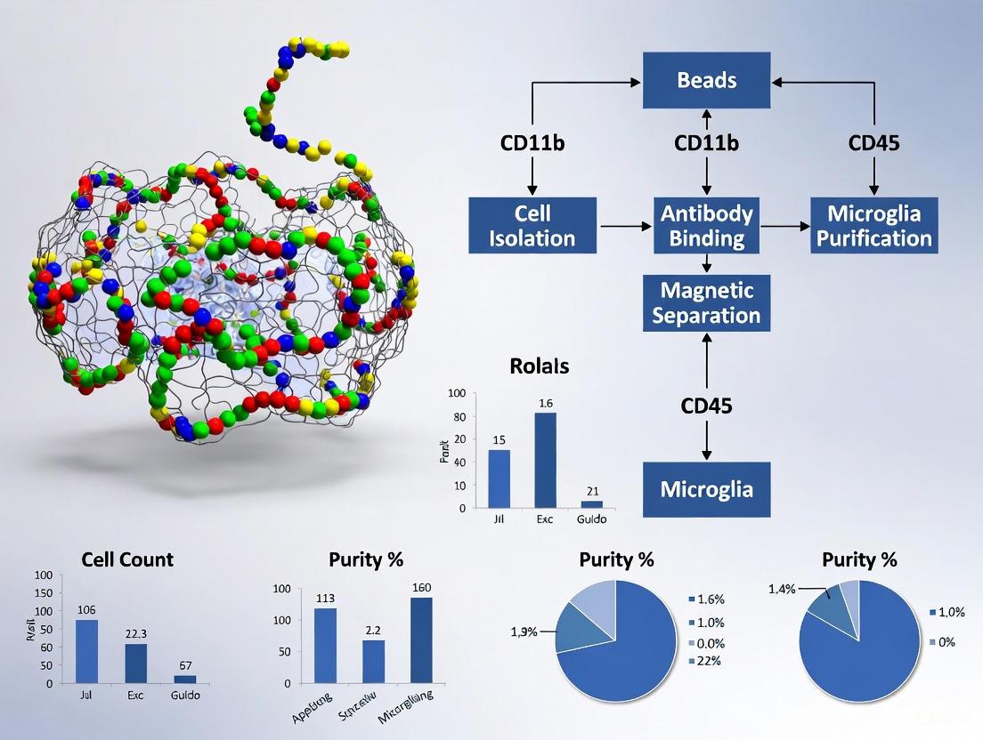
The following diagram illustrates the seamless integration of the bead conjugation chemistry with the subsequent cell isolation workflow, which is paramount for applications such as microglia purification.
Detailed Protocol for Bead Conjugation
This protocol provides a step-by-step guide for conjugating antibodies to magnetic beads, a critical preparatory step for immunomagnetic separation [2].
Materials and Reagents
- Magnetic Beads: Dynabeads M-270 Epoxy or similar.
- Antibody: Purified antibody against the target cell surface antigen (e.g., anti-CD11b for microglia) [4].
- Sodium Phosphate Buffer: 0.1 M, pH 7.4.
- Ammonium Sulfate: 3 M solution.
- Wash Buffers:
- 0.1 M sodium phosphate buffer (pH 7.4)
- 100 mM glycine·HCl (pH 2.5)
- 10 mM Tris-HCl (pH 8.8)
- 100 mM triethylamine (freshly prepared)
- Phosphate-buffered saline (PBS)
- PBS containing 0.5% Triton X-100
- Storage Buffer: PBS with 0.02% sodium azide (NaN₃).
- Equipment: Round-bottomed microcentrifuge tubes, magnetic particle concentrator, rotating wheel in a 30°C environment, tube shaker, micropipettor.
Step-by-Step Procedure
- Wash Beads: Weigh the required amount of magnetic beads (1-20 mg depending on scale) into a round-bottomed tube. Wash twice with 1 mL of 0.1 M sodium phosphate buffer (pH 7.4). Vortex for 30 seconds and mix for 15 minutes on a tube shaker at room temperature. Use the magnet to separate and remove the supernatant after each wash [2].
- Prepare Reaction Mixture: Resuspend the washed beads in the antibody solution. The total reaction volume should be approximately 20 µL per mg of beads. The table below provides guidelines for antibody amounts. Add sodium phosphate buffer to achieve the desired volume, and finally add 3 M ammonium sulfate to a final concentration of 1 M. Add the components in this order [2].
Table 1: Antibody Conjugation Guidelines per mg of Magnetic Beads
| Antibody Type | Recommended Amount | Notes |
|---|---|---|
| Commercial IgG | 10 µg | Suitable for most standard antibodies [2] |
| Purified, High-Affinity Custom Antibodies | 5 µg | Higher efficiency binding reduces quantity needed [2] |
| Saturation Point for M-270 Beads | ~7-8 µg | Maximum theoretical binding capacity [2] |
- Conjugate Antibodies: Incubate the reaction mixture overnight (approximately 16 hours) on a rotating wheel at 30°C. This extended incubation allows for maximal covalent coupling [2].
- Wash Conjugated Beads: The next morning, place the tube on a magnet and remove the supernatant. Wash the beads sequentially with the following buffers, using the magnet between each wash [2]:
- 1 mL of 0.1 M sodium phosphate buffer (pH 7.4)
- 1 mL of 100 mM glycine·HCl (quick wash)
- 1 mL of 10 mM Tris-HCl (pH 8.8)
- 1 mL of 100 mM triethylamine (freshly prepared; quick wash)
- Four washes with 1 mL of PBS
- 1 mL of PBS containing 0.5% Triton X-100 for 15 minutes
- Final wash with 1 mL of PBS
- Store Conjugated Beads: Resuspend the final bead pellet in PBS containing 0.02% sodium azide and store at 4°C. The conjugated beads should be used within 2-3 weeks for optimal performance, as isolation efficiency can decrease by approximately 40% after one month of storage [2].
Application in Primary Microglia Isolation
The isolation of primary microglia from the brain presents a significant challenge due to the delicate nature of these cells and the complexity of the neural tissue. Immunomagnetic separation using conjugated beads has emerged as a highly effective method to obtain pure, functional microglial populations from adult mice, which are more relevant for studying age-related neurodegenerative diseases than neonatal cells or cell lines [4].
Strategic Approach for Microglia
The key to successful microglia isolation lies in targeting specific cell surface markers. A common target is CD11b, a surface antigen highly expressed on microglia and other myeloid cells [4]. Antibodies against CD11b are conjugated to magnetic beads following the protocol above. When added to a single-cell suspension prepared from mouse brain tissue, these beads bind specifically to microglia. Subsequent magnetic separation pulls the CD11b-positive microglia out of the suspension, leaving behind neurons, astrocytes, oligodendrocytes, and other non-target cells [3]. This method, known as positive selection, directly isolates the cells of interest. It is often favored for its high specificity and purity. Comparative studies have shown that magnetic bead-based separation protocols can provide an optimal yield of functional microglial cells with minimal activation, making them suitable for downstream functional assays [4].
Comparison of Cell Isolation Strategies
Different research goals require different isolation strategies. The table below summarizes the main approaches using magnetic beads.
Table 2: Magnetic Cell Separation Strategies for Research
| Strategy | Principle | Outcome | Advantages | Considerations |
|---|---|---|---|---|
| Positive Isolation (Without Release) | Beads bind directly to target cells [1]. | Bead-bound cells are isolated [1]. | High purity; simple protocol. | Beads remain on cells, potentially interfering with some downstream applications [1]. |
| Positive Isolation (With Release) | Beads bind to target cells; a release buffer severs the bond [1]. | Bead-free, isolated cells [1]. | Cells are untouched by beads, ideal for functional studies and culture [1]. | Additional step required; potential for cell damage during release. |
| Negative Isolation | Beads bind to and remove ALL UNWANTED cells [1]. | Untouched target cells remain in supernatant [1]. | Target cells are completely free of antibodies and beads [1]. | Purity depends on comprehensive removal of all unwanted populations. |
| Cell Depletion | Beads remove a specific, abundant contaminating population [1]. | Sample enriched for rare target cells (e.g., CTCs) [1]. | Simplifies a complex sample matrix. | Is an enrichment step, not a full isolation. |
The Scientist's Toolkit
Table 3: Essential Reagents and Equipment for Immunomagnetic Separation
| Item | Function / Description | Example Use Case |
|---|---|---|
| Dynabeads M-270 Epoxy | Superparamagnetic, uniform beads with epoxy surface for covalent antibody conjugation [2]. | Foundation for creating custom conjugated beads for any target. |
| Anti-CD11b Antibody | Primary antibody targeting a surface marker highly expressed on microglia [4]. | Conjugation to beads for positive selection of microglia from a brain homogenate. |
| KingFisher Automation System | Automated magnetic particle processor that standardizes the isolation process [1]. | Increases reproducibility and throughput for processing multiple samples. |
| Magnetic Particle Concentrator | A magnet designed to separate beads from solution in standard microcentrifuge tubes [2]. | Essential for all manual washing and separation steps. |
| Sodium Phosphate Buffer (0.1 M, pH 7.4) | Provides the optimal alkaline pH for the epoxy-amine conjugation chemistry [2]. | Critical component of the conjugation reaction mixture. |
| Ammonium Sulfate (3 M) | Salt solution used to create a high-salt environment, promoting antibody-bead interaction [2]. | Added to the conjugation reaction to drive efficiency. |
Critical Factors for Success and Troubleshooting
Achieving high efficiency in both bead conjugation and cell isolation requires attention to several critical parameters. For the conjugation itself, the antibody-to-bead ratio is paramount; using more than the recommended amount can lead to unacceptable background from unbound antibody, while using too little results in suboptimal cell capture [2]. Furthermore, the purity and affinity of the antibody are crucial, as contaminants can conjugate to the beads and cause non-specific binding [2].
During the cell isolation phase, several factors must be optimized. Incubation time for cell capture is typically short; most specific binding occurs within the first 10-30 minutes, and prolonged incubation can increase non-specific binding [1]. Mixing conditions are also vital; gentle "slow" mixing preserves cell viability and isolation efficiency, whereas vigorous mixing can lead to significant cell loss and reduced viability [1]. Finally, the separation efficiency can be enhanced by incorporating multiple brief magnetic capture cycles (e.g., 2x) to ensure all bead-bound cells are retrieved from the solution [1]. Adhering to these optimized parameters ensures the reliable isolation of pure, viable microglia for downstream applications in neuroscience research.
Why Choose This Method? Key Advantages over FACS and Traditional Techniques
Immunomagnetic separation, specifically Magnetic-Activated Cell Sorting (MACS), provides a robust, accessible, and efficient method for isolating primary microglia from brain tissue. For researchers requiring high-purity isolation for sensitive downstream applications like proteomics, Fluorescence-Activated Cell Sorting (FACS) remains the gold standard, albeit with trade-offs in cost, speed, and technical demand [5]. This application note details the strategic advantages of MACS, provides a direct methodological comparison with FACS and traditional techniques, and delivers a validated protocol for obtaining high-quality microglia for research and drug development.
Microglia, the resident macrophages of the central nervous system, are pivotal players in brain development, homeostasis, and the pathogenesis of numerous neurological disorders [6] [7]. The isolation of pure, functionally intact primary microglia is therefore a cornerstone of neuroscience research. The choice of isolation method directly impacts cell yield, purity, viability, and phenotypic state, thereby influencing all subsequent experimental outcomes.
Immunomagnetic bead-based separation (MACS) has emerged as a premier technique that balances high quality with practical laboratory requirements. This document frames the use of MACS within a broader thesis on advanced cell isolation strategies, providing researchers with the evidence and protocols needed to implement this method effectively.
Comparative Analysis of Microglial Isolation Techniques
The selection of an isolation method involves balancing purity, yield, cost, speed, and technical requirements. The table below provides a quantitative and qualitative summary of the primary techniques available.
Table 1: Comprehensive Comparison of Microglia Isolation Methods
| Method | Reported Purity | Reported Cell Viability | Throughput & Speed | Key Advantages | Key Limitations/Laboratory Suitability |
|---|---|---|---|---|---|
| MACS (Immunomagnetic) | >90% (CD11b+ cells) [8] | >85% [9] | High; faster processing for single/multiple samples than FACS [9] | High yield; rapid protocol; minimal cell stress; suitable for subsequent cell culture; lower cost and equipment needs [9] [8] | Slight contamination with other myeloid cells/non-cellular proteins [9] [5] |
| FACS | Highly pure (e.g., using TMEM119) [6] | >85% [9] | Low; longer duration, especially for multiple samples [6] | Highest purity; ability to use multiple markers simultaneously (e.g., CD11b, CD45, TMEM119); excludes non-cellular debris definitively [6] [5] | Higher cost; requires specialized equipment and operator expertise; potential for greater ex vivo activation [6] |
| Differential Adhesion (Traditional) | Variable; requires validation | High [10] | Medium; culture-dependent (requires 1-2 weeks) [10] | Very low cost; requires only standard tissue culture equipment; simple to implement [10] | Lower initial purity; requires sub-culturing; yields less mature cells from neonatal tissue only [10] [4] |
| Density Gradient (Traditional) | Moderate to High | Variable; can be lower due to harsh centrifugation [4] | Medium | Effective myelin removal; no specialized antibodies required [4] | Lengthy procedure; low microglial yield; potential for excessive cell damage [4] |
Key Differentiator: Purity and Purity in Practice
While both MACS and FACS yield high purity, a critical distinction lies in the nature of the final product. MACS enriches for CD11b+ cells to a very high degree (>90%), making it excellent for most functional assays and culture work [8]. However, FACS can achieve a definitive population of microglia by using a combination of markers (e.g., CD11b+CD45loTMEM119+) and can exclude non-cellular debris, which is a known confounder in proteomic studies [6] [5]. Comparative proteomics revealed that FACS-isolated microglia had significantly less contamination from neuronal, astrocytic, and oligodendrocytic proteins compared to MACS-enriched samples [5].
Visualizing the Core MACS Workflow
The following diagram illustrates the streamlined, multi-stage process for isolating microglia from adult mouse brain using immunomagnetic beads.
Detailed Experimental Protocol: Immunomagnetic Isolation of Adult Mouse Microglia
This protocol is optimized for the isolation of microglia from adult mouse brain with high viability and purity, adapted from established methodologies [11] [8].
Reagents and Solutions
- Dissociation Kit: Neural Tissue Dissociation Kit (Papain-based or enzyme blends from Miltenyi Biotec).
- MACS Buffer: Phosphate-buffered saline (PBS), pH 7.2, supplemented with 0.5% bovine serum albumin (BSA) and 2 mM EDTA.
- Myelin Removal Reagent: 30% Percoll solution (9 parts 100% Percoll + 1 part 10x HBSS, diluted to 30% with 1x HBSS).
- Magnetic Microbeads: Anti-CD11b MicroBeads (Miltenyi Biotec, catalog # 130-093-636).
- MACS Columns: LS or MS columns paired with a suitable MACS separator (Miltenyi Biotec).
- Cell Culture Media: Dulbecco's Modified Eagle Medium (DMEM) or DMEM/F12, supplemented with 10% fetal bovine serum (FBS) and 1% penicillin/streptomycin.
Step-by-Step Procedure
Tissue Harvesting and Dissociation
- Euthanize an adult mouse according to approved institutional guidelines. Perfuse transcardially with ice-cold PBS to remove circulating blood cells.
- Rapidly dissect the brain and place it in ice-cold dissection buffer. Weigh the tissue.
- Mechanically dissociate the tissue using a gentleMACS Dissociator or by gentle trituration through fire-polished pipettes in the presence of the chosen enzymatic dissociation cocktail.
- Incubate the cell suspension for 15-35 minutes at 37°C with continuous agitation. Terminate the digestion with cold MACS buffer.
Single-Cell Suspension and Myelin Removal
- Pass the dissociated cell suspension through a 70 µm cell strainer to remove tissue debris.
- Centrifuge the filtrate at 300 x g for 10 minutes. Aspirate the supernatant.
- Resuspend the cell pellet thoroughly in 3-5 mL of 30% Percoll solution.
- Centrifuge the suspension at 700 x g for 10 minutes at 15°C with the brake disengaged.
- Carefully aspirate the top myelin layer and supernatant. Resuspend the cell pellet in an ample volume of MACS buffer and centrifuge again to wash.
Magnetic Labeling and Separation
- Resuspend the final cell pellet in 80 µL of cold MACS buffer per brain equivalent.
- Add 20 µL of anti-CD11b MicroBeads per brain equivalent. Mix well and incubate for 15 minutes in the refrigerator (2-8°C).
- Wash the cells by adding 1-2 mL of MACS buffer and centrifuging at 300 x g for 10 minutes. Aspirate the supernatant completely.
- Resuspend the cell pellet in 500 µL of MACS buffer.
- Place a MACS column in the magnetic field and rinse with 3 mL of MACS buffer.
- Apply the cell suspension to the column. Collect the flow-through containing the unlabeled, CD11b-negative cells.
- Wash the column 3 times with 3 mL of MACS buffer. The total effluent is the CD11b-depleted fraction.
- Remove the column from the magnetic field and place it over a clean collection tube.
- Pipette 5 mL of MACS buffer onto the column and firmly push the plunger through to elute the magnetically labeled CD11b+ microglia.
Post-Isolation Processing
- Centrifuge the eluted microglia at 300 x g for 10 minutes. Resuspend in complete culture medium for immediate culture or in an appropriate buffer for downstream molecular analyses.
- Determine cell viability and count using a hemocytometer and Trypan Blue exclusion.
The Scientist's Toolkit: Essential Research Reagent Solutions
Table 2: Key Reagents for Immunomagnetic Microglia Isolation
| Reagent / Kit | Function / Role | Example Product (Supplier) |
|---|---|---|
| Neural Tissue Dissociation Kit | Enzymatic breakdown of extracellular matrix to generate single-cell suspension. | Neural Tissue Dissociation Kit (Papain) (Miltenyi Biotec) |
| Anti-CD11b MicroBeads | Primary tool for immunomagnetic selection; antibodies conjugated to superparamagnetic beads bind specifically to microglial surface antigen CD11b. | CD11b MicroBeads, mouse (Miltenyi Biotec #130-093-636) |
| MACS Columns & Separator | Creates a high-gradient magnetic field to retain labeled cells while unlabeled cells pass through; the physical hardware for separation. | LS Columns / QuadroMACS Separator (Miltenyi Biotec) |
| Percoll | Density gradient medium for effective separation and removal of lipid-rich myelin debris, which can interfere with magnetic separation. | Percoll (Cytiva #17-0891-01) |
| CD11b Antibody (for validation) | Used in flow cytometry or immunocytochemistry to validate the purity of the isolated cell population post-MACS. | Anti-CD11b Antibody (clone M1/70) (BD Biosciences) |
Immunomagnetic bead separation using MACS technology represents a superior balance of performance and practicality for the routine isolation of primary microglia. Its key advantages—high yield, excellent viability, operational speed, and accessibility—make it an indispensable tool for researchers and drug development professionals aiming to study microglial biology in a context that closely reflects their in vivo state. While FACS is the method of choice for applications requiring the absolute highest purity, such as deep sequencing or advanced proteomics, MACS stands as the workhorse technique for the vast majority of functional, cultural, and biochemical assays.
CD11b, also known as integrin alpha M, is a transmembrane glycoprotein that serves as a reliable surface marker for identifying and isolating microglia, the resident immune cells of the central nervous system (CNS). As part of the integrin family, CD11b forms a heterodimer with the beta-2 subunit (CD18), resulting in the Mac-1 integrin, also referred to as complement receptor type 3 (CR3). This receptor is primarily expressed on the surface of myeloid cells, including monocytes, granulocytes, macrophages, and microglia [12]. The key function of CD11b is to mediate cell adhesion and migration through interactions with various ligands, including intercellular adhesion molecules (ICAMs) and complement component 3 (C3). Furthermore, CD11b plays a pivotal role in phagocytosis, the process by which cells engulf and eliminate foreign particles or cellular debris [12].
In the context of the CNS, microglia express CD11b in their resting state, with expression levels significantly increasing upon activation during neuroinflammatory processes [12] [13]. Resting microglia, characterized by a ramified morphology with small cell bodies and dynamic, branched processes, exhibit low levels of CD11b expression. However, when activated in response to pathological insults such as injury, infection, or neurodegenerative conditions, microglia undergo morphological transformation to an amoeboid or phagocytic phenotype and markedly upregulate CD11b expression [12]. This upregulation serves as a crucial indicator of microglial activation and their transition to a responsive state, enabling them to migrate to sites of CNS damage or inflammation, engage in phagocytic activity, present antigens, and produce various pro-inflammatory or anti-inflammatory factors [12].
The specificity of CD11b expression on microglia, combined with its accessibility as a cell surface antigen, makes it an ideal target for immunomagnetic separation techniques. Unlike astrocytes and neurons, for which antibodies recognizing extracellular epitopes of cell type-specific membrane proteins are not readily available, the consistent expression of CD11b on microglia enables efficient antibody-based separation of these cells from CNS tissues [8]. This approach allows researchers to obtain highly purified microglial populations without significant contamination from other neural cell types, facilitating precise analysis of microglial properties in various physiological and pathological conditions.
Technical Considerations for CD11b-Based Microglia Isolation
CD11b Expression and Specificity in Neural Tissue
When implementing CD11b-based microglia isolation protocols, researchers must consider several technical aspects to ensure successful outcomes. CD11b is expressed on microglia as well as other myeloid cells, but microglia can be distinguished by their characteristic CD11b+CD45lo profile, whereas infiltrating macrophages typically display a CD11b+CD45hi phenotype [14]. This distinction is particularly important in disease models involving blood-brain barrier disruption, where both resident microglia and peripheral macrophages may be present in CNS tissues [14].
The expression level of CD11b on microglia is not static but varies with their activation state. Bacterial lipopolysaccharide (LPS) and pro-inflammatory cytokines such as interferon-γ and interleukin-1β have been shown to induce increased CD11b expression through nitric oxide-dependent pathways [13]. This dynamic regulation means that the efficiency of CD11b-based isolation may vary depending on the physiological or pathological state of the microglia being targeted. The molecular mechanism behind this increased expression involves the nitric oxide-guanylate cyclase-cyclic GMP-protein kinase G pathway, ultimately leading to activation of cAMP response element-binding protein (CREB) that regulates CD11b expression [13].
Table 1: Key Surface Markers for Microglia Identification and Isolation
| Marker | Expression Profile | Utility in Isolation | Notes |
|---|---|---|---|
| CD11b | Myeloid cells; microglia (CD45lo); macrophages (CD45hi) | Primary isolation marker | Expression increases with activation [14] [13] |
| CD45 | Pan-leukocyte marker | Differentiation from other myeloid cells | Microglia: CD45lo; Infiltrating macrophages: CD45hi [14] |
| CD206 (MMR) | M2 alternatively activated microglia | Polarization assessment | Used in conjunction with M1 markers for activation state analysis [14] |
| FcγRII/III (CD16/32) | M1 classically activated microglia | Polarization assessment | Pro-inflammatory phenotype marker [14] |
Comparison of Myelin Removal Methods
A critical step in microglial isolation is the effective removal of myelin, which can interfere with downstream applications. Research has demonstrated that the choice of myelin removal method significantly affects both cell viability and yield [8].
Table 2: Comparison of Myelin Removal Methods for Microglia Isolation
| Method | Viability | Yield | Technical Considerations |
|---|---|---|---|
| 30% Percoll | Highest viability | Highest cell number | Density gradient centrifugation; requires optimization [8] |
| 0.9 mol/L Sucrose | Moderate viability | Moderate cell number | Hypertonic solution may affect cell integrity [8] |
| Anti-myelin Magnetic Beads | Good viability | Good cell number | Direct targeting of myelin; additional magnetic separation step [8] |
Among these methods, Percoll density gradient centrifugation has been shown to yield the highest viability and number of CD11b+ cells, making it the preferred choice for many applications [8]. However, the specific research objectives and available equipment may influence the selection of the most appropriate method.
Complete Immunomagnetic Separation Protocol for Microglia
Materials and Equipment
The following materials and equipment are required for successful immunomagnetic separation of microglia using CD11b targeting:
- Magnetic separator and appropriate columns (MS or LS columns depending on scale) [8]
- CD11b monoclonal antibody conjugated to magnetic beads (commercial sources available) [14] [8]
- Enzymatic digestion kit for neural tissue (e.g., Neural Tissue Dissociation Kit) [8]
- Myelin removal reagents (Percoll, sucrose, or anti-myelin magnetic beads) [8]
- Cell culture reagents including PBS, HBSS, IMAG buffer (PBS with 0.5% BSA and 2 mM EDTA) [8]
- Cell strainers (40 μm) [8]
- Centrifuge with temperature control capability
- Flow cytometry equipment for analysis and validation (optional but recommended) [14]
Step-by-Step Procedure
Tissue Collection and Preparation:
- Perfuse mice with ice-cold PBS to remove circulating blood cells [8].
- Dissect brain regions of interest and weigh the tissue.
- Mechanically dissociate tissue using a gentleMACS Dissociator or similar device if available.
Enzymatic Digestion:
- Use a Neural Tissue Dissociation Kit according to manufacturer's instructions [8].
- Incubate for 35 minutes at 37°C with periodic mixing.
- For sensitive applications, digestion can be performed on ice for a longer duration to preserve surface epitopes.
Myelin Removal:
- Resuspend dissociated cells in 30% Percoll solution [8].
- Centrifuge at 700 × g for 10 minutes at 4°C.
- Carefully aspirate the supernatant containing myelin debris.
- Wash cell pellet with HBSS and pass through a 40 μm cell strainer.
Immunomagnetic Labeling:
Magnetic Separation:
- Apply cell suspension to MS column placed in the magnetic field [8].
- Collect effluent (CD11b-negative cells) for downstream applications if desired.
- Wash column with IMAG buffer multiple times.
- Remove column from magnetic field and elute CD11b+ cells with appropriate buffer.
Post-Isolation Processing:
- Count cells using trypan blue exclusion or automated cell counter.
- Assess viability through Live/Dead staining if required [8].
- Proceed to downstream applications including cell culture, RNA extraction, protein analysis, or functional assays.
Microglia Isolation Workflow: This diagram illustrates the sequential steps for isolating microglia from mouse brain tissue using CD11b immunomagnetic beads.
Research Applications and Functional Assessment
Downstream Applications of Isolated Microglia
Microglia isolated via CD11b immunomagnetic separation can be utilized in numerous downstream applications that enable comprehensive analysis of their functional properties:
Flow Cytometric Analysis: Isolated microglia can be further characterized for activation state using additional surface markers. Researchers can assess M1/M2 polarization states by co-staining for markers such as FcγRII/III (M1) and CD206 (M2), generating an M1:M2 ratio that indicates the direction of the immune response [14]. This approach allows quantitative measurement of microglial polarization states without reliance on manual morphometric counting of immunohistochemistry slides [14].
Gene Expression Profiling: RNA extracted from purified microglia can be used for qRT-PCR analysis of inflammatory mediators, receptors, and other genes of interest. This application is particularly valuable for comparing gene expression patterns between microglia from different experimental conditions or disease models [8].
Protein Analysis: Isolated microglia can be lysed for Western blotting or used in ELISA to quantify protein expression levels and cytokine production. This enables researchers to directly correlate microglial activation states with specific protein expression patterns [8].
Functional Assays: Freshly isolated microglia can be used in various functional assays, including phagocytosis assays, migration assays, and cytokine secretion assays. These applications allow researchers to investigate the functional consequences of microglial activation in different pathological conditions.
Cell Culture Studies: Isolated microglia can be cultured for in vitro studies, either as pure populations or in co-culture systems with other neural cells. This approach enables investigation of cell-cell interactions and paracrine signaling mechanisms [15].
Assessment of Microglial Activation States
The immunomagnetic separation method using CD11b is suitable for isolating both quiescent and activated microglia without altering their phenotypic properties [8]. Research has demonstrated that microglia isolated from LPS-treated mice maintain their pro-inflammatory phenotype, as evidenced by upregulated levels of TNF-α, while microglia from control animals exhibit a quiescent phenotype [8]. This preservation of in vivo characteristics during the isolation process is crucial for obtaining biologically relevant data from subsequent analyses.
Table 3: Troubleshooting Common Issues in CD11b-Based Microglia Isolation
| Problem | Potential Causes | Solutions |
|---|---|---|
| Low cell yield | Incomplete tissue digestion, excessive myelin, insufficient antibody | Optimize digestion time/temperature; ensure proper myelin removal; titrate antibody concentration |
| Poor viability | Over-digestion, harsh myelin removal, prolonged processing | Reduce enzymatic digestion time; use Percoll method; maintain cold temperatures throughout |
| Low purity | Insufficient washing, non-specific binding | Increase wash steps; use cell strainers; optimize antibody concentration |
| Inconsistent results | Animal age variations, protocol deviations | Standardize animal age; strictly adhere to protocol steps; include internal controls |
The Scientist's Toolkit: Essential Research Reagents
Successful implementation of CD11b-based microglia isolation requires access to specific reagents and tools. The following table summarizes key components of the research toolkit for this application:
Table 4: Essential Research Reagents for CD11b-Based Microglia Isolation
| Reagent/Tool | Function | Examples/Specifications |
|---|---|---|
| CD11b Antibodies | Microglia identification and capture | Monoclonal antibodies conjugated to magnetic beads or fluorescent dyes [14] [8] |
| Magnetic Separator | Cell separation | MS or LS columns with appropriate magnets [8] |
| Enzymatic Digestion Kit | Tissue dissociation | Neural Tissue Dissociation Kit with optimized enzyme blends [8] |
| Myelin Removal Reagents | Debris clearance | Percoll (30%), sucrose (0.9 mol/L), or anti-myelin magnetic beads [8] |
| Cell Viability Assays | Quality assessment | Trypan blue exclusion, Live/Dead fixable stains [8] |
| Surface Markers for Characterization | Phenotype validation | CD45, CD206, FcγRII/III, other polarization markers [14] |
Immunomagnetic separation targeting CD11b represents a robust, efficient, and reliable method for isolating high-purity microglia from CNS tissues. This technique preserves the native phenotype of both quiescent and activated microglia, enabling accurate ex vivo analysis that reflects their in vivo state [8]. The availability of detailed protocols and commercial reagents makes this approach accessible to researchers investigating microglial function in various neurological disorders, injury models, and basic neuroimmunology research.
The versatility of CD11b-based isolation supports multiple downstream applications, including flow cytometric analysis, gene expression profiling, protein analysis, functional assays, and cell culture studies. By providing researchers with a method to obtain highly purified microglial populations without significant astrocyte or neuronal contamination [8], this technique facilitates precise investigation of microglial activities in any number of CNS pathologies or injuries. As research continues to elucidate the complex roles of microglia in CNS homeostasis and disease, CD11b-based immunomagnetic separation will remain an essential tool in the neuroscience research arsenal.
The Critical Role of Isolated Microglia in CNS Disease Modeling
Microglia, the resident macrophages of the central nervous system (CNS), play pivotal roles in brain development, homeostasis, and neuroinflammation. Their activation and dysfunction are implicated in numerous neurological disorders, making them essential targets for biomedical research. This application note outlines the principles and practical methodology for isolating primary microglia from adult mouse brain using immunomagnetic bead separation—a technique that yields high-purity populations suitable for sophisticated disease modeling, functional assays, and mechanistic studies. We provide detailed protocols, quantitative comparisons of methodological variations, and guidance for integrating isolated microglia into CNS disease modeling frameworks.
Microglia constitute approximately 5-10% of CNS glial cells and function as the primary immune sentinels of the brain [11]. Under physiological conditions, they continuously survey the microenvironment, manage synaptic connections, clear cellular debris, and respond to infection or injury [16]. In pathological states, microglia undergo morphological and functional changes, adopting distinct activation states that can either promote resolution or exacerbate damage [11] [14].
The transcriptomic and functional signatures of microglia vary significantly across developmental stages and between species [16] [17]. While microglial cell lines (e.g., BV-2, HMC3) offer convenience, they express few genes characteristic of adult microglia and are phenotypically distinct from primary cells [16] [17]. Similarly, neonatal primary microglia behave differently from their adult counterparts, limiting their utility for modeling age-related neurodegenerative diseases [16]. Therefore, obtaining high-purity primary microglia from adult animals is crucial for physiologically relevant studies of CNS pathology.
Principles of Immunomagnetic Microglia Isolation
Immunomagnetic separation leverages antibody-conjugated magnetic beads targeting specific cell surface antigens to isolate highly pure cell populations from heterogeneous suspensions. For microglia, this typically involves targeting the CD11b surface marker (integrin αM), which is expressed on microglia and other myeloid cells [11] [14].
Table 1: Key Markers for Microglia Identification and Characterization
| Marker | Expression Profile | Application |
|---|---|---|
| CD11b | Myeloid lineage cells (microglia, macrophages) | Primary isolation marker [11] [14] |
| CD45 | Microglia (CD45^low^), infiltrating macrophages (CD45^high^) | Distinguishing resident microglia from peripheral macrophages [11] [14] |
| TMEM119 | Specific to resident microglia | Identifying bona fide microglia (not infiltrating macrophages) [18] |
| Iba1 | Microglia/macrophages | Immunocytochemistry and morphological analysis [19] [20] |
| P2RY12 | Homeostatic microglia | Identifying non-activated, resting microglia [18] |
The fundamental advantage of immunomagnetic separation lies in its ability to isolate microglia with minimal activation and preserve their native state more effectively than other methods that involve prolonged culture or mechanical stress [11]. This technique enables researchers to obtain populations suitable for downstream applications including flow cytometric analysis, functional assays, and molecular profiling.
Comparative Methodologies and Performance Metrics
Multiple approaches exist for microglia isolation, each with distinct advantages and limitations. The choice of method depends on research objectives, available resources, and required purity and yield.
Table 2: Quantitative Comparison of Microglia Isolation Methods
| Method | Reported Purity | Reported Yield | Key Advantages | Key Limitations |
|---|---|---|---|---|
| Immunomagnetic Beads (MACS) | >90% [11] | Variable (depends on tissue input) | High specificity, minimal activation, compatibility with downstream applications [21] | Requires specific equipment, lower overall recovery compared to culture methods [21] |
| Flow Cytometric Sorting (FACS) | >95% [21] | Lower than MACS [21] | Highest purity, multi-parameter sorting | High cost, technical expertise, potential for cellular stress [21] |
| Differential Adherence/Shaking | ~73% (secondary), ~93% (tertiary) [20] | ~1% overall yield for tertiary cultures [20] | Cost-effective, simple equipment, establishes cultures | Lower purity, extended culture time may alter phenotype [21] [20] |
| Percoll Gradient | Comparable to MACS [22] | Comparable to MACS [22] | No antibodies required, effective myelin removal | Lengthy centrifugation, potential cell damage [16] |
The enzymatic digestion protocol significantly impacts microglial yield and viability. A systematic comparison found that accutase digestion produced the highest microglial yield with the lowest variance among tested enzymes, while Percoll-based myelin removal was superior for eliminating non-immune cells [22].
Detailed Protocol: Immunomagnetic Isolation of Adult Mouse Microglia
Reagents and Equipment
Table 3: Essential Research Reagent Solutions
| Reagent/Equipment | Function | Specifications |
|---|---|---|
| CD11b Microbeads | Immunomagnetic labeling of microglia | Anti-CD11b conjugated magnetic beads [11] [14] |
| MACS Separation Columns | Magnetic separation of labeled cells | LS or MS columns depending on cell number [11] |
| MACS Magnet | Creating magnetic field for separation | Appropriate for column type [11] |
| Enzyme Dissociation Kit | Tissue dissociation | Neural Tissue Dissociation Kit with papain or accutase [22] |
| AutoMACS Running Buffer | Buffer for magnetic separation | Miltenyi Biotech or equivalent [11] |
| Myelin Removal Beads | Optional myelin debris removal | Myelin Removal Beads II [11] |
| Viability Dye | Excluding dead cells | 7-AAD, propidium iodide, or similar [22] |
| Antibodies for Characterization | Phenotypic validation | Anti-CD11b, anti-CD45, appropriate fluorochromes [11] [22] |
Step-by-Step Procedure
Tissue Collection and Preparation
- Euthanize adult mouse (6-12 months old) using approved methods.
- Perform transcardial perfusion with ice-cold PBS to remove circulating blood cells [22].
- Rapidly extract brain and place in cold HBSS. Remove meninges carefully.
- Mechanically dissociate tissue with scalpel in cold HBSS.
Enzymatic Digestion
- Transfer tissue fragments to enzyme solution (2 mL accutase per brain) [22].
- Incubate for 30 minutes at 37°C with intermittent agitation.
- Add DMEM to inactivate enzymes and triturate 10-15 times with fire-polished pipette.
- Filter cell suspension through 70 μm cell strainer.
Myelin Removal (Optional but Recommended)
Immunomagnetic Labeling and Separation
- Centrifuge cell suspension at 300 × g for 10 minutes.
- Resuspend cell pellet in AutoMACS Running Buffer (80 μL per 10^7^ cells).
- Add CD11b Microbeads (20 μL per 10^7^ cells), mix well, and incubate for 15 minutes at 4°C.
- Wash cells with 1-2 mL buffer, centrifuge, and resuspend in 500 μL buffer.
- Place column in MACS separator and prepare with appropriate buffer.
- Apply cell suspension to column, collecting flow-through (CD11b^- negative fraction).
- Wash column 3 times with buffer.
- Remove column from magnet and elute CD11b^+^ cells with plunger.
Post-Isolation Processing
- Count cells using hemocytometer with trypan blue exclusion.
- Assess viability (>95% expected with optimal protocol).
- Plate cells for functional assays or process for immediate analysis.
Figure 1: Workflow for immunomagnetic isolation of primary microglia from adult mouse brain.
Quality Assessment and Validation
Purity and Viability Assessment
- Flow Cytometric Analysis: Stain cells with anti-CD11b and anti-CD45 antibodies. Microglia are typically CD11b^+^CD45^low^, while peripheral macrophages are CD11b^+^CD45^high^ [11] [14]. Expect >90% purity with optimized protocol.
- Viability Testing: Use trypan blue exclusion or fluorescent viability dyes (e.g., 7-AAD) during flow cytometry. Viability should exceed 95% with proper technique.
- Immunocytochemistry: Fix cells and stain with microglial markers (Iba1, P2RY12, TMEM119) to confirm identity and assess morphology [19] [20].
Functional Validation Assays
- Phagocytosis Assay: Incubate microglia with fluorescent latex beads for 2 hours. Analyze internalization by flow cytometry or confocal microscopy. >90% of microglia should phagocytose beads [23].
- Cytokine Secretion Profile: Stimulate microglia with LPS (100 ng/mL, 24 hours) and measure TNF-α, IL-6, MCP-1 secretion via ELISA. Compare to unstimulated controls [16] [23].
- Activation State Characterization: Evaluate M1/M2 polarization using surface markers (M1: FcγRII/III [CD16/32]; M2: CD206) [11] [14].
Applications in CNS Disease Modeling
Isolated primary microglia enable sophisticated disease modeling with direct translational relevance:
Neurodegenerative Disease Modeling (Alzheimer's, Parkinson's):
- Expose microglia to Aβ oligomers or α-synuclein fibrils
- Measure phagocytic capacity, inflammatory secretion, and gene expression
- Adult-derived microglia better model age-related diseases than neonatal cells [16]
Neuroinflammatory and Traumatic Conditions:
- Study microglial polarization states following cytokine exposure
- Investigate M1/M2 ratio shifts using flow cytometric analysis [11] [14]
- Model TBI responses by analyzing isolated microglia from injured brain regions
Drug Screening and Therapeutic Development:
- Test compound effects on microglial activation, phagocytosis, and neurotoxicity
- Human microglia show distinct responses to pharmacological substances compared to rodent cells [23] [17]
Figure 2: Experimental framework for modeling microglial functions and polarization states in CNS diseases.
Troubleshooting and Technical Considerations
Low Yield:
- Ensure complete tissue dissociation through optimized enzymatic digestion and mechanical trituration
- Verify perfusion efficiency to reduce blood cell contamination
- Test different enzyme combinations (accutase generally provides high yield) [22]
Reduced Viability:
- Minimize processing time; work quickly with cold solutions
- Avoid excessive mechanical force during trituration
- Use proper concentration of enzymes and timely inactivation
Low Purity:
- Confirm antibody specificity and concentration
- Ensure proper column preparation and washing
- Include myelin removal step for cleaner preparations
- Consider additional purification using CD45^low^ gating strategy [11]
Unanticipated Activation:
- Maintain cold conditions throughout isolation
- Use gentle dissociation methods
- Include appropriate control samples (unstimulated) for comparison
Immunomagnetic bead separation provides an effective methodology for isolating high-purity primary microglia from adult mouse brain, enabling physiologically relevant modeling of CNS diseases. The critical advantage of this approach lies in its ability to yield minimally activated cells that retain their native functional characteristics, supporting investigations into microglial polarization, phagocytic capacity, inflammatory secretion, and transcriptional regulation in neurological disorders. By implementing the detailed protocols and quality control measures outlined in this application note, researchers can establish robust, reproducible systems for studying microglial contributions to CNS pathophysiology and therapeutic intervention.
The selection of appropriate sample sources is a critical foundational step in neuroscience research, particularly in studies investigating microglia, the resident immune cells of the central nervous system. This application note examines the key considerations when choosing between mouse and human brain tissue for primary microglia isolation research, with specific focus on methodologies employing immunomagnetic beads. Understanding the anatomical, functional, and practical differences between these species is essential for designing physiologically relevant and translatable experiments, especially in the context of drug development for neurological disorders. We provide a comprehensive comparison of isolation techniques, morphological characteristics, and functional assessments to guide researchers in selecting the most appropriate model system for their specific research questions.
Comparative Neuroanatomy and Cellular Morphology
Fundamental structural differences exist between human and mouse brains that significantly impact experimental outcomes and translational potential. While basic circuit motifs show surprising conservation between species [24], important distinctions in cellular architecture and organization must be considered.
Table 1: Structural Comparison of Human and Mouse Neuronal Networks
| Parameter | Human Neurons | Mouse Neurons | Functional Implications |
|---|---|---|---|
| Pyramidal Soma Shape | Vertically triangular | Nearly spherical | Human neurons compatible with thicker, wider cortex [25] |
| Neurite Thickness | Thicker (~1.7x mouse) | Thinner | Human neurites more straight; mouse neurites more tortuous [25] |
| Neurite Curvature | Less tortuous | More tortuous (1.8x human) | Altered connectivity patterns; mouse connections more local [25] |
| Dendritic Spine Density | Lower | 2.6x higher in mice | Potential differences in synaptic integration [25] |
| Network Complexity | Highly complex with increased simplex dimension | Less complex | Human networks prioritize single-neuron complexity over density [26] |
| Inhibitory Circuit Motifs | Conserved Pvalb and Sst cell motifs | Strikingly similar to human | Supports translational relevance of mouse studies [24] |
Human pyramidal neurons form highly complex networks demonstrated by their increased number and simplex dimension compared to mice, despite human pyramidal cells being much sparser [26]. This greater dendritic complexity, a defining attribute of human pyramidal cells, may provide the human cortex with enhanced computational capacity and cognitive flexibility.
Decision Framework for Tissue Source Selection
Microglia Isolation Protocols Using Immunomagnetic Beads
Immunomagnetic Separation for Primary Microglia
Immunomagnetic bead separation has emerged as a powerful technique for isolating high-purity microglia from both human and mouse brain tissues. This method targets specific cell surface markers using antibody-magnetic bead conjugates, allowing for rapid and specific cell isolation [4]. The technique is particularly valuable for obtaining pure populations of microglia while preserving their native state and minimizing activation during the isolation process.
Table 2: Immunomagnetic Bead Isolation Protocols for Mouse and Human Microglia
| Protocol Step | Mouse Brain Tissue | Human Brain Tissue | Critical Considerations |
|---|---|---|---|
| Tissue Source | 6-month-old C57BL/6J mice [4] | Surgical biopsy or autopsy tissue [17] | Human tissue availability limited; mouse age affects microglial phenotype |
| Dissociation | Enzymatic (papain/DNase) and mechanical digestion [27] [17] | Enzymatic (papain/DNase) and mechanical digestion [17] | Enzymatic concentration and duration critical for viability |
| Bead Conjugation | Anti-CD11b magnetic beads [3] [4] | Anti-CD11b or tissue-specific markers | Antibody concentration and incubation time affect specificity |
| Magnetic Separation | Column-based separation in specific buffer systems | Column-based separation with adjusted flow rates | Flow rate controls purity vs. yield trade-off |
| Yield | ~1×10^6 cells per mouse brain (two cortices) [27] | Variable based on tissue mass and condition | Mouse yield more consistent; human yield highly variable |
| Purity Assessment | Flow cytometry for CD11b+ cells (>95% target) [4] | Immunocytochemistry for CD11b and Iba1 [17] | Multiple markers recommended for human tissue validation |
| Culture Maintenance | DMEM/F12 with M-CSF/GM-CSF [4] | Serum-supplemented DMEM with specific factors [17] | Media composition significantly affects phenotype retention |
Age-Specific Considerations for Microglia Isolation
Microglial phenotype and function change significantly with age, a critical consideration when selecting tissue sources. Aged microglia (from 18-month-old mice, approximately equivalent to humans aged 60 years) exhibit "inflammaging" - elevated baseline inflammation markers including higher expression of CD45, CD68, and MHC type II [27]. These age-related changes impact both isolation efficiency and experimental outcomes. Specialized protocols for aged microglia require specific growth media formulations that support continued survival and proliferation of adult and aged microglia, differing from standard neonatal isolation methods [27].
Morphological and Functional Characterization of Isolated Microglia
Quantitative Morphological Assessment
Advanced computational methods now enable high-throughput quantitative analysis of microglial morphology, providing objective assessment of activation states. StainAI represents a significant advancement in this field, leveraging deep learning to classify microglial morphology across whole-brain slices [28]. This approach can analyze millions of microglia across multiple slices, identifying subtle morphological changes that correlate with functional states.
Table 3: Quantitative Morphological Parameters for Microglial Characterization
| Morphological Parameter | Resting State Microglia | Activated State Microglia | Assessment Method |
|---|---|---|---|
| Fractal Dimension | Higher spatial complexity | Reduced complexity in chronic stress [29] | Fractal analysis |
| Cell Body Area | Smaller soma (≈148 µm² difference) [19] | Enlarged in activated states [19] [29] | Skeletal analysis |
| Branch Length | Extensive, long branches | Shortened in stress (≈315 µm difference) [19] [29] | Skeletal analysis |
| Branch Endpoints | Numerous endpoints | Fewer endpoints (≈23 fewer) [19] | Skeletal analysis |
| Process Complexity | Highly ramified | De-ramified, simplified branching | Sholl analysis [19] [29] |
| Activation Score | Lower weighted frequency | Higher in pathological states [28] | StainAI classification |
| Phagocytic Activity | Baseline CD68 expression | Significantly increased in stress [29] | CD68 immunofluorescence |
Functional Validation of Isolated Microglia
Beyond morphological assessment, functional validation is essential to confirm microglial behavior matches in vivo states. Key functional assays include phagocytic capacity measurement, cytokine secretion profiling in response to inflammatory stimuli (LPS, IL-4, IFN-γ), and metabolic activity assessment [17]. Comparative studies reveal that primary human microglia and iPSC-derived microglia display significantly higher levels of phagocytosis compared to mouse microglia or immortalized cell lines [17]. Additionally, notable species differences exist in inflammatory responses, with nitric oxide secretion observed only in mouse microglia following stimulation [17].
Research Reagent Solutions for Microglia Isolation
Table 4: Essential Research Reagents for Immunomagnetic Microglia Isolation
| Reagent/Category | Specific Examples | Function/Application | Species Compatibility |
|---|---|---|---|
| Digestion Enzymes | Papain (2.5 U/mL), DNase (10 U/mL) [17] | Tissue dissociation while preserving surface markers | Human & Mouse |
| Magnetic Beads | Anti-CD11b conjugated beads [3] [4] | Immunomagnetic separation of microglial populations | Human & Mouse |
| Culture Media | DMEM/F12 with GlutaMAX [4] | Base medium for microglial culture maintenance | Human & Mouse |
| Growth Factors | M-CSF (100 ng/mL), GM-CSF (100 ng/mL) [4] | Promote microglial survival and proliferation in culture | Primarily Mouse |
| Separation Columns | MACS columns or similar | Magnetic separation of bead-bound cells | Human & Mouse |
| Viability Markers | Trypan blue exclusion assay | Assessment of cell viability post-isolation | Human & Mouse |
| Purity Validation | CD11b, Iba1, PU.1 antibodies [17] | Immunostaining for microglial identity confirmation | Human & Mouse |
| Activation Assessment | CD45, CD68, MHC type II antibodies [27] | Detection of activation/inflammaging markers | Human & Mouse |
Immunomagnetic Bead Workflow for Microglia Isolation
The selection between mouse and human brain tissue for microglia isolation involves careful consideration of multiple factors, including research objectives, translational goals, resource availability, and technical expertise. Mouse models offer consistency, accessibility, and well-established protocols, while human tissue provides species-relevant data but with greater variability and accessibility challenges. Immunomagnetic bead separation has proven effective for both sources, yielding populations of sufficient purity for most downstream applications. When designing studies, researchers should align their model system with specific research questions—mouse models for fundamental mechanistic studies under controlled conditions, and human tissue for validation of clinically relevant findings. Cross-validation between species remains essential for ensuring translational relevance, particularly in drug development applications where species differences in microglial responses can significantly impact therapeutic efficacy.
Step-by-Step Protocol: Isolating High-Purity Microglia from Mouse and Human Brain
The isolation of high-purity primary microglia is a cornerstone of neuroscience research, enabling the study of neuroinflammation, neurodegenerative diseases, and drug mechanisms. Immunomagnetic bead-based separation has emerged as a powerful technique for obtaining these cells with exceptional purity and viability. The success of this method is critically dependent on the initial steps: careful tissue dissection and the creation of a high-quality single-cell suspension. This protocol details the optimized pre-isolation procedures essential for downstream applications, framed within a broader methodology for using immunomagnetic beads for primary microglia isolation research.
The Scientist's Toolkit: Essential Materials and Reagents
The following table catalogues the essential materials required for the tissue dissection and single-cell suspension preparation process.
Table 1: Key Research Reagent Solutions for Tissue Dissection and Dissociation
| Item | Function/Application | Example Catalog Number |
|---|---|---|
| Dissecting Tools (Scissors, Forceps) | Precise dissection of brain tissue and removal of meninges. [21] | Fine Science Tools: 14160-10, 11000-12 [21] |
| Cell Strainer (70 µm) | Removal of undissociated tissue clumps to generate a single-cell suspension. [21] | JETBIOFIL: CSS013070 [21] |
| Enzymatic Digestion Cocktail | Breaks down extracellular matrix to dissociate tissue. | Trypsin-EDTA (0.25%) [21], Collagenase/Dispase [30], DNase I [30] |
| Dissociation Buffer | Provides an ionic and pH-balanced environment for tissue processing. | Phosphate-buffered Saline (PBS) [21] |
| Culture Medium with Supplements | Supports cell survival during and after dissociation. | DMEM/F12 supplemented with B-27 [21] or Neurobasal medium with B-27 [30] |
| Density Gradient Medium | Enriches for microglia by separating cells based on density; removes myelin debris. [18] [31] | Percoll [18] [31] |
Workflow for Tissue Dissection and Single-Cell Suspension Creation
The process from whole brain to a single-cell suspension ready for immunomagnetic separation involves a series of critical, sequential steps. The following diagram outlines this comprehensive workflow.
Detailed Experimental Protocol
Tissue Dissection and Meninges Removal
- Euthanize the mouse according to approved institutional guidelines. Decapitate and carefully extract the whole brain into a dish containing cold, sterile PBS or Hanks' Balanced Salt Solution (HBSS). [18]
- Dissect the desired brain region (e.g., cortex, hippocampus, cerebellum) using fine dissection tools. Regional microdissection is valuable for studying microglial heterogeneity across different brain areas. [32]
- Remove the meninges completely. This is a critical step, as the meninges contain macrophages that can contaminate the microglial culture. Using fine ophthalmic forceps, gently peel away the meningeal layers from the surface of the brain tissue. [18] Incomplete meningeal removal is a major source of non-microglial macrophage contamination.
Mechanical and Enzymatic Tissue Dissociation
- Mechanical Disruption: Transfer the cleaned brain tissue to a fresh dish with a small volume of dissociation buffer. Use a sterile scalpel blade or scissors to mince the tissue into a fine slurry. [18]
- Enzymatic Digestion: Transfer the minced tissue into a tube containing an enzymatic digestion cocktail. A common and effective combination includes trypsin-EDTA (e.g., 0.25%) or a blend of collagenase/dispase supplemented with DNase I to digest DNA released from damaged cells. [21] [30]
- Incubate the tube at 37°C for 15-30 minutes, with gentle agitation or trituration every 5-10 minutes to aid dissociation.
Single-Cell Suspension Creation and Cleaning
- Quench the reaction by adding a large volume of cold culture medium containing serum (e.g., DMEM/F12 + 10% FBS) or a protease inhibitor to neutralize the enzymes. [21]
- Triturate the digested tissue vigorously using a serological pipette to further dissociate the chunks. Continue until no visible clumps remain.
- Filter the suspension by passing it through a sterile 70 µm cell strainer into a new 50 mL tube. This step removes any remaining tissue aggregates and creates a single-cell suspension. [21]
- Centrifuge the filtered suspension at 300-500 x g for 5-10 minutes. Carefully decant the supernatant.
- Resuspend the cell pellet in an appropriate buffer (e.g., PBS or MACS buffer) for the subsequent myelin removal step.
Optional but Recommended: Myelin Removal
For higher purity and better function in downstream applications, a density gradient centrifugation using Percoll is highly recommended to remove myelin debris, which is abundant in brain homogenates. [18] [31]
- Prepare a discontinuous Percoll gradient (e.g., 30% and 70% solutions in PBS).
- Layer the single-cell suspension carefully on top of the gradient.
- Centrifuge at 500-700 x g for 20-30 minutes at room temperature, with low brake setting.
- Collect the cell layer, which typically appears at the interface between the two densities. This fraction is enriched for microglia and other neural cells, with myelin debris pelleted at the bottom.
- Wash the collected cells with a large volume of buffer and centrifuge to remove residual Percoll before proceeding to immunomagnetic separation.
Critical Parameters for Success
The table below summarizes key quantitative and qualitative parameters that directly impact the yield, viability, and purity of the final microglial preparation.
Table 2: Critical Parameters for High-Quality Single-Cell Suspension Preparation
| Parameter | Optimal Condition / Quantitative Data | Impact on Experiment |
|---|---|---|
| Tissue Age | 9-day-old mice are optimal for tandem immunocapture of multiple neural cells. [18] [15] | Affects overall cellular yield and the ability to isolate other cell types (astrocytes, neurons) from the same brain. [18] |
| Meninges Removal | Complete removal is mandatory. | Incomplete removal is a primary source of contamination by peripheral macrophages, drastically reducing microglial purity. [18] |
| Enzyme Concentration & Time | Trypsin-EDTA (0.25%), 15-30 min at 37°C. [21] | Over-digestion reduces cell viability and surface antigen integrity; under-digestion reduces yield. [21] [30] |
| Post-Dissociation Processing Speed | Process tissue and cells quickly after dissociation. | Microglia are sensitive and can rapidly alter their transcriptome and phenotype ex vivo. [32] |
| Myelin Debris Removal | Percoll density gradient centrifugation. [18] [31] | Significantly improves microglial viability and function, and reduces background in downstream assays like flow cytometry. [31] |
The isolation of primary microglia is a cornerstone of neuroscience research, enabling the study of neuroinflammation, neurodegenerative diseases, and drug mechanisms. Enzymatic digestion of brain tissue is a critical first step in this process, directly impacting both the cell yield and functional viability of the isolated microglia. Achieving an optimal balance between complete tissue dissociation and preservation of delicate cell surface markers is particularly crucial when the downstream application involves immunomagnetic bead separation, a method prized for its high specificity and cost-effectiveness [33]. This application note details optimized enzymatic protocols designed to provide high-quality cell suspensions for reliable immunomagnetic separation of primary microglia from both neonatal and adult mouse brains.
The Role of Enzymatic Digestion in Microglial Isolation
Enzymatic digestion facilitates the breakdown of the extracellular matrix and intercellular connections within brain tissue, leading to a single-cell suspension. The choice of enzyme, its concentration, and incubation time are pivotal. Over-digestion can damage cell surface epitopes, which are essential for antibody binding during immunomagnetic separation, and reduce cell viability. Under-digestion results in low yield and clumping, hindering efficient separation [18] [34].
For studies intending to use immunomagnetic beads, preserving the integrity of surface markers like CD11b (for microglia) and ACSA-2 (for astrocytes) is non-negotiable [18] [33]. Furthermore, the age of the animal tissue (neonatal vs. adult) presents different challenges; adult brain tissue contains more myelin and connective tissue, often requiring more robust or combined enzymatic approaches [4] [34].
Quantitative Analysis of Enzymatic Methods
The table below summarizes key quantitative findings from studies that compared different enzymatic digestion strategies for microglia and astrocyte isolation.
Table 1: Comparative Analysis of Enzymatic Digestion Strategies for Brain Cell Isolation
| Enzymatic Combination | Animal Model / Age | Key Findings on Cell Yield & Viability | Recommended Downstream Application |
|---|---|---|---|
| Papain + Dispase II [34] | Adult Mouse | Highest combined yield of microglia, astrocytes, and infiltrating lymphocytes. Essential for detecting subtle glial activation (e.g., via LPS). | Flow cytometry, sequential cell isolation. |
| Papain alone [34] | Neonatal Mouse | Optimal for neonatal brain; addition of Dispase II provided no significant advantage. | Flow cytometry, general cell culture. |
| Enzyme Mix 1 & 2 (e.g., Papain-based) [33] | Adult Mouse (Young & Aged) | Compatible with sequential MACS; allowed high-purity isolation of microglia and astrocytes from the same brain for transcriptomics. | Magnetic-Activated Cell Sorting (MACS). |
| Combination of enzymes (unspecified) with mechanical dissociation [4] | Adult Mouse (Aging) | Modified protocol focused on minimizing activation, resulting in an optimal yield of functional microglial cells. | Functional assays, cell culture. |
| Trypsin [18] | General CNS Tissue | A commonly used enzyme for digesting intercellular proteins during the general isolation process. | General primary cell isolation. |
Recommended Protocols
Protocol 1: Balanced Digestion for Sequential MACS from Adult Mouse Brain
This protocol, optimized for sequential isolation of microglia and astrocytes from young and aged adult mice, is ideal for downstream transcriptomic analysis [33].
- Perfusion and Dissection: Following euthanasia, perform transcardiac perfusion with ice-cold PBS to remove blood cells. Dissect the brain and isolate the desired region (e.g., cortical and hippocampal tissue using a micro-punch).
- Tissue Preparation: Mince the tissue into small pieces and place them in a gentleMACS C Tube.
- Enzymatic Digestion:
- Add 1950 µL of Enzyme Mix 1 and 30 µL of Enzyme Mix 2 to the tube. The exact composition can vary but often includes a combination like papain and other neutral proteases.
- Attach the tube to a gentleMACS Octo Dissociator and run the pre-programmed dissociation protocol (e.g., 37CABDK02).
- Quenching and Filtration: Post-digestion, quench the enzymes by adding 10 mL of cold PBS. Pass the cell suspension through a 70 µm MACS SmartStrainer.
- Debris and RBC Removal: Centrifuge the filtrate and resuspend the pellet. Use a debris removal solution and a density gradient centrifugation step to remove myelin and red blood cells.
- Immunomagnetic Separation: The resulting single-cell suspension is now ready for sequential MACS, first for microglia (using anti-CD11b beads) and then for astrocytes (using anti-ACSA-2 beads) from the negative fraction [18] [33].
Protocol 2: Optimized Digestion for Adult Microglia with Enhanced Viability
This modified protocol emphasizes simplicity and high yield of functional microglia from adult mice, suitable for phagocytosis and inflammatory response assays [4].
- Dissection: Rapidly remove the brain and keep it cold in a medium containing antibiotics.
- Mechanical and Enzymatic Dissociation: The specific enzyme is not detailed, but the protocol highlights a modified dissociation process designed to be simpler and faster than compared methods.
- Centrifugation and Plating: After dissociation, the cell suspension is centrifuged, and the pellet is resuspended in a specific medium blend (50% DMEM/F-12 with GlutaMAX and 50% conditioned medium from mixed brain cells, supplemented with 10% FBS).
- Culture and Validation: Seed cells in a T25 flask. On day 2, supplement the medium with M-CSF and GM-CSF (100 ng/mL each) to support microglial survival and proliferation. Culture for 7 days, monitoring morphology and confirming purity via immunostaining for CD11b.
The Scientist's Toolkit: Key Research Reagent Solutions
Table 2: Essential Reagents for Enzymatic Digestion and Microglial Isolation
| Reagent / Material | Function / Application | Example |
|---|---|---|
| Papain | Proteolytic enzyme that breaks down extracellular matrix proteins; often a core component of digestion mixes. | Used in [34] and likely in [33]. |
| Dispase II | Neutral protease that dissociates cells by cleaving cell-surface proteins; effective in combination with papain for adult brain. | Used in combination with papain for adult mouse brain [34]. |
| MACS Cell Sorters | Equipment for high-specificity, high-throughput separation of cells using antibody-conjugated magnetic microbeads. | Used for sequential isolation of microglia and astrocytes [33]. |
| CD11b Microbeads | Antibody-conjugated magnetic beads for positive selection of microglia via the CD11b surface marker. | Key reagent for immunomagnetic isolation of microglia [18] [33]. |
| M-CSF & GM-CSF | Growth factors added to culture medium to support the survival and proliferation of isolated primary microglia. | Used at 100 ng/mL to culture microglia from adult mice [4]. |
| Percoll / Debris Removal Solution | Density gradient medium used to separate viable cells from myelin debris and dead cells after digestion. | Critical for obtaining a clean cell suspension post-digestion [33] [34]. |
Visualizing Microglial Signaling and Isolation Workflows
Microglial Signaling Pathways in Neuroinflammation
The following diagram illustrates key signaling pathways that govern microglial function, which can be studied using cells isolated with these protocols. Preserving these pathways during isolation is critical for physiologically relevant research.
Experimental Workflow for Microglial Isolation
This diagram outlines the complete experimental workflow from tissue dissociation to culture, highlighting the critical enzymatic digestion step.
Successful isolation of primary microglia for immunomagnetic bead research hinges on a meticulously optimized enzymatic digestion step. The data and protocols presented herein demonstrate that the choice between a single enzyme like papain for neonatal tissue or a combination like papain with dispase II for adult tissue significantly enhances the yield of viable, functional cells. By integrating these tailored enzymatic strategies with subsequent immunomagnetic separation, researchers can obtain highly pure microglial populations capable of yielding robust and translatable data in neuroscience and drug development.
Immunomagnetic separation using CD11b-conjugated beads is a cornerstone technique for the isolation of highly pure primary microglia. This critical binding step leverages the specific interaction between antibodies and the CD11b surface marker—an integrin highly expressed on microglia and other myeloid cells—to physically separate them from a mixed neural cell suspension. When executed with precision, this incubation is the foundation for obtaining reliable and reproducible data on microglial biology, neuroinflammation, and neurodegenerative disease mechanisms [18] [35]. This application note provides a detailed protocol and contextual framework for this essential procedure, enabling researchers to isolate microglia with preserved phenotypes for downstream functional and -omics analyses.
Detailed Protocol: The Binding Step
The following section outlines the core procedure for incubating a single-cell suspension with CD11b-conjugated magnetic beads. The prerequisite is a prepared single-cell suspension from brain tissue, obtained via enzymatic digestion and mechanical dissociation, with myelin removed [8].
Materials and Reagents
| Reagent/Material | Function & Specification |
|---|---|
| CD11b MicroBeads | Magnetic beads conjugated to anti-CD11b antibodies for specific microglial capture [35]. |
| Recommended Media | Buffered solution (e.g., DPBS without Ca²⁺/Mg²⁺) with 1-2% FBS and 1 mM EDTA [35]. |
| Rat Serum or BSA | Blocks non-specific antibody binding to Fc receptors on microglia and other immune cells [35]. |
| Magnetic Separator | Device to generate a strong magnetic field (e.g., MACS Separator) [8]. |
| Separation Columns | MS or LS columns placed within the magnet for cell separation (column-based method) [18]. |
| Polystyrene Tubes | Tubes for column-free separation methods [35]. |
| Cell Strainer | Removes cell clumps to ensure a single-cell suspension (e.g., 70 µm mesh) [35]. |
Step-by-Step Procedure
Prepare the Cell Suspension:
- Obtain a single-cell suspension from brain tissue and perform a cell count.
- Centrifuge the cell suspension (e.g., at 400 x g for 5 minutes) and thoroughly resuspend the pellet in a recommended buffer (e.g., 2% FBS in DPBS with 1 mM EDTA). A suggested starting volume is 1 mL per 100 x 10⁶ total cells [35].
Block Non-Specific Binding:
- Add rat serum to the cell suspension to a final concentration of 5% (e.g., 50 µL per 1 mL of cells). This critical step occupies Fc receptors, minimizing background binding and improving purity [35].
- Incubate for 5 minutes at room temperature (RT).
Incubate with CD11b-Conjugated Beads:
- Add the appropriate volume of CD11b MicroBeads directly to the cell suspension. The optimal bead-to-cell ratio must be determined empirically or as per the manufacturer's instructions.
- Mix thoroughly but gently to ensure uniform bead distribution.
- Incubate for 15 minutes at 4°C (or for 15 minutes at RT if following a refined column-free protocol) [35] [8]. The cooler temperature helps prevent capping and internalization of the antibody-antigen complex.
Wash and Resuspend:
- Add a larger volume (e.g., 10-20x the labeling volume) of buffer to the cell-bead mixture to wash away unbound beads.
- Centrifuge the suspension (e.g., at 400 x g for 5 minutes) and carefully decant the supernatant.
- Resuspend the cell pellet in a small volume of buffer suitable for the subsequent magnetic separation step.
Proceed to Magnetic Separation:
- The cell suspension is now ready for separation. For column-based methods, apply the cell suspension to a pre-rinsed column placed in the magnetic field. CD11b+ cells will be retained, while the negative fraction flows through [18] [8]. For column-free methods, place the entire tube in a magnet. CD11b+ cells will migrate to the tube wall, allowing the supernatant (negative fraction) to be carefully aspirated [35].
The following diagram illustrates the core workflow and the role of the binding step within the complete microglial isolation process.
Technical and Methodological Considerations
Quantitative Performance Data
The performance of immunomagnetic separation using CD11b beads can vary based on the specific protocol and the source of the tissue. The table below summarizes key metrics from published studies.
Table 1: Performance Metrics of CD11b-Based Microglial Isolation
| Protocol / Study | Reported Purity | Reported Yield | Key Methodological Notes |
|---|---|---|---|
| Refined Column-Free [35] | ~99% | Not specified for adult brain | Completion time reduced by half; allows use of red channel in fluorescence microscopy. |
| Immunomagnetic Separation (Adult Mouse) [8] | Highly purified CD11b+ population | Viability and yield highest with Percoll for myelin removal | Preserves in vivo phenotype; suitable for both quiescent and activated microglia. |
| Comparative Study (Five Methods) [36] | >90% (all methods) | Varies with method; adult acute isolation yield is lower | Neonatal mixed glial culture (MGC) protocols showed a larger transcriptional response to HMGB1 stimulus. |
Troubleshooting the Binding Step
- Low Purity: Ensure adequate Fc receptor blocking with rat serum or BSA. Titrate the amount of CD11b beads to optimize the signal-to-noise ratio and avoid non-specific binding. Using an optimized, refined protocol can significantly enhance purity [35].
- Low Yield: Verify the activity and specificity of the CD11b antibody on the beads. Do not over-triturate cells during preparation, as this can damage surface markers. Using tissue from neonatal mice generally provides a much higher yield than from adult mice [18] [36].
- Cell Activation: Maintain cells at 4°C during the incubation where possible to minimize cellular stress and activation. The immunomagnetic separation method itself, when performed correctly, does not activate microglia, allowing evaluation of their ex vivo state [8].
The Scientist's Toolkit
Table 2: Essential Research Reagent Solutions for CD11b-Based Isolation
| Item | Function in the Protocol |
|---|---|
| CD11b (ITGAM) Antibody | The primary capture agent; binds to the CD11b surface protein on microglia. |
| Magnetic MicroBeads | The solid-phase matrix conjugated to antibodies; enables physical separation in a magnetic field. |
| MACS Separation Columns | The platform where the positive and negative cell fractions are separated. |
| Neural Tissue Dissociation Kit | Prepares a high-viability single-cell suspension from the whole brain or specific regions. |
| Percoll or Debris Removal Solution | Critical for removing myelin debris from adult brain preparations, which improves purity and viability [8]. |
The incubation of a cell suspension with CD11b-conjugated beads is a definitive step in the immunomagnetic isolation of primary microglia. Meticulous attention to the details of blocking, incubation time, temperature, and bead-to-cell ratio is paramount to the success of subsequent experiments. By following this detailed protocol and considering the accompanying technical insights, researchers can consistently obtain high-purity microglial populations that faithfully represent their in vivo state, thereby strengthening the validity and translational potential of their findings in neuroscience and drug development.
Within primary microglia isolation research, the precise separation of these unique immune cells from the complex cellular environment of the brain is a fundamental prerequisite for downstream functional and phenotypic analyses. Immunomagnetic separation has emerged as a cornerstone technique for this purpose, enabling the isolation of a highly pure population of microglial cells based on their surface marker expression [8]. This application note details a standardized protocol for the column-based immunomagnetic separation of microglia, framed within a broader methodology that encompasses tissue dissociation and phenotypic validation. The ability to efficiently isolate microglia with preserved phenotypes is crucial for advancing our understanding of their role in central nervous system homeostasis, neuroinflammation, and neurodegenerative diseases [8] [37].
Technical Principles and Workflow
Immunomagnetic cell separation relies on the specific binding of antibody-coated magnetic beads to target cells, followed by their retention in a magnetic column placed within a strong magnetic field. While the entire process of microglial isolation begins with tissue harvesting and dissociation, the core magnetic separation steps—column setup, washing, and elution—are critical for achieving high purity and viability. Figure 1 below illustrates the complete workflow, from brain harvest to the final analysis of isolated microglia, with the core magnetic separation steps highlighted.
Figure 1. Complete Workflow for Immunomagnetic Isolation of Microglia. The process begins with brain harvest and tissue dissociation, followed by critical myelin removal. The core magnetic separation steps (setup, washing, and elution) are highlighted in blue, leading to the final isolation of microglia for phenotypic analysis.
Materials and Equipment
Research Reagent Solutions
The following table catalogues the essential reagents and equipment required for the immunomagnetic separation of microglia.
Table 1: Essential Reagents and Equipment for Immunomagnetic Separation
| Item | Function/Description | Example Source |
|---|---|---|
| CD11b MicroBeads | Magnetic beads conjugated to monoclonal anti-CD11b antibodies for specific targeting of microglia/macrophages. | Miltenyi Biotec [38] [8] |
| MACS Column | A column placed within a magnetic separator that retains magnetically labeled cells. LS or MS columns are commonly used. | Miltenyi Biotec [39] [8] |
| MACS Separator | A strong permanent magnet that creates a magnetic field for the column. | Miltenyi Biotec (e.g., QuadroMACS) [38] [8] |
| IMAG Buffer (PBS + 0.5% BSA + 2mM EDTA) | Buffer for dilution and washing of cells; preserves cell viability and prevents clumping. | Prepared in-lab [8] |
| Percoll Solution | Density gradient medium for the removal of myelin debris and dead cells prior to magnetic separation. | GE Healthcare [40] [8] [37] |
| Neural Tissue Dissociation Kit | Pre-optimized enzyme blends for the gentle and effective dissociation of brain tissue. | Miltenyi Biotec [38] [8] [37] |
Quantitative Comparison of Myelin Removal Methods
The method chosen for myelin removal prior to magnetic separation significantly impacts the viability and yield of the final microglial preparation. The following table summarizes data from a study that systematically compared three common techniques.
Table 2: Impact of Myelin Removal Method on Microglial Isolation [8]
| Myelin Removal Method | Relative Cell Viability | Relative Cell Yield | Key Observations |
|---|---|---|---|
| 30% Percoll Gradient | High | High | Highest viability and number of CD11b+ cells obtained; effective myelin removal. |
| 0.9 M Sucrose | Moderate | Moderate | Lower viability and yield compared to Percoll method. |
| Anti-Myelin Beads | Low | Low | Significant loss of CD11b+ cells during the myelin removal step. |
Step-by-Step Protocol: Magnetic Separation
Pre-Separation Steps
- Tissue Dissociation: Following euthanasia and perfusion with ice-cold PBS, dissect the brain tissue. Dissociate the tissue using a mechanical and enzymatic protocol, such as with the Adult Brain Dissociation Kit and a gentleMACS Octo Dissociator according to the manufacturer's instructions [38] [37].
- Myelin Removal: Resuspend the single-cell suspension in 30% isotonic Percoll and centrifuge at 700 × g for 10 minutes at 4°C. Carefully aspirate the myelin-containing supernatant [8]. Wash the cell pellet with HBSS or IMAG buffer and pass the suspension through a 70 μm cell strainer [37].
- Magnetic Labeling: Resuspend the cell pellet in IMAG buffer (e.g., 90 μL per 10^7 total cells). Add CD11b MicroBeads (e.g., 10 μL per 10^7 cells) and mix well. Incubate for 15 minutes in the refrigerator (2-8°C) [38] [8]. After incubation, wash the cells by adding 1-2 mL of buffer per 10^7 cells and centrifuge. Resuspend the cell pellet in a suitable volume of buffer (e.g., 500 μL to 1 mL).
Column Setup, Washing, and Elution
Column Selection Guide: The choice of MACS column depends on the total number of cells and the number of magnetically labeled cells. LS columns are suitable for up to 10^9 total cells and 10^7 labeled cells, while MS columns can handle up to 2×10^8 total cells and 10^7 labeled cells. The following diagram details the core magnetic separation procedure.
Figure 2. Core Steps of Magnetic Column Separation. After preparation, the labeled cell suspension is applied. Magnetically labeled microglia are retained in the column, while unlabeled cells pass through. After thorough washing, the target microglia are eluted once the column is removed from the magnetic field.
- Column Preparation: Place a suitable MACS column (e.g., LS or MS) in the magnetic field of the MACS separator. Rinse the column with 3 mL (for LS columns) or 500 μL (for MS columns) of IMAG buffer to prepare the matrix [8].
- Applying Cell Suspension: Apply the magnetically labeled cell suspension onto the top of the column reservoir. Allow the unlabeled cells to pass through by gravity or gentle pressure. Collect the effluent (flow-through); this fraction contains the unlabeled, non-target cells.
- Washing the Column: Without allowing the column to run dry, wash the column with the appropriate buffer volume (e.g., 3×3 mL for LS columns; 3×1 mL for MS columns). The wash steps remove any unbound or weakly bound cells, ensuring high purity of the final isolate.
- Elution of Target Cells: Remove the column from the magnetic separator and place it over a clean collection tube. Pipette an appropriate volume of buffer (e.g., 5 mL for LS columns; 1 mL for MS columns) onto the column and immediately flush out the magnetically retained cells by firmly applying the plunger supplied with the column [38] [8]. This eluted fraction contains the purified CD11b+ microglial cells.
Post-Isolation Analysis and Validation
The eluted microglia are now ready for downstream applications. Cell count and viability can be assessed using Trypan Blue exclusion [8]. To validate the success of the isolation and interrogate the activation state of the microglia, flow cytometric analysis is highly recommended.
- Staining for Phenotyping: Following isolation, stain the cells with fluorescently conjugated antibodies. Key markers include:
- CD11b (pan-myeloid marker) and CD45 (leukocyte common antigen) to confirm microglial identity (CD11b+/CD45low) and distinguish them from peripheral macrophages (CD11b+/CD45high) [41] [40].
- Activation Markers: To assess the polarization state, stain for M1 markers (e.g., FcγRII/III (CD16/32)) and M2 markers (e.g., CD206) [41] [14]. The ratio of M1 to M2 microglia can be a key readout in neuroinflammation studies [41].
- Gating Strategy to Manage Autofluorescence: Microglia exhibit significant autofluorescence, which can interfere with antibody signal detection. It is critical to include unstained and fluorescence-minus-one (FMO) controls to set appropriate gates and distinguish true positive signals from background [40].
Performance and Comparison with Other Methods
Table 3: Comparison of Microglia Isolation Techniques [4] [37]
| Isolation Method | Purity | Viability for Culture | Throughput | Relative Cost | Key Advantages/Disadvantages |
|---|---|---|---|---|---|
| Immunomagnetic Separation (MACS) | High [8] | High [8] | Moderate | Moderate | Advantages: High purity and viability; suitable for subsequent cell culture [8]. Disadvantages: Relies on a single surface marker (CD11b); magnetic beads may remain bound to cells [37]. |
| Fluorescence-Activated Cell Sorting (FACS) | Very High | Variable (can be low) [37] | Low | High | Advantages: Highest purity; multi-parameter sorting. Disadvantages: Can activate microglia [37]; requires specialized equipment; lower cell viability post-sort. |
| Adhesion-Based Protocols | Moderate | High | High | Low | Advantages: Simple, low-cost. Disadvantages: Lower purity; may select for a subpopulation of more adherent cells [4]. |
Troubleshooting Guide
Table 4: Common Issues and Proposed Solutions
| Problem | Potential Cause | Solution |
|---|---|---|
| Low Purity | Insufficient washing of the column; excessive cell load. | Ensure multiple wash steps are performed; do not exceed the recommended cell number for the column type. |
| Low Cell Yield | Inefficient tissue dissociation; over-digestion with enzymes; excessive mechanical force. | Optimize enzymatic digestion time and mechanical dissociation protocol [4] [37]. |
| Poor Cell Viability | Harsh tissue dissociation; prolonged processing time; bacterial contamination. | Keep cells and reagents cold; use pre-cooled equipment; work under sterile conditions for culture. |
The successful immunomagnetic bead isolation of primary microglia using CD11b+ selection represents a critical first step in obtaining a highly pure cell population [18] [39]. However, the subsequent post-isolation culture conditions are equally vital for preserving microglial identity, function, and responsiveness in vitro. Microglia are exquisitely sensitive to their environment, and suboptimal culture can lead to rapid phenotypic drift, activation, or loss of function, thereby undermining the value of a careful isolation [42] [6]. This protocol details evidence-based media formulations and culture techniques designed to maintain isolated microglia in a state that closely mirrors their in vivo physiology, enabling reliable downstream experimentation for drug discovery and basic research.
Media Formulations and Culture Conditions
The choice of base media, supplements, and growth factors significantly impacts microglial health, purity, and baseline activation state. The following table summarizes key media components and their recommended concentrations based on recent comparative studies.
Table 1: Recommended Media Components for Cultured Microglia
| Component | Recommended Concentration & Details | Primary Function | Key Considerations |
|---|---|---|---|
| Base Medium | DMEM/F-12 with GlutaMAX [4] or serum-free X-VIVO [42] | Provides nutritional and structural support. | X-VIVO lacks exogenous growth factors, reducing unintended stimulation [42]. |
| Serum | 10% Fetal Bovine Serum (FBS) [4] | Supports cell adhesion and survival. | Serum batches can vary; high concentrations may promote activation. |
| Growth Factors | GM-CSF: 0.5 ng/mL (neonatal) [42] or 100 ng/mL (adult) [4] [42]M-CSF: 100 ng/mL (adult) [4] [42] | Enhances microglial yield and survival in culture. | High GM-CSF concentrations (>5 ng/mL) can alter responsiveness to stimuli [42]. |
| Conditioned Medium | 50% conditioned medium from mixed brain cell cultures [4] | Provides a physiologically relevant mix of trophic factors. | Helps maintain a more in vivo-like phenotype. |
| Antibiotics | 1% Penicillin/Streptomycin [4] | Prevents bacterial contamination. | Standard for primary cell culture. |
Beyond the media composition, several other culture parameters are critical for success:
- Seeding Density: Plate cells at a density of ~200,000 cells per well in a 48-well plate for experimentation [42]. For expansion, a T25 flask is suitable for cells isolated from one mouse brain [4].
- Coating: Flasks and plates typically do not require coating [4]. However, for specific applications, Poly-D-Lysine (PDL) can be used to improve adherence [27].
- Incubation Environment: Standard cell culture conditions of 37°C and 5% CO₂ are used [43].
- Culture Duration and "Culture Shock": A 7-day adaptation period post-isolation allows microglia to recover a sub-reactive morphology [4]. However, prolonged culture (e.g., beyond 7-10 days) can induce "culture shock," altering transcriptional profiles and responsiveness [42]. Researchers should perform experiments as soon as possible after this recovery period to minimize phenotypic drift [18].
Experimental Protocols for Functional Validation
After establishing cultures, validating microglial function through key assays is essential. The following are detailed protocols for assessing purity, phagocytosis, and cytokine response.
Protocol: Assessment of Microglial Purity via Immunocytochemistry
Confirming culture purity is a critical first step after isolation and culture.
- Steps:
- Seed ~100,000 cells per well on glass coverslips in a 48-well plate 24 hours before staining [4].
- Fix cells with freshly prepared 4% Paraformaldehyde (PFA) in PBS for 15 minutes.
- Permeabilize and block cells using a solution containing 10% Bovine Serum Albumin (BSA) for 2 hours [43].
- Incubate cells overnight at 4°C with primary antibodies against microglial markers:
- The next day, wash cells and incubate with appropriate fluorescent secondary antibodies (e.g., BV421-conjugated) for 1-2 hours at room temperature if needed [6].
- Counterstain nuclei with DAPI and mount coverslips.
- Image using fluorescence microscopy. A successful immunomagnetic bead isolation followed by culture should yield >95% purity for CD11b+/Iba1+ cells [42] [23].
Protocol: Phagocytosis Assay Using Fluorescent Latex Beads
Phagocytosis is a core microglial function that can be tested with a bead-based assay.
- Steps:
- Culture isolated microglia in 48-well plates or on coverslips until they reach ~80% confluency.
- Incubate cells with fluorescent (e.g., FITC-conjugated) latex beads for 2 hours at 37°C and 5% CO₂ [23].
- After incubation, vigorously wash the cells with cold PBS to remove any beads that are adherent but not internalized.
- (Optional) For visualization by confocal microscopy, fix cells with 4% PFA [23].
- Quantify phagocytosis using flow cytometry (measuring a rightward shift in fluorescence intensity) or by counting internalized beads per cell via confocal microscopy [23]. Over 90% of pure, functional microglia should phagocytose beads under these conditions.
Protocol: Evaluating Cytokine Response to Stimulation
Testing the response to inflammatory stimuli validates microglial functionality.
- Steps:
- Serum-starve microglia by transferring them to serum-free X-VIVO medium or a similar low-serum formulation for several hours or overnight before stimulation [42].
- Treat cells with a pro-inflammatory stimulus. A common and relevant stimulus is HMGB1 (a DAMP), or classical activators like LPS (100 ng/mL) or IL-1β [42] [23].
- Incubate for a defined period (e.g., 6-24 hours).
- Collect cell culture supernatant and analyze for cytokine secretion (e.g., TNF-α, IL-1β, IL-6) using ELISA [42] [23].
- Alternatively, harvest cells for RNA extraction to assess changes in gene expression of these cytokines via qPCR or RNA sequencing [42] [43]. Functional microglia will show a significant upregulation of pro-inflammatory cytokines upon stimulation.
The Scientist's Toolkit: Key Research Reagent Solutions
Table 2: Essential Materials for Microglia Isolation and Culture
| Reagent / Kit | Specific Example (Supplier Catalog Number) | Function in Protocol |
|---|---|---|
| Immunomagnetic Beads | CD11b (Itgam) MicroBeads (Miltenyi Biotec) [18] [39] | Positive selection of microglia from a single-cell suspension. |
| Tissue Dissociation Kit | Neural Tissue Dissociation Kit (Miltenyi Biotec) [42] | Enzymatic digestion of brain tissue to create a single-cell suspension. |
| Myelin Removal Solution | Debris Removal Solution (Miltenyi Biotec) [42] | Removal of myelin debris which can interfere with downstream applications. |
| Recombinant Growth Factors | GM-CSF & M-CSF (R&D Systems) [4] [42] | Supports survival and proliferation of microglia in culture. |
| Microglial Marker Antibodies | Anti-CD11b (ThermoFisher Scientific) [4]; Anti-Iba1; Anti-TMEM119 (Abcam) [6] | Identification and purity assessment of isolated microglia. |
| Cell Strainer | 70 μm cell strainer (Corning) [42] [43] | Removal of cell clumps and tissue debris after dissociation. |
Workflow and Decision Pathway for Microglia Culture
The following diagram illustrates the critical steps and decision points in the post-isolation culture process, from receiving the isolated cells to conducting functional assays.
The journey from a successfully isolated population of CD11b+ microglia to a physiologically relevant in vitro model hinges on meticulously optimized culture conditions. By employing serum-free or carefully supplemented media, appropriate growth factors, and a defined culture period, researchers can mitigate "culture shock" and maintain microglial identity. The functional validation protocols outlined herein—assessing purity, phagocytosis, and cytokine release—are indispensable for confirming that the cultured cells retain their critical innate immune functions. Adherence to these application notes will ensure that microglia isolated via immunomagnetic beads serve as a robust and reliable platform for neuroscientific research and therapeutic discovery.
The study of central nervous system (CNS) physiology and pathology requires precise tools to investigate its composite cell types. Isolation of primary brain cells is essential for studying cellular behavior, signaling pathways, and disease mechanisms [18]. Traditional approaches often require multiple animal subjects to study different cell populations, introducing biological variability and complicating data interpretation. Tandem isolation protocols address this limitation by enabling the sequential separation of microglia, astrocytes, and neurons from a single brain sample [18] [44]. This methodological approach provides a powerful strategy for obtaining multiple CNS cell populations from the same biological context, thereby reducing inter-sample variability and animal use while facilitating direct comparative studies between cell types [44] [45]. The use of immunomagnetic beads specifically offers a versatile, cost-effective, and reliable technique for sequential cell isolation that is compatible with various downstream applications including transcriptomics, proteomics, and functional assays [44]. When framed within a broader thesis on primary microglia isolation, this tandem protocol provides a foundational methodology that enhances experimental consistency and enables more physiologically relevant investigations of neuroinflammatory processes and neurodegenerative diseases.
Principles of Immunomagnetic Separation
Immunomagnetic cell separation operates on the principle of using magnetic beads conjugated with antibodies specific to cell surface markers. When a cell suspension is incubated with these beads, target cells become labeled and can be retained in a magnetic field while unlabeled cells pass through [18]. This process enables both positive selection (isolation of cells expressing specific markers) and negative selection (depletion of unwanted cells) strategies. For neural cells, specific surface antigens provide the molecular handles for isolation: CD11b (also known as integrin alpha M or ITGAM) for microglia, ACSA-2 (astrocyte cell surface antigen-2) for astrocytes, and the absence of non-neuronal markers for neuronal enrichment [18] [44].
The sequential isolation process proceeds in a specific order based on relative abundance and marker specificity. Microglia are typically isolated first using CD11b-conjugated magnetic beads, followed by astrocytes using ACSA-2 antibodies, with neurons finally obtained through negative selection by depleting non-neuronal cells [18] [44]. This ordered approach maximizes yield and purity at each step. Compared to alternative methods like fluorescence-activated cell sorting (FACS), magnetic-activated cell sorting (MACS) offers advantages of faster processing, lower equipment costs, and reduced cell stress, though it may provide slightly lower purity in some applications [9].
Experimental Protocols
Materials and Reagents
Table 1: Key Reagents for Sequential Cell Isolation
| Reagent Name | Source/Example | Function in Protocol |
|---|---|---|
| Adult Brain Dissociation Kit | Miltenyi Biotec (#130-107-677) [44] | Enzymatic mixture for tissue dissociation |
| CD11b MicroBeads | Miltenyi Biotec (#130-093-634) [44] | Microglia isolation via positive selection |
| Anti-ACSA-2 MicroBead Kit | Miltenyi Biotec (#130-097-678) [44] | Astrocyte isolation via positive selection |
| Non-Neuronal Cell Biotin-Antibody Cocktail | Miltenyi Biotec [18] | Neuron isolation via negative selection |
| AstroMACS Separation Buffer | Miltenyi Biotec (#130-117-336) [44] | Buffer for maintaining astrocyte viability |
| MACS BSA Stock Solution | Miltenyi Biotec [44] | Protein component for separation buffers |
| FcR Blocking Reagent | Miltenyi Biotec [44] | Reduces non-specific antibody binding |
Table 2: Essential Equipment
| Equipment Name | Specification/Example | Application |
|---|---|---|
| gentleMACS Octo Dissociator | Miltenyi Biotec [44] | Standardized tissue dissociation |
| OctoMACS Separator | Miltenyi Biotec [44] | Magnetic separation for up to 8 samples |
| MS Columns | Miltenyi Biotec (#130-042-201) [44] | Columns for magnetic separation |
| MACS SmartStrainers | 70 µm [44] | Removal of cell clumps and tissue debris |
| Refrigerated Centrifuge | Capable of 300 × g [44] | Cell pelleting and washing steps |
Detailed Step-by-Step Protocol
Tissue Dissociation and Single-Cell Suspension Preparation
Euthanize the animal according to approved institutional guidelines and promptly remove the brain [4]. Rapid dissection is critical to prevent cell death and activation. Remove meninges carefully to avoid contamination with peripheral cells [18]. Place tissue in cold, sterile PBS or dissociation buffer to maintain viability.
Mechanically dissociate the brain tissue using a gentleMACS Dissociator according to manufacturer instructions or by gentle trituration through progressively smaller pipette tips [44]. Prepare enzymatic digestion mixtures fresh according to the Adult Brain Dissociation Kit protocol: combine Enzyme P with Buffer Z for Enzyme Mix 1, and Buffer Y with Enzyme A for Enzyme Mix 2 [44]. Incubate tissue with enzymatic mixtures at 37°C with continuous rotation for 30-45 minutes, monitoring dissociation visually.
After digestion, pass the cell suspension through a 70 µm MACS SmartStrainer to remove undissociated tissue clumps [44]. Centrifuge the filtrate at 300 × g for 10 minutes at 4°C. Resuspend the cell pellet in PB Buffer (PBS with 0.5% BSA and 2 mM EDTA, pH 7.2) [44]. For non-perfused tissues, add a red blood cell lysis step using 1X Red Blood Cell Removal Solution [44] [4]. Perform a cell count and viability assessment using trypan blue exclusion.
Sequential Magnetic-Activated Cell Sorting
Microglia Isolation Incubate the single-cell suspension with CD11b MicroBeads for 15 minutes at 4°C [44]. Use approximately 10-20 µL of bead slurry per 10^7 total cells. Place the labeled cell suspension on a pre-rinsed MS column positioned in the magnetic field. Collect the flow-through containing unlabeled cells (CD11b-negative fraction) for subsequent isolations. Wash the column three times with PB buffer, then remove the column from the magnetic field and elute the positively selected CD11b+ microglia by pushing plunger through the column with PB buffer [18] [44].
Astrocyte Isolation Take the CD11b-negative flow-through fraction and incubate with Anti-ACSA-2 MicroBeads for 15 minutes at 4°C [44]. Repeat the magnetic separation process using a fresh MS column. Collect the flow-through (CD11b-/ACSA-2- fraction) for neuronal isolation. Elute the ACSA-2+ astrocytes as described for microglia.
Neuron Isolation Incubate the CD11b-/ACSA-2- cell fraction with a non-neuronal cell biotin-antibody cocktail followed by incubation with antibiotic magnetic beads [18]. This negative selection approach depletes remaining non-neuronal cells. Pass the cell suspension through a MS column placed in the magnetic field. The unlabeled neurons pass through the column while non-neuronal cells are retained. Collect the flow-through which contains the enriched neuronal population.
Post-Isolation Processing and Culture
After isolation, centrifuge each cell fraction at 300 × g for 10 minutes and resuspend in appropriate culture media. For microglia, use DMEM/F-12 with GlutaMAX supplement containing 10% FBS, 1% penicillin/streptomycin, and supplemented with M-CSF (100 ng/mL) and GM-CSF (100 ng/mL) to support survival and proliferation [4]. Plate cells on poly-D-lysine coated vessels for improved adhesion [27]. For astrocytes and neurons, use specialized media formulations appropriate for each cell type. Cells typically reach maturity in approximately 14 days and can be maintained in culture for up to 30 days [27].
Quality Control and Validation
Purity Assessment and Cell Viability
Table 3: Cell Type Validation Markers
| Cell Type | Positive Markers | Negative Markers | Purity Expectations |
|---|---|---|---|
| Microglia | CD11b, IBA-1, TMEM119 [18] | GFAP, NeuN | >90% with MACS [44] [9] |
| Astrocytes | GFAP, ACSA-2, S100β [18] | CD11b, NeuN | >85% with MACS [44] |
| Neurons | MAP-2, NeuN, β-III-tubulin [18] | CD11b, GFAP | >80% with negative selection [18] |
Assess cell purity using immunocytochemistry or flow cytometry for the markers listed in Table 3 [4]. Determine cell viability using trypan blue exclusion, expecting >85% viability for properly isolated cells [9]. For transcriptomic applications, check RNA integrity numbers (RIN) to ensure RNA quality is maintained during the isolation process [44].
Quantitative Performance Metrics
Table 4: Expected Cell Yields from Mouse Brain
| Cell Type | Yield (3-month-old mouse) | Yield (18-month-old mouse) | Notes |
|---|---|---|---|
| Microglia | ~1 × 10^6 cells/brain [27] [44] | Slightly decreased in aged [4] | Varies with dissociation efficiency |
| Astrocytes | ~1.5 × 10^6 cells/brain [44] | Similar or slightly decreased | Highly region-dependent |
| Neurons | ~2-4 × 10^6 cells/brain [18] | Decreased in aged [18] | Varies by brain region |
Technical Considerations and Troubleshooting
Optimization for Specific Research Applications
Age Considerations: The protocol works effectively for both young (3-month) and aged (18-month) mice, though cell yields may decrease with advanced age [44] [4]. Aged microglia exhibit distinct phenotypes including higher baseline activation markers (CD45, CD68, MHC II) and should be considered separately from young cells in experimental designs [27].
Species and Strain Variations: While optimized for C57BL/6J mice, the protocol can be adapted for other strains and species with appropriate adjustments to antibody concentrations and enzymatic digestion times [18].
Downstream Applications: For RNA sequencing work, process cells quickly after isolation to minimize transcriptomic changes [44]. Using magnetic beads rather than FACS reduces cellular stress and may better preserve native transcriptional profiles despite slightly lower purity [9]. For functional assays, allow adequate recovery time (typically 7 days) after isolation before conducting experiments [4].
Common Challenges and Solutions
- Low cell viability: Ensure rapid processing, maintain cold temperatures during isolation, and use fresh enzyme mixtures. Avoid excessive mechanical force during trituration [4].
- Poor purity: Titrate antibody concentrations for specific tissue loads. Include Fc receptor blocking step to reduce non-specific binding [44]. Use fresh magnetic columns for each separation step.
- Low yield: Optimize enzymatic digestion time based on tissue age and region. Older animals may require slightly longer digestion times [4].
- Cellular activation: Work quickly to minimize ex vivo activation. Use chilled buffers and process tissue rapidly after dissection [4].
Research Reagent Solutions
Table 5: Essential Research Reagent Solutions
| Reagent Category | Specific Examples | Research Function |
|---|---|---|
| Magnetic Beads | Protein A, Protein G, Streptavidin-coated [46] [47] | Antibody immobilization for immunoprecipitation |
| Cell Separation Kits | Adult Brain Dissociation Kit (Miltenyi) [44] | Tissue dissociation into single cells |
| Cell Type-Specific Antibodies | Anti-CD11b, Anti-ACSA-2 [18] [44] | Cell surface marker recognition |
| Cell Culture Supplements | M-CSF, GM-CSF [4] | Support microglial survival and proliferation |
| Separation Buffers | PB Buffer, AstroMACS Buffer [44] | Maintain cell viability during processing |
This tandem isolation protocol provides an efficient methodology for obtaining multiple CNS cell populations from a single biological source. The sequential use of immunomagnetic beads for microglia, astrocytes, and neurons enables researchers to study cell-type-specific responses within the same experimental context, significantly enhancing the consistency and translational relevance of findings. When implemented with appropriate quality controls and consideration of technical variables, this approach serves as a powerful tool for advancing our understanding of CNS physiology and pathology in a reductionist model system.
Troubleshooting Low Yield and Purity: An Optimization Guide
Within the framework of a broader thesis on immunomagnetic bead separation for primary microglia isolation, achieving high cell viability is the most critical determinant for successful downstream applications. The isolation process itself, particularly the enzymatic digestion and mechanical handling of brain tissue, presents a significant challenge to cell survival [18]. Compromised viability not only reduces yield but can also alter the native phenotype of microglia, potentially skewing experimental results related to immune function, transcriptomics, and therapeutic screening [17] [8]. This application note provides detailed, evidence-based protocols and quantitative data to optimize tissue dissociation and handling, thereby preserving the viability and functional integrity of primary microglia for subsequent immunomagnetic purification.
The Impact of Isolation Methods on Microglial Viability
The chosen method for tissue dissociation and cell separation directly impacts the health and yield of isolated microglia. The following table summarizes a comparative analysis of different isolation approaches, highlighting their relative effects on viability.
Table 1: Comparison of Microglia Isolation Methods and Their Impact on Viability
| Method | Key Characteristic | Reported Impact on Viability & Yield | Suitability for Immunomagnetic Beads |
|---|---|---|---|
| Immunomagnetic Beads (CD11b+) | Antibody-based positive selection; highly specific [39]. | High viability and purity when combined with effective myelin removal [8]. | Native method. |
| Percoll Gradient | Density-based centrifugation for myelin and debris removal [18]. | Higher cell viability and yield compared to sucrose-based methods [8]. | Excellent pre-cleaning step before bead separation. |
| Shaking/Mixed Glial Culture | Relies on differential adhesion of microglia versus astrocytes [21]. | Can achieve high viability; yield and purity can be variable [4]. | Microglia must be detached, requiring an extra enzymatic step. |
| Direct Enzymatic Digestion (without culture) | Direct processing of tissue to single-cell suspension. | Risk of lower viability due to prolonged enzyme exposure; highly dependent on protocol optimization [4]. | Directly compatible, but requires careful optimization. |
Optimized Experimental Protocols
Protocol: Enzymatic Dissociation for Adult Mouse Brain Tissue
This protocol is optimized to minimize cellular stress during the critical initial phase of tissue processing [4] [8].
Reagents and Materials:
- Neural Tissue Dissociation Kit (e.g., from Miltenyi Biotec) or Trypsin-EDTA (0.25%) [21] [8].
- Hibernate A medium or PBS (ice-cold).
- DNase I.
- Wash Buffer: DMEM/F12 supplemented with 1x B-27 supplement [21].
Procedure:
- Rapid Harvesting and Perfusion: Following euthanasia, transcardially perfuse the mouse with ice-cold PBS to remove circulating blood cells, which can release harmful proteases [4] [8].
- Dissection and Meninges Removal: Rapidly dissect the brain and carefully remove the meninges. This step is crucial as meningeal tissue is a significant source of contamination [18].
- Mechanical Dicing: Place the brain in a petri dish with a small volume of cold Hibernate A medium. Using a sterile scalpel, mechanically dice the tissue into small, uniform fragments (~1-2 mm³) to increase the surface area for enzymatic action.
- Enzymatic Digestion:
- Transfer the tissue fragments to a tube containing the pre-warmed enzyme mix (e.g., papain or enzyme blend from a commercial kit, supplemented with DNase I).
- Critical Tip: Perform the enzymatic digestion for a controlled duration (typically 15-35 minutes) at 37°C with continuous, gentle rotation or agitation [8]. Avoid prolonged digestion, as it severely impacts viability [4].
- Mechanical Dissociation and Termination:
- After enzymatic digestion, triturate the tissue suspension 10-20 times using a fire-polished glass pipette or a large-bore tip. The motion should be smooth and gentle to avoid shearing cells.
- Inactivate the protease by adding a large volume of cold Wash Buffer (DMEM/F12 + B-27) containing serum or a trypsin inhibitor.
- Filtration and Washing: Filter the cell suspension through a 70 µm cell strainer to remove any remaining clumps [21]. Centrifuge the filtrate at 160-300 x g for 5 minutes at 4°C. Gently resuspend the cell pellet in an appropriate buffer for the subsequent myelin removal step.
Protocol: Myelin Removal and Immunomagnetic Separation
This tandem protocol ensures a clean cell suspension for efficient bead-based separation [18] [8].
Reagents and Materials:
- Percoll solution (30% in PBS or HBSS).
- MACS Buffer (PBS supplemented with 0.5% BSA and 2 mM EDTA, degassed).
- CD11b MicroBeads (or equivalent for other species).
- LS or MS Columns and a MACS Separator.
Procedure:
- Myelin Removal via Percoll Gradient:
- Resuspend the cell pellet from the previous step in 30% Percoll solution.
- Centrifuge at 700 x g for 10 minutes at room temperature with the brake off.
- Carefully aspirate the myelin-rich supernatant. The cell pellet will contain the microglia and other brain cells.
- Wash the pellet with HBSS or MACS buffer to remove residual Percoll.
- Immunomagnetic Labeling and Separation:
- Resuspend the cleaned cell pellet in MACS Buffer.
- Add CD11b MicroBeads and incubate for 15 minutes in the refrigerator (4-8°C).
- Wash the cells with excess MACS Buffer to remove unbound beads and centrifuge.
- Pass the cell suspension through a pre-wetted MACS column placed in the magnetic field. The CD11b+ microglia will be retained in the column.
- After washing the column with buffer, remove it from the magnet and elute the positively selected microglia into a collection tube.
Diagram: Experimental Workflow for High-Viability Microglia Isolation
The Scientist's Toolkit: Research Reagent Solutions
Table 2: Essential Reagents for Primary Microglia Isolation
| Reagent / Kit | Function / Application | Key Benefit |
|---|---|---|
| Neural Tissue Dissociation Kit | Enzymatic blend for gentle brain tissue dissociation. | Optimized for neural tissue; improves viability and yield over single enzymes [8]. |
| B-27 Supplement (Serum-Free) | Serum-free supplement for wash and culture media. | Enhances microglial survival and preserves function during isolation and culture [21]. |
| CD11b MicroBeads | Magnetic beads for positive selection of microglia. | Enables high-purity isolation directly from cell suspension; preserves phenotype [39] [8]. |
| Percoll | Density gradient medium for myelin debris removal. | Effectively cleans cell suspension, leading to higher microglial viability and column efficiency [8]. |
| Macrophage Colony-Stimulating Factor (M-CSF) | Cytokine added to microglial culture media. | Promotes microglial proliferation and survival in vitro after isolation [21] [48]. |
Discussion and Concluding Remarks
The journey from intact brain tissue to a purified population of viable microglia is fraught with challenges that can compromise cell health. A deep understanding of the sources of stress—primarily from prolonged enzymatic exposure and mechanical shear forces—is the first step toward mitigation [18] [4]. The protocols detailed herein, emphasizing rapid and cold tissue handling, controlled enzymatic digestion, and gentle mechanical techniques, are designed to address these specific vulnerabilities.
Integrating an effective myelin removal step, such as a Percoll gradient, prior to immunomagnetic separation is not merely optional but is a critical procedure for ensuring high viability [8]. A clean cell suspension prevents column clogging and reduces non-specific binding, allowing for efficient and gentle separation of CD11b+ microglia. By rigorously applying these optimized techniques, researchers can consistently obtain primary microglia that not only are numerous and pure but, most importantly, retain their native, unactivated phenotype and functional capabilities. This reliability is foundational for generating robust, translatable data in neuroscience and drug development research.
Immunomagnetic bead-based cell separation is a powerful technique widely used in neuroscience research for the isolation of specific primary brain cells, particularly microglia. This method relies on antibodies conjugated to magnetic beads that target specific cell-surface antigens, enabling the physical separation of target cells under a magnetic field. While this technique offers advantages such as ease of use, relatively low instrumentation costs, and compatibility with standard laboratory equipment, a significant challenge persists: non-specific binding of magnetic beads to non-target cells. This non-specific uptake compromises sample purity, potentially leading to inaccurate experimental results and difficulties in establishing pure cultures.
The issue of non-specific binding is particularly problematic when working with rare cell populations or when high-purity isolates are required for downstream applications such as transcriptomics, proteomics, or functional assays. For microglial research, where these cells constitute only 5-10% of the total brain cell population, achieving high purity is essential for studying their unique functions in central nervous system homeostasis, immunity, and disease pathogenesis. This application note outlines evidence-based strategies to minimize non-specific cell binding during immunomagnetic separation, with specific application to primary microglia isolation.
Background: Technical Challenges in Immunomagnetic Separation
Immunomagnetic separation techniques have revolutionized cell isolation protocols, but several technical challenges contribute to non-specific binding:
Cellular Sources of Contamination: During microglia isolation, the primary sources of contamination often include other myeloid lineage cells, monocytes, macrophages, and residual stromal cells that may non-specifically bind to magnetic beads. These contaminating cells can overgrow the target microglial cultures within days, compromising experimental outcomes [49] [36].
Methodological Limitations: Traditional immunomagnetic separation approaches may suffer from antibody cross-reactivity, non-specific bead uptake through phagocytic activity (particularly problematic for innate immune cells like microglia), and insufficient blocking of non-specific binding sites. The presence of magnetic beads on the cell surface may also potentially alter cellular properties, though recent research has demonstrated that these beads do not significantly affect electrophysiological properties such as ion channel function or membrane capacitance [50].
Strategic Approaches to Minimize Non-specific Binding
Bead and Antibody Optimization
The selection of appropriate bead systems and antibodies is fundamental to reducing non-specific interactions:
Bead Characteristics: Smaller magnetic beads (approximately 50nm in diameter) demonstrate reduced non-specific uptake compared to larger beads. The surface chemistry of the beads also significantly impacts non-specific binding; beads with hydrophilic coatings or specific functional groups (e.g., tosylactivated, streptavidin-coated) can be optimized for minimal background retention [49] [51].
Antibody Validation: Antibodies targeting well-established microglial markers such as CD11b (ITGAM) should be carefully validated for specificity. Titration experiments are essential to determine the optimal antibody concentration that maximizes specific binding while minimizing non-specific interactions. For human microglia, additional markers such as TMEM119 may provide improved specificity over CD11b alone [18] [7].
Table 1: Comparison of Cell Surface Markers for Microglia Isolation
| Marker | Specificity | Advantages | Limitations |
|---|---|---|---|
| CD11b (ITGAM) | Microglia, monocytes, macrophages | Well-characterized, strong expression | Lower specificity for pure microglial isolates |
| TMEM119 | Microglia-specific | High specificity for microglia | Potentially lower expression in some contexts |
| CX3CR1 | Microglia, some monocyte subsets | Functional relevance in signaling | Not entirely microglia-specific |
| P2RY12 | Microglia | High specificity for homeostatic microglia | Expression may change with activation |
Protocol Modifications and Technical Adjustments
Specific modifications to standard immunomagnetic separation protocols can significantly reduce non-specific binding:
Pre-clearing Steps: Implementing sequential negative selection steps before positive selection for microglia can substantially improve purity. For example, initial depletion of CD45+ leukocytes followed by positive selection of microglia significantly reduces contamination from other myeloid cells [49].
Buffer Optimization: The composition of separation and wash buffers critically influences non-specific binding. Including protein blockers such as bovine serum albumin (BSA) or fetal bovine serum (FBS) at optimized concentrations (typically 0.1-1%) helps saturate non-specific binding sites. Additionally, EDTA (1-2mM) can be included to reduce cell aggregation by chelating divalent cations involved in cell adhesion [49].
Incubation Parameters: Controlled incubation conditions—including temperature (4°C for reduced internalization), duration (30-60 minutes typically sufficient), and continuous gentle agitation—help maximize specific binding while minimizing non-specific interactions. Excessive incubation times can increase non-specific uptake, particularly for phagocytic cells like microglia [49] [51].
Integrated Separation Approaches
Combining immunomagnetic separation with additional purification methods can achieve higher purity levels:
Magnetic-Activated Cell Sorting (MACS) with Fluorescence-Activated Cell Sorting (FACS): While MACS offers rapid processing and high viability, subsequent FACS sorting can achieve higher purity for specific applications. Comparative studies show that MACS-isolated microglia may contain slight myeloid cell contamination but with higher efficiency, while FACS yields purer populations suitable for deep sequencing applications [9].
Density Gradient Centrifugation: Combining Percoll or other density gradient centrifugation with immunomagnetic separation can pre-enrich target populations before bead-based isolation. This approach is particularly valuable for avoiding enzymatic digestion that might affect cell viability or surface epitopes [18].
Advanced Microfluidic Platforms: Emerging technologies such as optically-induced dielectrophoresis (ODEP) can be combined with immunomagnetic separation to further refine cell purity based on differences in the dielectrophoretic properties of bead-bound versus unbound cells [52].
Detailed Experimental Protocol: Tandem Microglia Isolation with Purity Enhancement
This protocol outlines a sequential isolation approach for obtaining high-purity microglia from mouse brain tissue, incorporating specific strategies to minimize non-specific binding.
Materials and Reagents
- Magnetic Separation System: MACS separator and columns compatible with the manufacturer's system (e.g., Miltenyi Biotec)
- Magnetic Beads: Anti-CD11b microbeads (for microglia isolation)
- Dissection Solutions: Hibernate A medium, artificial cerebrospinal fluid, or phosphate-buffered saline (PBS)
- Enzymatic Dissociation Kit: Neural Tissue Dissociation Kit containing papain and DNase
- Blocking Solution: PBS containing 1% BSA and 2mM EDTA
- Wash Buffer: PBS with 0.5% BSA and 1mM EDTA
- Cell Strainers: 70μm nylon mesh filters
- Centrifuge Tubes: Pre-cooled 15mL and 50mL conical tubes
Step-by-Step Procedure
Tissue Dissociation
- Euthanize mouse according to approved ethical guidelines. For optimal microglial yield, 6-8 week old adult mice or P9-P10 neonatal pups are recommended.
- Perfuse transcardially with ice-cold heparinized saline to remove circulating blood cells, which reduces peripheral cell contamination.
- Dissect brain regions of interest and place in Hibernate A medium on ice.
- Mechanically dissociate tissue using a sterile scalpel, then transfer to enzymatic dissociation solution per manufacturer's instructions.
- Incubate for 15 minutes at 37°C with continuous rotation.
- Triturate the suspension 10 times with a fire-polished Pasteur pipette, then incubate for an additional 15 minutes.
- Perform final trituration and pass the cell suspension through a 70μm cell strainer to remove clumps and debris.
- Centrifuge at 400×g for 10 minutes and resuspend in blocking solution.
Pre-clearing Step (Negative Selection)
- Incubate cell suspension with Fc receptor blocking reagent (optional but recommended for reducing non-specific antibody binding).
- Apply cell suspension through a pre-wetted MACS column placed in the magnetic field without specific labeling to remove inherently sticky cells and debris.
- Collect flow-through and centrifuge at 400×g for 10 minutes.
Immunomagnetic Labeling with Optimization
- Resuspend cell pellet in blocking solution containing anti-CD11b microbeads at the manufacturer's recommended concentration (typically 10-20μL per 10^7 cells).
- Incubate for 30 minutes at 4°C (to reduce internalization) with gentle agitation.
- Add 10-20 volumes of wash buffer and centrifuge at 400×g for 10 minutes to remove unbound beads.
Magnetic Separation
- Resuspend cell pellet in an appropriate volume of wash buffer (500μL-1mL per 10^8 cells).
- Apply cell suspension to a pre-wetted MACS column placed in the magnetic field.
- Allow the negative fraction (unlabeled cells) to flow through, then wash the column three times with wash buffer.
- Remove the column from the magnetic field and elute the positively selected microglia into collection tube using plunger or appropriate elution buffer.
Post-separation Processing
- Centrifuge eluted cells at 400×g for 10 minutes and resuspend in appropriate culture medium.
- Assess cell viability using trypan blue exclusion or automated cell counters.
- Determine purity by flow cytometry using additional microglial markers (e.g., Iba1, CX3CR1, TMEM119) that differ from the isolation marker to avoid detection bias.
Troubleshooting Guidance
- Low Purity: Increase pre-clearing steps, optimize antibody titration, include additional wash steps, or implement a two-step negative/positive selection protocol.
- Poor Viability: Reduce enzymatic digestion time, optimize temperature conditions during separation, and use protein-supplemented buffers.
- Low Yield: Ensure proper tissue dissociation, verify antibody specificity and concentration, and avoid overloading MACS columns beyond their rated capacity.
Research Reagent Solutions
Table 2: Essential Materials for High-Purity Microglia Isolation
| Reagent/Material | Function | Specific Recommendations |
|---|---|---|
| CD11b Microbeads | Primary isolation reagent | Use at 4°C to reduce internalization; titrate for optimal concentration |
| BSA | Blocking non-specific binding | Use at 0.5-1% in buffers to saturate non-specific binding sites |
| EDTA | Reduce cell aggregation | Include at 1-2mM in all buffers to minimize clumping |
| Neural Tissue Dissociation Kit | Tissue processing | Pre-optimized enzyme mixtures preserve surface epitopes |
| Fc Receptor Blocking Reagent | Reduce non-specific antibody binding | Particularly important for myeloid cells with high Fc receptor expression |
| CD45 Microbeads | Negative selection | Deplete leukocytes before microglial isolation for higher purity |
Workflow Visualization
Achieving high-purity microglia isolates through immunomagnetic bead-based separation requires a multifaceted approach addressing multiple potential sources of non-specific binding. The strategies outlined in this application note—including bead and antibody optimization, protocol modifications with pre-clearing steps, buffer optimization, and integrated separation approaches—provide researchers with practical methods to significantly improve sample purity. Implementation of these evidence-based techniques will enhance the reliability of downstream applications including functional assays, -omics analyses, and in vitro modeling of microglial function in health and disease.
As the field advances, continued refinement of these methods and the development of increasingly specific microglial markers will further improve our ability to isolate pure microglial populations, thereby strengthening the translational relevance of basic research findings to human neurological disorders.
This application note provides a detailed analysis of how animal age and genetic background impact the yield and functionality of isolated primary microglia. We present comparative quantitative data and optimized protocols for researchers aiming to maximize cell yield for downstream applications in neuroscience and drug development, with a specific focus on immunomagnetic bead separation techniques.
Microglia, the resident macrophages of the central nervous system, play pivotal roles in brain homeostasis, development, and neuroinflammation [16]. The isolation of high-purity, functional primary microglia is essential for studying their function in health and disease. However, the yield and phenotype of isolated microglia are significantly influenced by the age and genetic background of the source animals [53]. This is particularly critical for research on age-related neurodegenerative diseases, where the use of neonatal microglia may not accurately replicate pathogenic processes [27]. This document provides a structured comparison of these variables and detailed protocols to guide researchers in selecting the most appropriate isolation strategy for their experimental goals.
Quantitative Impact of Animal Age on Microglia Yield and Function
The age of the source animal is one of the most critical factors determining the success of microglial isolation. The table below summarizes key differences between microglia isolated from neonatal and aged mice.
Table 1: Impact of Animal Age on Microglia Isolation and Phenotype
| Parameter | Neonatal (P0-P4) Microglia | Aged (18-24 month) Microglia | Citation |
|---|---|---|---|
| Cell Yield | Lower yield per brain | Higher yield and longer lifespan in culture | [53] |
| Baseline State | Not fully mature; different functional profile | "Primed" state; higher baseline inflammation ("inflammaging") | [27] [53] |
| Phagocytic Activity | Lower phagocytic capacity | Significantly enhanced phagocytosis | [53] |
| Morphology | Typical ramified shapes in culture | Larger cell bodies, more cytoplasmic inclusions | [53] |
| Inflammatory Profile | Distinct cytokine production | Heterogeneous cytokine production; higher CD11b baseline | [53] |
| Relevance to Disease | Less suitable for age-related neurodegenerative disease modeling | Ideal for modeling Alzheimer's, Parkinson's, and other age-related conditions | [27] [53] |
Experimental Evidence and Workflow for Aged Microglia Isolation
The distinct phenotypes outlined in Table 1 underscore the importance of age-matched models. The following workflow diagrams the process for isolating microglia from aged mice, which yields robust cultures suitable for functional assays.
Title: Isolation and Culture Workflow for Aged Microglia
Following this workflow, the culture of cells for approximately 7 days in medium supplemented with macrophage colony-stimulating factor (M-CSF) and granulocyte-macrophage colony-stimulating factor (GM-CSF) is crucial for allowing isolated microglia to recover a sub-reactive morphology and ensures high viability for subsequent experiments [16]. This protocol can yield approximately 1 x 10^6 viable cells per mouse brain (from two cortices) [27].
The Role of Genetic Background
While the search results provided do not offer extensive direct comparisons of different genetic backgrounds, the C57BL/6J strain is the most commonly used and well-characterized mouse strain for microglial isolation [16] [27] [53]. This consistency is vital for reproducibility. Researchers should note that genetic modifications (e.g., knockouts, transgenes) can significantly alter microglial yield and phenotype, and isolation protocols may require optimization for these specific models.
Immunomagnetic Bead-Based Isolation Protocol
Immunomagnetic separation offers a gentle and highly specific method for isolating microglia, preserving cell viability and reducing activation compared to other techniques like fluorescence-activated cell sorting (FACS) [37]. The following protocol is designed for use with adult or aged mouse brain tissue.
Detailed Protocol: Microglia Isolation Using Dynabeads
Principle: This protocol uses magnetic beads coated with antibodies against microglial surface markers (e.g., CD11b) for positive selection, ensuring high purity.
Reagents and Equipment:
- Dynabeads (e.g., Sheep anti-Mouse IgG, Pan Mouse IgG, or specific CD11b-coated beads) [54]
- Magnetic separation rack
- Cold DPBS
- Enzymatic dissociation kit (e.g., Miltenyi Neural Tissue Dissociation Kit)
- Culture medium (DMEM/F12 with 10% FBS, 1% P/S, M-CSF [100 ng/mL], GM-CSF [100 ng/mL])
Procedure:
- Tissue Dissociation: Harvest the brain and remove the cerebellum and brainstem. Chop the remaining tissue into small pieces. Dissociate the tissue using a validated enzymatic and mechanical dissociation protocol, such as the gentleMACS Octo Dissociator with Heaters program 37CABDK01 [37].
- Homogenate Processing: Transfer the homogenate to a Potter-Elvehjem tissue grinder and homogenize further with cold RPMI 1640 medium. Pass the suspension through a 70 µm cell strainer [37].
- Bind: Incubate the single-cell suspension with the appropriate pre-washed magnetic beads. The bead size is critical; larger beads (e.g., 4.5 µm) are typically used for cell isolation [54]. Follow the manufacturer's instructions for incubation time and temperature.
- Wash: Place the tube in a magnetic rack for 2-5 minutes. While the tube is on the magnet, carefully aspirate and discard the supernatant containing unbound cells and debris.
- Release/Wash: Remove the tube from the magnet and resuspend the bead-bound microglia in an appropriate culture medium. The bead-cell complex can be plated directly, or the cells can be released from the beads if the bead-antibody system is designed for it [54].
- Culture: Plate the isolated microglia in culture vessels. The provided medium, supplemented with M-CSF and GM-CSF, supports microglial survival and recovery [16]. Cells typically mature and are ready for experimentation after about 7-14 days in culture [16] [27].
Title: Immunomagnetic Bead Separation Steps
The Scientist's Toolkit: Research Reagent Solutions
Table 2: Essential Reagents for Microglial Isolation and Culture
| Reagent / Material | Function / Application | Examples / Specifications |
|---|---|---|
| Dynabeads | Immunomagnetic separation of microglia using surface markers (e.g., CD11b). Beads are uniform, superparamagnetic, and gentle on cells. | Pan Mouse IgG, Sheep anti-Mouse IgG, CELLection Kits [54] |
| Collagenase/DNase Mix | Enzymatic digestion of the extracellular matrix in brain tissue to create a single-cell suspension. | Miltenyi Neural Tissue Dissociation Kit (P) [37] |
| Percoll Gradient | Density gradient medium to separate microglia from myelin debris and other neural cells after tissue dissociation. | Discontinuous gradients (e.g., 70%/30%) are effective [16] [37] |
| M-CSF / GM-CSF | Essential growth factors added to culture medium to support the survival, proliferation, and health of isolated primary microglia. | Use at 100 ng/mL in culture medium [16] |
| CD11b Antibody | A common surface marker used for identifying and isolating microglia. | Used for immunostaining and can be targeted for bead coating [16] [37] |
| CX3CR1 / Siglec-H | More specific markers for confirming microglial identity and purity, helping to distinguish them from other macrophages. | Used in flow cytometry analysis [37] |
Maximizing the yield and relevance of primary microglia cultures requires careful consideration of source animals. Based on the data presented, the following conclusions can be drawn:
- For Yield and Long-Term Culture: Aged mice (18-24 months) provide a higher yield of microglia with a longer lifespan in culture compared to neonatal pups [53].
- For Disease Modeling: Aged microglia are the preferred model for studying age-related neurodegenerative diseases like Alzheimer's and Parkinson's disease, as they more accurately recapitulate the "inflammaging" phenotype [27] [53].
- For Isolation Technique: Immunomagnetic bead separation provides a gentle, efficient, and reproducible method for isolating highly pure microglia, which is superior to lengthy MACS procedures or potentially activating FACS protocols [54] [37].
Researchers are encouraged to use the C57BL/6J strain as a baseline and to rigorously validate their isolation outcomes using specific markers like CX3CR1 and Siglec-H to ensure population purity, particularly when working with genetically modified models [37].
The isolation of primary microglia via immunomagnetic beads represents a significant advancement in obtaining high-purity populations for neuroscience research. However, the isolation process itself presents cellular stressors that can compromise cell health, yield, and the preservation of native phenotypes. This application note details the strategic use of two key supplements—Macrophage Colony-Stimulating Factor (M-CSF) and B-27—within the context of a broader immunomagnetic separation workflow. By promoting survival and proliferation, these supplements are not merely additive components but are fundamental to ensuring that the high purity achieved through isolation translates into high-quality, physiologically relevant cultures for downstream applications.
Scientific Rationale: Mechanisms of Action for M-CSF and B-27
M-CSF: A Key Regulator of Microglial Viability and Homeostasis
Macrophage Colony-Stimulating Factor (M-CSF) is a cytokine critical for the survival, proliferation, and differentiation of cells in the mononuclear phagocyte lineage, including microglia [55]. Its action is mediated through binding to its receptor, CSF-1R, which initiates a cascade of downstream signaling events.
- Mechanism of Cell Survival: Upon M-CSF binding, CSF-1R undergoes autophosphorylation and recruits signaling molecules, notably the p85 subunit of phosphatidylinositol 3-kinase (PI3K) [56]. This leads to the activation of the serine/threonine kinase Akt (Protein Kinase B). The Akt pathway is a central regulator of cell survival, and its activation by M-CSF has been shown to suppress caspase-9 activation, thereby inhibiting apoptosis [56].
- Functional Consequences: In the context of microglia culture, the addition of M-CSF post-isolation promotes microglial proliferation and expansion in vitro [21]. It enhances the recovery of isolated cells and helps maintain a homeostatic state. Furthermore, M-CSF influences macrophage polarization, tending to promote an M2-like, anti-inflammatory, and tissue-repair phenotype [55]. This is crucial for studying microglia in a non-activated, baseline state.
B-27 Supplement: A Defined Complex for Neuronal and Glial Support
The B-27 supplement is a serum-free formulation originally optimized for the survival of hippocampal neurons [57]. It is a defined mixture of antioxidant enzymes, proteins, vitamins, hormones, and fatty acids combined in optimized ratios.
- Mechanism of Cell Survival: The primary mechanism of B-27 in supporting cell health is through the mitigation of oxidative stress and the provision of essential nutrients. Its antioxidants, such as superoxide dismutase and catalase, scavenge free radicals generated during cell metabolism and the stress of isolation, thereby reducing oxidative damage to lipids, proteins, and DNA [57].
- Functional Consequences in Microglia Culture: Although designed for neurons, B-27 is highly beneficial in mixed glial cultures used for microglia isolation. During the initial brain tissue dissociation and early mixed glial culture stage, the neuronal supplement B-27 was added to enhance cell survival and preserve microglial function [21]. It supports the overall health of the culture system, which in turn provides a more stable and supportive microenvironment for the microglia.
The following diagram illustrates the primary signaling pathway through which M-CSF promotes microglial survival.
Diagram 1: M-CSF Promotes Microglial Survival via the PI3K/Akt Pathway. This diagram illustrates the core signaling cascade where M-CSF binding to its receptor (CSF-1R) activates PI3K and subsequently Akt, leading to enhanced cell survival and suppression of apoptosis [55] [56].
Quantitative Data on Supplement Efficacy
The incorporation of M-CSF and B-27 into microglia culture protocols has demonstrated significant, measurable benefits across key parameters. The table below summarizes quantitative and qualitative findings from the literature regarding their roles.
Table 1: Functional Outcomes of M-CSF and B-27 Supplementation in Microglia Culture
| Supplement | Key Functions | Reported Outcomes in Culture | Typical Working Concentration |
|---|---|---|---|
| M-CSF | Promotes survival, proliferation, and expansion [21] [55]. | Markedly shortens isolation timeline; enables acquisition of usable primary microglia within 10 days [21]. | 100 ng/mL [4] |
| Activates PI3K/Akt survival pathway, suppressing apoptosis [56]. | Yields sufficient viable cells for Western blot, PCR, and proteomics without large mouse cohorts [21]. | ||
| Drives polarization towards a homeostatic, M2-like phenotype [55]. | Promotes a homeostatic microglial state, crucial for accurate physiological modeling [55]. | ||
| B-27 | Enhances overall cell survival during tissue dissociation and early culture [21]. | Preserves microglial function in the initial mixed glial culture stage [21]. | 1X (2% v/v) [21] |
| Mitigates oxidative stress via antioxidant components [57]. | Increases neuronal survival by >50% vs. competitors in neuronal contexts; improves outgrowth and network activity [57]. | ||
| Provides a defined mixture of hormones, proteins, and vitamins. | Serves as a critical component in simplified protocols for generating iPSC-derived microglia [58]. |
Integrated Protocols for Supplementation in Microglia Research
Protocol 1: Improved Primary Microglia Isolation from Neonatal Mice Using an Optimized Shaking Method
This protocol leverages a modified mixed glial culture approach, integrating M-CSF and B-27 to enhance yield and purity [21].
Materials & Reagents:
- Animals: C57BL/6J neonatal mice (P1-P3) [21].
- Basal Medium: DMEM/F-12 [21].
- Supplements: Fetal Bovine Serum (FBS), B-27 Supplement (50X), M-CSF [21].
- Solutions:
Procedure:
- Dissection and Dissociation: Dissect cerebral cortices from neonatal pups in cold PBS. Mechanically dissociate the tissue using a scalpel and then enzymatically digest with 0.25% trypsin-EDTA. Pass the resulting cell suspension through a 70-μm cell strainer [21].
- Mixed Glial Culture Initiation: Centrifuge the filtrate, resuspend the cell pellet in Wash Buffer (DMEM/F-12 + 1X B-27), and seed into T25 culture flasks. The B-27 supplement at this stage is critical for enhancing initial cell survival [21].
- Culture Maintenance: Culture the mixed glial cells for 7-10 days, changing the medium every 3 days. A confluent astrocyte monolayer with microglia growing on top will form.
- Microglial Isolation and Expansion: Subject the flasks to orbital shaking (e.g., 200 rpm for 2 hours) to detach the weakly adherent microglia. Collect the supernatant containing the microglia and centrifuge. Resuspend the microglial pellet in Culture Medium (DMEM/F-12 + 20% FBS) supplemented with M-CSF. Seed the cells into new culture vessels. The M-CSF promotes microglial proliferation and expansion over the subsequent days [21].
- Harvesting: After 2-3 days of expansion with M-CSF, the primary microglia are ready for experimentation. They can be detached using mild trypsinization for downstream applications.
Protocol 2: Simplified Generation of Human iPSC-Derived Microglia
This protocol highlights the use of M-CSF in generating microglia from induced pluripotent stem cells (iPSCs), a model that closely recapitulates human microglial biology [58].
Materials & Reagents:
- Cells: Human iPSCs.
- Cytokines: M-CSF, IL-34, TGFβ-1 [58].
- Basal Media: Commercially available hematopoietic differentiation media and microglial maturation media.
Procedure:
- Hematopoietic Progenitor Cell (HPC) Differentiation: Differentiate iPSCs toward mesodermal, hematopoietic lineage using a commercially available media system. This step produces non-adherent CD43+ hematopoietic progenitor cells with high purity and yield [58].
- Microglial Differentiation: Collect the non-adherent CD43+ HPCs and transfer them to microglial differentiation medium. This medium must contain the key cytokines M-CSF, IL-34, and TGFβ-1, which are essential for directing differentiation toward a homeostatic microglial phenotype [58].
- Maturation and Maintenance: Culture the cells in the microglial differentiation medium for several weeks, with periodic medium changes. The sustained presence of M-CSF and IL-34 supports the survival and maturation of the iPSC-derived microglia.
- Functional Validation: The resulting cells can be validated through phagocytosis assays, response to inflammatory stimuli like LPS, and transcriptomic analysis confirming a microglial signature [58].
The following workflow integrates immunomagnetic separation with the supplemental strategies discussed, providing a complete path from tissue to cultured microglia.
Diagram 2: Integrated Workflow for Primary Microglia Isolation and Culture. This diagram outlines the key steps from tissue processing to a stable microglia culture, highlighting the critical points for the addition of B-27 and M-CSF to optimize cell health and yield [21] [59] [4].
The Scientist's Toolkit: Essential Research Reagent Solutions
A successful microglia isolation and culture experiment depends on a well-characterized set of reagents. The following table lists essential materials and their functions.
Table 2: Key Reagents for Microglia Isolation and Culture
| Reagent / Material | Function / Application | Example Catalog Number |
|---|---|---|
| M-CSF (Recombinant) | Promotes survival and proliferation of isolated primary microglia or iPSC-derived microglia. | Sigma-Aldrich, SRP3221 [21] |
| B-27 Supplement (50X) | Serum-free supplement for enhanced cell survival during initial dissociation and culture; reduces oxidative stress. | Thermo Fisher, 17504044 [21] |
| CD11b MicroBeads | Antibody-conjugated magnetic beads for immunomagnetic separation of microglia from a single-cell suspension. | Miltenyi Biotec (Refer to manufacturer) [59] |
| DMEM/F-12 Medium | Basal medium for dissociating and culturing primary glial cells. | Gibco, 11320033 [21] |
| Percoll | Density gradient medium for effective removal of myelin debris and dead cells post-dissociation. | GE Healthcare (Refer to manufacturer) [59] [4] |
| IL-34 | Cytokine that acts via CSF-1R; used in combination with M-CSF to differentiate and maintain human iPSC-derived microglia. | PeproTech (Refer to manufacturer) [58] |
The integration of M-CSF and B-27 supplements into protocols for microglia isolation—particularly those based on immunomagnetic bead separation—is a decisive factor in transitioning from a mere cellular extraction to a robust culture system. M-CSF directly addresses the need for microglial viability and expansion through specific receptor-mediated signaling pathways, while B-27 provides a foundational support system that bolsters overall cellular health against the stresses of ex vivo life. By adopting these optimized application notes, researchers can consistently generate high-fidelity microglial cultures that are better equipped to yield translatable and physiologically relevant data in neuroscience and drug development.
The study of primary microglia is essential for advancing our understanding of neuroinflammation, neurodegenerative diseases, and neural development [4]. Immunomagnetic bead separation has emerged as a powerful technique for isolating high-purity microglia from brain tissue, enabling researchers to investigate the intrinsic properties of these cells without the phenotypic alterations associated with immortalized cell lines [10]. However, the successful culture of primary microglia is critically dependent on maintaining strict aseptic technique throughout the entire isolation and culture workflow. Even minor contaminations can compromise cell viability, alter activation states, and invalidate experimental results, potentially setting back research progress by weeks.
This application note provides a comprehensive framework for implementing critical aseptic techniques specifically within the context of immunomagnetic bead-based microglia isolation. We detail practical protocols and validation methods designed to help researchers maintain sterility from tissue dissection through cell culture, ensuring the recovery of pure, uncontaminated microglial populations suitable for downstream applications.
Experimental Workflow and Contamination Control Points
The diagram below illustrates the complete microglia isolation workflow using immunomagnetic beads, highlighting key stages where contamination risks are most significant and require strict aseptic intervention.
Figure 1: Microglia isolation workflow with critical contamination control points. Key aseptic techniques must be applied at each stage (red nodes) to prevent contamination.
Comprehensive Protocols and Methodologies
Pre-isolation Preparation: Establishing a Sterile Foundation
Proper preparation before beginning the isolation procedure is the most effective strategy for preventing contamination. All subsequent steps depend on this foundational work.
Sterile Workspace Preparation
- Perform all procedures within a Class II biological safety cabinet that has been properly sterilized with 70% ethanol and exposed to ultraviolet light for at least 30 minutes prior to use [4]
- Arrange all necessary equipment and reagents within the cabinet before starting to minimize disruptions to airflow during the procedure
- Prepare multiple sets of sterilized surgical instruments to allow for rotation during the dissection process, ensuring tools can be replaced with sterile alternatives if they contact non-sterile surfaces
Reagent and Media Preparation
- Filter-sterilize all media and solutions through 0.22-μm filters immediately before use [10]
- Prepare dissection medium containing antibiotics: Hanks' balanced salt solution with calcium, magnesium, glucose, 10 mM HEPES, and penicillin-streptomycin-amphotericin B solution [10]
- Pre-warm enzymatic digestion solutions (trypsin, trypsin inhibitor, DNase I) to 37°C in a water bath, ensuring the exterior of containers is wiped with 70% ethanol before introduction to the sterile cabinet
Tissue Dissection and Dissociation: Maintaining Sterility During Processing
The dissection phase presents high contamination risk due to frequent instrument handling and potential exposure to non-sterile tissue surfaces.
Aseptic Dissection Protocol
- Surface sterilization: Immerse euthanized mouse pup in 70% ethanol for 30 seconds before decapitation to reduce surface microbial load [10]
- Meningeal removal: Carefully separate brain hemispheres and roll across sterile weigh paper to completely remove meningeal layers, which represent a potential contamination source [10]
- Tissue processing: Transfer brain tissue to a 50-ml conical tube with cold dissection medium, maintaining tissue hydration while minimizing exposure to non-sterile environments
- * enzymatic digestion*: Add pre-warmed trypsin solution (2.5%) and incubate in a 37°C water bath for 15 minutes, inverting tubes once per minute to facilitate digestion [10]
Critical Aseptic Considerations
- Work quickly but methodically to minimize tissue exposure to ambient air
- Change instruments between different dissection steps or if contact with non-sterile surfaces occurs
- Perform all tissue manipulation within the sterile field of the biological safety cabinet
- Use sterile, low-binding pipettes and tips for all liquid transfers
Immunomagnetic Separation: Bead-Based Isolation with Sterility Maintenance
The immunomagnetic separation process involves multiple washing and incubation steps that must be performed aseptically to prevent introduction of contaminants.
CD11b+ Microglia Isolation Protocol
- Enzyme neutralization: After digestion, add trypsin inhibitor (1 mg/ml) and gently mix for 1 minute [10]
- DNase treatment: Add DNase I (10 mg/ml) to reduce viscosity from released DNA, then centrifuge for 5 minutes at 400 × g at 20°C [10]
- Myelin removal: Process cell suspension using Myelin Removal Beads II according to manufacturer's protocol to reduce debris that can interfere with separation [11]
- Immunomagnetic labeling: Incubate single-cell suspension with CD11b Microbeads for 15 minutes at 4°C, maintaining sterility during mixing and incubation steps [11]
- Magnetic separation: Place column in a suitable magnetic separator and wash with appropriate buffer, collecting the CD11b-enriched fraction for culture
Sterility Preservation Techniques
- Use sterile, pre-chilled buffers for all washing steps
- Work quickly during non-incubation steps to minimize sample exposure
- Perform all centrifugation steps with sterile tube covers or parafilm
- Filter all buffers through 0.22-μm filters immediately before use
Post-isolation Culture: Sustaining Aseptic Conditions
Once isolated, microglia are particularly vulnerable to contamination as they recover from the isolation process and adapt to culture conditions.
Primary Microglia Culture Protocol
- Seeding: Resuspend isolated microglia in culture medium (DMEM with 4.5 g/l glucose, l-glutamine, sodium pyruvate, 10% fetal bovine serum, and antibiotics) and seed into poly-L-lysine coated culture vessels [10]
- Initial culture: Place flask in a 5% CO₂ incubator at 37°C and allow cells to attach for 5-24 hours [10] [4]
- Medium supplementation: On day 2, supplement medium with macrophage colony-stimulating factor (M-CSF; 100 ng/mL) and granulocyte-macrophage colony-stimulating factor (GM-CSF; 100 ng/mL) to support microglial survival and proliferation [4]
- Medium changes: Replace culture medium every 3 days, using pre-warmed, sterile media to minimize thermal shock to cells [4]
Culture Maintenance and Monitoring
- Monitor cultures daily for signs of contamination (cloudy media, pH changes, unusual cellular morphology)
- Perform all medium changes within the biological safety cabinet using aseptic technique
- Maintain separate media aliquots for each cell isolation to prevent cross-contamination between preparations
- Document cell morphology and growth characteristics regularly to establish baseline behavior for detecting anomalies
Validation and Quality Control Methods
Purity Assessment and Contamination Detection
Rigorous quality control is essential to confirm both the purity of isolated microglia and the absence of contamination.
Flow Cytometric Purity Assessment
- Prepare isolated microglia for flow cytometry by staining with CD11b-FITC and GFAP-Alexa Fluor 647 antibodies [10]
- Analyze using an LSRII cytometer, acquiring at least 10,000 CD11b+ singlets per sample [11]
- Establish purity benchmarks: successful isolations typically yield >90% CD11b+ cells with minimal GFAP+ astrocyte contamination [10]
Microbiological Contamination Testing
- Regularly culture aliquots of conditioned media on bacterial and fungal growth plates
- Monitor cultures microscopically for signs of contamination, including:
- Rapid pH changes (yellowing of phenol red indicator)
- Cloudy media appearance
- Unusual particle movement (bacterial swimming)
- Fungal hyphae formation
Functional Validation of Isolated Microglia
Beyond purity assessment, functional validation ensures that isolation procedures and aseptic techniques have preserved normal microglial biology.
Phagocytosis Assay
- Incubate isolated microglia with FITC-labeled Aβ1–42 peptide (100 nM) for 30 minutes at 37°C [10]
- Analyze by flow cytometry to measure fluorescence intensity as an indicator of phagocytic capability
- Compare to established positive controls to verify normal functional capacity
Morphological Assessment
- Document microglial morphology using phase-contrast microscopy at day 1, 4, and 7 of culture [4]
- Expect to observe characteristic ramified morphology with small, static cell bodies and dynamic, branched processes in resting state [11]
- Note any atypical morphologies that might indicate cellular stress or activation
Research Reagent Solutions
The table below details essential reagents and materials required for successful immunomagnetic isolation of microglia, along with their specific functions in the protocol.
Table 1: Essential reagents and materials for immunomagnetic microglia isolation
| Reagent/Material | Function | Specifications |
|---|---|---|
| CD11b Microbeads | Immunomagnetic separation of microglia | Magnetic beads conjugated to CD11b antibody [11] |
| Myelin Removal Beads II | Debris removal | Magnetic beads for myelin depletion from neural cell suspensions [11] |
| Neural Dissociation Kit | Tissue dissociation | Enzyme mixture for gentle brain tissue dissociation [11] |
| Poly-L-lysine | Surface coating | Enhances cell adhesion to culture vessels [10] |
| DMEM Culture Medium | Cell culture | Dulbecco's Modified Eagle Medium with 4.5 g/l glucose, l-glutamine, sodium pyruvate [10] |
| Fetal Bovine Serum | Culture supplement | 10% concentration for microglia culture [10] |
| Penicillin-Streptomycin-Amphotericin B | Antimicrobial protection | Prevents bacterial and fungal contamination [10] |
| M-CSF & GM-CSF | Cell survival factors | 100 ng/mL each to support microglial survival and proliferation [4] |
Troubleshooting Common Contamination Issues
Despite meticulous technique, contamination incidents may occur. The table below outlines common contamination problems, their likely causes, and evidence-based solutions.
Table 2: Troubleshooting guide for common contamination issues in microglia isolation
| Problem | Potential Causes | Solutions | Prevention Strategies |
|---|---|---|---|
| Bacterial Contamination | Non-sterile reagents, improper technique, contaminated water bath | Discard culture, implement more rigorous sterilization protocols, use antibiotic-antimycotic supplements | Filter-sterilize all reagents, regularly clean water baths, verify autoclave function |
| Fungal Contamination | Spores in ventilation systems, contaminated incubators | Discard culture, thoroughly clean incubators with antifungal agents | Regular HEPA filter maintenance, incubator sterilization cycles, antimycotic supplements |
| Mycoplasma Contamination | Contaminated serum, cross-contamination from other cell lines | Discard culture, test new serum batches, implement quarantine for new cell lines | Use certified mycoplasma-free serum, regular mycoplasma testing, separate media preparations |
| Low Cell Viability | Over-digestion with enzymes, excessive mechanical force, contamination | Optimize digestion time and enzyme concentration, gentle pipetting, verify sterility | Perform viability counts during protocol, use gentle dissociation methods, pre-test enzyme batches |
Maintaining strict aseptic technique throughout the microglia isolation workflow is not merely a procedural requirement but a fundamental determinant of experimental success. The protocols and quality control measures outlined herein provide a comprehensive framework for preventing contamination while obtaining high-purity microglia suitable for sophisticated downstream applications. By integrating these evidence-based practices into routine laboratory procedures, researchers can significantly enhance the reliability and reproducibility of studies investigating microglial function in health and disease.
Achieving batch-to-batch consistency is a critical challenge in biomedical research, particularly in techniques like the isolation of primary microglia. Reproducible results are the cornerstone of scientific validity, enabling reliable data interpretation and facilitating peer validation. For research involving primary brain cells, consistency is paramount as variability can significantly impact the study of cellular behavior, signaling pathways, and disease mechanisms [18]. Immunomagnetic bead-based isolation has emerged as a powerful tool to address these challenges, allowing for the high-purity separation of specific cell types like microglia from complex neural tissue. This protocol outlines standardized methods for isolating primary microglia using immunomagnetic beads, focusing on controls and practices that ensure reliable, consistent outcomes across experimental batches.
The Critical Need for Standardization in Primary Cell Isolation
Primary cells, including microglia, are preferred for research as they retain the characteristics of their original tissue, making experimental results more translatable to pre-clinical and clinical scenarios [18]. However, unlike immortalized cell lines, primary cells have a limited lifespan and are inherently more variable. Each isolation may not render identical results to the previous one, necessitating phenotypic characterization of each batch to minimize inconsistencies [18]. Key factors influencing batch-to-batch variability include:
- Biological Source: Age, gender, and species of the donor animal introduce inherent biological variability [18].
- Isolation Procedure: Enzymatic digestion times, mechanical disruption force, and technician skill can affect cell yield and viability [18].
- Culture Conditions: Subtle changes in pH, CO₂, medium formulation, and substrate coating can alter cell health and phenotype [18].
Standardizing protocols is therefore not optional but essential for generating statistically significant and scientifically sound data.
Application Note: Immunomagnetic Bead Isolation of Primary Microglia
Immunomagnetic separation leverages antibodies conjugated to magnetic microbeads to target specific cell surface antigens, enabling the physical separation of the bound cells in a magnetic field [18]. This method is renowned for its high specificity and purity.
Experimental Protocol: Tandem Isolation of Microglia, Astrocytes, and Neurons
This detailed protocol allows for the sequential isolation of multiple neural cell types from a single brain tissue sample, maximizing resource use and enabling comparative studies [18].
Principle: A single-cell suspension from brain tissue is sequentially subjected to positive selection for microglia (using CD11b beads), then astrocytes (using ACSA-2 beads), and finally, neurons are obtained by negative selection [18].
Table: Key Research Reagent Solutions for Immunomagnetic Separation
| Item | Function & Specification |
|---|---|
| Anti-CD11b Microbeads | Magnetic beads conjugated to an antibody against the CD11b (ITGAM) surface protein for positive selection of microglia [18] [60]. |
| Anti-ACSA-2 Microbeads | Magnetic beads for the positive selection of astrocytes via the Astrocyte Cell Surface Antigen-2 [18]. |
| Non-Neuronal Cell Biotin-Antibody Cocktail | A mixture of antibodies for depleting non-neuronal cells, enabling the purification of neurons by negative selection [18]. |
| Magnetic Cell Sorting (MACS) Buffer | A buffer, often containing EDTA and bovine serum albumin, used to resuspend cells and perform washes to prevent clumping and non-specific binding [61] [60]. |
| Magnetic Separation Column & Magnet | A column placed within a strong magnetic field to retain bead-bound cells while allowing unbound cells to pass through [18] [61]. |
Step-by-Step Workflow:
- Preparation of Single-Cell Suspension: Begin with a single-cell suspension obtained from dissociated mouse brain tissue (e.g., from 9-day-old pups as per the referenced protocol). The meninges must be carefully removed prior to dissociation [18].
- Microglia Isolation (CD11b+ Selection):
- Resuspend the bulk brain cell suspension in MACS buffer. For an adult mouse brain yielding ~3x10⁶ cells, use 90 μL of buffer [60].
- Add 10 μL of anti-CD11b magnetic beads per 1x10⁷ cells. If the cell yield is lower, use 10 μL as a minimum volume [60].
- Incubate the cell-bead mixture for 15 minutes at 4°C to allow the beads to bind to CD11b-positive microglia [60].
- Add 2 mL of MACS buffer to wash the cells, then centrifuge at 300 x g for 10 minutes at 4°C. Aspirate the supernatant [60].
- Resuspend the cell pellet in 500 μL of MACS buffer.
- Place the magnetic separation column in the magnetic field and pre-rinse it with buffer.
- Apply the cell suspension to the column through a 70 μm cell strainer to remove clumps.
- Wash the column three times with 500 μL of MACS buffer. The flow-through contains the CD11b-negative cells (a mix of astrocytes, neurons, etc.).
- Remove the column from the magnetic field and flush out the retained CD11b-positive microglia into a fresh tube using 1 mL of an appropriate growth medium, such as Astrocyte Growth Medium (AGM) [60].
- Astrocyte Isolation (ACSA-2+ Selection):
- Take the CD11b-negative flow-through from the previous step and centrifuge it.
- Incubate the cell pellet with anti-ACSA-2 magnetic beads.
- Pass this new mixture through a fresh magnetic column. The ACSA-2-positive astrocytes will be retained on the column and can be eluted similarly to the microglia [18].
- Neuron Isolation (Negative Selection):
- The flow-through from the astrocyte separation step, which is now depleted of CD11b-positive and ACSA-2-positive cells, is incubated with the non-neuronal cell biotin-antibody cocktail and magnetic beads.
- When this mixture is passed through a magnetic column, the labeled non-neuronal cells will be retained. The flow-through from this column will contain the purified neurons [18].
- Post-Isolation Processing:
- Centrifuge the isolated microglial fraction at 300 x g for 10 minutes, aspirate the supernatant, and resuspend the cell pellet in 300 μL of culture medium [60].
- Count live cells using a hemocytometer and Trypan Blue exclusion to assess viability [60].
- Perform flow cytometry or immunocytochemistry to determine the purity of the isolation using markers like Iba1, CD45, or PU.1 [60].
The following diagram illustrates this sequential isolation workflow.
Figure 1: Tandem Immunomagnetic Isolation Workflow for Primary Brain Cells.
Quality Control and Validation of Isolated Microglia
To ensure batch-to-batch consistency, every isolation must be rigorously validated. The following table summarizes key quality control metrics and expected outcomes based on established protocols.
Table: Quality Control Metrics for Isolated Primary Microglia
| Parameter | Method of Assessment | Expected Outcome / Benchmark |
|---|---|---|
| Purity | Flow Cytometry (e.g., CD11b, CD45) [60] or Immunocytochemistry (e.g., Iba1, PU.1) [23] [60] | >95% purity for human microglia [23]. Representative gating for mouse microglia shown in flow cytometry [60]. |
| Viability | Trypan Blue Exclusion [60] | High viability, specific benchmark should be established by the lab (e.g., >90%). |
| Cell Yield | Hemocytometer or Automated Cell Counter | Varies by protocol and tissue source. Protocol II yielded significantly higher viable cells than Protocol I in a comparative study [60]. |
| Phenotypic Markers | Immunocytochemistry / Flow Cytometry | Expression of typical microglial markers (CD68, M-CSFR, DAP12) and low basal levels of activation markers (e.g., HLA-DR, DP, DQ) [23]. |
| Functional Assay: Phagocytosis | Incubation with Fluorescent Latex Beads & Flow Cytometry/Confocal Microscopy | >90% of microglia should phagocytose beads within 2 hours [23]. |
| Functional Assay: Inflammatory Response | Stimulation with LPS/IFNγ and Cytokine Measurement (ELISA/Luminex) | Significant secretion of IP-10 upon IFNγ stimulation; induction of IL-8, MCP-1, and IL-6 by LPS [23]. |
Strategies for Ensuring Batch-to-Batch Consistency
Standardizing the protocol is the first step; maintaining consistency requires a comprehensive quality assurance approach. The following diagram outlines the key pillars for achieving reproducible results.
Figure 2: Pillars of Reproducible Primary Microglia Isolation.
- Standardized Reagents and Materials: Use consistent, validated lots of critical reagents like immunomagnetic beads and antibodies [62]. The age, gender, and strain of the animal source should be kept constant [18].
- Robust and Documented Process: Create and strictly adhere to detailed Standard Operating Procedures (SOPs) that cover every step, from tissue dissection to final cell plating. Control critical parameters such as enzymatic digestion time and temperature [63].
- Comprehensive Quality Control: Implement rigorous checks for every batch. This goes beyond simple cell counting to include purity assessment (e.g., via flow cytometry) and, periodically, functional assays like phagocytosis or cytokine secretion in response to stimuli [23].
- Data-Driven Refinement: Maintain detailed batch records. Using principles of Statistical Process Control (SPC) to track key metrics like yield and purity over time can help identify process drifts before they lead to batch failure [63].
Standardizing protocols for the isolation of primary microglia using immunomagnetic beads is not merely a technical exercise but a fundamental requirement for rigorous and reproducible neuroscience research. By implementing the detailed tandem isolation protocol, adhering to stringent quality control measures, and embracing a culture of continuous monitoring and documentation, researchers can significantly minimize batch-to-batch variability. This approach ensures that experimental results are reliable, comparable across different laboratories, and truly reflective of the biological phenomena under investigation, thereby strengthening the foundation for discoveries in brain function and disease.
Ensuring Success: Validation, Functional Assays, and Method Comparison
In neuroimmunology and drug development, the precise identification of microglia, the resident immune cells of the central nervous system (CNS), is paramount for understanding their role in brain homeostasis, neuroinflammation, and neurodegenerative diseases. A significant challenge in this field has been the reliable distinction between resident microglia and infiltrating peripheral macrophages, as both cell types share common lineage and several surface markers. The combination of Ionized calcium-binding adapter molecule 1 (IBA1) and Transmembrane protein 119 (TMEM119) has emerged as a powerful dual-marker approach to overcome this obstacle [64] [65]. IBA1 provides exceptional visualization of microglial morphology, while TMEM119 serves as a highly specific marker for homeostatic microglia, absent from peripherally-derived macrophages [65]. This application note details the integrated methodology for microglia isolation via immunomagnetic beads and subsequent confirmation of cellular identity using IBA1 and TMEM119 immunofluorescence staining, providing researchers and drug development professionals with a robust framework for their neuroimmunology projects.
Background and Principle
Microglia are the primary immune sentinels of the CNS, constantly surveying the parenchyma and responding to environmental insults such as trauma, infection, or disease [64]. Under physiological conditions, they exhibit a ramified morphology with a small soma and extensive processes, distributed in a regular mosaic-like pattern [64]. The activation of microglia is a hallmark of neuroinflammatory and neurodegenerative diseases, but their common myeloid lineage with peripheral macrophages has historically complicated the specific study of microglial functions.
The principle behind using IBA1 and TMEM119 in tandem leverages their complementary strengths:
- IBA1: An intracellular protein that binds calcium and is upregulated in activated microglia and macrophages. It is indispensable for visualizing the complete cell architecture, including fine processes, allowing for detailed analysis of density, distribution, and morphological changes [64] [66].
- TMEM119: A type I transmembrane protein specifically expressed on homeostatic microglia in the CNS. It is not expressed by infiltrating macrophages, other brain-resident cells, or other immune cells, making it a critical tool for discriminating between these populations [64] [65].
When combined, these markers enable the specific identification of resident microglia (IBA1+/TMEM119+) and their differentiation from infiltrating myeloid cells (IBA1+/TMEM119-) in mouse brain tissue [64]. However, it is crucial to note that TMEM119 expression can be downregulated in disease-associated microglia (DAM) in conditions like Alzheimer's disease and ischemic stroke, which should be considered during experimental design and data interpretation [67] [66] [65].
Materials and Reagents
Research Reagent Solutions
Table 1: Essential reagents and materials for microglia isolation and staining.
| Item | Function/Description |
|---|---|
| CD11b (ITGAM) Microbeads | Magnetic beads conjugated to anti-CD11b antibodies for positive selection of microglia via immunomagnetic separation [59] [39]. |
| Anti-IBA1 Antibody | Primary antibody targeting the ionized calcium-binding adapter molecule 1 for microglia/macrophage morphology visualization [64]. |
| Anti-TMEM119 Antibody | Primary antibody targeting the transmembrane protein 119 for specific labelling of resident microglia [64]. |
| Percoll or Sucrose Solution | Density gradient media for myelin removal and cell purification post-tissue dissociation [59] [18]. |
| Magnetic Separation Columns | Columns placed in a strong magnetic field for retaining labelled cells during the isolation process [59] [39]. |
| Fluorophore-Conjugated Secondary Antibodies | Species-specific antibodies conjugated to fluorescent dyes (e.g., Alexa Fluor) for visualizing primary antibody binding [64]. |
Integrated Methodology
This section provides a consolidated protocol, from isolating microglia from adult mouse brain to confirming their identity and phenotype through immunofluorescence staining.
Part A: Isolation of Microglia using Immunomagnetic Beads
The following protocol for microglia isolation is adapted from established methodologies [59] [39] and is designed to preserve the native phenotype of the cells.
- Tissue Dissociation: Euthanize an adult mouse (e.g., C57BL/6J) following approved institutional guidelines. Perfuse transcardially with cold phosphate-buffered saline (PBS) to remove circulating blood cells. Dissect the brain and remove the meninges carefully. Mechanically dissociate the tissue in cold Hanks' Balanced Salt Solution (HBSS) using a sterile pestle or pipette. This is followed by enzymatic digestion using a papain-based system or trypsin (e.g., 0.25% trypsin-EDTA for 10 minutes) to obtain a single-cell suspension [18] [68].
- Myelin Removal and Cell Preparation: Inactivate the protease by adding complete culture medium (e.g., DMEM/F-12 with 10% FBS). Centrifuge the cell suspension (e.g., at 1200 rpm for 15 minutes) and resuspend the pellet. Remove myelin debris using a 30% Percoll gradient centrifugation or a sucrose solution (0.9 mol/L) [59]. The Percoll method is reported to yield higher cell viability and number [59].
- Immunomagnetic Separation (MACS): Resuspend the washed cell pellet in MACS buffer (e.g., PBS pH 7.2, 0.5% BSA, 2mM EDTA). Incubate the suspension with CD11b (ITGAM) microbeads for 15-30 minutes at 4°C. Wash the cells to remove unbound beads and pass the suspension through a magnetic separation column placed in a MACS separator. The CD11b⁺ cells (microglia) are retained in the column. After washing, remove the column from the magnetic field and elute the positively selected microglia [59] [39]. The resulting population is highly pure and suitable for downstream applications like cell culture, RNA/protein extraction, or flow cytometry.
Part B: Immunofluorescence Staining for IBA1 and TMEM119
The following staining protocol is ideal for fixed brain sections or cultured cells on coverslips to confirm microglial identity and assess morphology [64].
- Sample Fixation and Permeabilization: For tissue sections, perfuse and post-fix brains in 4% paraformaldehyde (PFA) for 24 hours, then cryoprotect in 30% sucrose. Section the tissue (e.g., 20-40 µm thick) using a cryostat. For cultured cells, fix with 4% PFA for 15 minutes. Permeabilize cells with 0.1-0.3% Triton X-100 in PBS for 10-15 minutes to allow antibody penetration.
- Blocking: Incubate samples in a blocking solution (e.g., 2-5% normal serum from the species of the secondary antibody, with or without 1% BSA) for 1 hour at room temperature to reduce non-specific binding.
- Primary Antibody Incubation: Apply a mixture of primary antibodies—such as rabbit anti-IBA1 and goat anti-TMEM119—diluted in blocking buffer. Incubate overnight at 4°C in a humidified chamber.
- Secondary Antibody Incubation: Wash the samples thoroughly with PBS to remove unbound primary antibodies. Incubate with appropriate fluorophore-conjugated secondary antibodies (e.g., Alexa Fluor 488-donkey anti-goat and Alexa Fluor 555-donkey anti-rabbit) for 1-2 hours at room temperature, protected from light.
- Counterstaining and Mounting: Wash again and counterstain nuclei with DAPI (1 µg/mL) for 5 minutes. Mount sections or coverslips with an anti-fade mounting medium.
- Image Acquisition and Analysis: Visualize and acquire images using a fluorescence or confocal microscope. Analyze parameters such as microglial density, morphology (ramification, cell body size), and TMEM119 expression intensity. Co-localization analysis can distinguish TMEM119-positive microglia from TMEM119-negative, IBA1-positive infiltrating macrophages.
Diagram 1: Integrated workflow for microglia isolation and identity confirmation.
Data Interpretation and Marker Dynamics
The successful application of this protocol allows for a nuanced analysis of microglial status. In healthy tissue, the vast majority of IBA1-positive cells will co-express TMEM119, displaying a highly ramified morphology [64] [65]. However, in pathological contexts, the expression dynamics of these markers can shift, providing critical functional insights.
Table 2: Interpretation guide for IBA1 and TMEM119 staining patterns in different contexts.
| Staining Pattern | Cellular Identity | Morphology | Biological Context |
|---|---|---|---|
| IBA1+ / TMEM119+ | Resident Homeostatic Microglia | Ramified (small soma, long processes) | Normal brain physiology [64] [65]. |
| IBA1+ / TMEM119- | Infiltrating Peripheral Macrophages OR Disease-Associated Microglia (DAM) | Often amoeboid | Blood-brain barrier disruption (infiltrating macrophages) [64] or proximity to injury core (e.g., in ischemic stroke, Alzheimer's plaques) where TMEM119 is downregulated [67] [66]. |
| IBA1 (High) / TMEM119 (Low) | Activated Resident Microglia | Less ramified, hypertrophic soma | Neuroinflammatory response to injury, disease, or infection. TMEM119 downregulation indicates loss of homeostatic signature [67] [66] [65]. |
Applications and Limitations
The integrated protocol of immunomagnetic isolation followed by IBA1/TMEM119 staining is a powerful tool for:
- Preclinical Drug Development: Precisely evaluating the effect of therapeutics on microglia-specific responses versus general myeloid cell infiltration in models of neurodegenerative diseases (e.g., Alzheimer's disease, Parkinson's disease) [66] [65].
- Basic Research: Investigating the distinct roles of microglia and macrophages in brain homeostasis, aging, and various CNS disorders including stroke, traumatic brain injury, and multiple sclerosis [64] [67] [65].
- Phenotypic Screening: Characterizing and quantifying changes in microglial density, distribution, and activation state in response to genetic or pharmacological manipulations.
However, researchers must be aware of key limitations:
- TMEM119 Downregulation: TMEM119 is a marker for homeostatic microglia. Its expression is significantly reduced or lost in activated microglia in various disease models, such as ischemic stroke, Alzheimer's disease, and experimental autoimmune encephalomyelitis (EAE) [67] [66] [65]. Relying solely on TMEM119 to distinguish microglia from macrophages in these contexts can lead to misinterpretation.
- Complementary Markers: It is strongly recommended to include additional microglial markers, such as the purinergic receptor P2RY12 (which is also downregulated in activation), for a more comprehensive phenotypic characterization [66] [65].
- Cell Isolation Effects: The process of tissue dissociation and isolation can potentially activate microglia. Including immediate early gene expression analysis or comparing with staining in fixed intact tissue can help control for this [59].
The combination of immunomagnetic bead-based isolation and IBA1/TMEM119 immunofluorescence staining provides researchers with a robust methodology to obtain high-purity microglia and unambiguously confirm their identity while distinguishing them from peripheral macrophages. This approach is indispensable for deconvoluting the specific contributions of resident microglia to neuroinflammatory and neurodegenerative processes. By being mindful of the dynamic nature of TMEM119 expression and employing a multi-marker strategy, scientists can generate highly reliable data, accelerating the discovery of microglia-specific pathways and targets for CNS drug development.
Microglia, the resident macrophages of the central nervous system (CNS), play critical roles in brain development, homeostasis, and neuroinflammation. The isolation and accurate identification of pure microglial populations are fundamental for studying their functions in health and disease. The CD11b+CD45lo immunophenotype has become a standard surface marker profile for distinguishing resident microglia from peripherally-derived macrophages, which typically exhibit CD11b+CD45hi expression. This application note details standardized protocols for the immunomagnetic isolation of primary microglia and subsequent flow cytometric assessment of population purity using CD11b and CD45 markers, providing a critical framework for researchers investigating microglial biology in neuroinflammatory and neurodegenerative disease contexts.
Key Principles of Microglial Identification
Microglia account for the majority of CNS mononuclear phagocytes and are characterized as CD11b+CD45lo cells, while a smaller population of CD11b+CD45hi cells represents either peripherally-derived CNS-infiltrating macrophages or activated microglia that have upregulated CD45 [69]. This distinction is critical because these subpopulations demonstrate unique transcriptomic profiles and functional characteristics, including differential phagocytic capacities for materials such as amyloid β fibrils in Alzheimer's disease models [69].
The CD11b marker (integrin alpha M, ITGAM) forms a heterodimer with CD18 to form Mac-1 (macrophage-1 antigen) and is expressed on monocytes, macrophages, granulocytes, and most NK cells [70]. When using CD11b for microglial identification, researchers should note that its intensity varies at different maturation stages, with brightest expression on mature cells [70].
Technical Comparison of Microglial Isolation Methods
Myelin Removal Techniques
Myelin removal is a critical step in microglial isolation, significantly impacting cell viability and yield. The following table compares three common approaches:
Table 1: Comparison of Myelin Removal Methods for Microglial Isolation
| Method | Viability | Cell Yield | Technical Notes |
|---|---|---|---|
| 30% Percoll | Highest viability | Highest number of CD11b+ cells | Most effective for reducing cellular damage [71] |
| 0.9 mol/L Sucrose | Moderate viability | Moderate cell yield | Alternative density gradient medium [71] |
| Anti-Myelin Magnetic Beads | Good viability | Good cell yield | Specific myelin depletion; requires specialized beads [71] |
MACS vs. FACS for Microglial Isolation
Both Magnetic Activated Cell Sorting (MACS) and Fluorescence Activated Cell Sorting (FACS) can effectively isolate microglial populations with high viability (>85%):
Table 2: Comparison of MACS and FACS for Microglial Isolation
| Parameter | MACS | FACS |
|---|---|---|
| Purity | Slight myeloid cell contamination | Higher purity microglia |
| Efficiency | Slightly higher | Slightly lower |
| Processing Speed | Faster for single or multiple samples | Slower |
| Best Applications | General functional studies | Deep sequencing, applications requiring highest purity |
| Technical Requirements | Magnetic separator and columns | Flow cytometer with sorting capability |
MACS processing is typically faster than FACS for both single and multiple samples, while FACS can yield purer microglial populations, making it preferable for techniques like RNA sequencing [9].
Comprehensive Protocol for Immunomagnetic Microglia Isolation
Materials and Reagents
Table 3: Essential Research Reagent Solutions for Microglial Isolation
| Reagent/Material | Function | Specifications |
|---|---|---|
| CD11b MicroBeads | Immunomagnetic selection | Anti-CD11b conjugated magnetic beads |
| MS or LS Columns | Magnetic separation | Size selection based on cell number |
| Neural Tissue Dissociation Kit | Tissue digestion | Enzyme blend for CNS tissue |
| Percoll Solution (30%) | Myelin removal | Density gradient medium |
| IMAG Buffer | Cell suspension | PBS with 0.5% BSA and 2 mM EDTA |
| Anti-CD11b Antibody | Cell labeling | Primary antibody for detection |
| Anti-CD45 Antibody | Cell labeling | Differentiation marker |
| MACs Separator | Magnetic separation | Creates magnetic field for column |
Step-by-Step Procedure
Tissue Preparation: Euthanize adult mice via cervical dislocation and transcardially perfuse with ice-cold PBS to remove circulating blood cells. Rapidly dissect brain tissues and keep them cold in medium containing antibiotics to prevent cell death and activation [71] [4].
Enzymatic Dissociation: Use a neural tissue dissociation kit according to manufacturer instructions. Mechanically dissociate brain tissue and incubate with enzymatic mixture for 35 minutes at 37°C. For more sensitive applications, digestion can be performed on ice with extended time [71].
Myelin Removal: Resuspend dissociated cells in 30% Percoll solution and centrifuge at 700 × g for 10 minutes. Carefully remove the supernatant containing myelin debris and wash the cell pellet with HBSS [71].
Immunomagnetic Labeling: Resuspend cells in IMAG buffer and incubate with PE-conjugated anti-CD11b antibodies for 10 minutes at 4°C. After washing, incubate cells with anti-PE magnetic beads for 15 minutes [71].
Magnetic Separation: Place the cell suspension on pre-rinsed MS columns in the magnetic field. Wash columns thoroughly with IMAG buffer, then remove the columns from the separator and elute the CD11b+ fraction [71].
Cell Culture (Optional): Seed isolated microglia in T25 culture flasks using DMEM/F-12 with GlutaMAX supplement and 50% conditioned medium from mixed brain cells, supplemented with 10% FBS and 1% penicillin/streptomycin. On day 2, add M-CSF (100 ng/mL) and GM-CSF (100 ng/mL) to support microglial survival and proliferation [4].
Flow Cytometric Purity Assessment
Staining Protocol for CD11b+CD45lo Analysis
Cell Preparation: Acutely isolate CD11b+ cells via immunomagnetic separation or prepare single-cell suspensions from brain tissue without prior isolation.
Surface Staining: Resuspend cells in flow cytometry buffer and incubate with fluorochrome-conjugated antibodies against CD11b and CD45 for 10 minutes at 4°C. Include viability dyes to exclude dead cells from analysis.
Fixation: Fix cells with 1-4% paraformaldehyde for 15 minutes if analysis cannot be performed immediately.
Data Acquisition: Acquire data using a flow cytometer capable of detecting the chosen fluorochromes. A minimum of 10,000 events in the microglial gate is recommended for robust analysis.
Gating Strategy:
- Exclude doublets using FSC-H vs FSC-A
- Gate on live cells using viability dye exclusion
- Identify CD11b+ population
- Distinguish CD45lo (microglia) from CD45hi (infiltrating macrophages) subsets
Flow Cytometry Panel Design
For optimal resolution of microglial populations, the following fluorochrome panel is recommended:
Table 4: Recommended Fluorochrome Panel for Microglial Purity Assessment
| Marker | Fluorochrome | Purpose | Population |
|---|---|---|---|
| Viability | Fixable viability dye | Exclude dead cells | All cells |
| CD11b | FITC, PE, or APC | Microglial identification | Microglia and macrophages |
| CD45 | PE-Cy7, PerCP-Cy5.5, or AF700 | Differentiation marker | Distinguish microglia (lo) vs macrophages (hi) |
| Optional: P2RY12 | BV421 or PacBlue | Microglia-specific marker | Confirm resident microglia |
The combination of CD11b and CD45 enables clear discrimination of resident microglia (CD11b+CD45lo) from infiltrating myeloid cells (CD11b+CD45hi), with the CD45lo population typically exhibiting ≤5% contamination in pure isolates [69].
Critical Experimental Considerations
Population Heterogeneity and Activation States
Microglia exist in multiple activation states, reflected in surface marker expression changes. During neuroinflammation, microglia can upregulate CD45, potentially blurring the distinction between resident and infiltrating cells [72]. Additional markers such as P2RY12, TMEM119, or CX3CR1 can provide confirmation of microglial identity, as these are more selectively expressed on resident microglia [72].
Technical Validation
To validate your gating strategy:
- Include fluorescence-minus-one (FMO) controls for proper gate placement
- Use isotype controls to assess non-specific binding
- Consider intravenous injection of dyes like PKH67 prior to sacrifice to label peripherally-derived cells that have recently infiltrated the CNS [72]
- Validate with microglial depletion approaches using CSF1R inhibitors like PLX5622 [72]
Impact of Isolation Method on Phenotype
The chosen isolation method can significantly impact microglial phenotype. Immunomagnetic separation without long-term culture preserves microglial characteristics more effectively than approaches requiring extended in vitro maintenance [71]. enzymatic digestion protocols should be optimized to minimize activation, as prolonged digestion can alter surface marker expression [4].
Applications in Disease Modeling
The CD11b+CD45lo purity assessment is particularly valuable in neurodegenerative disease models. In Alzheimer's disease models, CD11b+CD45high CNS MPs demonstrate enhanced phagocytic capacity for amyloid β fibrils compared to CD45low microglia, highlighting the functional importance of accurately distinguishing these populations [69]. In glioblastoma models, spatial distribution differences emerge, with resident microglia predominantly localized to peritumoral regions while infiltrating macrophages accumulate in perivascular areas [73].
Standardized immunomagnetic isolation followed by flow cytometric purity assessment of CD11b+CD45lo populations provides a robust methodological framework for microglial research. The protocols outlined herein enable reproducible isolation of high-purity microglia with preserved phenotypes, facilitating accurate investigation of microglial functions in health and disease. Proper implementation of these techniques allows researchers to confidently distinguish resident microglia from infiltrating myeloid populations, a critical consideration for studies of neuroinflammation, neurodegeneration, and CNS pathology.
In the study of neuroimmunology and neurodegenerative diseases, the isolation of pure primary microglia is a critical first step. Utilizing immunomagnetic beads targeted against specific cell surface antigens allows for the efficient and selective separation of microglia from mixed neural cell cultures [3]. Following isolation, a core component of microglial phenotypic validation involves assessing key functional responses: phagocytic capacity and reaction to lipopolysaccharide (LPS) challenge. Phagocytosis, the process of engulfing and clearing cellular debris and pathogens, is a fundamental microglial housekeeping function [7]. Concurrently, the response to LPS, a potent toll-like receptor 4 (TLR4) agonist, reveals the inflammatory propensity of these cells. A comprehensive functional validation protocol must therefore integrate both assays to fully characterize the isolated microglia's state, be it homeostatic, activated, or tolerant [74] [75]. This application note details a standardized methodology for validating microglial function through simultaneous evaluation of phagocytosis and cytokine response to LPS, framed within the context of a research workflow that begins with immunomagnetic bead-based isolation.
Theoretical Background: Microglial Activation, Phagocytosis, and LPS Response
Microglia, the resident macrophages of the central nervous system, are highly dynamic cells. Under homeostatic conditions, they constantly survey their environment, with their activity tonically inhibited by neuronal signals such as the CD200-CD200R1 and CX3CL1-CX3CR1 interactions [7]. Upon detecting damage or pathogen-associated molecular patterns (PAMPs), they undergo rapid activation, a key component of which is the initiation of phagocytosis. This process is regulated by receptors like TREM2 and the vitronectin receptor, which trigger cytoskeletal reorganization and the engulfment of targets [7].
A classic activator used in research is LPS, which binds to TLR4 and typically triggers a robust pro-inflammatory cytokine response, including the production of TNF-α, IL-6, and IL-1β [76]. However, the relationship between LPS challenge, cytokine release, and phagocytic activity is not always straightforward. The magnitude of the LPS-elicited cytokine response does not necessarily predict antimicrobial capacity [74]. For instance, prior exposure to certain TLR ligands can induce a state of "endotoxin tolerance," characterized by a suppressed cytokine response to subsequent LPS challenge, yet this state can be associated with enhanced or preserved phagocytic function and host resistance to infection [74]. Furthermore, the effect of LPS on phagocytosis itself can be complex and is modulated by other signaling pathways; in some macrophage phenotypes, LPS-induced prostaglandin E2 (PGE2) can even suppress phagocytosis via the EP4 receptor [75]. Therefore, a multifaceted validation approach is essential to accurately define microglial phenotype and function.
Table 1: Key Functional Assays for Microglial Validation
| Functional Assay | What It Measures | Interpretation and Significance |
|---|---|---|
| Phagocytosis Assay | The capacity of microglia to engulf fluorescent beads or other particles. | A core housekeeping function. Can be upregulated in activated states, but chronic or excessive phagocytosis may contribute to pathology. |
| LPS Challenge (Cytokine Response) | The production of pro-inflammatory cytokines (e.g., TNF-α, IL-6, IL-1β) following TLR4 activation. | Indicates the inflammatory potential of the cells. A hyper-response suggests pro-inflammatory (M1-like) bias; endotoxin tolerance shows a suppressed response. |
| LPS Challenge (Phagocytosis Modulation) | The effect of LPS pre-treatment on subsequent phagocytic ability. | Reveals complex phenotype-specific regulation. LPS may increase phagocytosis in M2-like macrophages, but not in M1-like, via PGE2 signaling [75]. |
| Metabolic Profiling | Measurements of glycolysis and oxidative phosphorylation. | Protective TLR ligand priming induces sustained augmentation of phagocyte metabolism, essential for increased antimicrobial function [74]. |
Materials and Reagents
Research Reagent Solutions
The following table compiles essential reagents and materials required for the successful isolation and functional validation of primary microglia.
Table 2: Essential Reagents for Microglia Isolation and Functional Validation
| Item | Function / Application | Specific Example / Target |
|---|---|---|
| Immunomagnetic Beads | Sequential immunocapture of microglia, astrocytes, and neurons from the same brain. | Anti-ACSA-2 for astrocytes; anti-microglial surface antigens (e.g., complement receptor, integrins) [3]. |
| Cell Culture Media | Maintenance and growth of isolated primary neural cells. | MEM or DMEM supplemented with fetal bovine serum (FBS), penicillin, and streptomycin [77]. |
| Lipopolysaccharide (LPS) | Potent TLR4 agonist used to challenge microglia and induce an inflammatory response. | Ultrapure LPS from E. coli 0111:B4; used at concentrations ranging from 10-100 ng/ml [74] [76]. |
| Fluorescent Latex Beads | Particles used to quantify phagocytic activity. Can be coated to target specific receptors. | Polystyrene latex beads (~1 µm diameter), uncoated, or coated with IgG (for FcγR-mediated phagocytosis) or poly-L-lysine (for non-specific uptake) [76]. |
| Cytokine Detection Kit | Quantification of cytokine secretion in response to LPS challenge. | Multiplex bead-based array (e.g., Bio-Plex) or ELISA for TNF-α, IL-1β, IL-6 [74] [76]. |
| Cytochalasin D | Inhibitor of actin polymerization; used as a negative control to confirm phagocytosis is energy- and cytoskeleton-dependent. | Used at ~10 µM to inhibit bead uptake [76]. |
| Non-Steroidal Anti-Inflammatory Drugs (NSAIDs) | Cyclooxygenase (COX) inhibitors; used to investigate the role of PGE2 in modulating LPS effects on phagocytosis. | Blocking COX can increase phagocytosis in inflammatory (M1-like) macrophages [75]. |
Experimental Protocol
The following diagram outlines the comprehensive workflow from brain dissection to data analysis for the functional validation of isolated microglia.
Step-by-Step Procedures
Microglia Isolation via Immunomagnetic Beads
This protocol is adapted from the method described by Leites et al. for the sequential isolation of microglia, astrocytes, and neurons from the same mouse brain [3].
- Brain Dissociation: Isolate and dissociate the brain from 9-day-old mouse pups (e.g., C57BL/6) to create a single-cell suspension using a neural tissue dissociation kit and gentle mechanical trituration.
- Sequential Immunocapture:
- Astrocyte Removal: First, incubate the mixed cell suspension with anti-ACSA-2 conjugated magnetic beads to negatively select or remove astrocytes. Deplete the bead-bound astrocytes using a strong magnet.
- Microglia Isolation: Incubate the resulting astrocyte-depleted suspension with anti-microglial surface antigen (e.g., anti-integrin β2) conjugated magnetic beads.
- Washing: Place the tube on a magnet rack for 1-2 minutes. Carefully remove the supernatant (containing neurons and other non-target cells) without disturbing the bead-bound microglia.
- Elution: Resuspend the bead-microglia complex in complete cell culture media. The microglia can be cultured with the beads attached or detached using specific detachment reagents depending on the bead system.
- Culture: Seed the isolated microglia into tissue culture plates pre-coated with poly-D-lysine. Maintain cells in a humidified incubator (37°C, 5% CO₂) using appropriate microglial culture media, often supplemented with macrophage colony-stimulating factor (M-CSF) [74]. Allow cells to recover for at least 24-48 hours before functional assays.
Phagocytosis Assay Using Fluorescent Beads
This protocol measures the phagocytic capacity of the isolated microglia, which can be influenced by their activation state [75] [76].
- Preparation: Pre-treat microglia as required (e.g., with 100 ng/mL LPS for 24 hours, with or without a COX inhibitor like a NSAID). Include a control group pre-treated with 10 µM Cytochalasin D for 1 hour to inhibit actin polymerization and confirm the phagocytosis is active.
- Bead Opsonization (Optional): To study receptor-specific phagocytosis, opsonize 1 µm fluorescent latex beads by incubating with 1000 µg/mL human IgG (for Fcγ receptor-mediated uptake) or poly-L-lysine (for scavenger receptor-mediated uptake) for 1 hour at 37°C. Centrifuge and resuspend beads in culture media.
- Assay Execution: Add the prepared fluorescent beads to the microglial cultures at a multiplicity of ~100 beads per cell. Incubate for 2-4 hours in the dark at 37°C.
- Quenching and Washing: Carefully aspirate the media and wash the cells twice with cold PBS to remove non-adherent beads. To quench the fluorescence of any externally adhered (but not internalized) beads, add a trypan blue solution (0.2% in PBS) for 1 minute, then wash again with PBS.
- Analysis: Fix the cells with 4% paraformaldehyde for 15 minutes. The number of internalized beads per cell can be quantified using high-content imaging systems or flow cytometry. Analyze at least 100 cells per condition.
Cytokine Response to LPS Challenge
This protocol assesses the inflammatory response of microglia upon TLR4 activation [74] [76].
- Stimulation: Treat the isolated microglia with a range of LPS concentrations (e.g., 1, 10, 100 ng/mL) derived from E. coli 0111:B4 in serum-free media. Include a vehicle control. Incubate for 6-24 hours.
- Sample Collection: At the end of the stimulation period, collect the cell culture supernatant. Centrifuge at 300 × g for 10 minutes at 4°C to remove any cellular debris. Aliquot and store the clarified supernatant at -80°C until analysis.
- Cytokine Quantification: Use a multiplex bead-based immunoassay (e.g., Bio-Plex) or standard ELISA kits to measure the concentrations of key pro-inflammatory cytokines, including TNF-α, IL-1β, and IL-6, in the supernatant according to the manufacturer's instructions.
Signaling Pathways in Microglial Function
The functional outcomes measured in these assays are the result of complex intracellular signaling events. The diagram below integrates key pathways regulating microglial phagocytosis and the LPS-induced cytokine response, highlighting points of crosstalk like the inhibitory role of PGE2.
Anticipated Results and Data Interpretation
The following table summarizes typical experimental outcomes from the functional validation assays, illustrating how different microglial treatments or phenotypes can be distinguished.
Table 3: Anticipated Results for Microglial Functional Assays under Different Conditions
| Experimental Condition | Phagocytic Index (Beads/Cell) | TNF-α Secretion (pg/mL) | IL-6 Secretion (pg/mL) | Functional Phenotype Interpretation |
|---|---|---|---|---|
| Resting Microglia (Vehicle) | Baseline (e.g., 10-20) | Low (e.g., 50-200) | Low (e.g., 100-500) | Homeostatic, surveillant state. |
| LPS Priming (100 ng/mL, 24h) | ↑ Increased [76] | ↑↑ High (e.g., >5000) | ↑↑ High (e.g., >5000) | Classically activated (M1-like), pro-inflammatory. |
| LPS + COX Inhibitor | ↑↑ Further Increased [75] | Variable | Variable | Relief of PGE2-mediated suppression reveals enhanced phagocytic capacity. |
| Endotoxin Tolerant State | ↑ Increased or Maintained [74] | ↓ Suppressed [74] | ↓ Suppressed [74] | Tolerized cytokine response but retained/improved antimicrobial function. |
| Cytochalasin D Control | ↓ Drastically Reduced (near zero) [76] | Unaffected by assay | Unaffected by assay | Confirms phagocytosis is an active process. |
Interpretation Guidelines
- Correlation is not Causation: A high cytokine response to LPS does not automatically predict high phagocytic activity, and vice-versa. The two functions can be uncoupled, as seen in endotoxin tolerance [74].
- Phenotype-Specific Effects: The effect of LPS on phagocytosis is phenotype-dependent. While LPS can directly increase phagocytosis in M2-like macrophages, it may fail to do so in M1-like macrophages due to concurrent high PGE2 production, which acts as a brake via the EP4 receptor [75].
- Metabolic State as a Unifying Feature: Protective TLR ligand priming induces a sustained metabolic phenotype characterized by elevated glycolysis and oxidative phosphorylation, which supports increased phagocytic capacity regardless of the cytokine response [74]. Integrating metabolic measurements (e.g., Seahorse Analyzer) can provide a more holistic view of microglial function.
This application note provides a detailed comparative analysis of three principal methods for the isolation of primary microglia: immunomagnetic separation, Percoll gradient centrifugation, and the shaking method. The isolation of high-purity, functional microglia is a critical step in neuroscientific research, particularly in the study of neuroinflammation, neurodegenerative diseases, and drug discovery. Each technique offers distinct advantages and limitations in terms of purity, yield, cellular activation state, and technical demand. Framed within the broader context of utilizing immunomagnetic beads for primary microglia research, this document provides structured quantitative data, detailed experimental protocols, and visual workflows to guide researchers in selecting and optimizing the most appropriate method for their specific experimental requirements.
The table below summarizes the core principles, key performance metrics, and primary applications of the three isolation methods.
Table 1: Comparative Overview of Microglia Isolation Methods
| Method | Fundamental Principle | Reported Purity | Reported Yield | Key Advantages | Key Limitations | Ideal Application |
|---|---|---|---|---|---|---|
| Immunomagnetic | Antibody-conjugated magnetic beads bind specific surface antigens (e.g., CD11b) for positive selection [11] [14]. | >95% [14] | High cell recovery post-enrichment [11]. | High purity; precise selection of specific cell populations; suitable for subsequent flow cytometric analysis [11] [14]. | Higher cost; potential for antibody-mediated cell activation; requires specific equipment. | Flow cytometry, transcriptomics, studies requiring defined cell populations (e.g., M1/M2 polarization) [11] [14]. |
| Percoll Gradient | Density-based separation using a pre-formed or in-situ silica colloid gradient [78] [34]. | Varies with protocol optimization. | Varies with protocol and tissue type. | No antibody exposure; suitable for simultaneous isolation of multiple cell types (e.g., microglia, astrocytes, lymphocytes) [34]. | Lower purity vs. immunomagnetic; requires optimization of osmolality and centrifugation parameters [78] [34]. | Simultaneous isolation of multiple neural cell types; studies avoiding antibody binding. |
| Shaking | Exploits differential adhesion of microglia compared to other glial cells (e.g., astrocytes) in mixed culture [79] [23]. | >95% [23] | ~1 million cells/adult mouse brain [79]. | Low technical and cost barrier; simple protocol; yields functional, responsive cells [79] [23]. | Requires long-term culture (≥9 days); lower yield from autopsy tissue [23]. | General functional assays (phagocytosis, cytokine release), labs without specialized separation equipment [79] [23]. |
Detailed Experimental Protocols
Immunomagnetic Bead Separation for Microglia and Myeloid Cells
This protocol is adapted from methods used to isolate microglia/macrophages from mouse brain after traumatic brain injury for flow cytometric analysis [11] [14].
Materials & Reagents:
- Neural Tissue Dissociation Kit (e.g., from Miltenyi Biotec)
- GentleMACS Dissociator or similar mechanical dissociator
- CD11b Microbeads (e.g., Miltenyi Biotec)
- MACS Separator and MS or LS Columns
- AutoMACS Running Buffer (or PBS pH 7.2, 0.5% BSA, 2mM EDTA)
- Myelin Removal Beads II (Optional, for enhanced purity)
Procedure:
- Tissue Dissociation: Sacrifice and perfuse the mouse with cold PBS. Excise the brain and dissect into desired regions (e.g., ipsilateral/contralateral hemispheres). Process tissue using a Neural Tissue Dissociation Kit according to manufacturer's instructions, using a GentleMACS Dissociator. Program settings may require optimization; one referenced method used three runs of program #1 and two runs of program #2 [11].
- Myelin Removal (Optional): To increase purity and reduce debris, incubate the single-cell suspension with Myelin Removal Beads II and pass through a MACS column placed in the magnetic field. The flow-through contains the myelin-depleted cell suspension [11].
- Immunomagnetic Labeling: Centrifuge the cell suspension and resuspend the pellet in AutoMACS Running Buffer. Add CD11b Microbeads (typically 10-20 µL per 10^7 cells) and incubate for 15 minutes in the refrigerator (4-8°C).
- Magnetic Separation: Wash the cells to remove unbound beads, resuspend in buffer, and apply the cell suspension to a MACS column placed in the separator. The magnetic field retains the CD11b+ cells (microglia and other myeloid cells).
- Collection: Remove the column from the magnet and flush out the retained CD11b+ cells with buffer. This fraction is your highly enriched microglial population.
- Downstream Analysis: The enriched cells are now ready for flow cytometric analysis (e.g., staining for CD45, CD11b, FcγRII/III [M1], CD206 [M2]), culture, or other applications [11] [14].
Percoll Gradient Centrifugation for Microglia
This protocol combines enzymatic dissociation with density separation for the simultaneous isolation of microglia, astrocytes, and infiltrating lymphocytes [34].
Materials & Reagents:
- Papain Dissociation System
- Dispase II
- DNase I
- Percoll
- HBSS or PBS
- HEPES Buffer
Procedure:
- Tissue Dissociation: Dissect the brain and subject it to enzymatic and mechanical dissociation. The cited study found that a combination of papain and dispase II, followed by DNase I incubation, provided the best balanced yield for microglia, astrocytes, and lymphocytes from adult mouse brain [34].
- Percoll Solution Preparation: Prepare a stock isotonic Percoll (SIP) by mixing 9 parts Percoll with 1 part 10x PBS. Then, dilute the SIP with 1x PBS or HBSS to create the desired working concentration (e.g., 30% Percoll). Note: Osmolality is critical and must be maintained with saline or culture medium [78] [34].
- Gradient Centrifugation: Layer the single-cell suspension carefully on top of a 30% Percoll solution. The study compared 30% and 30-70% gradients and found the 30% gradient provided higher cell recovery [34].
- Centrifugation: Centrifuge at 500-600 x g for 10-15 minutes at room temperature, with low brake setting. Fixed-angle rotors are generally preferred over swinging bucket rotors for gradient formation [78].
- Cell Collection: After centrifugation, microglia and other mononuclear cells will form a distinct band at the sample/30% Percoll interface. Carefully aspirate this layer.
- Washing: Wash the harvested cells with PBS or culture medium to remove residual Percoll. The cell pellet is now enriched for microglia and can be used for downstream applications.
Shaking Method for Microglia from Adult Mouse Brain
This simple protocol yields highly pure microglia without the need for antibodies or density gradients, based on differential adhesion [79].
Materials & Reagents:
- DMEM/F-12 complete media
- PBS
- 0.25% Trypsin-EDTA
- RBC Lysis Buffer
- Poly-D-lysine-coated T-flasks
Procedure:
- Tissue Preparation and Dissociation: Anesthetize and decapitate the mouse. Remove the brain and mesh it through a cell strainer into complete DMEM/F-12 media.
- Centrifugation: Centrifuge the cell suspension at 1200 rpm for 15 minutes. This is a critical adjustment from neonatal protocols due to the lipid-rich nature of adult brains [79].
- Trypsinization and RBC Lysis: Resuspend the pellet in PBS and centrifuge again. Digest the pellet with 0.25% trypsin-EDTA for 10 minutes. Stop the reaction with complete media, centrifuge, and lyse red blood cells using RBC lysis buffer.
- Plating and Culture: Wash the cell pellet three times with PBS, resuspend in complete media, and seed onto poly-D-lysine-coated flasks. Change the media every three days.
- Microglial Detachment: On day 9 in culture, shake the flasks at 240 rpm for 2 hours at 37°C to detach the less-adherent microglia.
- Collection and Seeding: Collect the supernatant and centrifuge. The resulting cell pellet contains highly pure microglia, which can be plated for experiments. Yield is approximately 1 million cells per adult mouse brain [79].
Workflow Visualization
The following diagrams illustrate the key procedural steps for each isolation method.
Diagram 1: Microglia isolation method workflows.
The Scientist's Toolkit: Essential Research Reagents and Materials
The following table lists key reagents and materials essential for successfully implementing the described microglia isolation protocols.
Table 2: Essential Reagents and Materials for Microglia Isolation
| Item | Function / Application | Example / Specification |
|---|---|---|
| CD11b Microbeads | Immunomagnetic positive selection of microglia and myeloid cells. | Clone M1/70, typically used with MACS system [11] [14]. |
| MACS Separator & Columns | Magnetic separation hardware for immunomagnetic protocols. | LS or MS Columns (Miltenyi Biotec). |
| Percoll | Silica colloid for forming density gradients to separate cells based on buoyant density. | Must be diluted to isotonic conditions with saline or medium [78] [34]. |
| Poly-D-Lysine | Coating for culture surfaces to enhance adhesion of mixed glial cells for the shaking method. | Critical for adhering adult brain cells in the simple protocol [79]. |
| Papain/Dispase II | Enzymatic blend for effective tissue dissociation prior to Percoll separation. | Combination found optimal for adult brain dissociation [34]. |
| DNase I | Prevents cell clumping during dissociation by digesting DNA released from damaged cells. | Used in conjunction with dissociation enzymes [34]. |
| Flow Cytometry Antibodies | Characterizing and quantifying isolated microglia (e.g., purity, polarization). | CD11b, CD45, FcγRII/III (CD16/32, M1), CD206 (M2) [11] [14]. |
| GentleMACS Dissociator | Standardized mechanical dissociation of neural tissue. | Ensures reproducible single-cell suspensions [11]. |
Method Selection and Concluding Remarks
The choice of isolation method is a critical determinant of experimental success and should align with specific research goals and technical constraints.
- For High-Purity, Phenotype-Specific Studies: Immunomagnetic separation is unparalleled. Its ability to provide highly pure populations ready for sophisticated downstream analysis like flow cytometric quantification of M1/M2 polarization makes it ideal for detailed mechanistic studies of microglial function in health and disease [11] [14].
- For Simultaneous Isolation of Multiple Cell Types or Avoiding Antibodies: Percoll gradient centrifugation is a powerful tool. It allows for the co-isolation of microglia, astrocytes, and infiltrating lymphocytes from the same sample, providing a broader view of the neuroimmune environment without the potential activation from antibody binding [34].
- For Simplicity, Low Cost, and Functional Assays: The shaking method is highly effective. It provides functionally responsive microglia suitable for a wide range of assays including phagocytosis, cytokine secretion, and response to inflammatory stimuli like LPS and α-synuclein fibrils, with minimal requirement for specialized equipment [79] [23].
In the context of a thesis focused on immunomagnetic bead technology, this comparison underscores that while immunomagnetic separation is a powerful and precise tool, its application must be justified against the project's need for purity versus the simplicity and lower cost of alternative methods. The protocols and data provided herein serve as a foundational guide for making these critical methodological decisions in primary microglia research.
Microglia, the resident macrophages of the central nervous system (CNS), are essential guardians of brain homeostasis, dynamically surveilling the microenvironment and responding to pathological challenges [80]. In neurodegenerative diseases, including Alzheimer's disease (AD) and Parkinson's disease (PD), microglial activation is a hallmark feature, though their roles are complex and dualistic [81]. Historically, microglial activation has been categorized into a binary classification: the pro-inflammatory, neurotoxic M1 phenotype and the anti-inflammatory, neuroprotective M2 phenotype [82]. The M1 phenotype, induced by stimuli like interferon-gamma (IFN-γ) and lipopolysaccharide (LPS), secretes pro-inflammatory cytokines such as TNF-α, IL-1β, and IL-6, and upregulates surface markers like CD32 and CD86 [82]. Conversely, the M2 phenotype, activated by interleukins IL-4, IL-10, or IL-13, promotes tissue repair and resolution of inflammation by releasing anti-inflammatory factors like IL-10, TGF-β, and neurotrophic factors [82].
However, this simplistic M1/M2 dichotomy, largely derived from in vitro studies, fails to capture the vast heterogeneity and functional plasticity of microglial responses in vivo [80] [83]. With advancements in single-cell technologies, numerous disease-specific microglial states have been identified, such as disease-associated microglia (DAM) in AD models, which exhibit a unique transcriptional signature not conforming to the classical M1/M2 framework [80]. Despite the limitations of this model, characterizing the M1/M2 activation profile remains a valuable initial approach for evaluating microglial function in health and disease, providing a foundational framework for screening therapeutic compounds and understanding basic neuroimmune mechanisms [82].
This Application Note provides a detailed protocol for the isolation of primary microglia from the adult mouse brain using immunomagnetic beads, followed by comprehensive methods for polarizing these cells into M1 and M2 states and characterizing their phenotypic profiles. The protocol is designed to be performed in tandem with the isolation of astrocytes and neurons from the same mouse brain, maximizing the yield and utility of precious biological samples [15] [18].
Materials and Methods
Research Reagent Solutions
The following table lists the key reagents essential for the successful isolation and phenotypic characterization of primary microglia.
Table 1: Essential Research Reagents for Microglia Isolation and Phenotyping
| Reagent / Material | Function / Application | Specific Example / Target |
|---|---|---|
| Immunomagnetic Beads | Physical separation of specific cell types from a mixed suspension using antibodies and a magnetic field [18]. | CD11b (ITGAM) microbeads for positive selection of microglia [15] [18]. |
| CD11b Antibody | Recognizes a surface protein (integrin alpha M) common to microglia and other myeloid cells; used for immunocapture and purity assessment [18] [4]. | PE-conjugated anti-CD11b for flow cytometry or immunocytochemistry [4]. |
| Polarizing Inducers | Cytokines and agents used to direct microglia toward specific activation states in vitro [82]. | M1: IFN-γ (100 ng/mL), LPS (100 ng/mL). M2: IL-4 (20 ng/mL), IL-13 (20 ng/mL) [82]. |
| Phenotypic Markers (M1) | Antibodies for detecting pro-inflammatory, cytotoxic microglial state. | iNOS, CD16/32, CD86, MHC-II [82]. |
| Phenotypic Markers (M2) | Antibodies for detecting anti-inflammatory, reparative microglial state. | Arg-1, CD206, Ym1/2, FIZZ1 [82]. |
| Enzymatic Digestion Mix | Dissociates solid brain tissue into a single-cell suspension by breaking down extracellular proteins. | Papain-based neural tissue dissociation kit or trypsin [18]. |
| Percoll Gradient | Density-based centrifugation medium for enriching microglia and removing myelin debris [4]. | Discontinuous gradients (e.g., 30%/70%) for separating cell types post-dissociation [18]. |
Tandem Isolation of Microglia, Astrocytes, and Neurons
The following workflow illustrates the sequential isolation of the three major neural cell types from a single mouse brain sample, ensuring comparative studies from an identical biological source.
Figure 1: Workflow for the sequential isolation of microglia, astrocytes, and neurons from the same mouse brain using immunomagnetic beads.
Protocol Steps
- Tissue Dissociation: Euthanize the mouse and rapidly extract the brain. Remove the meninges carefully to avoid contamination. Mechanically dissociate the tissue and subject it to enzymatic digestion using a papain-based neural dissociation kit according to the manufacturer's instructions to obtain a single-cell suspension [18] [4].
- Myelin Debris Removal (Optional but Recommended): Pellet the cell suspension and resuspend in a predefined volume of PBS. Layer the cell suspension onto a pre-formed discontinuous Percoll gradient (e.g., 30%/70%). Centrifuge at high speed for 20-30 minutes. Carefully collect the microglia-enriched layer at the interface and wash with PBS [4].
- Sequential Immunomagnetic Separation:
- Microglia Isolation: Resuspend the cell pellet in a buffer containing anti-CD11b (ITGAM) microbeads. Incubate, then wash to remove unbound beads. Pass the cell suspension through a magnetic column placed in a separator. The CD11b+ microglia are retained in the column. Remove the column from the magnetic field and flush out the positively selected microglia [15] [18].
- Astrocyte Isolation: Take the unbound cell fraction (flow-through) from the previous step and incubate it with anti-ACSA-2 (Astrocyte Cell Surface Antigen-2) microbeads. Repeat the magnetic separation process to isolate ACSA-2+ astrocytes [18].
- Neuron Isolation: The remaining cell fraction is incubated with a biotinylated antibody cocktail against non-neuronal cells. Subsequent addition of antibiotic microbeads labels all non-neuronal cells. Passing this through a magnetic column results in a flow-through of purified, untouched neurons via negative selection [18].
- Cell Culture: Seed the isolated microglia in culture flasks coated with poly-D-lysine. Maintain cells in Dulbecco’s Modified Eagle Medium/Nutrient Mixture F-12 (DMEM/F-12) supplemented with 10% Fetal Bovine Serum (FBS), 1% penicillin/streptomycin, and macrophage colony-stimulating factor (M-CSF; 100 ng/mL) to support microglial survival and proliferation. Culture at 37°C in a 5% CO₂ humidified incubator [4].
Polarization and Phenotypic Characterization of Microglia
Once isolated and allowed to stabilize in culture, primary microglia can be directed toward specific activation states for functional analysis. The diagram below summarizes the key signaling pathways and markers associated with M1 and M2 polarization.
Figure 2: Signaling pathways and marker expression in M1 and M2 microglial polarization.
Protocol for Microglia Polarization
- Stimulation: After microglia have adhered and stabilized in culture (typically 24-48 hours post-seeding), replace the culture medium with fresh medium containing polarizing agents.
- For M1 Phenotype: Treat cells with 100 ng/mL IFN-γ and 100 ng/mL LPS [82].
- For M2 Phenotype: Treat cells with 20 ng/mL IL-4 and 20 ng/mL IL-13 [82].
- Include a control group with culture medium only (unstimulated/resting microglia).
- Incubate the cells for 12-48 hours, depending on the downstream analysis.
Protocol for Characterizing Activation Profiles
A multi-modal approach is recommended to confidently define the activation state.
Gene Expression Analysis (qRT-PCR): Extract total RNA from polarized and control microglia. Synthesize cDNA and perform quantitative real-time PCR (qRT-PCR) using primers for key markers.
- Table 2: Key Molecular Markers for M1/M2 Profiling
Phenotype Key Marker Function / Significance M1 iNOS (Nos2) Produces nitric oxide (NO), a key neurotoxic mediator [82]. CD16/32 (Fcgr3/Fcgr2) Pro-inflammatory Fc receptors; surface markers for M1 state [82]. TNF-α (Tnf) Potent pro-inflammatory cytokine [82]. M2 Arginase-1 (Arg1) Competes with iNOS for arginine, promotes polyamine synthesis for repair [82]. CD206 (Mrc1) Mannose receptor; characteristic surface marker for M2 state [82]. Ym1/2 (Chil3) Secreted protein involved in tissue remodeling [82]. IL-10 (Il10) Key anti-inflammatory cytokine [82]. Protein-Level Analysis (Immunocytochemistry/Cytometry): For surface markers (CD11b, CD16/32, CD206), detach the cells and incubate with fluorochrome-conjugated antibodies for analysis by flow cytometry. For intracellular proteins (iNOS, Arg-1), fix and permeabilize cells before antibody staining and analysis by flow cytometry or immunocytochemistry/confocal microscopy [4] [23].
- Functional Assays:
- Phagocytosis Assay: Incubate cells with fluorescent latex beads for 2 hours. Analyze by flow cytometry or confocal microscopy to determine the percentage of cells that have phagocytosed beads and the mean number of beads per cell [23].
- Cytokine Secretion Profile: Collect cell culture supernatants from polarized and control microglia. Use enzyme-linked immunosorbent assays (ELISAs) or multiplex bead-based arrays to quantify the secretion of cytokines such as TNF-α and IL-6 (M1) and IL-10 and TGF-β (M2) [23].
Results and Data Interpretation
Expected Outcomes and Representative Data
Following the described protocols, researchers can expect to obtain high-purity microglial cultures suitable for polarization studies. The table below summarizes the expected quantitative changes in key markers following M1 and M2 polarization.
Table 3: Expected Quantitative Changes in Microglial Markers Post-Polarization
| Analysis Method | Target | Unstimulated (Baseline) | M1-Polarized (Expected Change) | M2-Polarized (Expected Change) |
|---|---|---|---|---|
| qRT-PCR (Fold Change) | iNOS (Nos2) | 1.0 | >10-50 fold increase [82] | No change or decrease |
| Arg-1 (Arg1) | 1.0 | No change or decrease | >10-30 fold increase [82] | |
| TNF-α (Tnf) | 1.0 | >20-100 fold increase [82] | No change or decrease | |
| CD206 (Mrc1) | 1.0 | No change or decrease | >5-20 fold increase [82] | |
| Flow Cytometry (% Positive Cells) | CD16/32 | Low (<10%) | High (>70-90%) [82] | Low (<10%) |
| CD206 | Low (<10%) | Low (<10%) | High (>60-80%) [82] | |
| ELISA (Secreted Cytokines, pg/mL) | TNF-α | Low (e.g., <100) | High (e.g., >1000-5000) [23] | Low (e.g., <100) |
| IL-10 | Low (e.g., <50) | Low (e.g., <50) | High (e.g., >200-1000) [23] | |
| Functional Assay | Phagocytic Index | Baseline (100%) | Variable (can be impaired or enhanced) [80] | Often enhanced [80] |
When comparing isolation techniques, the immunomagnetic bead approach demonstrates significant advantages in purity and yield from adult animals, which is critical for reliable downstream phenotyping.
Table 4: Comparison of Microglia Isolation Methods from Adult Mice
| Parameter | Immunomagnetic Beads (CD11b+) | Percoll Gradient | Adherence-Based Methods |
|---|---|---|---|
| Reported Purity | >95% [18] [4] | ~90% [4] | Variable, lower purity [4] |
| Reported Yield (Cells/Brain) | High (~3-5 x 10⁵) [4] | Moderate [4] | Lower [4] |
| Relative Speed | Fast (~2-3 hours) [18] | Slow (long centrifugation) [4] | Moderate (requires days of culture) |
| Risk of Activation | Moderate (antibody binding) [4] | Moderate (extensive processing) [4] | High (serum, prolonged culture) [4] |
| Key Advantage | High purity and specificity; tandem isolation possible [15] [18] | Avoids antibody cost; good for myelin removal [18] | Technically simple; no specialized equipment |
| Key Disadvantage | Cost of beads/antibodies; potential receptor binding interference | Lower purity; can cause cell damage [4] | Low yield; high contamination risk; significant activation |
Discussion
The protocol outlined herein provides a robust framework for isolating primary microglia and evaluating their classical M1/M2 activation states. The use of immunomagnetic beads for CD11b-positive selection is a cornerstone of this methodology, offering high purity and yield from a single animal, which is essential for reducing inter-experimental variability and obtaining biologically relevant data [15] [18] [4]. The ability to perform a tandem isolation of astrocytes and neurons from the same brain further enhances the translational value of this approach, allowing for the generation of isogenic co-culture systems to study cell-cell interactions [15].
A critical consideration for any researcher employing these models is the acknowledgment that the M1/M2 paradigm represents extremes on a spectrum of activation. In vivo, microglia display a vast array of context-dependent states, such as Disease-Associated Microglia (DAM) in Alzheimer's disease, which possess a unique transcriptional profile not fully described by M1 or M2 labels [80] [83]. Therefore, data generated from in vitro polarization studies should be interpreted as a valuable, but simplified, representation of microglial function. Characterizing a panel of markers, as recommended in this protocol, is crucial for building a confident profile rather than relying on a single marker.
Potential technical challenges include the inherent reactivity of microglia. Every step, from dissection to culture conditions, must be optimized to minimize unintended activation [4]. The age of the animal is also a key factor; microglia from neonatal mice behave differently than those from adult or aged mice, which is particularly important when modeling age-related neurodegenerative diseases [18] [4]. Furthermore, while CD11b is a standard marker for microglia isolation, researchers should be aware that it is also expressed on infiltrating peripheral macrophages, which can be a source of contamination in disease models with blood-brain barrier impairment [18]. Using additional markers like TMEM119 or P2RY12 in the characterization phase can help confirm the identity of isolated cells as resident microglia [18].
This application note details a standardized and effective workflow for the immunomagnetic bead-based isolation of primary microglia and their subsequent characterization within the M1/M2 phenotypic framework. By providing detailed protocols for cell separation, polarization, and multi-parametric analysis, this guide equips researchers with the tools to generate consistent and reliable in vitro data. As the field moves beyond simplistic dichotomies toward a more nuanced understanding of microglial heterogeneity, the methods described here serve as a solid foundation. They enable the screening of therapeutic compounds, such as nutraceuticals that aim to shift microglia from a detrimental M1 to a protective M2 state [82], and provide a essential technical springboard for deeper investigation using high-resolution tools like single-cell RNA sequencing and spatial transcriptomics.
Microglia, the resident immune cells of the central nervous system, play crucial roles in brain homeostasis, neuroinflammation, and neurodegenerative diseases [84]. Studying their function requires isolation methods that preserve cellular integrity and physiological relevance for downstream applications. Immunomagnetic bead-based separation has emerged as a powerful technique for obtaining high-purity microglia populations suitable for transcriptomic analysis and co-culture studies [33]. This application note evaluates the readiness of magnetic-activated cell sorting (MACS) for primary microglia isolation within the context of a broader thesis on immunomagnetic bead technology, providing detailed protocols and analytical frameworks for researchers and drug development professionals.
The sequential isolation of microglia and astrocytes from the same biological source allows for the study of cellular interactions within the same experimental context, reducing animal numbers and increasing data robustness [33]. This approach is particularly valuable for investigating neuroinflammatory mechanisms in age-related neurodegeneration, where microglia and astrocytes exhibit temporally overlapping immune responses [33]. As the field moves toward more personalized medicine approaches, including patient-specific microglia models [85], the reliability and standardization of isolation techniques become increasingly critical for successful pre-clinical studies.
Comparative Analysis of Microglia Isolation Platforms
Various platforms exist for microglia isolation, each with distinct advantages and limitations for downstream applications. The table below summarizes key technical attributes of prominent isolation methods.
Table 1: Platform Comparison for Microglia Isolation and Culture
| Method | Purity | Yield | Viability | Key Advantages | Limitations | Suitability for Co-culture | Suitability for Omics |
|---|---|---|---|---|---|---|---|
| MACS (Adult mouse brain) | High (>95% Iba1+ cells) [79] | ~1 million cells/adult mouse brain [79] | High with optimized centrifugation [79] | Cost-effective, quick processing (4h), minimal specialized equipment [33] | Lower yield from aged brains [33] | Excellent for sequential isolation of multiple cell types [38] [33] | High RNA quality for sequencing [33] |
| Flow Cytometry (FACS) | High [33] | Requires large starting cell number [33] | Reduced due to processing [33] | High purity, multiple parameters | Expensive equipment, potential cell damage [33] | Limited by cell number recovery | Moderate (processing may affect integrity) |
| Primary Culture (Neonatal) | High with shaking protocol [79] | High [17] | High [17] | Established protocol, high yield | Developmental differences from adult [17] | Excellent for established co-culture systems | Possible developmental bias |
| Immortalized Cell Lines (HMC3) | N/A (clonal) | Unlimited | High [86] | Unlimited expansion, low cost | Genetically altered, poor representation of in vivo signatures [17] [87] | Convenient but less physiologically relevant | Possible genetic artifact interference |
| iPSC-Derived Microglia | High with optimized protocols [87] | Variable depending on protocol [87] | High [85] | Patient-specific, human genetic background | Time-consuming (≥40 days), high cost, technically challenging [87] [85] | Excellent for human-specific co-culture models | High, patient-specific transcriptomics |
Table 2: Functional Characterization of Isolated Microglia Across Models
| Model System | Phagocytosis Capacity | Inflammatory Response | Key Marker Expression | Species-Specific Limitations |
|---|---|---|---|---|
| Primary Mouse Microglia | Yes [79] | Responsive to LPS/α-syn PFF [79] | Iba1+, CD68+ [79] | Murine vs. human immune gene differences [87] |
| Primary Human Microglia | High [17] | Significant inflammatory secretions [17] | Iba1+, CD45+, PU.1+ [17] | Limited availability, rapid phenotype loss in culture [85] |
| iPSC-Derived Microglia | High [17] | Most significant inflammatory secretions [17] | TMEM119, P2RY12 (when properly differentiated) [85] | Variable differentiation efficiency [85] |
| HMC3 Cell Line | Moderate [17] | Distinct secretome vs. primary [17] | May lack key microglial markers [17] | Does not fully recapitulate primary human microglia [17] [86] |
MACS-Based Microglia Isolation Protocol
Materials and Reagents
Table 3: Key Research Reagent Solutions for Microglia Isolation
| Reagent/Catalog Item | Function | Example Source |
|---|---|---|
| CD11b MicroBeads | Immunomagnetic selection of microglia | Miltenyi Biotec [38] |
| Anti-ACSA-2 MicroBeads | Astrocyte selection (sequential isolation) | Miltenyi Biotec [38] |
| Adult Brain Dissociation Kit | Enzymatic tissue dissociation | Miltenyi Biotec [38] |
| Poly-D-lysine | Substrate coating for culture surfaces | Sigma-Aldrich [38] |
| MACS BSA | Buffer additive to reduce non-specific binding | Miltenyi Biotec [38] |
| Debris Removal Solution | Gradient purification to remove myelin debris | Miltenyi Biotec [33] |
| LS Columns | Magnetic separation columns | Miltenyi Biotec [38] |
| Neurobasal Medium + B27 | Neuronal culture medium supplement | Thermo Fisher Scientific [38] |
Sequential Microglia and Astrocyte Isolation Procedure
The following protocol has been optimized for sequential isolation of microglia and astrocytes from young and aged adult mouse brains for downstream transcriptomic analysis [33]:
Preparatory Steps (Day Before):
- Prepare borate buffer (0.1 M, pH 8.5) and dilute poly-D-lysine (PDL) to 0.01 mg/mL
- Coat tissue culture plates with PDL solution and incubate overnight at 4°C
- Prepare Neurobasal Medium supplemented with B27 and store at 4°C [38]
Day of Procedure:
- Euthanasia and Perfusion:
- Euthanize mouse via CO₂ chamber followed by transcardiac perfusion with ice-cold PBS
- Perfuse at 5 mL/min for approximately 2 minutes until liver shows visible color change [33]
Brain Removal and Dissection:
- Decapitate and carefully remove the skull using micro-dissecting scissors
- Extract whole brain and place on ice-cold plate
- Hemisect brain and dissect desired regions (cortex, hippocampus, etc.)
- Mince tissue into small pieces using sterile scalpel [33]
Tissue Dissociation:
- Transfer minced tissue to gentleMACS C Tube containing enzyme mix
- Run gentleMACS Octo Dissociator with program 37CABDK02
- Resuspend sample in cold PBS and filter through 70μm MACS SmartStrainer [33]
Debris and Red Blood Cell Removal:
- Centrifuge filtrate at 300× g for 10 minutes at 4°C
- Resuspend pellet in PBS and add debris removal solution
- Overlay with additional PBS and centrifuge at 300× g for 10 minutes at 4°C
- Aspirate top two phases and resuspend cell pellet in PBS [33]
Magnetic Labeling and Separation:
Sequential Astrocyte Isolation:
- Take flow-through from microglia isolation and incubate with Anti-ACSA-2 MicroBeads
- Repeat magnetic separation process to isolate astrocytes [38]
Figure 1: Workflow for Sequential Isolation of Microglia and Astrocytes
Critical Signaling Pathways in Microglia Function
Understanding key signaling pathways is essential for designing functional assays and interpreting omics data from isolated microglia. The diagram below illustrates major pathways regulating microglia chemotaxis and phagocytosis.
Figure 2: Key Signaling Pathways Regulating Microglia Function
Functional Validation Assays
Phagocytosis Assay
Phagocytic function is a critical indicator of microglial health and activation state. The following protocol validates this function in isolated microglia:
- Plate isolated microglia on poly-D-lysine-coated glass coverslips
- Treat cells with latex beads-rabbit IgG-FITC complex at 1:200 dilution for 30 minutes
- For stimulated conditions, pre-treat with 1 μg/mL LPS for 1 hour under serum-free conditions
- Wash cells three times with warm serum-free medium
- Fix with 4% paraformaldehyde for 15 minutes
- Process for Iba1 immunostaining to identify microglia
- Image using fluorescence microscopy and quantify phagocytic activity [79]
Inflammatory Response Assessment
Microglial activation in response to inflammatory stimuli can be measured through nitric oxide production and cytokine secretion:
- Treat isolated microglia with 1 μg/mL LPS or 25 nM preformed α-synuclein fibrils in serum-free media
- Incubate for 24-48 hours at 37°C with 5% CO₂
- Measure nitric oxide production via Griess assay
- Analyze cytokine secretion (IL-6, TNFα, IL-1β) via ELISA or multiplex assays
- Detect iNOS expression via immunostaining with anti-iNOS antibodies (1:200) [79]
Flow Cytometric Engulfment Assay (FEAST)
For high-throughput quantification of synaptic engulfment, the FEAST platform offers several advantages:
- Isolate microglia via Dounce homogenization at 4°C to prevent ex vivo engulfment
- Add inhibitor cocktail (Dynasore, Pitstop 2, Wortmannin, Cytochalasin D) to block phagocytosis and endocytosis
- Include Bafilomycin A1 to inhibit lysosomal acidification
- Fix cells and stain with antibodies against synaptic proteins (SNAP-25, SYN1)
- Analyze by flow cytometry with appropriate gating strategies and controls [88]
Applications in Disease Modeling
Alzheimer's Disease Modeling
Patient-specific microglia models are particularly valuable for Alzheimer's disease research:
- Generate monocyte-derived microglia-like cells (MDMi) from patient PBMCs
- Culture in 3D Matrigel systems to enhance physiological relevance
- Co-culture with neuro-glial cells (ReNcell VM) to mimic brain microenvironment
- Challenge with amyloid-β and monitor cytokine responses
- Test anti-inflammatory drugs (dasatinib, spiperone) for personalized screening approaches [85]
Aging Studies
The sequential MACS protocol has been validated in both young (3-month) and aged (18-month) mice:
- Isolate microglia from aged mouse brains using optimized debris removal steps
- Account for reduced yield in aged tissue (approximately 30-40% reduction)
- Compare transcriptomic profiles between young and aged microglia
- Analyze age-related changes in neuroinflammatory pathways [33]
Immunomagnetic bead-based isolation of primary microglia using MACS technology provides a robust platform for obtaining high-purity cell populations suitable for downstream omics and co-culture studies. The sequential isolation protocol enables researchers to study multiple neural cell types from the same biological source, reducing experimental variability and enhancing data quality. When combined with appropriate functional validation assays and disease-relevant model systems, this approach supports both basic mechanistic studies and pre-clinical drug development applications. As the field advances toward more personalized medicine approaches, standardized microglia isolation protocols will play an increasingly important role in understanding patient-specific responses in neurodegenerative diseases.
Conclusion
Immunomagnetic bead isolation stands as a powerful, reproducible method for obtaining high-purity primary microglia, crucial for dissecting their role in CNS health and disease. This technique, when properly optimized and validated, provides cells that closely mirror in vivo function, offering a significant advantage over immortalized cell lines. Future directions should focus on standardizing protocols for human tissue, improving isolation efficiency from aged models relevant to neurodegeneration, and integrating these cells into advanced human-relevant systems like co-cultures and organoids to better predict therapeutic outcomes.
References
- 1. Magnetic Cell Separation: How to Automate Cell Isolation [https://www.thermofisher.com/blog/behindthebench/magnetic-cell-separation-how-to-automate-cell-isolation/]
- 2. Conjugation of Magnetic Beads for Immunopurification ... [https://pmc.ncbi.nlm.nih.gov/articles/PMC6666400/]
- 3. Protocol for the isolation and culture of microglia ... [https://pubmed.ncbi.nlm.nih.gov/38206816/]
- 4. Microglia isolation from aging mice for cell culture [https://www.frontiersin.org/journals/cellular-neuroscience/articles/10.3389/fncel.2023.1082180/full]
- 5. Flow-cytometric microglial sorting coupled with quantitative proteomics identifies moesin as a highly-abundant microglial protein with relevance to Alzheimer’s disease | Molecular Neurodegeneration | Full Text [https://molecularneurodegeneration.biomedcentral.com/articles/10.1186/s13024-020-00377-5]
- 6. Isolation and culture of microglia - PMC [https://pmc.ncbi.nlm.nih.gov/articles/PMC6510657/]
- 7. Validation of flow cytometry and magnetic bead-based ... [https://pmc.ncbi.nlm.nih.gov/articles/PMC4662612/]
- 8. Efficient isolation of live microglia with preserved phenotypes ... [https://jneuroinflammation.biomedcentral.com/articles/10.1186/1742-2094-9-147]
- 9. Methodological comparison of FACS and MACS isolation of enriched microglia and astrocytes from mouse brain - ScienceDirect [https://www.sciencedirect.com/science/article/pii/S0022175920301186]
- 10. A simplified procedure for isolation of primary murine microglia - PMC [https://pmc.ncbi.nlm.nih.gov/articles/PMC9761776/]
- 11. Immunomagnetic Enrichment and Flow Cytometric ... [https://pmc.ncbi.nlm.nih.gov/articles/PMC3809889/]
- 12. CD11b: Macrophages and Microglia Marker [https://www.assaygenie.com/cd11b-microglia-and-macrophages?srsltid=AfmBOopbk6Z9XOYavXeRBUXvYWD_JSdMLpGGB54oHU2TAm-dd4Y8y1DX]
- 13. Up-regulation of Microglial CD11b Expression by Nitric Oxide [https://www.sciencedirect.com/science/article/pii/S0021925820724403]
- 14. Immunomagnetic enrichment and flow cytometric ... [https://www.sciencedirect.com/science/article/abs/pii/S0165027013002665]
- 15. Protocol for the isolation and culture of microglia ... [https://www.sciencedirect.com/science/article/pii/S2666166723007712]
- 16. Microglia isolation from aging mice for cell culture [https://pmc.ncbi.nlm.nih.gov/articles/PMC9893793/]
- 17. In vitro models of microglia: a comparative study [https://www.nature.com/articles/s41598-025-99867-z]
- 18. Isolation of Primary Brain Cells: Challenges and Solutions [https://pmc.ncbi.nlm.nih.gov/articles/PMC12320896/]
- 19. Comparisons of quantitative approaches for assessing ... [https://pmc.ncbi.nlm.nih.gov/articles/PMC9616881/]
- 20. Quantitative morphometric and cell-type-specific ... [https://www.sciencedirect.com/science/article/pii/S2667242121000087]
- 21. An Improved Method for Microglia Extraction [https://bio-protocol.org/cn/bpdetail?id=5530&type=0]
- 22. Proposed practical protocol for flow cytometry analysis of ... [https://pmc.ncbi.nlm.nih.gov/articles/PMC9626753/]
- 23. Isolation of highly enriched primary human microglia for ... [https://www.nature.com/articles/srep19371]
- 24. Comparing mouse and human brains - PMC [https://pmc.ncbi.nlm.nih.gov/articles/PMC10332809/]
- 25. Structural differences between human and mouse neurons ... [https://www.nature.com/articles/s41598-025-10912-3]
- 26. Of mice and men: Dendritic architecture differentiates ... [https://www.sciencedirect.com/science/article/pii/S2589004225011897]
- 27. Novel In Vitro Culture of Microglia from Aged Mice [https://pmc.ncbi.nlm.nih.gov/articles/PMC12365726/]
- 28. StainAI: quantitative mapping of stained microglia ... - Nature [https://www.nature.com/articles/s42003-025-07926-y]
- 29. Quantitative analysis of microglia morphological changes ... [https://www.sciencedirect.com/science/article/pii/S0361923023002861]
- 30. Optimized protocols for the simultaneous isolation of ... [https://www.sciencedirect.com/science/article/abs/pii/S0165027025002122]
- 31. Flow Cytometry and Single-Cell Analysis for Characterizing ... [https://pubmed.ncbi.nlm.nih.gov/41115126/?utm_source=FeedFetcher&utm_medium=rss&utm_campaign=None&utm_content=14mzXgUgXxOhz72eR8oGB-fhcdG9zbPCC1041iU1Zt22GVtQAD&fc=None&ff=20251024010210&v=2.18.0.post22+67771e2]
- 32. Rapid and robust isolation of microglia and vascular cells ... [https://www.sciencedirect.com/science/article/pii/S2405844024118695]
- 33. Sequential Isolation of Microglia and Astrocytes from ... [https://www.mdpi.com/2409-9279/5/5/77]
- 34. Dissociation of neonatal and adult mice brain for ... [https://www.sciencedirect.com/science/article/pii/S2451830120300017]
- 35. Rapid and Refined CD11b Magnetic Isolation of Primary ... [https://pmc.ncbi.nlm.nih.gov/articles/PMC5555434/]
- 36. Impact of microglia isolation and culture methodology on ... [https://pmc.ncbi.nlm.nih.gov/articles/PMC11000335/]
- 37. Development of a novel purification protocol to isolate and ... [https://pmc.ncbi.nlm.nih.gov/articles/PMC9493764/]
- 38. Protocol for the isolation and culture of microglia ... [https://pmc.ncbi.nlm.nih.gov/articles/PMC10825338/]
- 39. Protocol Isolation of Microglia from Mouse or Human Tissue [https://www.sciencedirect.com/science/article/pii/S2666166720300228]
- 40. of Isolation and Analysis of Protein Expression by Flow... Microglia [https://bio-protocol.org/en/bpdetail?id=4091&type=0]
- 41. Immunomagnetic enrichment and flow cytometric ... - PubMed [https://pubmed.ncbi.nlm.nih.gov/23928152/]
- 42. Impact of microglia isolation and culture methodology on transcriptional profile and function | Journal of Neuroinflammation [https://link.springer.com/article/10.1186/s12974-024-03076-w]
- 43. Mapping the Plasticity of Morphology, Molecular Properties ... [https://www.frontiersin.org/journals/cellular-neuroscience/articles/10.3389/fncel.2021.811061/full]
- 44. Sequential Isolation of Microglia and Astrocytes from Young ... [https://pmc.ncbi.nlm.nih.gov/articles/PMC9610580/]
- 45. Protocol to isolate oligodendrocytes, microglia, endothelial ... [https://pubmed.ncbi.nlm.nih.gov/40832248/]
- 46. Immunoprecipitation Protocol (Magnetic Beads) [https://www.cellsignal.com/learn-and-support/protocols/protocol-ip-magnetic?srsltid=AfmBOooJ6R80Q_0LrWQPb5v4xyfZY4f6hfTN_vZ-tpR86W_G9F3R2uHk]
- 47. Immunoprecipitation with Magnetic Beads [https://www.thermofisher.com/us/en/home/life-science/protein-biology/protein-assays-analysis/immunoprecipitation.html]
- 48. Novel protocol for the isolation of highly purified neonatal ... [https://www.sciencedirect.com/science/article/abs/pii/S0165027021003551]
- 49. Mitigating the non-specific uptake of immunomagnetic ... [https://www.nature.com/articles/s42003-021-02732-8]
- 50. Immunomagnetic separation is a suitable method for ... [https://pmc.ncbi.nlm.nih.gov/articles/PMC7781520/]
- 51. Immunomagnetic separation methods for the isolation of ... [https://www.sciencedirect.com/science/article/abs/pii/S0022175901003726]
- 52. The Combination of Immunomagnetic Bead-Based Cell ... [https://www.frontiersin.org/journals/bioengineering-and-biotechnology/articles/10.3389/fbioe.2020.00921/full]
- 53. Age and sex differences in primary microglia culture [https://www.sciencedirect.com/science/article/abs/pii/S0165027021002946]
- 54. Dynabeads Magnetic Bead Separation Technology [https://www.thermofisher.com/us/en/home/brands/product-brand/dynal.html]
- 55. Macrophage colony-stimulating factor and its role in the tumor ... [https://pmc.ncbi.nlm.nih.gov/articles/PMC11027505/]
- 56. Macrophage Colony-stimulating Factor Promotes Cell ... [https://www.sciencedirect.com/science/article/pii/S0021925819552279]
- 57. B-27 Supplement: The Standard for Neuronal Cell Culture [https://www.thermofisher.com/us/en/home/brands/gibco/gibco-b-27-supplement.html]
- 58. Development and validation of a simplified method to ... [https://molecularneurodegeneration.biomedcentral.com/articles/10.1186/s13024-018-0297-x]
- 59. Efficient isolation of live microglia with preserved phenotypes ... [https://pubmed.ncbi.nlm.nih.gov/22742584/]
- 60. Protocol I: Isolation of adult microglia using magnetic sorting [https://bio-protocol.org/exchange/minidetail?id=19040132&type=30]
- 61. Video: Isolating Murine Primary Oligodendrocytes Using ... [https://www.jove.com/v/31117/isolating-murine-primary-oligodendrocytes-using-immunomagnetic-beads]
- 62. Phytochemical Analytical Standards for Accurate Data [https://www.iroatech.com/blog/why-phytochemical-analytical-standards-are-vital-for-accurate-data-analysis/]
- 63. Ensuring Batch-to-Batch Consistency: Quality Assurance [https://tristarintermediates.org/blog/quality-assurance-batch-consistency-2/]
- 64. Immunofluorescence Staining Using IBA1 and TMEM119 ... [https://pubmed.ncbi.nlm.nih.gov/31710033/]
- 65. TMEM119 (transmembrane protein 119) and Microglia [https://biospective.com/resources/tmem119-and-microglia]
- 66. Co-expression patterns of microglia markers Iba1 ... [https://www.sciencedirect.com/science/article/pii/S0969996122000754]
- 67. Can quantifying morphology and TMEM119 expression ... [https://pmc.ncbi.nlm.nih.gov/articles/PMC7901206/]
- 68. A simple protocol for isolating microglia from adult mous... [https://www.degruyterbrill.com/document/doi/10.1515/nipt-2023-0014/html?lang=en&srsltid=AfmBOor4KMmRZBhpk2r7sfr2FStALd1gWzep2IPf_228rAGVM4gjRAuh]
- 69. Differential Phagocytic Properties of CD45 low Microglia ... [https://www.frontiersin.org/journals/immunology/articles/10.3389/fimmu.2018.00405/full]
- 70. What Are CD11b And CD11c and What ... [https://www.cytometry.org/web/q_view.php?id=512&filter=Interpretation%20and%20Clinical%20Application]
- 71. Efficient isolation of live microglia with preserved phenotypes ... [https://pmc.ncbi.nlm.nih.gov/articles/PMC3418565/]
- 72. High-parameter cytometry unmasks microglial cell spatio ... [https://jneuroinflammation.biomedcentral.com/articles/10.1186/s12974-021-02214-y]
- 73. Cellular and molecular identity of tumor-associated ... [https://pmc.ncbi.nlm.nih.gov/articles/PMC5741820/]
- 74. The Cytokine Response to LPS Does Not Predict the Host ... [https://pmc.ncbi.nlm.nih.gov/articles/PMC5380530/]
- 75. Phagocytosis is differentially regulated by LPS in M1 [https://www.sciencedirect.com/science/article/pii/S1098882325000516]
- 76. Phagocytosis of microparticles increases responsiveness ... [https://www.nature.com/articles/s41598-021-86202-5]
- 77. Silica-coated magnetic nanoparticles activate microglia and ... [https://particleandfibretoxicology.biomedcentral.com/articles/10.1186/s12989-021-00420-3]
- 78. Percoll Gradient Centrifugation: What to consider when ... [https://labsup.net/blogs/blog/percoll-gradient-centrifugation-what-to-consider-when-isolating-cells-subcellular-particles-and-virus?srsltid=AfmBOoogMEhY84gDAfNuwiDIdd3ORdxvctoFjM-jeULX2E3bYriiqh5p]
- 79. A simple protocol for isolating microglia from adult mouse brain [https://pmc.ncbi.nlm.nih.gov/articles/PMC10474378/]
- 80. Microglia in neurodegenerative diseases: mechanism and ... [https://www.nature.com/articles/s41392-023-01588-0]
- 81. The dual role of microglia in Alzheimer's disease [https://pmc.ncbi.nlm.nih.gov/articles/PMC11983507/]
- 82. The dual face of microglia (M1/M2) as a potential target in ... [https://www.frontiersin.org/journals/aging/articles/10.3389/fragi.2023.1231706/full]
- 83. Microglia states and nomenclature: A field at its crossroads [https://www.sciencedirect.com/science/article/pii/S0896627322009539]
- 84. Primary microglia cell cultures in translational research [https://www.sciencedirect.com/science/article/pii/S0168165624000750]
- 85. Advanced patient-specific microglia cell models for pre-clinical ... [https://jneuroinflammation.biomedcentral.com/articles/10.1186/s12974-024-03037-3]
- 86. Comparative Analysis of HMC3 and C20 Microglial Cell Lines ... [https://pmc.ncbi.nlm.nih.gov/articles/PMC11982601/]
- 87. Recent Advances in Microglia Modelling to Address ... [https://pmc.ncbi.nlm.nih.gov/articles/PMC9140031/]
- 88. FEAST: A flow cytometry-based toolkit for interrogating ... [https://www.nature.com/articles/s41467-023-41448-7]